Abstract
The forward (kon) and reverse (koff) rate constants of drug–target interactions have important implications for therapeutic efficacy. Hence, time-resolved assays capable of measuring these binding rate constants may be informative to drug discovery efforts. Here, we used an ion channel activation assay to estimate the kons and koffs of four dopamine D2 receptor (D2R) agonists; dopamine (DA), p-tyramine, (R)- and (S)-5-OH-dipropylaminotetralin (DPAT). We further probed the role of the conserved serine S1935.42 by mutagenesis, taking advantage of the preferential interaction of (S)-, but not (R)-5-OH-DPAT with this residue. Results suggested similar koffs for the two 5-OH-DPAT enantiomers at wild-type (WT) D2R, both being slower than the koffs of DA and p-tyramine. Conversely, the kon of (S)-5-OH-DPAT was estimated to be higher than that of (R)-5-OH-DPAT, in agreement with the higher potency of the (S)-enantiomer. Furthermore, S1935.42A mutation lowered the kon of (S)-5-OH-DPAT and reduced the potency difference between the two 5-OH-DPAT enantiomers. Kinetic Kds derived from the koff and kon estimates correlated well with EC50 values for all four compounds across four orders of magnitude, strengthening the notion that our assay captured meaningful information about binding kinetics. The approach presented here may thus prove valuable for characterizing D2R agonist candidate drugs.
1. Introduction
G protein-coupled receptors (GPCRs) are ubiquitously expressed in the human body and targeted by over 30% of all FDA-approved drugs [1]. Recently published structures of catecholamine receptors, which belong to the rhodopsin-like GPCRs (also known as class A [1]), are consistent with important polar interactions between orthosteric agonist amine groups and a conserved aspartate (D3.32) in the transmembrane segment (TM) 3 and between agonist electronegative groups (such as hydroxyls) and three conserved serine (S) residues in TM) 5; S5.42, S5.43, and S5.46 [2,3,4] (superscript numbers represent the Ballesteros–Weinstein numbering scheme [5]). Dopamine receptors form a subgroup of catecholamine receptors and are further divided into D1-like (D1 and D5) and D2-like (D2, D3, and D4) receptors based on sequence homology and G protein coupling specificity; the former being coupled mainly to stimulatory Gs proteins whereas the latter signal through inhibitory Gi/o proteins [6]. In addition to classical G protein-dependent pathways evoking adenylate cyclase inhibition, potassium channel activation, and calcium channel inhibition, activated D2-like receptors are known to elicit a number of other downstream responses, such as extracellular signal-regulated kinase (ERK) phosphorylation and Akt dephosphorylation, the latter of which is induced via beta-arrestin2 recruitment to the receptor [7]. The dopamine D2 receptor (D2R) functions as an autoreceptor on dopaminergic terminals and is found postsynaptically in many brain regions, notably including the striatum, where it is prominently expressed on medium spiny neurons and forms heteromers with several other GPCRs, such as adenosine A2A receptors [8]. D2R has been implicated in several physiological processes such as locomotion, working memory and reward learning, and the regulation of prolactin secretion. In agreement, D2R is an important target for treating neurological, psychiatric, and endocrine disorders, including Parkinson’s disease, schizophrenia, and hyperprolactinemia [6,7]. Dopamine (DA) binding affinity and functional potency at D2R has been demonstrated to be highly dependent on S1935.42, as alanine (A) mutation of this residue reduces DA affinity and potency by 80- to 800-fold [9,10,11,12,13]. In contrast, the affinity of the weak partial agonist, p-tyramine, which lacks the meta-hydroxyl of dopamine (Figure 1A), is decreased less than twofold by the same mutation [9,14]. Certain synthetic, structurally constrained agonists, such as monohydroxylated N,N-dipropyl-2-amino-tetralins (DPATs), are believed to interact with only one of the TM5 serine residues in a stereospecific manner. For example, (S)-5-OH-DPAT has been postulated to form an important hydrogen bond with S1935.42, whereas its stereoisomer, (R)-5-OH-DPAT, instead prefers interacting with S1975.46 [14,15]. Interestingly, S1935.42A mutation has been found to have a differential impact on the efficacies of certain synthetic agonists depending on the signaling pathway examined [11].
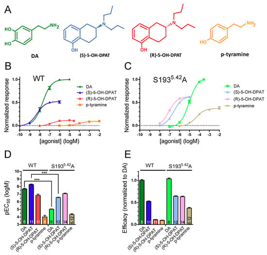
Figure 1.
Concentration-response relationships of agonists at WT D2R and S1935.42A. Concentration–response curves for GIRK activation elicited by application of DA, (S)-5-OH-DPAT, (R)-5-OH-DPAT, and p-tyramine (structures shown in panel (A)) in oocytes co-expressing GIRK1/4 subunits and RGS4 with (B) WT D2R and (C) D2R S1935.42A. (D) Agonist pEC50s at WT and S1935.42A D2R: pEC50 for DA (WT: pEC50 = 7.70 ± 0.07, n = 5; S1935.42A: pEC50 = 5.03 ± 0.04, n = 8), (S)-5-OH-DPAT (WT: pEC50 = 8.28 ± 0.08, n = 11; S1935.42A: pEC50 = 6.56 ± 0.05, n = 12), (R)-5-OH-DPAT (WT: pEC50 = 6.85 ± 0.16, n = 11; S1935.42A: pEC50 = 7.07 ± 0.08, n = 14), and p-tyramine (WT: pEC50 = 4.00 ± 0.21, n = 6; S1935.42A: pEC50 = 4.32 ± 0.12, n = 6–12). Comparison of pEC50s using two-way ANOVA yielded significant main effects of agonist (F(3, 65) = 281.8) and of the S1935.42A mutation (F(1, 65) = 139.2), as well as a significant interaction between these two factors (F(3, 65) = 79.98, p < 0.001 for each main effect). Sidak’s multiple comparisons test further revealed that the pEC50s of DA and (S)-5-OH-DPAT, but not p-tyramine and (R)-5-OH-DPAT, differed significantly between WT and mutant D2R, as indicated by asterisks; ***, p < 0.001. (E) Relative efficacies at WT D2R and S1935.42A for DA (WT: 1.00 ± 0.02; S1935.42A: 1.04 ± 0.04), (S)-5-OH-DPAT (WT: 0.52 ± 0.01; S1935.42A: 0.64 ± 0.01), (R)-5-OH-DPAT (WT: 0.11 ± 0.01; S1935.42A: 0.63 ± 0.02), and p-tyramine (WT: 0.09 ± 0.01; S1935.42A: 0.37 ± 0.01). WT and S1935.42A responses were normalized to the response evoked by 1 µM and 300 µM DA, respectively. The efficacy values were obtained from the fitted parameter Top, from the corresponding concentration-response curves (see Materials and Methods). The number of oocytes used for each condition is indicated on the bars in (D,E) and corresponds to the number recorded to generate the data points plotted in (B,C). Experiments were performed using a buffer perfusion rate of 1 mL/min. Data are presented as mean ± SEM.
The network of contacts between the binding pocket and the ligand governs ligand affinity by determining the association (kon) and dissociation rate constants (koff) of the ligand–receptor complex. Interestingly, agonist binding kinetics may influence the efficacy and downstream signaling preferences (bias) [16,17,18]. In addition, the kinetics of ligand binding has garnered increasing attention as a way to optimize in vivo receptor occupancy, ensuring therapeutic efficacy and avoiding drug-induced side effects [19,20]. Agonist binding kinetics are thus of interest both for therapeutic ligand design and from a basic science perspective. Many GPCRs, including D2R, are known to exist in two distinct affinity states with regard to agonist binding. In general, the high-affinity state is favored by the binding of a G protein to the receptor and is considered to be the functional, signaling state of the receptor, whereas the low-affinity state predominates in the absence of bound G protein [21,22]. Previous investigations of aminergic GPCRs have investigated the kinetics of agonist binding to isolated membranes [17,23], measured agonist-induced incorporation of radiolabeled guanine nucleotide analogues [24], and the activation rates of receptors and G proteins using fluorescently labeled constructs [25,26].
To our knowledge, estimates of association (kon) and dissociation (koff) rate constants for D2R agonists at the G protein-coupled signaling state have not been previously reported from live cells. Thus, in the present investigation, we examined (S)- and (R)-5-OH-DPAT, DA, and p-tyramine using G protein-coupled inward rectifier potassium (GIRK) channel currents in Xenopus oocytes as readout. GIRK channels are opened by Gβγ subunits released from Gi/o protein trimers activated by agonist-stimulated GPCRs [27]. To examine the role of S1935.42 for kon and koff of these D2R agonists, both WT and S1935.42A mutant D2R constructs were used. kon and koff were estimated from the time courses of activation and deactivation of agonist-induced GIRK currents upon agonist application and washout, respectively. The experimental data was also compared with molecular dynamics simulations based on the active-state structure of D2R in complex with an inhibitory G protein and bound to the agonist, bromocriptine [3]. Our results suggest that contacts with S1935.42 have a relatively greater impact on kon, compared to koff, for both DA and (S)-5-OH-DPAT, whereas the binding kinetics of p-tyramine and (R)-5-OH-DPAT were only marginally affected by S1935.42A mutation. In agreement, the two enantiomers of 5-OH-DPAT were distinguished mainly by their kon rates at the WT D2R and displayed similar kinetics at the S1935.42A mutant receptor.
2. Results
2.1. Phenethylamine and DPAT Potencies and Efficacies at WT and S1935.42A D2R
Following agonist application to Xenopus oocytes expressing D2R and GIRK1/4 subunits, Gβγ subunits released from activated Gi/o protein heterotrimers open the GIRK channels. Co-expression of the regulator of G protein signaling-4 (RGS4) accelerates the GTP hydrolysis rate at the Gαi/o subunit and increases the G protein cycle’s turnover rate, such that the time course of the GIRK current more closely follows the time course of D2R agonist occupancy [27,28]. Here, we used the GIRK current in oocytes voltage-clamped at −80 mV and superfused with a high-K+ buffer (25 mM KCl) as a readout of agonist binding to D2R.
Thus, increasing agonist concentrations were applied consecutively to voltage-clamped oocytes expressing WT D2R, RGS4, and GIRK1/4. In each oocyte, the elicited current responses were normalized to the response to a maximally effective concentration of DA (1 µM) to yield concentration-response relationships. To avoid oocyte deterioration during data acquisition, as well as to minimize the potential effects of current rundown [29] or receptor desensitization on the results, concentration–response data was acquired for only one agonist per oocyte. A representative trace of currents measured during p-tyramine concentration–response data acquisition is shown in Supplementary Figure S1). The tested agonists displayed the following rank order of potencies: (S)-5-OH-DPAT > DA > (R)-5-OH-DPAT > p-tyramine (Figure 1A,B), in agreement with previously published data on these agonists [14,30]. In oocytes expressing S1935.42A mutant D2R, RGS4, and GIRK1/4, responses were normalized to the response to 300 µM DA (concentrations of 1 mM and above were found to block GIRK channels; data not shown) and the rank order of agonist potencies were: (R)-5-OH-DPAT > (S)-5-OH-DPAT > DA > p-tyramine (Figure 1C). The differences in rank order of potency between WT and S1935.42A mutant D2R were driven mainly by the 467- and 52-fold reductions in DA and (S)-5-OH-DPAT potency at S1935.42A (Figure 1D). The DA-normalized agonist efficacies increased 5.7- and 4.1-fold for (R)-5-OH-DPAT and p-tyramine, respectively, at S1935.42A (Figure 1E). The strategy of acquiring concentration-response data for only one agonist per oocyte is unlikely to have had much impact on the estimation of relative efficacies since receptor reserve is low or absent under the conditions used [28,31]. In agreement with this notion, when averaging data across all experiments (Supplementary Figure S2), the relative current response amplitudes evoked by maximally effective concentrations of DA, and the three partial agonists via WT and mutant receptors, are similar to the relative efficacies shown in Figure 1E.
2.2. (R)- and (S)-5-OH-DPAT Dissociation Rates Are Similar, But Slower Than Those of DA and p-Tyramine at WT D2R
The deactivation time course of agonist-induced GIRK currents upon agonist washout has been shown to reflect agonist residence time at the receptor; i.e., 1/koff [32,33]. We obtained such estimates of koff by analyzing the washout phase following short (13 s) applications of submaximally effective agonist concentrations (see legend of Figure 2) in oocytes co-expressing WT D2R with GIRK1/4 and RGS4. GIRK responses elicited by both (R)- and (S)-5-OH-DPAT decayed with a similar time course upon washout, but slower than responses elicited by DA and p-tyramine (Figure 2A,D). To test the notion that the slower decay kinetics of GIRK responses to (R)- and (S)-5-OH-DPAT were due to a longer residence time at the receptor rather than to accumulation of these lipophilic agonists in the oocyte membrane, we also measured GIRK response decay kinetics when agonist availability to the D2R was terminated by the addition of an antagonist in the continued presence of agonist. As can be seen in Figure 2B, the time courses of response decay upon addition of 1 µM of the D2R antagonist, haloperidol, were similar to the washout-induced decay rates for (R)- and (S)-5-OH-DPAT. As is also apparent, the DA-induced GIRK response decayed faster under these conditions as well (see Supplementary Table S1 for quantification of the haloperidol-induced response decay kinetics). While haloperidol has been reported to block GIRK channels directly, thus potentially confounding the response decay rates measured here, this effect occurs with an IC50 of 41 µM and channel block at 1 µM is negligible [34,35]. In agreement, we have verified that haloperidol at 3 µM induces only 5.9 ± 0.2% (n = 4) direct GIRK current block in our hands. The effect at the D2R receptor is much more potent and 1 µM haloperidol fully blocks the GIRK response to even 100 nM DA [28]. Thus, the effect of haloperidol reported here most likely reflects action at the D2R, rather than at the GIRK channel.
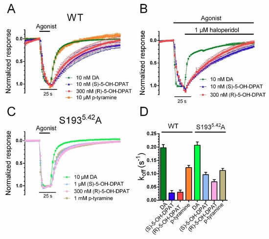
Figure 2.
Kinetics of GIRK current deactivation following agonist washout was used to estimate agonist koff. (A) Response decay time courses following washout of DA, (S)-5-OH-DPAT, (R)-5-OH-DPAT, and p-tyramine from oocytes co-expressing WT D2R with GIRK1/4 and RGS4. (B) Terminating the agonist-induced response by application of 1 µM haloperidol revealed similar rates of decay as observed in the agonist washout experiments presented in (A). (C) Response decay time constants following washout of DA, (S)-5-OH-DPAT, (R)-5-OH-DPAT, and p-tyramine from oocytes co-expressing S1935.42A D2R with GIRK1/4 and RGS4. (D) Estimated dissociation rate constants at WT D2R and S1935.42A for DA (WT: 0.197 ± 0.012 s−1, n = 6; S1935.42A: 0.207 ± 0.012 s−1, n = 8), (S)-5-OH-DPAT (WT: 0.028 ± 0.010 s−1, n = 4; S1935.42A: 0.096 ± 0.009 s−1, n = 6), (R)-5-OH-DPAT (WT: 0.030 ± 0.008 s−1, n = 5; S1935.42A: 0.069 ± 0.008 s−1, n = 6), and p-tyramine (WT: 0.123 ± 0.008 s−1, n = 7; S1935.42A: 0.112 ± 0.008 s−1, n = 5), determined by fitting exponential functions to the agonist washout phases. For DA and p-tyramine, the first 42 s, and for (S)- and (R)-5-OH-DPATs, the first 104 s following agonist washout were used to fit the exponential functions at WT D2R. 10 nM DA, 10 nM (S)-5-OH-DPAT, 300 nM (R)-5-OH-DPAT, and 10 µM p-tyramine were used for these experiments at the WT receptor. At D2R S1935.42A, 10 µM DA, 1 µM (S)-5-OH-DPAT, 300 nM (R)-5-OH-DPAT, and 1 mM p-tyramine were used, and the first 24 s of the washout were used to fit the exponential functions for all agonists. Experiments were performed using a perfusion rate of 4.5 mL/min. Data are presented as mean ± SEM.
The time course of solution exchange around the oocyte under the conditions used here was also tested by switching from a low (1 mM) to a high (25 mM) K+ extracellular solution and monitoring the rate of change of basal GIRK current amplitudes. These experiments revealed a mean time constant of >2 s−1 (Supplementary Figure S3); greater than the fastest rates of GIRK current deactivation and activation (see below) analyzed here. While the koff values were in the same range for DA and p-tyramine at both WT D2R and the S1935.42A mutant, the mutation increased the decay rates of both (R)- and (S)-5-OH-DPAT-induced responses, such that they approached the koffs of DA- and p-tyramine (Figure 2C,D).
2.3. Estimated Association Rates of (R)- and (S)-5-OH-DPAT Differ at WT D2R but Are Similar at the S1935.42A Mutant
The nearly identical response deactivation time courses obtained with (R)- and (S)-5-OH-DPAT suggest that the higher affinity of (S)-5-OH-DPAT may result from a difference in kon, rather than in koff. We, therefore, investigated the time courses of GIRK current activation at different concentrations of agonist in oocytes co-expressing WT D2R or S1935.42A mutant D2R with RGS4 and GIRK1/4. At low agonist concentrations, evoking response time courses well below the solution exchange rate, the observed activation rate, kobs, was linearly dependent on the agonist concentration. At higher agonist concentrations, kobs appeared to saturate (Supplementary Figure S4), as previously observed for the rates of G protein- and receptor activation [25,36].
A straight line was fitted to the data over the linear range of the relation between kobs and agonist concentration and the slope was taken as a measure of kon. As could be expected based on the previously obtained koffs and EC50s, the estimated kons of (S)-5-OH-DPAT and DA were higher than those of (R)-5-OH-DPAT and p-tyramine. Compared to the WT receptor, kon estimates for DA and (S)-5-OH-DPAT were reduced at the S1935.42A mutant (Figure 3A,B,D). In contrast, (R)-5-OH-DPAT, and to a lesser extent, p-tyramine, demonstrated slightly increased kon values at the S1935.42A D2R (Figure 3B–D). Consequently, the kon estimates of (R)- and (S)-5-OH-DPAT were similar at the S1935.42A mutant. To relate mutation-induced changes in estimated binding rates to the corresponding EC50 shifts (see Figure 1), kinetic Kd values were calculated from the ratios of koff and kon (Table 1). pEC50s for each agonist were plotted against the respective kinetic pKd values, demonstrating a significant correlation (Figure 3E).
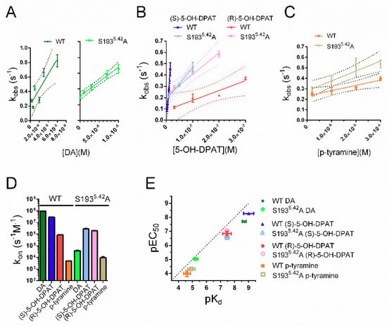
Figure 3.
Estimation of agonist kon at WT and S1935.42A mutant D2R. Individual concentrations of (A) DA, (B) (S)-5-OH-DPAT and (R)-5-OH-DPAT, and (C) p-tyramine were added to oocytes co-expressing WT D2R or D2R S1935.42A with RGS4 and GIRK1/4. The rates of rise (kobs) of the resulting current responses have been plotted against the corresponding agonist concentrations and linear fits (solid lines) and their 95% confidence bands (dotted lines) are shown. Experiments were performed using a perfusion rate of 4.5 mL/min. (D) Summary statistics for kon, estimated from the slopes of the linear fits to kobs shown in (A), (B), and (C). Shown are mean kon estimates for DA (WT: 9.70 ± 1.23 × 10⁷ s−1 × M−1, S1935.42A: 3.69 ± 0.48 × 10⁴ s−1 × M−1), (S)-5-OH-DPAT (WT: 2.86 ± 0.31 × 10⁷ s−1 × M−1, S1935.42A: 2.82 ± 0.34 × 10⁶ s−1 × M−1), (R)-5-OH-DPAT (WT: 8.65 ± 1.21 × 10⁵ s−1 × M−1, S1935.42A: 1.95 ± 0.15 × 10⁶ s−1 × M−1), and p-tyramine (WT: 4.94 ± 1.15 × 10³ s−1 × M−1, S1935.42A: 9.41 ± 2.34 × 10³ s−1 × M−1). Note the logarithmic y-axis. (E) Relations between kinetic pKds, derived from koff and kon, and pEC50s, obtained from the concentration–response experiments shown in Figure 1, at WT D2R and S1935.42A. The correlation between pEC50s and kinetic pKds for all four agonists at both WT and S1935.42A mutant D2R was statistically significant (Spearman’s r = 0.9048, p = 0.0046) for all four pairs. Data are presented as mean ± SEM.

Table 1.
Agonist EC50s, estimated forward (kon) and reverse (koff) rate constants, and kinetic Kds at WT and S1935.42A mutant D2R.
2.4. Structural Determinants of Mutation-Induced Changes in Compound Kinetics
In order to gain structural insight into how S1935.42A mutation impacts agonist–receptor interactions, we simulated the active conformation of D2R in complex with (R)-5-OH-DPAT, (S)-5-OH-DPAT, and DA. The binding mode of each compound was approximated by clustering the generated simulation frames based on the ligand coordinates and studying the most populated cluster. Both DA and (S)-5-OH-DPAT were observed to form simultaneous contacts with S1935.42 and histidine (H) 3646.55 (Figure 4A,B). Seeing as these two compounds formed extensive interactions with S1935.42, it is likely that altering the polar character of this residue would impair the binding of both compounds to D2R. In contrast to DA and (S)-5-OH-DPAT, (R)-5-OH-DPAT primarily forms polar interactions with S1975.46 and does not interact extensively with S1935.42 (Figure 4C). Indeed, simulating the studied compound within the S1935.42A D2R reveals that the mutation does not impact the primary binding mode of the compound. (Figure 4D).
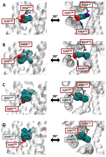
Figure 4.
Binding modes of studied compounds within the WT D2R and the S1935.42A mutant receptor. Simulations of (A) DA, (B) (S)-5-OH-DPAT, and (C) (R)-5-OH-DPAT in complex with the WT D2R and (D) (R)-5-OH-DPAT in complex with the S1935.42A D2R were clustered based on the root-mean-square deviation (RMSD) of the ligand. For each of the systems, the poses of the ligand within the most populated cluster are depicted in licorice. For each of the binding modes, the studied position 1935.42, as well as the residues that formed polar interactions with the ligand, are also shown in licorice. Polar interactions are highlighted by red lines.
3. Discussion
In the present study, we used the kinetics of agonist-induced response activation and deactivation in a GIRK channel-based electrophysiology assay to derive estimates of binding rate constants of four D2R agonists. We also aimed to investigate how agonist interactions with a conserved serine, S1935.42, in the D2R orthosteric binding pocket affect these rate constants. Since agonist kon has often been assumed to be diffusion-limited [37,38], a particularly interesting finding is that the differential potencies/affinities of the two 5-OH-DPAT enantiomers appear to stem from differences in kon, rather than koff. The electrophysiology assay employed here has the advantage of using unmodified proteins and has been reported to demonstrate a very close temporal relation with G protein activation, as measured by fluorescence resonance energy transfer between labeled G protein subunits [39]. While this readout of agonist binding is an indirect one, our previous analysis of antipsychotic binding rates, also using GIRK currents as a proxy for D2R agonist occupancy, yielded results in very good agreement with radioligand- and fluorescence-based binding studies [28,40]. Similarly, other investigators have used oocyte-based GIRK assays to make inferences about the structural details of DA-D2R interactions [41,42].
However, we are aware that the temporal resolution of our assay is limited both by the rate of buffer exchange around the relatively large oocyte, as well as by the kinetics of G protein turnover. By co-expressing RGS4 and using high perfusion rates, the conditions of the washout-induced deactivation experiments were optimized to reflect agonist dissociation rates [12,27]. We also used conditions where there is little or no receptor reserve [28,43] in order for the concentration-response relationships of GIRK activation to more directly reflect D2R occupancy by agonist. In agreement with this notion, our measured EC50 value for DA (20 nM) is in good agreement with high-affinity site Ki values for DA binding to D2R reported from radioligand competition studies [14,44,45,46,47].
Here, we observed a linear dependence of GIRK current activation rates on agonist concentration in the lower, submaximally effective range, but not at higher concentrations (see Supplementary Figure S4). Likewise, rates of α2A receptor activation and G protein activation by muscarinic receptors were found to increase linearly at low, subsaturating agonist concentrations but showed a hyperbolic, saturating relationship at higher concentrations [25,36]. The maximal rates of G protein activation have also been found to differ between different agonists at the α2A adrenergic receptor [48], presumably explaining differences in efficacy. Here, we deliberately used the linear range of the relation between kobs and agonist concentration for kon estimation.
Comparing agonist EC50s with kinetic Kd values calculated from the kon and koff estimates, we found a good correlation for all four agonists at both the WT and S1935.42A mutant receptors. However, the kinetic Kd values were found to systematically overestimate the potency/affinity of the tested agonists (see Table 1). Although the Y-axis intercepts of the kobs—[agonist] plots were generally in good agreement with the koff estimates obtained from GIRK current deactivation time courses, plotting the Y intercepts against koffs from washout experiments reveals a tendency of the former values to be higher (Supplementary Figure S5). Thus, we cannot exclude the possibility that buffer exchange limited our koff estimates, leading to the overestimation of ligand potency. In agreement with this notion, we were previously able to record rates of DA-evoked GIRK current inhibition by competitive D2R antagonists of just above 0.4 s−1—twice as fast as the response decay rates observed here [28]. Finally, the rate of GTP hydrolysis at the G protein would, even in the presence of RGS4, restricted our ability to accurately quantify very rapid rates of agonist dissociation.
S1935.42 in D2R has consistently been reported to be crucial for high-affinity binding of DA and of (S)- but not (R)-5-OH-DPAT, nor of p-tyramine [9,14,15]. This notion is supported by the electrophysiology data and molecular dynamics simulations presented here. Accordingly, the simulated binding modes for DA and (S)-5-OH-DPAT revealed prominent polar contacts with S1935.42, and S1935.42A mutation was found to decrease experimentally determined kon estimates and potencies for both DA and (S)-5-OH-DPAT. Conversely, the mutation marginally increased (R)-5-OH-DPAT kon, such that the kons of both enantiomers were similar at the mutant receptor. S1935.42A mutation slightly increased p-tyramine kon while having a negligible effect on koff. Somewhat surprisingly, DA koff was also unaffected by S1935.42A mutation. This finding should, however, be interpreted with caution due to the limiting effects of buffer exchange and GTP hydrolysis noted above and the fact that the DA koff estimates were the fastest in the dataset. Thus, the apparent lack of effect of S1935.42A mutation on DA koff may be due to insufficient temporal resolution in our experimental system. Alternatively, there may indeed be no appreciable effect of the mutation on DA koff, putatively due to the receptor-agonist-G protein complex undergoing isomerization after initial agonist binding and receptor activation, such that S1935.42A no longer forms important contacts with the agonist.
Furthermore, the mutation also increased the relative efficacies of (R)-5-OH-DPAT and p-tyramine, while the relative efficacy of (S)-5-remained virtually unchanged. However, since agonist efficacy was determined by normalization to the maximal DA-induced response, another possible interpretation would be that the efficacies of DA and (S)-5-OH-DPAT decreased, while those of (R)-5-OH-DPAT and p-tyramine were less affected. The effects of S1935.42A mutation on all four agonists are summarized in Table 2.

Table 2.
Relative changes in agonist potencies, forward (kon) and reverse (koff) rate constants and calculated kinetic potencies at the S1935.42A mutant, as compared to WT D2R.
Interestingly, the koffs of (R)- and (S)-5-OH-DPAT were similar at the WT D2R and were affected by mutation to a similar extent despite their differential interactions with S1935.42. It thus appears that interaction with S1935.42 may slow dissociation of both (R)- and (S)-5-OH-DPAT from the receptor. As expected, the results of our molecular dynamics simulations suggest that the main binding mode of (R)-5-OH-DPAT is not altered by the S1935.42A mutation (Figure 4C,D). Thus, the observed shifts of (R)-5-OH-DPAT kinetics appear to be mediated by a change in the ligand entry/exit pathway. Indeed, a detailed analysis of the simulation data of (R)-5-OH-DPAT in complex with the WT D2R suggests that apart from the main binding mode involving S1975.46 (Figure 5A,B, in green), the existence of an additional meta-stable binding mode (Figure 5A,C, in red). As this binding mode depends on polar contacts between (R)-5-OH-DPAT and S1935.42, it would be absent in the S1935.42A mutant receptor. We speculate that the meta-stable binding mode forms a step in the binding/unbinding process of (R)-5-OH-DPAT. Before dissociating from D2R, (R)-5-OH-DPAT would thus assume the meta-stable conformation, from which it would either proceed to complete dissociation, or return to the main binding mode (Figure 5D, left panel). Hence, the presence of this meta-stable binding mode could slow ligand dissociation. On the other hand, the lack of the meta-stable binding mode in the S1935.42A mutant D2R would permit fast exchange between the bound and unbound conformations, which would explain the increase in (R)-5-OH-DPAT koff observed at the S1935.42A mutant (Figure 5D, right panel).
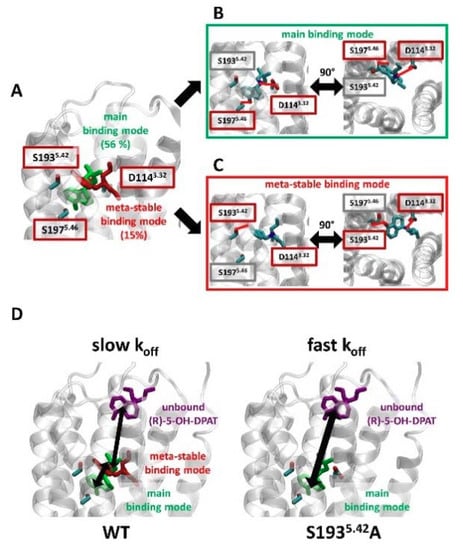
Figure 5.
(R)-5-OH-DPAT forms a meta-stable binding mode with S1935.42. (A) Clustering simulations of (R)-5-OH-DPAT in complex with WT D2R, based on the RMSD of the ligand, reveal two binding modes. The main binding mode was maintained over 56% of the simulation frames (green) and a meta-stable binding mode was maintained over 15% of the simulation frames (red). (B,C). For each of the binding modes, the studied position S1935.42, as well as residues that form polar interactions with the ligand, are shown. These polar interactions are highlighted with red lines. (D) A model explaining the slow Koff values observed for the unbinding of (R)-5-OH-DPAT from WT D2R. Before dissociating from the receptor (purple conformation), (R)-5-OH-DPAT bound in the main binding mode (green) assumes a meta-stable binding mode (red). When bound in this meta-stable binding mode, (R)-5-OH-DPAT can revert to the main binding mode or proceed to an unbound conformation. Hence, the dissociation of (R)-5-OH-DPAT is effectively slowed down by S1935.42. In comparison, at the S1935.42A mutant receptor, the absence of the meta-stable binding mode permits fast exchange between the bound and unbound conformations of (R)-5-OH-DPAT.
Interactions with S1935.42 have long been known to be crucial for affinities and functional potencies of several D2R agonist ligands. Unexpectedly, our results from GIRK channel activation experiments suggest that these contacts may have a greater impact on agonist association rate constants (kons) rather than on agonist dissociation rate constants (koffs). Optimization of therapeutic or diagnostic ligand binding kinetics is important for optimal target engagement in vivo [19,20,49]. With the advantage of using unmodified proteins, ligands and living cells, the approach presented here may therefore be of interest for estimation of binding kinetics of drug candidates at the D2R and other GPCRs capable of activating GIRK channels.
4. Materials and Methods
4.1. Molecular Biology
cDNA encoding the wildtype (WT) and S1935.42AA human dopamine D2S (short isoform; from Dr. Marc Caron, Duke University, NC, USA) receptors were in pXOOM (provided by Dr. Søren-Peter Olesen, University of Copenhagen, Denmark) whereas RGS4 (from the cDNA Resource Center, Bloomsburg, PA, USA; www.cdna.org, access date 1 March 2021) and GIRK1/4 (a gift from Dr. Terence Hebert, McGill University, Montreal, QC, Canada) were in pcDNA3.1+. The S1935.42A point mutation of the D2R was made using the QuickChange Lightning kit (Agilent Technologies, Santa Clara, CA, USA), according to the manufacturer’s instructions, and confirmed by DNA sequencing of the entire insert. Plasmids were linearized using suitable restriction enzymes (WT D2R, S1935.42A mutant D2R, and RGS4: XhoI; GIRK1 and GIRK4: NotI), followed by in vitro transcription using the T7 mMessage mMachine kit (Ambion, Austin, TX, USA). cRNA concentration and purity were determined by spectrophotometry.
4.2. Oocyte Preparation
Oocytes from the African clawed toad, Xenopus laevis, were surgically isolated as described previously [12]. The procedure was approved by the Swedish National Board for Laboratory Animals and the Animal Welfare Ethical Committee in Stockholm (approval number N245/15). Following one day of incubation at 12 °C, oocytes were injected with 50 nL containing 0.2 ng D2R cRNA, 40 ng of RGS4, and 1 ng of each GIRK1 and GIRK4 cRNA using the Nanoject II (Drummond Scientific, Broomall, PA, USA).
4.3. Electrophysiological Methods
Injected cells were incubated for another 6 days at 12 °C in modified Barth’s solution (MBS), composed of (in mM): 88 NaCl, 1 KCl, 2.4 NaHCO3, 15 HEPES, 0.33 Ca(NO3)2, 0.41 CaCl2, 0.92 MgSO4, and 2.5 sodium pyruvate, supplemented with 25 U/mL penicillin and 25 µg/mL streptomycin and adjusted to pH 7.6 with NaOH. Electrophysiological recordings were performed at 22 ℃ using the eight-channel, two-electrode voltage-clamp OpusXpress 6000A (Molecular Devices, San José, CA, USA) [50]. Continuous perfusion was maintained at either 1 (concentration–response experiments) or 4.5 (for estimation of kinetic parameters) mL/min. Data were acquired at membrane potentials of −80 mV and sampled at 156 Hz using the OpusXpress 1.10.42 (Molecular Devices) software. To increase the inward rectifier potassium channel current at negative potentials, a high-potassium extracellular buffer was used (in mM: 64 NaCl, 25 KCl, 0.8 MgCl2, 0.4 CaCl2, 15 HEPES, and 1 ascorbic acid, adjusted to pH 7.4 with NaOH), yielding a K+ reversal potential of about −40 mV. In experiments with 1 mM KCl buffer, the NaCl concentration was adjusted to 88 mM. Ascorbic acid was included to prevent the spontaneous oxidation of DA. For concentration-response experiments, each oocyte was first exposed to 1 µM (WT) or 300 µM (S1935.42A) DA, evoking a maximal D2R-mediated response. After washout of DA, an initial stabilization period of 60 s where 25 KCl buffer was perfused was followed by applications of three to five increasing concentrations of agonist, which were added consecutively at 60 s intervals (see Supplementary Figure S1 for a representative current trace showing the responses to increasing concentrations of p-tyramine). For each concentration of agonist, the relative current amplitude after 60 s (when a steady-state response had been achieved) of agonist perfusion was plotted to generate the concentration-response relationships. Oocytes were selected for electrophysiology recordings based on having holding currents at −40 mV of less than 0.5 µA. Likewise, recordings where holding currents at −40 mV were greater than 0.5 µA after data acquisition at −80 mV were discarded.
4.4. Ligands
DA and p-tyramine were from Sigma-Aldrich (St. Louis, MO, USA) and were prepared fresh on each day of experiments and dissolved directly in the recording buffer. (R)- and (S)-5-OH-DPAT (Axon MedChem BV, Groeningen, The Netherlands) were dissolved in DMSO at 10 mM and subsequently diluted into the recording buffer at the desired concentrations. Likewise, haloperidol (Abcam Chemicals, Cambridge, UK) was dissolved at 10 mM in DMSO and diluted in recording buffer to the final concentration. The maximum final concentration of DMSO used in any experiment was 0.1% v/v.
4.5. Data Analysis
Electrophysiology concentration-response data was initially processed in Clampfit 10.6 (Molecular Devices) by subtracting the basal (agonist-independent) current and quantifying the current amplitude evoked by each concentration of agonist. Agonist concentration–response relationships were analyzed by fitting sigmoidal functions using nonlinear regression in GraphPad Prism 8 (GraphPad Software, San Diego, CA, USA). The following equation was used for fitting:
where Y is the GIRK current response normalized to the response to a maximally effective concentration of DA, Top is the maximal response of the agonist in question, and X is the logarithm of agonist concentration. Top and its SEM were used to plot the efficacy values shown in Figure 1E.
Y = Top/(1 + 10 (LogEC50 − X))
Recordings of GIRK response deactivation were transferred to Matrix Laboratory 2018b (MathWorks, Natick, MA, USA), peak normalized and time-averaged. Data are reported as mean ± SEM throughout the manuscript. Monoexponential functions were fitted to the relevant intervals of individual traces corresponding to response decay following agonist washout (see Section 2), outputting the deactivation time constant, τdeact. For DA and p-tyramine, the first 42 s, and for (S)- and (R)-5-OH-DPATs, the first 104 s following agonist washout were used to fit the exponential functions at WT D2R. 10 nM DA, 10 nM (S)-5-OH-DPAT, 300 nM (R)-5-OH-DPAT, and 10 µM p-tyramine were used for these experiments at the WT receptor. At the S1935.42A mutant, 10 µM DA, 1 µM (S)-5-OH-DPAT, 300 nM (R)-5-OH-DPAT, and 1 mM p-tyramine were used, and the first 24 s of washout were used to fit the exponential functions for all agonists. Estimates of the dissociation rate constant, koff, were obtained as 1/τdeact.
Association rate constant estimates were derived from recordings of GIRK current activation in response to applications of various agonist concentrations. Monoexponential functions were fit to cover 80% of the current increase in response to agonist using Levenberg–Marquardt least-squares fitting in Clampfit 10, outputting the activation time constant, τact. The observed activation rate (kobs) was defined as 1/τact, and subsequently used for estimation of the association rate constant as the slope of the dependence of kobs on agonist concentration, over the range of concentrations where this relation was linear (see also [28]), using the following relation:
where [A] is the agonist concentration, kon the association rate constant, and koff the dissociation rate. However, rather than using Equation (2), koff was calculated separately from the response decay time constant upon agonist washout, τdeact, as described above.
kobs = [A] × kon + koff
Kinetic Kds were calculated as:
Kd = koff/kon
Statistical analysis was performed using GraphPad Prism 8. p < 0.05 was chosen as the significance limit.
4.6. Molecular Dynamics Simulations
The simulated complexes were generated by docking the studied compounds into the structure of D2R in an active conformation [PDB code: 6VMS] [3]. For docking, we used the standard docking method available in the MOE package (www.chemcomp.com, access date 1 March 2021). The poses were selected based on the docking score, as well as available mutational data [9,10,11,13,14]. The generated WT complex was oriented in the membrane using coordinates obtained from the OPM database (Positioning of proteins in membranes: a computational approach), and solvated with TIP3 waters, using the CHARMM-GUI server [51]. The ionic strength of the system was kept at 0.15 M using NaCl ions. The S1935.42A mutation was introduced using the CHARMM-GUI pipeline.
Simulations were carried out using the ACEMD simulation package [52]. Ligand parameters were assigned by ParamChem from the CGenFF force field [53,54]. Parameters for other system components were obtained from CHARMM36m [55] and CHARMM36 force fields [56]. In the simulation protocol, we adhere to the guidelines of the GPCRmd consortium [57].
The systems were first relaxed during 100 ns of simulations under constant pressure and temperature (NPT) with a time step of 2 fs, with harmonic constraints applied to the protein backbone and ligand heavy atoms. The temperature was maintained at 310 K using the Langevin thermostat [58] and pressure was kept at 1 bar using the Berendsen barostat [59]. The equilibration run was followed by two parallel 400 ns production runs in conditions of constant volume and temperature (NVT) with a 4-fs time step. No constraints were applied during this stage. In all simulations, we used van der Waals and short-range electrostatic interactions with a cutoff of 9 Å and the particle mesh Ewald method [60] for long-range electrostatic interactions. The resulting simulation frames were analyzed using VMD [61] and tools available within.
Supplementary Materials
The following are available online at https://www.mdpi.com/article/10.3390/ijms22084078/s1, Figure S1: Representative trace illustrating the protocol for concentration-response experiments, showing the current response to application of increasing concentrations of p-tyramine as well as the voltage-clamp protocol used, Figure S2: Mean current amplitudes elicited via WT and S1935.42A D2R by maximally effective concentrations of the different agonists used in the present study, Figure S3: Solution exchange rate at the oocyte membrane as measured by switching from low- to high-K+ buffer, Figure S4: kobs saturation at high DA concentrations, Figure S5: Comparison of koff estimates obtained from agonist washout-induced response decay and from kobs y-intercepts, Table S1: Response decay rates upon haloperidol antagonism.
Author Contributions
Conceptualization, K.S., J.S., R.Å. and T.M.S.; methodology, K.S. and R.Å.; software, R.Å., T.M.S. and J.S.; validation, R.Å. and K.S.; formal analysis, R.Å. and H.Z.; investigation, R.Å. and T.M.S.; resources, K.S. and J.S.; data curation, R.Å. and T.M.S.; writing—original draft preparation, R.Å.; writing—review and editing, K.S. and T.M.S.; visualization, R.Å. and T.M.S.; supervision, K.S. and J.S.; project administration, K.S.; funding acquisition, K.S. All authors have read and agreed to the published version of the manuscript.
Funding
This research was funded by Åhlén-stiftelsen, grant number mB3 h18, and Magnus Bergvalls stiftelse, grant number 2018-02980 (to K.S.). K.S. is currently a fellow at the Wallenberg Center for Molecular Medicine at Umeå University.
Institutional Review Board Statement
The study was conducted according to the guidelines of the Declaration of Helsinki and approved by the Swedish National Board for Laboratory Animals and the Animal Welfare Ethical Committee in Stockholm (approval number N245/15, approval date 17/12-2015). Animal experiments took place at Karolinska Institutet, Sweden.
Informed Consent Statement
Not applicable.
Data Availability Statement
The data presented in this study are available in the article and in the Supplementary Material.
Conflicts of Interest
The authors declare no conflict of interest. The funders had no role in the design of the study; in the collection, analyses, or interpretation of data; in the writing of the manuscript, or in the decision to publish the results.
References
- Hauser, A.S.; Attwood, M.M.; Rask-Andersen, M.; Schiöth, H.B.; Gloriam, D.E. Trends in GPCR drug discovery: New agents, targets and indications. Nat. Rev. Drug Discov. 2017, 16, 829–842. [Google Scholar] [CrossRef]
- Ring, A.M.; Manglik, A.; Kruse, A.C.; Enos, M.D.; Weis, W.I.; Garcia, K.C.; Kobilka, B.K. Adrenaline-activated structure of β2-adrenoceptor stabilized by an engineered nanobody. Nature 2013, 502, 575–579. [Google Scholar] [CrossRef] [PubMed]
- Yin, J.; Chen, K.M.; Clark, M.J.; Hijazi, M.; Kumari, P.; Bai, X.C.; Sunahara, R.K.; Barth, P.; Rosenbaum, D.M. Structure of a D2 dopamine receptor-G-protein complex in a lipid membrane. Nature 2020, 584, 125–129. [Google Scholar] [CrossRef]
- Zhuang, Y.; Xu, P.; Mao, C.; Wang, L.; Krumm, B.; Zhou, X.E.; Huang, S.; Liu, H.; Cheng, X.; Huang, X.P.; et al. Structural insights into the human D1 and D2 dopamine receptor signaling complexes. Cell 2021, 184, 931–942.e18. [Google Scholar] [CrossRef]
- Ballesteros, J.; Weinstein, H. Integrated methods for the construction of three-dimensional models and computational probing of structure-function relations in G protein-coupled receptors. In Methods in Neurosciences; Sealfon, S., Ed.; Elsevier: Amsterdam, The Netherlands, 1995; Volume 25, pp. 366–428. [Google Scholar]
- Missale, C.; Nash, S.R.; Robinson, S.W.; Jaber, M.; Caron, M.G. Dopamine receptors: From structure to function. Physiol. Rev. 1998, 78, 189–225. [Google Scholar] [CrossRef] [PubMed]
- Beaulieu, J.M.; Gainetdinov, R.R. The physiology, signaling, and pharmacology of dopamine receptors. Pharmacol. Rev. 2011, 63, 182–217. [Google Scholar] [CrossRef]
- Fernandez-Duenas, V.; Ferre, S.; Ciruela, F. Adenosine A2A-dopamine D2 receptor heteromers operate striatal function: Impact on Parkinson’s disease pharmacotherapeutics. Neural Regen. Res. 2018, 13, 241–243. [Google Scholar] [PubMed]
- Cox, B.A.; Henningsen, R.A.; Spanoyannis, A.; Neve, R.L.; Neve, K.A. Contributions of conserved serine residues to the interactions of ligands with dopamine D2 receptors. J. Neurochem. 1992, 59, 627–635. [Google Scholar] [CrossRef] [PubMed]
- Wiens, B.L.; Nelson, C.S.; Neve, K.A. Contribution of serine residues to constitutive and agonist-induced signaling via the D2S dopamine receptor: Evidence for multiple, agonist-specific active conformations. Mol. Pharmacol. 1998, 54, 435–444. [Google Scholar] [CrossRef]
- Fowler, J.C.; Bhattacharya, S.; Urban, J.D.; Vaidehi, N.; Mailman, R.B. Receptor conformations involved in dopamine D(2L) receptor functional selectivity induced by selected transmembrane-5 serine mutations. Mol. Pharmacol. 2012, 81, 820–831. [Google Scholar] [CrossRef] [PubMed]
- Sahlholm, K.; Barchad-Avitzur, O.; Marcellino, D.; Gómez-Soler, M.; Fuxe, K.; Ciruela, F.; Arhem, P. Agonist-specific voltage sensitivity at the dopamine D2S receptor—Molecular determinants and relevance to therapeutic ligands. Neuropharmacology 2011, 61, 937–949. [Google Scholar] [CrossRef] [PubMed]
- Woodward, R.; Coley, C.; Daniell, S.; Naylor, L.H.; Strange, P.G. Investigation of the role of conserved serine residues in the long form of the rat D2 dopamine receptor using site-directed mutagenesis. J. Neurochem. 1996, 66, 394–402. [Google Scholar] [CrossRef]
- Coley, C.; Woodward, R.; Johansson, A.M.; Strange, P.G.; Naylor, L.H. Effect of multiple serine/alanine mutations in the transmembrane spanning region V of the D2 dopamine receptor on ligand binding. J. Neurochem. 2000, 74, 358–366. [Google Scholar] [CrossRef]
- Malmberg, A.; Nordvall, G.; Johansson, A.M.; Mohell, N.; Hacksell, U. Molecular basis for the binding of 2-aminotetralins to human dopamine D2A and D3 receptors. Mol. Pharmacol. 1994, 46, 299–312. [Google Scholar] [PubMed]
- Strasser, A.; Wittmann, H.J.; Seifert, R. Binding Kinetics and Pathways of Ligands to GPCRs. Trends Pharmacol. Sci. 2017, 38, 717–732. [Google Scholar] [CrossRef] [PubMed]
- Klein Herenbrink, C.; Sykes, D.A.; Donthamsetti, P.; Canals, M.; Coudrat, T.; Shonberg, J.; Scammells, P.J.; Capuano, B.; Sexton, P.M.; Charlton, S.J.; et al. The role of kinetic context in apparent biased agonism at GPCRs. Nat. Commun. 2016, 7, 10842. [Google Scholar] [CrossRef] [PubMed]
- Wacker, D.; Wang, S.; McCorvy, J.D.; Betz, R.M.; Venkatakrishnan, A.J.; Levit, A.; Lansu, K.; Schools, Z.L.; Che, T.; Nichols, D.E.; et al. Crystal Structure of an LSD-Bound Human Serotonin Receptor. Cell 2017, 168, 377–389.e12. [Google Scholar] [CrossRef]
- Copeland, R.A. The drug-target residence time model: A 10-year retrospective. Nat. Rev. Drug Discov. 2016, 15, 87–95. [Google Scholar] [CrossRef]
- IJzerman, A.P.; Guo, D. Drug-Target Association Kinetics in Drug Discovery. Trends Biochem. Sci. 2019, 44, 861–871. [Google Scholar] [CrossRef]
- van Wieringen, J.P.; Booij, J.; Shalgunov, V.; Elsinga, P.; Michel, M.C. Agonist high- and low-affinity states of dopamine D(2) receptors: Methods of detection and clinical implications. Naunyn Schmiedebergs Arch. Pharmacol. 2013, 386, 135–154. [Google Scholar] [CrossRef]
- Strange, P.G. Agonist binding, agonist affinity and agonist efficacy at G protein-coupled receptors. Br. J. Pharmacol. 2008, 153, 1353–1363. [Google Scholar] [CrossRef] [PubMed]
- Kara, E.; Lin, H.; Strange, P.G. Co-operativity in agonist binding at the D2 dopamine receptor: Evidence from agonist dissociation kinetics. J. Neurochem. 2010, 112, 1442–1453. [Google Scholar] [CrossRef]
- Roberts, D.J.; Lin, H.; Strange, P.G. Investigation of the mechanism of agonist and inverse agonist action at D2 dopamine receptors. Biochem. Pharmacol. 2004, 67, 1657–1665. [Google Scholar] [CrossRef]
- Ilyaskina, O.S.; Lemoine, H.; Bünemann, M. Lifetime of muscarinic receptor-G-protein complexes determines coupling efficiency and G-protein subtype selectivity. Proc. Natl. Acad. Sci. USA 2018, 115, 5016–5021. [Google Scholar] [CrossRef] [PubMed]
- Sungkaworn, T.; Jobin, M.L.; Burnecki, K.; Weron, A.; Lohse, M.J.; Calebiro, D. Single-molecule imaging reveals receptor-G protein interactions at cell surface hot spots. Nature 2017, 550, 543–547. [Google Scholar] [CrossRef] [PubMed]
- Dascal, N.; Kahanovitch, U. The Roles of Gbetagamma and Galpha in Gating and Regulation of GIRK Channels. Int. Rev. Neurobiol. 2015, 123, 27–85. [Google Scholar] [PubMed]
- Sahlholm, K.; Zeberg, H.; Nilsson, J.; Ögren, S.O.; Fuxe, K.; Århem, P. The fast-off hypothesis revisited: A functional kinetic study of antipsychotic antagonism of the dopamine D2 receptor. Eur. Neuropsychopharmacol. 2016, 26, 467–476. [Google Scholar] [CrossRef] [PubMed]
- Vorobiov, D.; Levin, G.; Lotan, I.; Dascal, N. Agonist-independent inactivation and agonist-induced desensitization of the G protein-activated K+ channel (GIRK) in Xenopus oocytes. Pflug. Arch. 1998, 436, 56–68. [Google Scholar] [CrossRef]
- Payne, S.L.; Johansson, A.M.; Strange, P.G. Mechanisms of ligand binding and efficacy at the human D2(short) dopamine receptor. J. Neurochem. 2002, 82, 1106–1117. [Google Scholar] [CrossRef]
- Agren, R.; Arhem, P.; Nilsson, J.; Sahlholm, K. The Beta-Arrestin-Biased Dopamine D2 Receptor Ligand, UNC9994, Is a Partial Agonist at G-Protein-Mediated Potassium Channel Activation. Int. J. Neuropsychopharmacol. 2018, 21, 1102–1108. [Google Scholar] [CrossRef]
- Benians, A.; Leaney, J.L.; Tinker, A. Agonist unbinding from receptor dictates the nature of deactivation kinetics of G protein-gated K+ channels. Proc. Natl. Acad. Sci. USA 2003, 100, 6239–6244. [Google Scholar] [CrossRef] [PubMed]
- Bünemann, M.; Bücheler, M.M.; Philipp, M.; Lohse, M.J.; Hein, L. Activation and deactivation kinetics of alpha 2A- and alpha 2C-adrenergic receptor-activated G protein-activated inwardly rectifying K+ channel currents. J. Biol. Chem. 2001, 276, 47512–47517. [Google Scholar] [CrossRef] [PubMed]
- Kobayashi, T.; Ikeda, K.; Kumanishi, T. Inhibition by various antipsychotic drugs of the G-protein-activated inwardly rectifying K(+) (GIRK) channels expressed in xenopus oocytes. Br. J. Pharmacol. 2000, 129, 1716–1722. [Google Scholar] [CrossRef] [PubMed]
- Heusler, P.; Newman-Tancredi, A.; Castro-Fernandez, A.; Cussac, D. Differential agonist and inverse agonist profile of antipsychotics at D2L receptors coupled to GIRK potassium channels. Neuropharmacology 2007, 52, 1106–1113. [Google Scholar] [CrossRef]
- Vilardaga, J.P.; Steinmeyer, R.; Harms, G.S.; Lohse, M.J. Molecular basis of inverse agonism in a G protein-coupled receptor. Nat. Chem. Biol. 2005, 1, 25–28. [Google Scholar] [CrossRef] [PubMed]
- Benians, A.; Leaney, J.L.; Milligan, G.; Tinker, A. The dynamics of formation and action of the ternary complex revealed in living cells using a G-protein-gated K+ channel as a biosensor. J. Biol. Chem. 2003, 278, 10851–10858. [Google Scholar] [CrossRef]
- Tummino, P.J.; Copeland, R.A. Residence time of receptor-ligand complexes and its effect on biological function. Biochemistry 2008, 47, 5481–5492. [Google Scholar] [CrossRef] [PubMed]
- Bünemann, M.; Frank, M.; Lohse, M.J. Gi protein activation in intact cells involves subunit rearrangement rather than dissociation. Proc. Natl. Acad. Sci. USA 2003, 100, 16077–16082. [Google Scholar] [CrossRef]
- Sykes, D.A.; Lane, J.R.; Szabo, M.; Capuano, B.; Javitch, J.A.; Charlton, S.J. Reply to ‘Antipsychotics with similar association kinetics at dopamine D2 receptors differ in extrapyramidal side-effects’. Nat. Commun. 2018, 9, 3568. [Google Scholar] [CrossRef]
- Torrice, M.M.; Bower, K.S.; Lester, H.A.; Dougherty, D.A. Probing the role of the cation-pi interaction in the binding sites of GPCRs using unnatural amino acids. Proc. Natl. Acad. Sci. USA 2009, 106, 11919–11924. [Google Scholar] [CrossRef]
- Daeffler, K.N.; Lester, H.A.; Dougherty, D.A. Functionally important aromatic-aromatic and sulfur-pi interactions in the D2 dopamine receptor. J. Am. Chem. Soc. 2012, 134, 14890–14896. [Google Scholar] [CrossRef]
- Sahlholm, K.; Marcellino, D.; Nilsson, J.; Fuxe, K.; Arhem, P. Voltage-sensitivity at the human dopamine D2S receptor is agonist-specific. Biochem. Biophys. Res. Commun. 2008, 377, 1216–1221. [Google Scholar] [CrossRef]
- De Lean, A.; Kilpatrick, B.F.; Caron, M.G. Dopamine receptor of the porcine anterior pituitary gland. Evidence for two affinity states discriminated by both agonists and antagonists. Mol. Pharmacol. 1982, 22, 290–297. [Google Scholar]
- Marcellino, D.; Kehr, J.; Agnati, L.F.; Fuxe, K. Increased affinity of dopamine for D(2) -like versus D(1) -like receptors. Relevance for volume transmission in interpreting PET findings. Synapse 2012, 66, 196–203. [Google Scholar] [CrossRef]
- Richfield, E.K.; Penney, J.B.; Young, A.B. Anatomical and affinity state comparisons between dopamine D1 and D2 receptors in the rat central nervous system. Neuroscience 1989, 30, 767–777. [Google Scholar] [CrossRef]
- Skinbjerg, M.; Namkung, Y.; Halldin, C.; Innis, R.B.; Sibley, D.R. Pharmacological characterization of 2-methoxy-N-propylnorapomorphine’s interactions with D2 and D3 dopamine receptors. Synapse 2009, 63, 462–475. [Google Scholar] [CrossRef] [PubMed]
- Nikolaev, V.O.; Hoffmann, C.; Bünemann, M.; Lohse, M.J.; Vilardaga, J.P. Molecular basis of partial agonism at the neurotransmitter alpha2A-adrenergic receptor and Gi-protein heterotrimer. J. Biol. Chem. 2006, 281, 24506–24511. [Google Scholar] [CrossRef]
- Vauquelin, G. Effects of target binding kinetics on in vivo drug efficacy: Koff, kon and rebinding. Br. J. Pharmacol. 2016, 173, 2319–2334. [Google Scholar] [CrossRef] [PubMed]
- Papke, R.L.; Stokes, C. Working with OpusXpress: Methods for high volume oocyte experiments. Methods 2010, 51, 121–133. [Google Scholar] [CrossRef] [PubMed]
- Jo, S.; Kim, T.; Iyer, V.G.; Im, W. CHARMM-GUI: A web-based graphical user interface for CHARMM. J. Comput. Chem. 2008, 29, 1859–1865. [Google Scholar] [CrossRef]
- Harvey, M.J.; Giupponi, G.; Fabritiis, G.D. ACEMD: Accelerating Biomolecular Dynamics in the Microsecond Time Scale. J. Chem. Theory Comput. 2009, 5, 1632–1639. [Google Scholar] [CrossRef]
- Vanommeslaeghe, K.; MacKerell, A.D., Jr. Automation of the CHARMM General Force Field (CGenFF) I: Bond perception and atom typing. J. Chem. Inf. Model. 2012, 52, 3144–3154. [Google Scholar] [CrossRef]
- Vanommeslaeghe, K.; Raman, E.P.; MacKerell, A.D., Jr. Automation of the CHARMM General Force Field (CGenFF) II: Assignment of bonded parameters and partial atomic charges. J. Chem. Inf. Model. 2012, 52, 3155–3168. [Google Scholar] [CrossRef] [PubMed]
- Huang, J.; Rauscher, S.; Nawrocki, G.; Ran, T.; Feig, M.; de Groot, B.L.; Grubmuller, H.; MacKerell, A.D., Jr. CHARMM36m: An improved force field for folded and intrinsically disordered proteins. Nat. Methods 2017, 14, 71–73. [Google Scholar] [CrossRef]
- Klauda, J.B.; Venable, R.M.; Freites, J.A.; O’Connor, J.W.; Tobias, D.J.; Mondragon-Ramirez, C.; Vorobyov, I.; MacKerell, A.D., Jr.; Pastor, R.W. Update of the CHARMM all-atom additive force field for lipids: Validation on six lipid types. J. Phys. Chem. B 2010, 114, 7830–7843. [Google Scholar] [CrossRef] [PubMed]
- Rodriguez-Espigares, I.; Torrens-Fontanals, M.; Tiemann, J.K.S.; Aranda-Garcia, D.; Ramirez-Anguita, J.M.; Stepniewski, T.M.; Worp, N.; Varela-Rial, A.; Morales-Pastor, A.; Medel-Lacruz, B.; et al. GPCRmd uncovers the dynamics of the 3D-GPCRome. Nat. Methods 2020, 17, 777–787. [Google Scholar] [CrossRef]
- Grest, G.S.; Kremer, K. Molecular dynamics simulation for polymers in the presence of a heat bath. Phys. Rev. A Gen. Phys. 1986, 33, 3628–3631. [Google Scholar] [CrossRef]
- Berendsen, H.J.C.; Postma, J.P.M.; Van Gunsteren, W.F.; DiNola, A.; Haak, J.R. Molecular dynamics with coupling to an external bath. J. Chem. Phys. 1984, 81, 3684–3690. [Google Scholar] [CrossRef]
- Darden, T.; York, D.; Pedersen, L. Particle mesh Ewald: An N⋅log(N) method for Ewald sums in large systems. J. Chem. Phys. 1993, 98, 10089–10092. [Google Scholar] [CrossRef]
- Humphrey, W.; Dalke, A.; Schulten, K. VMD: Visual molecular dynamics. J. Mol. Graph. 1996, 14, 33–38. [Google Scholar] [CrossRef]
Publisher’s Note: MDPI stays neutral with regard to jurisdictional claims in published maps and institutional affiliations. |
© 2021 by the authors. Licensee MDPI, Basel, Switzerland. This article is an open access article distributed under the terms and conditions of the Creative Commons Attribution (CC BY) license (https://creativecommons.org/licenses/by/4.0/).