Article

The Mitogen-Activated Protein Kinase PlMAPK2 Is Involved in Zoosporogenesis and Pathogenicity of Peronophythoralitchii

Guangdong Province Key Laboratory of Microbial Signals and Disease Control, Department of Plant Pathology, South China Agricultural University, Guangzhou 510642, China
*
Author to whom correspondence should be addressed.
Int. J. Mol. Sci. 2021, 22(7), 3524; https://doi.org/10.3390/ijms22073524
Submission received: 29 January 2021 / Revised: 21 March 2021 / Accepted: 24 March 2021 / Published: 29 March 2021
(This article belongs to the Section Molecular Genetics and Genomics)

Abstract

:
As an evolutionarily conserved pathway, mitogen-activated protein kinase (MAPK) cascades function as the key signal transducers that convey information by protein phosphorylation. Here we identified PlMAPK2 as one of 14 predicted MAPKs encoding genes in the plant pathogenic oomycete Peronophythora litchii. PlMAPK2 is conserved in P. litchii and Phytophthora species. We found that PlMAPK2 was up-regulated in sporangium, zoospore, cyst, cyst germination and early stage of infection. We generated PlMAPK2 knockout mutants using the CRISPR/Cas9 method. Compared with wild-type strain, the PlMAPK2 mutants showed no significant difference in vegetative growth, oospore production and sensitivity to various abiotic stresses. However, the sporangium release was severely impaired. We further found that the cleavage of the cytoplasm into uninucleate zoospores was disrupted in the PlMAPK2 mutants, and this developmental phenotype was accompanied by reduction in the transcription levels of PlMAD1 and PlMYB1 genes. Meanwhile, the PlMAPK2 mutants exhibited lower laccase activity and reduced virulence to lychee leaves. Overall, this study identified a MAPK that is critical for zoosporogenesis by regulating the sporangial cleavage and pathogenicity of P. litchii, likely by regulating laccase activity.

1. Introduction

Oomycetes are fungus-like eukaryotic microorganisms which are evolutionarily close to photosynthetic algae, but are distant from fungi [1,2]. Oomycete contains a large number of economically significant pathogens in agriculture, forestry and the ecosystem. For example, Phytophthora infestans, Phytophthora capsici, Phytophthora sojae and Peronophythora litchii severely damage potato, soybean, cucurbits and lychee, respectively [3,4]. Among them, lychee downy blight caused by P. litchii is the most destructive disease of lychee and results in crop losses ranging from 30% to 80% annually [5].
As evolutionarily conserved signaling pathways, mitogen-activated protein kinase (MAPK) cascades function as key signal transducers in plants, animals, fungi and oomycetes, which utilize protein phosphorylation/dephosphorylation cycles to channel information [6]. In all eukaryotes, MAPK cascades generally consist of three interlinked protein kinases (PKs) [7]. Activated MAPK kinases (MAP3Ks) first phosphorylate two Ser and/or Thr residues of MAPK kinases (MAP2Ks). Activated MAP2Ks trigger MAPK activation at the well-conserved threonine-x-tyrosine (TXY) motif [6,8]. Activated MAPKs can phosphorylate downstream substrates, regulating their functions [6].
MAPK cascades participate in the response to diverse stresses, developmental processes and infection of fungi and oomycetes [9,10]. In P. sojae, PsMAPK1 is involved in vegetative growth, zoosporogenesis and virulence [11]. Silencing of PsSAK1 led to defects in zoospore viability and infection of soybean [12]. PsMPK7 is required for stress tolerance, detoxification of reactive oxygen species (ROS), cyst germination, sexual reproduction and pathogenicity [13]. Silencing of P. litchii MAPK10 led to defects in mycelial growth, sporulation, laccase activity and plant infection [10]. These studies demonstrated that MAPK signaling is critical for development and pathogenicity of plant pathogenic oomycete; however, the MAPK pathways in oomycetes remain largely unknown.
In this study, we identified the P. litchii MAPK2 gene, a homologue of Saccharomyces cerevisiae Fus3, conserved in P. litchii and Phytophthora species. PlMAPK2 is up-regulated in asexual stages and early stage of infection. Knockout of PlMAPK2 impaired the zoospore release, due to failure of the sporangial cleavage. PlMAPK2 is also involved in the pathogenicity and laccase activity of P. litchii.

2. Results

2.1. PlMAPK2 Is Conserved in P. litchii and Phytophthora Species and Up-Regulated in Asexual Development and Early Stage of Infection

Bioinformatic analysis showed that P. litchii encodes 14 MAPKs (Table S2). Here we characterized the functions of P. litchii mitogen-activated protein kinase, PlMAPK2. We searched the PlMAPK2 orthologs in 13 Phytophthora species and found PlMAPK2 orthologs in all tested species (with 91–96% similarities), indicating that PlMAPK2 orthologs are highly conserved in Phytophthora species (Figure 1A and Figure S1; Table S2); however, their function has not been reported. PlMAPK2 is a MAPK with the conserved TEY motif within the catalytic domain of the serine/threonine kinase located in 88-415 amino acid in the full length of 455 amino acids (Figure 1A). A phylogenetic tree was constructed based on the protein sequences of PlMAPK2 and the orthologs from oomycetes and fungi, showing that PlMAPK2 likely belong to the Kss1/Fus3-type MAPK with Saccharomyces cerevisiae Fus3 (Figure S1).
Transcriptional analysis found that PlMAPK2 is up-regulated in sporangium (SP), cyst (CY), germination of cyst (GC) and early stage of infection (Figure 1B). These results suggest that PlMAPK2 might function in asexual development and infection stages.

2.2. Generation of PlMAPK2 Knockout Mutants Using CRISPR/Cas9 Genome Editing Technology

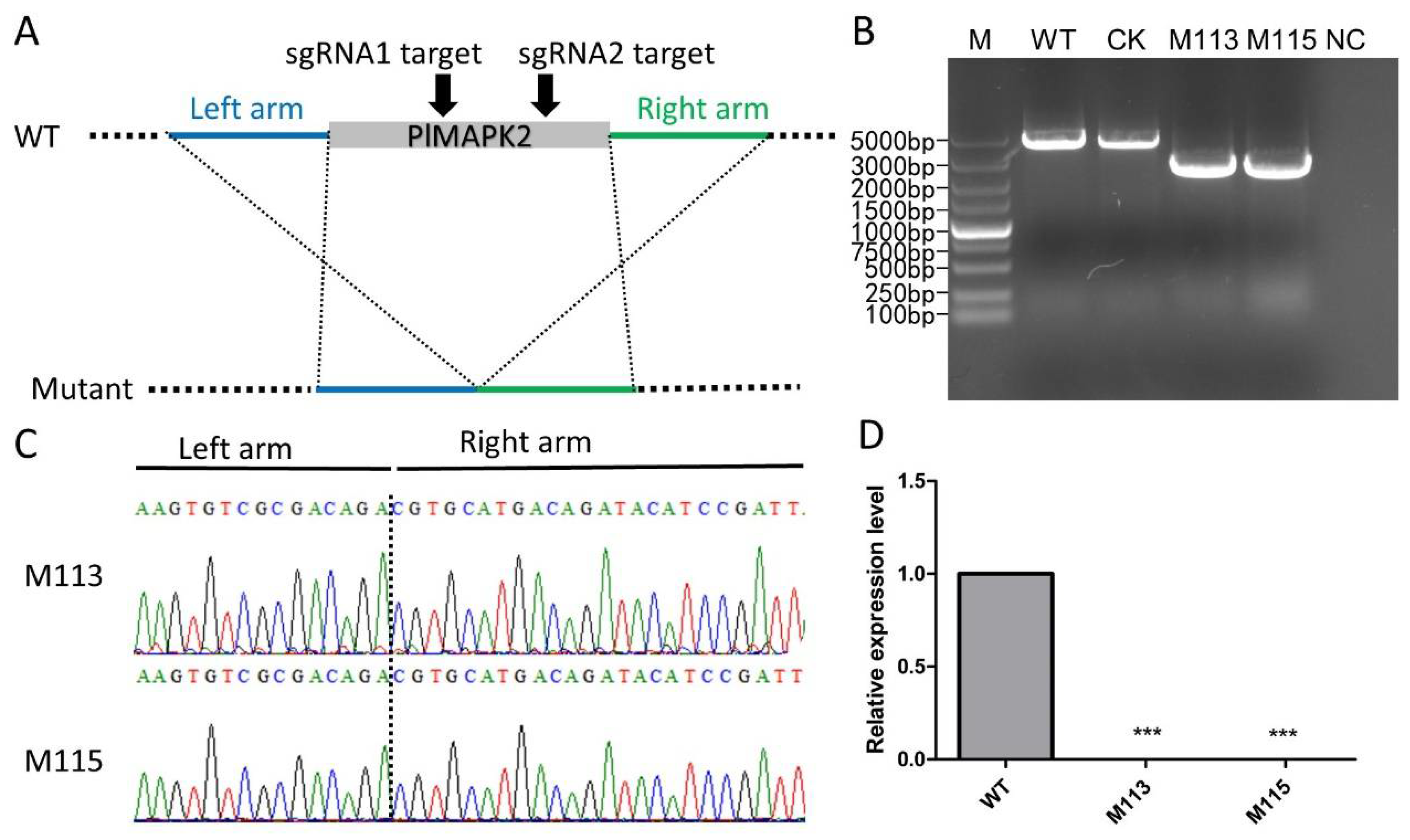
To investigate function of PlMAPK2, we generated PlMAPK2 knockout mutants using the CRISPR/Cas9 system. Single-guide RNAs were designed to disrupt the PlMAPK2 coding region (Figure 2A). The transformants were screened by G418 resistance and then verified by genomic PCR and sequencing (Figure 2B,C). Finally, two PlMAPK2 mutants (M113, M115) were obtained, and a transformant that failed to acquire PlMAPK2 mutation was selected as the control (CK). We also confirmed the transcription of PlMAPK2 in WT (wild-type stain, SHS3) and PlMAPK2 mutants, and our results showed that PlMAPK2 did not express in M113 and M115 (Figure 2D). These results demonstrated that PlMAPK2 was knocked out in M113 and M115.

2.3. Knockout of PlMAPK2 Affects Zoosporogenesis of P. litchii

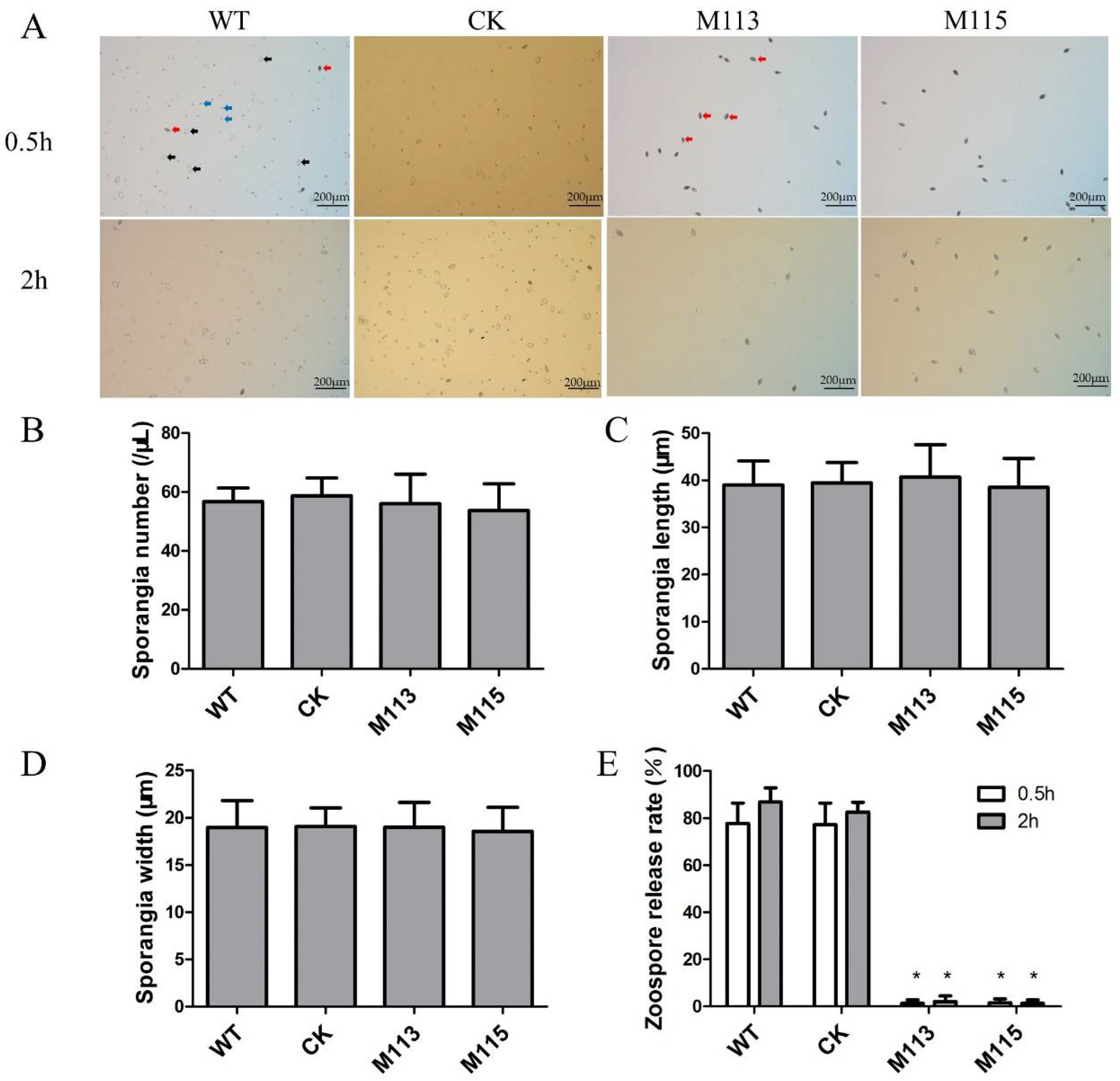
At the asexual life cycle stages, oomycetes produce sporangia, which then release zoospores. To investigate the function of PlMAPK2 at the asexual development, the PlMAPK2 mutants, CK and WT, were cultured in CJA medium for 5 days, and then the sporangia were collected and calculated. We found that the knockout of PlMAPK2 did not affect the production of sporangia (Figure 3A,B). The sporangia produced by PlMAPK2 mutants showed similar lengths and widths compared with WT (Figure 3C,D). Zoospore release rates were observed 0.5 and 2 h after collecting the sporangia in water. Around 87% of WT sporangia released zoospores. However, only about 2% of M113 or M115 sporangia released zoospores, in 2 h (Figure 3A,E). Therefore, our results indicated that PlMAPK2 affected the zoospores release, but not the production of sporangia.

2.4. PlMAPK2 Is Required for Sporangial Cleavage during Zoospore Development

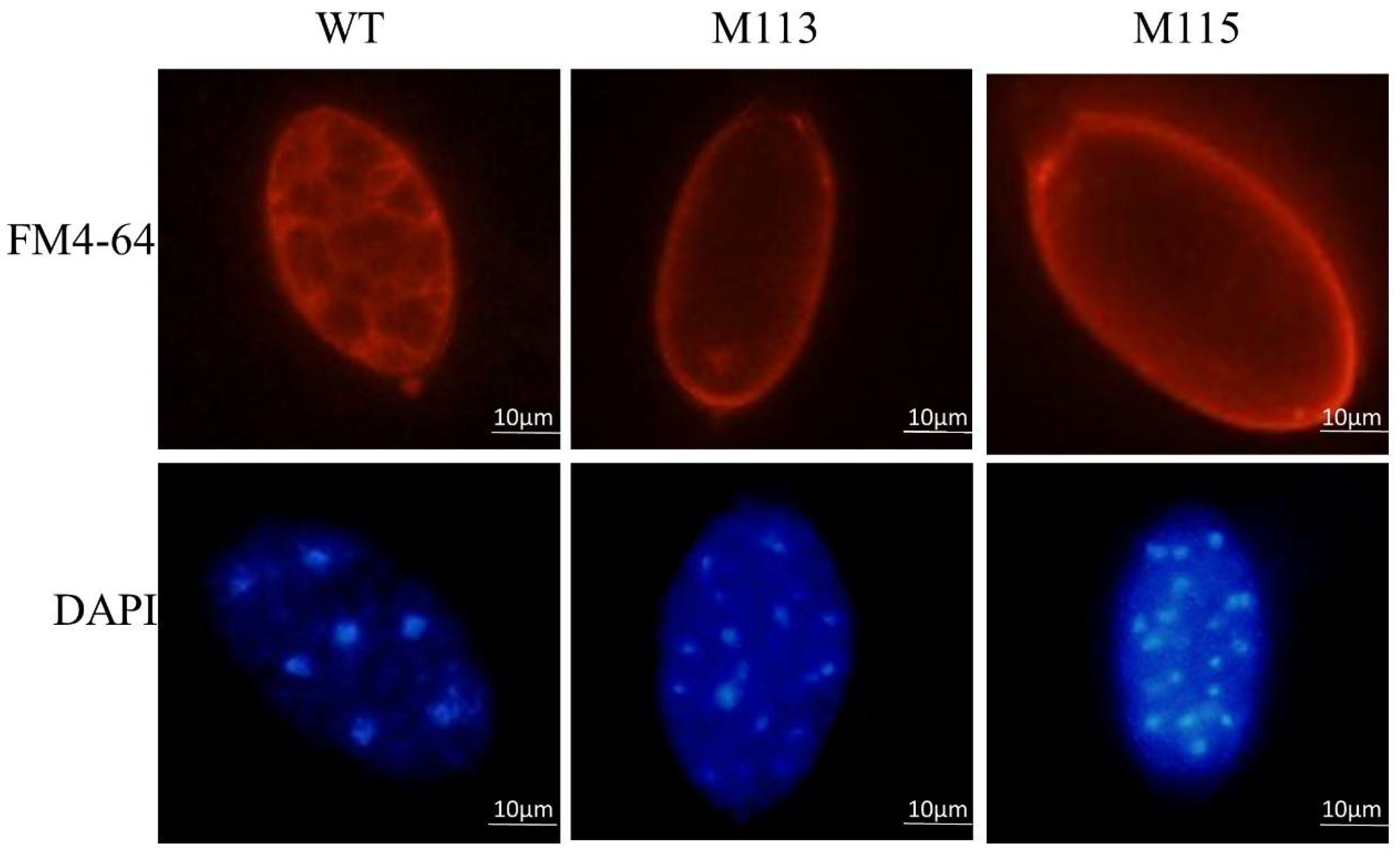
Zoospore development involves cleavage of the sporangial cytoplasm by nucleus-enveloping membrane networks [14], we next assessed sporangial cleavage in WT and PlMAPK2 mutants by FM4-64 and DAPI staining. We found that the sporangial cleavage was impaired in PlMAPK2 mutants. The nuclei within the WT sporangia were regularly spaced and the cytoplasm differentiated to form developed zoospores. In contrast, the sporangium cytoplasm of the PlMAPK2 mutants remained undifferentiated and nuclei remained disordered (Figure 4), so there were few developed zoospores formed and released in PlMAPK2 mutants. Our results demonstrated that deletion of PlMAPK2 impaired the sporangial cleavage during zoospore development, leading to defects of zoospore release.

2.5. PlMAD1 and PlMYB1 Were Down-Regulated in PlMAPK2 Mutants

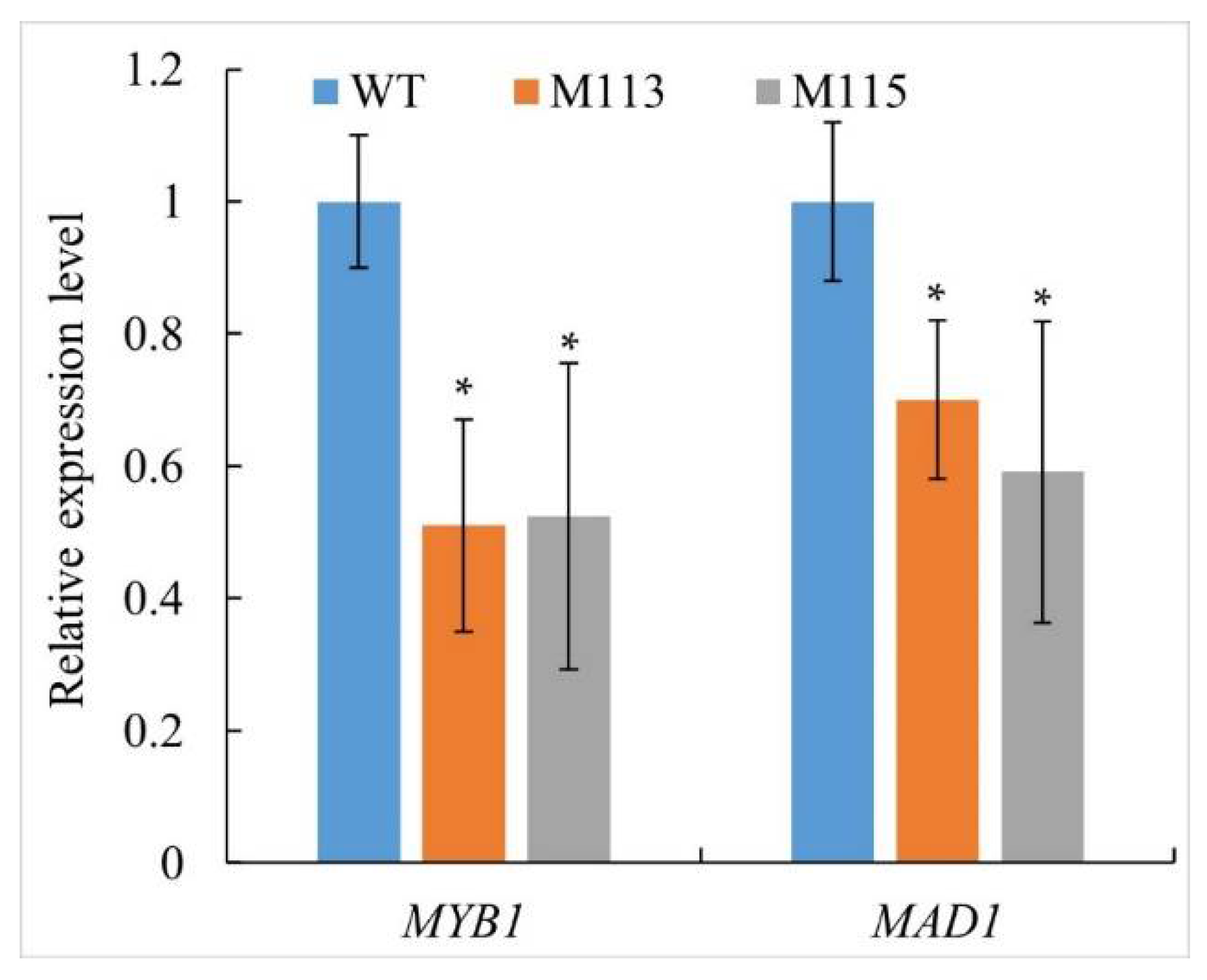
P. sojae is phylogenetically close to P. litchii. In P. sojae, a MADS-box transcription factor PsMAD1 and a Myb transcription factor PsMYB1 were reported to be involved in cleavage of sporangium and virulence [15,16]. We identified the P. litchii orthologs of MAD1 and MYB1, named as PlMAD1 and PlMYB1, respectively (Table S2). These proteins share sequence identities of 80 and 82% respectively with their counterparts in P. sojae. Here, the total RNA was extracted from sporangia of WT and PlMAPK2 mutants. Then, we evaluated the transcriptional levels of those two genes at the stage of sporangia by qRT-PCR. Our results showed that PlMAD1 and PlMYB1 were down-regulated in PlMAPK2 mutants (Figure 5), suggesting that PlMAPK2 affects transcriptional levels of PlMAD1 and PlMYB1.

2.6. PlMAPK2 Is Required for Full Virulence and Regulates Laccase Activity in P. litchii

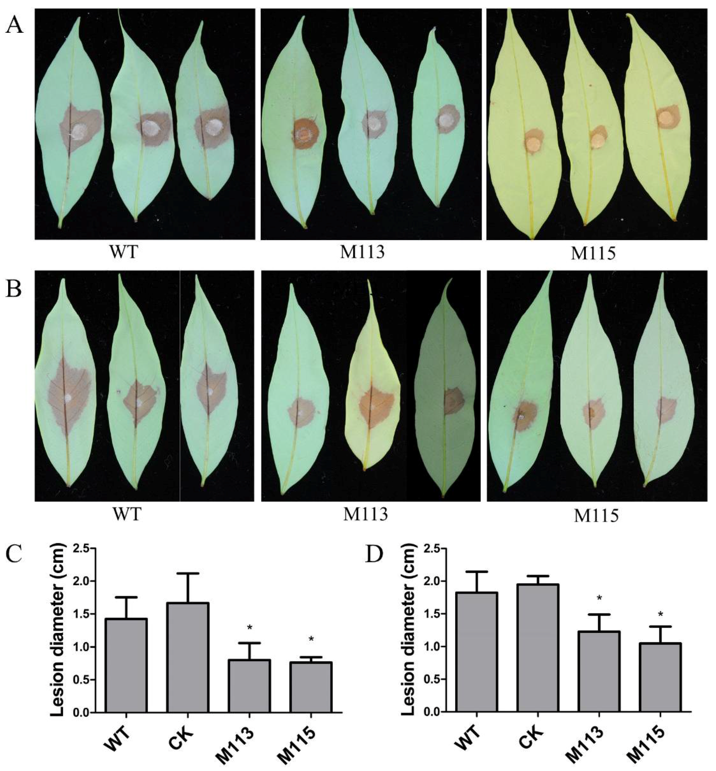
PlMAPK2 is up-regulated in the early stage of infection; therefore, PlMAPK2 might be critical for infection. The virulence of WT and PlMAPK2 mutants were tested on lychee leaves. Our results showed that the lesion length of PlMAPK2 mutants is reduced by 33–54% compared with WT (Figure 6). These results indicate that PlMAPK2 is required for the full virulence of P. litchii.
In fungi, laccases participate in the oxidation of antibiotics such as flavonoids and phytoalexins and contribute to the virulence of pathogen [17]. In oomycetes, the laccase activity is also associated with the plant infection [5,10,18]. Here we tested the laccase activity of PlMAPK2 mutants and found that these mutants showed decreased laccase activity (Figure 7A). Therefore, we conclude that PlMAPK2 is critical for the virulence and regulates laccase activity in P. litchii.
We further examined the transcriptional levels of 8 laccase-encoding genes, and found that 3 of them, Pl104952, Pl106183 and Pl111417, were dramatically down-regulated in PlMAPK2 mutants, while Pl103272 and Pl106923 were slightly up-regulated in PlMAPK2 mutants (Figure 7B); the transcription level of Pl106181, Pl 106924 and Pl111416 showed no significant difference in PlMAPK2 mutant. We inferred that PlMAPK2 affected the transcription levels of these laccase-encoding genes.

2.7. Assessment of Growth and Stress Response in the PlMAPK2 Mutants

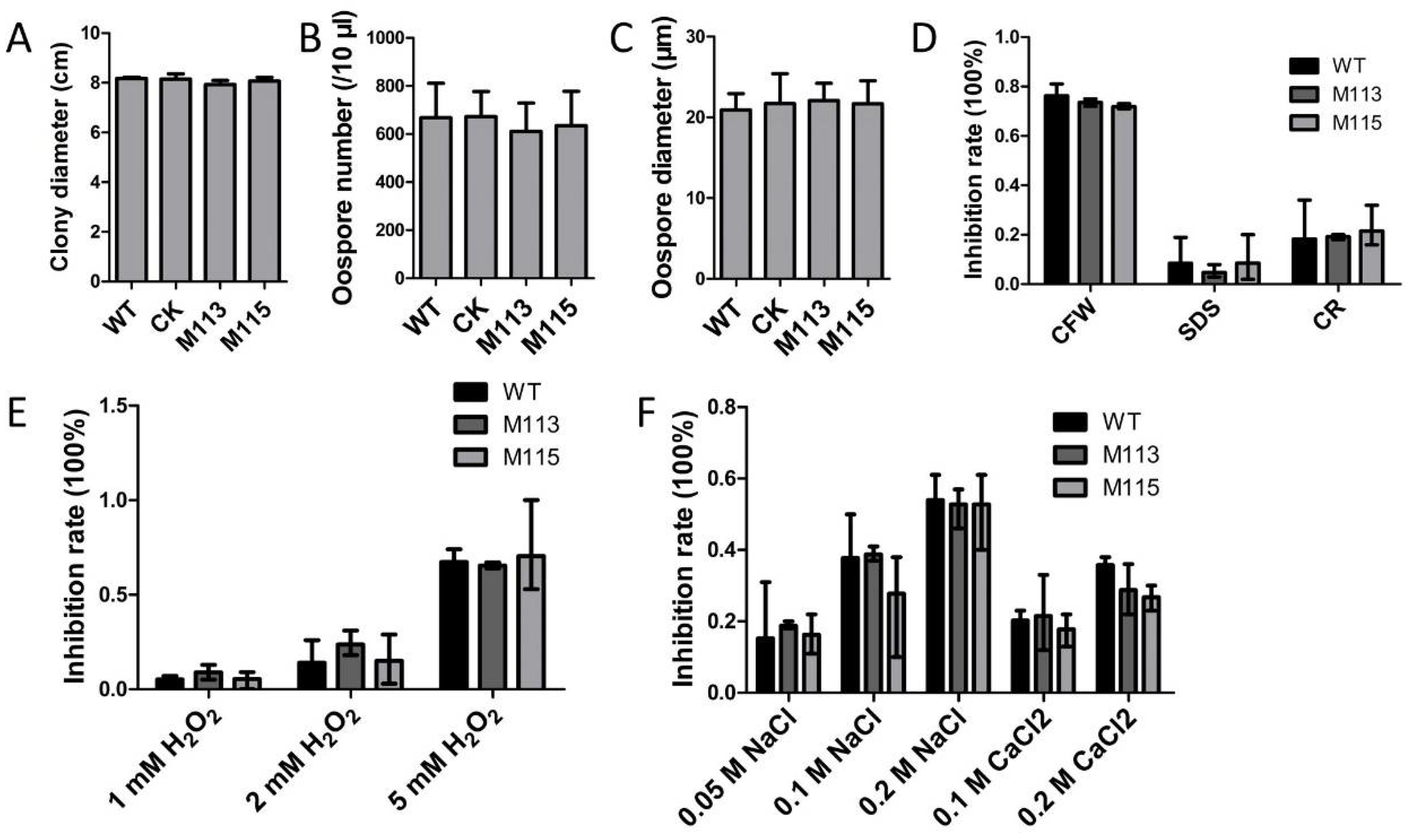
PlMAPK2 showed 53% similarity with Fus3 of S. cerevisiae. In S. cerevisiae, Fus3 involved in filamentous growth and mating [6]. Here, we tested the growth, production of sexual spores (oospores) and responses to various stresses (CFW, SDS, CR, H2O2, NaCl and CaCl2). Compared to the WT, the mutants showed no significant difference of growth rate on CJA medium. The PlMAPK2 mutants were exposed to various stresses, and we found that PlMAPK2 mutants showed no significance in responses to various stresses comparing with WT (Figure 8). We further found that the oospore size and production of the PlMAPK2 mutants were also similar to that of WT (Figure 8 and Figure S2). These results suggested that deletion of PlMAPK2 did not affect the filamentous growth, responses to various stresses, or sexual reproduction in P. litchii.

3. Discussion

MAPK cascades are widespread and conserved in plants, fungi and oomycetes. However, the function of MAPKs in development and pathogenicity of oomycetes are rarely known. In oomycetes, PsMAPK1, PsSAK1, PsMPK7 and MAPK10 showed various functions in vegetative growth, zoospore viability, stress tolerance, detoxification of reactive oxygen species, cyst germination, sexual reproduction, laccase activity and pathogenicity [10,11,12,13]. In this study, we identified the PlMAPK2 and found that its orthologs in oomycete are functional unknown MAPKs. We found that PlMAPK2 plays a key role in zoosporogenesis by regulating the sporangial cleavage, PlMAPK2 also contributes to the pathogenicity and is associated with laccase activity of this pathogen. This study firstly reported a MAPK required for sporangia cleavage in oomycete, providing new insight into the functions of MAPK signaling pathway in oomycete life cycle.
Zoospore release is important for plant pathogenic oomycete life cycle, which is associated with the sporangial cytoplasm cleavage and the assembly of flagella [14]. CDC14 orthologs have been revealed to function in sporulation in pathogenic oomycetes P. infestans and P. sojae [19,20]. MAD-box and MYB transcription factors were also shown to be essential for sporangial cleavage in P. sojae [15,16]. Here, our results showed that PlMAPK2 mutation led to defects in sporangial cleavage and down-regulation of PlMAD1 and PlMYB1. Therefore, we hypothesize that PlMAPK2 regulates zoospore release through transcription factors including MAD1 and MYB1. However, further studies are needed to reveal the zoospore formation process.
In S. cerevisiae, MAPK kinase cascades always directly regulate transcription factors via phosphorylation [6], therefore indirectly modulating the transcription of down-stream genes. In oomycetes, silencing of a P. sojae MAPK-encoding gene, PsSAK1, decreased the transcriptional level of PsMYB1. Here, we found that the expression of PlMAD1 and PlMYB1 was down-regulated in PlMAK2 mutants (Figure 5), which suggested that PlMAPK2 might regulate zoosporogenesis and virulence by regulating PlMAD1 and PlMYB1 at expression level. Additional experiments are needed to clarify how MAPK signaling regulates sporangial development and the activation of pathogenic pathways in oomycetes.
Knockout of PlMAPK2 decreased the pathogenicity, the laccase activity and interfered the transcription of several laccase-encoding genes in P. litchii. These results are consistent with previous reports that laccase activity is important for plant pathogenic fungi and oomycetes [10,21]. Further, we analyzed the expressions of laccase-encoding genes; three of them, Pl104952, Pl106183 and Pl111417, are significantly down-regulated in the PlMAPK2 mutants (Figure 7B). We speculate that PlMAPK2 might regulate laccase activity by affecting the transcription of laccase-encoding genes in P. litchii. PlMAPK10 also regulated the transcription level of another laccase-encoding gene [10]. These results suggested that different MAPKs might specifically regulate corresponding laccase-encoding genes.
In S. cerevisiae, Fus3 was involved in mating and filamentous growth [6], and in Magnaporthe oryzae, Pmk1, the homologue of Fus3, was involved in appressorium formation and penetration and invasive growth [8]. In P. litchii, PlMAPK2 showed highest similarity with Fus3 of S. cerevisiae, but PlMAPK2 was not associated with growth, sexual development and responses to various tolerance stresses of P. litchii (Figure 8). We found that PlMAPK2 is important for the zoospore release and virulence of this pathogen (Figure 3). These results indicated that sequence and functional differentiation occurred in fungi and oomycete MAPK cascades for their specific development and pathogenic processes.

4. Materials and Methods

4.1. Sequences and Phylogenetic Analysis

Genome sequence of P. litchii was obtained from NCBI (BioProject ID: PRJNA290406) [22]. Genome sequence of Pythium species (Pythium vexans DAOM BR484, Pythium aphanidermatum DAOM BR444, Pythium irregulare DAOM BR486) were obtained from the Pythium genome database (http://protists.ensembl.org/index.html, accessed on 6 July 2020). Other oomycete and fungal sequences were obtained from NCBI (http://www.ncbi.nlm.nih.gov, accessed on 9 June 2020). The obtained protein sequences were submitted to NCBI-CDD (http://www.ncbi.nlm.nih.gov/Structure/cdd, accessed on 9 July 2020) to identify conserved domains. All sequences used in this study were listed in Table S2.
Sequence alignments were created using ClustalW program [23] and the phylogenetic tree was constructed with MEGA 6.0 program using a neighbor-joining algorithm with 1000 bootstrap replicates [24].

4.2. Strains and Culture Conditions

Peronophythora litchii strain SHS3 were locally maintained on CJA medium (juice from 200 g carrot topped up to 1 L, 15 g/L agar for solid media) [25] at 25 °C in the dark.

4.3. Transcriptional Analysis

Total RNA was extracted using the total RNA kit (catalog [cat.] number R6834-01; Omega). Samples included mycelia (MY), sporangia (SP), zoospores (ZO), cysts (CY), germination of cysts (GC) and 1.5–24 h post inoculation. The first-strand cDNA was synthesized from total RNA by oligo(dT) priming using a Moloney murine leukemia virus (MMLV) reverse transcriptase kit (number S28025-014; Invitrogen, Carlsbad, CA, USA). Transcription of PlMAPK2 was analyzed with qRT-PCR (Quantitative Reverse transcription PCR) assay using primers MAPK2-qRTF and MAPK2-qRTR on ABI Prism 7500 Fast real-time PCR System (Applied Biosystems, Foster City, CA, USA). P. litchii actin gene was used as loading control and the relative fold change was calculated using the 2∆∆Ct method [26]. Transcription of PlMAD1 (P. litchii MAD-box transcription factor 1), PlMYB1 (P. litchii MYB transcription factor 1) and laccase-encoding genes were also analyzed as described above, primers were listed in Supplementary Table S1.

4.4. CRISPR/Cas9 Editing for PlMAPK2

To generate PlMAPK2 knockout transformants, P. litchii protoplasts were transformed using the polyethylene glycol (PEG)–CaCl2-mediated method as described previously [25,27,28]. The plasmids, pYF2-PsNLS-hSpCas9, pYF2.3G-RibosgRNA::PlMAPK2 and pBluescript II KS(+)::PlMAPK2 were cotransformed into protoplasts of P. litchii (Figure 1A and Figure S3). We designed two sgRNAs (sgRNA1 and sgRNA2) that target PlMAPK2 using the protocol described previously [27], then checking their RNA secondary structure online (http://rna.urmc.rochester.edu/RNAstructureWeb/Servers/Predict1/Predict1.html, accessed on 9 July 2020). The transformed protoplasts were regenerated overnight, and the recovered mycelia were selected on CJA medium with 30 μg/mL G418. After 2–3 days, the primary transformants were transferred to new selective medium and identified by subsequent genomic PCR (Polymerase chain reaction) and sequencing analyses. The primers used to genomic PCR are listed in Table S1.

4.5. Analysis of Sporangium, Zoospore and Oospore Development

Sporangia were harvested by flooding the mycelia, which had been cultured on CJA media for 5 days, with sterile water, and then filtering the suspension through a 100 μm strainer [25]. The suspension was incubated with sterile distilled water at 16 °C for 2 h to release zoospores. For oospore observation, P. litchii strains were inoculated on CJA medium for 10 days, then the production and diameter of oospores were calculated.

4.6. Chemical Dyes with FM4-64 and DAPI (4’,6-Diamidino-2-Phenylindole, Dilactate)

Sporangium were visualized by the blue-fluorescent nucleic acid stain 4′, 6-diamidino-2-phenylindole (DAPI), dilactate (D3571, Invitrogen, USA) and then viewed with Olympus BX53F microscope as described previously [15]. Red-fluorescent FM 4-64 dye (T13320, Invitrogen, USA) was used to analyze the zoospore membrane in sporangium [16].

4.7. Pathogenicity Test

Five-mm mycelium plugs of the PlMAPK2 mutants were inoculated on the abaxial surface of lychee leaves in a dark box and then were placed in a climate room under 80% humidity in the dark at 25 °C; lesion lengths were measured after 48 h. Inoculation with sporangia: the sporangia were collected as described above and we adjusted the concentration of sporangia to 10 sporangia per μL, then 10 μL of sporangium suspension were inoculated on abaxial surface of the leaves, the lesion lengths were measured after 48 h. WT and CK strains were used as controls.

4.8. Laccase Activity Assays

The laccase activity was analyzed following the previously described procedure [18]. The experiments were repeated three times independently, with three technical replicates each time.

5. Conclusions

In summary, we characterized the functions of PlMAPK2 in P. litchii with CRISPR/Cas9-mediated genome editing method. PlMAPK2 plays a key role in zoosporogenesis, virulence and regulation of laccase activity and transcription of PlMAD1, PlMYB1 in P. litchii. This study provided new insight into the functions of MAPK signaling in zoosporogenesis and pathogenicity of oomycete, and identified MAPK2 as a potential fungicide target for oomycete pathogen control.

Supplementary Materials

The following are available online at https://www.mdpi.com/article/10.3390/ijms22073524/s1, Table S1. The primers used in this study. Table S2. Sequences used in this study. Figure S1 Phylogenetic analysis of putative PlMAPK2 orthologs based on the amino acid sequences (Table S2). The neighbor-joining tree was constructed by MEGA 6.0 using the neighbor-joining algorithms with 1000 bootstrap replications [23]. Bootstrap values greater than 50 were displayed. Figure S2 PlMAPK2 is not associated with growth and responses to various stresses. (A) WT and PlMAPK2 mutants were inoculated on CJA medium, photographs were taken 5 days after inoculation. (B) WT and PlMAPK2 mutants were inoculated on Plich medium with or without indicated chemicals (350 μg/mL CFW, 25 μg/mL SDS, 100 μg/mL CR, 1 mM H2O2, 2 mM H2O2, 5 mM H2O2 0.05 mM NaCl, 0.1 mM NaCl, 0.2 mM NaCl, 0.1 mM CaCl2 and 0.2 mM CaCl2.). Photographs were taken 5 days after inoculation. Figure S3 Structure of pBluescript II KS(+)::MAPK2. Left arm and right arm were inserted in pBluescript II KS(+) plasmid.

Author Contributions

Conceptualization, J.H., P.X., Y.D. and G.K.; funding acquisition, G.K.; investigation, J.H., P.X., W.H., J.W., Q.Z., W.Y., W.L., J.S., L.J. and T.G.; supervision, G.K.; writing—original draft, J.H.; writing—review and editing, P.X., Y.D., M.L., Z.J. and G.K. All authors have read and agreed to the published version of the manuscript.

Funding

This work was supported by grants to GK from the Natural Science Foundation of Guangdong Province, China (2020A1515011335) and the National Natural Science Foundation of China (31701771).

Institutional Review Board Statement

Not applicable.

Informed Consent Statement

Not applicable.

Data Availability Statement

Not applicable.

Acknowledgments

We thank Brett Tyler (Oregon State University, United States) for the plasmids.

Conflicts of Interest

The authors declare no conflict of interest.

References

  1. Cavalier-Smith, T.; Chao, E.E.; Lewis, R. Multigene phylogeny and cell evolution of chromist infrakingdom Rhizaria: Contrasting cell organisation of sister phyla Cercozoa and Retaria. Protoplasma 2018, 255, 1517–1574. [Google Scholar] [CrossRef] [Green Version]
  2. Tyler, B.M.; Tripathy, S.; Zhang, X.; Dehal, P.; Jiang, R.H.; Aerts, A.; Arredondo, F.D.; Baxter, L.; Bensasson, D.; Beynon, J.L.; et al. Phytophthora genome sequences uncover evolutionary origins and mechanisms of Pathogenesis. Science 2006, 313, 1261–1266. [Google Scholar] [CrossRef] [PubMed] [Green Version]
  3. Kamoun, S.; Furzer, O.; Jones, J.D.G.; Judelson, H.S.; Ali, G.S.; Dalio, R.J.D.; Roy, S.G.; Schena, L.; Zambounis, A.; Panabières, F.; et al. The Top 10 oomycete pathogens in molecular plant pathology. Mol. Plant Pathol. 2015, 16, 413–434. [Google Scholar] [CrossRef] [PubMed]
  4. Kong, G.; Li, T.; Huang, W.; Li, M.; Shen, W.; Jiang, L.; Hsiang, T.; Jiang, Z.; Xi, P. Detection of Peronophythora litchii on lychee by loop-mediated isothermal amplification assay. Crop. Prot. 2021, 139, 105370. [Google Scholar] [CrossRef]
  5. Kong, G.; Chen, Y.; Deng, Y.; Feng, D.; Jiang, L.; Wan, L.; Li, M.; Jiang, Z.; Xi, P. The Basic Leucine Zipper Transcription Factor PlBZP32 Associated with the Oxidative Stress Response Is Critical for Pathogenicity of the Lychee Downy Blight Oomycete Peronophythora litchii. mSphere 2020, 5, e00261–e00320. [Google Scholar] [CrossRef]
  6. Hamel, L.-P.; Nicole, M.-C.; Duplessis, S.; Ellis, B.E. Mitogen-Activated Protein Kinase Signaling in Plant-Interacting Fungi: Distinct Messages from Conserved Messengers. Plant Cell 2012, 24, 1327–1351. [Google Scholar] [CrossRef] [Green Version]
  7. Widmann, C.; Gibson, S.; Jarpe, M.B.; Johnson, G.L. Mitogen-Activated Protein Kinase: Conservation of a Three-Kinase Module from Yeast to Human. Physiol. Rev. 1999, 79, 143–180. [Google Scholar] [CrossRef]
  8. Jiang, C.; Zhang, X.; Liu, H.; Xu, J.-R. Mitogen-activated protein kinase signaling in plant pathogenic fungi. PLoS Pathog. 2018, 14, e1006875. [Google Scholar] [CrossRef] [PubMed] [Green Version]
  9. González-Rubio, G.; Fernández-Acero, T.; Martín, H.; Molina, M. Mitogen-Activated Protein Kinase Phosphatases (MKPs) in Fungal Signaling: Conservation, Function, and Regulation. Int. J. Mol. Sci. 2019, 20, 1709. [Google Scholar] [CrossRef] [Green Version]
  10. Jiang, L.; Situ, J.; Deng, Y.Z.; Wan, L.; Xu, D.; Chen, Y.; Xi, P.; Jiang, Z. PlMAPK10, a Mitogen-Activated Protein Kinase (MAPK) in Peronophythora litchii, Is Required for Mycelial Growth, Sporulation, Laccase Activity, and Plant Infection. Front. Microbiol. 2018, 9, 426. [Google Scholar] [CrossRef] [Green Version]
  11. Li, A.; Zhang, M.; Wang, Y.; Li, D.; Liu, X.; Tao, K.; Ye, W.; Wang, Y. PsMPK1, an SLT2-type mitogen-activated protein kinase, is required for hyphal growth, zoosporogenesis, cell wall integrity, and pathogenicity in Phytophthora sojae. Fungal Genet. Biol. 2014, 65, 14–24. [Google Scholar] [CrossRef]
  12. Li, A.; Wang, Y.; Tao, K.; Dong, S.; Huang, Q.; Dai, T.; Zheng, X.; Wang, Y. PsSAK1, a Stress-Activated MAP Kinase of Phytophthora sojae, Is Required for Zoospore Viability and Infection of Soybean. Mol. Plant-Microbe Interact. 2010, 23, 1022–1031. [Google Scholar] [CrossRef] [Green Version]
  13. Gao, J.; Cao, M.; Ye, W.; Li, H.; Kong, L.; Zheng, X.; Wang, Y. PsMPK7, a stress-associated mitogen-activated protein kinase (MAPK) in Phytophthora sojae, is required for stress tolerance, reactive oxygenated species detoxification, cyst germination, sexual reproduction and infection of soybean. Mol. Plant Pathol. 2014, 16, 61–70. [Google Scholar] [CrossRef] [PubMed]
  14. Judelson, H.S.; Blanco, F.A. The spores of Phytophthora: Weapons of the plant destroyer. Nat. Rev. Genet. 2005, 3, 47–58. [Google Scholar] [CrossRef] [PubMed]
  15. Lin, L.; Ye, W.; Wu, J.; Xuan, M.; Li, Y.; Gao, J.; Wang, Y.; Wang, Y.; Dong, S.; Wang, Y. The MADS-box Transcription Factor PsMAD1 Is Involved in Zoosporogenesis and Pathogenesis of Phytophthora sojae. Front. Microbiol. 2018, 9, 2259. [Google Scholar] [CrossRef] [PubMed]
  16. Zhang, M.; Lu, J.; Tao, K.; Ye, W.; Li, A.; Liu, X.; Kong, L.; Dong, S.; Zheng, X.; Wang, Y. A Myb Transcription Factor of Phytophthora sojae, Regulated by MAP Kinase PsSAK1, Is Required for Zoospore Development. PLoS ONE 2012, 7, e40246. [Google Scholar] [CrossRef]
  17. Cázares-García, S.V.; Vázquez-Garcidueñas, M.S.; Vázquez-Marrufo, G. Structural and Phylogenetic Analysis of Laccases from Trichoderma: A Bioinformatic Approach. PLoS ONE 2013, 8, e55295. [Google Scholar] [CrossRef] [Green Version]
  18. Sheng, Y.; Wang, Y.; Meijer, H.J.; Yang, X.; Hua, C.; Ye, W.; Tao, K.; Liu, X.; Govers, F.; Wang, Y. The heat shock transcription factor PsHSF1 of Phytophthora sojae is required for oxidative stress tolerance and detoxifying the plant oxidative burst. Environ. Microbiol. 2015, 17, 1351–1364. [Google Scholar] [CrossRef]
  19. Fong, A.M.V.A.; Judelson, H.S. Cell cycle regulator Cdc14 is expressed during sporulation but not hyphal growth in the fungus-like oomycete Phytophthora infestans. Mol. Microbiol. 2003, 50, 487–494. [Google Scholar] [CrossRef] [PubMed]
  20. Zhao, W.; Yang, X.; Dong, S.; Sheng, Y.; Wang, Y.; Zheng, X. Transient silencing mediated by in vitro synthesized double-stranded RNA indicates that PsCdc14 is required for sporangial development in a soybean root rot pathogen. Sci. China Life Sci. 2011, 54, 1143–1150. [Google Scholar] [CrossRef] [Green Version]
  21. Guo, M.; Chen, Y.; Du, Y.; Dong, Y.; Guo, W.; Zhai, S.; Zhang, H.; Dong, S.; Zhang, Z.; Wang, Y.; et al. The bZIP Transcription Factor MoAP1 Mediates the Oxidative Stress Response and Is Critical for Pathogenicity of the Rice Blast Fungus Magnaporthe oryzae. PLoS Pathog. 2011, 7, e1001302. [Google Scholar] [CrossRef] [Green Version]
  22. Ye, W.; Wang, Y.; Shen, D.; Li, D.; Pu, T.; Jiang, Z.; Zhang, Z.; Zheng, X.; Tyler, B.M.; Wang, Y. Sequencing of the Litchi Downy Blight Pathogen Reveals It Is a Phytophthora Species with Downy Mildew-Like Characteristics. Mol. Plant-Microbe Interact. 2016, 29, 573–583. [Google Scholar] [CrossRef] [PubMed] [Green Version]
  23. Thompson, J.D.; Higgins, D.G.; Gibson, T.J. CLUSTAL W: Improving the sensitivity of progressive multiple sequence alignment through sequence weighting, position-specific gap penalties and weight matrix choice. Nucleic Acids Res. 1994, 22, 4673–4680. [Google Scholar] [CrossRef] [PubMed] [Green Version]
  24. Tamura, K.; Stecher, G.; Peterson, D.; Filipski, A.; Kumar, S. MEGA6: Molecular evolutionary genetics analysis version 6.0. Mol. Biol. Evol. 2013, 30, 2725–2729. [Google Scholar] [CrossRef] [PubMed] [Green Version]
  25. Jiang, L.; Ye, W.; Situ, J.; Chen, Y.; Yang, X.; Kong, G.; Liu, Y.; Tinashe, R.J.; Xi, P.; Wang, Y.; et al. A Puf RNA-binding protein encoding gene PlM90 regulates the sexual and asexual life stages of the litchi downy blight pathogen Peronophythora litchii. Fungal Genet. Biol. 2017, 98, 39–45. [Google Scholar] [CrossRef] [PubMed]
  26. Livak, K.J.; Schmittgen, T.D. Analysis of relative gene expression data using real-time quantitative PCR and the 2(-Delta Delta C(T)) Method. Methods 2001, 25, 402–408. [Google Scholar] [CrossRef]
  27. Fang, Y.; Tyler, B.M. Efficient disruption and replacement of an effector gene in the oomycete Phytophthora sojae using CRISPR/Cas9. Mol. Plant Pathol. 2016, 17, 127–139. [Google Scholar] [CrossRef]
  28. Situ, J.; Jiang, L.; Fan, X.; Yang, W.; Li, W.; Xi, P.; Deng, Y.; Kong, G.; Jiang, Z. An RXLR effector PlAvh142 from Peronophythora litchii triggers plant cell death and contributes to virulence. Mol. Plant Pathol. 2020, 21, 415–428. [Google Scholar] [CrossRef] [Green Version]
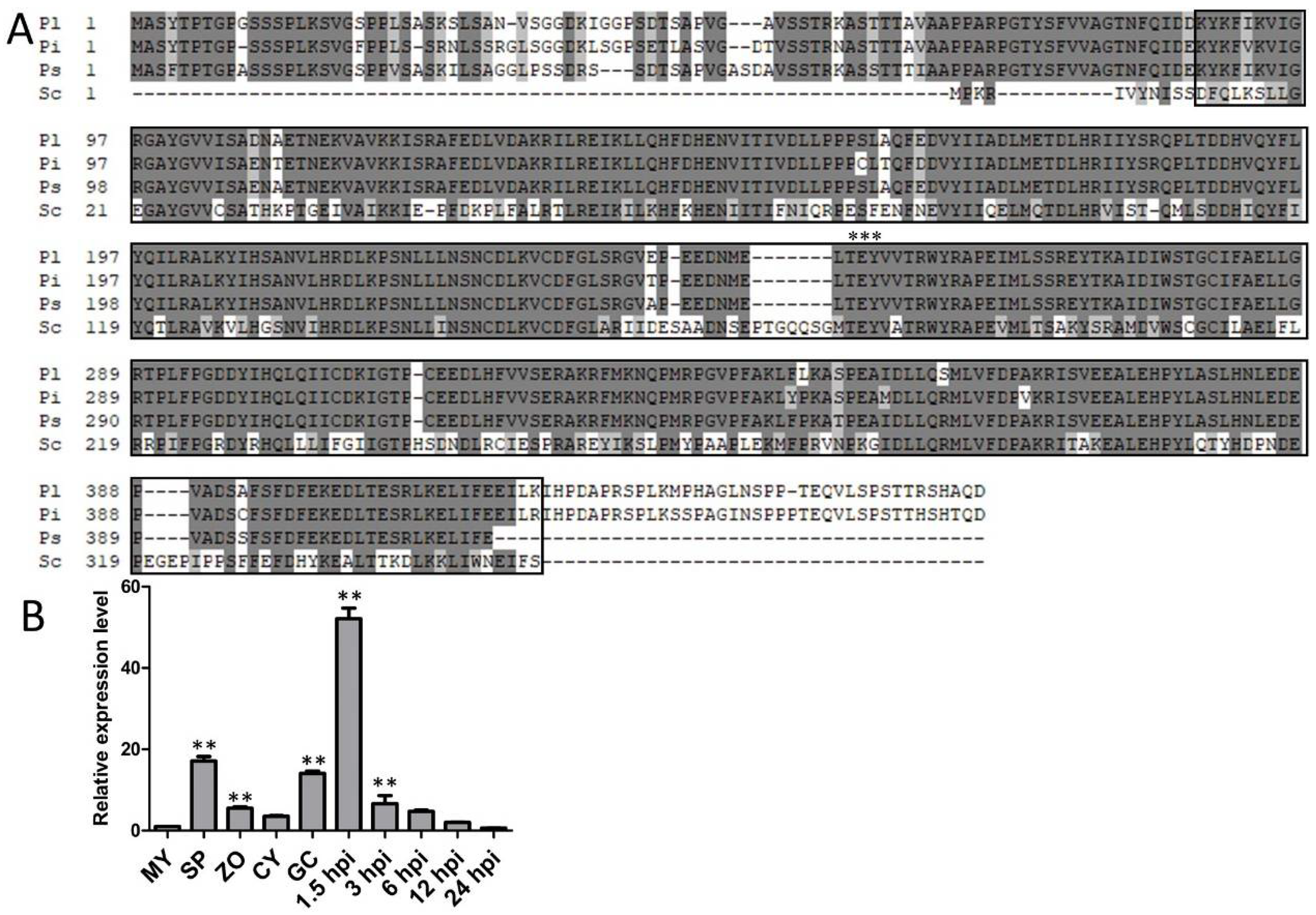
Figure 1. Identification of PlMAPK2 in P. litchii. (A) Amino acid sequences alignment of PlMAPK2 (Pl) and its orthologs from P. infestans (Pi), P. sojae (Ps) and S. cerevisiae (Sc). The black boxes and three black asterisks represent catalytic domain of the serine/threonine kinase and conserved TEY motif, respectively. (B) Expression pattern of PlMAPK2 during the asexual life cycle and infection stages was analyzed by quantitative reverse transcription PCR (qRT-PCR). MY: Mycelia; SP: sporangia; ZO: zoospore; CY: cyst; GC: germination of cyst; hpi: hours post inoculation. Relative expression levels were calculated using the MY values as reference. Asterisks indicate significant difference (** p < 0.01) based on Bonferroni’s multiple comparison test. These experiments were repeated three times.
Ijms 22 03524 g001
Figure 2. CRISPR/Cas9-mediated deletion of PlMAPK2 in P. litchii. (A) Schematic representation of the strategy of CRISPR/Cas9-mediated mutagenesis of PlMAPK2. Two single-guide RNAs targeted the PlMAPK2 gene sequence (indicated by black arrows). The PlMAPK2 mutants were identified by genomic PCR (B) and sequencing (C). (D) PlMAPK2 was not expressed in PlMAPK2 mutants. WT: wild type; CK: the transformant failed to acquire PIMAPK2 mutation; NC: negative control (distilled water as template). “***” indicates significant difference (t-test, p < 0.001). This experiment was repeated three times.
Ijms 22 03524 g002
Figure 3. Knockout of PlMAPK2 decreased the zoospores release rate. PlMAPK2 mutants, CK (the transformant failed to acquire PIMAPK2 mutation) and WT (wild type) were cultured on CJA medium for 7 days and sporangia were collected and used for releasing zoospores. (A) Photographs were taken 0.5 and 2 h after releasing zoospores. Scale bar = 200 μm. Black arrows indicate the representative released sporangia; red arrows indicate representative unreleased sporangia; blue arrows indicate representative zoospores. (B) The sporangia numbers were calculated. (C,D) The sporangia lengths and widths were measured. (E) The zoospore release rates of PlMAPK2 mutants, CK and WT were calculated after 0.5 and 2 h. Asterisks indicate significant difference between WT and PlMAPK2 mutants (t-test, p < 0.01). These experiments were replicated three times.
Ijms 22 03524 g003
Figure 4. Assessment of nuclei distribution and cytoplasm cleavage within sporangia. The nuclei in sporangia were detected by DAPI staining. Plasma membrane stained by FM4-64. Representative microscopic images for WT (wild type) or the mutants were displayed (n ≥ 9). Scale bar = 10 μm.
Ijms 22 03524 g004
Figure 5. PlMAD1 and PlMYB1 is down-regulated in sporangia of PlMAPK2 mutants, the expression of PlMAD1 and PlMYB1 were analyzed by qRT-PCR. WT (wild type) was used as control. Asterisks indicate significant difference (t-test, p < 0.01). These experiments were replicated three times.
Ijms 22 03524 g005
Figure 6. Infection assays with WT and the PlMAPK2 mutants. The virulence of PlMAPK2 mutants were tested on lychee leaves, with WT (wild type) as the positive control. Mycelium plugs (A) and sporangia (B) were inoculated on the lychee leaves, respectively. Photographs were taken 48 h after inoculation. (C,D) The lesion lengths were calculated 2 days after inoculation, corresponding to (A,B), respectively. CK: the transformant failed to acquire PIMAPK2 mutation. Asterisks indicate significant difference (t-test, p < 0.01). These experiments were replicated three times, 5 leaves were calculated for each strain each time.
Ijms 22 03524 g006
Figure 7. PlMAPK2 regulates laccase activity. (A) Mycelial mats of the aforementioned strains were inoculated on media containing 0.2 mM ABTS for 10 days. (B) The expression of 8 laccase-encoding genes were analyzed by qRT-PCR. Asterisks indicate significant difference (t-test, p < 0.01). WT: wild type. The experiments contain three independent biological repeats, each of which contained three replicas.
Ijms 22 03524 g007
Figure 8. Assessment of mycelia growth, sexual reproduction and responses to various stresses. (A) Colony diameters of WT (wild type) or the PlMAPK2 mutants were measured 5 days after inoculation on CJA medium. (B) Oospore number at 10 days after inoculation on CJA medium. (C) Oospores diameters of WT and PlMAPK2 mutants at 10 days after inoculation on CJA medium. (D) Inhibition rates of WT and PlMAPK2 mutants on Plich medium supplemented with 350 μg/mL CFW, 25 μg/mL SDS or 100 μg/mL CR. (E) Inhibition rates of WT and PlMAPK2 mutants on Plich medium with 1, 2 and 5 mM H2O2. (F) Inhibition rates of WT and PlMAPK2 mutants on Plich medium with 0.05 mM NaCl, 0.1 mM NaCl, 0.2 mM NaCl, 0.1 mM CaCl2 and 0.2 mM CaCl2. CK: the transformant failed to acquire PIMAPK2 mutation. These experiments were repeated three times, with three replicas each time.
Ijms 22 03524 g008
Publisher’s Note: MDPI stays neutral with regard to jurisdictional claims in published maps and institutional affiliations.