Review

Clinical Perspective on Proteomic and Glycomic Biomarkers for Diagnosis, Prognosis, and Prediction of Pancreatic Cancer

by
Randa G. Hanna-Sawires
1,
Jorinde H. Schiphuis
1,
Manfred Wuhrer
2,
Hans F. A. Vasen
3,
Monique E. van Leerdam
3,
Bert A. Bonsing
1,
Wilma E. Mesker
1,
Yuri E. M. van der Burgt
2,* and
Rob A. E. M. Tollenaar
1
1
Department of Surgery, Leiden University Medical Center, 2300 RC Leiden, The Netherlands
2
Center of Proteomics and Metabolomics, Leiden University Medical Center, 2300 RC Leiden, The Netherlands
3
Department of Gastroenterology, Leiden University Medical Center, 2300 RC Leiden, The Netherlands
*
Author to whom correspondence should be addressed.
Int. J. Mol. Sci. 2021, 22(5), 2655; https://doi.org/10.3390/ijms22052655
Submission received: 27 January 2021 / Revised: 26 February 2021 / Accepted: 2 March 2021 / Published: 6 March 2021
(This article belongs to the Special Issue Recent Advances of Proteomics Applied to Cancer and Human Diseases)

Abstract

:
Pancreatic ductal adenocarcinoma (PDAC) is known as a highly aggressive malignant disease. Prognosis for patients is notoriously poor, despite improvements in surgical techniques and new (neo)adjuvant chemotherapy regimens. Early detection of PDAC may increase the overall survival. It is furthermore foreseen that precision medicine will provide improved prognostic stratification and prediction of therapeutic response. In this review, omics-based discovery efforts are presented that aim for novel diagnostic and prognostic biomarkers of PDAC. For this purpose, we systematically evaluated the literature published between 1999 and 2020 with a focus on protein- and protein-glycosylation biomarkers in pancreatic cancer patients. Besides genomic and transcriptomic approaches, mass spectrometry (MS)-based proteomics and glycomics of blood- and tissue-derived samples from PDAC patients have yielded new candidates with biomarker potential. However, for reasons discussed in this review, the validation and clinical translation of these candidate markers has not been successful. Consequently, there has been a change of mindset from initial efforts to identify new unimarkers into the current hypothesis that a combination of biomarkers better suits a diagnostic or prognostic panel. With continuing development of current research methods and available techniques combined with careful study designs, new biomarkers could contribute to improved detection, prognosis, and prediction of pancreatic cancer.

1. Introduction

In the previous decade, the increase in pancreatic ductal adenocarcinoma (PDAC) incidences has been higher than for other cancers. Currently, PDAC is the 11th most common cancer in the world [1]. Moreover, PDAC diagnosis implies one of the most unfavorable prognoses with a five year overall survival rate between 5–8% as a result of its aggressive tumor behavior with extensive local and metastatic spread. Often PDACs are in advanced stage upon diagnosis (80%) and curative treatment is no longer possible [2]. Available intensive treatment regimens with chemo(radio)therapy and/or surgery is associated with severe complications and side effects, resulting in an impaired quality of life [3]. Understanding tumor biology and knowledge on correlations between clinicopathologic parameters and progress of disease could provide leads for precision medicine. For example, response to therapy can be anticipated and support a decision to, in some cases, alternative options for treatment [4]. Early detection of PDAC provides a window of opportunity where treatment with curative intention is possible [5]. So far, only CA 19-9 is routinely applied as a tumor marker. However, its clinical value is limited to post-treatment follow-up and surveillance [6]. The Dutch Nationwide Program for the detection of pancreatic cancer in high-risk individuals recently reported that screening by means of imaging is insufficient and that biomarker research is warranted [7]. The clinical decision pathway for sporadic PDAC patients as well as for individuals with a hereditary risk is depicted in Figure 1. Within this care pathway, an urgent need for novel biomarkers for screening and diagnostic purposes is broadly acknowledged, and consequently, numerous exploratory studies have been performed following different methods. Better understanding of the natural course of PDAC might allow improvements in current surveillance protocols, thereby possibly improving oncological outcome. Therefore, biomarkers that can provide this information could be of great clinical benefit. An alternative method for current screening modalities could be reliable and specific biomarkers detectable in peripheral blood to detect PDAC in its earliest stage.
In this review, candidate biomarkers for PDAC detection, prognostication, and prediction are summarized, with emphasis on proteins and protein glycosylation. Most efforts have followed mass spectrometry (MS)-based proteomics or glycomics strategies [8,9,10,11,12,13]. The use of biomarkers in clinical pathways will be discussed and recommendations for further exploratory biomarker research are given.

1.1. Sporadic Pancreatic Cancer

The worldwide incidence of pancreatic cancer is estimated at 8.1 per 100,000 person-years and 6.9 deaths per 100,000 person-years [14]. There is great geographic variation in incidence, approximately 55% of all new cases are registered in well-developed countries [15]. In some scenarios, pancreatic cancer is projected to turn into one of the leading causes of cancer death by 2050 [16]. The two main types of pancreatic neoplasia are: adenocarcinoma (almost 93%) and endocrine tumors of the pancreas (5%). The vast majority of pancreatic cancer patients concerns sporadic pancreatic cancer [17].
Development of pancreatic cancer starts with the transformation of pancreatic cells into precursor lesions, followed by malignant degeneration. Typical precursor lesions of the pancreas are pancreatic intra-epithelial neoplasia (PanIN) and intraductal papillary mucinous neoplasm (IPMN). Not all precursor lesions develop into PDAC [18]. The true cause of malignant degeneration into PDAC remains unknown [19]. Multiple potential risk factors have been identified such as smoking, alcohol, obesity, diabetes, genetics, lifestyle, and chronic pancreatitis [16,20,21,22,23]. Chronic pancreatitis and various benign lesions can mimic PDAC and consequently lead to misclassification and overtreatment. It has been reported that 5–11% of patients who underwent pancreatic resection for presumed pancreatic cancer had benign lesions [24,25]. Identifying (a set of) biomarkers that could accurately differentiate between PDAC and benign lesions could therefore be of great benefit to clinicians.
Eighty percent of patients diagnosed with pancreatic cancer present with incurable disease [2]. The remaining 20% qualify for a curative resection. Even for those who undergo pancreatic surgery and achieve successful resection with clear resection margins (R0), survival remains low [26]. The 5-year survival rate for this group of patients is estimated at 10% [27]. Modern pancreatic cancer treatment regimens aim at combining different modalities: chemotherapy, radiotherapy, and surgery. Depending on diagnostic staging, various combinations of these modalities are applied. However, failure rates still remain high [28,29]. Hence, it is essential to develop novel cancer biomarkers that could provide clinicians with upfront knowledge on therapy response and prognosis, promoting personalized medicine [30].

1.2. Hereditary Pancreatic Cancer

Hereditary factors play a role in about 10% of all pancreatic cancers, yet in only 3%, an underlying gene defect can be found. Management protocols for patients with increased risk for familial pancreatic cancer are determined by the International Cancer of the Pancreas Screening Consortium (CAPS). CAPS defined certain groups that would benefit from surveillance [31]. The most common cancer syndrome for pancreatic cancer is Familial Atypical Multiple Mole Melanoma (FAMMM) syndrome, caused by a CDKN2A germline mutation. These individuals have a familial predisposition for developing cutaneous melanoma as well as PDAC [32]. Another known hereditary cancer type is the Peutz–Jeghers syndrome, an autosomal dominant disease caused by a germline mutation in the STK11 gene. Patients with the Peutz–Jeghers syndrome are at higher risk of developing both gastrointestinal and extra-gastrointestinal malignancies (e.g., pancreas, esophageal, and breast carcinoma) [33,34]. Other known predisposition syndromes with an increased risk of PDAC are BRCA1 and BRCA2; hereditary breast and ovarian cancer syndrome, Lynch syndrome, and ATM mutation syndrome [35,36]. In 85–90% of familial pancreatic cancer cases, no genetic mutation is found to explain the familial predisposition [22,37]. This group of patients is often referred to as familial pancreatic cancer (FPC) patients [38]. Overall, the lifetime risk of developing PDAC for individuals with hereditary risk (either carrying a genetic mutation or with familial predisposition) is 5–36% [17]. Pancreatic surveillance programs for such high-risk individuals may offer opportunities for the early detection of PDAC or relevant precursor lesions in individuals with a familial predisposition to PDAC [39]. Vasen et al. demonstrated that surveillance of individuals with a CDKN2A germline mutation that developed PDAC could more frequently be treated with curative intent (e.g., more surgical resections (75%), which resulted in an increased five years survival (24%) [17]).

1.3. Diagnosis of Pancreatic Ductal Adenocarcinoma (PDAC)

Current diagnostic methods consist of imaging techniques such as computed tomography (CT) and magnetic resonance imaging (MRI) often combined with endoscopic ultrasonography (EUS) and fine-needle-aspiration (FNA) to acquire pathological diagnosis. CT is generally able to evaluate possibilities for resection, whilst it has limited diagnostic accuracy (specificity 79%) [37]. MRI combined with cholangiopancreatography (MRCP), provides a sensitivity of 84% and 97% specificity for the detection of pancreatic cancer [40]. EUS can visualize the pancreas from nearby, enabling portrayal of small focal lesions. Canto and colleagues compared different surveillance modalities in a blinded setting. EUS detected 79%, whereas CT and MRI detected 13.8% and 77%, respectively [41]. For screening purposes, a combination of MRI and EUS is generally applied to detect early stage PDAC, however, the diagnostic success of surveillance programs using these modalities differs [39,42].

1.4. Treatment Strategies for PDAC

Pancreatic cancer treatment regimens currently consist of combinations of surgery, chemotherapy, and radiotherapy [43]. Preferred regimens differ per country, hospital, and depend on the stage of the disease. Until today, treatment with curative intent at least includes surgical resection. Additionally, (neo)adjuvant chemo(radio)therapy is given to improve survival rates [44] Depending on tumor location, there are different types of surgical procedures [45]. A pancreaticoduodenectomy (Whipple’s procedure) is performed for tumors located in the head of the pancreas whereas distal pancreatectomy is performed in the case of a tumor in the pancreas body or tail [44]. Pancreaticoduodenectomy is considered a high-risk surgical procedure. Due to centralization of surgical procedures for PDAC and ongoing improvements in perioperative care and technical focus, the mortality has decreased from 20% to 2–4% over the last years [28,46]. However, morbidity remains high (30–40%), while fast recovery is important for patients to start adjuvant chemotherapy to improve oncological outcome [28]. Up to 40% of patients do not qualify for adjuvant therapy due to surgical complications [47]. Since adjuvant chemotherapy is considered important for improving oncological outcome, interest has grown to provide chemotherapy before surgical resection (neoadjuvant therapy) in an effort to increase the percentage of patients receiving chemotherapy. In addition, neoadjuvant chemotherapy/chemoradiotherapy may also improve R0 resection rate, which in turn translates into significantly higher survival rates compared to incomplete (R1) resections [48,49,50,51,52,53,54]. The role of radiation therapy, adjuvant, or neoadjuvant, in pancreatic cancer treatment is unclear and is still the subject of research [55].
In an effort to minimize surgical morbidity and mortality, interest in minimally invasive techniques for pancreatic surgery has grown, first with the introduction of laparoscopic pancreatic resection, followed by robot-assisted pancreaticoduodenectomy. Numerous studies have shown that thus far, minimally invasive techniques do not improve oncological outcomes. They do show a shorter hospital stay in some studies, but more randomized controlled trials and prospective studies are needed to support current findings [46,48,49,50,51,52]. Thus far, minimally invasive techniques failed to show improved oncological outcomes.
Patients with PDAC not feasible for treatment with curative intent are usually offered palliative chemotherapy combined with symptom control. Current preferred palliative chemotherapy is FOLFIRINOX, a combination of oxaliplatin, irinotecan, fluorouracil, and leucovorin. It has a survival advantage compared to the previous standard treatment (gemcitabine) of 11.1 months versus 6.8 months, respectively. However, it has increased toxicity. The response rate to palliative chemotherapy is 11.8–31.8% with a median survival of eight months [56]. In conclusion, treatments for patients with PDAC, either with curative or palliative intent, come with complications/toxicity, resulting in a decreased quality of life and still provide poor oncological outcomes. Therefore, there is a need for biomarkers that provide information on prognosis of survival and prediction therapy response. This will enable effective personalized patient treatment regimens resulting in optimal oncological outcomes and prevention of unnecessary toxic treatments.

2. Current Biomarkers for Detection of Pancreatic Ductal Adenocarcinoma (PDAC)

The most frequently used biomarker and the most extensively evaluated marker for PDAC is carbohydrate antigen 19-9 (CA 19-9) [57]. CA 19-9 is a Lewis Antigen and belongs to the class of mucin-1 (MUC-1) proteins. It is commonly used as a biomarker for monitoring (or follow-up) purposes such as treatment response and disease recurrence As a biomarker, CA 19-9 exhibits two major limitations: (1) CA 19-9 can occasionally and transiently be elevated in patients with benign diseases, and (2) it has a poor predictive value of 72.3% [58]. In a study by Kim and colleagues, 70,940 asymptomatic patients were screened using CA 19-9 with a standard cut-off at 37 U/mL. Only four of the 1063 cases with elevated CA 19-9 had pancreatic cancer [59]. Xing and colleagues performed a meta-analysis on the diagnostic value of CA 19-9. A total of 13 studies were included and a poor specificity (68–80%) and a sensitivity of 80% was reported [60]. The accuracy of CA19-9 as a diagnostic marker is not sufficient to be used in high risk individuals or in the general population.
The second most used diagnostic biomarker for PDAC is carcinoembryonic antigen (CEA), a glycoprotein that was originally identified as a biomarker for colorectal cancer and has since been evaluated as a diagnostic marker for several cancers [61,62]. Poruk and colleagues performed a meta-analysis on CEA as a diagnostic marker for PDAC. A total of 23 studies were included. Most cases included PDAC, but benign pancreatic disease patients were also analyzed. Meta-analysis showed a mean sensitivity of 44.2% (p ≤ 0.001) and specificity of 84.8% (p = 0.29). The difference in sensitivity and specificity is explained by the different analytical technique using mean versus median estimates. In the same meta-analysis, CA19-9 had a mean sensitivity of 78.2% (p ≤ 0.001) and specificity of 82.8% (p ≤ 0.001) [57]. CA 19-9 nor CEA possess the desired accuracy in order to use these markers for screening in asymptomatic high risk or general populations [63]. In clinical practice, CA 19-9 and CEA are used in combination with other diagnostic tools as a follow-up marker or for directing treatment decisions [57].

3. The Pursuit of Novel Diagnostic PDAC Biomarkers

PDAC diagnosis needs to be discerned from chronic pancreatitis and other benign and (pre)malignant diseases of the pancreas to allow precise patient selection for curative surgery. However, diagnosis by CT/MRI is not perfectly specific and cytology/histology not always possible to obtain, and as a result, approximately 5–11% of the patients receive surgical overtreatment [24,25,64]. A clinical need to explore new methods for diagnosing PDAC and differentiate from benign pancreatic diseases and improve patient selection preoperatively is therefore apparent. In addition, detection of PDAC at an earlier stage increases the possibility for treatment with curative intent, which in turn might translate into improved overall survival [17,65]. A recent review on sixteen pancreatic cancer screening studies of high-risk individuals showed that early detection resulted in a higher curative resection rate (60% vs. 25%, p = 0.011) and a longer median survival (14.5 months vs. four months, p < 0.001) [66].
The need for an improved diagnostic test for pancreatic cancer is also high for patients with increased inherited risk. Such a test should meet the specific requirements and should exhibit suitable sensitivity and specificity specifications that would render screening modalities feasible. Multiple studies have been performed aiming at a test that combines CA 19-9 with other protein biomarkers. For example, Park and colleagues described the performance of CA 19-9 together with apolipoprotein A-IV and metalloproteinase-1 in a cohort of 182 individuals consisting of 42 early stage (I/II) PDAC patients, 74 advanced stage (III/IV) PDAC patients, 31 pancreatitis patients, and 35 healthy controls. The sensitivity was 86% at fixed 90% specificity versus a sensitivity of 71% for CA 19-9 alone [67]. Alternatively, Kim and colleagues used plasma samples of 81 PDAC patients and 80 healthy controls and found that elevated levels of plasma thrombospondin-2 (TSP-2) can distinguish PDAC patients from healthy controls with a sensitivity of 87% and a specificity of 98% when combined with CA 19-9 [68]. Furthermore, Liu and colleagues described a novel potential biomarker panel consisting of: apolipoprotein E (APOE), inter-alpha-trypsin inhibitor heavy chain H3 (ITIH3), apolipoprotein A-I (APOA1), and apolipoprotein L1 (APOL1), combined with CA19-9. The cohort consisted of 80 PDAC patients, 30 patients with benign pancreatic disease (e.g., pancreatitis, benign tumor), and 40 healthy controls. This panel showed a statistically significantly improved sensitivity (95%) and specificity (94.1%), outperforming CA19-9 for the diagnosis of PDAC. Despite these promising results, these studies included a limited number of cases and require further evidence to replicate and validate these results in screening settings [69]. The same is true for other exploratory studies that have been performed using retrospective clinical sample cohorts (body fluids such as plasma, serum, and pancreatic juice) in a case-control setup. Candidate markers require replication in larger sample cohorts [70,71,72,73,74,75,76]. However, such validation of candidate markers is difficult to achieve due to an often long follow up time and limitations with regard to patient consent. So far, none of the biomarker candidates have been translated into clinical practice. Whereas initially technical imprecision and complexity of the applied MS-based strategies were held responsible for this disappointing outcome, it soon became clear that the lack of a thoughtful study design and standard protocols may have influenced successful biomarker development. In addition, it should be kept in mind that tumors exhibit a heterogeneous character due to the various cell types within their microenvironment, and that clonal evolution can lead to various cellular subpopulations.

3.1. Biomarker Discovery Studies by Mass Spectrometry (MS)-Based Proteomics and Glycomics

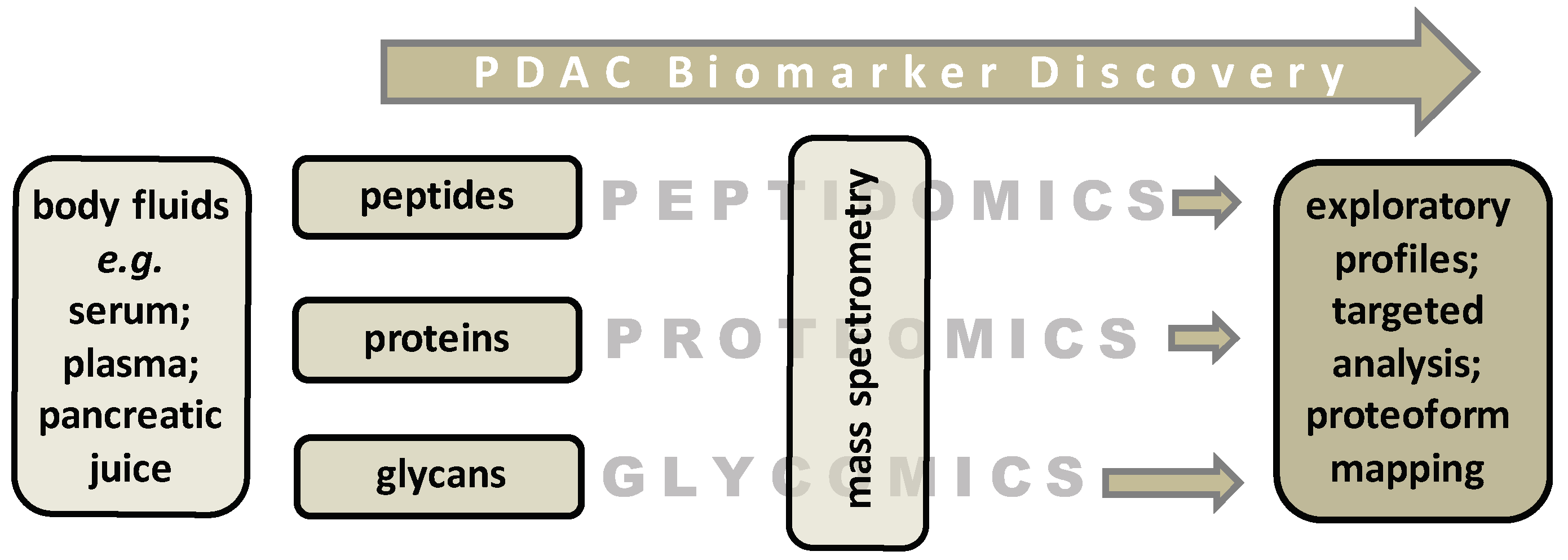
Recently, we reported on N-glycome analysis that allowed differentiation of PDAC patients from healthy controls with a combination of three so-called derived glycosylation traits (antennarity, sialylation, and fucosylation with an area-under-the-curve of 0.81–0.88) [9]. This study is a first step in the pursuit of novel glycan markers that made use of earlier technological developments in proteomics. Initially, both high-end and automated analytical strategies that have been developed for MS-based proteomics purposes were applied to measure peptide and protein levels in biological samples with the analytical robustness required for clinical application [77] (summarized in Figure 2).
In these early MS-based proteomics studies, patient serum samples were used to evaluate the profiling potential of body fluids with minimal sample workup. For example, Velstra and colleagues were able to discriminate a peptide/protein signature of pancreatic cancer patients from healthy individuals using ultrahigh-resolution matrix-assisted laser desorption/ionization (MALDI) time-of-flight MS. In that study, serum samples were obtained from sporadic pancreatic cancer patients (N = 89) and healthy volunteers (N = 185) and divided into a calibration and validation set. The discriminating profile showed a sensitivity of 78% and a specificity of 89%, validated with a sensitivity of 74% and a specificity of 91% [78]. This work was continued by combining MALDI with ultrahigh resolution instrumentation that provides more detailed peptide signatures [79]. Differentiating peptides were identified from thrombin light chain, fibrinogen alpha, platelet factor 4, complement C3f, and factor XIIIa protein. This signature was further used in a study to classify hereditary PDAC cases in a surveillance cohort from control samples (discriminant score of 0.26 vs. 0.016; p = 0.001). All individuals had a CDKN2A germline mutation and were enrolled in the pancreatic surveillance program [80]. A combination of these peptides could be useful for future clinical applications, but replication and validation on a larger cohort is still needed.
Reproducibility of biomarker discovery studies for PDAC screening remains challenging and the availability of validated results is limited [81,82]. Therefore, the proteomics field has moved to a standardized approach that results in improved reproducibility, although it involves more work before MS-analysis. A recent study by Jiyoung Park and co-workers performed 1000 biomarker candidate research on 134 plasma samples using multiple reaction monitoring MS. From the 1000 potential biomarkers that have previously been studied, a thorough selection was made based on previous study outcomes. Only biomarker candidates that previously showed an AUC > 0.60 were selected, leaving them with 176 proteins to test. The cohort consisted of 50 PDAC patients, 34 precursor lesions patients (IPMN), and 50 healthy volunteers. Following relative quantification and triplicate analyses, 54 proteins showed a AUC > 0.60 to discriminate between PDAC and healthy volunteers. Afterward, a cross-platform validation study was performed with 1008 plasma samples to validate the previously analyzed 54 proteins. A multimarker panel was found consisting of leucine-rich alpha-2 glycoprotein (LRGI), transthyretin (TTR) and CA 19-9 with a sensitivity of 82.5% and a specificity of 92.1%. It exceeded the performance of CA 19-9 alone in that same study by 10%. The sensitivity for distinguishing surgically resectable early stage PDAC from advanced stages in this study was 64% [83]. Although these results look promising, one should consider false positive results leading to unnecessary diagnostics as well as false negative results leading to missed cases in high risk populations. Moreover, samples were obtained at five different locations (medical centers) and potential differences in pre-analysis could have an influence on the results.
Alternatively, the currently applied protein routine markers can be re-visited, that is in-depth analysis can be performed to shed light on their precise molecular structures. It is hypothesized that so-called proteoform profiles of existing (clinically relevant) markers may provide an additional structural layer to quantitative levels of individual proteins with potential for patient stratification [84,85]. In this context, protein glycosylation provides an interesting source of potential leads. Protein glycosylation is an enzymatically regulated process in which glycans covalently attach to specific amino acids in proteins. It plays an important role in many biological mechanisms such as cell adhesion, protein folding, trafficking, pathogen recognition, and immune response. At least 50% of cell proteins are found to be glycosylated [86]. The first knowledge on glycosylation and the association with cancer already dates from over 50 years ago. However, during the last decade, the field of glycobiology has evolved enormously [86,87,88,89]. The aberrant glycosylation profile on the surface of cancer cells has been found to be of potential diagnostic value toward evaluating tumor progression [90]. Mucin (MUC) proteins, often described as CA 19-9 protein carriers, are found to be of influence on pancreatic cancer tumorigenesis, invasiveness, and metastasis. However, applicability is often described to be limited due to several reasons: expression in benign diseases (e.g., pancreatitis), low predictive value in asymptomatic patients (0.5–0.9%), and varying specificity (70–90%) and sensitivity (68–91%) [91,92,93].
From an alternative body fluid material, namely extracellular vesicles, Glypican 1 and Glycoprotein 2 were detected in a study by Melo and colleagues [94]. However, the sensitivity and specificity of Glypican 1 only or Glypican 1 and Glycoprotein 2 combined was insufficient to differentiate between malignant and benign disease [95]. SPAN-1, a high molecular weight glycoprotein, was first described in 1990 as a potential diagnostic marker. Kiriyama and colleagues studied the sera of 64 PDAC patients, 90 with other types of cancers, 254 non-malignant patients, and 55 healthy controls. Even though it showed a sensitivity and specificity of 81.3% and 75.6%, respectively, it also showed high false-positive elevations in liver cirrhosis (53.8%) and chronic hepatitis (26.3%) and was therefore not clinically applicable [96]. SPAN-1 was later investigated as a potential predictive marker. Nigjeh and colleagues investigated the possible value of N-glycosylated peptides in human plasma for early detection of PDAC. They indicated that the level of N-glycosylated peptides derived from galectin-3 binding proteins (LGALS3BP) were frequently elevated in plasma from PDAC patients [97]. Krishnan and colleagues also investigated the potential of altered glycosylation of serum proteins. They provided preliminary evidence of altered glycosylation of several serum proteins (e.g., α-1-antitrypsin, haptoglobin, α-1-acid glycoprotein 1) prior to pancreatic cancer diagnosis, but stated that further investigation of these proteins as early biomarkers is indicated [98]. Nie and colleagues analyzed 179 serum samples of patients with pancreatic cancer (N = 37), chronic pancreatitis (N = 30), diabetes (N = 30), obstructive jaundice (N = 22), pancreatic cysts (N = 30), and healthy controls (N = 30). A combination of α-1-antichymotrypsin (AACT), thrombospondin-1 (THBS1), and haptoglobin (HPT) outperformed CA 19−9 in distinguishing pancreatic cancer from normal controls (AUC = 0.95), patients with diabetes (AUC = 0.89), pancreatic cysts (AUC = 0.82), and chronic pancreatitis (AUC = 0.90). However, in both studies (Krishnan et al. and Nie et al.), acute-phase proteins were used and not specific cancer-related markers [99]. Therefore, Kontro and colleagues recently studied N-glycopeptide levels in serum of PDAC and acute pancreatitis patients compared to healthy controls. An increase in sialylated N-glycopeptides was found in both PDAC and acute pancreatitis patients. Mainly N-glycopeptides derived from acute-phase proteins and immunoglobulins were found: HPT, α-1-antitrypsin (A1AT), transferrin, ceruloplasmin, α-1-acid-glycoprotein (AGP), fetuin A, and immunoglobulins [100]. The potential of glycopeptides has been further pursued in glycoproteomics studies [101]. In a recent discovery study from Aronsson and colleagues, a glycosylation profile of 1000 serum proteins was reported for eight PDAC patients and eight healthy controls. A panel including CA 19-9, IL.17E, B7.1, and DR6 showed a promising AUC of 0.988 at 100% sensitivity at 90% specificity for differentiating between PDAC and controls [102].
The described potential glycoproteins observed in patient serum could be powerful biomarkers for detection of PDAC. However, little is known on the site specific glycoproteome changes associated with PDAC. Another limitation of glycoproteins is that most of them are neither pancreatic cancer nor cancer specific [89]. To further increase specificity and improve clinical applicability, ongoing method development and validation studies are essential.

3.2. Prognostic Markers

There is a spectrum of biological aggressiveness of PDAC. Identification of markers that are involved in PDAC tumor progression and aggressiveness may help to select adequate treatment strategies and to retrieve knowledge on how patients will respond to therapy and in some cases perform in the absence of therapy [103]. Numerous biomarker studies have been performed over the years with the aim to discover potential prognostic biomarkers for different types of cancer (e.g., colorectal, ovarian, and breast cancer) [104,105,106]. Up to now, prognostic proteomics or glycoproteomic studies have not been performed widely in PDAC patients. Jenkinson and co-workers proposed thrombospondin-1 (TSP-1) as a biomarker for early diagnosis of PDAC. Independent blood samples were collected from patients with PDAC (N = 152), chronic pancreatitis (N = 57), type 2 diabetes (N = 13, benign biliary disease (N = 20), and healthy individuals (N = 56). TSP-1 were generally detected at a lower level as far as 24 months before diagnosis of PDAC. Additionally, low TSP-1 levels were associated with a poor prognosis, making this a potential prognostic marker [107]. Apart from the potential diagnostic value of CEA, which has been researched widely, Boeck and colleagues described CEA as a potential prognostic marker. The CEA levels of 78 PDAC patients were monitored during treatment. Patients were sub-grouped into stable disease (SD), partial response (PR), or progressive disease (PD). The median CEA levels after eight weeks of treatment were 2.6 ng/mL in patients with SD or PR, and 18.1 ng/mL in patients with PD (p = 0.002). However, the median decrease of CEA levels in patients with response to therapy was not significantly different from patients with PD (p = 0.078). In the same study, cytokeratin-19 was described as a potential prognostic marker for advanced pancreatic cancer. Pre-treatment cytokeratin-19 levels were independently associated with performance status (p = 0.04) and disease stage (p = 0.0001) [108]. In other studies, the prognostic value of CEA has often been researched mainly using immunoassays. A meta-analysis by Meng and colleagues on 11 studies described an association between high level of CEA and worse overall survival (HR, 1.43; 95% CI, 1.31–1.56) [109]. Other than the above-mentioned studies, studies on cell-free circulating tumor DNA (ctDNA) have been performed using exome sequencing. Additionally, biomarker studies using tissue microarrays and immunochemistry are often performed. These methods do not fall within the scope of this review [110]. Therefore, these studies have been left out of consideration.

3.3. Predictive Markers

Biomarkers that provide information on responsiveness to therapy (selective patient treatment) and consequently prevent or terminate unsuccessful treatment regimens with substantial potential for morbidity are not available. Ideally, these markers would also allow monitoring the patient’s response to chemotherapy and provide evidence to guide clinicians in their conversations with patients [111]. So far, only a few predictive protein biomarker studies for PDAC have been performed. SPAN-1, a glycoprotein, emerged as a potential diagnostic marker in 1990 and appeared to be unsuccessful. In 2012, Tsutsumi and colleagues monitored the CA 19-9 and SPAN-1 levels as a potential predictor of tumor progression during chemotherapy. SPAN-1 showed a specificity of 90% prior to treatment. Blood samples and CT scans were performed every four weeks during treatment with gemcitabine in a cohort of 90 PDAC patients. Treatment failure was found in 59% of the patients by using SPAN-1 and 61% for CA 19-9. However, combined with CA19-9, it showed an earlier treatment failure of 72% of all patients with treatment failure, significantly better than CA 19-9 alone (p = 0.004) [112]. Several studies have described CA 19-9 as a predictive marker of unresectable disease when its level is high. In a retrospective cohort study consisting of 49 resected PDAC patients and 122 unresected PDAC patients, the CA 19-9 level prior to treatment was evaluated. The CA 19-9 levels were significantly lower in the resected group than in the group with unresectable disease (p < 0.001). Additionally, the serum CA 19-9 level accurately predicted resectability in 91% of patients. However, CA 19-9 carries a limit, because patients missing the Lewis group lack secretion of CA 19-9 (5–10% of all patients) [113]. A similar study was performed to assess the preoperative C-reactive protein level to albumin ratio (CAR) as a predictor of overall survival after pancreatic resection for PDAC. In a cohort of 136 patients, with a median follow up of 16.8 months, a high pre-operative CAR appeared to be an independent predictor of poor overall survival (p = 0.03) [114]. Further validation and prospective studies of the above-mentioned markers are warranted to determine the clinical value.

4. Summary and Conclusions

Despite great efforts in biomarker research, up to this day, only CA 19-9 is FDA approved (Food and Drug Administration) with limited diagnostic value. In this review, current detection and treatment strategies are presented as well as recent protein-based biomarker research, as summarized in a tabular overview (Table 1). From a clinical perspective, there are three main targets for improvement: early detection to provide a window of opportunity where treatment with curative intent is possible; upfront knowledge on individual patient prognosis; and knowledge on response to (chemo)therapy. Prognostic and predictive knowledge can contribute to better therapy selection and work toward personalized medicine. As described throughout this review, recent progress has been made in the discovery of a panel of biomarkers instead of one unimarker. Worldwide, biomarker research is more and more evolving into this approach.
With regard to novel pancreatic cancer biomarkers, there have been significant improvements in both the reported performance of the biomarkers (sensitivity and specificity) and the design of the studies (use of appropriate samples and populations and use of independent training and test samples). Unfortunately, most if not all, candidate biomarker panels have not been confirmed in independent validation sets, especially not in a screening setting. MS technology has matured and robust platforms are now available for quantitative biomarker measurements in discovery studies. These platforms allow the field of proteomics and glycomics to enter the next phase of MS applications by bridging between basic discovery and clinical verification. Further developments are needed to evaluate the performance of a candidate marker when aiming for implementation as a clinical test. For screening of either a high-risk population or an average-risk population for PDAC, blood-based biomarkers require a high specificity for the general population to avoid high numbers of false positives and a high sensitivity for high-risk groups. Several of the protein biomarkers discussed in this manuscript show sensitivities in the range of 50% to 60% at 95% specificity. At a high specificity, which may be required for screening, the reported sensitivities are considerably reduced. So far, protein based prognostic and predictive biomarker research for PDAC has been shown to be limited. For the identification of a potential prognostic or predictive biomarker panel, more research with large patient populations, the use of independent validation sets, and the application of standardized techniques is necessary.

Author Contributions

R.G.H.-S., J.H.S., W.E.M., B.A.B., H.F.A.V., M.E.v.L., M.W., R.A.E.M.T. and Y.E.M.v.d.B. contributed to the content of this review and were all involved in the writing process. R.A.E.M.T. and W.E.M. initiated the start and literature search and proposed the content and focus. Y.E.M.v.d.B. and R.G.H.-S. made the Table and the Figures. All authors have read and agreed to the published version of the manuscript.

Funding

This work was supported by ZonMW (project number: 50-53125-98-031 R.G. Hanna-Sawires) and the “Bollenstreek fund for early detection of cancer”, Lisse, The Netherlands.

Acknowledgments

The authors wish to thank J.W. Schoones of the University Library at the Leiden University Medical Center, The Netherlands for the extensive literature search and valuable discussions.

Conflicts of Interest

The authors declare no conflict of interest.

References

  1. Ilic, M.; Ilic, I. Epidemiology of pancreatic cancer. World J. Gastroenterol. 2016, 28, 9694–9705. [Google Scholar] [CrossRef] [PubMed]
  2. Kardosh, A.; Lichtensztajn, D.Y.; Gubens, M.A.; Kunz, P.L.; Fisher, G.A.; Clarke, C.A. Long-Term Survivors of Pancreatic Cancer: A California Population-Based Study. Pancreas 2018, 47, 958–966. [Google Scholar] [CrossRef] [PubMed]
  3. Raman, P.; Maddipati, R.; Lim, K.H.; Tozeren, A. Pancreatic cancer survival analysis defines a signature that predicts outcome. PLoS ONE 2018, 13, e0201751. [Google Scholar] [CrossRef]
  4. Alyass, A.; Turcotte, M.; Meyre, D. From big data analysis to personalized medicine for all: Challenges and opportunities. BMC Med. Genom. 2015, 8, 1–12. [Google Scholar] [CrossRef] [PubMed] [Green Version]
  5. Lennon, A.M.; Wolfgang, C.L.; Canto, M.I.; Klein, A.P.; Herman, J.M.; Goggins, M.; Fishman, E.K.; Kamel, I.; Weiss, M.J.; Diaz, L.A.; et al. The Early Detection of Pancreatic Cancer: What Will It Take to Diagnose and Treat Curable Pancreatic Neoplasia? Cancer Res. 2014, 74, 3381–3389. [Google Scholar] [CrossRef] [PubMed] [Green Version]
  6. Ansari, D.; Chen, B.C.; Dong, L.; Zhou, M.T.; Andersson, R. Pancreatic cancer: Translational research aspects and clinical implications. World J. Gastroenterol. 2012, 18, 1417–1424. [Google Scholar] [CrossRef] [PubMed]
  7. Overbeek, K.; Levink, I.; Konings, I.; Harinck, F.; Koopmann, B.; Ausems, M.; Wagner, A.; Fockens, P.; Koerkamp, G.; Besselink, M.; et al. 12 Years of Prospective Pancreatic Cancer Surveillance: Results of the Dutch Nationwide Program in High-Risk Individuals. Gastroenterology 2019, 156, S-756–S-757. [Google Scholar] [CrossRef]
  8. Harsha, H.C.; Kandasamy, K.; Ranganathan, P.; Rani, S.; Ramabadran, S.; Gollapudi, S.; Balakrishnan, L.; Dwivedi, S.B.; Telikicherla, D.; Selvan, L.D.N.; et al. A Compendium of Potential Biomarkers of Pancreatic Cancer. PLoS Med. 2009, 6, e1000046. [Google Scholar] [CrossRef] [PubMed]
  9. Vreeker, G.C.M.; Hanna-Sawires, R.G.; Mohammed, Y.; Bladergroen, M.R.; Nicolardi, S.; Dotz, V.; Nouta, J.; Bonsing, B.A.; Mesker, W.E.; Van Der Burgt, Y.E.M.; et al. Serum N -Glycome analysis reveals pancreatic cancer disease signatures. Cancer Med. 2020, 9, 8519–8529. [Google Scholar] [CrossRef] [PubMed]
  10. Pan, S.; Brentnall, T.A.; Chen, R. Proteomics analysis of bodily fluids in pancreatic cancer. Proteomics 2015, 15, 2705–2715. [Google Scholar] [CrossRef] [PubMed] [Green Version]
  11. Kim, H.; Park, J.; Wang, J.I.; Kim, Y. Recent advances in proteomic profiling of pancreatic ductal adenocarcinoma and the road ahead. Expert Rev. Proteom. 2017, 14, 963–971. [Google Scholar] [CrossRef]
  12. Root, A.; Allen, P.; Tempst, P.; Yu, K. Protein Biomarkers for Early Detection of Pancreatic Ductal Adenocarcinoma: Progress and Challenges. Cancers 2018, 10, 67. [Google Scholar] [CrossRef] [Green Version]
  13. Ansari, D.; Torén, W.; Zhou, Q.; Hu, D.; Andersson, R. Proteomic and genomic profiling of pancreatic cancer. Cell Biol. Toxicol. 2019, 35, 333–343. [Google Scholar] [CrossRef] [PubMed] [Green Version]
  14. Xiao, A.Y.; Tan, M.L.; Wu, L.M.; Asrani, V.M.; Windsor, J.A.; Yadav, D.; Petrov, M.S. Global incidence and mortality of pancreatic diseases: A systematic review, meta-analysis, and meta-regression of population-based cohort studies. Lancet Gastroenterol. Hepatol. 2016, 1, 45–55. [Google Scholar] [CrossRef]
  15. Ferlay, J.; Soerjomataram, I.; Dikshit, R.; Eser, S.; Mathers, C.; Rebelo, M.; Parkin, D.M.; Forman, D.; Bray, F. Cancer incidence and mortality worldwide: Sources, methods and major patterns in GLOBOCAN 2012. Int. J. Cancer 2014, 136, E359–E386. [Google Scholar] [CrossRef] [PubMed]
  16. Korc, M.; Jeon, C.Y.; Edderkaoui, M.; Pandol, S.J.; Petrov, M.S. Tobacco and alcohol as risk factors for pancreatic cancer. Best Pr. Res. Clin. Gastroenterol. 2017, 31, 529–536. [Google Scholar] [CrossRef]
  17. Vasen, H.; Ibrahim, I.; Ponce, C.G.; Slater, E.P.; Matthäi, E.; Carrato, A.; Earl, J.; Robbers, K.; Van Mil, A.M.; Potjer, T.; et al. Benefit of Surveillance for Pancreatic Cancer in High-Risk Individuals: Outcome of Long-Term Prospective Follow-Up Studies From Three European Expert Centers. J. Clin. Oncol. 2016, 34, 2010–2019. [Google Scholar] [CrossRef] [PubMed] [Green Version]
  18. Ralph, H.; Hruban, A.M.M.B.B.S.; Scott, E.; Kern, M.D.; Goggins, M.D. Precursors to Pancreatic Cancer. Gastroenterol. Clin. North Am. 2007, 36, 831–836. [Google Scholar]
  19. Weinrich, M.; Bochow, J.; Kutsch, A.L.; Alsfasser, G.; Weiss, C.; Klar, E.; Rau, B.M. High compliance with guideline recommendations but low completion rates of adjuvant chemotherapy in resected pancreatic cancer: A cohort study. Ann. Med. Surg. (Lond.) 2018, 32, 32–37. [Google Scholar] [CrossRef] [PubMed]
  20. Carreras-Torres, R.; Johansson, M.; Gaborieau, V.; Haycock, P.C.; Wade, K.H.; Relton, C.L.; Martin, R.M.; Smith, G.D.; Brennan, P. The Role of Obesity, Type 2 Diabetes, and Metabolic Factors in Pancreatic Cancer: A Mendelian Randomization Study. J. Natl. Cancer Inst. 2017, 109. [Google Scholar] [CrossRef] [PubMed] [Green Version]
  21. Anand, P.; Kunnumakkara, A.B.; Sundaram, C.; Harikumar, K.B.; Tharakan, S.T.; Lai, O.S.; Sung, B.; Aggarwal, B.B. Cancer is a Preventable Disease that Requires Major Lifestyle Changes. Pharm. Res. 2008, 25, 2200. [Google Scholar] [CrossRef] [Green Version]
  22. Carrera, S.; Sancho, A.; Azkona, E.; Azkuna, J.; Lopez-Vivanco, G. Hereditary pancreatic cancer: Related syndromes and clinical perspective. Hered. Cancer Clin. Pr. 2017, 15, 1–9. [Google Scholar] [CrossRef] [PubMed]
  23. Andersen, D.K.; Andren-Sandberg, A.; Duell, E.J.; Goggins, M.; Korc, M.; Petersen, G.; Smith, J.; Whitcomb, D. Pancreatitis-diabetes-pancreatic cancer: Summary of an NIDDK-NCI workshop. Pancreas 2013, 42, 1227–1237. [Google Scholar] [CrossRef] [PubMed] [Green Version]
  24. Abraham, S.C.; Wilentz, R.E.; Yeo, C.J.; Sohn, T.A.; Cameron, J.L.; Boitnott, J.K.; Hruban, R.H. Pancreaticoduodenectomy (Whipple Resections) in Patients Without Malignancy: Are They All ‘Chronic Pancreatitis’? Am. J. Surg. Pathol. 2003, 27, 110–120. [Google Scholar] [CrossRef] [PubMed]
  25. Kim, M.; Jang, K.M.; Kim, J.H.; Jeong, W.K.; Kim, S.H.; Kang, T.W.; Kim, Y.K.; Cha, D.I.; Kim, K. Differentiation of mass-forming focal pancreatitis from pancreatic ductal adenocarcinoma: Value of characterizing dynamic enhancement patterns on contrast-enhanced MR images by adding signal intensity color mapping. Eur. Radiol. 2016, 27, 1722–1732. [Google Scholar] [CrossRef] [PubMed]
  26. Choi, J.; Nipp, R.; Tramontano, A.; Ali, A.; Zhan, T.; Pandharipande, P.; Dowling, E.; Ferrone, C.; Hong, T.; Schrag, D.; et al. Neoadjuvant FOLFIRINOX for Patients with Borderline Resectable or Locally Advanced Pancreatic Cancer: Results of a Decision Analysis. Oncologist 2019, 24, 945. [Google Scholar] [CrossRef] [PubMed] [Green Version]
  27. Conroy, T.; Hammel, P.; Hebbar, M.; Abdelghani, B.M.; Wei, A.C.; Raoul, J.L.; Choné, L.; Francois, E.; Artru, P.; Biagi, J.J.; et al. FOLFIRINOX or Gemcitabine as Adjuvant Therapy for Pancreatic Cancer. N. Engl. J. Med. 2018, 379, 2395–2406. [Google Scholar] [CrossRef] [PubMed]
  28. McEvoy, S.H.; Lavelle, L.P.; Hoare, S.M.; O’Neill, A.C.; Awan, F.N.; Malone, D.; Ryan, E.R.; McCann, J.W.; Heffernan, E.J. Pancreaticoduodenectomy: Expected post-operative anatomy and complications. Br. J. Radiol. 2014, 87, 20140050. [Google Scholar] [CrossRef] [PubMed] [Green Version]
  29. Garrido-Laguna, I.; Hidalgo, M. Pancreatic cancer: From state-of-the-art treatments to promising novel therapies. Nat. Rev. Clin. Oncol. 2015, 12, 319–334. [Google Scholar] [CrossRef] [PubMed]
  30. Jackson, S.E.; Chester, J.D. Personalised cancer medicine. Int. J. Cancer 2015, 137, 262–266. [Google Scholar] [CrossRef]
  31. Goggins, M.; Overbeek, K.A.; Brand, R.; Syngal, S.; Del Chiaro, M.; Bartsch, D.K.; Bassi, C.; Carrato, A.; Farrell, J.; Fishman, E.K.; et al. Management of patients with increased risk for familial pancreatic cancer: Updated recommendations from the International Cancer of the Pancreas Screening (CAPS) Consortium. Gut 2020, 69, 7–17. [Google Scholar] [CrossRef] [Green Version]
  32. Vasen, H.F.; Wasser, M.; Van Mil, A.; Tollenaar, R.A.; Konstantinovski, M.; Gruis, N.A.; Bergman, W.; Hes, F.J.; Hommes, D.W.; Offerhaus, G.J.A.; et al. Magnetic Resonance Imaging Surveillance Detects Early-Stage Pancreatic Cancer in Carriers of a p16-Leiden Mutation. Gastroenterology 2011, 140, 850–856. [Google Scholar] [CrossRef] [PubMed]
  33. Giardiello, F.M.; Brensinger, J.D.; Tersmette, A.C.; Goodman, S.N.; Petersen, G.M.; Booker, S.V.; Cruz–Correa, M.; Offerhaus, J.A. Very high risk of cancer in familial Peutz–Jeghers syndrome. Gastroenterology 2000, 119, 1447–1453. [Google Scholar] [CrossRef] [PubMed] [Green Version]
  34. van Lier, M.G.F.; Wagner, A.; Mathus-Vliegen, E.M.H.; Kuipers, E.J.; Steyerberg, E.W.; van Leerdam, M.E. High Cancer Risk in Peutz–Jeghers Syndrome: A Systematic Review and Surveillance Recommendations. Am. J. Gastroenterol. 2010, 105, 1258–1264. [Google Scholar] [CrossRef] [PubMed]
  35. Roberts, N.J.; Klein, A.P. Genome-wide sequencing to identify the cause of hereditary cancer syndromes: With examples from familial pancreatic cancer. Cancer Lett. 2013, 340, 227–233. [Google Scholar] [CrossRef] [PubMed] [Green Version]
  36. Whitcomb, D.C.; Shelton, C.A.; Brand, R.E. Genetics and Genetic Testing in Pancreatic Cancer. Gastroenterology 2015, 149, 1252–1264.e4. [Google Scholar] [CrossRef] [PubMed]
  37. Overbeek, K.A.; Cahen, D.L.; Canto, M.I.; Bruno, M.J. Surveillance for neoplasia in the pancreas. Best Pr. Res. Clin. Gastroenterol. 2016, 30, 971–986. [Google Scholar] [CrossRef] [PubMed] [Green Version]
  38. Hruban, R.H.; Canto, M.I.; Goggins, M.; Schulick, R.; Klein, A.P. Update on Familial Pancreatic Cancer. Adv. Surg. 2010, 44, 293–311. [Google Scholar] [CrossRef] [PubMed] [Green Version]
  39. Canto, M.I.; Harinck, F.; Hruban, R.H.; Offerhaus, G.J.; Poley, J.W.; Kamel, I.; Nio, Y.; Schulick, R.S.; Bassi, C.; Kluijt, I.; et al. International Cancer of the Pancreas Screening (CAPS) Consortium summit on the management of patients with increased risk for familial pancreatic cancer. Gut 2012, 62, 339–347. [Google Scholar] [CrossRef] [PubMed]
  40. Healey, P.R. MRI and MRCP for Diagnosis and Staging of Pancreatic Cancer. Adv. Struct. Saf. Stud. 2018, 2018, 711–734. [Google Scholar] [CrossRef]
  41. Canto, M.I.; Hruban, R.H.; Fishman, E.K.; Kamel, I.R.; Schulick, R.; Zhang, Z.; Topazian, M.; Takahashi, N.; Fletcher, J.; Petersen, G.; et al. Frequent Detection of Pancreatic Lesions in Asymptomatic High-Risk Individuals. Gastroenterology 2012, 142, 796–804. [Google Scholar] [CrossRef] [Green Version]
  42. Templeton, A.W.; Brentnall, T.A. Screening and Surgical Outcomes of Familial Pancreatic Cancer. Surg. Clin. North Am. 2013, 93, 629–645. [Google Scholar] [CrossRef] [PubMed]
  43. Heinrich, S.; Lang, H. Neoadjuvant Therapy of Pancreatic Cancer: Definitions and Benefits. Int. J. Mol. Sci. 2017, 18, 1622. [Google Scholar] [CrossRef] [PubMed] [Green Version]
  44. McGuigan, A.; Kelly, P.; Turkington, R.C.; Jones, C.; Coleman, H.G.; McCain, R.S. Pancreatic cancer: A review of clinical diagnosis, epidemiology, treatment and outcomes. World J. Gastroenterol. 2018, 24, 4846–4861. [Google Scholar] [CrossRef] [PubMed]
  45. Riviere, D.; Gurusamy, K.S.; Kooby, D.A.; Vollmer, C.M.; Besselink, M.G.H.; Davidson, B.R.; Van Laarhoven, C.J.H.M. Laparoscopic versus open distal pancreatectomy for pancreatic cancer. Cochrane Database Syst. Rev. 2016, 4, CD011391. [Google Scholar] [CrossRef] [PubMed] [Green Version]
  46. Gooiker, G.A.; Lemmens, V.E.P.P.; Besselink, M.G.; Busch, O.R.; Bonsing, B.A.; Molenaar, I.Q.; Tollenaar, R.A.E.M.; De Hingh, I.H.J.T.; Wouters, M.W.J.M. Impact of centralization of pancreatic cancer surgery on resection rates and survival. BJS 2014, 101, 1000–1005. [Google Scholar] [CrossRef] [PubMed]
  47. Ghosn, M.; Kourie, H.R.; El Rassy, E.; Haddad, F.G.; Hanna, C.; El Karak, F.; Nasr, D. Where does chemotherapy stands in the treatment of ampullary carcinoma? A review of literature. World J. Gastrointest. Oncol. 2016, 8, 745–750. [Google Scholar] [CrossRef] [PubMed]
  48. Demir, I.E.; Jäger, C.; Schlitter, A.M.; Konukiewitz, B.; Stecher, L.; Schorn, S.; Tieftrunk, E.; Scheufele, F.; Calavrezos, L.; Schirren, R.; et al. R0 Versus R1 Resection Matters after Pancreaticoduodenectomy, and Less after Distal or Total Pancreatectomy for Pancreatic Cancer. Ann. Surg. 2018, 268, 1058–1068. [Google Scholar] [CrossRef]
  49. Shin, S.H.; Kim, Y.J.; Song, K.B.; Jun, E.; Hwang, D.W.; Lee, J.H.; Park, K.M. Totally laparoscopic or robot-assisted pancreaticoduodenectomy versus open surgery for periampullary neoplasms: Separate systematic reviews and meta-analyses. Surg. Endosc. 2016, 31, 3459–3474. [Google Scholar] [CrossRef] [PubMed]
  50. Torphy, R.J.; Friedman, C.; Halpern, A.; Chapman, B.C.; Ahrendt, S.S.; McCarter, M.M.; Edil, B.H.; Schulick, R.D.; Gleisner, A. Comparing Short-term and Oncologic Outcomes of Minimally Invasive Versus Open Pancreaticoduodenectomy Across Low and High Volume Centers. Ann. Surg. 2019, 270, 1147–1155. [Google Scholar] [CrossRef] [PubMed]
  51. Palanivelu, C.; Senthilnathan, P.; Sabnis, S.C.; Babu, N.S.; Gurumurthy, S.S.; Vijai, N.A.; Nalankilli, V.P.; Raj, P.P.; Parthasarathy, R.; Rajapandian, S. Randomized clinical trial of laparoscopic versus open pancreatoduodenectomy for periampullary tumours. BJS 2017, 104, 1443–1450. [Google Scholar] [CrossRef] [PubMed]
  52. Stauffer, J.A.; Coppola, A.; Villacreses, D.; Mody, K.; Johnson, E.; Li, Z.; Asbun, H.J. Laparoscopic versus open pancreaticoduodenectomy for pancreatic adenocarcinoma: Long-term results at a single institution. Surg. Endosc. 2016, 31, 2233–2241. [Google Scholar] [CrossRef] [PubMed]
  53. Chiorean, E.G.; Coveler, A.L. Pancreatic cancer: Optimizing treatment options, new, and emerging targeted therapies. Drug Des. Dev. Ther. 2015, 9, 3529–3545. [Google Scholar] [CrossRef] [Green Version]
  54. Labori, K.J.; Lassen, K.; Hoem, D.; Grønbech, J.E.; Søreide, J.A.; Mortensen, K.; Smaaland, R.; Sorbye, H.; Verbeke, C.; Dueland, S. Neoadjuvant chemotherapy versus surgery first for resectable pancreatic cancer (Norwegian Pancreatic Cancer Trial-1 (NorPACT-1))-study protocol for a national multicentre randomized controlled trial. BMC Surg. 2017, 17, 94. [Google Scholar] [CrossRef] [PubMed]
  55. Landau, E.; Kalnicki, S. The Evolving Role of Radiation in Pancreatic Cancer. Surg. Clin. N. Am. 2018, 98, 113–125. [Google Scholar] [CrossRef] [PubMed]
  56. Conroy, T.; Desseigne, F.; Ychou, M.; Bouché, O.; Guimbaud, R.; Bécouarn, Y.; Adenis, A.; Raoul, J.; Gourgou-Bourgade, S.; de la Fouchardière, C.; et al. FOLFIRINOX versus Gemcitabine for Metastatic Pancreatic Cancer. N. Engl. J. Med. 2011, 364, 1817–1825. [Google Scholar] [CrossRef] [PubMed] [Green Version]
  57. Poruk, K.; Brown, K.; Mulvihill, J.D.; Boucher, K.M.; Scaife, C.L.; Firpo, M.A.; Mulvihill, S.J. The Clinical Utility of CA 19-9 in Pancreatic Adenocarcinoma: Diagnostic and Prognostic Updates. Curr. Mol. Med. 2013, 13, 340–351. [Google Scholar] [PubMed]
  58. Scarà, S.; Scatena, R. Biochemical and Clinical Aspects. Adv. Exp. Med. Biol. 2015, 867, 247–260. [Google Scholar]
  59. Kim, J.E.; Lee, J.K.; Paik, S.W.; Rhee, J.C.; Choi, K.W. Clinical usefulness of carbohydrate antigen 19-9 as a screening test for pancreatic cancer in an asymptomatic population. J. Gastroenterol. Hepatol. 2004, 19, 182–186. [Google Scholar] [CrossRef] [PubMed]
  60. Xing, H.; Wang, J.; Wang, Y.; Tong, M.; Hu, H.; Huang, C.; Li, D. Diagnostic Value of CA 19-9 and Carcinoembryonic Antigen for Pancreatic Cancer: A Meta-Analysis. Gastroenterol. Res. Pr. 2018, 2018, 1–9. [Google Scholar] [CrossRef] [Green Version]
  61. Hatakeyama, K.; Wakabayashi-Nakao, K.; Ohshima, K.; Sakura, N.; Yamaguchi, K.; Mochizuki, T. Novel protein isoforms of carcinoembryonic antigen are secreted from pancreatic, gastric and colorectal cancer cells. BMC Res. Notes 2013, 6, 381. [Google Scholar] [CrossRef] [PubMed] [Green Version]
  62. Beauchemin, N.; Arabzadeh, A. Carcinoembryonic antigen-related cell adhesion molecules (CEACAMs) in cancer progression and metastasis. Cancer Metastasis Rev. 2013, 32, 643–671. [Google Scholar] [CrossRef]
  63. Homma, T.R. The study of the mass screening of persons without symptoms and of the screening of outpatients with gastrointestinal complaints or icterus for pancreatic cancer in Japan, using CA19-9 and elastase-1 or ultrasonography. Int. J. Pancreatol. 1991, 9, 119–124. [Google Scholar]
  64. Detlefsen, S.; de Vos, J.D.; Tanassi, J.T.; Heegaard, N.H.H.; Fristrup, C.; de Muckadell, O.B.S. Value of anti-plasminogen binding peptide, anti-carbonic anhydrase II, immunoglobulin G4, and other serological markers for the differentiation of autoimmune pancreatitis and pancreatic cancer. Medicine 2018, 97, e11641. [Google Scholar] [CrossRef] [PubMed]
  65. Matsuno, E.S.; Fukuyama, S.; Motoi, F.; Sunamura, M.; Isaji, S.; Imaizumi, T.; Okada, S.; Kato, H.; Suda, K.; Nakao, A.; et al. Pancreatic Cancer Registry in Japan 20 years of experience. Pancreas 2004, 28, 219–230. [Google Scholar] [CrossRef] [PubMed]
  66. Lu, X.C.; Wan, X.Y.; Zhu, H.T.; Yu, C.H.; Li, Y.M. Screening for pancreatic cancer in familial high-risk individuals: A systematic review. World J Gastroenterol. 2015, 21, 8678–8686. [Google Scholar] [CrossRef] [PubMed]
  67. Park, J.; Lee, P.K.J.; Park, H.D.; Kim, J.W.; Woo, H.I.; Lee, K.H.; Lee, K.T.; Lee, J.K.; Park, J.O.; Park, Y.S.; et al. Large-scale clinical validation of biomarkers for pancreatic cancer using a mass spectrometry-based proteomics approach. Oncotarget 2017, 28, 42761–42771. [Google Scholar] [CrossRef] [PubMed] [Green Version]
  68. Kim, J.; Bamlet, W.R.; Oberg, A.L.; Chaffee, K.G.; Donahue, G.; Cao, X.J.; Chari, S.; Garcia, B.A.; Petersen, G.M.; Zaret, K.S. Detection of early pancreatic ductal adenocarcinoma with thrombospondin-2 and CA19-9 blood markers. Sci. Transl. Med. 2017, 9, eaah5583. [Google Scholar] [CrossRef] [PubMed] [Green Version]
  69. Liu, X.; Zheng, W.; Wang, W.; Shen, H.; Liu, L.; Lou, W.; Wang, X.; Yang, P. A new panel of pancreatic cancer biomarkers discovered using a mass spectrometry-based pipeline. Br. J. Cancer 2017, 117, 1846–1854. [Google Scholar] [CrossRef]
  70. Bradley, S.D.; Talukder, A.H.; Lai, I.; Davis, R.; Alvarez, H.; Tiriac, H.; Zhang, M.; Chiu, Y.; Melendez, B.; Jackson, K.R.; et al. Vestigial-like 1 is a shared targetable cancer-placenta antigen expressed by pancreatic and basal-like breast cancers. Nat. Commun. 2020, 11, 1–12. [Google Scholar] [CrossRef]
  71. Chung, K.H.; Lee, J.C.; Cho, I.K.; Kim, J.; Jang, W.; Yoo, B.C.; Hwang, J.H. Serum fibrinogen as a diagnostic and prognostic biomarker for pancreatic ductal adenocarcinoma. Pancreatology 2020, 20, 1465–1471. [Google Scholar] [CrossRef] [PubMed]
  72. Sahni, S.; Krisp, C.; Molloy, M.P.; Nahm, C.; Maloney, S.; Gillson, J.; Gill, A.J.; Samra, J.; Mittal, A. PSMD11, PTPRM and PTPRB as novel biomarkers of pancreatic cancer progression. Biochim. Biophys. Acta (BBA) Gen. Subj. 2020, 1864, 129682. [Google Scholar] [CrossRef] [PubMed]
  73. Mandili, G.; Follia, L.; Ferrero, G.; Katayama, H.; Hong, W.; Momin, A.A.; Capello, M.; Giordano, D.; Spadi, R.; Satolli, M.A.; et al. Immune-Complexome Analysis Identifies Immunoglobulin-Bound Biomarkers That Predict the Response to Chemotherapy of Pancreatic Cancer Patients. Cancers 2020, 12, 746. [Google Scholar] [CrossRef] [Green Version]
  74. Zhou, Q.; Andersson, R.; Hu, D.; Bauden, M.; Sasor, A.; Bygott, T.; Pawlowski, K.; Pla, I.; Marko-Varga, G.; Ansari, D. Alpha-1-acid glycoprotein 1 is upregulated in pancreatic ductal adenocarcinoma and confers a poor prognosis. Transl. Res. 2019, 212, 67–79. [Google Scholar] [CrossRef] [PubMed]
  75. Wu, X.; Zhang, Z.; Chen, X.; Xu, Y.; Yin, N.; Yang, J.; Zhu, D.; Li, D.; Zhou, J. A Panel of Three Biomarkers Identified by iTRAQ for the Early Diagnosis of Pancreatic Cancer. Proteom. Clin. Appl. 2019, 13, e1800195. [Google Scholar] [CrossRef] [PubMed]
  76. Shih, H.C.; Chang, M.C.; Chen, C.H.; Tsai, I.L.; Wang, S.Y.; Kuo, Y.P.; Chang, Y.T.; Chen, C.H. High accuracy differentiating autoimmune pancreatitis from pancreatic ductal adenocarcinoma by immunoglobulin G glycosylation. Clin. Proteom. 2019, 16, 1. [Google Scholar] [CrossRef] [PubMed] [Green Version]
  77. Pham, T.V.; Piersma, S.R.; Oudgenoeg, G.; Jimenez, C.R. Label-free mass spectrometry-based proteomics for biomarker discovery and validation. Expert Rev. Mol. Diagn. 2012, 12, 343–359. [Google Scholar] [CrossRef] [PubMed]
  78. Velstra, B.; Bonsing, B.A.; Mertens, B.J.; Burgt, Y.E.M.; Huijbers, A.; Vasen, H.; Mesker, W.E.; Deelder, A.M.; Tollenaar, R.A.E.M. Detection of pancreatic cancer using serum protein profiling. HPB 2013, 15, 602–610. [Google Scholar] [CrossRef] [Green Version]
  79. Nicolardi, S.; Velstra, B.; Mertens, B.J.; Bonsing, B.; Mesker, W.E.; Tollenaar, R.A.; Deelder, A.M.; Van Der Burgt, Y.E. Ultrahigh resolution profiles lead to more detailed serum peptidome signatures of pancreatic cancer. Transl. Proteom. 2014, 2, 39–51. [Google Scholar] [CrossRef] [Green Version]
  80. Potjer, T.P.; Mertens, B.J.; Nicolardi, S.; Van Der Burgt, Y.E.M.; Bonsing, B.A.; Mesker, W.E.; Tollenaar, R.A.E.M.A.; Vasen, H.F. Application of a Serum Protein Signature for Pancreatic Cancer to Separate Cases from Controls in a Pancreatic Surveillance Cohort. Transl. Oncol. 2016, 9, 242–247. [Google Scholar] [CrossRef] [PubMed] [Green Version]
  81. Kinsinger, C.R.; Apffel, J.; Baker, M.; Bian, X.; Borchers, C.H.; Bradshaw, R.; Brusniak, M.Y.; Chan, D.W.; Deutsch, E.W.; Domon, B.; et al. Recommendations for mass spectrometry data quality metrics for open access data (corollary to the Amsterdam principles). Proteom. Clin. Appl. 2011, 5, 580–589. [Google Scholar] [CrossRef] [Green Version]
  82. Anne, K.C.; Werner, V.; Per, E.J.; Søren, C.; Ole, M.; Torben, A.; Kruse, O.N.J.; Jonna, S.M. Reproducibility of Mass Spectrometry Based Protein Profiles for Diagnosis of Breast Cancer across Clinical Studies: A Systematic Review. J. Proteome Res. 2008, 7, 1395–1402. [Google Scholar]
  83. Park, J.C.Y.; Namkung, J.; Yi, S.G.; Kim, H.; Yu, J.; Kim, Y.; Kwon, M.S.; Kwon, W.; Oh, D.Y.; Kim, S.W.; et al. Diagnostic performance enhancement of pancreatic cancer using proteomic multimarker panel. Oncotarget 2017, 8, 93117–93130. [Google Scholar] [CrossRef] [Green Version]
  84. van der Burgt, Y.E.M.; Cobbaert, C.M. Proteoform Analysis to Fulfill Unmet Clinical Needs and Reach Global Standardization of Protein Measurands in Clinical Chemistry Proteomics. Clin. Lab. Med. 2018, 38, 487–497. [Google Scholar] [CrossRef] [Green Version]
  85. Jimenez-Luna, C.; Torres, C.; Ortiz, R.; Dieguez, C.; Martinez-Galan, J.; Melguizo, C.; Prados, J.C.; Caba, O. Proteomic biomarkers in body fluids associated with pancreatic cancer. Oncotarget 2018, 9, 16573–16587. [Google Scholar] [CrossRef]
  86. Pan, S.A.; Brentnall, T.; Chen, R. Glycoproteins and glycoproteomics in pancreatic cancer. World J. Gastroenterol. 2016, 22, 9288–9299. [Google Scholar] [CrossRef] [PubMed]
  87. Kailemia, M.J.; Park, D.; Lebrilla, C.B. Glycans and glycoproteins as specific biomarkers for cancer. Anal. Bioanal. Chem. 2017, 409, 395–410. [Google Scholar] [CrossRef] [PubMed]
  88. Kuzmanov, U.; Kosanam, H.; Diamandis, E.P. The sweet and sour of serological glycoprotein tumor biomarker quantification. BMC Med. 2013, 11, 31. [Google Scholar] [CrossRef] [PubMed] [Green Version]
  89. Llop, E.; Guerrero, P.; Duran, A.; Barrabés, S.; Massaguer, A.; Ferri, M.J.; Albiol-Quer, M.; De Llorens, R.; Peracaula, R. Glycoprotein biomarkers for the detection of pancreatic ductal adenocarcinoma. World J. Gastroenterol. 2018, 24, 2537–2554. [Google Scholar] [CrossRef] [PubMed]
  90. Park, H.M.; Hwang, M.P.; Kim, Y.W.; Kim, K.J.; Jin, J.M.; Kim, Y.H.; Yang, Y.H.; Lee, K.H.; Kim, Y.G. Mass spectrometry-based N-linked glycomic profiling as a means for tracking pancreatic cancer metastasis. Carbohydr. Res. 2015, 413, 5–11. [Google Scholar] [CrossRef] [PubMed]
  91. Hollingsworth, M.A.; Swanson, B.J. Mucins in cancer: Protection and control of the cell surface. Nat. Rev. Cancer 2004, 4, 45–60. [Google Scholar] [CrossRef] [PubMed]
  92. Kaur, S.; Kumar, S.; Momi, N.; Sasson, A.R.; Batra, S.K. Mucins in pancreatic cancer and its microenvironment. Nat. Rev. Gastroenterol. Hepatol. 2013, 10, 607–620. [Google Scholar] [CrossRef] [Green Version]
  93. Nagata, K.; Horinouchi, M.; Saitou, M.; Higashi, M.; Nomoto, M.; Goto, M.; Yonezawa, S. Mucin expression profile in pancreatic cancer and the precursor lesions. J. Hepato-Biliary-Pancreatic Surg. 2007, 14, 243–254. [Google Scholar] [CrossRef]
  94. Melo, S.A.; Luecke, L.B.; Kahlert, C.; Fernández, A.F.; Gammon, S.T.; Kaye, J.; LeBleu, V.S.; Mittendorf, E.A.; Weitz, J.; Rahbari, N.N.; et al. Glypican-1 identifies cancer exosomes and detects early pancreatic cancer. Nat. Cell Biol. 2015, 523, 177–182. [Google Scholar] [CrossRef] [Green Version]
  95. Lucien, F.; Lac, V.; Billadeau, D.D.; Borgida, A.; Gallinger, S.; Leong, H.S. Glypican-1 and glycoprotein 2 bearing extracellular vesicles do not discern pancreatic cancer from benign pancreatic diseases. Oncotarget 2019, 10, 1045–1055. [Google Scholar] [CrossRef] [PubMed] [Green Version]
  96. Kiriyama, S.; Hayakawa, T.; Kando, T.; Shibata, T.; Kitagawa, M.; Ono, H.; Sakai, Y. Usefulness of a new tumor marker, span-1, for the diagnosis of pancreatic cancer. Cancer 1990, 65, 1557–1561. [Google Scholar] [CrossRef]
  97. Nigjeh, E.N.; Chen, R.; Allen-Tamura, Y.; Brand, R.E.; Brentnall, T.A.; Pan, S. Spectral library-based glycopeptide analysis-detection of circulating galectin-3 binding protein in pancreatic cancer. Proteom. Clin. Appl. 2017, 11, 1700064. [Google Scholar] [CrossRef]
  98. Krishnan, S.; Whitwell, H.J.; Cuenco, J.; Gentry-Maharaj, A.; Menon, U.; Pereira, S.P.; Gaspari, M.; Timms, J.F. Evidence of Altered Glycosylation of Serum Proteins Prior to Pancreatic Cancer Diagnosis. Int. J. Mol. Sci. 2017, 18, 2670. [Google Scholar] [CrossRef] [PubMed] [Green Version]
  99. Nie, S.; Lo, A.; Wu, J.; Zhu, J.; Tan, Z.; Simeone, D.M.; Anderson, M.A.; Shedden, K.A.; Ruffin, M.T.; Lubman, D.M. Glycoprotein Biomarker Panel for Pancreatic Cancer Discovered by Quantitative Proteomics Analysis. J. Proteome Res. 2014, 13, 1873–1884. [Google Scholar] [CrossRef] [PubMed]
  100. Kontro, H.; Joenväärä, S.; Haglund, C.; Renkonen, R. Comparison of sialylated N -glycopeptide levels in serum of pancreatic cancer patients, acute pancreatitis patients, and healthy controls. Proteomics 2014, 14, 1713–1723. [Google Scholar] [CrossRef] [PubMed]
  101. Tan, Z.; Yin, H.; Nie, S.; Lin, Z.; Zhu, J.; Ruffin, M.T.; Anderson, M.A.; Simeone, D.M.; Lubman, D.M. Large-Scale Identification of Core-Fucosylated Glycopeptide Sites in Pancreatic Cancer Serum Using Mass Spectrometry. J. Proteome Res. 2015, 14, 1968–1978. [Google Scholar] [CrossRef] [PubMed] [Green Version]
  102. Aronsson, L.; Andersson, R.; Bauden, M.; Andersson, B.; Bygott, T.; Ansari, D. High-density and targeted glycoproteomic profiling of serum proteins in pancreatic cancer and intraductal papillary mucinous neoplasm. Scand. J. Gastroenterol. 2018, 53, 1597–1603. [Google Scholar] [CrossRef] [PubMed]
  103. Le, N.; Sund, M.; Vinci, A.; Beyer, G.; Javed, M.A.; Krug, S.; Neessee, A.; Schober, M. Prognostic and predictive markers in pancreatic adenocarcinoma. Dig. Liver Dis. 2016, 48, 223–230. [Google Scholar] [CrossRef] [PubMed] [Green Version]
  104. de Vroome, S.W.; Girondo, M.R.; van der Burgt, Y.E.M.; Mesker, W.E.; Tollenaar, R.A.E.M.; Wuhrer, M. Serum N-glycome alterations in colorectal cancer associate with survival. Oncotarget 2018, 9, 30610–30623. [Google Scholar] [CrossRef] [PubMed] [Green Version]
  105. Anugraham, M.; Jacob, F.; Everest-Dass, A.V.; Schoetzau, A.; Nixdorf, S.; Hacker, N.F.; Fink, D.; Heinzelmann-Schwarz, V.; Packer, N.H. Tissue glycomics distinguish tumour sites in women with advanced serous adenocarcinoma. Mol. Oncol. 2017, 11, 1595–1615. [Google Scholar] [CrossRef] [Green Version]
  106. (Saldova), R.F.; Haakensen, V.D.; Rødland, E.; Walsh, I.; Stockmann, H.; Engebraaten, O.; Børresen-Dale, A.L.; Rudd, P.M. Serum N-glycome alterations in breast cancer during multimodal treatment and follow-up. Mol. Oncol. 2017, 11, 1361–1379. [Google Scholar] [CrossRef] [Green Version]
  107. Jenkinson, C.; Elliott, V.L.; Evans, A.; Oldfield, L.; Jenkins, R.E.; O’Brien, D.P.; Apostolidou, S.; Gentry-Maharaj, A.; Fourkala, E.O.; Jacobs, I.J.; et al. Decreased Serum Thrombospondin-1 Levels in Pancreatic Cancer Patients Up to 24 Months Prior to Clinical Diagnosis: Association with Diabetes Mellitus. Clin. Cancer Res. 2016, 22, 1734–1743. [Google Scholar] [CrossRef] [PubMed] [Green Version]
  108. Boeck, S.; Wittwer, C.; Heinemann, V.; Haas, M.; Kern, C.; Stieber, P.; Nagel, D.; Holdenrieder, S. Cytokeratin 19-fragments (CYFRA 21-1) as a novel serum biomarker for response and survival in patients with advanced pancreatic cancer. Br. J. Cancer 2013, 108, 1684–1694. [Google Scholar] [CrossRef] [PubMed] [Green Version]
  109. Meng, Q.; Shi, S.; Liang, C.; Liang, D.; Xu, W.; Ji, S.; Zhang, B.; Ni, Q.; Xu, J.; Yu, X. Diagnostic and prognostic value of carcinoembryonic antigen in pancreatic cancer: A systematic review and meta-analysis. OncoTargets Ther. 2017, 10, 4591–4598. [Google Scholar] [CrossRef] [Green Version]
  110. Cheng, H.; Liu, C.; Jiang, J.; Luo, G.; Lu, Y.; Jin, K.; Guo, M.; Zhang, Z.; Xu, J.; Liu, L.; et al. Analysis of ctDNA to predict prognosis and monitor treatment responses in metastatic pancreatic cancer patients. Int. J. Cancer 2017, 140, 2344–2350. [Google Scholar] [CrossRef] [PubMed]
  111. van der Sijde, F.; Vietsch, E.E.; Mustafa, D.A.M.; Besselink, M.G.; Groot, K.B.; van Eijck, C.H.J. Circulating Biomarkers for Prediction of Objective Response to Chemotherapy in Pancreatic Cancer Patients. Cancers. 2019, 11, 93. [Google Scholar] [CrossRef] [PubMed] [Green Version]
  112. Tsutsumi, K.; Kawamoto, H.; Hirao, K.; Sakakihara, I.; Yamamoto, N.; Noma, Y.; Fujii, M.; Kato, H.; Ogawa, T.; Ishida, E.; et al. Monitoring of CA19-9 and SPan-1 can facilitate the earlier confirmation of progressing pancreatic cancer during chemotherapy. Pancreatol. 2012, 12, 409–416. [Google Scholar] [CrossRef] [PubMed] [Green Version]
  113. Santucci, N.; Facy, O.; Ortega-Deballon, P.; Lequeu, J.B.; Rat, P.; Rat, P. CA 19–9 predicts resectability of pancreatic cancer even in jaundiced patients. Pancreatol. 2018, 18, 666–670. [Google Scholar] [CrossRef] [PubMed]
  114. Ikuta, S.; Aihara, T.; Yamanaka, N. Preoperative C-reactive protein to albumin ratio is a predictor of survival after pancreatic resection for pancreatic ductal adenocarcinoma. Asia-Pacific J. Clin. Oncol. 2019, 15, e109–e114. [Google Scholar] [CrossRef] [PubMed]
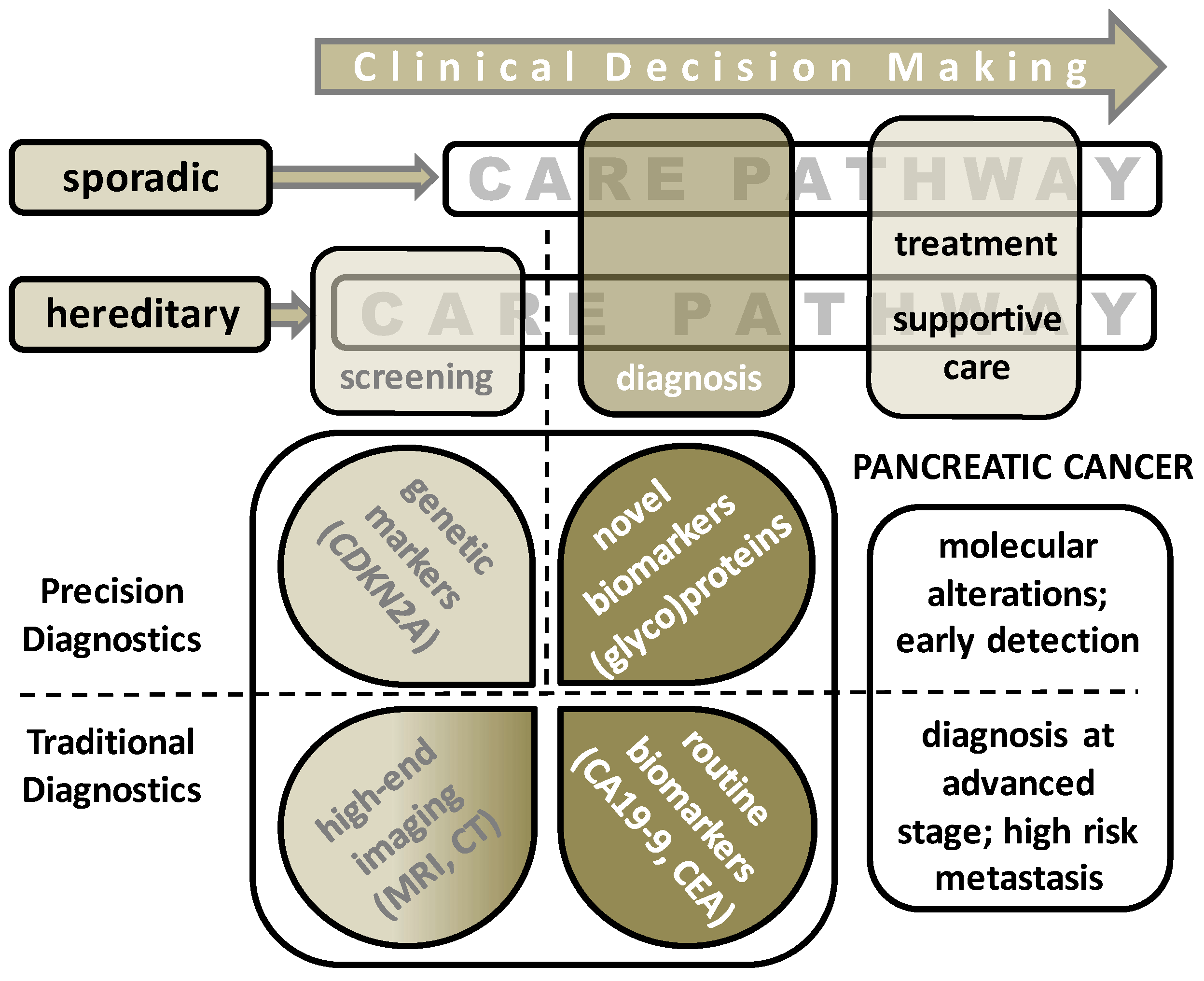
Figure 1. Clinical decision making pathway depicted for sporadic pancreatic ductal adenocarcinoma (PDAC) patients and individuals with a hereditary background (family history or gene mutation carriers). Surveillance of the latter group was performed with high-end imaging approaches such as MRI and EUS. Diagnosis of PDAC is commonly based on the same imaging technology combined with current routine markers, with the aid of cytology and histology diagnostics as is discussed in this review. Various ongoing approaches are summarized that search for novel (glyco)protein biomarkers.
Ijms 22 02655 g001
Figure 2. Overview of previous and ongoing PDAC biomarker discovery studies. This review has focused on three types of biomolecules that are obtained from patient body fluids. Potential biomarkers identified from tissue-based samples were out-of-scope. Without detailing the various mass spectrometry technologies, the applied strategies can either have an exploratory or a targeted nature. A third strategy, namely proteoform mapping, is explained in the main text.
Ijms 22 02655 g002
Table 1. Overview of diagnostic, prognostic, and predictive markers.
MarkerTypeStudy Size
N
Sensitivity
(%)
Specificity
(%)
MethodologyReference
CA 19-9 (meta-analysis of 13 studies)Diagnostic25–641 PDAC72–8668–80Routine DiagnosticsH. Xing et al.
CEA (meta-analysis of 23 studies)Diagnostic17–123 PDAC
15–58 BD
38–5082–91Routine DiagnosticsK. Poruk et al.
CA 19-9 combined with:
-
Apolipoprotein A-IV
-
Metalloproteinase-1
Diagnostic42 early stage (I/II) PDAC
72 advanced stage (III/IV) PDAC
31 pancreatitis
35 controls
8690Routine Diagnostics & MS-based ProteomicsJ. Park et al.
CA 19-9 combined with:
-
plasma thrombosondin-2
Diagnostic81 PDAC
80 controls
8798Routine Diagnostics & ELISAJ. Kim et al.
CA 19-9 combined with:
-
Apolipoprotein E
-
ITIH3
-
Apolipoprotein A-I
-
Apolipoprotein L-1
Diagnostic80 PDAC
30 BD
40 controls
9594Routine Diagnostics & MS-based ProteomicsX. Liu et al.
Peptide signature:
-
Thrombin light chain
-
Fibrinogen alpha
-
Platelet factor 4
-
Complement C3f
-
Factor XIIIa proteins
DiagnosticCalibration set:
50 PDAC
110 controls
Validation set:
39 PDAC
75 controls
78


74
89


91
MS-based PeptidomicsB. Velstra et al.
S. Nicolardi et al.
Multimarker panel:
-
Alpha-2-Glycoprotein
-
Transthyretin
-
CA 19-9
Diagnostic50 PDAC
34 PL
50 controls
8292MS-based ProteomicsJ. Park et al.
SPAN-1Diagnostic64 PDAC
90 other cancers
254 BD
55 controls
8176Radioimmuno assayS. Kiriyama et al.
Multimarker panel:
-
α -1-antitrypsin
-
haptoglobin
-
α-1-acid glycoprotein 1
Diagnostic154 PDAC
154 controls
Not reportedNot reportedMS-based ProteomicsS. Krishnan et al.
Multimarker panel:
-
α -1-antichymotrypsin
-
thrombospondin-1
-
haptoglobin
Diagnostic37 PDAC
30 CP
30 DMII
30 PC
22 OJ
30 controls
9178MS-based ProteomicsS. Nie et al.
Thrombospondin-1Prognostic152 PDAC
57 CP
13 DMII
20 BD
56 controls
Not reportedNot reportedMS-based ProteomicsJenkinson et al.
CEAPrognostic Not reportedNot reportedRoutine DiagnosticsS. Boeck et al.
Cytokeratin-19Prognostic Not reportedNot reportedRoutine DiagnosticsS. Boeck et al.
SPAN-1Predictive 9077immunoassayK. Tsutsumi et al.
CA 19-9Predictive 8581Routine DiagnosticsN. Santucci et al.
CARPredictive Not reportedNot reportedRoutine DiagnosticsS. Ikuta et al.
CA 19-9: Carbohydrate Antigen 19-9; CEA: Carcinoembryonic Antigen; PDAC: pancreatic ductal adenocarcinoma; ITIH3: inter-alpha trypsin inhibitor heavy chain H3; BD: benign disease; PL: precursor lesions; CP: chronic pancreatitis; DMII: type II diabetes; OJ: obstructive jaundice; CAR: C-reactive protein level to albumin ratio.
Publisher’s Note: MDPI stays neutral with regard to jurisdictional claims in published maps and institutional affiliations.