Review

Conventional and Unconventional Mechanisms by which Exocytosis Proteins Oversee β-cell Function and Protection

by
Diti Chatterjee Bhowmick
,
Miwon Ahn
,
Eunjin Oh
,
Rajakrishnan Veluthakal
and
Debbie C. Thurmond
*
Department of Molecular and Cellular Endocrinology, Beckman Research Institute of City of Hope, Duarte, CA 91010, USA
*
Author to whom correspondence should be addressed.
These authors contributed equally to this work.
Int. J. Mol. Sci. 2021, 22(4), 1833; https://doi.org/10.3390/ijms22041833
Submission received: 18 January 2021 / Revised: 2 February 2021 / Accepted: 8 February 2021 / Published: 12 February 2021
(This article belongs to the Special Issue Journey inside the Beta Cells in Type 2 Diabetes)

Abstract

:
Type 2 diabetes (T2D) is one of the prominent causes of morbidity and mortality in the United States and beyond, reaching global pandemic proportions. One hallmark of T2D is dysfunctional glucose-stimulated insulin secretion from the pancreatic β-cell. Insulin is secreted via the recruitment of insulin secretory granules to the plasma membrane, where the soluble N-ethylmaleimide-sensitive factor attachment protein receptors (SNAREs) and SNARE regulators work together to dock the secretory granules and release insulin into the circulation. SNARE proteins and their regulators include the Syntaxins, SNAPs, Sec1/Munc18, VAMPs, and double C2-domain proteins. Recent studies using genomics, proteomics, and biochemical approaches have linked deficiencies of exocytosis proteins with the onset and progression of T2D. Promising results are also emerging wherein restoration or enhancement of certain exocytosis proteins to β-cells improves whole-body glucose homeostasis, enhances β-cell function, and surprisingly, protection of β-cell mass. Intriguingly, overexpression and knockout studies have revealed novel functions of certain exocytosis proteins, like Syntaxin 4, suggesting that exocytosis proteins can impact a variety of pathways, including inflammatory signaling and aging. In this review, we present the conventional and unconventional functions of β-cell exocytosis proteins in normal physiology and T2D and describe how these insights might improve clinical care for T2D.

1. Introduction

Diabetes mellitus is a complex and heterogeneous disease characterized by progressive loss of function in the insulin-secreting pancreatic β-cells. Diabetes can be largely classified into type 1 (T1D) and type 2 (T2D), with worldwide occurrence rates of ~5% and ~95%, respectively [1]. Based on the 2020 Standards of Medical Care in Diabetes from the American Diabetes Association, T1D involves autoimmune β-cell destruction, whereas T2D is characterized by progressive loss of β-cell insulin secretion frequently on the background of insulin resistance [2]. Hence, although the pathogenesis of T1D and T2D is mediated by immune vs. metabolic stress, respectively, the common outcome of both T1D and T2D is the loss of functional β-cell mass and resulting hyperglycemia.
Histological analysis of the pancreatic islets from T2D human donors shows a 40% average reduction in the β-cell mass (range 25–60%), increased amyloid deposits and β-cell apoptosis, reduced insulin content, and unaltered α-cell mass as compared with non-diabetic control pancreas samples [3,4,5,6,7,8,9]. Interestingly, although multiple studies have reported a substantial reduction in β-cell function (~80%) at the onset of T2D [8,10,11], this early loss of function is not coupled with the dramatic loss of β-cell mass that has been observed later in patients with a longer disease history [6,8]. Thus, β-cell dysfunction without β-cell loss is thought to be an early player in T2D pathogenesis.
T2D is associated with considerable morbidity and a significant decrease in lifespan. Several therapies are available for T2D treatment, such as metformin, metformin plus GLP-1 analogs, insulin plus metformin, and pioglitazone. However, none has been successful in preventing β-cell dysfunction and demise over time (reviewed in [1,12]). Hence, alternative therapeutic approaches to preserve or restore β-cell function remain highly sought-after.
There is growing recognition for the importance of exocytosis proteins, such as soluble N-ethylmaleimide-sensitive factor attachment protein receptors (SNAREs) and their regulators, in improving β-cell insulin secretion and peripheral insulin-stimulated glucose uptake. The importance of exocytosis proteins has also been demonstrated in other diseases, such as cancer and neuronal disorders [1,13,14,15,16,17], providing clues that these proteins could be drug targets. More recently, the beneficial role of the SNARE protein Syntaxin 4 (STX4) in promoting whole-body glucose homeostasis, healthspan, and longevity has been unveiled [18,19,20]. In this review, we highlight current advances that have clarified the role of exocytosis proteins in preserving β-cell health and function, with a particular focus on the SNARE protein STX4.

2. Exocytosis Proteins and β-Cell Function

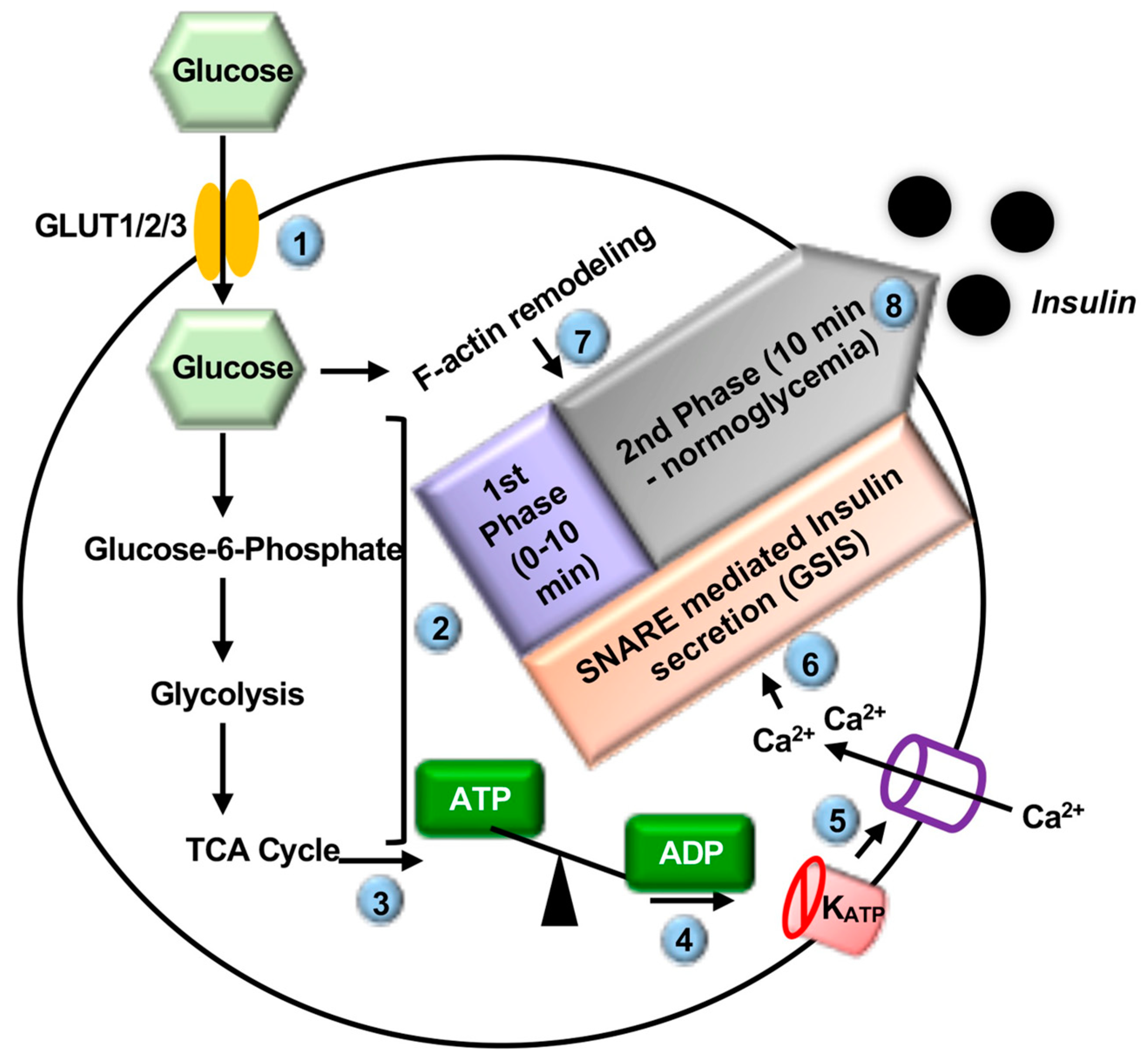
2.1. Introduction to Insulin Secretion

Insulin is a peptide hormone biosynthesized and secreted from the pancreatic β-cell and is the master regulator of glucose homeostasis. Glucose-stimulated insulin secretion (GSIS) refers to the exocytosis of insulin from the insulin secretory granules (ISGs) of the pancreatic β-cell in response to elevated blood glucose concentrations (Figure 1). Extracellular glucose enters the β-cell via the plasma membrane (PM)-localized GLUT1/2/3 transporter proteins (GLUT2 in rodents, GLUT1/3 in humans) [21,22,23,24]. Once intracellular, glucose is rapidly metabolized, thereby increasing the ATP/ADP ratio. This results in the closing of PM-localized ATP-sensitive potassium channels (KATP), depolarization of the PM, opening of voltage-sensitive Ca2+ channels at the PM, and influx of Ca2+ into the β-cell [25,26]. Elevated intracellular Ca2+ leads to SNARE-mediated ISG-PM priming and fusion, the formation of the fusion pore, and the release of insulin cargo extracellularly into the circulation [27,28].
Both in vitro and in vivo experiments have demonstrated that insulin is released from the β-cell in a pulsatile fashion, unlike the continuous release that is characteristic of other endocrine cells [29,30,31]. Early electron microscopy studies revealed that a small portion of the ISG pool is located within 100–200 nm of the β-cell PM, while the rest of the ISGs are located distal to the PM (reviewed in ref [32]). Based on this observation and similar knowledge from observations of neurotransmitter release from neurons, it was hypothesized that the membrane-proximal pre-docked ISGs constitute a readily releasable pool (RRP) of ISGs, which are released as the first response to glucose stimulation (reviewed in ref [32]). The distal storage pool of ISGs was similarly hypothesized to remain in the cytoplasm of the β-cell and await recruitment to the PM during depletion of the RRP (reviewed in [33]). This concept fits well with the observation that GSIS is biphasic. The first phase of GSIS is a transient insulin spike, lasting ~10 min, coinciding with the release of the RRP [34,35,36]. A sustained second phase of GSIS then ensues and can continue for hours until normal blood glucose levels are restored [37,38]. This second phase of GSIS requires the recruitment of the distal pool of ISGs (including newcomer ISGs), a process requiring the dynamic reorganization of the actin cytoskeleton [39].
ISG exocytosis is tightly regulated by the SNAREs, which are highly conserved proteins that closely resemble the vesicle fusion mechanism in neurons as well as exocrine, hematopoietic, and endocrine cells. SNARE proteins involved in ISG exocytosis are primarily categorized into two types: (i) Vesicle or “v”-SNAREs on the ISGs and (ii) target or “t”-SNAREs on the PM. Both v-SNARE and t-SNARE proteins bring the vesicle and target membranes (PM in this case) into proximity for fusion. Ultrastructural studies of the SNARE complex have revealed that one v-SNARE (VAMP2 in the β-cell) binds with two cognate t-SNARE proteins ((Syntaxin 1/4 (STX1/STX4) and SNAP23/25 in the β-cell)) in a heterotrimeric 1:1:1 ratio to form the SNARE core complex [40,41,42,43,44,45], which ultimately leads to membrane fusion and insulin release from the β-cell.

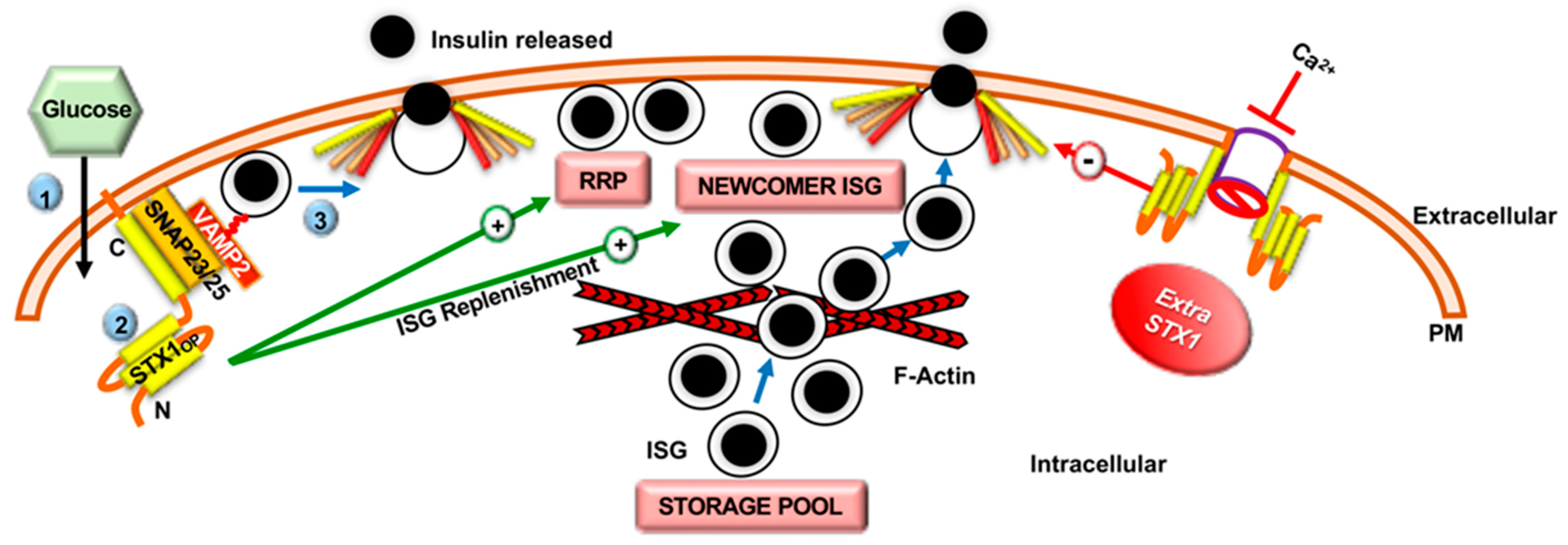
2.2. Role of Syntaxin Proteins in SNARE-Mediated Insulin Secretion

The importance of STX proteins in GSIS was first revealed by the study using anti-STX1 antibodies and global STX1 knockout (KO) mice, which showed a requirement for STX1 in insulin exocytosis and docking of the ISG RRP [46]. In line with these observations, a recent study using β-cell-specific STX1 KO (β-STX1 KO) mice revealed a new role for STX1 in both ISG exocytosis and replenishment. The β-STX1 KO mice showed impaired first- and second-phase GSIS, linked to severe reductions in the ISG RRP, as well as reduced recruitment of the distal ISG pool [47] (Figure 2). Intriguingly, in disagreement with the knowledge gathered from STX1 knockout (KO) mice in the context of the β-cell function, mice overexpressing STX1 in the islet β-cells showed decreased insulin exocytosis and perturbed activity of L-type Ca2+ channels, contributing to whole-body glucose intolerance [48]. These cumulative results indicate that STX1 has a narrow window of efficacy in the β-cell, wherein too little STX1 impairs GSIS, and too much STX1 causes novel interactions that impair β-cell function (Figure 2). STX1 overexpression in neurosecretory cells has a similar effect, indicating that the effects of excess STX1 on cellular function are similar across secretory cell types [49]. Interestingly, an early study demonstrated that cleavage of STX1 by botulinum toxin only reduced β-cell GSIS by 25% [50]. This result indicated that a STX1-independent insulin secretory complex must be present in the β-cells, possibly comprised of alternate STX isoforms.
Indeed, the pancreatic β-cell expresses four PM-localized STX isoforms: STX1, STX2, STX3, and STX4 (reviewed in [1,32]). The importance of STX4 was revealed using global heterozygous KO of STX4 (−/+) in mice, which reduced first-phase GSIS by 50% and was fully rescued by overexpression of recombinant STX4 [51]. In contrast to the glucose intolerance observed in STX1A-overexpressing mice, STX4-transgenic mice with 2–5-fold overexpression of STX4 in the skeletal muscle, adipose tissue, and pancreatic tissues, showed improved glucose homeostasis and islet function [52]. Indeed, as little as a 2-fold increase in islet STX4 expression in the STX4-transgenic mice increased insulin secretion by ~35% during both phases of GSIS (Figure 3), indicating that STX4 positively regulates both phases of GSIS [51]. Consistent with these animal studies, shRNA mediated knockdown (KD) of endogenous STX4 in primary human islets followed by determination of GSIS by islet perifusion assay showed a reduction in both phases of GSIS by ~40–42% [53], supporting the existence of a key role for STX4 in GSIS in both mice and humans. Further analyses of single ISG behavior using patch-clamp capacitance measurements and total internal reflection fluorescence microscopy revealed that STX4-KD β-cells are characterized by severe loss of the RRP and impaired mobilization of ISGs to the inner PM surface, potentially explaining the loss of both phases of GSIS [53]. Interestingly, the authors also reported a concomitant reduction in the exocytotic protein Munc18c (46%), but not other exocytotic proteins, in the STX4-KD human islets (77% KD), which again highlighted the various roles of STX4 in regulating both phases of insulin secretion [53]. This same dependency of Munc18c level relative to STX4 alteration was also seen in mouse models of STX4 KO or overexpression [54,55]. Marked reduction in STX4 protein in the T2D donor islets (~70% reduction) and significant improvement of GSIS in otherwise secretion-deficient T2D islets following STX4 replenishment holds the key to the therapeutic potential of STX4 in diabetes treatment [20].
In addition to STX1 and STX4, STX3 participates in second-phase GSIS via interacting with the R-type/Cav2.3 Ca2+ channel α1 subunit and regulates exocytosis of newcomer ISGs [56,57]. In contrast to STX1, STX3, and STX4, STX2 is known to be an inhibitory SNARE protein in mammals as global STX2 KO mice have enhanced recruitment of newcomer ISGs in their β-cells that enhances GSIS [58]. Taken together, these results provide evidence for the important but non-identical roles of the four PM-localized STX isoforms in the β-cells that collectively regulate insulin secretion contributing to the maintenance of glucose homeostasis.

2.3. Role of SNARE-Associated Proteins in Insulin Secretion

The SNARE-associated proteins Sec1/Munc18 and double C2-domain-containing protein (DOC2) assist in the assembly and formation of the core SNARE complex (reviewed in [1,32]). Understanding the molecular regulators of insulin exocytosis can drive therapeutic discovery by clarifying which molecules are critical for each step in the pathway.
Sec1/Munc18 has three isoforms: Munc18a (also called Munc18-1), Munc18b, and Munc18c [59,60], which interact with specific PM-localized STX proteins to regulate exocytosis. All three Munc18 isoforms are expressed in the islet β-cell [51,54,61]. Munc18a and Munc18b bind to STX1, STX2, and STX3, whereas Munc18c only interacts with and regulates STX4 [62,63,64,65]. In the β-cell, Munc18a regulates pre-docked ISG fusion via its interaction with STX1-SNAP25-VAMP2 and thereby influences first-phase GSIS [66]. Recently, STX1 was shown to be recruited to the ISG docking site in a Munc18a-bound conformation in rat clonal β-cells, which provides the mechanical insight for the requirement of both the STX1 and Munc18a for granule docking in the β-cell [67]. In line with these, reduced Munc18a level in the T2D islets depicts the contributory role of Munc18a in the development of β-cell dysfunction during diabetes [20]. Similar to Munc18a, Munc18b has also been shown to regulate SNARE complexes. However, unlike Munc18a, Munc18b appears to regulate STX3-based SNARE complexes, and via interaction with VAMP8-based newcomer ISGs in particular [68,69]. These distinct SNARE complex and ISG population binding and regulatory specificities of the Munc18 family of proteins depict extraordinarily exquisite control metering ISG exocytosis in pancreatic β-cells [69].
The importance of Munc18c was revealed using pancreatic islets isolated from Munc18c (−/+) heterozygous KO mice and RNAi-mediated KD of endogenous Munc18c in a mouse clonal β-cell line. These Munc18c-deficient cells demonstrated selective deficits in second-phase GSIS and decreased STX4 accessibility to VAMP2 [59]. Also, electron microscopy revealed fewer ISGs juxtaposed to the PM in the Munc18c-depleted cells, indicating a functional requirement for Munc18c to mobilize ISGs during GSIS [59]. Consistent with the studies using rodent β-cells, studies using dispersed primary human islets with lentivirus-mediated knockdown of endogenous Munc18c confirmed Munc18c’s involvement in the exocytosis of predocked and newcomer ISG pools, as well as its requirement for both the first and second phases of GSIS [60]. However, Munc18c is not found in the SNARE complex in β-cells per se. Instead, in response to glucose, Munc18c becomes tyrosine-phosphorylated and transiently dissociates from STX4, presumably transitioning STX4 from its closed to open and activated conformation, thereby indirectly facilitating SNARE complex formation [70,71] (Figure 3). Munc18c-STX4 complexes are further regulated by the competitive binding of DOC2b to Munc18c, whereby phosphorylation of Munc18c at tyrosine 219 acts as a molecular switch to reduce its binding affinity for STX4 and enhance binding to DOC2b [71] (Figure 3). Taken together, increasing evidence points towards the crucial functional contribution of different isoforms of Munc18 proteins in regulating β-cell function. This is again reinforced by the fact that while most of the non-β-cells express either one or two dominant Munc18 isoforms, the pancreatic β-cell expresses all three Munc18 isoforms and each of them contributes towards the regulation of GSIS in an overlapping yet independent manner.
DOC2 has two main isoforms, DOC2a and DOC2b; DOC2a is primarily expressed in the pancreatic islets and neurons and DOC2b is ubiquitously expressed [72,73,74]. Islets from DOC2a KO mice have normal islet function [75], but DOC2b KO mouse islets have defects in both phases of GSIS [75,76]. Moreover, transgenic mice with DOC2b overexpression in the pancreas, skeletal muscle, and adipose tissue showed improved whole-body glucose tolerance and peripheral insulin sensitivity, and isolated islets showed enhanced GSIS [77]. Accordingly, inducible β-cell-specific DOC2b overexpressing transgenic mice exhibited improved whole-body glucose tolerance and enhanced islet GSIS, as well as resistance to the diabetogenic stimulus multiple-low-dose streptozotocin (STZ), which causes loss of β-cell mass and glucose intolerance [78]. DOC2b is a 46–50 kDa protein comprised of an N-terminal Munc13-interacting domain (MID) and C-terminal tandem Ca2+ and phospholipid-binding C2 domains (C2A and C2B). DOC2b interacts with Munc18a and Munc18c via its C2A and C2B domains, respectively [79], and appears to serve as a scaffolding platform for transient binding of Sec1/Munc18 proteins to facilitate SNARE complex formation and GSIS [79] (Figure 3). However, it remains unclear whether this scaffolding mechanism, presumed to impact ISG exocytosis and hence β-cell function, also underlies DOC2b’s ability to protect β-cell mass. Indeed, since β-cell dysfunction is now recognized as a precursor to loss of β-cell mass, this remains an intriguing possibility.

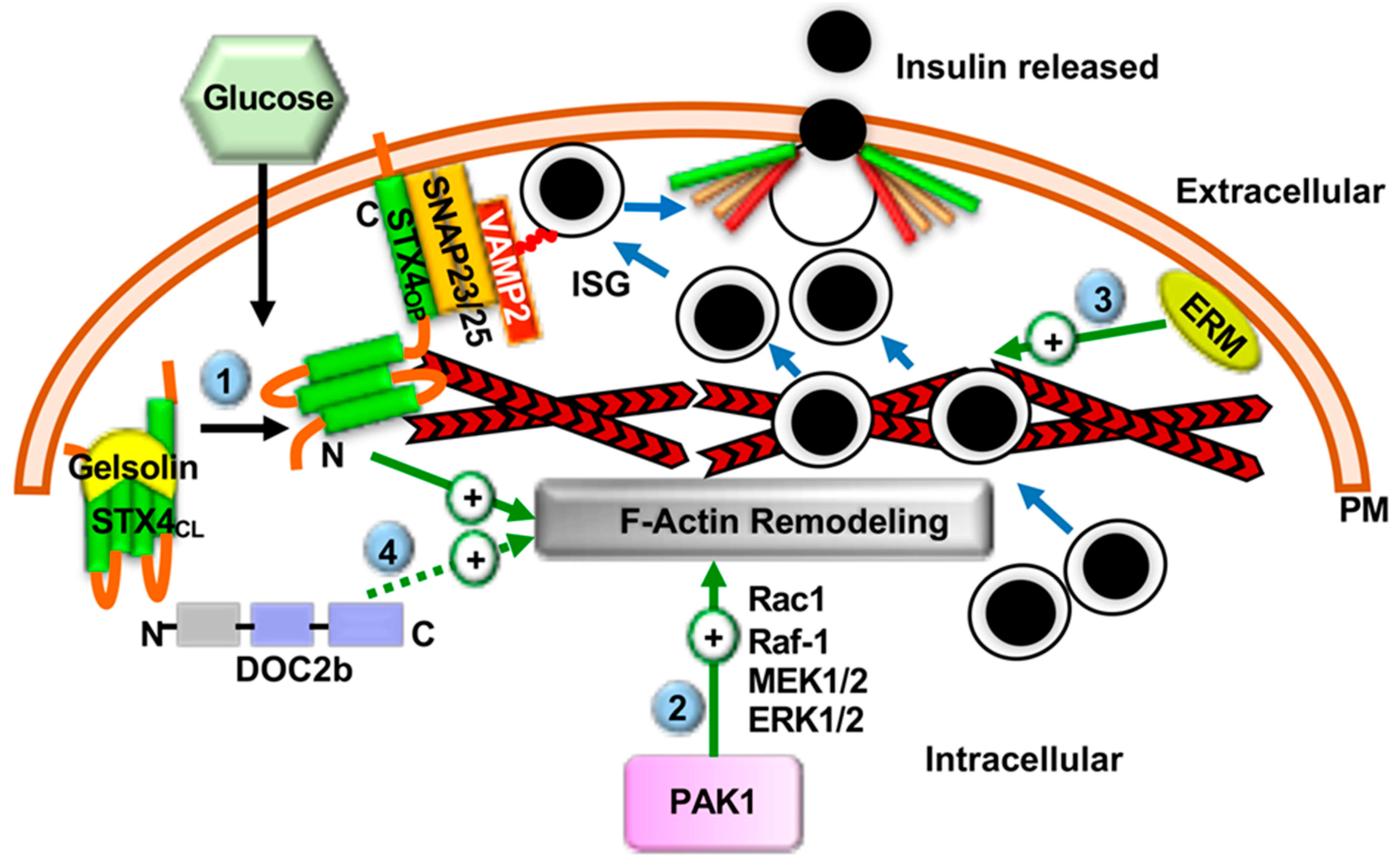
2.4. F-Actin Remodeling in Insulin Secretion

Prior to SNARE complex assembly and GSIS, filamentous actin (F-actin) remodeling plays an important role in ISG recruitment [80,81,82,83] (Figure 4). For example, while F-actin associates in macromolecular complexes with multiple t-SNAREs and prevents ISG movement towards the PM in unstimulated β-cells [84,85,86], glucose stimulation transiently induces F-actin remodeling and disrupts the t-SNARE: F-actin interaction, permitting ISG access to the t-SNARE docking sites at the PM. DOC2b and STX4, in particular, are noted for their abilities to forge novel interactions with cytoskeletal proteins. In vitro studies, have confirmed that F-actin directly interacts only with STX4, via a unique amino-terminal α-spectrin-like domain within STX4. On the other hand, F-actin interacts indirectly with other SNARE proteins, such as STX1, STX2, STX3, t-SNARE SNAP25, VAMP2, and another v-SNARE VAMP8 [84,86,87,88,89,90].
The F-actin network also acts as a binding platform for additional proteins required for GSIS [84,85,86], such as the Ca2+-activated F-actin severing/capping protein gelsolin. Gelsolin directly regulates STX4-mediated GSIS in the β-cell [84]. In absence of Ca2+, gelsolin prevents the formation of the SNARE complex via direct binding to STX4. Glucose-stimulated Ca2+ influx causes a conformational change that dissociates gelsolin from STX4, thereby facilitating SNARE complex formation and ISG exocytosis [84] (Figure 4). Hence, the gelsolin-STX4 interaction restrains unsolicited insulin release from β-cells in the absence of the appropriate glucose stimulus.
The role of DOC2b in the remodeling of cytoskeletal proteins came from a cancer study using cervical cancer cells showing replenishment of DOC2b, which is otherwise reduced in cancer cell lines, resulting in increased cytoskeletal remodeling and reduced cell migration leading to decreased cancer cell growth [16]. In line with this, both STX4 and DOC2b is known to associate with microtubule-associated Tctex-1 type proteins, reinforcing the regulatory contribution of both of these exocytosis proteins in cytoskeletal remodeling [84,91,92]. Recently, DOC2b was shown to undergo tyrosine-phosphorylation at tyrosine 301, within its functionally indispensable C2B domain following insulin stimulation in the skeletal muscle, resulting in its association with microtubule protein kinesin light chain 1 (KLC1) [93]. KLC1 is a cytoskeletal motor protein with a proposed role in protein/vesicle translocation. The association of DOC2b-KLC1 is critical for its stimulatory role in glucose uptake in the skeletal muscle [93]. However, the precise contribution of DOC2b in cytoskeletal remodeling in the β-cell during insulin exocytosis is not known and calls for future investigation (Figure 4).
The link between β-cell glucose stimulation and F-actin remodeling involves the small Rho family GTPases Cdc42 and Rac1 [94,95,96,97]. Glucose-mediated activation of Cdc42 leads to activation of PAK1, which initiates a signaling cascade via activation of Rac1, Raf-1, MEK1/2, and ERK1/2 to induce F-actin remodeling and recruitment of ISGs to the PM for second-phase GSIS [98,99,100] (Figure 4). Consistent with these studies, PAK1 protein levels are reduced by ~80% in the islets of humans with T2D, compared with non-diabetics, suggesting that deficiency of PAK1 or defects in PAK1 signaling may correlate to T2D susceptibility [98,99,101]. Very recent studies point to roles for the adaptor proteins APPL1 and APPL2 in F-actin remodeling via suppressing the Rac GTPase activating protein 1 (RacGAP1)-mediated inhibition of Rac1 activation [102]. While Rac1 is implicated only in second phase GSIS [103,104], APPL2 regulated both phases of GSIS. This might indicate a second role or binding partner for APPL2 that will need to be identified to reconcile this apparent disconnect. Interestingly, APPL1, which has the same domain organization and high protein sequence homology as APPL2, augments first-phase GSIS via upregulating the mRNA and the protein levels of SNARE proteins STX1, SNAP25, and VAMP2, indicating that these similar APPL isoforms play complementary functional roles to support insulin exocytosis [105]. As the underlying mechanism, APPL1 deficiency attenuated insulin-stimulated Akt activation in both primary pancreatic islets and rat β-cells, whereas adenovirus-mediated expressions of APPL1 rescued GSIS [105]. These results altogether revealed that APPL1 links insulin-stimulated Akt activation to GSIS by upregulating the expression of the core exocytotic machinery proteins in the β-cell [105].
The ERM scaffolding proteins, Ezrin, Radixin, and Moesin, have also been implicated as regulators of F-actin remodeling in GSIS. Each ERM protein contains an F-actin binding domain and a PIP2 binding domain [106,107,108]. All three ERM proteins are expressed in the mouse β-cell, with radixin being the most abundant in mouse islets and β-cells [109]. Glucose stimulation activates the ERM proteins via Ca2+-dependent phosphorylation; the activated phosphorylated ERM proteins translocate to the cell periphery where they link PIP2 to F-actin, a step essential for trafficking and docking of ISGs to the β-cell PM [109] (Figure 4). Hence, whereas Cdc42 signaling via PAK1 regulates the initial glucose response that supports F-actin remodeling and second phase GSIS, ERM proteins appear to be involved in the later stages of the signaling cascade. Reduced ERM protein activity has been observed in diabetic ob/ob mouse islets [109], consistent with a model wherein loss of ERM protein function contributes to T2D pathogenesis.
Recent advancements in high spatiotemporal resolution live-cell imaging are likely to facilitate linking these events to provide the first comprehensive understanding of the molecular interactions between proteins on a physiologically relevant timescale underlying the cytoskeletal changes with the coordinated assembly of the exocytosis machinery in secretory cells [110,111,112]. Toward this goal, two-photon fluorescence lifetime imaging of primed SNARE complexes in the β-cell has revealed that SNARE complexes are unassembled in the unstimulated state, and stimulation leads to slow assembly of SNARE complexes [113]. This finding supports prior models wherein DOC2b, Munc18c, and transient F-actin disassembly cause STX4 to take on a conformational state conducive to glucose-stimulated assembly with SNAP25 and VAMP2 for GSIS.
In addition to the above-mentioned proteins and their signaling mechanisms for regulating GSIS, insulin exocytosis from the β-cell is also regulated by secretion-potentiating agents like Glucagon-like-peptide-1 (GLP-1) and cAMP releasing agents [114]. GLP-1 and its analogs have widely been used as therapeutic agents for T2D [115,116]. GLP-1 is an incretin hormone, secreted from the intestinal L-cells in response to glucose and other ingested nutrients and boosts insulin secretion via Glucagon-like-peptide-1 receptor (GLP-1R) in a glucose-dependent manner (reviewed in [117]). Previous studies showed that the insulinotropic effects of GLP-1 depend on the cAMP-Epac2 signaling pathway and is capable of restoring first phase insulin secretory defects in diabetic β-cells [118,119,120]. Interestingly, recent studies point towards the beneficial role of GLP-1 signaling in potentiating insulin secretion under glucotoxic conditions via actin cytoskeleton remodeling [121,122]. As an underlying mechanism, it has been shown that in the INS 832/13 cells (rat clonal beta-cell line) GLP-1 mediated improvement insulin exocytosis under glucotoxic condition is associated with partial restoration of actin dynamics and ISG fusion during both the phases of GSIS, via a preferential involvement of Epac2 signaling in the first phase and protein kinase A (PKA) signaling in the second phase of insulin exocytosis [122].
GLP-1 stimulates PKA-dependent phosphorylation of synaptotagmin-7, a crucial Ca2+ sensor affiliated with SNARE-mediated exocytosis, thereby boosting glucose- and Ca2+-stimulated insulin secretion [123]. Liraglutide, a GLP-1 analog, was reported to modulate plasma membrane-raft clustering in RIN-m5f β-cells, possibly impacting functionality of raft-embedded SNARE protein isoforms STX1, SNAP25, and VAMP2 [124]. Indeed, earlier studies showed GLP-1 to enhance insulin secretion via mobilizing and docking an increased number of ISGs at the plasma membrane and accelerating sequential ISG—ISG fusions [118]. In line with this, a study using wild type and truncated form of SNAP-25 overexpressing INS-1 cells (rat clonal β-cell line) revealed the requirement of fully functional SNAP-25 in GLP-1 mediated boosting of insulin release, specifically at the level of mobilization of RRP of ISG granules [125]. Additionally, cAMP potentiation via GLP-1 is also known to rescue priming defects caused by Munc13-1 deficiency via Epac and PKA signaling pathways, again confirming the crucial role of GLP-1 signaling in SNARE-mediated insulin release from the β-cell [119]. Interestingly, it has also been reported that VAMP8, a SNARE protein that is known to play a nonessential role in the membrane fusion processes because of functional redundancy contributed by other VAMPs, partially mediates the insulinotrophic effect of GLP-1 [69].

2.5. The Ability of SNARE Proteins to Restore β-Cell Function in Diabetes

Human T2D islet β-cells show alterations in the expression levels of SNAREs and SNARE-associated proteins, and rodent models of diabetes show similar changes [20,126,127,128,129,130]. For example, the levels of STX1, STX4, and Munc18a are reduced in T2D human islets compared to non-diabetic islets [20]. In line with this, low DOC2b transcript levels have been reported in the islets of diabetic mice, consistent with a link between loss of DOC2b function and pathogenicity of diabetes [131,132]. It is conceivable that compromised levels of SNARE proteins limit the exocytosis of insulin from β-cells, creating an ISG bottleneck at the most distal step of GSIS. From a therapeutic standpoint, it is important to understand whether the restoration of SNARE proteins is sufficient to restore GSIS. Furthermore, it is important to clarify whether restoring certain SNAREs is sufficient for GSIS. This was addressed initially in the Goto-Kakizaki (GK) rat, a non-obese T2D model characterized by reduced expression of STX1 and SNAP-25 in pancreatic islets [128]. Adenovirus-mediated restoration of STX1 and SNAP25 protein levels was sufficient to improve GSIS, indicating that the SNARE proteins are critical for reversing T2D [128]. Similarly, restoration of STX4 to T2D human islets, which are deficient in STX1, SNAP25, and STX4, was sufficient to restore GSIS to levels seen in age/gender-matched non-diabetic human islets, showing that GSIS can be restored without restoring the levels of all SNARE proteins [20].
Enrichment of individual SNARE accessory proteins can be similarly effective, as DOC2b overexpression in the β-cells of mice prevents the diabetogenic effects of STZ treatment on whole-body glucose tolerance and loss of β-cell mass [78]. Interestingly, while upregulation of STX4 or DOC2b expression in β-cells protects against β-cell damage and dysfunction in diabetogenic mouse models [18,19,78], upregulation of STX1 or Munc18c expression dysregulates GSIS [48,55,133]. As discussed in the previous section, these disparate outcomes are potentially linked to differences in binding partners of these SNAREs and SNARE regulators, which can impact actin cytoskeletal remodeling (e.g., STX4) or interfere with ion channel functions (e.g., STX1). SNARE stoichiometry is also important, since SNARE core complexes are composed in a specific 1:1:1 ratio, and loss/gain of any member of the complex can cause unexpected interactions with other proteins, as was the case with upregulated STX1, which disrupts ion channel function [48]. In summary, these results suggest that very specific isoforms of individual SNAREs can rescue the loss of GSIS but that the balance of SNARE protein levels is critical for maintaining and boosting glucose homeostasis; knowing which ones can be safely overexpressed is key to positive therapeutic outcomes.

3. Exocytosis Proteins and β-Cell Protection

Peripheral insulin resistance during pre-T2D places an immense workload on the β-cells to release an extraordinary volume of insulin. To maintain normal glycemic levels, the β-cell mass increases as a compensatory mechanism in response to the insulin resistance induced workload; though, whether the increased mass of β-cells function normally remains in debate [134]. Chronic insulin resistance eventually overwhelms the capacity of the β-cells. When the β-cell can no longer produce enough insulin, glucose homeostasis is disrupted and loss of β-cells ensues primarily via mechanisms such as apoptosis and de-differentiation. Hence, proteins that can protect against β-cell loss are candidates for therapeutic development. Towards this, the ability of exocytosis proteins to protect β-cells from this fate is discussed in this section.

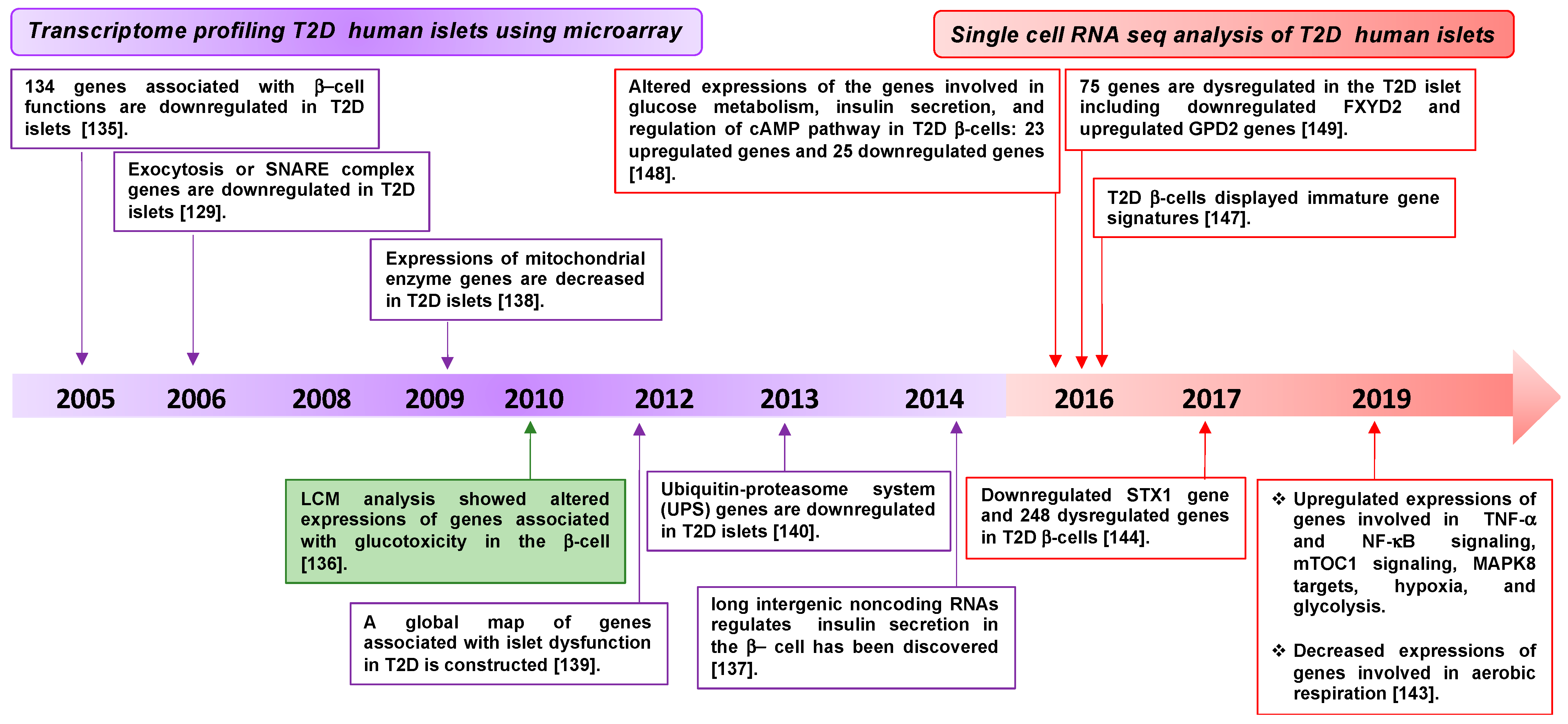
3.1. Diabetes-Associated Genes Revealed by Transcriptomic Profiling of β-Cells

Transcriptomic profiling has been used to gain novel insight into mechanisms underlying the dysfunction of pancreatic islet β-cells during T2D. Human islet cell transcriptomic analysis of T2D and non-diabetic donors identified multiple genes, such as HNF4α, IR, IRS2, Akt2, and ARNT, as differentially expressed [135]. In addition, microarray analysis of T2D human islet tissue collected using laser capture microdissection (LCM) has revealed downregulation of the genes associated with β-cell function, relative to non-diabetic controls [136]. The candidate genes include those involved in insulin secretion, such as the SNARE and SNARE accessory genes (SNAP-25, VAMP2, STX1, Munc18a, Munc13-1, and synaptophysin). Long intergenic noncoding RNAs and genes involved in metabolic activity and protein homeostasis, such as mitochondrial genes and genes in the ubiquitin-proteasome system, respectively, are also included [129,135,137,138,139,140] (Figure 5 and Table 1).
Given the altered levels of other factors such as STX4 and DOC2b reported in T2D human islets it was surprising that these genes were not detected in the transcriptomic profiling. One possibility for the discrepancy is that unlike the other SNARE genes detected, STX4 and DOC2b are widely expressed across cell types, and that transcriptomics is generally performed using IEQs, or “islet equivalents”, which are known to contain non-islet cells to varying degrees. The contribution of STX4 and DOC2b from non-islet cells in the IEQs would effectively dilute any deficiencies present in the islets. Comparison analyses of STX4 and DOC2b levels in hand-picked T2D human islets vs. IEQs will be required to test this hypothesis.
In addition, β-cell heterogeneity [141] makes it challenging to accurately interpret the whole-islet transcriptome data and may complicate the identification of the precise molecular mechanisms for islet dysfunction in T2D. To overcome this limitation, single-cell RNA sequencing approaches are now used to identify differentially expressed genes and gene groups between non-diabetic and T2D donor islets [142,143,144,145,146,147,148,149] (Figure 5 and Table 1). A single-cell RNA sequencing study identified 248 differentially expressed genes between non-diabetic and T2D donor β-cells, which confirmed a strong correlation between the expression of β-cell-specific genes and T2D. Among the genes decreased by T2D were the insulin and STX1 genes [126,143,144]. In line with these studies, a recent temporal transcriptomic and proteomic analysis of the pancreatic islets procured from T2D rats demonstrated attenuated levels of exocytosis factors like STX1 and STXBP1 [150]. Indeed, one study identified an altered β-cell gene signature in the T2D donor islets. For example, the expression of genes involved in insulin secretion was attenuated, but there was increased expression of hypoxia genes, MAPK8 targets, mTORC1 signaling genes, TNF-α mediated NF-ĸB signaling genes, and immature β-cell gene signatures, indicating de-differentiation [143]. A recent single-cell data highlighted that genes known to be involved in aging are also related to stress signaling in the pancreas [151] (Figure 5 and Table 1). Cumulatively, the results of these transcriptomic studies are consistent with β-cell stress, dysfunction, and dedifferentiation being the key differences between T2D and non-diabetic islets.

3.2. Regulation of Exocytosis Proteins by Diabetogenic Stressors

It is well established that genetic and environmental factors, such as obesity and inflammation, accelerate the development of T2D. New insights, as discussed in the following paragraphs, have implicated regulatory cross-talks between exocytosis factors and the diabetogenic risk factors in the process of β-cell disruption during T2D.

3.2.1. Regulation of Exocytosis Proteins and Obesity

The causes of T2D are thought to be chronic exposure to high levels of circulating FFA and glucose. High glucose exposure, in vitro and in vivo, was shown to rapidly reduce the protein levels of the v-SNARE proteins VAMP2 and VAMP3, but not the t-SNARE proteins STX1, SNAP25, or SNAP23, in rodent models [152]. The effects of elevated glucose levels on the other SNARE proteins and the effects of FFA exposure remain to be determined. A recent study showed that overexpression of fatty acid translocase CD36, which is upregulated in obese T2D patients, diminishes SNARE protein expression, and disrupts ISG docking and GSIS in human islet cultures [153]. The authors established a link between overexpression of CD36 and reduced expression of the exocytotic proteins SNAP25, Munc18a, and VAMP2, potentially via a CD36-mediated attenuation of the nPKC-IRS-PI3K/AKT signaling pathway [153]. An antibody to CD36 reversed the dysfunctional GSIS associated with CD36 overexpression, indicating that CD36 is a candidate therapeutic target for restoring SNARE protein levels and GSIS during obesity-associated T2D [153].

3.2.2. Regulation of Exocytosis Proteins and Immune Signaling

Crosstalk between the innate immune system and GSIS was established via detection of an unexpected interaction between the complement system protein CD59 and the SNARE proteins VAMP2 and STX1 in INS-1 β-cells [154]. Otherwise, in non-β-cells, CD59 was known to protect cells from complement-mediated membrane perforation and cell death by translocating to the PM and inhibiting the formation of the membrane attack complex (MAC) [155]. However, in β-cells, the silencing of CD59 decreased GSIS by destabilizing lipid rafts [154]. Furthermore, enzymatic removal of extracellular CD59 did not suppress GSIS, suggesting that an intracellular pool of CD59 is important for regulating β-cell function [154]. This finding indicates that loss of CD59 may both enhance complement-mediated cell killing and directly disrupt GSIS through different mechanisms, making CD59 a potential target for preserving β-cell function and protecting against immune attack. Recently, INS-1 β-cells were shown to contain a cytosolic non-GPI-anchored form of CD59, which facilitates GSIS via interacting with VAMP2 and STX1 [154,156]. Consistent with a role for CD59 as a link between T2D and immunity, another group identified that glycated CD59, which is an inactivated form of CD59 caused by exposure to high blood glucose levels, is elevated in the blood of T2D patients and is decreased by insulin treatment [157]. Thus, CD59 is an intriguing candidate biomarker for T2D as well as a possible therapeutic candidate. However, the precise mechanism linking immunity to GSIS via the SNARE proteins needs further investigation, which may reveal additional candidates as clinical targets.

3.3. SNARE Proteins Can Modulate Inflammatory Signals in the β-Cell

Earlier studies have demonstrated a link between low-grade chronic inflammation due to mononuclear cells and T2D [158,159,160,161,162]. Macrophage infiltration has been observed in human T2D pancreatic islets and the islets of diabetes models such as db/db mice and GK rats, as well as pre-T2D models such as C57BL/6 mice fed a high-fat diet. Furthermore, the T2D milieu, which includes elevated FFA plus glucose, drives the production of chemokines, and chemokines expressed by β-cells attract macrophages for infiltration [163,164,165]. Inflammatory factors such as IL-1β, Fas, and NF-κB, and ER stress factors, are important contributors to β-cell dysfunction in T2D. IL-1β and TNFα are the predominant inflammatory cytokines produced by pro-inflammatory M1 macrophages in T2D islets and are known to upregulate stress-related pathways via NF-κB signaling in the islet β-cells [160,166]. Hence, from the above discussion, it is apparent that pancreatic β-cell inflammation positively contributes to the β-cell stress and death in T2D [167]. Mitigating islet inflammation and augmenting β-cell function could be one of the most effective therapeutic strategies to combat complex diseases like diabetes. In line with this, certain SNARE proteins have recently been shown to mitigate islet inflammation, as is discussed below.
The transcription factor NF-κB plays a central role in regulating β-cell inflammation in T2D by inducing many inflammation-related genes, including the characteristic CXCL9 and CXCL10 chemokine ligands of T2D [168,169,170]. Overexpression of STX4 in β-cells was found to not only increase ISG exocytosis but also suppress NF-κB signaling by preventing its translocation to the nuclear compartment [18] (Figure 6). Most recently, STX4 was shown to block NF-κB nuclear translocation by associating with NF-κB inhibitor IκBβ, preventing its degradation and retaining the associated p50-NF-κB in the cytoplasm (Figure 6) [171]. This establishes an unconventional mechanistic cross-talk between exocytosis factors and inflammatory pathway components in β-cells.
STX4 levels were reduced in pro-inflammatory cytokine exposed human islets [18], identifying STX4 itself as a target of cytokines. Work from the cancer field identified STX4 serine 78 as a potential phosphorylation site that targets STX4 for proteasomal degradation; mutation of this serine to alanine resulted in a stabilized form of STX4 [172]. Extrapolating this to the β-cell, the IKKβ subunit, in particular, was implicated as a kinase for STX4, given that an IKKβ inhibitor was able to stabilize the STX4 protein [171]. Furthermore, expression of a Ser78Ala-STX4 mutant suppressed cytokine-induced IκBβ protein degradation, NF-κB nuclear translocation, and CXCL9 expression. Work from the neuronal field points to yet another post-translational modification possibility for STX4 regulation, S-nitrosylation. S-nitrosylation of STX1 at cysteine 145 destabilized its binding with Munc18a to promote synaptic vesicle exocytosis [173]; S-nitrosylation of STX4 at cysteine 141 similarly augments insulin exocytosis in β-cells [174]. These findings highlight the complexity of differential post-transcriptional modifications that can influence the function and resilience of β-cells in the context of β-cell stressors.
In addition to the regulatory role for STX4 in β-cell inflammatory signaling, DOC2b ameliorates β-cell stress during diabetes. Global heterozygous DOC2b+/– KO mice are highly susceptible to STZ treatment, with increased β-cell apoptosis and a smaller β-cell mass compared to their wild type littermates [78]. By contrast, inducible β-cell-specific DOC2b overexpressing mice retain functional β-cell mass following STZ treatment [77,78]. DOC2b enrichment protects β-cells from cytokine-induced apoptosis and ameliorates ER stress, and a truncated portion of the DOC2b protein containing only the two C2 domains is needed to confer this resilience [78]. Post-translational modification of tyrosine 301 within the C2B domain of DOC2b was found to confer resilience to stressed skeletal muscle cells [93], and may function similarly in β-cells. Examination of nonconventional binding interactions, as well as their susceptibilities to stress-induced post-transcriptional/translational modifications, will provide insight into how the SNARE complexes function and how to stabilize them to confer β-cell resilience against diabetogenic stress.

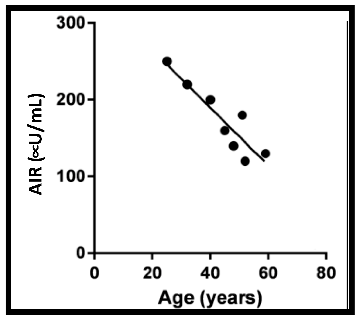
3.4. Exocytosis Proteins and Aging

The risk of developing T2D begins to rise around age 45 and rises considerably after age 65 [175]. Aging is an important risk factor for T2D and several aging mechanisms, such as chronic inflammation, macromolecular dysfunction, and cellular senescence, have also been implicated in the generation of insulin resistance [176]. T2D is also a risk factor for a shortened lifespan [177,178]. Age-associated defects in β-cell GSIS have been demonstrated in both rodents and humans [179,180]. Moreover, islet transplantation outcomes are worse when the islets are from aged donors compared with those from young donors [181], possibly related to diminished peak first-phase GSIS in more aged human islets (Figure 7). Despite a strong correlation between aging and T2D, the mechanism of age-related deterioration in functional β-cell mass is not clear. β-cell mass is controlled by the balance of apoptosis and proliferation. In both rodents and humans, aging limits the regenerative capacity of β-cells, and decreases β-cell proliferation in response to increased metabolic demands [182,183]. High glucose induces apoptosis in human β-cells via inducing the expression of Fas receptor and activating caspases 8 and 3 [184]. β-cell apoptosis is elevated 3–10-fold in T2D compared to non-diabetic individuals, as detected by TUNEL staining [3]. Additionally, cellular senescence could contribute to the functional decline of β-cells during aging. Cellular stressors increase with age, leading to the accumulation of senescent β-cells [185]. The subpopulation of senescent cells was increased in human islets by age and this portion was enriched in T2D human islets compared to age-matched non-diabetic donors [186]. Senescent β-cells are abnormally large and are characterized by upregulated expression of p16INK4a and the anti-apoptotic molecule Bcl2, along with senescence-associated β-galactosidase, as well as the senescence-associated secretory phenotype (SASP) such as IL-6, IL-8, MCP-1 (monocyte chemoattractant protein-1), PAI-1 (plasminogen-activated inhibitor-1), and IGF1R [179,186,187,188], although marker expression is notably heterogeneous. Consistent with the coupling of T2D with early aging, induction of insulin resistance increases the expression of aging markers [179]. It has been proposed that β-cell senescence during aging impairs the expression of genes relevant to β-cell identity and cellular function [179,186]. Moreover, senescence-associated cytokines could trigger an inflammatory response, exacerbating β-cell dysfunction, and demise. Hence, decreasing the senescent population in β-cells is thought to improve β-cells function and glucose regulation [186]. However, contradicting this widely accepted view, β-cell-specific overexpression of p16INK4a in mice improved GSIS, rather than impaired GSIS [189]. As a result, while it is clear that p16INK4a exerts complex influences on β-cells, the underly- ing mechanistic details must await further study.
STX4 protein, a key regulator of GSIS and found in reduced quantities in T2D human islets, is similarly reduced in the pancreata of aged mice [19]. Global STX4 overexpression (2–5-fold STX4 overexpression detected in skeletal muscle, adipose, and pancreas) extended lifespan by ~35%; underlying this was the retention of youthful insulin sensitivity and GSIS capacity, even in the face of diet-induced obesity stress, indicating an anti-aging role for STX4 beyond conventional exocytosis function [19]. Mechanistically, although classical aging-related genes such as Sirt1, mTOR, and aging-related inflammatory factors TNFα or IL-6 were unchanged, phosphorylated Foxo1 was significantly decreased in the pancreata of the aged STX4 transgenic mice [19]. Evaluation of senescence in islet β-cells from the long-lived STX4 mice will be an important step forward in interrogating the mechanistic link among exocytosis proteins, T2D, and aging. It has been reported that activation of NF-κB signaling in normal somatic cells enhances aging and accelerates senescence via upregulation of SASP associated genes and downregulation of genes for cell cycle progression [190]. Given the recently discovered role of STX4 in attenuating IĸBβ degradation and thereby blunting NF-κB signaling [18,171], it is conceivable that STX4 may impact β-cell senescence and proteostasis via an IĸBβ-NF-κB-dependent mechanism. In summary, exciting recent discoveries are pointing towards a previously unexplored, unconventional function for classical exocytosis proteins, establishing them as mediators of healthy aging and resistance to T2D.

4. Future Perspectives

The primary aim for T2D treatment is to attain and maintain whole-body glucose homeostasis, via boosting pancreatic β-cell function and mitigating the stressful workload impinged by persistent peripheral insulin resistance. Several SNARE and SNARE-associated proteins are promising therapeutic candidates, including STX4 and DOC2b. The mRNA and protein levels of both these proteins are reduced in T2D islet β-cells, suggesting that their deficiency may contribute to the pathogenesis of T2D. However, while STX4 nor DOC2b has been reported as T2D susceptibility genes per se, the STX4 gene associates with BMI (http://type2diabetesgenetics-old.org/). Intriguingly, STX4 was identified in an in silico phenome–interactome analysis, a method that prioritized candidates according to their physical interactions at the protein level with other proteins involved in type 1 diabetes. STX4 was in the top 10 in a list of genes predicted to be likely disease genes in T1D, including the insulin (INS) gene. Further development of these proteins as drug targets will reveal whether they can rescue β-cell dysfunction in the clinical setting.
Based on the promising beneficial contribution of extra STX4 and DOC2b in regulating whole-body glucose homeostasis and the reduced level of these two key exocytosis factors in T2D β-cell, it is imperative to explore the ways to induce their protein levels in the β-cell. The major technical challenge for the induction of targeted protein expression in the β-cell is the complex 3D structure of islets. Several approaches are now being tested including small activating RNA (saRNA) mediated induction of endogenous proteins, use of adeno-associated virus vectors (AAV) as a delivery system [191,192]. Another alternative therapeutic approach is the transplantation of pancreatic islets harboring enhanced levels of STX4 or DOC2b into T2D patients [193]. Future studies are required to identify and optimize ways to enrich levels of these two key exocytosis proteins in the β-cell. It is worth mentioning here that at this time other drugs that modulate insulin exocytosis to protect β-cells are upstream of SNARES. For example, Sulfonylureas (chlorpropamide, glipizide, etc.) increase insulin secretion via binding to ATP-dependent K+ channel [1,194]; GLP-1 agonists (liraglutide, exenatide) boost insulin secretion via binding to GLIP-1 receptor in the β-cell [1,195]; and additionally, inhibitors of Thioredoxin-interacting protein (TXNIP), promote insulin production and GLP-1 signaling via regulation of microRNA, are emerging as a potential treatment option for diabetes [196,197]. However, since all of these above-mentioned drugs work upstream of SNAREs, there may be effects beyond the distal steps of insulin exocytosis, again implying the significance of developing drugs for specific modulations of SNARE and SNARE regulatory proteins in the β-cell.
It is important to mention here that not all SNARE proteins are candidates for therapeutic development, as extra STX1 in the β-cell interferes with Ca2+ channel gating and impairs GSIS. Also, extra STX2 would be detrimental in T2D β-cells because of its inhibitory function. These data again remind us of the essential requirement for the proper stoichiometry during SNARE complex assembly and successful GSIS. It is imperative to suggest that the exocytosis factors that are conferring beneficial function when overexpressed are possibly doing so via indirectly influencing specific signaling pathways, such as STX4 mediated inhibition of NF-κB nuclear translocation, or possibly via direct protein-protein interactions. One such example is the direct interaction of STX4 with key proteins of the actin cytoskeleton, including F-actin itself, in the β-cell. These findings altogether suggest that our growing understanding of exocytosis proteins is opening up new avenues for T2D treatment.

Author Contributions

All authors participated in the writing, review and editing. All authors have read and agreed to the published version of the manuscript.

Funding

This study was supported by grants from the National Institutes of Health (DK067912, DK112917, and DK102233 to D.C.T.), a JDRF postdoctoral fellowship (to D.C.B., 3-PDF-2020-934-A-N), and the Wanek Project to Cure Type 1 Diabetes at the City of Hope.

Acknowledgments

Nancy Linford provided editing assistance.

Conflicts of Interest

The authors declare no conflict of interest.

References

  1. Aslamy, A.; Thurmond, D.C. Exocytosis proteins as novel targets for diabetes prevention and/or remediation? Am. J. Physiol. Regul. Integr. Comp. Physiol. 2017, 312, R739–R752. [Google Scholar] [CrossRef] [Green Version]
  2. ADA’s Primary Care Advisory Group (PCAG). American Diabetes Association. Standards of Medical Care in Diabetes—2020 Abridged for Primary Care Providers. Clin. Diabetes 2020, 38, 10–38. [Google Scholar] [CrossRef] [Green Version]
  3. Butler, A.E.; Janson, J.; Bonner-Weir, S.; Ritzel, R.; Rizza, R.A.; Butler, P.C. Beta-cell deficit and increased beta-cell apoptosis in humans with type 2 diabetes. Diabetes 2003, 52, 102–110. [Google Scholar] [CrossRef] [PubMed] [Green Version]
  4. Del Guerra, S.; Lupi, R.; Marselli, L.; Masini, M.; Bugliani, M.; Sbrana, S.; Torri, S.; Pollera, M.; Boggi, U.; Mosca, F.; et al. Functional and molecular defects of pancreatic islets in human type 2 diabetes. Diabetes 2005, 54, 727–735. [Google Scholar] [CrossRef] [Green Version]
  5. Hanley, S.C.; Austin, E.; Assouline-Thomas, B.; Kapeluto, J.; Blaichman, J.; Moosavi, M.; Petropavlovskaia, M.; Rosenberg, L. {beta}-Cell mass dynamics and islet cell plasticity in human type 2 diabetes. Endocrinology 2010, 151, 1462–1472. [Google Scholar] [CrossRef] [PubMed] [Green Version]
  6. Rahier, J.; Guiot, Y.; Goebbels, R.M.; Sempoux, C.; Henquin, J.C. Pancreatic beta-cell mass in European subjects with type 2 diabetes. Diabetes Obes. Metab. 2008, 10 (Suppl. 4), 32–42. [Google Scholar] [CrossRef]
  7. Sakuraba, H.; Mizukami, H.; Yagihashi, N.; Wada, R.; Hanyu, C.; Yagihashi, S. Reduced beta-cell mass and expression of oxidative stress-related DNA damage in the islet of Japanese Type II diabetic patients. Diabetologia 2002, 45, 85–96. [Google Scholar] [CrossRef] [PubMed] [Green Version]
  8. Eizirik, D.L.; Pasquali, L.; Cnop, M. Pancreatic beta-cells in type 1 and type 2 diabetes mellitus: Different pathways to failure. Nat. Rev. Endocrinol. 2020, 16, 349–362. [Google Scholar] [CrossRef]
  9. Henquin, J.C.; Rahier, J. Pancreatic alpha cell mass in European subjects with type 2 diabetes. Diabetologia 2011, 54, 1720–1725. [Google Scholar] [CrossRef] [PubMed] [Green Version]
  10. Cnop, M.; Vidal, J.; Hull, R.L.; Utzschneider, K.M.; Carr, D.B.; Schraw, T.; Scherer, P.E.; Boyko, E.J.; Fujimoto, W.Y.; Kahn, S.E. Progressive loss of beta-cell function leads to worsening glucose tolerance in first-degree relatives of subjects with type 2 diabetes. Diabetes Care 2007, 30, 677–682. [Google Scholar] [CrossRef] [Green Version]
  11. Utzschneider, K.M.; Prigeon, R.L.; Faulenbach, M.V.; Tong, J.; Carr, D.B.; Boyko, E.J.; Leonetti, D.L.; McNeely, M.J.; Fujimoto, W.Y.; Kahn, S.E. Oral disposition index predicts the development of future diabetes above and beyond fasting and 2-h glucose levels. Diabetes Care 2009, 32, 335–341. [Google Scholar] [CrossRef] [PubMed] [Green Version]
  12. Lytrivi, M.; Castell, A.L.; Poitout, V.; Cnop, M. Recent Insights into Mechanisms of beta-Cell Lipo- and Glucolipotoxicity in Type 2 Diabetes. J. Mol. Biol. 2020, 432, 1514–1534. [Google Scholar] [CrossRef] [PubMed]
  13. Eastwood, S.L.; Cotter, D.; Harrison, P.J. Cerebellar synaptic protein expression in schizophrenia. Neuroscience 2001, 105, 219–229. [Google Scholar] [CrossRef]
  14. Fatemi, S.H.; Earle, J.A.; Stary, J.M.; Lee, S.; Sedgewick, J. Altered levels of the synaptosomal associated protein SNAP-25 in hippocampus of subjects with mood disorders and schizophrenia. Neuroreport 2001, 12, 3257–3262. [Google Scholar] [CrossRef]
  15. Galvez, J.M.; Forero, D.A.; Fonseca, D.J.; Mateus, H.E.; Talero-Gutierrez, C.; Velez-van-Meerbeke, A. Evidence of association between SNAP25 gene and attention deficit hyperactivity disorder in a Latin American sample. Atten. Defic. Hyperact. Disord. 2014, 6, 19–23. [Google Scholar] [CrossRef]
  16. Kabekkodu, S.P.; Bhat, S.; Radhakrishnan, R.; Aithal, A.; Mascarenhas, R.; Pandey, D.; Rai, L.; Kushtagi, P.; Mundyat, G.P.; Satyamoorthy, K. DNA promoter methylation-dependent transcription of the double C2-like domain beta (DOC2B) gene regulates tumor growth in human cervical cancer. J. Biol. Chem. 2014, 289, 10637–10649. [Google Scholar] [CrossRef] [Green Version]
  17. Perrotta, C.; Bizzozero, L.; Cazzato, D.; Morlacchi, S.; Assi, E.; Simbari, F.; Zhang, Y.; Gulbins, E.; Bassi, M.T.; Rosa, P.; et al. Syntaxin 4 is required for acid sphingomyelinase activity and apoptotic function. J. Biol. Chem. 2010, 285, 40240–40251. [Google Scholar] [CrossRef] [PubMed] [Green Version]
  18. Oh, E.; Ahn, M.; Afelik, S.; Becker, T.C.; Roep, B.O.; Thurmond, D.C. Syntaxin 4 Expression in Pancreatic Beta-Cells Promotes Islet Function and Protects Functional Beta-Cell Mass. Diabetes 2018, 67, 2626–2639. [Google Scholar] [CrossRef] [Green Version]
  19. Oh, E.; Miller, R.A.; Thurmond, D.C. Syntaxin 4 Overexpression Ameliorates Effects of Aging and High-Fat Diet on Glucose Control and Extends Lifespan. Cell Metab. 2015, 22, 499–507. [Google Scholar] [CrossRef] [PubMed] [Green Version]
  20. Oh, E.; Stull, N.D.; Mirmira, R.G.; Thurmond, D.C. Syntaxin 4 up-regulation increases efficiency of insulin release in pancreatic islets from humans with and without type 2 diabetes mellitus. J. Clin. Endocrinol. Metab. 2014, 99, E866–E870. [Google Scholar] [CrossRef] [PubMed] [Green Version]
  21. McCulloch, L.J.; van de Bunt, M.; Braun, M.; Frayn, K.N.; Clark, A.; Gloyn, A.L. GLUT2 (SLC2A2) is not the principal glucose transporter in human pancreatic beta cells: Implications for understanding genetic association signals at this locus. Mol. Genet. Metab. 2011, 104, 648–653. [Google Scholar] [CrossRef]
  22. Thorens, B.; Gerard, N.; Deriaz, N. GLUT2 surface expression and intracellular transport via the constitutive pathway in pancreatic beta cells and insulinoma: Evidence for a block in trans-Golgi network exit by brefeldin A. J. Cell Biol. 1993, 123, 1687–1694. [Google Scholar] [CrossRef]
  23. De Vos, A.; Heimberg, H.; Quartier, E.; Huypens, P.; Bouwens, L.; Pipeleers, D.; Schuit, F. Human and rat beta cells differ in glucose transporter but not in glucokinase gene expression. J. Clin. Invest. 1995, 96, 2489–2495. [Google Scholar] [CrossRef] [PubMed] [Green Version]
  24. Pingitore, A.; Ruz-Maldonado, I.; Liu, B.; Huang, G.C.; Choudhary, P.; Persaud, S.J. Dynamic Profiling of Insulin Secretion and ATP Generation in Isolated Human and Mouse Islets Reveals Differential Glucose Sensitivity. Cell. Physiol. Biochem. 2017, 44, 1352–1359. [Google Scholar] [CrossRef] [PubMed] [Green Version]
  25. Ashcroft, F.M.; Harrison, D.E.; Ashcroft, S.J. Glucose induces closure of single potassium channels in isolated rat pancreatic beta-cells. Nature 1984, 312, 446–448. [Google Scholar] [CrossRef] [PubMed]
  26. Cook, D.L.; Hales, C.N. Intracellular ATP directly blocks K+ channels in pancreatic B-cells. Nature 1984, 311, 271–273. [Google Scholar] [CrossRef] [PubMed]
  27. Rorsman, P.; Ashcroft, F.M.; Trube, G. Single Ca channel currents in mouse pancreatic B-cells. Pflugers Archiv 1988, 412, 597–603. [Google Scholar] [CrossRef] [PubMed]
  28. Satin, L.S.; Cook, D.L. Voltage-gated Ca2+ current in pancreatic B-cells. Pflugers Archiv 1985, 404, 385–387. [Google Scholar] [CrossRef] [PubMed]
  29. Alvarez de Toledo, G.; Montes, M.A.; Montenegro, P.; Borges, R. Phases of the exocytotic fusion pore. FEBS Lett. 2018, 592, 3532–3541. [Google Scholar] [CrossRef] [PubMed] [Green Version]
  30. Nunemaker, C.S.; Satin, L.S. Episodic hormone secretion: A comparison of the basis of pulsatile secretion of insulin and GnRH. Endocrine 2014, 47, 49–63. [Google Scholar] [CrossRef] [Green Version]
  31. Song, S.H.; McIntyre, S.S.; Shah, H.; Veldhuis, J.D.; Hayes, P.C.; Butler, P.C. Direct measurement of pulsatile insulin secretion from the portal vein in human subjects. J. Clin. Endocrinol. Metab. 2000, 85, 4491–4499. [Google Scholar] [CrossRef] [PubMed]
  32. Thurmond, D.C.; Gaisano, H.Y. Recent Insights into Beta-cell Exocytosis in Type 2 Diabetes. J. Mol. Biol. 2020, 432, 1310–1325. [Google Scholar] [CrossRef] [PubMed]
  33. Gaisano, H.Y. Here come the newcomer granules, better late than never. Trends Endocrinol. Metab. 2014, 25, 381–388. [Google Scholar] [CrossRef]
  34. Barg, S.; Eliasson, L.; Renstrom, E.; Rorsman, P. A subset of 50 secretory granules in close contact with L-type Ca2+ channels accounts for first-phase insulin secretion in mouse beta-cells. Diabetes 2002, 51 (Suppl. 1), S74–S82. [Google Scholar] [CrossRef] [Green Version]
  35. Daniel, S.; Noda, M.; Straub, S.G.; Sharp, G.W. Identification of the docked granule pool responsible for the first phase of glucose-stimulated insulin secretion. Diabetes 1999, 48, 1686–1690. [Google Scholar] [CrossRef] [PubMed]
  36. Olofsson, C.S.; Gopel, S.O.; Barg, S.; Galvanovskis, J.; Ma, X.; Salehi, A.; Rorsman, P.; Eliasson, L. Fast insulin secretion reflects exocytosis of docked granules in mouse pancreatic B-cells. Pflugers Archiv 2002, 444, 43–51. [Google Scholar] [CrossRef] [PubMed]
  37. Curry, D.L.; Bennett, L.L.; Grodsky, G.M. Dynamics of insulin secretion by the perfused rat pancreas. Endocrinology 1968, 83, 572–584. [Google Scholar] [CrossRef]
  38. Porte, D., Jr.; Pupo, A.A. Insulin responses to glucose: Evidence for a two pool system in man. J. Clin. Invest. 1969, 48, 2309–2319. [Google Scholar] [CrossRef] [Green Version]
  39. Cui, J.; Wang, Z.; Cheng, Q.; Lin, R.; Zhang, X.M.; Leung, P.S.; Copeland, N.G.; Jenkins, N.A.; Yao, K.M.; Huang, J.D. Targeted inactivation of kinesin-1 in pancreatic beta-cells in vivo leads to insulin secretory deficiency. Diabetes 2011, 60, 320–330. [Google Scholar] [CrossRef] [PubMed] [Green Version]
  40. Baumert, M.; Maycox, P.R.; Navone, F.; De Camilli, P.; Jahn, R. Synaptobrevin: An integral membrane protein of 18,000 daltons present in small synaptic vesicles of rat brain. EMBO J. 1989, 8, 379–384. [Google Scholar] [CrossRef]
  41. Gembal, M.; Gilon, P.; Henquin, J.C. Evidence that glucose can control insulin release independently from its action on ATP-sensitive K+ channels in mouse B cells. J. Clin. Invest. 1992, 89, 1288–1295. [Google Scholar] [CrossRef] [Green Version]
  42. Kiraly-Borri, C.E.; Morgan, A.; Burgoyne, R.D.; Weller, U.; Wollheim, C.B.; Lang, J. Soluble N-ethylmaleimide-sensitive-factor attachment protein and N-ethylmaleimide-insensitive factors are required for Ca2+-stimulated exocytosis of insulin. Biochem. J. 1996, 314 Pt 1, 199–203. [Google Scholar] [CrossRef] [Green Version]
  43. Lang, J. Molecular mechanisms and regulation of insulin exocytosis as a paradigm of endocrine secretion. Eur. J. Biochem. 1999, 259, 3–17. [Google Scholar] [CrossRef] [PubMed]
  44. Sollner, T.; Bennett, M.K.; Whiteheart, S.W.; Scheller, R.H.; Rothman, J.E. A protein assembly-disassembly pathway in vitro that may correspond to sequential steps of synaptic vesicle docking, activation, and fusion. Cell 1993, 75, 409–418. [Google Scholar] [CrossRef]
  45. Sollner, T.; Whiteheart, S.W.; Brunner, M.; Erdjument-Bromage, H.; Geromanos, S.; Tempst, P.; Rothman, J.E. SNAP receptors implicated in vesicle targeting and fusion. Nature 1993, 362, 318–324. [Google Scholar] [CrossRef] [PubMed]
  46. Martin, F.; Moya, F.; Gutierrez, L.M.; Reig, J.A.; Soria, B. Role of syntaxin in mouse pancreatic beta cells. Diabetologia 1995, 38, 860–863. [Google Scholar] [CrossRef] [PubMed] [Green Version]
  47. Liang, T.; Qin, T.; Xie, L.; Dolai, S.; Zhu, D.; Prentice, K.J.; Wheeler, M.; Kang, Y.; Osborne, L.; Gaisano, H.Y. New Roles of Syntaxin-1A in Insulin Granule Exocytosis and Replenishment. J. Biol. Chem. 2017, 292, 2203–2216. [Google Scholar] [CrossRef] [PubMed] [Green Version]
  48. Lam, P.P.; Leung, Y.M.; Sheu, L.; Ellis, J.; Tsushima, R.G.; Osborne, L.R.; Gaisano, H.Y. Transgenic mouse overexpressing syntaxin-1A as a diabetes model. Diabetes 2005, 54, 2744–2754. [Google Scholar] [CrossRef] [PubMed] [Green Version]
  49. Watanabe, T.; Fujiwara, T.; Komazaki, S.; Yamaguchi, K.; Tajima, O.; Akagawa, K. HPC-1/syntaxin 1A suppresses exocytosis of PC12 cells. J. Biochem. 1999, 125, 685–689. [Google Scholar] [CrossRef] [PubMed]
  50. Land, J.; Zhang, H.; Vaidyanathan, V.V.; Sadoul, K.; Niemann, H.; Wollheim, C.B. Transient expression of botulinum neurotoxin C1 light chain differentially inhibits calcium and glucose induced insulin secretion in clonal beta-cells. FEBS Lett. 1997, 419, 13–17. [Google Scholar] [CrossRef] [Green Version]
  51. Spurlin, B.A.; Thurmond, D.C. Syntaxin 4 facilitates biphasic glucose-stimulated insulin secretion from pancreatic beta-cells. Mol. Endocrinol. 2006, 20, 183–193. [Google Scholar] [CrossRef] [PubMed] [Green Version]
  52. Spurlin, B.A.; Park, S.Y.; Nevins, A.K.; Kim, J.K.; Thurmond, D.C. Syntaxin 4 transgenic mice exhibit enhanced insulin-mediated glucose uptake in skeletal muscle. Diabetes 2004, 53, 2223–2231. [Google Scholar] [CrossRef] [Green Version]
  53. Xie, L.; Zhu, D.; Dolai, S.; Liang, T.; Qin, T.; Kang, Y.; Xie, H.; Huang, Y.C.; Gaisano, H.Y. Syntaxin-4 mediates exocytosis of pre-docked and newcomer insulin granules underlying biphasic glucose-stimulated insulin secretion in human pancreatic beta cells. Diabetologia 2015, 58, 1250–1259. [Google Scholar] [CrossRef] [PubMed] [Green Version]
  54. Oh, E.; Spurlin, B.A.; Pessin, J.E.; Thurmond, D.C. Munc18c heterozygous knockout mice display increased susceptibility for severe glucose intolerance. Diabetes 2005, 54, 638–647. [Google Scholar] [CrossRef] [PubMed] [Green Version]
  55. Spurlin, B.A.; Thomas, R.M.; Nevins, A.K.; Kim, H.J.; Kim, Y.J.; Noh, H.L.; Shulman, G.I.; Kim, J.K.; Thurmond, D.C. Insulin resistance in tetracycline-repressible Munc18c transgenic mice. Diabetes 2003, 52, 1910–1917. [Google Scholar] [CrossRef] [PubMed] [Green Version]
  56. Zhu, D.; Koo, E.; Kwan, E.; Kang, Y.; Park, S.; Xie, H.; Sugita, S.; Gaisano, H.Y. Syntaxin-3 regulates newcomer insulin granule exocytosis and compound fusion in pancreatic beta cells. Diabetologia 2013, 56, 359–369. [Google Scholar] [CrossRef] [PubMed]
  57. Xie, L.; Dolai, S.; Kang, Y.; Liang, T.; Xie, H.; Qin, T.; Yang, L.; Chen, L.; Gaisano, H.Y. Syntaxin-3 Binds and Regulates Both R- and L-Type Calcium Channels in Insulin-Secreting INS-1 832/13 Cells. PLoS ONE 2016, 11, e0147862. [Google Scholar] [CrossRef] [PubMed]
  58. Zhu, D.; Xie, L.; Kang, Y.; Dolai, S.; Bondo Hansen, J.; Qin, T.; Xie, H.; Liang, T.; Rubin, D.C.; Osborne, L.; et al. Syntaxin 2 Acts as Inhibitory SNARE for Insulin Granule Exocytosis. Diabetes 2017, 66, 948–959. [Google Scholar] [CrossRef] [PubMed] [Green Version]
  59. Oh, E.; Thurmond, D.C. Munc18c depletion selectively impairs the sustained phase of insulin release. Diabetes 2009, 58, 1165–1174. [Google Scholar] [CrossRef] [PubMed] [Green Version]
  60. Zhu, D.; Xie, L.; Karimian, N.; Liang, T.; Kang, Y.; Huang, Y.C.; Gaisano, H.Y. Munc18c mediates exocytosis of pre-docked and newcomer insulin granules underlying biphasic glucose stimulated insulin secretion in human pancreatic beta-cells. Mol. Metab. 2015, 4, 418–426. [Google Scholar] [CrossRef] [PubMed]
  61. Tomas, A.; Meda, P.; Regazzi, R.; Pessin, J.E.; Halban, P.A. Munc 18-1 and granuphilin collaborate during insulin granule exocytosis. Traffic 2008, 9, 813–832. [Google Scholar] [CrossRef] [PubMed]
  62. Garcia, E.P.; Gatti, E.; Butler, M.; Burton, J.; De Camilli, P. A rat brain Sec1 homologue related to Rop and UNC18 interacts with syntaxin. Proc. Natl. Acad. Sci. USA 1994, 91, 2003–2007. [Google Scholar] [CrossRef] [PubMed] [Green Version]
  63. Tamori, Y.; Kawanishi, M.; Niki, T.; Shinoda, H.; Araki, S.; Okazawa, H.; Kasuga, M. Inhibition of insulin-induced GLUT4 translocation by Munc18c through interaction with syntaxin4 in 3T3-L1 adipocytes. J. Biol. Chem. 1998, 273, 19740–19746. [Google Scholar] [CrossRef] [Green Version]
  64. Tellam, J.T.; Macaulay, S.L.; McIntosh, S.; Hewish, D.R.; Ward, C.W.; James, D.E. Characterization of Munc-18c and syntaxin-4 in 3T3-L1 adipocytes. Putative role in insulin-dependent movement of GLUT-4. J. Biol. Chem. 1997, 272, 6179–6186. [Google Scholar] [CrossRef] [Green Version]
  65. Thurmond, D.C.; Ceresa, B.P.; Okada, S.; Elmendorf, J.S.; Coker, K.; Pessin, J.E. Regulation of insulin-stimulated GLUT4 translocation by Munc18c in 3T3L1 adipocytes. J. Biol. Chem. 1998, 273, 33876–33883. [Google Scholar] [CrossRef] [PubMed] [Green Version]
  66. Oh, E.; Kalwat, M.A.; Kim, M.J.; Verhage, M.; Thurmond, D.C. Munc18-1 regulates first-phase insulin release by promoting granule docking to multiple syntaxin isoforms. J. Biol. Chem. 2012, 287, 25821–25833. [Google Scholar] [CrossRef] [PubMed] [Green Version]
  67. Yin, P.; Gandasi, N.R.; Arora, S.; Omar-Hmeadi, M.; Saras, J.; Barg, S. Syntaxin clusters at secretory granules in a munc18-bound conformation. Mol. Biol. Cell 2018, 29, 2700–2708. [Google Scholar] [CrossRef]
  68. Lam, P.P.; Ohno, M.; Dolai, S.; He, Y.; Qin, T.; Liang, T.; Zhu, D.; Kang, Y.; Liu, Y.; Kauppi, M.; et al. Munc18b is a major mediator of insulin exocytosis in rat pancreatic beta-cells. Diabetes 2013, 62, 2416–2428. [Google Scholar] [CrossRef] [PubMed] [Green Version]
  69. Zhu, D.; Zhang, Y.; Lam, P.P.; Dolai, S.; Liu, Y.; Cai, E.P.; Choi, D.; Schroer, S.A.; Kang, Y.; Allister, E.M.; et al. Dual role of VAMP8 in regulating insulin exocytosis and islet beta cell growth. Cell Metab. 2012, 16, 238–249. [Google Scholar] [CrossRef] [Green Version]
  70. Oh, E.; Thurmond, D.C. The stimulus-induced tyrosine phosphorylation of Munc18c facilitates vesicle exocytosis. J. Biol. Chem. 2006, 281, 17624–17634. [Google Scholar] [CrossRef] [Green Version]
  71. Jewell, J.L.; Oh, E.; Bennett, S.M.; Meroueh, S.O.; Thurmond, D.C. The tyrosine phosphorylation of Munc18c induces a switch in binding specificity from syntaxin 4 to Doc2beta. J. Biol. Chem. 2008, 283, 21734–21746. [Google Scholar] [CrossRef] [PubMed] [Green Version]
  72. Ke, B.; Oh, E.; Thurmond, D.C. Doc2beta is a novel Munc18c-interacting partner and positive effector of syntaxin 4-mediated exocytosis. J. Biol. Chem. 2007, 282, 21786–21797. [Google Scholar] [CrossRef] [PubMed] [Green Version]
  73. Sakaguchi, G.; Orita, S.; Maeda, M.; Igarashi, H.; Takai, Y. Molecular cloning of an isoform of Doc2 having two C2-like domains. Biochem. Biophys. Res. Commun. 1995, 217, 1053–1061. [Google Scholar] [CrossRef]
  74. Verhage, M.; de Vries, K.J.; Roshol, H.; Burbach, J.P.; Gispen, W.H.; Sudhof, T.C. DOC2 proteins in rat brain: Complementary distribution and proposed function as vesicular adapter proteins in early stages of secretion. Neuron 1997, 18, 453–461. [Google Scholar] [CrossRef] [Green Version]
  75. Li, J.; Cantley, J.; Burchfield, J.G.; Meoli, C.C.; Stockli, J.; Whitworth, P.T.; Pant, H.; Chaudhuri, R.; Groffen, A.J.; Verhage, M.; et al. DOC2 isoforms play dual roles in insulin secretion and insulin-stimulated glucose uptake. Diabetologia 2014, 57, 2173–2182. [Google Scholar] [CrossRef] [PubMed]
  76. Ramalingam, L.; Oh, E.; Yoder, S.M.; Brozinick, J.T.; Kalwat, M.A.; Groffen, A.J.; Verhage, M.; Thurmond, D.C. Doc2b is a key effector of insulin secretion and skeletal muscle insulin sensitivity. Diabetes 2012, 61, 2424–2432. [Google Scholar] [CrossRef] [Green Version]
  77. Ramalingam, L.; Oh, E.; Thurmond, D.C. Doc2b enrichment enhances glucose homeostasis in mice via potentiation of insulin secretion and peripheral insulin sensitivity. Diabetologia 2014, 57, 1476–1484. [Google Scholar] [CrossRef] [Green Version]
  78. Aslamy, A.; Oh, E.; Olson, E.M.; Zhang, J.; Ahn, M.; Moin, A.S.M.; Tunduguru, R.; Salunkhe, V.A.; Veluthakal, R.; Thurmond, D.C. Doc2b Protects beta-Cells Against Inflammatory Damage and Enhances Function. Diabetes 2018, 67, 1332–1344. [Google Scholar] [CrossRef] [Green Version]
  79. Ramalingam, L.; Lu, J.; Hudmon, A.; Thurmond, D.C. Doc2b serves as a scaffolding platform for concurrent binding of multiple Munc18 isoforms in pancreatic islet beta-cells. Biochem. J. 2014, 464, 251–258. [Google Scholar] [CrossRef] [PubMed] [Green Version]
  80. Li, G.; Rungger-Brandle, E.; Just, I.; Jonas, J.C.; Aktories, K.; Wollheim, C.B. Effect of disruption of actin filaments by Clostridium botulinum C2 toxin on insulin secretion in HIT-T15 cells and pancreatic islets. Mol. Biol. Cell 1994, 5, 1199–1213. [Google Scholar] [CrossRef] [PubMed] [Green Version]
  81. Somers, G.; Blondel, B.; Orci, L.; Malaisse, W.J. Motile events in pancreatic endocrine cells. Endocrinology 1979, 104, 255–264. [Google Scholar] [CrossRef] [PubMed]
  82. Wilson, J.R.; Ludowyke, R.I.; Biden, T.J. A redistribution of actin and myosin IIA accompanies Ca(2+)-dependent insulin secretion. FEBS Lett. 2001, 492, 101–106. [Google Scholar] [CrossRef] [Green Version]
  83. Orci, L.; Gabbay, K.H.; Malaisse, W.J. Pancreatic beta-cell web: Its possible role in insulin secretion. Science 1972, 175, 1128–1130. [Google Scholar] [CrossRef]
  84. Kalwat, M.A.; Wiseman, D.A.; Luo, W.; Wang, Z.; Thurmond, D.C. Gelsolin associates with the N terminus of syntaxin 4 to regulate insulin granule exocytosis. Mol. Endocrinol. 2012, 26, 128–141. [Google Scholar] [CrossRef] [PubMed] [Green Version]
  85. Wang, Z.; Thurmond, D.C. Mechanisms of biphasic insulin-granule exocytosis—Roles of the cytoskeleton, small GTPases and SNARE proteins. J. Cell Sci. 2009, 122, 893–903. [Google Scholar] [CrossRef] [Green Version]
  86. Kalwat, M.A.; Thurmond, D.C. Signaling mechanisms of glucose-induced F-actin remodeling in pancreatic islet beta cells. Exp. Mol. Med. 2013, 45, e37. [Google Scholar] [CrossRef] [Green Version]
  87. Thurmond, D.C.; Gonelle-Gispert, C.; Furukawa, M.; Halban, P.A.; Pessin, J.E. Glucose-stimulated insulin secretion is coupled to the interaction of actin with the t-SNARE (target membrane soluble N-ethylmaleimide-sensitive factor attachment protein receptor protein) complex. Mol. Endocrinol. 2003, 17, 732–742. [Google Scholar] [CrossRef] [PubMed] [Green Version]
  88. Band, A.M.; Ali, H.; Vartiainen, M.K.; Welti, S.; Lappalainen, P.; Olkkonen, V.M.; Kuismanen, E. Endogenous plasma membrane t-SNARE syntaxin 4 is present in rab11 positive endosomal membranes and associates with cortical actin cytoskeleton. FEBS Lett. 2002, 531, 513–519. [Google Scholar] [CrossRef] [Green Version]
  89. Jewell, J.L.; Luo, W.; Oh, E.; Wang, Z.; Thurmond, D.C. Filamentous actin regulates insulin exocytosis through direct interaction with Syntaxin 4. J. Biol. Chem. 2008, 283, 10716–10726. [Google Scholar] [CrossRef] [PubMed] [Green Version]
  90. Nevins, A.K.; Thurmond, D.C. A direct interaction between Cdc42 and vesicle-associated membrane protein 2 regulates SNARE-dependent insulin exocytosis. J. Biol. Chem. 2005, 280, 1944–1952. [Google Scholar] [CrossRef] [PubMed] [Green Version]
  91. Nagano, F.; Orita, S.; Sasaki, T.; Naito, A.; Sakaguchi, G.; Maeda, M.; Watanabe, T.; Kominami, E.; Uchiyama, Y.; Takai, Y. Interaction of Doc2 with tctex-1, a light chain of cytoplasmic dynein. Implication in dynein-dependent vesicle transport. J. Biol. Chem. 1998, 273, 30065–30068. [Google Scholar] [CrossRef] [PubMed] [Green Version]
  92. Shimoda, Y.; Okada, S.; Yamada, E.; Pessin, J.E.; Yamada, M. Tctex1d2 Is a Negative Regulator of GLUT4 Translocation and Glucose Uptake. Endocrinology 2015, 156, 3548–3558. [Google Scholar] [CrossRef] [PubMed] [Green Version]
  93. Zhang, J.; Oh, E.; Merz, K.E.; Aslamy, A.; Veluthakal, R.; Salunkhe, V.A.; Ahn, M.; Tunduguru, R.; Thurmond, D.C. DOC2B promotes insulin sensitivity in mice via a novel KLC1-dependent mechanism in skeletal muscle. Diabetologia 2019, 62, 845–859. [Google Scholar] [CrossRef] [Green Version]
  94. Daniel, S.; Noda, M.; Cerione, R.A.; Sharp, G.W. A link between Cdc42 and syntaxin is involved in mastoparan-stimulated insulin release. Biochemistry 2002, 41, 9663–9671. [Google Scholar] [CrossRef] [PubMed]
  95. Kowluru, A.; Metz, S.A. Regulation of guanine-nucleotide binding proteins in islet subcellular fractions by phospholipase-derived lipid mediators of insulin secretion. Biochim. Biophys. Acta 1994, 1222, 360–368. [Google Scholar] [CrossRef]
  96. Kowluru, A.; Seavey, S.E.; Li, G.; Sorenson, R.L.; Weinhaus, A.J.; Nesher, R.; Rabaglia, M.E.; Vadakekalam, J.; Metz, S.A. Glucose- and GTP-dependent stimulation of the carboxyl methylation of CDC42 in rodent and human pancreatic islets and pure beta cells. Evidence for an essential role of GTP-binding proteins in nutrient-induced insulin secretion. J. Clin. Invest. 1996, 98, 540–555. [Google Scholar] [CrossRef] [PubMed] [Green Version]
  97. Nevins, A.K.; Thurmond, D.C. Glucose regulates the cortical actin network through modulation of Cdc42 cycling to stimulate insulin secretion. Am. J. Physiol. Cell Physiol. 2003, 285, C698–C710. [Google Scholar] [CrossRef] [Green Version]
  98. Kalwat, M.A.; Yoder, S.M.; Wang, Z.; Thurmond, D.C. A p21-activated kinase (PAK1) signaling cascade coordinately regulates F-actin remodeling and insulin granule exocytosis in pancreatic beta cells. Biochem. Pharmacol. 2013, 85, 808–816. [Google Scholar] [CrossRef] [PubMed] [Green Version]
  99. Wang, Z.; Oh, E.; Clapp, D.W.; Chernoff, J.; Thurmond, D.C. Inhibition or ablation of p21-activated kinase (PAK1) disrupts glucose homeostatic mechanisms in vivo. J. Biol. Chem. 2011, 286, 41359–41367. [Google Scholar] [CrossRef] [PubMed] [Green Version]
  100. Wang, Z.; Oh, E.; Thurmond, D.C. Glucose-stimulated Cdc42 signaling is essential for the second phase of insulin secretion. J. Biol. Chem. 2007, 282, 9536–9546. [Google Scholar] [CrossRef] [Green Version]
  101. Ahn, M.; Yoder, S.M.; Wang, Z.; Oh, E.; Ramalingam, L.; Tunduguru, R.; Thurmond, D.C. The p21-activated kinase (PAK1) is involved in diet-induced beta cell mass expansion and survival in mice and human islets. Diabetologia 2016, 59, 2145–2155. [Google Scholar] [CrossRef] [Green Version]
  102. Wang, B.; Lin, H.; Li, X.; Lu, W.; Kim, J.B.; Xu, A.; Cheng, K.K.Y. The adaptor protein APPL2 controls glucose-stimulated insulin secretion via F-actin remodeling in pancreatic beta-cells. Proc. Natl. Acad. Sci. USA 2020, 117, 28307–28315. [Google Scholar] [CrossRef]
  103. Asahara, S.; Shibutani, Y.; Teruyama, K.; Inoue, H.Y.; Kawada, Y.; Etoh, H.; Matsuda, T.; Kimura-Koyanagi, M.; Hashimoto, N.; Sakahara, M.; et al. Ras-related C3 botulinum toxin substrate 1 (RAC1) regulates glucose-stimulated insulin secretion via modulation of F-actin. Diabetologia 2013, 56, 1088–1097. [Google Scholar] [CrossRef] [PubMed] [Green Version]
  104. Li, J.; Luo, R.; Kowluru, A.; Li, G. Novel regulation by Rac1 of glucose- and forskolin-induced insulin secretion in INS-1 beta-cells. Am. J. Physiol. Endocrinol. Metab. 2004, 286, E818–E827. [Google Scholar] [CrossRef] [PubMed]
  105. Cheng, K.K.; Lam, K.S.; Wu, D.; Wang, Y.; Sweeney, G.; Hoo, R.L.; Zhang, J.; Xu, A. APPL1 potentiates insulin secretion in pancreatic beta cells by enhancing protein kinase Akt-dependent expression of SNARE proteins in mice. Proc. Natl. Acad. Sci. USA 2012, 109, 8919–8924. [Google Scholar] [CrossRef] [PubMed] [Green Version]
  106. Niggli, V.; Andreoli, C.; Roy, C.; Mangeat, P. Identification of a phosphatidylinositol-4,5-bisphosphate-binding domain in the N-terminal region of ezrin. FEBS Lett. 1995, 376, 172–176. [Google Scholar] [CrossRef] [Green Version]
  107. Ponuwei, G.A. A glimpse of the ERM proteins. J. Biomed. Sci. 2016, 23, 35. [Google Scholar] [CrossRef] [PubMed] [Green Version]
  108. Turunen, O.; Wahlstrom, T.; Vaheri, A. Ezrin has a COOH-terminal actin-binding site that is conserved in the ezrin protein family. J. Cell Biol. 1994, 126, 1445–1453. [Google Scholar] [CrossRef] [PubMed] [Green Version]
  109. Lopez, J.P.; Turner, J.R.; Philipson, L.H. Glucose-induced ERM protein activation and translocation regulates insulin secretion. Am. J. Physiol. Endocrinol. Metab. 2010, 299, E772–E785. [Google Scholar] [CrossRef] [PubMed] [Green Version]
  110. Fu, J.; Githaka, J.M.; Dai, X.; Plummer, G.; Suzuki, K.; Spigelman, A.F.; Bautista, A.; Kim, R.; Greitzer-Antes, D.; Fox, J.E.M.; et al. A glucose-dependent spatial patterning of exocytosis in human beta-cells is disrupted in type 2 diabetes. JCI Insight 2019, 5. [Google Scholar] [CrossRef]
  111. Ferri, G.; Digiacomo, L.; Lavagnino, Z.; Occhipinti, M.; Bugliani, M.; Cappello, V.; Caracciolo, G.; Marchetti, P.; Piston, D.W.; Cardarelli, F. Insulin secretory granules labelled with phogrin-fluorescent proteins show alterations in size, mobility and responsiveness to glucose stimulation in living beta-cells. Sci. Rep. 2019, 9, 2890. [Google Scholar] [CrossRef]
  112. Makhmutova, M.; Liang, T.; Gaisano, H.; Caicedo, A.; Almaca, J. Confocal Imaging of Neuropeptide Y-pHluorin: A Technique to Visualize Insulin Granule Exocytosis in Intact Murine and Human Islets. J. Vis. Exp. 2017. [Google Scholar] [CrossRef] [PubMed] [Green Version]
  113. Takahashi, N.; Sawada, W.; Noguchi, J.; Watanabe, S.; Ucar, H.; Hayashi-Takagi, A.; Yagishita, S.; Ohno, M.; Tokumaru, H.; Kasai, H. Two-photon fluorescence lifetime imaging of primed SNARE complexes in presynaptic terminals and beta cells. Nat. Commun. 2015, 6, 8531. [Google Scholar] [CrossRef] [PubMed] [Green Version]
  114. Huypens, P.; Ling, Z.; Pipeleers, D.; Schuit, F. Glucagon receptors on human islet cells contribute to glucose competence of insulin release. Diabetologia 2000, 43, 1012–1019. [Google Scholar] [CrossRef] [PubMed] [Green Version]
  115. Ahren, B. GLP-1 for type 2 diabetes. Exp. Cell Res. 2011, 317, 1239–1245. [Google Scholar] [CrossRef] [PubMed] [Green Version]
  116. Nadkarni, P.; Chepurny, O.G.; Holz, G.G. Regulation of glucose homeostasis by GLP-1. Prog. Mol. Biol. Transl. Sci. 2014, 121, 23–65. [Google Scholar] [CrossRef] [Green Version]
  117. Meloni, A.R.; DeYoung, M.B.; Lowe, C.; Parkes, D.G. GLP-1 receptor activated insulin secretion from pancreatic beta-cells: Mechanism and glucose dependence. Diabetes Obes. Metab. 2013, 15, 15–27. [Google Scholar] [CrossRef] [Green Version]
  118. Kwan, E.P.; Gaisano, H.Y. Glucagon-like peptide 1 regulates sequential and compound exocytosis in pancreatic islet beta-cells. Diabetes 2005, 54, 2734–2743. [Google Scholar] [CrossRef] [PubMed]
  119. Kwan, E.P.; Xie, L.; Sheu, L.; Ohtsuka, T.; Gaisano, H.Y. Interaction between Munc13-1 and RIM is critical for glucagon-like peptide-1 mediated rescue of exocytotic defects in Munc13-1 deficient pancreatic beta-cells. Diabetes 2007, 56, 2579–2588. [Google Scholar] [CrossRef] [PubMed] [Green Version]
  120. Dolz, M.; Movassat, J.; Bailbe, D.; Le Stunff, H.; Giroix, M.H.; Fradet, M.; Kergoat, M.; Portha, B. cAMP-secretion coupling is impaired in diabetic GK/Par rat beta-cells: A defect counteracted by GLP-1. Am. J. Physiol. Endocrinol. Metab. 2011, 301, E797–E806. [Google Scholar] [CrossRef] [Green Version]
  121. Kong, X.; Yan, D.; Sun, J.; Wu, X.; Mulder, H.; Hua, X.; Ma, X. Glucagon-like peptide 1 stimulates insulin secretion via inhibiting RhoA/ROCK signaling and disassembling glucotoxicity-induced stress fibers. Endocrinology 2014, 155, 4676–4685. [Google Scholar] [CrossRef] [PubMed]
  122. Quinault, A.; Gausseres, B.; Bailbe, D.; Chebbah, N.; Portha, B.; Movassat, J.; Tourrel-Cuzin, C. Disrupted dynamics of F-actin and insulin granule fusion in INS-1 832/13 beta-cells exposed to glucotoxicity: Partial restoration by glucagon-like peptide 1. Biochim. Biophys. Acta 2016, 1862, 1401–1411. [Google Scholar] [CrossRef] [PubMed]
  123. Wu, B.; Wei, S.; Petersen, N.; Ali, Y.; Wang, X.; Bacaj, T.; Rorsman, P.; Hong, W.; Sudhof, T.C.; Han, W. Synaptotagmin-7 phosphorylation mediates GLP-1-dependent potentiation of insulin secretion from beta-cells. Proc. Natl. Acad. Sci. USA 2015, 112, 9996–10001. [Google Scholar] [CrossRef] [PubMed] [Green Version]
  124. Gleizes, C.; Kreutter, G.; Abbas, M.; Kassem, M.; Constantinescu, A.A.; Boisrame-Helms, J.; Yver, B.; Toti, F.; Kessler, L. beta cell membrane remodelling and procoagulant events occur in inflammation-driven insulin impairment: A GLP-1 receptor dependent and independent control. J. Cell. Mol. Med. 2016, 20, 231–242. [Google Scholar] [CrossRef] [Green Version]
  125. Vikman, J.; Svensson, H.; Huang, Y.C.; Kang, Y.; Andersson, S.A.; Gaisano, H.Y.; Eliasson, L. Truncation of SNAP-25 reduces the stimulatory action of cAMP on rapid exocytosis in insulin-secreting cells. Am. J. Physiol. Endocrinol. Metab. 2009, 297, E452–E461. [Google Scholar] [CrossRef] [Green Version]
  126. Andersson, S.A.; Olsson, A.H.; Esguerra, J.L.; Heimann, E.; Ladenvall, C.; Edlund, A.; Salehi, A.; Taneera, J.; Degerman, E.; Groop, L.; et al. Reduced insulin secretion correlates with decreased expression of exocytotic genes in pancreatic islets from patients with type 2 diabetes. Mol. Cell. Endocrinol. 2012, 364, 36–45. [Google Scholar] [CrossRef]
  127. Gaisano, H.Y.; Ostenson, C.G.; Sheu, L.; Wheeler, M.B.; Efendic, S. Abnormal expression of pancreatic islet exocytotic soluble N-ethylmaleimide-sensitive factor attachment protein receptors in Goto-Kakizaki rats is partially restored by phlorizin treatment and accentuated by high glucose treatment. Endocrinology 2002, 143, 4218–4226. [Google Scholar] [CrossRef] [PubMed] [Green Version]
  128. Nagamatsu, S.; Nakamichi, Y.; Yamamura, C.; Matsushima, S.; Watanabe, T.; Ozawa, S.; Furukawa, H.; Ishida, H. Decreased expression of t-SNARE, syntaxin 1, and SNAP-25 in pancreatic beta-cells is involved in impaired insulin secretion from diabetic GK rat islets: Restoration of decreased t-SNARE proteins improves impaired insulin secretion. Diabetes 1999, 48, 2367–2373. [Google Scholar] [CrossRef]
  129. Ostenson, C.G.; Gaisano, H.; Sheu, L.; Tibell, A.; Bartfai, T. Impaired gene and protein expression of exocytotic soluble N-ethylmaleimide attachment protein receptor complex proteins in pancreatic islets of type 2 diabetic patients. Diabetes 2006, 55, 435–440. [Google Scholar] [CrossRef] [Green Version]
  130. Tsunoda, K.; Sanke, T.; Nakagawa, T.; Furuta, H.; Nanjo, K. Single nucleotide polymorphism (D68D, T to C) in the syntaxin 1A gene correlates to age at onset and insulin requirement in Type II diabetic patients. Diabetologia 2001, 44, 2092–2097. [Google Scholar] [CrossRef] [PubMed] [Green Version]
  131. Aslamy, A.; Oh, E.; Ahn, M.; Moin, A.S.M.; Chang, M.; Duncan, M.; Hacker-Stratton, J.; El-Shahawy, M.; Kandeel, F.; DiMeglio, L.A.; et al. Exocytosis Protein DOC2B as a Biomarker of Type 1 Diabetes. J. Clin. Endocrinol. Metab. 2018, 103, 1966–1976. [Google Scholar] [CrossRef] [PubMed]
  132. Keller, M.P.; Choi, Y.; Wang, P.; Davis, D.B.; Rabaglia, M.E.; Oler, A.T.; Stapleton, D.S.; Argmann, C.; Schueler, K.L.; Edwards, S.; et al. A gene expression network model of type 2 diabetes links cell cycle regulation in islets with diabetes susceptibility. Genome Res. 2008, 18, 706–716. [Google Scholar] [CrossRef] [PubMed] [Green Version]
  133. Kwan, E.P.; Xie, L.; Sheu, L.; Nolan, C.J.; Prentki, M.; Betz, A.; Brose, N.; Gaisano, H.Y. Munc13-1 deficiency reduces insulin secretion and causes abnormal glucose tolerance. Diabetes 2006, 55, 1421–1429. [Google Scholar] [CrossRef] [PubMed] [Green Version]
  134. Salunkhe, V.A.; Veluthakal, R.; Kahn, S.E.; Thurmond, D.C. Novel approaches to restore beta cell function in prediabetes and type 2 diabetes. Diabetologia 2018, 61, 1895–1901. [Google Scholar] [CrossRef] [PubMed] [Green Version]
  135. Gunton, J.E.; Kulkarni, R.N.; Yim, S.; Okada, T.; Hawthorne, W.J.; Tseng, Y.H.; Roberson, R.S.; Ricordi, C.; O’Connell, P.J.; Gonzalez, F.J.; et al. Loss of ARNT/HIF1beta mediates altered gene expression and pancreatic-islet dysfunction in human type 2 diabetes. Cell 2005, 122, 337–349. [Google Scholar] [CrossRef] [PubMed] [Green Version]
  136. Marselli, L.; Thorne, J.; Dahiya, S.; Sgroi, D.C.; Sharma, A.; Bonner-Weir, S.; Marchetti, P.; Weir, G.C. Gene expression profiles of Beta-cell enriched tissue obtained by laser capture microdissection from subjects with type 2 diabetes. PLoS ONE 2010, 5, e11499. [Google Scholar] [CrossRef] [PubMed] [Green Version]
  137. Fadista, J.; Vikman, P.; Laakso, E.O.; Mollet, I.G.; Esguerra, J.L.; Taneera, J.; Storm, P.; Osmark, P.; Ladenvall, C.; Prasad, R.B.; et al. Global genomic and transcriptomic analysis of human pancreatic islets reveals novel genes influencing glucose metabolism. Proc. Natl. Acad. Sci. USA 2014, 111, 13924–13929. [Google Scholar] [CrossRef] [PubMed] [Green Version]
  138. MacDonald, M.J.; Longacre, M.J.; Langberg, E.C.; Tibell, A.; Kendrick, M.A.; Fukao, T.; Ostenson, C.G. Decreased levels of metabolic enzymes in pancreatic islets of patients with type 2 diabetes. Diabetologia 2009, 52, 1087–1091. [Google Scholar] [CrossRef] [Green Version]
  139. Taneera, J.; Lang, S.; Sharma, A.; Fadista, J.; Zhou, Y.; Ahlqvist, E.; Jonsson, A.; Lyssenko, V.; Vikman, P.; Hansson, O.; et al. A systems genetics approach identifies genes and pathways for type 2 diabetes in human islets. Cell Metab. 2012, 16, 122–134. [Google Scholar] [CrossRef] [Green Version]
  140. Bugliani, M.; Liechti, R.; Cheon, H.; Suleiman, M.; Marselli, L.; Kirkpatrick, C.; Filipponi, F.; Boggi, U.; Xenarios, I.; Syed, F.; et al. Microarray analysis of isolated human islet transcriptome in type 2 diabetes and the role of the ubiquitin-proteasome system in pancreatic beta cell dysfunction. Mol. Cell. Endocrinol. 2013, 367, 1–10. [Google Scholar] [CrossRef]
  141. Blodgett, D.M.; Redick, S.D.; Harlan, D.M. Surprising Heterogeneity of Pancreatic Islet Cell Subsets. Cell Syst. 2016, 3, 330–332. [Google Scholar] [CrossRef] [PubMed]
  142. Baron, M.; Veres, A.; Wolock, S.L.; Faust, A.L.; Gaujoux, R.; Vetere, A.; Ryu, J.H.; Wagner, B.K.; Shen-Orr, S.S.; Klein, A.M.; et al. A Single-Cell Transcriptomic Map of the Human and Mouse Pancreas Reveals Inter- and Intra-cell Population Structure. Cell Syst. 2016, 3, 346–360.e4. [Google Scholar] [CrossRef] [PubMed] [Green Version]
  143. Fang, Z.; Weng, C.; Li, H.; Tao, R.; Mai, W.; Liu, X.; Lu, L.; Lai, S.; Duan, Q.; Alvarez, C.; et al. Single-Cell Heterogeneity Analysis and CRISPR Screen Identify Key Beta-Cell-Specific Disease Genes. Cell Rep. 2019, 26, 3132–3144.e7. [Google Scholar] [CrossRef] [PubMed] [Green Version]
  144. Lawlor, N.; George, J.; Bolisetty, M.; Kursawe, R.; Sun, L.; Sivakamasundari, V.; Kycia, I.; Robson, P.; Stitzel, M.L. Single-cell transcriptomes identify human islet cell signatures and reveal cell-type-specific expression changes in type 2 diabetes. Genome Res. 2017, 27, 208–222. [Google Scholar] [CrossRef] [PubMed]
  145. Li, J.; Klughammer, J.; Farlik, M.; Penz, T.; Spittler, A.; Barbieux, C.; Berishvili, E.; Bock, C.; Kubicek, S. Single-cell transcriptomes reveal characteristic features of human pancreatic islet cell types. EMBO Rep. 2016, 17, 178–187. [Google Scholar] [CrossRef] [PubMed]
  146. Muraro, M.J.; Dharmadhikari, G.; Grun, D.; Groen, N.; Dielen, T.; Jansen, E.; van Gurp, L.; Engelse, M.A.; Carlotti, F.; de Koning, E.J.; et al. A Single-Cell Transcriptome Atlas of the Human Pancreas. Cell Syst. 2016, 3, 385–394.e3. [Google Scholar] [CrossRef] [Green Version]
  147. Wang, Y.J.; Schug, J.; Won, K.J.; Liu, C.; Naji, A.; Avrahami, D.; Golson, M.L.; Kaestner, K.H. Single-Cell Transcriptomics of the Human Endocrine Pancreas. Diabetes 2016, 65, 3028–3038. [Google Scholar] [CrossRef] [Green Version]
  148. Xin, Y.; Kim, J.; Okamoto, H.; Ni, M.; Wei, Y.; Adler, C.; Murphy, A.J.; Yancopoulos, G.D.; Lin, C.; Gromada, J. RNA Sequencing of Single Human Islet Cells Reveals Type 2 Diabetes Genes. Cell Metab. 2016, 24, 608–615. [Google Scholar] [CrossRef] [Green Version]
  149. Segerstolpe, A.; Palasantza, A.; Eliasson, P.; Andersson, E.M.; Andreasson, A.C.; Sun, X.; Picelli, S.; Sabirsh, A.; Clausen, M.; Bjursell, M.K.; et al. Single-Cell Transcriptome Profiling of Human Pancreatic Islets in Health and Type 2 Diabetes. Cell Metab. 2016, 24, 593–607. [Google Scholar] [CrossRef] [Green Version]
  150. Hou, J.; Li, Z.; Zhong, W.; Hao, Q.; Lei, L.; Wang, L.; Zhao, D.; Xu, P.; Zhou, Y.; Wang, Y.; et al. Temporal Transcriptomic and Proteomic Landscapes of Deteriorating Pancreatic Islets in Type 2 Diabetic Rats. Diabetes 2017, 66, 2188–2200. [Google Scholar] [CrossRef] [Green Version]
  151. Enge, M.; Arda, H.E.; Mignardi, M.; Beausang, J.; Bottino, R.; Kim, S.K.; Quake, S.R. Single-Cell Analysis of Human Pancreas Reveals Transcriptional Signatures of Aging and Somatic Mutation Patterns. Cell 2017, 171, 321–330.e14. [Google Scholar] [CrossRef] [Green Version]
  152. Torrejon-Escribano, B.; Escoriza, J.; Montanya, E.; Blasi, J. Glucose-dependent changes in SNARE protein levels in pancreatic beta-cells. Endocrinology 2011, 152, 1290–1299. [Google Scholar] [CrossRef] [PubMed] [Green Version]
  153. Nagao, M.; Esguerra, J.L.S.; Asai, A.; Ofori, J.K.; Edlund, A.; Wendt, A.; Sugihara, H.; Wollheim, C.B.; Oikawa, S.; Eliasson, L. Potential Protection Against Type 2 Diabetes in Obesity Through Lower CD36 Expression and Improved Exocytosis in beta-Cells. Diabetes 2020, 69, 1193–1205. [Google Scholar] [CrossRef] [PubMed]
  154. Krus, U.; King, B.C.; Nagaraj, V.; Gandasi, N.R.; Sjolander, J.; Buda, P.; Garcia-Vaz, E.; Gomez, M.F.; Ottosson-Laakso, E.; Storm, P.; et al. The complement inhibitor CD59 regulates insulin secretion by modulating exocytotic events. Cell Metab. 2014, 19, 883–890. [Google Scholar] [CrossRef] [PubMed] [Green Version]
  155. Wu, G.; Hu, W.; Shahsafaei, A.; Song, W.; Dobarro, M.; Sukhova, G.K.; Bronson, R.R.; Shi, G.P.; Rother, R.P.; Halperin, J.A.; et al. Complement regulator CD59 protects against atherosclerosis by restricting the formation of complement membrane attack complex. Circ. Res. 2009, 104, 550–558. [Google Scholar] [CrossRef] [PubMed] [Green Version]
  156. Golec, E.; Rosberg, R.; Zhang, E.; Renstrom, E.; Blom, A.M.; King, B.C. A cryptic non-GPI-anchored cytosolic isoform of CD59 controls insulin exocytosis in pancreatic beta-cells by interaction with SNARE proteins. FASEB J. 2019, 33, 12425–12434. [Google Scholar] [CrossRef] [PubMed] [Green Version]
  157. Ghosh, P.; Vaidya, A.; Sahoo, R.; Goldfine, A.; Herring, N.; Bry, L.; Chorev, M.; Halperin, J.A. Glycation of the complement regulatory protein CD59 is a novel biomarker for glucose handling in humans. J. Clin. Endocrinol. Metab. 2014, 99, E999–E1006. [Google Scholar] [CrossRef] [PubMed]
  158. Boni-Schnetzler, M.; Meier, D.T. Islet inflammation in type 2 diabetes. Semin. Immunopathol. 2019, 41, 501–513. [Google Scholar] [CrossRef] [PubMed] [Green Version]
  159. Donath, M.Y.; Shoelson, S.E. Type 2 diabetes as an inflammatory disease. Nat. Rev. Immunol. 2011, 11, 98–107. [Google Scholar] [CrossRef] [PubMed]
  160. Eguchi, K.; Manabe, I.; Oishi-Tanaka, Y.; Ohsugi, M.; Kono, N.; Ogata, F.; Yagi, N.; Ohto, U.; Kimoto, M.; Miyake, K.; et al. Saturated fatty acid and TLR signaling link beta cell dysfunction and islet inflammation. Cell Metab. 2012, 15, 518–533. [Google Scholar] [CrossRef] [Green Version]
  161. Larsen, C.M.; Faulenbach, M.; Vaag, A.; Volund, A.; Ehses, J.A.; Seifert, B.; Mandrup-Poulsen, T.; Donath, M.Y. Interleukin-1-receptor antagonist in type 2 diabetes mellitus. N. Engl. J. Med. 2007, 356, 1517–1526. [Google Scholar] [CrossRef] [PubMed] [Green Version]
  162. Sauter, N.S.; Thienel, C.; Plutino, Y.; Kampe, K.; Dror, E.; Traub, S.; Timper, K.; Bedat, B.; Pattou, F.; Kerr-Conte, J.; et al. Angiotensin II induces interleukin-1beta-mediated islet inflammation and beta-cell dysfunction independently of vasoconstrictive effects. Diabetes 2015, 64, 1273–1283. [Google Scholar] [CrossRef] [PubMed] [Green Version]
  163. Ehses, J.A.; Perren, A.; Eppler, E.; Ribaux, P.; Pospisilik, J.A.; Maor-Cahn, R.; Gueripel, X.; Ellingsgaard, H.; Schneider, M.K.; Biollaz, G.; et al. Increased number of islet-associated macrophages in type 2 diabetes. Diabetes 2007, 56, 2356–2370. [Google Scholar] [CrossRef] [PubMed] [Green Version]
  164. Kamata, K.; Mizukami, H.; Inaba, W.; Tsuboi, K.; Tateishi, Y.; Yoshida, T.; Yagihashi, S. Islet amyloid with macrophage migration correlates with augmented beta-cell deficits in type 2 diabetic patients. Amyloid 2014, 21, 191–201. [Google Scholar] [CrossRef] [PubMed] [Green Version]
  165. Richardson, S.J.; Willcox, A.; Bone, A.J.; Foulis, A.K.; Morgan, N.G. Islet-associated macrophages in type 2 diabetes. Diabetologia 2009, 52, 1686–1688. [Google Scholar] [CrossRef] [PubMed] [Green Version]
  166. Ortis, F.; Naamane, N.; Flamez, D.; Ladriere, L.; Moore, F.; Cunha, D.A.; Colli, M.L.; Thykjaer, T.; Thorsen, K.; Orntoft, T.F.; et al. Cytokines interleukin-1beta and tumor necrosis factor-alpha regulate different transcriptional and alternative splicing networks in primary beta-cells. Diabetes 2010, 59, 358–374. [Google Scholar] [CrossRef] [PubMed] [Green Version]
  167. Tsalamandris, S.; Antonopoulos, A.S.; Oikonomou, E.; Papamikroulis, G.A.; Vogiatzi, G.; Papaioannou, S.; Deftereos, S.; Tousoulis, D. The Role of Inflammation in Diabetes: Current Concepts and Future Perspectives. Eur. Cardiol. 2019, 14, 50–59. [Google Scholar] [CrossRef] [Green Version]
  168. Eldor, R.; Yeffet, A.; Baum, K.; Doviner, V.; Amar, D.; Ben-Neriah, Y.; Christofori, G.; Peled, A.; Carel, J.C.; Boitard, C.; et al. Conditional and specific NF-kappaB blockade protects pancreatic beta cells from diabetogenic agents. Proc. Natl. Acad. Sci. USA 2006, 103, 5072–5077. [Google Scholar] [CrossRef] [PubMed] [Green Version]
  169. Malle, E.K.; Zammit, N.W.; Walters, S.N.; Koay, Y.C.; Wu, J.; Tan, B.M.; Villanueva, J.E.; Brink, R.; Loudovaris, T.; Cantley, J.; et al. Nuclear factor kappaB-inducing kinase activation as a mechanism of pancreatic beta cell failure in obesity. J. Exp. Med. 2015, 212, 1239–1254. [Google Scholar] [CrossRef]
  170. Taniguchi, K.; Karin, M. NF-kappaB, inflammation, immunity and cancer: Coming of age. Nat. Rev. Immunol. 2018, 18, 309–324. [Google Scholar] [CrossRef]
  171. Veluthakal, R.O.E.; Ahn, M.; Chatterjee-Bhowmick, D.; Thurmond, D.C. Syntaxin 4 mediates NF-kB signaling and chemokine ligand expression via specific interaction with IkBβ. Diabetes 2021. Online ahead of print. [Google Scholar] [CrossRef]
  172. Perrotta, C.; Cervia, D.; Di Renzo, I.; Moscheni, C.; Bassi, M.T.; Campana, L.; Martelli, C.; Catalani, E.; Giovarelli, M.; Zecchini, S.; et al. Nitric Oxide Generated by Tumor-Associated Macrophages Is Responsible for Cancer Resistance to Cisplatin and Correlated with Syntaxin 4 and Acid Sphingomyelinase Inhibition. Front. Immunol. 2018, 9, 1186. [Google Scholar] [CrossRef] [PubMed] [Green Version]
  173. Palmer, Z.J.; Duncan, R.R.; Johnson, J.R.; Lian, L.Y.; Mello, L.V.; Booth, D.; Barclay, J.W.; Graham, M.E.; Burgoyne, R.D.; Prior, I.A.; et al. S-nitrosylation of syntaxin 1 at Cys(145) is a regulatory switch controlling Munc18-1 binding. Biochem. J. 2008, 413, 479–491. [Google Scholar] [CrossRef] [Green Version]
  174. Wiseman, D.A.; Kalwat, M.A.; Thurmond, D.C. Stimulus-induced S-nitrosylation of Syntaxin 4 impacts insulin granule exocytosis. J. Biol. Chem. 2011, 286, 16344–16354. [Google Scholar] [CrossRef] [Green Version]
  175. Centers for Disease Control and Prevention. National Diabetes Statistics Report, 2020; Centers for Disease Control and Prevention: Atlanta, GA, USA, 2020; pp. 1–21.
  176. Chow, H.M.; Shi, M.; Cheng, A.; Gao, Y.; Chen, G.; Song, X.; So, R.W.L.; Zhang, J.; Herrup, K. Age-related hyperinsulinemia leads to insulin resistance in neurons and cell-cycle-induced senescence. Nat. Neurosci. 2019, 22, 1806–1819. [Google Scholar] [CrossRef] [PubMed]
  177. Wright, A.K.; Kontopantelis, E.; Emsley, R.; Buchan, I.; Sattar, N.; Rutter, M.K.; Ashcroft, D.M. Life Expectancy and Cause-Specific Mortality in Type 2 Diabetes: A Population-Based Cohort Study Quantifying Relationships in Ethnic Subgroups. Diabetes Care 2017, 40, 338–345. [Google Scholar] [CrossRef] [PubMed] [Green Version]
  178. Leal, J.; Gray, A.M.; Clarke, P.M. Development of life-expectancy tables for people with type 2 diabetes. Eur. Heart J. 2009, 30, 834–839. [Google Scholar] [CrossRef] [PubMed]
  179. Aguayo-Mazzucato, C.; van Haaren, M.; Mruk, M.; Lee, T.B., Jr.; Crawford, C.; Hollister-Lock, J.; Sullivan, B.A.; Johnson, J.W.; Ebrahimi, A.; Dreyfuss, J.M.; et al. beta Cell Aging Markers Have Heterogeneous Distribution and Are Induced by Insulin Resistance. Cell Metab. 2017, 25, 898–910.e5. [Google Scholar] [CrossRef] [PubMed] [Green Version]
  180. Westacott, M.J.; Farnsworth, N.L.; St Clair, J.R.; Poffenberger, G.; Heintz, A.; Ludin, N.W.; Hart, N.J.; Powers, A.C.; Benninger, R.K.P. Age-Dependent Decline in the Coordinated Ca(2+) and Insulin Secretory Dynamics in Human Pancreatic Islets. Diabetes 2017, 66, 2436–2445. [Google Scholar] [CrossRef] [PubMed] [Green Version]
  181. Niclauss, N.; Bosco, D.; Morel, P.; Demuylder-Mischler, S.; Brault, C.; Milliat-Guittard, L.; Colin, C.; Parnaud, G.; Muller, Y.D.; Giovannoni, L.; et al. Influence of donor age on islet isolation and transplantation outcome. Transplantation 2011, 91, 360–366. [Google Scholar] [CrossRef]
  182. Scaglia, L.; Cahill, C.J.; Finegood, D.T.; Bonner-Weir, S. Apoptosis participates in the remodeling of the endocrine pancreas in the neonatal rat. Endocrinology 1997, 138, 1736–1741. [Google Scholar] [CrossRef]
  183. Gregg, B.E.; Moore, P.C.; Demozay, D.; Hall, B.A.; Li, M.; Husain, A.; Wright, A.J.; Atkinson, M.A.; Rhodes, C.J. Formation of a human beta-cell population within pancreatic islets is set early in life. J. Clin. Endocrinol. Metab. 2012, 97, 3197–3206. [Google Scholar] [CrossRef] [PubMed]
  184. Maedler, K.; Spinas, G.A.; Lehmann, R.; Sergeev, P.; Weber, M.; Fontana, A.; Kaiser, N.; Donath, M.Y. Glucose induces beta-cell apoptosis via upregulation of the Fas receptor in human islets. Diabetes 2001, 50, 1683–1690. [Google Scholar] [CrossRef] [PubMed] [Green Version]
  185. Van Deursen, J.M. The role of senescent cells in ageing. Nature 2014, 509, 439–446. [Google Scholar] [CrossRef] [PubMed] [Green Version]
  186. Aguayo-Mazzucato, C.; Andle, J.; Lee, T.B., Jr.; Midha, A.; Talemal, L.; Chipashvili, V.; Hollister-Lock, J.; van Deursen, J.; Weir, G.; Bonner-Weir, S. Acceleration of beta Cell Aging Determines Diabetes and Senolysis Improves Disease Outcomes. Cell Metab. 2019, 30, 129–142.e4. [Google Scholar] [CrossRef] [PubMed]
  187. Thompson, P.J.; Shah, A.; Ntranos, V.; Van Gool, F.; Atkinson, M.; Bhushan, A. Targeted Elimination of Senescent Beta Cells Prevents Type 1 Diabetes. Cell Metab. 2019, 29, 1045–1060.e10. [Google Scholar] [CrossRef]
  188. Chandra, T.; Ewels, P.A.; Schoenfelder, S.; Furlan-Magaril, M.; Wingett, S.W.; Kirschner, K.; Thuret, J.Y.; Andrews, S.; Fraser, P.; Reik, W. Global reorganization of the nuclear landscape in senescent cells. Cell Rep. 2015, 10, 471–483. [Google Scholar] [CrossRef] [PubMed]
  189. Helman, A.; Klochendler, A.; Azazmeh, N.; Gabai, Y.; Horwitz, E.; Anzi, S.; Swisa, A.; Condiotti, R.; Granit, R.Z.; Nevo, Y.; et al. p16(Ink4a)-induced senescence of pancreatic beta cells enhances insulin secretion. Nat. Med. 2016, 22, 412–420. [Google Scholar] [CrossRef] [Green Version]
  190. Vaughan, S.; Jat, P.S. Deciphering the role of nuclear factor-kappaB in cellular senescence. Aging (Albany NY) 2011, 3, 913–919. [Google Scholar] [CrossRef]
  191. Flores, R.R.; Zhou, L.; Robbins, P.D. Expression of IL-2 in beta cells by AAV8 gene transfer in pre-diabetic NOD mice prevents diabetes through activation of FoxP3-positive regulatory T cells. Gene Ther. 2014, 21, 715–722. [Google Scholar] [CrossRef]
  192. Flotte, T.R.; Trapnell, B.C.; Humphries, M.; Carey, B.; Calcedo, R.; Rouhani, F.; Campbell-Thompson, M.; Yachnis, A.T.; Sandhaus, R.A.; McElvaney, N.G.; et al. Phase 2 clinical trial of a recombinant adeno-associated viral vector expressing alpha1-antitrypsin: Interim results. Hum. Gene Ther. 2011, 22, 1239–1247. [Google Scholar] [CrossRef] [PubMed] [Green Version]
  193. Sampaio, M.S.; Kuo, H.T.; Bunnapradist, S. Outcomes of simultaneous pancreas-kidney transplantation in type 2 diabetic recipients. Clin. J. Am. Soc. Nephrol. 2011, 6, 1198–1206. [Google Scholar] [CrossRef] [PubMed] [Green Version]
  194. Kalra, S.; Bahendeka, S.; Sahay, R.; Ghosh, S.; Md, F.; Orabi, A.; Ramaiya, K.; Al Shammari, S.; Shrestha, D.; Shaikh, K.; et al. Consensus Recommendations on Sulfonylurea and Sulfonylurea Combinations in the Management of Type 2 Diabetes Mellitus—International Task Force. Indian J. Endocrinol. Metab. 2018, 22, 132–157. [Google Scholar] [CrossRef] [PubMed]
  195. Palmer, S.C.; Tendal, B.; Mustafa, R.A.; Vandvik, P.O.; Li, S.; Hao, Q.; Tunnicliffe, D.; Ruospo, M.; Natale, P.; Saglimbene, V.; et al. Sodium-glucose cotransporter protein-2 (SGLT-2) inhibitors and glucagon-like peptide-1 (GLP-1) receptor agonists for type 2 diabetes: Systematic review and network meta-analysis of randomised controlled trials. BMJ 2021, 372, m4573. [Google Scholar] [CrossRef] [PubMed]
  196. Thielen, L.; Chen, J.; Xu, G.; Jing, G.; Grayson, T.; Jo, S.; Shalev, A. Novel Small Molecule TXNIP Inhibitor Protects Against Diabetes. Diabetes 2018, 67, 87-OR. [Google Scholar] [CrossRef]
  197. Thielen, L.; Shalev, A. Diabetes pathogenic mechanisms and potential new therapies based upon a novel target called TXNIP. Curr. Opin. Endocrinol. Diabetes Obes. 2018, 25, 75–80. [Google Scholar] [CrossRef]
Figure 1. The steps of glucose stimulated insulin secretion (GSIS). Glucose enters the pancreatic β-cell via the GLUT1 or 3 (human)/GLUT2 (rodent) transporter (❶) and is rapidly metabolized via glycolysis and the tricarboxylic acid (TCA) cycle (❷). This increases the ATP/ADP ratio (❸), thereby closing the plasma membrane (PM)-localized ATP sensitive potassium channels (KATP) (❹), resulting in depolarization of the PM, opening of voltage-sensitive Ca2+ channels at the PM (❺), and influx of Ca2+ into the β-cell. Increased Ca2+ (❻), as well as glucose-induced F-actin remodeling (❼), results in soluble N-ethylmaleimide-sensitive factor attachment protein receptor (SNARE)-mediated fusion of insulin secretory granules to the PM and biphasic insulin release from the β-cell (❽). Steps are indicated by (circled numbers).
Ijms 22 01833 g001
Figure 2. Role of STX1 in regulating insulin secretory granule (ISG) Pools and GSIS. During first–phase GSIS (0–10 min following glucose entry) (❶), three SNARE proteins: The STX1 open form (STX1OP), SNAP23/25, and VAMP2, assemble to form a heterotrimeric SNARE complex (❷) in the β-cell. This leads to fusion of the ISGs with the plasma membrane and release of insulin cargo into the extracellular space (❸). STX1 positively regulates the readily releasable pool (RRP) and newcomer pool of ISGs in the β-cell. Overexpression of STX1 decreases Ca2+ channel activity by physically interacting with it and thereby reduces GSIS. Blue arrows depict ISG movements, “+” arrows depict positive regulatory roles, “-“ arrow depicts a negative regulatory role. Steps are indicated by (circled numbers).
Ijms 22 01833 g002
Figure 3. Roles of Syntaxin 4 in regulating ISG Pools and GSIS. (A) During the first (glucose stimulation 0–10min) (❶) and (B) second phases (glucose stimulation >10 min) (❺) of GSIS, (A, B) glucose stimulation tyrosine phosphorylates Munc18c and tyrosine phosphorylated Munc18c transiently switches its binding from STX4 to DOC2b (❷, ❻) in the β-cell. This results in the transition of STX4 from its closed (STX4CL) to open (STX4OP) conformation and the assembly of the three SNARE proteins to form a heterotrimeric complex: STX4, SNAP25 or SNAP23 and VAMP2 (❸, ❼) in the β-cell. This leads to SNARE-mediated ISG fusion with the PM and release of insulin cargo into the extracellular space (❹, ❽). STX4 positively regulates the RRP of ISGs. STX4 also positively regulates ISG refilling or newcomer pool of ISGs, possibly via its direct interaction with F-actin in the β-cell. Extra STX4 increases the amplitude of both phases of GSIS and enhances glucose homeostasis in vivo. Blue arrows depict ISG movements, “+” arrows depict positive regulatory roles. Steps are indicated by (circled numbers).
Ijms 22 01833 g003
Figure 4. Role of SNAREs and SNARE-associated proteins in F-actin remodeling and GSIS. In the β-cell glucose stimulation dissociates gelsolin from the STX4. This results in the transition of STX4 from its closed (STX4CL) to open (STX4OP) conformation and F-actin remodeling via direct interaction with the F-actin network (❶). F-actin remodeling increases ISG movement towards PM and SNARE-mediated insulin secretion. Glucose-induced activation of PAK1 facilitates F-actin remodeling and recruitment of ISGs to the PM to support the second phase of insulin release, via activation of Rac1, Raf-1, MEK1/2, and ERK1/2 (❷). Glucose stimulation activates ezrin-radixin-moesin (ERM) proteins and activated ERM translocate to the PM and positively regulates ISG docking via interacting with the F-actin network (❸). Hypothetical positive regulatory role of DOC2b in F-actin remodeling in the β-cell (❹). Blue arrows depict ISG movements, “+” arrows depict positive regulatory roles. F-actin remodeling branches are depicted by (circled numbers).
Ijms 22 01833 g004
Figure 5. Timeline of human islet transcriptome analysis details from the diabetic and non-diabetic donors. Transcriptome profiling via microarray, GWAS and laser capture (LCM) analyses between 2006 and 2014 elucidated the first associations between human T2D genes and changes attributed to β-cell function. After 2016, analyses transitioned largely to single-cell RNA sequencing (seq), providing β-cell specific changes. [numbers] refer to the list of references.
Ijms 22 01833 g005
Figure 6. Role of STX4 in modulating inflammatory signals in the β-cell. STX4 binds to IĸBβ and prevents IL-1β and TNFα-induced proteasomal degradation of IĸBβ, thereby reducing p50-NF-ĸB nuclear translocation and suppressing gene expression of T2D-associated chemokine ligands CXCL9 and CXCL10. “T” arrow and red “X” denote inhibitory functions.
Ijms 22 01833 g006
Figure 7. Inverse association between the peak amplitude of first phase GSIS and human islet donor age. (R2 = 0.84, p = 0.0014). Non-diabetic human donor islets were cultured overnight upon arrival and hand-picked to eliminate non-islet debris. Islets were evaluated by perifusion analyses. First-phase insulin release/acute insulin release (AIR) was quantified in 8 sets of donor islets across a 40-year span of ages.
Ijms 22 01833 g007
Table 1. Transcriptomic studies of β-cell dysfunction in T2D.
MaterialTechniqueGenes IdentifiedReference
Human T2D isletsMicroarray and qRT-PCRDysregulation of 370 genes (134 genes downregulated associated with β-cell function [e.g., HNF4a, IR, IRS2, AKT2, ARNT]).[135]
Human T2D isletsMicroarray and qRT-PCRDecreased expression of exocytotic SNARE complex proteins (STX1A, SNAP25, VAMP2, Munc18-1, Munc13-1, Synaptophysin).[129]
Human T2D isletsMicroarray and qRT-PCRDecreased expression of metabolic enzymes (mitochondrial enzymes). [138]
Human T2D pancreasMicroarray of islets captured by LCMIncreased expression of genes associated with glucotoxicity. Decreased expression of exocytotic protein, SNAP25.[136]
Human T2D isletsMicroarray, qRT-PCR and SNP arrayA global map of genes associated with islet dysfunction. Decreased expression of genes related to insulin secretion (KCNJ11, WFS1, SLCA2A, JAZF1, G6PC2). [139]
Human T2D isletsMicroarray, qRT-PCRDownregulation of ubiquitin-proteasome system genes.[140]
Human T2D isletsRNA-seqDecreased expression of long intergenic noncoding RNAs (LOC283177) which positively associate with insulin exocytosis.[137]
Human T2D isletsSingle-cell RNA-seqDysregulation of 48 genes which are involved in sensing and metabolism of glucose and regulating cAMP pathways, correlated with insulin secretion.[148]
Human T2D isletsSingle-cell RNA-seqDysregulation of 75 genes; downregulation of FXYD2 (unlike the findings in ref 4) and upregulation of GPD2.[149]
Human T2D isletsSingle-cell RNA-seqA more immature gene signature found in T2D β-cells[147]
Human T2D isletsSingle-cell RNA-seqDownregulation of 248 genes in T2D β-cells, including STX1A.[144]
GK T2D ratRNA-seq and TMT-based proteomicsDownregulation of STX1A and STXBP1.[150]
Human T2D isletsSingle-cell RNA-seqT2D β-cells with increased TNF-α signaling via NF-κB, MAPK8 targets, mTOC1 signaling, hypoxia, glycolysis, proteasome pathway and decreased aerobic cellular respiration compared to non-diabetic β-cells.[143]
GK, Goto-Kakizaki; LCM, laser capture microdissection; RNA-seq, RNA sequencing; qRT-PCR, quantitative reverse transcriptase PCR; T2D, type 2 diabetes.
Publisher’s Note: MDPI stays neutral with regard to jurisdictional claims in published maps and institutional affiliations.

