Article

Genotypic Homogeneity in Distinctive Transforming Growth Factor-Beta Induced (TGFBI) Protein Phenotypes

1
Department of Ophthalmology, Kangwon National University School of Medicine, Chuncheon 24289, Korea
2
Department of Ophthalmology, Kangwon National University Hospital, Chuncheon 24289, Korea
3
Singapore Eye Research Institute, Singapore 169856, Singapore
4
Singapore National Eye Centre, Singapore 168751, Singapore
5
Ophthalmology and Visual Sciences Academic Clinical Program, Duke-NUS Graduate Medical School, Singapore 169857, Singapore
*
Author to whom correspondence should be addressed.
Int. J. Mol. Sci. 2021, 22(3), 1230; https://doi.org/10.3390/ijms22031230
Submission received: 9 January 2021 / Revised: 20 January 2021 / Accepted: 20 January 2021 / Published: 27 January 2021
(This article belongs to the Special Issue Genetics of Eye Disease)

Abstract

:
Background: To evaluate the distribution of the transforming growth factor-beta induced (TGFBI) corneal dystrophies in a multi-ethnic population in Singapore, and to present the different phenotypes with the same genotype. Methods: This study included 32 patients. Slit lamp biomicroscopy was performed for each patient to determine the disease phenotype. Genomic DNA was extracted from the blood samples and the 17 exons of the TGFBI gene were amplified by PCR and sequenced bi-directionally for genotype analysis. Results: Regarding phenotypes, the study patients comprised 11 (34.4%; 8 with R555W and 3 with R124H mutation) patients with granular corneal dystrophy type 1 (GCD1), 6 (18.8%; 5 with R124H and 1 with R124C mutation) patients with GCD2, 13 (40.6%; 7 with R124C, 2 with H626R, 2 with L550P, 1 with A620D and 1 with H572R) patients with lattice corneal dystrophy (LCD) and 2 (6.3%; 1 with R124L and 1 with R124C) patients with Reis–Bückler corneal dystrophy. Regarding genotype, R124H mutation was associated with GCD2 (5 cases; 62.5%) and GCD1 (3 cases; 37.5%). R124C mutation was associated with LCD (7 cases; 87.5%) and GCD2 (1 case; 12.5%). All the 8 cases (100%) of R555W mutation were associated with GCD1. Conclusions: Although the association between genotype and phenotype was good in most cases (65.7%; 21 of 32 patients), genotype/phenotype discrepancy was observed in a significant number.

1. Introduction

Corneal dystrophies represent a group of hereditary, bilateral and non-inflammatory conditions characterized by the progressive accumulation of abnormal deposits in different layers of the cornea, which causes corneal opacity and visual impairment [1,2]. Mutations in the transforming growth factor β-induced (TGFBI) gene on chromosome 5q31 result in the https://paperpile.com/c/Lr4V24/3N6Nproduction and extracellular accumulation of mutated abnormal TGFBI protein [3,4], which leads to superficial and stromal corneal dystrophies [2].
Munier et al. [4] first recognized the associations between the phenotypes and genotypes for corneal dystrophies caused by TGFBI gene mutations, as follows: p.Arg124Leu(R124L) for Reis–Bücklers corneal dystrophy (RBCD), p.Arg555Gln (R555Q) for Thiel-Behnke corneal dystrophy (TBCD), p.Arg555Trp (R555W) for granular corneal dystrophy type 1 (GCD1), p.Arg124His (R124H) for granular corneal dystrophy type 2 (GCD2) and p.Arg124Cys (R124C) for lattice corneal dystrophy type 1 (LCD1) [4]. However, classification of various types of TGFBI corneal dystrophies based on clinical findings has remained challenging because there is not a one-to-one correspondence between phenotypic appearances and genetic mutations [5].
In 2008, the International Committee for Classification of Corneal Dystrophies (IC3D) introduced a new classification system for corneal dystrophies by integrating information on genotypes as well as phenotype and pathology [6]. The IC3D further updated the classification in 2015 that incorporated new clinical, histopathologic and genetic information [7], dividing the dystrophies into epithelial and subepithelial dystrophies, epithelial-stromal TGFBI dystrophies including RBCD, TBCD, GCD1, GCD2 and LCD, stromal dystrophies and endothelial dystrophies, according to the corneal layer primarily involved [7].
Previous studies have suggested that a strong correlation between phenotypic change and genotypic mutations for the majority of TGFBI corneal dystrophies including GCD and LCD [8,9,10]. However, there are mutation hot spots e.g., at codon R124 and R555 that are responsible for majority of cases [8]. Due to this, there are more reports of discrepancy between genotype and phenotype [11,12,13,14]. Hence, the same genotype can cause a large variation in phenotypes [10,14,15], likewise the same phenotypical presentation has been shown to be caused by multiple different genotypic mutations [12,13,16,17].
In this study, we aimed to evaluate the distribution of the phenotypes and genotypes of the TGFBI mutations in Singapore, a nation with a multi-ethnic population and to determine the genotype/phenotype correlation of the diseases.

2. Patients and Methods

2.1. Patients and Blood Sample Collection

This study was approved by the Singhealth Centralised Institutional Review Board and adhered to the tenets of the Declaration of Helsinki. A consecutive series of unrelated 32 patients who were clinically diagnosed as having TGFBI corneal dystrophies at Singapore National Eye Centre from 1999 to 2019 were retrospectively enrolled. All the 32 patients underwent TGFBI gene analysis.
Slit lamp biomicroscopy and anterior segment photography was performed to evaluate and document the depth, size, shape and distribution of the corneal deposits. Anterior segment optical coherence tomography (AS-OCT; Carl Zeiss Meditec, Jena, Germany) was also done to assess depth of the lesions. Approximately 5 mL of blood were collected from patients in EDTA tubes. Blood samples were kept frozen at −20 °C until DNA was extracted. Written informed consent were obtained from all patients prior to blood collection. Ethical approval for the collection of blood samples was granted by the Singhealth Institutional Review Board.

2.2. PCR Amplification Reaction and Sequencing

Genomic DNA was extracted from peripheral blood leukocytes of each patient using the Nucleon® blood extraction kit (Tepnel, Manchester, UK). The 17 exons of TGFBI gene were amplified by Polymerase Chain Reaction (PCR) using the primers as described previously (Table 1) [18]. The PCR products were purified using PCR clean-up kits (Axygen Biosciences, Union City, CA, USA) and sequenced using Big Dye Terminator v3.1 chemistry (Applied Biosystems, Foster City, CA, USA). Bi-directional sequencing of the amplicons was carried out on an ABI prism 3100 genetic analyzer (Applied Biosystems). The obtained nucleotide sequences were analyzed for base-pair changes using the Lasergene V.8.0 software (DNASTAR, Inc., Madison, WI, USA).
The original published TGFBI cDNa sequence (GenBank accession no. NM_000358) was used for sequence comparison. Numbering of the base pair change for identification of mutations was based on the original reference sequence with +1 corresponding to the A of the translation initiation codon ATG. For 8 of the reported patients, whole exome sequencing (WES) was performed at 3 billion, Inc (Seoul, South Korea), using genomic DNA isolated from the patient’s whole blood with informed consent obtained from the patient.

3. Results

Distribution of Genotypes and Phenotypes

Thirty-two patients with an average age of 61.4 ± 18.1 years (mean ± SD; range, 22–94) were included in the study. Regarding the genetic mutations, a total of nine mutations in the TGFBI gene, i.e., R124H, R124C and R124L in the first fasciclin 1 (FAS1) domain and, R555W, R555Q, H262R, H572R, A620D and L550P in the fourth FAS1 domain were detected.
Regarding the phenotypes of TGFBI corneal dystrophy, the subjects comprised 11 (34.4%) patients with GCD1, 6 (18.8%) patients with GCD2, 13 (40.6%) patients with LCD, 2 (6.3%) patients with RBCD. Regarding the distribution of TGFBI mutations in each phenotype of corneal dystrophy, patients with GCD1 consisted of 3 patients with R124H and 8 patients with R555W, and those with GCD2 comprised 5 patients with R124H and 1 patient with R124C. Patients with LCD comprised 7 patients with R124C, 2 patients with H626R, 2 patients with L550P, 1 patient with A620D and 1 patient with H572R. For 2 patients with RBCD, 1 patient had R124L mutation and 1 patient had R555Q mutation (Table 2). Table 3 shows the distribution of corneal dystrophies among the different ethnicities, which suggests possible distinct ethnic clustering of several mutations, although sample size was small. GCD1 with R555W mutation was the most common dystrophy found in patients of Indian ethnicity, whereas LCD with R124C mutation was the prominent dystrophy amongst patients of Malay ethnicity. The Chinese population showed a heterogeneous mix of TGFBI corneal dystrophies, in which GCD1 was the most common, followed by GCD2, LCD and RBCD (Table 2).
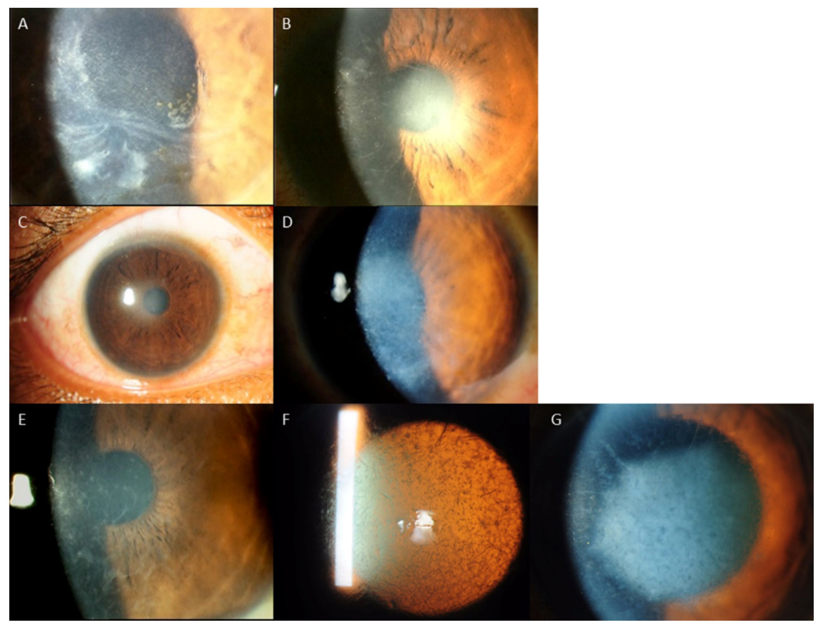
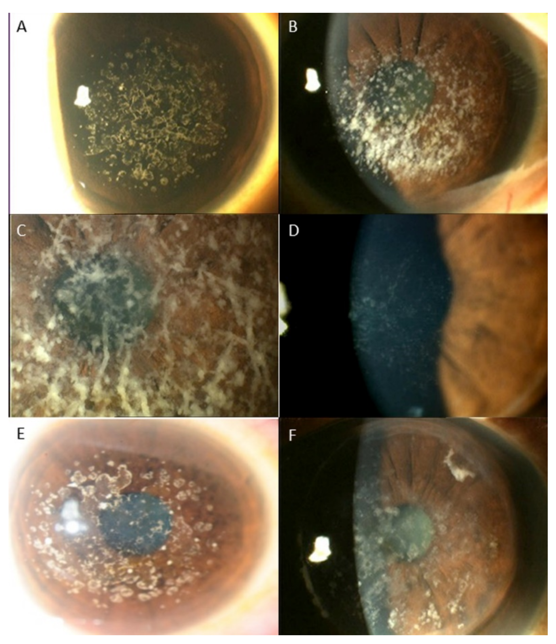
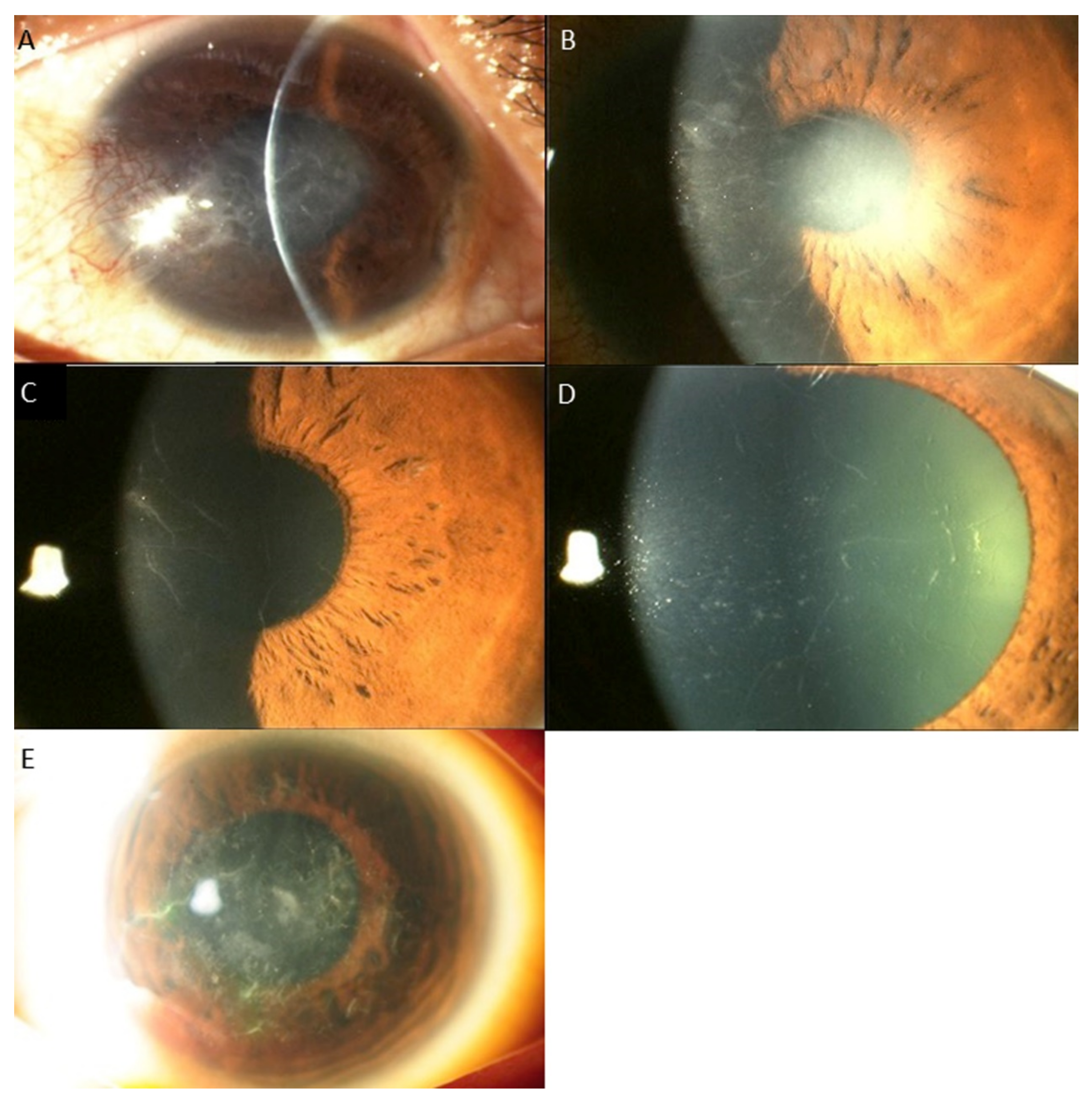
Regarding the distribution of TGFBI mutations, the mutation of the codon of R124 (17 cases, 53.1%) was the most common, followed by the mutation of the locus R555 (9 cases, 28.1%). The R124H mutation (8 cases) was associated with GCD1 in 3 cases (37.5%) and GCD2 in 5 cases (62.5%). In cases associated with R124H mutation, clinical findings varied from typical findings of GCD1, such as bread crumb like lesions with clear intervening stroma that do not extend to the periphery, to lesions typically seen in GCD2, i.e., discoid lesions and star shaped, spiky deposits (Figure 1), The R124C mutation (8 cases) caused LCD in 7 cases (87.5%) and GCD2 in 1 case (12.5%). In LCD cases associated with R124C mutation, the phenotypic alterations ranged from subepithelial haze only, to central subepithelial haze together with feathery opacities, thin lattice lines, white dots and flecks, or a combination of these. In 1 case of the R124H mutation, lattice lines with multiple grayish white dots and some spiky deposits were observed (Figure 2). The R555W mutation led to GCD1 in all 8 cases (100%). However, large variation of phenotypic changes were found, i.e., discoid lesions with central clearing, white dots and bands, star shaped whitish lesions and dots with hazy intervening stroma, translucent specks or a mixture of grayish translucent specks and denser whitish dots and bands (Figure 3). One case with the R555Q mutation was associated with RBCD. The other mutations, including H626R, A620D, L550P and H572R were all associated with classic manifestations of LCD, such as, similar refractive lines, dots and subepithelial haze of varying severity LCD (Table 4, Figure 4) [2].

4. Discussion

The present study demonstrated that the mutations in the codons of R124 and R555, such as, R124H, R555W and R124C were the most common mutations in a multi-ethnic population in Singapore, corroborating well with the results of previous studies in different countries [2,5,8,19,20,21,22,23,24]. The predominance of R124H, R555W and R124C mutations in various geographical locations worldwide indicates that these mutations may represent mutation hot spots in the gene [2]. The differences in phenotypes and genotypes among different ethnicities in Singapore in the present study suggest that the frequency of TGFBI corneal dystrophies can vary among different races, although this study included relatively small number of patients. Other studies have also suggested the possible differences in frequency of TGFBI corneal dystrophies in different areas around the world [8,9,10,21,24,25,26,27,28,29,30].
Studies in East Asian countries including Korea and Japan showed striking predominance of R124H mutation and GCD2 [21,25,26,27,28], whereas studies in Chinese population suggested the prevalence of R124C, R555W and R124H mutations were comparable [10,31]. Studies in India demonstrated higher proportion of R124C and R555W mutations, whereas R124H mutation was relatively rare [29,30]. The distribution of expressivity of TGFBI corneal dystrophies in Singaporeans, a multi-ethnic population comprising Chinese, Indian, Malaysian and others, might reflect these differences. Further studies with larger patient group are needed for evaluation of the influence of ethnicities on expressivity of TGFBI corneal dystrophies.
In general, the results of this study indicated moderate genotype/phenotype correlation in TGFBI corneal dystrophies, such as, R555W and GCD1, R124H and GCD2, and R124C and LCD, which corroborate with previous studies [2,5,8,9,10,23,32]. R555W and R124H mutations shows strong association with GCD1 and GCD2, respectively, and 99% of the genotypic mutations led to corresponding phenotypic changes [2,9,10,27,28]. R124C mutation also has close correlation with LCD [2,23,32]. However, the results also revealed that the discrepancy between phenotypes and genotypes can also exist, i.e., the same mutation can result in different phenotypes and similar phenotypes can be caused by various mutations [5]. In this study, R124H mutation was associated with GCD1 in 2 cases, R124C was associated with GCD2 in 1 case, and R124L was associated with RBCD instead of TBCD in 1 case. In addition, there was a large variation in clinical changes even in the same phenotypes associated with the same genotypic mutations (Figure 1, Figure 2 and Figure 3). LCD phenotype was associated with various mutations, i.e., H626R, A620D, L550P, H572R as well as R124C (Figure 4), suggesting that these mutations can lead to increased propensity for the formation and accumulation of amyloid fibrils [5].
Previous studies have shown that R124C mutation can also be associated with GCD2, RBCD and TBCD, although the mutation is most commonly associated with LCD [12,13,16,17,33]. Cases with subepithelial granular aggregates as well as stromal lattice deposits in association with R124C gene mutation have been reported [13,33], and this phenomenon was also observed in one patient of the present study. Moreover, R124C mutation was also reported to be associated to RBCD [16,17] and TBCD [12]. Conversely, LCD can be caused by many different mutations of TGFBI gene, as shown in this study [2,5]. A620D mutation was also found to be associated with LCD in a study in Korea [34]. Studies in other countries have also shown H626R mutation can lead to LCD with marked phenotypic heterogeneity [9,35]. However, H626R mutation was also reported to be able to cause a significantly different phenotype that was clinically similar to TBCD and RBCD [36,37], which also suggests that the same mutations can result in various phenotypes [5]. In addition to the mutations observed in this study, a large variety of mutations, e.g., A546T, L569Q, T621P, L527R and P542R, which are in the fourth FAS1 domain were demonstrated to be associated with LCD [27,28,38], suggesting that these mutations can also have high capacity for the amyloidogenesis [5].
In the present study, R124L and R555Q mutations were found in RBCD cases, although the mutations are typically associated with RBCD and TBCD, respectively [2]. A study in Brazil also reported that RBCD could be caused by R555Q and R124L [38]. Zeng et al. [39] showed that R124L and R555Q can also be associated with GCD1 and GCD2, which also support possible discrepancy between genotype and phenotype in TGFBI corneal dystrophies [5].
Regarding the mechanism underlying this possible discrepancy, a role of interplay of genetic and environmental factors has been suggested [10,14], i.e., modifications from other genes and environmental circumstances might have influence on penetrance and expressivity of the mutated genes [10,11,14,15]. Protein production at a local level in the cornea might be modulated by other genetic factors that might affect the abundance of TGFBI protein or the function of the protein processing pathway [10,11,14,15]. For instance, contact lens wear was suggested to be a possible anti-aggravation factor and to prevent progression and recurrence of corneal deposits in patients with GCD2 [14,40]. Although TGFBI corneal dystrophies are autosomal dominant hereditary disorders, R124H heterozytes have been shown to tend to show less severe corneal opacity than homozygotes [2,11,41,42]. R124H heterozygotes can sometimes have incomplete penetrance, which may lead to resemblance to the phenotype of GCD1, e.g., granular deposits without lattice opacities [10,11,43,44]. Song et al. [44] reported a case with the heterozygous R124H mutation in which no corneal opacities were observed, suggesting the mutation can even have non-penetrance, which can have important implication for refractive surgery in phenotypically normal patients with TGFBI mutation [44]. Three patients with phenotype of GCD1 in association with R124H mutations were also observed in the present study, conceivably due to the reduced penetrance. Among the 8 patients with R124H mutation in the present study, zygosity data were available in 4 patients, all the 4 patients were heterozygote, in which 3 had GCD2 and 1 had GCD1.
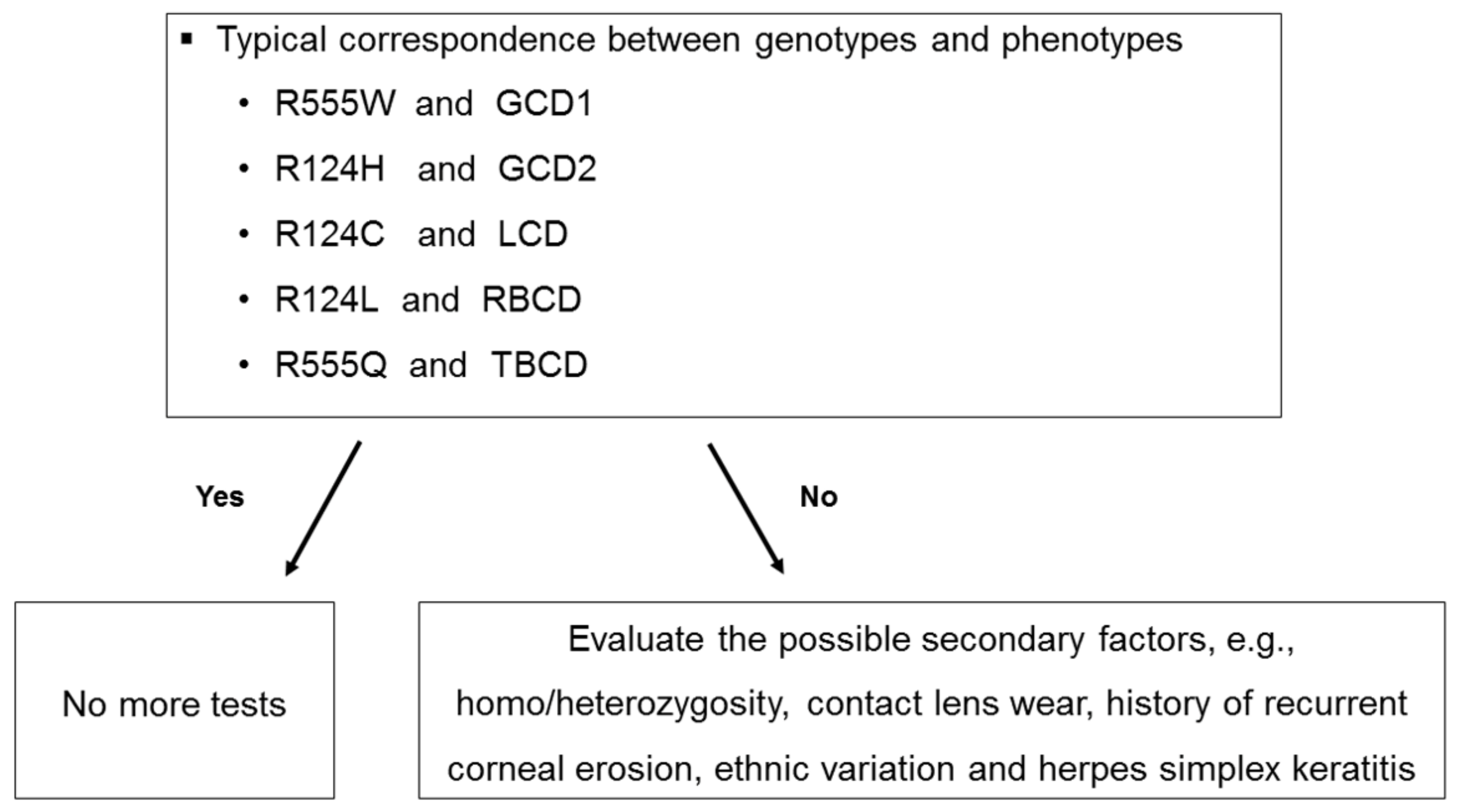
Disturbance of genotype/phenotype correlation in some cases may suggest that the scheme for nomenclature and classification of the TGFBI corneal dystrophies is still imperfect [5], indicating that development of a modified classification system [2]. It can also be considered to check the presence of typical correspondence between genotype and phenotype, and if the discrepancy is found, then evaluate the possible secondary factors, i.e., homo/heterozygosity, contact lens wear, history of corneal erosion, ethnic variation and herpes simplex keratitis (Figure 5).
The limitations of this study are as follows: (1) This study did not include the pathologic examination of the cornea specimen. Further studies that include histopathologic analysis of the corneal samples are needed for the investigation of the pathophysiology underlying the association between genotype and phenotype of the corneal dystrophies. (2) This study included relatively small sample size, which may not be sufficient for the evaluation of the genotype/phenotype correlation and influence of the ethnicity. Thus, we believe further studies with larger population are needed to develop a repository of cases for evaluation of these in detail. (3) We cannot completely rule out the possibility that incorrect phenotypic and genotypic description could cause misclassification of the subtype. Scientific phenotypical/genotypical relationship of the TGFBI corneal dystrophies may only be possible in forthcoming prospective studies in which corneal presentation is thoroughly recorded in direct and indirect illumination and by pharmacologically dilated pupil and focusing the epithelium, the stroma and the endothelium, and the minimum of five patients in three generations in one family are explored.
In conclusion, we have shown the distribution of genotype and phenotype of TGFBI corneal dystrophies in a multi-ethnic population in Singapore. Although genotype/phenotype correlation was moderate, discrepancy was observed in cases, which warrant further studies for elucidation of the pathophysiology underlying these association.

Author Contributions

Conceptualization, J.S.M.; data curation, V.A.; formal analysis, S.B.H.; investigation, S.B.H. and V.A.; methodology, C.W.W. and S.R.N.; supervision, J.S.M.; writing—original draft, S.B.H.; writing—review and editing, V.A., C.W.W., S.R.N. and J.S.M. All authors have read and agreed to the published version of the manuscript.

Funding

This research received no external funding.

Institutional Review Board Statement

This study was approved by the Singhealth Centralised Instituitional Review Board and adhered to the tenets of the Declaration of Helsinki. Ethical approval for the collection of blood samples was granted by the Singhealth Institutional Review Board.

Informed Consent Statement

Written Informed consent was obtained from all subjects involved in the study.

Data Availability Statement

The data presented in this study are available on request from the corresponding author. The data are not publicly available due to privacy of the study subjects.

Acknowledgments

The authors acknowledge 3 Billion, Inc., Seoul, Republic of Korea for providing TGFBI mutational analysis reports for some of the patients.

Conflicts of Interest

The authors declare no conflict of interest.

References

  1. Zhang, T.; Yan, N.; Yu, W.; Liu, Y.; Liu, G.; Wu, X.; Lian, J.; Liu, X. Molecular genetics of Chinese families with TGFBI corneal dystrophies. Mol. Vis. 2011, 17, 380–387. [Google Scholar] [PubMed]
  2. Lakshminarayanan, R.; Chaurasia, S.S.; Anandalakshmi, V.; Chai, S.M.; Murugan, E.; Vithana, E.N.; Beuerman, R.W.; Mehta, J.S. Clinical and genetic aspects of the TGFBI-associated corneal dystrophies. Ocul. Surf. 2014, 12, 234–251. [Google Scholar] [CrossRef] [PubMed]
  3. Kheir, V.; Cortes-Gonzalez, V.; Zenteno, J.C.; Schorderet, D.F. Mutation update: TGFBI pathogenic and likely pathogenic variants in corneal dystrophies. Hum. Mutat. 2019, 40, 675–693. [Google Scholar] [CrossRef] [PubMed] [Green Version]
  4. Munier, F.L.; Korvatska, E.; Djemaï, A.; Le Paslier, D.; Zografos, L.; Pescia, G.; Schorderet, D.F. Kerato-epithelin mutations in four 5q31-linked corneal dystrophies. Nat. Genet. 1997, 15, 247–251. [Google Scholar] [CrossRef]
  5. Han, K.E.; Choi, S.I.; Kim, T.I.; Maeng, Y.S.; Stulting, R.D.; Ji, Y.W.; Kim, E.K. Pathogenesis and treatments of TGFBI corneal dystrophies. Prog. Retin. Eye Res. 2016, 50, 67–88. [Google Scholar] [CrossRef] [PubMed]
  6. Weiss, J.S.; Møller, H.U.; Lisch, W.; Kinoshita, S.; Aldave, A.J.; Belin, M.W.; Kivelä, T.; Busin, M.; Munier, F.L.; Seitz, B.; et al. The IC3D classification of the corneal dystrophies. Cornea 2008, 27 (Suppl. 2), S1–S83. [Google Scholar]
  7. Weiss, J.S.; Møller, H.U.; Aldave, A.J.; Seitz, B.; Bredrup, C.; Kivelä, T.; Munier, F.L.; Rapuano, C.J.; Nischal, K.K.; Kim, E.K.; et al. IC3D classification of corneal dystrophies—Edition 2. Cornea 2015, 34, 117–159. [Google Scholar] [CrossRef] [Green Version]
  8. Evans, C.J.; Davidson, A.E.; Carnt, N.; Rojas Lopez, K.E.; Veli, N.; Thaung, C.M.; Tuft, S.J.; Hardcastle, A.J. Genotype-Phenotype Correlation for TGFBI Corneal Dystrophies Identifies p.(G623D) as a Novel Cause of Epithelial Basement Membrane Dystrophy. Investig. Ophthalmol. Vis. Sci. 2016, 57, 5407–5414. [Google Scholar] [CrossRef] [Green Version]
  9. Nowinska, A.K.; Wylegala, E.; Janiszewska, D.A.; Dobrowolski, D.; Aragona, P.; Roszkowska, A.M.; Puzzolo, D. Genotype-phenotype correlation of TGFBI corneal dystrophies in Polish patients. Mol. Vis. 2011, 17, 2333–2342. [Google Scholar]
  10. Hou, Y.C.; Wang, I.J.; Hsiao, C.H.; Chen, W.L.; Hu, F.R. Phenotype-genotype correlations in patients with TGFBI-linked corneal dystrophies in Taiwan. Mol. Vis. 2012, 18, 362–371. [Google Scholar]
  11. Cao, W.; Ge, H.; Cui, X.; Zhang, L.; Bai, J.; Fu, S.; Liu, P. Reduced penetrance in familial Avellino corneal dystrophy associated with TGFBI mutations. Mol. Vis. 2009, 15, 70–75. [Google Scholar] [PubMed]
  12. Chang., L.; Zhiqun, W.; Shijing, D.; Chen, Z.; Qingfeng, L.; Li, L.; Xuguang, S. Arg124Cys mutation of the TGFBI gene in 2 Chinese families with Thiel-Behnke corneal dystrophy. Arch. Ophthalmol. 2009, 127, 641–644. [Google Scholar] [CrossRef] [PubMed] [Green Version]
  13. Patel, D.A.; Chang, S.-H.; Harocopos, G.J.; Vora, S.C.; Thang, D.H.; Huang, A.J.W. Granular and lattice deposits in corneal dystrophy caused by R124C mutation of TGFBIp. Cornea 2010, 29, 1215–1222. [Google Scholar] [CrossRef] [PubMed] [Green Version]
  14. Han, K.E.; Choi, S.I.; Chung, W.S.; Maeng, Y.S.; Stulting, R.D.; Ji, Y.W.; Kim, E.K. Extremely varied phenotypes in granular corneal dystrophy type 2 heterozygotes. Mol. Vis. 2012, 18, 1755–1762. [Google Scholar] [PubMed]
  15. Watanabe, H.; Hashida, Y.; Tsujikawa, K.; Tsujikawa, M.; Maeda, N.; Inoue, Y.; Yamamoto, S.; Tano, Y. Two patterns of opacity in corneal dystrophy caused by the homozygous BIG-H3 R124H mutation. Am. J. Ophthalmol. 2001, 132, 211–216. [Google Scholar] [CrossRef]
  16. Yang, Q.-N.; Zhao, Y.-W.; Guo, L.-H.; Yan, N.H.; Liu, X.Y.; Cai, S.P. Arg124Cys mutation of the TGFBI gene in a Chinese pedigree of Reis-Bücklers corneal dystrophy. Int. J. Ophthalmol. 2011, 4, 235–238. [Google Scholar]
  17. Ma, K.; Liu, G.; Yang, Y.; Yu, M.; Sui, R.; Yu, W.; Chen, X.; Deng, Y.; Yan, N.; Cao, G.; et al. TGFBI gene mutation analysis in a Chinese pedigree of Reis-Bücklers corneal dystrophy. Mol. Vis. 2010, 16, 556–561. [Google Scholar]
  18. Aldave, A.J.; Rayner, S.A.; Kim, B.T.; Prechanond, A.; Yellore, V.S. Unilateral lattice corneal dystrophy associated with the novel His572del mutation in the TGFBI gene. Mol. Vis. 2006, 12, 142–146. [Google Scholar]
  19. Fujiki, K.; Nakayasu, K.; Kanai, A. Corneal dystrophies in Japan. J. Hum. Genet. 2001, 46, 431–435. [Google Scholar] [CrossRef]
  20. Lee, J.H.; Cristol, S.M.; Kim, W.C.; Chung, E.S.; Tchah, H.; Kim, M.S.; Nam, C.M.; Cho, H.S.; Kim, E.K. Prevalence of granular corneal dystrophy type 2 (Avellino corneal dystrophy) in the Korean population. Ophthalmic Epidemiol. 2010, 17, 160–165. [Google Scholar] [CrossRef]
  21. Mashima, Y.; Yamamoto, S.; Inoue, Y.; Imamura, Y.; Yamada, M.; Ogata, T.; Kudoh, J.; Shimizu, N. Association of autosomal dominantly inherited corneal dystrophies with BIGH3 gene mutations in Japan. Am. J. Ophthalmol. 2000, 130, 516–517. [Google Scholar] [CrossRef]
  22. Chao-Shern, C.; DeDionisio, L.A.; Jang, J.H.; Chan, C.C.; Thompson, V.; Christie, K.; Nesbit, M.A.; McMullen, C.B.T. Evaluation of TGFBI corneal dystrophy and molecular diagnostic testing. Eye 2019, 33, 874–881. [Google Scholar] [CrossRef] [PubMed] [Green Version]
  23. Zhao, F.; Liu, Y.; Guan, T. Analysis of TGFBI Gene Mutations in Three Chinese Families with Corneal Dystrophy. J. Ophthalmol. 2019, 2019, 6769013. [Google Scholar] [CrossRef] [PubMed] [Green Version]
  24. Lakshminarayanan, R.; Vithana, E.N.; Chai, S.M.; Chaurasia, S.S.; Saraswathi, P.; Venkatraman, A.; Rojare, C.; Venkataraman, D.; Tan, D.; Aung, T.; et al. A novel mutation in transforming growth factor-beta induced protein (TGFβIp) reveals secondary structure perturbation in lattice corneal dystrophy. Br. J. Ophthalmol. 2011, 95, 1457–1462. [Google Scholar] [CrossRef]
  25. Fujiki, K.; Hotta, Y.; Nakayasu, K.; Yamaguchi, T.; Kato, T.; Uesugi, Y.; Ha, N.T.; Endo, S.; Ishida, N.; Lu, W.N.; et al. Six different mutations of TGFBI (betaig-h3, keratoepithelin) gene found in Japanese corneal dystrophies. Cornea 2000, 19, 842–845. [Google Scholar] [CrossRef]
  26. Yoshida, S.; Kumano, Y.; Yoshida, A.; Hisatomi, T.; Matsui, H.; Nishida, T.; Ishibashi, T.; Matsui, T. An analysis of BIGH3 mutations in patients with corneal dystrophies in the Kyushu district of Japan. Jpn. J. Ophthalmol. 2002, 46, 469–471. [Google Scholar] [CrossRef]
  27. Cho, K.J.; Mok, J.W.; Na, K.S.; Rho, C.R.; Byun, Y.S.; Hwang, H.S.; Hwang, K.Y.; Joo, C.K. TGFBI gene mutations in a Korean population with corneal dystrophy. Mol. Vis. 2012, 18, 2012–2021. [Google Scholar]
  28. Song, J.S.; Lim, D.H.; Chung, E.S.; Chung, T.Y.; Ki, C.S. Mutation Analysis of the TGFBI Gene in Consecutive Korean Patients with Corneal Dystrophies. Ann. Lab. Med. 2015, 35, 336–340. [Google Scholar] [CrossRef] [Green Version]
  29. Paliwal, P.; Sharma, A.; Tandon, R.; Sharma, A.; Vajpayee, R.B. TGFBI mutation screening and genotype-phenotype correlation in north Indian patients with corneal dystrophies. Mol. Vis. 2010, 16, 1429–1438. [Google Scholar]
  30. Chakravarthi, S.V.V.K.; Kannabiran, C.; Sridhar, M.S.; Vemuganti, G.K. TGFBI gene mutations causing lattice and granular corneal dystrophies in Indian patients. Investig. Ophthalmol. Vis. Sci. 2005, 46, 121–125. [Google Scholar] [CrossRef]
  31. Yang, J.; Han, X.; Huang, D.; Yu, L.; Zhu, Y.; Tong, Y.; Zhu, B.; Li, C.; Weng, M.; Ma, X.; et al. Analysis of TGFBI gene mutations in Chinese patients with corneal dystrophies and review of the literature. Mol. Vis. 2010, 16, 1186–1193. [Google Scholar]
  32. Wang, X.; Ying, M.; Fu, C.; Wang, Y.; Li, N. TGFBI gene mutations analysis in Chinese families with corneal dystrophies. Mol. Med. Rep. 2017, 15, 3198–3202. [Google Scholar] [CrossRef] [PubMed]
  33. Edelstein, S.L.; Huang, A.J.; Harocopos, G.J.; Waltman, S.R. Genotype of lattice corneal dystrophy (R124C mutation in TGFBI) in a patient presenting with features of avellino corneal dystrophy. Cornea 2010, 29, 698–700. [Google Scholar] [CrossRef] [PubMed]
  34. Jung, J.W.; Kim, S.A.; Kang, E.M.; Kim, T.I.; Cho, H.S.; Kim, E.K. Lattice corneal dystrophy type IIIA with hyaline component from a novel A620P mutation and distinct surgical treatments. Cornea 2014, 33, 1324–1331. [Google Scholar] [CrossRef] [PubMed]
  35. Munier, F.L.; Frueh, B.E.; Othenin-Girard, P.; Uffer, S.; Cousin, P.; Wang, M.X.; Héon, E.; Black, G.C.; Blasi, M.A.; Balestrazzi, E.; et al. BIGH3 mutation spectrum in corneal dystrophies. Investig. Ophthalmol. Vis. Sci. 2002, 43, 949–954. [Google Scholar]
  36. Wheeldon, C.E.; de Karolyi, B.H.; Patel, D.V.; Sherwin, T.; McGhee, C.N.; Vincent, A.L. A novel phenotype-genotype relationship with a TGFBI exon 14 mutation in a pedigree with a unique corneal dystrophy of Bowman’s layer. Mol. Vis. 2008, 14, 1503–1512. [Google Scholar]
  37. Liskova, P.; Klintworth, G.K.; Bowling, B.L.; Filipec, M.; Jirsova, K.; Tuft, S.J.; Bhattacharya, S.S.; Hardcastle, A.J.; Ebenezer, N.D. Phenotype associated with the H626P mutation and other changes in the TGFBI gene in Czech families. Ophthalmic Res. 2008, 40, 105–108. [Google Scholar] [CrossRef]
  38. Solari, H.P.; Ventura, M.P.; Perez, A.B.; Sallum, J.M.; Burnier, M.N., Jr.; Belfort, R., Jr. TGFBI gene mutations in Brazilian patients with corneal dystrophy. Eye 2007, 21, 587–590. [Google Scholar] [CrossRef] [Green Version]
  39. Zeng, L.; Zhao, J.; Chen, Y.; Zhao, F.; Li, M.; Chao-Shern, C.; Moore, T.; Marshall, J.; Zhou, X. TGFBI Gene Mutation Analysis of Clinically Diagnosed Granular Corneal Dystrophy Patients Prior to PTK: A Pilot Study from Eastern China. Sci. Rep. 2017, 7, 596. [Google Scholar] [CrossRef] [Green Version]
  40. Roters, S.; Severin, M.; Konen, W.; Krieglstein, G.K. Treatment of granular dystrophy with soft contact lenses. Ophthalmologica 2004, 218, 70–72. [Google Scholar] [CrossRef]
  41. Okada, M.; Yamamoto, S.; Inoue, Y.; Watanabe, H.; Maeda, N.; Shimomura, Y.; Ishii, Y.; Tano, Y. Severe corneal dystrophy phenotype caused by homozygous R124H keratoepithelin mutations. Investig. Ophthalmol. Vis. Sci. 1998, 39, 1947–1953. [Google Scholar]
  42. Fujiki, K.; Hotta, Y.; Nakayasu, K.; Kanai, A. Homozygotic patient with betaig-h3 gene mutation in granular dystrophy. Cornea 1998, 17, 288–292. [Google Scholar] [CrossRef] [PubMed]
  43. Konishi, M.; Yamada, M.; Nakamura, Y.; Mashima, Y. Varied appearance of cornea of patients with corneal dystrophy associated with R124H mutation in the BIGH3 gene. Cornea 1999, 18, 424–429. [Google Scholar] [CrossRef] [PubMed]
  44. Kim, J.W.; Kim, H.M.; Song, J.S. Phenotypic non-penetrance in granular corneal dystrophy type II. Graefes Arch. Clin. Exp. Ophthalmol. 2008, 246, 1629–1631. [Google Scholar] [CrossRef]
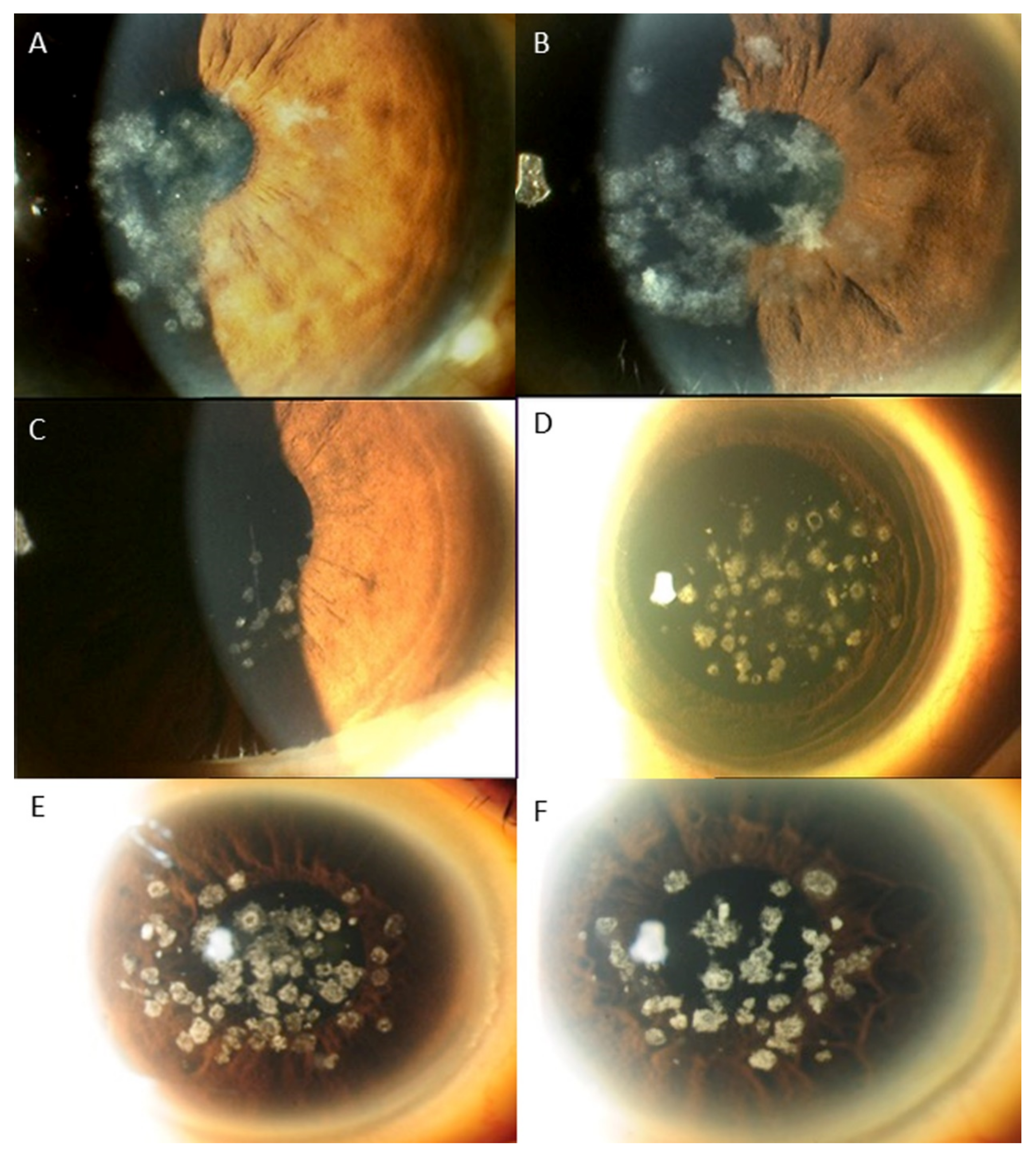
Figure 1. The phenotypic variations associated with mutation at R124H: (A): Classic bread crumb like whitish lesions in the anterior stroma that can be seen GCD1; (B): Similar to A but a few lesions have stellate pattern that is seen in GCD2; (C): Few translucent dots that can be seen in GCD1; (D): multiple grayish white dots, some with moth eaten central clearing and some spiky deposits that can be seen in GCD 2; (E): classic bread crumb like lesions with clear intervening stroma that can be seen in GCD1; (F): similar bread crumb like lesions interspersed with some spiky deposits that can be seen in GCD2.
Ijms 22 01230 g001
Figure 2. Different phenotypes of LCD1 associated with the same R124C mutation: (A): subepithelial haze and feathery opacities; (B): Thin lattice lines with subepithelial haze; (C): Central subepithelial haze only; (D): Central subepithelial haze with white dots and few lattice lines; (E): Dots and fleck like deposits; (F,G): Lattice lines with multiple grayish white dots and some spiky deposits (GCD2).
Ijms 22 01230 g002
Figure 3. Different phenotypes of GCD1 associated with the same R555W mutation: (A): discoid lesions with central clearing; (B): White dots of varying densities, some coalescing to form white bands; (C): Star-shaped whitish lesions and dots with hazy intervening stroma; (D): Specks of translucent dots; (E): bread crumb like lesions sparing the peripheral cornea; (F): Grayish specks centrally and denser white dots that coalesce in the peripheral cornea.
Ijms 22 01230 g003
Figure 4. LCD1 phenotypes associated with 5 different mutations demonstrating similar phenotypic change, i.e., similar refractive lines, dots and subepithelial haze of varying severity. (A): H572R, (B): R124C, (C): H626R, (D): L550P, (E): A620D.
Ijms 22 01230 g004
Figure 5. Flowchart for the genotype/phenotype correlation of TGFBI corneal dystrophies. The presence of typical correspondence between genotype and phenotypes can be checked, and if the discrepancy is found, the possible secondary factors, such as, homo/heterozygosity, contact lens wear, history of recurrent corneal erosion, ethnic variation and herpes simplex keratitis, can be evaluated.
Ijms 22 01230 g005
Table 1. Primers for the TGFBI DNA sequencing.
ExonForward PrimerReverse PrimerProduct Size (bp)
1CGGAGGCGCTCTCACTTCCCGAGCCCCGACTACCTGACC265
2GGGAGTCATTAAAGTGGGGTGGAAGCTTGGTCTCCTGGCTGGTTAC99
3CAACTTAGTGGAGAGGGGCCAGACTCTCTCCCACCATTCCCTTCC206
4GCCATCCCTCCTTCTGTCTTCTGCCGGGCAGACGGAGGTCATC217
5ACTGACACCCTGTCCTTCCTCCTAGCCCACACATGGAACAGAAATG261
6CTGCTCATCCTTGCTGCTTCTCTAGAGTTCCTGCTAGGCCCCTCTT249
7TCTGTGGGGAGTGCCAGAGTCCAAATGAGGCAGCAGCAGGA234
8TGGACCCTGACTTGACCTGAGTCAAAGGATGGCAGAAGAGATGGTG311
9CCCTGGGGTGGATGAATGATAAAGCCTCCAGGGACAATCTAACAGG251
10ATTGCAGGAGCACATCTCTCTGGGCTTCCCAGGAGCATGATTTAGG230
11GCCCCTCGTGGAAGTATAACCAGATCCCACTCCAGCATGACCACT248
12TGACAGGTGACATTTTCTGTGTGTGGGGCCCTGAGGGATCACTACTTT224
13TGACCAGGCTAATTACCATTCTTGGCAGCCTTTGATTTGCAGGACACT210
14TGCTCTACTTTCAACCACTACTCTGCCAACTGCCACATGAAGAAAAGG198
15TCACTCTGGTCAAACCTGCCTTTCCTCTATGGCCCAAACAGAGGAC183
16GCCATTGTCATAAGCAGTTGCAGATACAGCAGATGGCAGGCTTGG176
17TGGGGAGATCTGCACCTATTTGAGGTCAGCACACTGTACCATGCAC710
Table 2. The distribution of genotypes of TGFBI mutations in each phenotype of TGFBI corneal dystrophy.
PhenotypeAssociated TGFBI Mutations (Genotypes)
Granular corneal dystrophy (GCD), type 1 (n = 11)R124H (n = 3)
R555W (n = 8)
Granular corneal dystrophy (GCD), type 2 (n = 6)R124H (n = 5)
R124C (n = 1)
Lattice corneal dystrophy (LCD) (n = 13)R124C (n = 7)
H626R (n = 2)
L550P (n = 2)
A620D (n = 1)
H572R (n = 1)
Reis-Buckler dystrophy (RBCD) (n = 2)R124L (n = 1)
R555Q (n = 1)
Table 3. Phenotypes and genotypes of TGFBI corneal dystrophy in various ethnicities.
EthnicityPhenotypeAssociated TGFBI Mutations
Chinese (n = 16)GCD1 (n = 5)R124H (n = 3)
R555W (n = 2)
GCD2 (n = 5)R124H (n = 4)
R124C (n = 1)
LCD (n = 4)R124C (n = 1)
A620D (n = 1)
H626R (n = 2)
RBCD (n = 2)R124L (n = 1)
R555Q (n = 1)
Malay (n = 6)LCD (n = 6)R124C (n = 6)
Indian (n = 5)GCD1 (n = 5)R555W (n = 5)
Others (n = 5)GCD1 (n = 1)R555W (n = 1)
GCD2 (n =1)R124H (n = 1)
LCD (n = 3)L550P (n = 2)
H572R (n = 1)
Table 4. The distribution of phenotypes of TGFBI corneal dystrophies in each genotype of TGFBI mutations.
LocusTGFBI MutationsPhenotype
R124 (n = 17)R124H (n = 8)GCD1 (n = 3)
GCD 2 (n = 5)
R124C (n = 8)GCD2 (n = 1)
LCD (n = 7)
R124L (n = 1)RBCD (n = 1)
R555 (n = 9)R555W (n = 8)GCD1 (n = 8)
R555Q (n = 1)RBCD (n = 1)
Others (n = 6)H626R (n = 2)LCD
A620D (n = 1)LCD
L550P (n = 2)LCD
H572R (n = 1)LCD
Publisher’s Note: MDPI stays neutral with regard to jurisdictional claims in published maps and institutional affiliations.