Review

The Communication between Ocular Surface and Nasal Epithelia in 3D Cell Culture Technology for Translational Research: A Narrative Review

by
Malik Aydin
1,2,†,
Jana Dietrich
3,†,
Joana Witt
4,
Maximiliane S. C. Finkbeiner
5,
Jonas J.-H. Park
6,
Stefan Wirth
2,
Christine E. Engeland
5,
Friedrich Paulsen
3,7,*,‡ and
Anja Ehrhardt
5,*,‡
1
Laboratory of Experimental Pediatric Pneumology and Allergology, Center for Biomedical Education and Research, School of Life Sciences (ZBAF), Department of Human Medicine, Faculty of Health, Witten/Herdecke University, 58448 Witten, Germany
2
Center for Child and Adolescent Medicine, Center for Clinical and Translational Research (CCTR), Helios University Hospital Wuppertal, 42283 Wuppertal, Germany
3
Institute of Functional and Clinical Anatomy, Friedrich Alexander University Erlangen-Nürnberg, 91054 Erlangen, Germany
4
Department of Ophthalmology, University Hospital Düsseldorf, Heinrich-Heine-University, 40225 Düsseldorf, Germany
5
Virology and Microbiology, Center for Biomedical Education and Research (ZBAF), Department of Human Medicine, Faculty of Health, Witten/Herdecke University, 58453 Witten, Germany
6
Department of Otorhinolaryngology, Kath. Krankenhaus Hagen gGmbH, St.-Josefs-Hospital, Faculty of Health, Department of Human Medicine, Witten/Herdecke University, 58097 Hagen, Germany
7
Department of Topographic Anatomy and Operative Surgery, Sechenov University, 119146 Moscow, Russia
*
Authors to whom correspondence should be addressed.
Malik Aydin and Jana Dietrich contributed equally to this work and share the first authorship.
Anja Ehrhardt and Friedrich Paulsen contributed equally to this work and share the senior authorship.
Int. J. Mol. Sci. 2021, 22(23), 12994; https://doi.org/10.3390/ijms222312994
Submission received: 13 October 2021 / Revised: 25 November 2021 / Accepted: 26 November 2021 / Published: 30 November 2021
(This article belongs to the Special Issue Development of Cell Culture Technology: Molecular Aspects and Beyond)

Abstract

:
There is a lack of knowledge regarding the connection between the ocular and nasal epithelia. This narrative review focuses on conjunctival, corneal, ultrastructural corneal stroma, and nasal epithelia as well as an introduction into their interconnections. We describe in detail the morphology and physiology of the ocular surface, the nasolacrimal ducts, and the nasal cavity. This knowledge provides a basis for functional studies and the development of relevant cell culture models that can be used to investigate the pathogenesis of diseases related to these complex structures. Moreover, we also provide a state-of-the-art overview regarding the development of 3D culture models, which allow for addressing research questions in models resembling the in vivo situation. In particular, we give an overview of the current developments of corneal 3D and organoid models, as well as 3D cell culture models of epithelia with goblet cells (conjunctiva and nasal cavity). The benefits and shortcomings of these cell culture models are discussed. As examples for pathogens related to ocular and nasal epithelia, we discuss infections caused by adenovirus and measles virus. In addition to pathogens, also external triggers such as allergens can cause rhinoconjunctivitis. These diseases exemplify the interconnections between the ocular surface and nasal epithelia in a molecular and clinical context. With a final translational section on optical coherence tomography (OCT), we provide an overview about the applicability of this technique in basic research and clinical ophthalmology. The techniques presented herein will be instrumental in further elucidating the functional interrelations and crosstalk between ocular and nasal epithelia.

1. Historical Introduction into the Ocular and Nasal Communication

Can one cry on command? Can one shed tears without crying? Are there false tears? From a purely physiological point of view, the answer is simple: when someone cries, saline tear fluid flows down the cheek [1,2]. Most of this is formed by the lacrimal glands located in the temporal upper quadrant on and at the eye bulb and passes as ‘used tear fluid’ through the draining tear ducts, which consist of the lacrimal puncta, lacrimal tubules, lacrimal sac, and nasolacrimal duct, into the nose or the lower nasal duct [3,4]. Thus, colloquially, ‘Rotz (=snot) & Wasser (=water) heulen (=cry)’ is commonly used as a traditional idiom in German culture, which also corresponds to crying the eyes out [1].
Thus, even this simple idiom implies a connection between the eye and nose.
Until well into the 16th century, it was assumed that tears were a product of the brain [3]. It was not until 1574 that the Italian Giovanni Battista Carcano Leone described the draining tear ducts quite precisely [5]. Together with the investigations of the Dane Niels Stensen (1662), after whom the main excretory duct of the parotid gland is named (Stenon’s duct, Ductus Stenonius), on tear secretion, a plausible understanding of the entire lacrimal system emerged [6]. In 1755, this was completed by the German anatomist and botanist Johann Gottfried Zinn after which the ZINN tendon ring (annulus tendineus communis), the common origin of all the external eye muscles except the obliquus inferior muscle, is named [7,8].
Nevertheless, the mechanisms that ensure tear transport through the draining lacrimal ducts—and thereby link eye and nose—are still not fully understood. Although there are currently elaborated morphological studies, unfortunately, there is still a significant lack of functional insights. The numerous existing hypotheses reflect the complex structure of the nasolacrimal ducts and show that tear transport is a complex process [4,9].
Considering these complex mechanisms and the close communication between the eyes and the nose, with this review, we aim at presenting distinct cell culture methods, which were previously published, and highlighting their applicability in translational contexts. The interconnection between the ocular and nasal epithelia is important to study the etiopathogenesis of diseases affecting these tissues and to develop therapeutic algorithms. A summary on current cell culture models of these epithelia and important technical notes and nuances will guide experimentalists to work with these models in their laboratories. Moreover, an introduction to current 3D cell culture techniques for conjunctival, corneal, nasolacrimal, and nasal epithelial cells are an important advancement discussed herein. In addition to the morphological preservation of these difficult-to-cultivate but valuable cells in a physiological context, some disease pathologies affecting these tissues will be illustrated. Exemplarily, adenoviruses (AdV) and measles virus (MeV) as important pathogens but also allergens during the pathogenesis of rhinoconjunctivitis are discussed. At the end, we will provide an experimental outlook, which may serve as a scaffold for scientists aiming to work with these cell culture models to study physiological and pathological aspects of ocular and nasal epithelia.

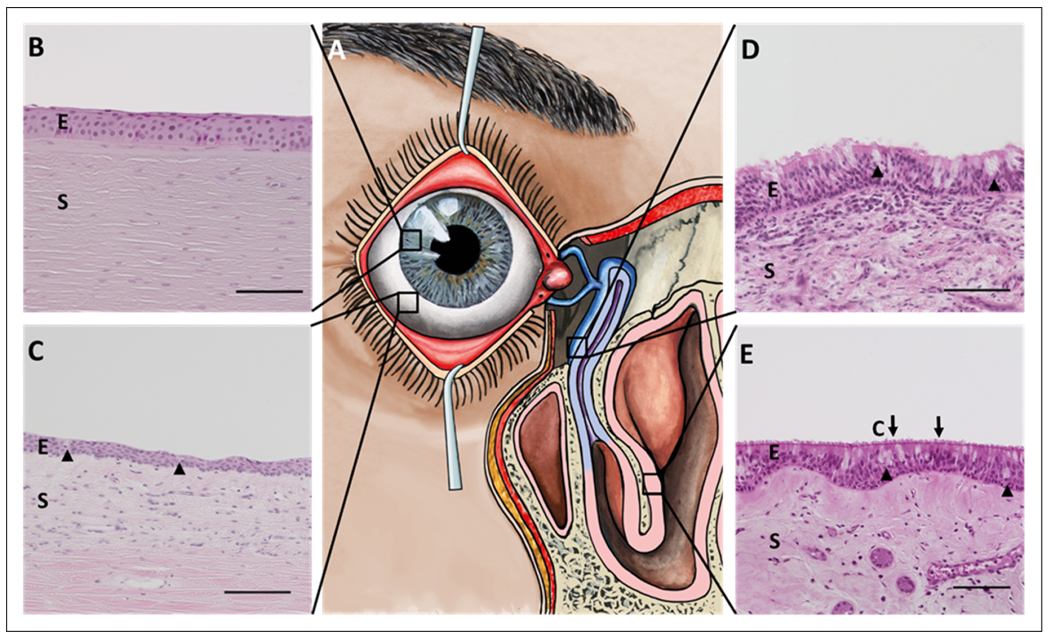
2. Morphology and Physiology of the Ocular Surface Epithelium, Nasolacrimal Ducts, and Nasal Epithelium

This section summarizes the native anatomical morphology as well as the physiological function of the ocular surface epithelium (cornea and conjunctiva), the efferent tear ducts (nasolacrimal ducts), and the nasal cavity (Figure 1A). These anatomical structures are in direct physical connection with each other and represent the physiological tear out-flow from the formation of the tear film at the ocular surface via the drainage through the nasolacrimal ducts to the end point of the ’used tear fluid´ in the nasal cavity [3,4]. Tears can be considered a carrier of biological information exchanged between the eye and the nose, a tool of communication. The tear film covers the ocular surface epithelium. It provides nutrients, hydration, and oxygen, smoothens the epithelial surface, forms an essential part of the refractive optical system, and protects against pathogens (e.g., viruses) [10]. Of note, the retina and vitreous body are not directly addressed in this review. However, the retina represents a particular layer of the eye and their corresponding retinal pathologies including retinitis pigmentosa punctata albescens (RPA) which may lead to vision loss and blindness (reviewed in [11,12]) [13]. Exemplarily, the work of Donato and colleagues examines variants in four known genes that trigger RPA and provides important clues to the etiopathogenesis of the disease [14].

2.1. Cornea

The cornea is a specialized optical tissue with transparent properties that enables light transmission into the inner eye. The cornea covers the central area of the ocular surface (our window to the outside world) and extends to the limbus, the transition zone between the cornea and the conjunctiva. With regard to the communication between the ocular surface and the nose, the cornea lies at the beginning of the communication.
Current understanding indicates that the cornea includes five different layers [15]:
  • Corneal epithelium
  • Bowman´s membrane
  • Stroma (with Dua’s layer)
  • Descemet´s membrane
  • Corneal endothelium
The corneal epithelium itself consists of five to six layers of non-keratinized squamous epithelial cells that can be further divided into basal, wing, and superficial cells (from posterior to anterior) (Figure 1B) [16].
The superficial cells are lined by microplicae at the tips of which the glycocalyx is anchored, mainly in the form of the membrane-anchored mucins MUC1, MUC4, and MUC16 (see below), which are part of the mucosal component of the tear film. The epithelial cells are connected through tight junctions, building an effective barrier against pathogens, environmental debris, and fluids [10]. Progenitor cells, also defined as limbal stem cells, are located in the limbal area and continuously replace cells lost through physiological turnover by differentiation and maturation processes [16,17]. The basal cells of the corneal epithelium rest on a basement membrane, which is attached to the Bowman’s membrane posteriorly. The corneal stroma consists of highly regular and orthogonally arranged lamellae composed of type I, type V, and type VI collagen fibers [18]. In addition, keratocytes are sparsely distributed within this collagenous matrix [19]. Located posterior to the stroma is the Descemet membrane, which serves as the basement membrane for the corneal endothelium. The endothelium consists of a single layer of hexagonal cells that prevent passive diffusion of the aqueous humor into the stroma and thus stromal swelling [20].
This basement membrane encompasses the extracellular matrix secreted by the corneal endothelial cells. Furthermore, the posterior corneal endothelium consists of a single layer of hexagonal cells that prevent passive diffusion of the aqueous humor into the stroma and thus stromal swelling [20]. Due to their limited regenerative capacity, endothelial cell loss needs to be compensated by the expansion (polymegathism) of neighboring cells [20,21]. Of note, when the enlarged endothelium is no longer able to maintain fluid balance, the resulting stromal swelling leads to loss of transparency, corneal edema, and visual impairment [20].

2.2. Conjunctiva

The conjunctiva is a transparent mucous membrane that covers the main part of the ocular surface. It consists of an epithelium with secretory goblet cells, covered by the tear film, and an underlying stroma (Figure 1C). Similar to the cornea, the conjunctiva is at the beginning of the communication between the ocular surface and the nose. Since the conjunctiva actively forms parts of the tear film, especially the mucin component, messenger substances could be secreted here, which then have an effect on the nasal epithelia.
The conjunctival epithelium in general is formed by two main cell types: the conjunctival epithelial cells and the conjunctival goblet cells. The epithelium is a multilayered unkeratinized squamous epithelium with microplicae (just as in the cornea, the microplicae are covered with a felt of membrane-bound mucins, MUC1, MUC4, and MUC16 [22]) and can be subdivided into basal, intermediate, and superficial cells. The conjunctival goblet cells produce secretory mucins, part of the mucin component of the tear film lubricating the ocular surface and thus maintaining the integrity of the entire eye [23,24,25]. The stroma of the conjunctiva consists of loose connective tissue. It contains defense cells, mast cells, and abundant blood vessels, which form a marginal loop network at the limbus corneae. In about one third of adult people, it harbors the conjunctiva-associated lymphoid tissue (CALT) that plays an important role in the immune control of the eye by activating the immune system in case of pathogen infiltration by reacting mainly with the differentiation of immunoglobulin (Ig)A producing plasma cells [26,27].
The conjunctiva begins at the corneoscleral limbus, extends to the fornix of the eye, and covers the inner layer of the eyelid to the lid margin. As each of these different regions have specific characteristics, the conjunctiva can be divided into three areas:
  • Bulbar conjunctiva
  • Fornical conjunctiva
  • Palpebral conjunctiva
In detail, each area is exposed to different stress factors and has a distinct primary function. Thus, in each area, the non-keratinized epithelium exhibits a slightly different structure in terms of epithelial cell shape and stratification as well as goblet cell density [28]. The bulbar conjunctiva covers the sclera of the eyeball and is loosely attached to it. Here, the conjunctival epithelium consists of a stratified squamous epithelium with six to nine layers [29,30]. The fornical conjunctiva represents the transition between the bulbar and palpebral conjunctiva. In this area, the conjunctiva is highly elastic and forms folds that ensure the flexible movement of the eyeball and represents a reservoir for the tear film, as the excretory ducts of the lacrimal glands are located there [31]. In addition, the cylindrical epithelium reaches two to three layers. Moreover, the palpebral conjunctiva can be subdivided into the marginal, tarsal, and orbital conjunctiva, with the tarsal conjunctiva tightly attached to the tarsal plate and the marginal conjunctiva close to the lid margin. The tarsal conjunctiva consists of two to three layers of cuboidal epithelium, whereas the marginal conjunctiva is a stratified squamous epithelium [29,30].
Goblet cells are found in all areas of the conjunctiva, but with unequal distribution, with a higher density in the inferior than the superior conjunctiva as well as at the nasal side [28]. In areas of low density, goblet cells occur as solitary cells, while in areas of higher density, additional clusters are detected. Functionally, goblet cells produce and secrete gel-forming mucins (especially MUC5AC), but also trefoil factor family (TFF) peptides such as TFF3 [24,32]. Both are essential constituents of the aqueous component of the tear film. The conjunctival goblet cells as well as the conjunctival epithelium are derived from the same bi-potent progenitor cells [33]. However, it is still under discussion whether these progenitor cells are distributed uniformly or locally throughout the conjunctiva [33,34,35].

2.3. Draining Tear Ducts

The efferent ducts build the direct physical connection between the eye and the nose. They form the lacrimal drainage system and are responsible for the transportation of the tears from the ocular surface to the nasal cavity [4,36]. The ‘used tear fluid’ is collected at the nasal side of the eye and is drained into the inferior nasal meatus of the nose through the lacrimal puncta (upper and lower), the lacrimal canaliculi (upper and lower), the lacrimal sac, and the nasolacrimal duct [3,4]. The lacrimal puncta, as well as the lacrimal canaliculi, consist of a multilayered non-keratinized squamous epithelium. In contrast, the lacrimal sac and the nasolacrimal duct are lined by a double-layered non-keratinized epithelium that rests on a broad basement membrane (Figure 1D) [36].
The superficial epithelial layer of the lacrimal sac and the nasolacrimal duct consists of columnar epithelial cells with microvilli that are attached to the basal-cell layer. Within the epithelium of the lacrimal sac and the nasolacrimal duct, goblet cells are integrated as solitary cells or as intraepithelial mucous glands [36]. These goblet cells have been shown to produce and secrete mucins, particularly MUC5B and MUC2 but also MUC5AC, as well as TFF peptides, e.g., TFF1 and TFF3 [32,37].
Particularly in the lower section of the nasolacrimal duct, individual kinocilia-bearing epithelial cells are also present in the epithelium [36]. Absorption experiments in rabbits show that components of the transported tear fluid can be reabsorbed in the nasolacrimal duct [38], (reviewed in [39]), which allows the hypothesis that reabsorption may represent a feedback signal for tear production. Mucosa-associated-lymphoid tissue (MALT) is referred to as TALT = tear duct-associated lymphoid tissue in the draining tear ducts, and is also present in approximately one-third of adult subjects [40,41], (reviewed in [39]). After its description, it was combined with the CALT under the name EALT = eye associated-lymphoid tissue [26]. In addition to free intra- and subepithelially localized defense cells, the epithelium of the lacrimal sac and nasolacrimal duct also produces a whole arsenal of antimicrobial peptides that serve the immune defense in the ‘closed’ (located in a bony canal) system of the draining lacrimal ducts [42]. The epithelium of the lacrimal sac and nasolacrimal duct lies on a vascular system of specialized vessels that is comparable to an erectile tissue [43] and is functionally involved in tear transport [44].

2.4. Nasal Cavity

The nasal cavity represents the internal part of the nose and is separated into two almost symmetrical halves by the nasal septum (Figure 1E). The nasal cavity is the recipient of biological information through the tears that enter the inferior nasal meatus from the nasolacrimal duct. Biological material that has entered the tear film at the ocular surface is now released into the nasal cavity. Besides the entry of biological material with the tears, the nasal cavity is also the entry site for inhaled air and serves the sense of smell, filtering, warming, and humidifying the inhaled air while absorbing water, gases, and particles. It acts as a physical barrier against debris and pathogens [45]. Structurally, the nasal cavity can be divided into three different areas:
  • Cutaneous region
  • Olfactory region
  • Respiratory region
In detail, the nasal inlet is covered by the keratinized, multilayered squamous epithelium (epidermis). Here, sweat and sebaceous glands, as well as vibrissae hairs, are located. The olfactory region is a smaller area in the superior nasal cavity and is lined with the olfactory epithelium [46]. The main area is built by the respiratory epithelium composed of a typical epithelium and a transitional epithelium. Both types consist of basal and superficial cells. The anterior third of the nasal cavity is lined by the transitional epithelium that is characterized by stratified, non-ciliated, cuboidal to low columnar cells with microvilli [45]. In contrast, the typical respiratory epithelium consists of stratified, ciliated columnar epithelial cells [45]. In this area, the synchronized movement of the cilia (kinocilia) serves to transport the covering mucus and all bound particles and pathogens towards the pharynx. Similar to the nasolacrimal ducts, the respiratory epithelium rests on a broad basement membrane [46]. Below the epithelium and attached to its basement membrane is the nasal stroma, which contains fibroblasts, immune cells such as lymphocytes and mast cells as well as a dense specialized vasculature in the form of a cavernous body, nerves, and submucosal (nasal) glands [45]. The nasal glands together with intraepithelial goblet cells produce the seromucous secretion of the nasal mucosa that moisten the nasal epithelium, bind and transport foreign particles. These goblet cells have been shown to produce and secrete mucins, particularly MUC5AC and MUC2, as well as TFF peptides, e.g., TFF 1 and TFF3 [47,48,49,50]. The goblet cells are distributed throughout the respiratory epithelium with a higher density in the posterior than in the anterior part [51].

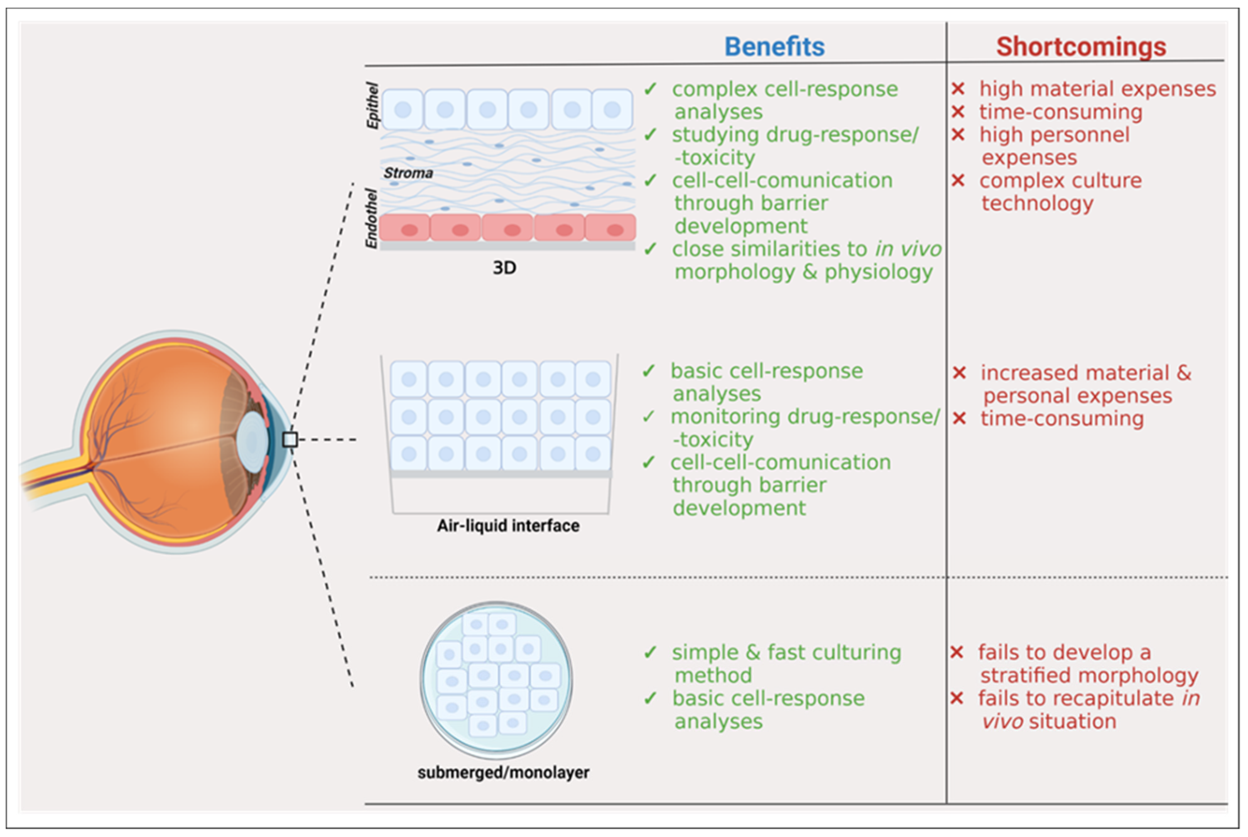
3. Development of 3D Cell Culture Models

Vision loss but also chronic nasal symptoms affect a person’s quality of life [52,53]. Therefore, it is important to advance research in this area to provide better treatment options and alternatives to such patients. With regard to this, 3D cell culture models provide a basis for better understanding of the pathogenesis of diseases and may result in the development of therapies for such patients [54]. In addition, cell-cell or cell-matrix interactions can be studied [54,55]. Due to the 3D cell culture methodology, the different cell types have good access to nutrients and oxygen in this in vitro environment [54,56]. Organotypic cell culture models arrived in cell culture banks several years ago. Their advantage is that cells are extracted from tissues or from biopsies and seeded on semipermeable membranes after few passages [57,58]. In the appropriate cell culture media with growth factors and other supplements, the cells differentiate and acquire some organotypic phenotype, wherein cell-cell or cell-matrix interactions can be studied, as well as pathogenesis of e.g., viral infection [54,57,58,59,60,61,62,63]. Therefore, the following sections first describe the current 3D models for each compartment individually. For investigating the connection and communication between the ocular surface and the nasal epithelia, either (i) complex 3D models combining the current individual models could be used, or (ii) the same experimental question could be investigated simultaneously on the individual 3D models and the results then correlated.

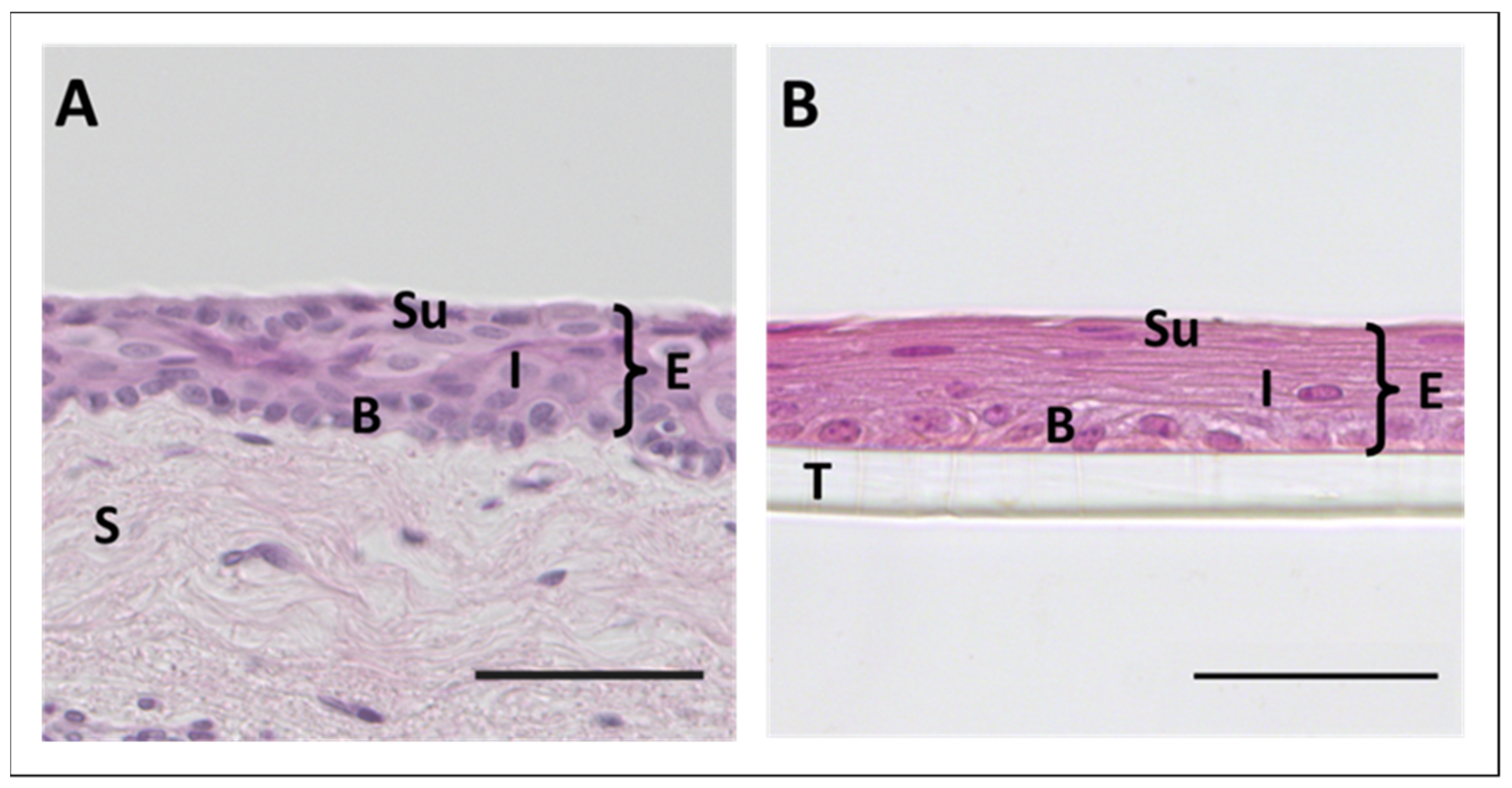
3.1. 3D Cell Culture of Corneal Epithelium

The cornea is transparent and non-vascularized [54]. It is anatomically composed of five distinct layers [15,54] with different cell types (Section 2.1) (reviewed in [54]). A 3D in vitro model of the cornea would therefore need to account for these different cell types and layers, which is not easy to implement [54]. The cornea functions as a refracting lens, which focuses the visible light and acts as a protector of the inner part of the eye (reviewed in [64,65]). Morphologically, between the anterior corneal epithelial and the posterior corneal endothelial parts, the corneal stroma constitutes the main layer (reviewed in [64]). To date, there is increasing awareness of the complexity of the corneal stroma and hierarchical organization of the collagen fibrils in the stroma. Previously, these were not or insufficiently well studied, and it is important to explore this ultrastructural part of the eye in more detail (reviewed in [64,65]) [66,67,68,69,70]. As described below in more detail, optical coherence tomography (OCT) is an important technique in visualizing also the ultrastructure of the cornea [71,72]. Exemplarily, Napoli and colleagues have used the OCT to explore the corneal stromal striae in Ovis aries [73].
Epithelial stroma cultures, epithelial stroma cultures with nerves or corneal endothelial stroma cultures were already described by different authors (reviewed in [54]). In the first culture type, cell-cell interactions between corneal fibroblasts and epithelial cells are analyzed, which can also be used as a so-called wound and healing model of the cornea [54]. 3D cell culture models with a single cell type can be established for example within an air-liquid interface model, and drug effects, injury, or chemical noxious agents can be studied [74,75,76,77], (reviewed in [54]). Pluripotent stem cells can differentiate into any cell type and acquire so-called organ-like structures [78]. Organoid models are certainly an important milestone in experimental research, as these so-called mini-organs incorporate an in vivo-like morphology and physiology. Organoids are defined as a cell cluster of organ-specific cell types originating from stem or progenitor cells that self-organize and group to resemble an organ or organ-like structures [78]. Due to their organ-like structures in culture dishes, hypotheses related to organ physiology and dysfunction can be studied in vitro [78]. Moreover, drug testing can be also performed in such models at a scale that cannot normally be applied in human trials [78]. Foster and colleagues were finally able to culture corneal organoids from human induced-pluripotent stem cells (iPSCs) containing the known three different cell types with the corresponding epithelial, stromal, and endothelial cell marker expression [54,79]. In detail, first, iPSCs were induced/stimulated, which consequently led to neural induction and finally to induction at day 12 and maturation at day 20. This was followed by corneal selection at day 30 and eventual maturation at >120 days [79]. In addition, so-called organ chips may also represent an interesting model, which can be alternatively used in the laboratory to study eye disorders in vitro [52,80]. Rötzer et al. (2019) recently presented an interesting ex vivo slice culture model using ‘thicker’ (300 µm) tissue sections of donor eyelids in culture. They were able to study the regulation of cell cohesion in meibocytes on the slice cultures for up to 6 days [81]. A similar model could be envisioned for the cornea. For all in vivo cultivations, one should be aware of how important mechanical influences on culture conditions are as well. Hampel et al. (2018) showed that shear stress as it occurs at the corneal epithelium during blinking exerts marked effects on corneal epithelial cells, such as changes in cellular morphology and the expression of cell junctions [82].
Figure 2 summarizes the benefits and shortcomings of distinct corneal cell culture models.

3.2. 3D Cell Culture of Epithelia with Goblet Cells

The epithelium of the conjunctiva, lacrimal sac, nasolacrimal duct, and nasal epithelia contain specialized mucus-secreting goblet cells. Moreover, nasal mucosa grafts were used for conjunctiva reconstruction after extensive damage with satisfactory results [84,85]. Depending on the site of tissue harvest in the nasal cavity, as well as in the conjunctiva, the amount of goblet cells can be ‘chosen’ [28,51]. Therefore, cell culture conditions may be similar, in particular with regard to isolation, differentiation, and/or maintenance of goblet cells. In this section, challenges, and achievements to establish 3D in vitro cultivation of epithelia with goblet cells are compiled.
As the conjunctiva covers most of the ocular surface area, conjunctival disorders can severely affect the quality of life [86,87,88]. Therapies based on reconstruction of the conjunctiva using 3D cell culture techniques, with conjunctival cells expanded in vitro on suitable matrices, are gaining increasing attention. However, the efficient culture of conjunctival cells and, in particular, differentiation and/or preservation of mature conjunctival cells and goblet cells pose special requirements for cultivation. Special attention should be paid to the choice of starting material when 3D cell cultures are established, as the three conjunctival regions show anatomical differences in terms of epithelial cell shape, stratification, and goblet cell density (see Section 2.2). In order to represent the physiological morphology in higher concordance, cell culture models should be established in 3D. To verify an ‘organ-like’ phenotype of cultured conjunctival cells, stratification (Figure 3) as well as cytokeratin 4, 13, and 19 expression are established as markers for mature conjunctival epithelial cells, while MUC5AC is the most common marker for identifying conjunctival goblet cells. A summary of current 3D conjunctiva and nasal cell culture models is presented in Table 1.
To study conjunctival cells, various species are used as donors including rabbits, bovids, and humans [89,90,91,92]. In addition, described cell culture conditions vary greatly with regard to isolation, medium, carrier matrices, and differentiation protocols. In most cases, isolation was carried out by enzymatic homogenization of the conjunctiva with subsequent seeding [90,91,92,93], but also attempts were made to use whole conjunctival pieces and isolate the cells by simply letting the explant culture grow out on a suitable matrix [89,94]. The majority of studies use medium based on Dulbecco’s Modified Eagle Medium (DMEM)/F12 nutrient mix with fetal bovine serum, insulin, and epidermal growth factor (EGF) as additives among others for the expansion of conjunctival cells (see Table A1, Appendix A). For differentiation into mature conjunctival epithelial cells and into goblet cells, special medium compositions as well as methods of air-liquid interface were evaluated, or both were applied in combination.
Another approach to study conjunctival cell biology and effects of various therapies, but not as therapies per se, is the organ culture of conjunctiva pieces. In one study, it was shown that human-derived conjunctiva pieces could be cultivated long-term on gelatin sponges [95]. After 14 days of culture, a new conjunctival epithelium was formed with the expression of cytokeratin 19 and the presence of goblet cells. Of course, the mechanical stress on the tissue exerted under physiological conditions (blinking) should also be considered here and, ideally, the culture should be cultivated under mechanical stress, e.g., by means of a cell stretcher [95].
Table 1. 3D cell culture conditions of conjunctival epithelium.
#Tissue SourceGoblet CellsCell Culture ConditionsRef.
1Rabbit-derived conjunctivayes
  • New Zealand white rabbit-derived palpebral and bulbar conjunctiva
  • Isolation of conjunctival progenitor cells by homogenization of cells using collagenase and Trypsin-EDTA
  • Expansion of conjunctival cells at 1 × 10⁷ cells/mL in bioprinted micro-constructs in conjunctival stem cells (CjSC)-medium
  • Goblet cell differentiation was initiated by a special goblet cell differentiation medium for seven days
  • No efforts were made to analyze the presence of mature conjunctival epithelium
  • Goblet cells were detected by positive MUC5AC
[90]
2Human-derived conjunctiva
  • Human donor-derived bulbar conjunctiva after cryopreservation for up to six months
[89]
no
  • Isolation and expansion of conjunctival progenitor cells on feeder cells in conjunctival epithelial cells (CEC) medium by explant culture
  • Expansion and differentiation by seeding 4.4 × 10⁵ cell/cm2 onto a decellularized porcine conjunctiva in a cell crown, submerged cultivation in CEC medium for 7 days followed by cultivation at air-liquid interface in differentiation medium
  • Mature conjunctival cells were detected by stratification, morphology, and positive cytokeratin 19 staining
  • No goblet cells were detected by MUC5AC staining
few
  • Isolation on decellularized porcine conjunctiva
  • Isolation and expansion of conjunctival cells by direct outgrowth from an explant piece onto a scaffold
  • Expansion on decellularized porcine conjunctiva placed in a cell crown, submerged cultivation in CEC medium for 7 days followed by cultivation at air-liquid interface in differentiation medium
  • Mature conjunctival cells were detected by stratification, morphology, and positive cytokeratin 19 expression
  • Goblet cells were detected by positive MUC5AC staining
3Bovine-derived conjunctivano
  • Bovine-derived bulbar conjunctiva
  • Isolation of conjunctival epithelial cells by homogenization of cells using dispase and Trypsin-EDTA
  • Expansion of conjunctival cells at 5 × 10⁵ cells/cm2 on collagen-coated transwell inserts in complete growth medium
  • For 3D culture, cells were maintained at the air-liquid interface from day 5 onwards
  • Mature conjunctival cells were detected by stratification, morphology, and cytokeratin 4 and 13 expression
  • Goblet cells were not detected
[91]
4Rabbit-derived conjunctivayes
  • New Zealand white rabbit-derived palpebral and fornical conjunctiva
  • Isolation of conjunctival progenitor cells by homogenization of cells using dispase II and Trypsin-EDTA
  • Expansion of conjunctival cells onto a collagen-coated cell culture dish in growth medium to sub-confluence
  • For 3D culture, conjunctival cells were seeded at 1.1 × 10⁶ cells/cm2 in a temperature-responsive culture dish, and after 4 days, a second cell layer was seeded at 1.1 × 10⁶ cells/cm2
  • Mature conjunctival cells were detected by stratification, morphology, and cytokeratin 4 and 19 expression
  • Goblet cells were detected by positive MUC5AC staining
[92]
5Human-derived conjunctivayes
  • Human donor-derived biopsy specimens from superior temporal bulbar conjunctiva
  • Isolation of conjunctival cells by homogenization using protease and differential attachment to plastic cell culture dishes
  • Expansion of cells at 1.4 × 10³ cells/cm2 on plastic culture dishes in bronchial epithelial growth medium (BEGM)
  • For 3D culture, cells were seeded at 2.1 × 10⁴ cells/cm2 onto transwell inserts in a mixed medium (BEGM/DMEM) and maintained at air-liquid interface from day 5 onwards
  • Mature conjunctival cells were detected by stratification, morphology, and positive cytokeratin 19 staining
  • Goblet cells were detected by positive MUC5AC staining
[96]
6Human-derived conjunctiva
  • Human donor-derived conjunctiva from 6 different regions in initial experiments; the inferior fornical conjunctiva, as the best source of conjunctival stem cells, was then used in further 3D cell culture experiments
  • Isolation of conjunctival cells by homogenization using Trypsin-EDTA
[93]
no
  • Expansion on amniotic membrane at 1.7 × 10⁴ cells/cm2 with/without feeder cells in control medium (K) or without feeder cells in XerumFree (XF) or keratinocyte (SFM) medium for up to 7 days
  • Mature conjunctival cells were detected by stratification, morphology, and positive cytokeratin 13 and 19 staining when cultured in K or XF; cells in SFM failed to attach to the amniotic membrane
  • Goblet cells were not detected
no-few
  • Expansion on fibrin glue gel at 1.7 × 10⁴ cells/cm2 with feeder cells in control medium (K) or without feeder cells in XerumFree medium (XF) for up to 7 days
  • Mature conjunctival cells were detected by stratification, morphology, and positive cytokeratin 13 and 19 staining when cultured in K or XF
  • Goblet cells were not detected by MUC5AC staining, but few MUCS5AC transcripts were detected by qPCR
7Human-derived conjunctiva
  • Human donor-derived bulbar conjunctiva
  • Isolation of conjunctival cells (epithelial cells and fibroblasts) by explant culture on plastic dishes in epithelial cell culture medium
  • Preparation of a fibrin scaffold with 1 × 10⁵ fibroblasts/mL incorporated into the scaffold and 1 × 10⁵ epithelial cells/cm2 seeded onto the scaffold surface 24h after polymerization
[94]
Few-no
  • Cultures of the scaffolds submerged in epithelial cell culture medium
  • Mature conjunctival cells were detected by stratification, morphology, and positive cytokeratin 19 staining
  • Goblet cells were detected by lectin staining of glycoconjugates (HPA) and MUC5AC ELISA at day 7, but no goblet cells were detected at day 14 by lectin staining of glycoconjugates (HPA)
  • Cultures of the scaffolds were maintained at the air-liquid interface from day 3 onwards in epithelial cell culture medium
  • Mature conjunctival cells were detected by stratification, morphology, and positive cytokeratin 19 staining
  • Goblet cells were detected by lectin staining of glycoconjugates (HPA) and MUC5AC ELISA at day 7
DMEM: Dulbecco’s modified eagle medium, EDTA: ethylenediaminetetraacetic acid, ELISA: enzyme-linked immunosorbent assay, HPA: Helix pomatia agglutinin, MUC5AC: mucin 5AC, qPCR: quantitative real time polymerase chain reaction.
Different 3D cell cultures with nasal epithelial cells were previously described in the literature. Recent works by our group providing detailed information on the corresponding cell culture models (submerged, spheroid, organotypic 3D cultures, etc.) with the corresponding clinical applications were published previously [57,58,59]. Among others, the advantages of nasal epithelial cells over bronchial cells are discussed there. Because of their ease of collection (in contrast to bronchial cells, which are obtained by complex bronchoscopies), scientists can readily obtain nasal samples from patients and healthy donors and apply complex cell culture methods (reviewed in [58]). In addition, cell culture media, either commercially available or self-made, allow practical implementation of such cell cultures ex vivo (reviewed in [58]). Studies using nasal epithelial samples from patients with cystic fibrosis (CF) ensure important molecular insights [97]. Keegan and Brewington recently wrote about using these insights to guide individualized therapy for CF patients in their review [97]. The authors discuss that preclinical testing of drugs in such nasal epithelial cell cultures of CF patients should be performed to provide appropriate personalized therapy with Cystic Fibrosis Transmembrane Conductance Regulator (CFTR) modulators, because not every CF patient shows the same response to these therapeutics [97]. The establishment of nasal epithelial organoids may allow the implementation from simple dose-effect approaches to complex vector use with the major aim to help individuals who are suffering from serious diseases e.g., CF, nasal tumors.
To the best of our knowledge, no study has reported the isolation, cultivation, or investigation of nasolacrimal epithelial cells. This could possibly be due to the limited accessibility of the nasolacrimal duct within its bony canal (nasolacrimal canal). Based on the similarities between the epithelial structures, we hypothesize that the cell culture technologies may be similar. However, this needs to be verified in future experiments.

3.3. Optical Coherence Tomography as a Useful Non-Invasive Technique for Ocular Surface Imaging and Disease Exploring

For several years, the increasing use of non-invasive methods to visualize structural depths and three-dimensional structures of the eye have been growing. In particular, OCT takes on an important role in ophthalmology (reviewed in [98]).
In detail, the OCT is a non-invasive, non-contact, high-resolution optical methodology that can generate real-time cross-sectional images [98,99,100,101,102]. Currently, increasing adaptations and modifications of OCT are taking place, in which fast and better-resolution images can be generated [100]. These changes and technological achievements are due to a certain transition for several years between the traditional time-domain (TD) to Fourier-domain OCT (FD-OCT) methodology [100].
The FD-OCT technique uses the spectrum of reflected light to generate an image, which displays structural information on the ocular surface, cornea, and retina [100]. In contrast, previous OCT techniques used time-domain detection, an interferometer, and a scanning arm as well as a light-source of a low-coherence nature (reviewed in [103]). Napoli and colleagues summarized the usability of this instrument in their recent review [100], mapping the transition from the traditional to FD-OCT method. In addition, the same research group also tested the clinical applicability of OCT and highlighted its use in different areas, including in forensic medicine, where the authors studied postmortem ocular changes [98,101,104,105]. In another article, the same research group administered sodium carboxymethylcellulose over the cornea in healthy and dry eye patients and successfully measured adhesiveness using FD-OCT [106]. Moreover, Messner and colleagues demonstrated dry eye disease in a mouse model using OCT and fluorescein staining [107]. They concluded that fluorescein staining can detect defects of the corneal surface, but OCT can also provide information on the depth of the defect and the close layers [107].
In summary, OCT can provide important clinical information and was shown to be useful to date. As was also shown in appropriate animal models, the device is useful in basic research [72,73,108,109,110]. Such new technological achievements will certainly complement the complex culture experiments in the future. Thus, there should be a constant exchange of information with basic researchers and ophthalmologists who may develop further establishments and adaptations of OCT.

4. Clinically Relevant Molecular Applications of Conjunctival, Nasolacrimal, and Nasal 3D Cell Culture Models

In the previous section, we described the current state-of-the-art 3D cell culture technology for corneal, conjunctival, nasolacrimal, and nasal epithelial cells. These technologies can be used to perform functional studies that provide insight into common and distinct pathologies in these complex structures. Therefore, in this section we will exemplify three clinically relevant pathologies, namely measles virus infection, adenovirus infection, and allergies, which can affect both the eye and the nose.

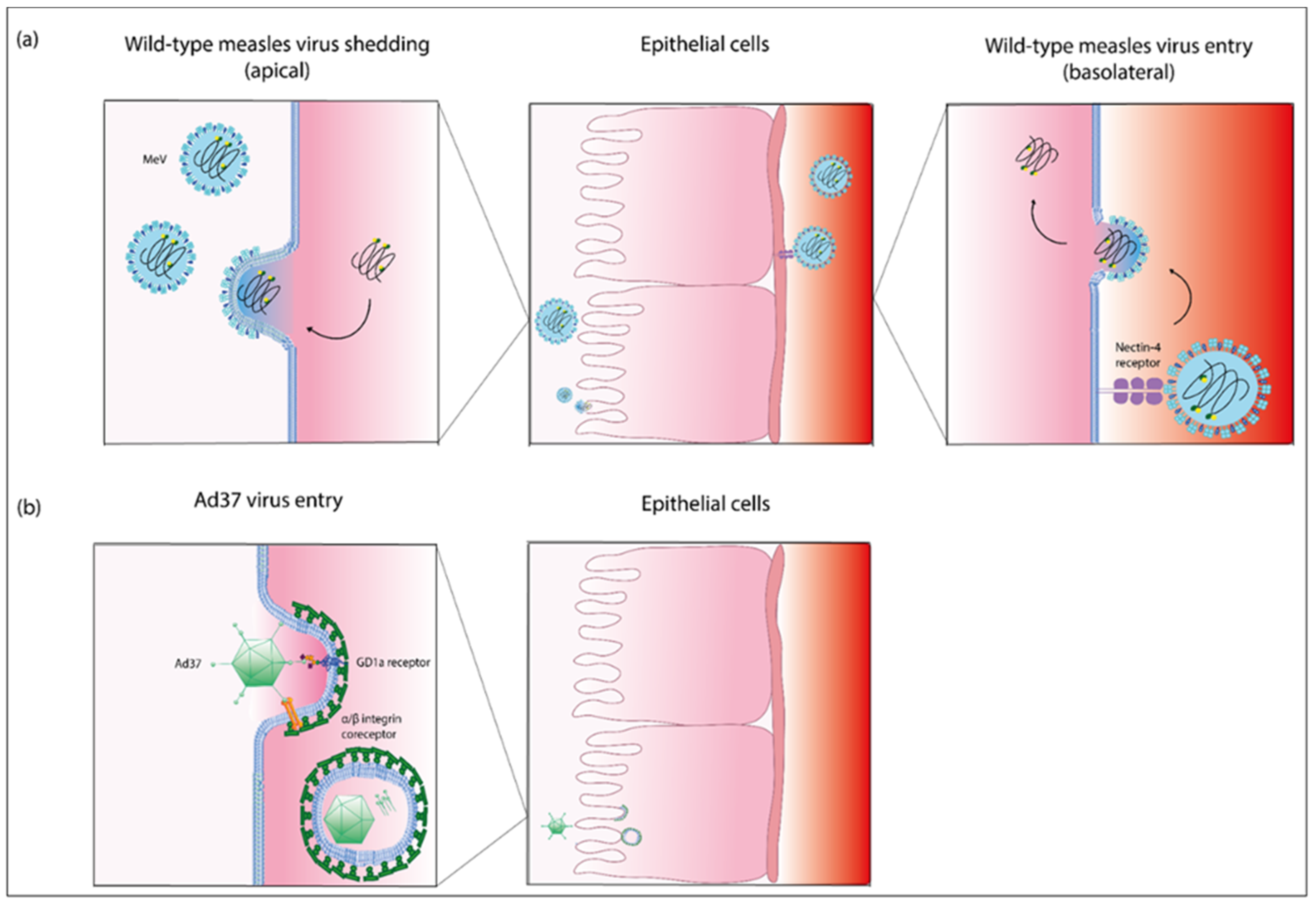
4.1. Pathogenesis of Measles Virus Infection in the Eye and the Nasolacrimal Tract

Measles virus (MeV) is the causative agent of measles, one of the most transmissible human diseases. MeV is a negative-strand RNA virus of the Paramyxoviridae family [111]. The MeV genome has a size of approximately 16 kb; the size of the pleiomorphic virions ranges from 100 to 300 nm [112,113]. The entry receptors for pathogenic wild-type MeV are CD150/SLAM, expressed on immune cells, and epithelial nectin-4/PVRL4 [114]. After receptor binding, MeV fuses at the plasma membrane, and its exclusively cytoplasmic replication cycle is initiated (Figure 4a). MeV is transmitted through the respiratory route, and immune cells in the upper respiratory tract represent the primary target cells. MeV infection then spreads via immune cells within the lymphoid tissue, followed by viremia and systemic spread of the virus. In epithelia, infection occurs at the basolateral membrane; the virus then spreads from cell to cell and finally buds at the apical membrane [111,115,116]. Several studies on measles pathogenesis employ the macaque infection model [117], but human epithelial cell cultures have also been used [115,118]. Analyses of samples acquired during measles infection of children are also published [119,120]. These investigations yielded insights into the spread of MeV within the eye and nose.
Conjunctivitis and coryza are hallmarks of measles [116] and shedding via the nasal mucosa is assumed to contribute to the high transmissibility of the virus [121]. Clinically, an ocular infection with MeV is thought to be acquired following viremia, leading to conjunctivitis and epithelial keratitis [122,123]. Measles infection is a major cause of childhood blindness in unvaccinated individuals, particularly in malnourished children [111]. During measles infection, MeV can be localized in ocular tissue [124] and its genomic RNA is detected in tear samples [125]. Detection of MeV antigen in epithelial cells within measles-associated conjunctival lesions was also reported previously [120,123]. In biopsies without conjunctival lesions, the antigen was only detected in the subepithelial tissue [120]. Syncytial giant cells as a characteristic feature of measles infection were also found in conjunctival swab samples during the early phase of the disease [119]. However, it is argued that these giant cells and ocular symptoms may represent sequelae of inflammation and the ensuing immune response [111,126].
Corneal rim epithelial cells can be infected with MeV after disruption of epithelial integrity [126] and eye protection as well as administration of measles convalescent serum into the conjunctival sac was reported to reduce the risk of measles infection [127,128,129]. This led some authors to the conclusion that the conjunctiva is a route of measles entry [127,128]. Nevertheless, ocular transmission is certainly not a main route of measles transmission, as opposed to transmission via respiratory droplets and aerosols [129].
In the macaque model of measles infection, high titers of cell-associated as well as cell-free virus were found in nasal swabs [130], and large groups of infected cells were observed in the nasal epithelium [131]. The release of infectious virus from macaque nasal epithelial cultures after both apical and basolateral inoculation has been reported [118]. These studies indicate that the nasal epithelium may play an important role in measles transmission. Syncytia were also observed in nasal secretions of children with measles [119,132]. A recent study using human airway epithelial cultures demonstrated transmission of infectious virus from epithelial to immune cells via dislodged epithelial infectious foci [133]. These findings support the notion of measles transmission via nasal secretions and sneezing. The pathogenesis of measles in the eye and nose is documented and studied in preclinical models and using clinical specimens. However, a distinct role for the communication between these organs in measles pathogenesis has not been established to date.

4.2. Pathogenesis of Adenovirus Infection in the Eye and the Nasolacrimal Tract

Human adenoviruses (HAdV) comprise more than 100 different types (http://hadvwg.gmu.edu/, accessed on 1 September 2021), which are divided into seven know species, HAdV-A to HAdV-G. As pathogens, adenoviruses cause a multitude of clinical diseases. Depending on the virus type, infections most frequently affect the respiratory and the gastrointestinal tract, as well as the eyes. An effective treatment is lacking, and the mortality rate in immune-compromised patients can amount to up to 70% [134]. HAdV infections in the eye manifest as either follicular conjunctivitis, pharyngoconjunctival fever (PCF), or epidemic keratoconjunctivitis (EKC) involving the cornea (reviewed in [135]). EKC represents one of the most frequent ocular diseases and is mainly caused by species D adenoviruses including HAdV-8, HAdV-37, HAdV-53, HAdV-54, HAdV-56, HAdV-64 (previously known as HAdV-19a), HAdV-82, and HAdV-85 [135]. Among these, HAdV-8, HAdV-19, and HAdV-37 cause the most severe conjunctivitis [136]. Pharyngoconjunctival fever (PCF) is mainly caused by HAdV-3 from species B, and HAdV-4 from species E is associated with PCF [137,138]. This infection mainly affects children, and outbreaks can occur in schools, kindergartens, and summer camps. Patients suffering from PCF usually present with mild symptoms such as follicular conjunctivitis, rhinitis, pharyngitis, and fever.
There is a limited number of studies analyzing HAdV infection in the nasolacrimal ducts and nasal epithelial cells. One study analyzed the susceptibility of primary human nasal epithelial cells to a broad range of different HAdV [139] and found that predominantly HAdV-5 derived from species C yielded robust transduction of these primary cells. Other non-species C viruses showed low transduction rates [139,140].
To study the pathogenicity of HAdVs associated with infections in the eye and efferent tear ducts, virus entry can be analyzed in respective infection models of the 3D cell culture model. Virus tropism is determined by primary receptors, and there is evidence in the literature that EKC-associated HAdV utilizes GD1a glycan [141] and CD46 [142] as an entry receptor. Furthermore, it was shown that various EKC-associated HAdVs utilize sialic acid containing glycans as cellular receptors [143]. In contrast, PCF-associated HAdVs enter cells utilizing the desmoglein 2 receptor (HAdV-3) [144] and the coxsackie- and adenovirus receptor (CAR, HAdV-4) [145]. Transduction of nasal epithelial cells was most efficient with HAdV-5 [139,140], which enters the target cell utilizing the CAR receptor [146]. After binding of the adenovirus fiber knob protein protruding from the adenovirus capsid to the primary receptor, the arginine-glycine-aspartic acid (RGD) motif of the adenovirus penton protein interacts with integrins of the host cell. It was shown that, for instance, HAdV-37 binds to αVβ1 and α3β1 integrins for infection of corneal cells [147], which is followed by clathrin-mediated endocytosis (Figure 4b).
To study the pathogenesis of HAdV at the ocular surface or the nasolacrimal tract further, host species infection models in which infectious virus progeny can be observed were explored. Utilizing porcine corneal cell culture [148] and a three-dimensional culture model based on human corneal stromal fibroblasts [149], infections in the eye were analyzed. Moreover, alternative infection models for the eye immortalized human corneal epithelial cells were studied [150,151]. Furthermore, HAdV infection was explored in submerged 2D and organotypic 3D primary human nasal epithelial cells [59]. However, to date, studies investigating conjunctival epithelial and efferent tear duct epithelial susceptibility are missing.
Figure 4. Entry and exit pathway of measles virus and entry pathway of adenovirus type 37. (a) Schematic representation of the entry and exit process of wild-type measles virus (MeV) in epithelia. MeV is transmitted to epithelial cells via the receptor nectin-4 located at the basolateral membrane. Entry occurs via membrane fusion. Progeny MeV particles bud from the apical surface of epithelial cells into the lumen [152,153]. This panel (a) was adapted from [111]. (b) Schematic representation of the entry pathway of adenovirus 37 (Ad37). Ad37 enters epithelial cells by binding the ganglioside (GD1a) receptor and α/β integrins as co-receptors. Cell entry occurs by clathrin-mediated endocytosis [141,154]. This panel (b) was adapted from [135]. Abbreviations: Ad, adenovirus; MeV, wild-type, i.e., pathogenic measles virus; GD1a, ganglioside. This figure was created with Adobe Inc. (2021) Adobe illustrator. Retrieved from https://adobe.com/products/illustrator, Dublin, Ireland, software purchased on 6 April 2021, link accessed last 6 April 2021, software last accessed on 12 October 2021).
Ijms 22 12994 g004

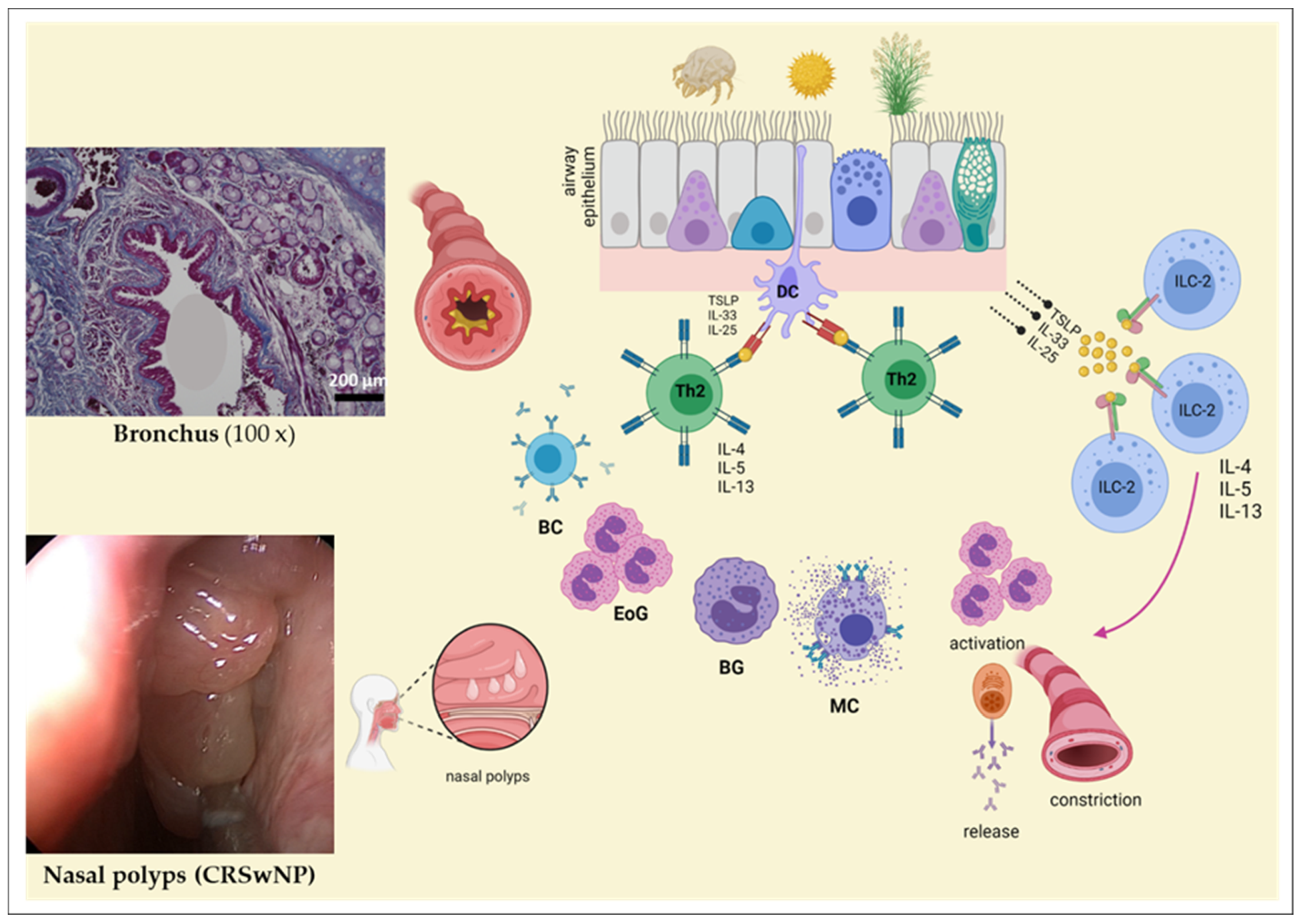
4.3. Allergen Exposure in Ocular and Nasolacrimal Systems

The ocular and nasal epithelia may be affected in the setting of allergies [155,156]. This is a clinical example of how both compartments can be affected locally and how they are interconnected via allergen exposure (reviewed in [157]) [155,156]. Patients suffering from ocular allergic symptoms commonly have rhinitis symptoms, and both can be linked with asthma as well (reviewed in [158,159]. An effective medical treatment would reduce both ocular and nasal local symptoms simultaneously (reviewed in [160]) [155]. External triggers such as allergens can lead to rhino-conjunctival symptoms. About a quarter of the world’s population suffers from allergic diseases including hay fever, asthma, or atopic dermatitis [161], and circa 10–20% have an allergic rhinitis [162]. Epidemiologically, the highest disease rates are noticed in developed, westernized countries [163].
Rhinitis is characterized by runny nose/rhinorrhea, nasal congestion, itching, and/or sneezing due to underlying inflammation of the nasal mucosa [161,163,164,165]. Patients with an allergic rhinitis usually have conjunctivitis and asthma [161,166]. Allergic rhinitis is caused by an IgE-mediated reaction to inhaled substances [161]. Different guidelines are used for the classification of allergic/non-allergic rhinitis (for allergic rhinitis, the Allergic Rhinitis and its Impact on Asthma (ARIA) guidelines are commonly used) [161,163,167,168]. Nevertheless, there are still no molecular parameters which have been translated into clinical practice that can predict the type and severity of allergic rhinitis [161]. Allergies/atopy (allergic inflammation) can lead to a chronic rhinosinusitis (reviewed in [169,170]). Furthermore, allergens can also affect the eyes, which can be also a manifestation of allergic sensitization and can also appear together with rhinitis (=Allergic rhinoconjunctivitis) [171]. The ocular allergy affects the eyelid, the conjunctiva, and the cornea [172] and is observed either seasonally or as persistent symptoms throughout the year [172,173]. Due to the stimulation of the ocular and nasal mucosa by distinct allergens (e.g., outdoor allergens), clinically, increased tearing, redness, itching and swelling, runny or stuffy nose, as well as increased sneezing are characteristically observed [172,173,174].
The underlying molecular mechanism is a T helper 2 (Th2)-polarized immune response, which is characterized by the secretion of cytokines such as interleukin (IL)-4, -5, or -13 (reviewed in [175,176]). When an allergen hits the epithelium, pro-inflammatory cytokines/alarmins such as IL-33, IL-25, or thymic stromal lymphopoeitin (TSLP) (reviewed in [175,176]) are secreted. On the one hand, these proteins can prime naïve T helper cells through dendritic cells via the MHC-II complex, which, as a result, then differentiate into mature Th2 effector cells (reviewed in [175,176]). On the other hand, these cytokines can also directly stimulate Innate Lymphoid Cells Type 2 (ILC-2), which are characterized by the release of type 2 cytokines (reviewed in [161,177,178,179]). These cytokines, in turn, then activate other immune cells, including, for example, B cells, basophil and/or eosinophil granulocytes, and mast cells (reviewed in [175,180]). Other lymphocytes such as regulatory T cells [177,180,181] and Th17 cells [182,183] also play an important role in this signaling cascade. Clinically, patients suffer from distinct local and systemic reactions due to an increased mucus production and inflammation (Figure 5).
A persistent Th2 inflammation can lead to the development of chronic sinusitis with nasal polyps (reviewed in [184]) (Figure 5). Due to the increased prevalence rate of patients with allergic rhinitis [185], which also influences quality of life [53], there must be more attention drawn to the search of biomarkers. Thus, it is necessarily important to investigate the development of new and complex cell culture technologies to increase the impact of the experimental performance and to implement disease-related hypotheses efficiently in vitro culture models.
Figure 5. The influence of distinct triggers on the airway epithelium. Several triggers can affect or harm the epithelium, which consequently lead to a complex signaling cascade including the activation of T helper 2 (Th2) cells and the release of Th2 cytokines. Following cytokine secretion, several types of immune cells e.g., B cells (BC), eosinophil granulocytes (EoG), basophil granulocytes (BG), or mast cells (MC) are activated. In addition, through the direct stimulation of innate lymphoid cells type 2 (ILC-2), interleukin (IL)-4, IL-5, and/or IL-13 are also released. Clinically, through the activation of these cellular actors, mucus production, airway inflammation, and bronchoconstriction are observed in the bronchi. In addition, chronic inflammation in the nasal cavity leads to the development of nasal polyps. This figure was adapted from [175,176,186,187,188] and was created with BioRender.com. (left) Nasal polyps of a patient with a chronic rhinosinusitis and nasal polyps (CRSwNP). In this figure, polyps of the nasal mucosa are depicted from a patient with a chronic rhinosinusitis and allergy, who suffered from recurrent rhinitis and impediment of nasal breathing.
Ijms 22 12994 g005

5. Conclusions and Experimental Outlook

As evident from what is discussed above, the detailed morphological knowledge of the ocular and nasal epithelia stands in stark contrast to the lack of functional studies explaining physiological states and pathological alterations in these tissues and their interconnections. As one example, how infections (whether bacterial or viral) do not readily ‘jump over’ from the eye to the draining lacrimal ducts and from there to the nasal mucosa or take the reverse route, but usually remain localized, is incompletely understood. One reason could be the different morphology of the epithelial cells with their differential expression of receptors and distinct properties, which preclude pathogen spread across the regional epithelial borders. With the advent of modern technology and the invention of important methodological elements that are also used in the therapy of diseases, mammalian cell cultures represent essential tools to perform first pilot as well as complex experiments in vitro. Due to the high incidence of diseases associated with ocular and nasal epithelia, e.g., atopic disorders [189,190], advanced cell culture models representing these structures are key to advancing related approaches in molecular medicine. Starting from so-called submerged cultures, followed by 3D models to organ-like formats, different culture models are currently described in the literature. Technical improvements and refinements can make the testing of highly complex mechanistic hypotheses feasible ex vivo. This includes the investigation of the pathomolecular role of allergens (such as house dust mites or so-called ‘outdoor allergens’), but also the effects of viruses on corneal, conjunctival, efferent tear duct, and nasal epithelial cells. Exemplarily, AdV and MeV are important, as they can lead from harmless local symptoms to serious systemic diseases (reviewed in [111,191]). With the use of these cell culture technologies, entry receptors for AdV and MeV can be studied at the molecular level. In addition, AdV can be used in a next step as a potential gene therapy tool in these cell cultures with the intention to treat diseases curatively (reviewed in [192]). These viruses may also be used to develop novel anti-cancer agents for ocular and nasal malignancies with high medical need, since both MeV and AdV exhibit oncolytic properties [192,193,194,195,196]. Interestingly, the nose has already been positively evaluated to function as a potential vaccination site. Immunization via the nasal route is drawing increased interest, and the nasal cultures introduced in this review may provide a useful tool to study such vaccines [197,198,199]. Nasal and ocular cells can be used to perform co-cultures and thus interaction studies between cellular compartments, which can provide an additional layer of complexity, e.g., by enabling immunological observations. Practically, those co-cultures can be set up either with both nasal and ocular cells or co-cultures with immune cells, e.g., T lymphocytes. Moreover, also epigenetic and methylation patterns of (stimulated) nasal and ocular cells may provide key information for the characterization of potential molecular barcodes, from which mechanistic hypotheses regarding clinical phenomena can be established. First pilot experiments already demonstrate the functionality and robustness of these cultures with external stimulants including AdV or Dermatophagoides pteronyssinus (=house dust mite) [59,140]. Further establishment and more widespread use of the sophisticated techniques introduced herein to study the ocular and nasal epithelia will highlight their utility for medical progress for treatment and prevention of diseases with ocular and nasal pathologies.

Author Contributions

Conceptualization, all authors; methodology, M.A., J.D., J.W., M.S.C.F. and J.J.-H.P.; validation, all authors; formal analysis, all authors; investigation, A.E. and F.P.; resources, S.W., A.E. and F.P.; data curation, M.A. and J.D.; writing—original draft preparation, all authors; writing—review and editing, all authors; visualization, M.A., J.D., J.W., M.S.C.F. and J.J.-H.P.; supervision, C.E.E., A.E. and F.P.; project administration, S.W., C.E.E., A.E. and F.P.; funding acquisition, S.W., A.E. and F.P. All authors have read and agreed to the published version of the manuscript.

Funding

This research was funded by the Internal Research Grant of the Faculty of Health of Witten/Herdecke University, Germany (project number 2021-02) and in part by German Research Foundation (DFG) grant PA738/15-1.

Institutional Review Board Statement

Not applicable.

Informed Consent Statement

Not applicable.

Data Availability Statement

This review did not report any data.

Acknowledgments

We thank Matthias Hock for reviewing the OCT section.

Conflicts of Interest

The authors declare no conflict of interest. The funders had no role in the design of the study; in the collection, analyses, or interpretation of data; in the writing of the manuscript; or in the decision to publish the results.

Appendix A

Table A1. Cell culture medium composition.
MediumCompositionRef.
1CjSC Medium
  • DMEM/F-12 (3:1)
  • 10% fetal bovine serum
  • 1% penicillin-streptomycin
  • 1× insulin-transferrin-selenium
  • 10 ng/mL epidermal growth factor
  • 400 ng/mL hydrocortisone
  • 0.1 nM cholera toxin
  • 2 nM 3,3´,5-triiodo-L-thyronine
  • 10 µM Y27632
  • 1 µM A83-01
  • 1 µM DMH1
[90]
Goblet cell differentiation medium
  • Keratinocyte SFM
  • 1% penicillin-streptomycin
  • 50 µg/mL bovine pituitary extract
  • 5 ng/mL epidermal growth factor
  • 100 ng/mL bone morphogenetic protein 4
  • 10 ng/mL fibroblast growth factor 10
  • 100 ng/mL interleukin 13
  • 1 µM A83-01
2CEC Medium
  • DMEM/F-12 (1:1)
  • 10% fetal bovine serum
  • 1% penicillin-streptomycin
  • 5 µg/mL transferrin
  • 10 ng/mL epidermal growth factor
  • 400 ng/mL hydrocortisone
  • 0.1 nM cholera toxin
  • 0.075% sodium bicarbonate
  • 0.18 mM adenin
  • 5 µg/mL insulin
  • 2 nM 3,3´,5-triiodo-L-thyronine
[89]
Differentiation medium
  • DMEM/F-12 (1:1)
  • 10% fetal bovine serum
  • 1% penicillin-streptomycin
  • 5 µg/mL transferrin
  • 0.5 ng/mL epidermal growth factor
  • 400 ng/mL hydrocortisone
  • 0.1 nM cholera toxin
  • 0.075% sodium bicarbonate
  • 0.18 mM adenin
  • 5 µg/mL insulin
  • 2 nM 3,3´,5-triiodo-L-thyronine
3Complete growth medium
  • DMEM/F-12 (1:1)
  • 10% fetal bovine serum
  • 1% penicillin-streptomycin
  • 2 mM L-glutamine
  • 10 ng/mL epidermal growth factor
  • 2 µg/mL hydrocortisone
  • 0.1 µg/mL cholera toxin
  • 50 µg/mL gentamycin
  • 5 µg/mL insulin
[91]
4Growth medium
  • DMEM/F-12 (1:1)
  • 10% fetal bovine serum
  • 1% penicillin-streptomycin
  • 5 µg/mL insulin
  • 5µg/mL transferrin
  • 5 ng/mL selenium
  • 10 ng/mL epidermal growth factor
  • 10 ng/mL nerve growth factor
5Bronchial epithelial growth medium
  • Bronchial epithelial growth medium (BEGM)
  • 5 µg/mL insulin
  • 500 ng/mL hydrocortisone
  • 500 ng/mL epinephrine
  • 6.5 ng/mL triiodo-thyronine
  • 10 ng/mL transferrin
  • 10 ng/mL retinoic acid
  • 0.13 mg/mL bovine pituitary extract
  • 50 µg/mL:50 ng/mL gentamycin:amphotericin
  • 10 ng/mL epidermal growth factor
  • 0.15 mg/mL bovine serum albumin
[96]
Mixture Medium
  • BEGM:DMEM (1:1)
  • 5 µg/mL insulin
  • 500 ng/mL hydrocortisone
  • 500 ng/mL epinephrine
  • 6.5 ng/mL triiodo-thyronine
  • 10 ng/mL transferrin
  • 10 ng/mL retinoic acid
  • 0.13 mg/mL bovine pituitary extract
  • 50 µg/mL:50 ng/mL gentamycin:amphotericin
  • 0.5 ng/mL epidermal growth factor
  • 0.15 mg/mL bovine serum albumin
6Control medium (K)
  • DMEM/F-12 (2:1)
  • 10% fetal bovine serum
  • 50 µg/mL penicillin-streptomycin
  • 4 mM glutamine
  • 5 µg/mL insulin
  • 0.4 µg/mL hydrocortisone
  • 0.18 mM adenine
  • 8.1 µg/mL cholera toxin
  • 2 mM triiodo-thyronine
  • 10 ng/mL epidermal growth factor
[93]
XerumFree medium (XF)
  • DMEM/F-12 (2:1)
  • 10% XerumFree XF205
  • 50 µg/mL penicillin-streptomycin
  • 4 mM glutamine
  • 5 µg/mL insulin
  • 0.4 µg/mL hydrocortisone
  • 0.18 mM adenine
  • 8.1 µg/mL cholera toxin
  • 2 mM triiodo-thyronine
  • 10 ng/mL epidermal growth factor
Keratinocyte serum Free medium (SFM)
  • Keratinocyte SFM
  • 50 µg/mL bovine pituitary extract
  • 5 ng/mL epidermal growth factor
7Epithelial cell culture medium
  • DMEM/F-12
  • 10% human serum
  • 25 µg/mL fungizone
  • 5000 U penicillin-streptomycin
  • 1 µg/mL insulin
  • 0.5 µg/mL hydrocortisone
  • 2 ng/mL epidermal growth factor
[94]

References and Notes

  1. Available online: https://www.tagesspiegel.de/gesellschaft/panorama/emotionen-traenen-sind-echt-weinen-kann-man-lernen/1137512.html. (accessed on 10 September 2021).
  2. Messmer, E.M. Emotionale Tränen. Der Ophthalmol. 2009, 106, 593–602. [Google Scholar] [CrossRef]
  3. Paulsen, F. The human nasolacrimal ducts. Adv. Anat. Embryol. Cell Biol. 2003, 170, 1–106. [Google Scholar]
  4. Paulsen, F.; Garreis, F.; Schicht, M.; Brauer, L.; Ali, M.J.; Sel, S. Anatomy and physiology of the nasolacrimal ducts. HNO 2016, 64, 354–366. [Google Scholar] [CrossRef] [PubMed]
  5. Leone, G. In Altero de Musculis Palpebrarum Atque Oculorum Motibus Deservientibus, Accurate Disseritur. Anatomici libri II, Ticini.
  6. De Ductu Salivali Novo, Saliva, Ductibus Oculorum Aquosis et Muore Oculi Aqueo. In Quibus Varia Oris Oculorium et Narium Vasa Discributu, Novique Salivae Lacymae et Muci Fontis Deteguntur et Novum Norilissimi Bilsii des Lymphae Notu et usu Comentrum Examinatur et Rehictur; Obervationes Anatomicae: Leyden, The Netherlands, 1662.
  7. Descriptio Anatomica Oculi Humani. Göttingen, Germany, 1755.
  8. Die bedeutende Publikation von Johann Gottfried Zinn (1727–1759)“Descriptio anatomica oculi humani” (1755). Klin. Monbl. Augenheilkund. 2021, 238, 1018–1020. [CrossRef] [PubMed]
  9. Ali, M.J.; Zetzsche, M.; Scholz, M.; Hahn, D.; Gaffling, S.; Heichel, J.; Hammer, C.M.; Brauer, L.; Paulsen, F. New insights into the lacrimal pump. Ocul. Surf. 2020, 18, 689–698. [Google Scholar] [CrossRef] [PubMed]
  10. Willcox, M.D.P.; Argueso, P.; Georgiev, G.A.; Holopainen, J.M.; Laurie, G.W.; Millar, T.J.; Papas, E.B.; Rolland, J.P.; Schmidt, T.A.; Stahl, U.; et al. TFOS DEWS II Tear Film Report. Ocul. Surf. 2017, 15, 366–403. [Google Scholar] [CrossRef] [PubMed] [Green Version]
  11. Hartong, D.T.; Berson, E.L.; Dryja, T.P. Retinitis pigmentosa. Lancet 2006, 368, 1795–1809. [Google Scholar] [CrossRef]
  12. Pagano, G.; Pallardo, F.V.; Lyakhovich, A.; Tiano, L.; Trifuoggi, M. Mitigating the pro-oxidant state and melanogenesis of Retinitis pigmentosa: By counteracting mitochondrial dysfunction. Cell Mol. Life Sci. 2021, 78, 7491–7503. [Google Scholar] [CrossRef] [PubMed]
  13. Bielmeier, C.B.; Roth, S.; Schmitt, S.I.; Boneva, S.K.; Schlecht, A.; Vallon, M.; Tamm, E.R.; Ergun, S.; Neueder, A.; Braunger, B.M. Transcriptional Profiling Identifies Upregulation of Neuroprotective Pathways in Retinitis Pigmentosa. Int. J. Mol. Sci. 2021, 22, 6307. [Google Scholar] [CrossRef] [PubMed]
  14. Donato, L.; Abdalla, E.M.; Scimone, C.; Alibrandi, S.; Rinaldi, C.; Nabil, K.M.; D’Angelo, R.; Sidoti, A. Impairments of Photoreceptor Outer Segments Renewal and Phototransduction Due to a Peripherin Rare Haplotype Variant: Insights from Molecular Modeling. Int. J. Mol. Sci. 2021, 22, 3484. [Google Scholar] [CrossRef] [PubMed]
  15. DelMonte, D.W.; Kim, T. Anatomy and physiology of the cornea. J. Cataract. Refract. Surg. 2011, 37, 588–598. [Google Scholar] [CrossRef] [PubMed]
  16. Asbell, P.; Brocks, D. Cornea Overview. 2010.
  17. Hamrah, P.; Sahin, A. Limbus and corneal epithelium. In Ocular Surface Disease: Cornea, Conjunctiva and Tear Film; Elsevier: Amsterdam, The Netherlands, 2013; pp. 29–33. [Google Scholar]
  18. Michelacci, Y.M. Collagens and proteoglycans of the corneal extracellular matrix. Braz. J. Med. Biol. Res. 2003, 36, 1037–1046. [Google Scholar] [CrossRef] [Green Version]
  19. Hay, E.D. Development of the vertebrate cornea. Int. Rev. Cytol. 1980, 63, 263–322. [Google Scholar] [PubMed]
  20. Joyce, N.C. Proliferative capacity of corneal endothelial cells. Exp. Eye Res. 2012, 95, 16–23. [Google Scholar] [CrossRef] [PubMed] [Green Version]
  21. Bourne, W.M.; McLaren, J.W. Clinical responses of the corneal endothelium. Exp. Eye Res. 2004, 78, 561–572. [Google Scholar] [CrossRef] [PubMed]
  22. Gipson, I.K. Distribution of mucins at the ocular surface. Exp. Eye Res. 2004, 78, 379–388. [Google Scholar] [CrossRef]
  23. Jumblatt, M.M.; McKenzie, R.W.; Jumblatt, J.E. MUC5AC mucin is a component of the human precorneal tear film. Investig. Ophthalmol. Vis. Sci. 1999, 40, 43–49. [Google Scholar]
  24. McKenzie, R.W.; Jumblatt, J.E.; Jumblatt, M.M. Quantification of MUC2 and MUC5AC transcripts in human conjunctiva. Investig. Ophthalmol. Vis. Sci. 2000, 41, 703–708. [Google Scholar]
  25. Paulsen, F.P.; Berry, M.S. Mucins and TFF peptides of the tear film and lacrimal apparatus. Prog. Histochem. Cytochem. 2006, 41, 1–53. [Google Scholar] [CrossRef] [PubMed]
  26. Knop, E.; Knop, N. [Eye-associated lymphoid tissue (EALT) is continuously spread throughout the ocular surface from the lacrimal gland to the lacrimal drainage system]. Ophthalmologe 2003, 100, 929–942. [Google Scholar] [CrossRef] [PubMed]
  27. Steven, P.; Gebert, A. Conjunctiva-associated lymphoid tissue-current knowledge, animal models and experimental prospects. Ophthalmic. Res. 2009, 42, 2–8. [Google Scholar] [CrossRef]
  28. Gipson, I.K. Goblet cells of the conjunctiva: A review of recent findings. Prog. Retin. Eye Res. 2016, 54, 49–63. [Google Scholar] [CrossRef] [PubMed] [Green Version]
  29. Tsubota, K.; Tseng, S.C.; Nordlund, M.L. Anatomy and physiology of the ocular surface. In Ocular Surface Disease Medical and Surgical Management; Springer: Berlin/Heidelberg, Germany, 2002; pp. 3–15. [Google Scholar]
  30. Forrester, J.V.; Dick, A.D.; McMenamin, P.G.; Roberts, F.; Pearlman, E. Anatomy of the eye and orbit. Eye Basic Sci. Pract. 2002, 2, 66–90. [Google Scholar]
  31. Obata, H. Anatomy and histopathology of the human lacrimal gland. Cornea 2006, 25, S82–S89. [Google Scholar] [CrossRef] [PubMed]
  32. Paulsen, F.P.; Hinz, M.; Schaudig, U.; Thale, A.B.; Hoffmann, W. TFF peptides in the human efferent tear ducts. Investig. Ophthalmol. Vis. Sci. 2002, 43, 3359–3364. [Google Scholar]
  33. Pellegrini, G.; Golisano, O.; Paterna, P.; Lambiase, A.; Bonini, S.; Rama, P.; De Luca, M. Location and clonal analysis of stem cells and their differentiated progeny in the human ocular surface. J. Cell Biol. 1999, 145, 769–782. [Google Scholar] [CrossRef] [PubMed]
  34. Nagasaki, T.; Zhao, J. Uniform distribution of epithelial stem cells in the bulbar conjunctiva. Investig. Ophthalmol. Vis. Sci. 2005, 46, 126–132. [Google Scholar] [CrossRef] [PubMed]
  35. Stewart, R.M.; Sheridan, C.M.; Hiscott, P.S.; Czanner, G.; Kaye, S.B. Human conjunctival stem cells are predominantly located in the medial canthal and inferior forniceal areas. Investig. Ophthalmol. Vis. Sci. 2015, 56, 2021–2030. [Google Scholar] [CrossRef] [PubMed] [Green Version]
  36. Paulsen, F.; Thale, A.; Kohla, G.; Schauer, R.; Rochels, R.; Parwaresch, R.; Tillmann, B. Functional anatomy of human lacrimal duct epithelium. Anat. Embryol. 1998, 198, 1–12. [Google Scholar] [CrossRef] [PubMed]
  37. Paulsen, F.P.; Corfield, A.P.; Hinz, M.; Hoffmann, W.; Schaudig, U.; Thale, A.B.; Berry, M. Characterization of mucins in human lacrimal sac and nasolacrimal duct. Investig. Ophthalmol. Vis. Sci. 2003, 44, 1807–1813. [Google Scholar] [CrossRef] [PubMed] [Green Version]
  38. Paulsen, F.P.; Foge, M.; Thale, A.B.; Tillmann, B.N.; Mentlein, R. Animal model for the absorption of lipophilic substances from tear fluid by the epithelium of the nasolacrimal ducts. Investig. Ophthalmol. Vis. Sci. 2002, 43, 3137–3143. [Google Scholar]
  39. Paulsen, F.P.; Schaudig, U.; Thale, A.B. Drainage of tears: Impact on the ocular surface and lacrimal system. Ocul. Surf. 2003, 1, 180–191. [Google Scholar] [CrossRef]
  40. Paulsen, F.P.; Paulsen, J.I.; Thale, A.B.; Tillmann, B.N. Mucosa-associated lymphoid tissue in human efferent tear ducts. Virchows Arch. 2000, 437, 185–189. [Google Scholar] [CrossRef] [PubMed]
  41. Paulsen, F.P.; Schaudig, U.; Maune, S.; Thale, A.B. Loss of tear duct-associated lymphoid tissue in association with the scarring of symptomatic dacryostenosis. Ophthalmology 2003, 110, 85–92. [Google Scholar] [CrossRef]
  42. Paulsen, F.P.; Pufe, T.; Schaudig, U.; Held-Feindt, J.; Lehmann, J.; Schroder, J.M.; Tillmann, B.N. Detection of natural peptide antibiotics in human nasolacrimal ducts. Investig. Ophthalmol. Vis. Sci. 2001, 42, 2157–2163. [Google Scholar]
  43. Paulsen, F.P.; Thale, A.B.; Hallmann, U.J.; Schaudig, U.; Tillmann, B.N. The cavernous body of the human efferent tear ducts: Function in tear outflow mechanism. Investig. Ophthalmol. Vis. Sci. 2000, 41, 965–970. [Google Scholar] [CrossRef] [PubMed] [Green Version]
  44. Ayub, M.; Thale, A.B.; Hedderich, J.; Tillmann, B.N.; Paulsen, F.P. The cavernous body of the human efferent tear ducts contributes to regulation of tear outflow. Investig. Ophthalmol. Vis. Sci. 2003, 44, 4900–4907. [Google Scholar] [CrossRef] [PubMed] [Green Version]
  45. Harkema, J.R.; Carey, S.A.; Wagner, J.G. The nose revisited: A brief review of the comparative structure, function, and toxicologic pathology of the nasal epithelium. Toxicol. Pathol. 2006, 34, 252–269. [Google Scholar] [CrossRef]
  46. Mygind, N.; Dahl, R. Anatomy, physiology and function of the nasal cavities in health and disease. Adv. Drug Deliv. Rev. 1998, 29, 3–12. [Google Scholar] [CrossRef]
  47. Groneberg, D.A.; Peiser, C.; Dinh, Q.T.; Matthias, J.; Eynott, P.R.; Heppt, W.; Carlstedt, I.; Witt, C.; Fischer, A.; Chung, K.F. Distribution of respiratory mucin proteins in human nasal mucosa. Laryngoscope 2003, 113, 520–524. [Google Scholar] [CrossRef] [PubMed]
  48. Fahy, J.V.; Dickey, B.F. Airway mucus function and dysfunction. N. Engl. J. Med. 2010, 363, 2233–2247. [Google Scholar] [CrossRef] [PubMed] [Green Version]
  49. dos Santos Silva, E.; Ulrich, M.; Döring, G.; Botzenhart, K.; Gött, P. Trefoil factor family domain peptides in the human respiratory tract. J. Pathol. 2000, 190, 133–142. [Google Scholar] [CrossRef]
  50. Ali, M.S.; Pearson, J.P. Upper airway mucin gene expression: A review. Laryngoscope 2007, 117, 932–938. [Google Scholar] [CrossRef] [PubMed]
  51. Tos, M. Distribution of mucus producing elements in the respiratory tract. Differences between upper and lower airway. Eur. J. Respir. Dis. Suppl. 1983, 128, 269–279. [Google Scholar] [PubMed]
  52. Manafi, N.; Shokri, F.; Achberger, K.; Hirayama, M.; Mohammadi, M.H.; Noorizadeh, F.; Hong, J.; Liebau, S.; Tsuji, T.; Quinn, P.M.J.; et al. Organoids and organ chips in ophthalmology. Ocul. Surf. 2021, 19, 1–15. [Google Scholar] [CrossRef] [PubMed]
  53. Blaiss, M.S.; Hammerby, E.; Robinson, S.; Kennedy-Martin, T.; Buchs, S. The burden of allergic rhinitis and allergic rhinoconjunctivitis on adolescents: A literature review. Ann. Allergy Asthma Immunol. 2018, 121, 43–52.e43. [Google Scholar] [CrossRef] [Green Version]
  54. Shiju, T.M.; Carlos de Oliveira, R.; Wilson, S.E. 3D in vitro corneal models: A review of current technologies. Exp. Eye Res. 2020, 200, 108213. [Google Scholar] [CrossRef] [PubMed]
  55. Edmondson, R.; Broglie, J.J.; Adcock, A.F.; Yang, L. Three-dimensional cell culture systems and their applications in drug discovery and cell-based biosensors. Assay Drug Dev. Technol. 2014, 12, 207–218. [Google Scholar] [CrossRef] [Green Version]
  56. Duval, K.; Grover, H.; Han, L.H.; Mou, Y.; Pegoraro, A.F.; Fredberg, J.; Chen, Z. Modeling Physiological Events in 2D vs. 3D Cell Culture. Physiology 2017, 32, 266–277. [Google Scholar] [CrossRef] [PubMed]
  57. Aydin, M.; Naumova, E.A.; Bellm, A.; Behrendt, A.K.; Giachero, F.; Bahlmann, N.; Zhang, W.; Wirth, S.; Ehrhardt, A.; Arnold, W.H.; et al. The mystery behind the nostrils-technical clues for successful nasal epithelial cell cultivation. Ann. Anat. 2021, 238, 151748. [Google Scholar] [CrossRef] [PubMed]
  58. Aydin, M.; Naumova, E.A.; Bellm, A.; Behrendt, A.K.; Giachero, F.; Bahlmann, N.; Zhang, W.; Wirth, S.; Paulsen, F.; Arnold, W.H.; et al. From Submerged Cultures to 3D Cell Culture Models: Evolution of Nasal Epithelial Cells in Asthma Research and Virus Infection. Viruses 2021, 13, 387. [Google Scholar] [CrossRef] [PubMed]
  59. Aydin, M.; Naumova, E.A.; Paulsen, F.; Zhang, W.; Gopon, F.; Theis, C.; Lutz, S.; Ehrke-Schulz, E.; Arnold, W.H.; Wirth, S.; et al. House Dust Mite Exposure Causes Increased Susceptibility of Nasal Epithelial Cells to Adenovirus Infection. Viruses 2020, 12, 1151. [Google Scholar] [CrossRef] [PubMed]
  60. Chioni, A.M.; Bajwa, R.T.; Grose, R. 3D Organotypic Culture Model to Study Components of ERK Signaling. Methods Mol. Biol. 2017, 1487, 255–267. [Google Scholar] [CrossRef] [PubMed]
  61. Chioni, A.M.; Grose, R. Organotypic modelling as a means of investigating epithelial-stromal interactions during tumourigenesis. Fibrogenesis Tissue Repair 2008, 1, 8. [Google Scholar] [CrossRef] [PubMed] [Green Version]
  62. McKay, T.B.; Ford, A.; Wang, S.; Cairns, D.M.; Parker, R.N.; Deardorff, P.M.; Ghezzi, C.E.; Kaplan, D.L. Assembly and Application of a Three-Dimensional Human Corneal Tissue Model. Curr. Protoc. Toxicol. 2019, 81, e84. [Google Scholar] [CrossRef]
  63. McKay, T.B.; Karamichos, D.; Hutcheon, A.E.K.; Guo, X.; Zieske, J.D. Corneal Epithelial-Stromal Fibroblast Constructs to Study Cell-Cell Communication in Vitro. Bioengineering 2019, 6, 110. [Google Scholar] [CrossRef] [PubMed] [Green Version]
  64. Espana, E.M.; Birk, D.E. Composition, structure and function of the corneal stroma. Exp. Eye Res. 2020, 198, 108137. [Google Scholar] [CrossRef]
  65. Meek, K.M.; Knupp, C. Corneal structure and transparency. Prog. Retin. Eye Res. 2015, 49, 1–16. [Google Scholar] [CrossRef] [Green Version]
  66. Akhtar, S.; Petrovski, G.; Albert, R.; Alkanaan, A.; Kirat, O.; Khan, A.D.; Almubrad, T. Ultrastructure and 3D transmission electron tomography of collagen fibrils and proteoglycans of swollen human corneal stroma. Histol. Histopathol. 2019, 34, 91–102. [Google Scholar] [CrossRef]
  67. Alkanaan, A.; Barsotti, R.; Kirat, O.; Khan, A.; Almubrad, T.; Akhtar, S. Collagen fibrils and proteoglycans of peripheral and central stroma of the keratoconus cornea-Ultrastructure and 3D transmission electron tomography. Sci. Rep. 2019, 9, 19963. [Google Scholar] [CrossRef]
  68. Meek, K.M.; Leonard, D.W. Ultrastructure of the corneal stroma: A comparative study. Biophys. J. 1993, 64, 273–280. [Google Scholar] [CrossRef] [Green Version]
  69. Akhtar, S.; Alkatan, H.; Kirat, O.; Almubrad, T. Ultrastructural and three-dimensional study of post-LASIK ectasia cornea. Microsc. Res. Tech. 2014, 77, 91–98. [Google Scholar] [CrossRef] [PubMed]
  70. Akhtar, S.; Alkatan, H.M.; Kirat, O.; Khan, A.A.; Almubrad, T. Collagen Fibrils and Proteoglycans of Macular Dystrophy Cornea: Ultrastructure and 3D Transmission Electron Tomography. Microsc. Microanal. 2015, 21, 666–679. [Google Scholar] [CrossRef]
  71. Annadanam, A.; Stoeger, C.G.; Galloway, J.D.; Hikes, M.T.; Jun, A.S. Optical Coherence Tomography Assessment of the Cornea During Corneal Swelling: Should the Term "Descemet Membrane Folds" Be Reconsidered? Cornea 2019, 38, 754–757. [Google Scholar] [CrossRef] [PubMed]
  72. Liu, A.S.; Brown, D.M.; Conn, R.E.; McNabb, R.P.; Pardue, M.T.; Kuo, A.N. Topography and pachymetry maps for mouse corneas using optical coherence tomography. Exp. Eye Res. 2020, 190, 107868. [Google Scholar] [CrossRef] [PubMed]
  73. Napoli, P.E.; Nioi, M.; d’Aloja, E.; Loy, F.; Fossarello, M. The architecture of corneal stromal striae on optical coherence tomography and histology in an animal model and in humans. Sci. Rep. 2020, 10, 19861. [Google Scholar] [CrossRef] [PubMed]
  74. Kaluzhny, Y.; Kinuthia, M.W.; Lapointe, A.M.; Truong, T.; Klausner, M.; Hayden, P. Oxidative stress in corneal injuries of different origin: Utilization of 3D human corneal epithelial tissue model. Exp. Eye Res. 2020, 190, 107867. [Google Scholar] [CrossRef]
  75. Kaluzhny, Y.; Kinuthia, M.W.; Truong, T.; Lapointe, A.M.; Hayden, P.; Klausner, M. New Human Organotypic Corneal Tissue Model for Ophthalmic Drug Delivery Studies. Investig. Ophthalmol. Vis. Sci. 2018, 59, 2880–2898. [Google Scholar] [CrossRef] [PubMed] [Green Version]
  76. Garcia, I.; Capallere, C.; Arcioni, M.; Brulas, M.; Plaza, C.; Meyrignac, C.; Bauza, E.; Botto, J.M. Establishment and performance assessment of an in-house 3D Reconstructed Human Cornea-Like Epithelium (RhCE) as a screening tool for the identification of liquid chemicals with potential eye hazard. Toxicol. In Vitro 2019, 61, 104604. [Google Scholar] [CrossRef] [PubMed]
  77. da Silva, A.C.G.; Chialchia, A.R.; de Castro, E.G.; MRL, E.S.; Arantes, D.A.C.; Batista, A.C.; Kitten, G.T.; Valadares, M.C. A new corneal epithelial biomimetic 3D model for in vitro eye toxicity assessment: Development, characterization and applicability. Toxicol. In Vitro 2020, 62, 104666. [Google Scholar] [CrossRef] [PubMed]
  78. Lancaster, M.A.; Knoblich, J.A. Organogenesis in a dish: Modeling development and disease using organoid technologies. Science 2014, 345, 1247125. [Google Scholar] [CrossRef]
  79. Foster, J.W.; Wahlin, K.; Adams, S.M.; Birk, D.E.; Zack, D.J.; Chakravarti, S. Cornea organoids from human induced pluripotent stem cells. Sci. Rep. 2017, 7, 41286. [Google Scholar] [CrossRef]
  80. Van Meenen, J.; Ni Dhubhghaill, S.; Van den Bogerd, B.; Koppen, C. An Overview of Advanced In Vitro Corneal Models: Implications for Pharmacological Testing. Tissue Eng. Part B Rev. 2021. [Google Scholar] [CrossRef]
  81. Rotzer, V.; Melega, F.; Garreis, F.; Paulsen, F.; Waschke, J. E-Cadherin Is Important for Meibomian Gland Function as Revealed by a New Human ex Vivo Slice Culture Model. Am. J. Pathol. 2019, 189, 1559–1568. [Google Scholar] [CrossRef] [PubMed] [Green Version]
  82. Hampel, U.; Garreis, F.; Burgemeister, F.; Essel, N.; Paulsen, F. Effect of intermittent shear stress on corneal epithelial cells using an in vitro flow culture model. Ocul. Surf. 2018, 16, 341–351. [Google Scholar] [CrossRef] [PubMed]
  83. Ronkko, S.; Vellonen, K.S.; Jarvinen, K.; Toropainen, E.; Urtti, A. Human corneal cell culture models for drug toxicity studies. Drug Deliv. Transl. Res. 2016, 6, 660–675. [Google Scholar] [CrossRef] [PubMed] [Green Version]
  84. Kim, J.H.; Chun, Y.S.; Lee, S.H.; Mun, S.K.; Jung, H.S.; Lee, S.H.; Son, Y.; Kim, J.C. Ocular surface reconstruction with autologous nasal mucosa in cicatricial ocular surface disease. Am. J. Ophthalmol. 2010, 149, 45–53.e42. [Google Scholar] [CrossRef] [PubMed]
  85. Wenkel, H.; Rummelt, V.; Naumann, G.O. Long term results after autologous nasal mucosal transplantation in severe mucus deficiency syndromes. Br. J. Ophthalmol. 2000, 84, 279–284. [Google Scholar] [CrossRef] [PubMed] [Green Version]
  86. Bielory, L.; Delgado, L.; Katelaris, C.H.; Leonardi, A.; Rosario, N.; Vichyanoud, P. ICON: Diagnosis and management of allergic conjunctivitis. Ann. Allergy Asthma Immunol. 2020, 124, 118–134. [Google Scholar] [CrossRef] [PubMed] [Green Version]
  87. Gomez-Henao, C.M.; Herrera-Morales, C.I.; Ramirez-Giraldo, R.; Cardona-Villa, R. Quality of life and clinical characterization of patients with vernal keratoconjunctivitis in a pediatric population in Colombia. Allergol. Immunopathol. 2018, 46, 370–377. [Google Scholar] [CrossRef]
  88. Mikhail, E.; Azizoglu, S.; Gokhale, M.; Suphioglu, C. Questionnaires Assessing the Quality of Life of Ocular Allergy Patients. J. Allergy Clin. Immunol. Pract. 2020, 8, 2945–2952. [Google Scholar] [CrossRef]
  89. Witt, J.; Dietrich, J.; Mertsch, S.; Schrader, S.; Spaniol, K.; Geerling, G. Decellularized porcine conjunctiva as an alternative substrate for tissue-engineered epithelialized conjunctiva. Ocul. Surf. 2020, 18, 901–911. [Google Scholar] [CrossRef] [PubMed]
  90. Zhong, Z.; Deng, X.; Wang, P.; Yu, C.; Kiratitanaporn, W.; Wu, X.; Schimelman, J.; Tang, M.; Balayan, A.; Yao, E. Rapid bioprinting of conjunctival stem cell micro-constructs for subconjunctival ocular injection. Biomaterials 2021, 267, 120462. [Google Scholar] [CrossRef] [PubMed]
  91. Paladino, G.; Marino, C.; Mule, S.L.T.; Civiale, C.; Rusciano, D.; Enea, V. Cytokeratin expression in primary epithelial cell culture from bovine conjunctiva. Tissue Cell 2004, 36, 323–332. [Google Scholar] [CrossRef] [PubMed]
  92. Yao, Q.; Zhu, M.; Chen, J.; Shao, C.; Yan, C.; Wang, Z.; Fan, X.; Gu, P.; Fu, Y. Reconstruction of conjunctival epithelium-like tissue using a temperature-responsive culture dish. Mol. Vis. 2015, 21, 1113. [Google Scholar] [PubMed]
  93. Bertolin, M.; Breda, C.; Ferrari, S.; Van Acker, S.I.; Zakaria, N.; Di Iorio, E.; Migliorati, A.; Ponzin, D.; Ferrari, B.; Lužnik, Z. Optimized protocol for regeneration of the conjunctival epithelium using the cell suspension technique. Cornea 2019, 38, 469–479. [Google Scholar] [CrossRef] [PubMed]
  94. Garcia-Posadas, L.; Soriano-Romani, L.; Lopez-Garcia, A.; Diebold, Y. An engineered human conjunctival-like tissue to study ocular surface inflammatory diseases. PLoS ONE 2017, 12, e0171099. [Google Scholar] [CrossRef] [PubMed]
  95. Rosellini, A.; Papini, S.; Giannarini, C.; Nardi, M.; Revoltella, R.P. Human conjunctival epithelial precursor cells and their progeny in 3D organotypic culture. Int. J. Dev. Biol. 2002, 51, 739–743. [Google Scholar] [CrossRef] [Green Version]
  96. Chung, S.-H.; Lee, J.H.; Yoon, J.-H.; Lee, H.K.; Seo, K.Y. Multi-layered culture of primary human conjunctival epithelial cells producing MUC5AC. Exp. Eye Res. 2007, 85, 226–233. [Google Scholar] [CrossRef] [PubMed]
  97. Keegan, D.E.; Brewington, J.J. Nasal Epithelial Cell-Based Models for Individualized Study in Cystic Fibrosis. Int. J. Mol. Sci. 2021, 22, 4448. [Google Scholar] [CrossRef] [PubMed]
  98. Nioi, M.; Napoli, P.E.; Demontis, R.; Locci, E.; Fossarello, M.; d’Aloja, E. Morphological analysis of corneal findings modifications after death: A preliminary OCT study on an animal model. Exp. Eye Res. 2018, 169, 20–27. [Google Scholar] [CrossRef]
  99. Huang, D.; Swanson, E.A.; Lin, C.P.; Schuman, J.S.; Stinson, W.G.; Chang, W.; Hee, M.R.; Flotte, T.; Gregory, K.; Puliafito, C.A.; et al. Optical coherence tomography. Science 1991, 254, 1178–1181. [Google Scholar] [CrossRef] [PubMed] [Green Version]
  100. Napoli, P.E.; Nioi, M.; Mangoni, L.; Gentile, P.; Braghiroli, M.; d’Aloja, E.; Fossarello, M. Fourier-Domain OCT Imaging of the Ocular Surface and Tear Film Dynamics: A Review of the State of the Art and an Integrative Model of the Tear Behavior During the Inter-Blink Period and Visual Fixation. J. Clin. Med. 2020, 9, 668. [Google Scholar] [CrossRef] [PubMed] [Green Version]
  101. Nioi, M.; Napoli, P.E.; Mayerson, S.M.; Fossarello, M.; d’Aloja, E. Optical coherence tomography in forensic sciences: A review of the literature. Forensic Sci. Med. Pathol. 2019, 15, 445–452. [Google Scholar] [CrossRef] [PubMed]
  102. Podoleanu, A.G. Optical coherence tomography. J. Microsc. 2012, 247, 209–219. [Google Scholar] [CrossRef] [PubMed] [Green Version]
  103. Fujimoto, J.; Swanson, E. The Development, Commercialization, and Impact of Optical Coherence Tomography. Investig. Ophthalmol. Vis. Sci. 2016, 57, OCT1–OCT13. [Google Scholar] [CrossRef] [PubMed] [Green Version]
  104. Napoli, P.E.; Nioi, M.; d’Aloja, E.; Fossarello, M. Post-Mortem Corneal Thickness Measurements with a Portable Optical Coherence Tomography System: A Reliability Study. Sci. Rep. 2016, 6, 30428. [Google Scholar] [CrossRef] [PubMed] [Green Version]
  105. Napoli, P.E.; Nioi, M.; Gabiati, L.; Laurenzo, M.; De-Giorgio, F.; Scorcia, V.; Grassi, S.; d’Aloja, E.; Fossarello, M. Repeatability and reproducibility of post-mortem central corneal thickness measurements using a portable optical coherence tomography system in humans: A prospective multicenter study. Sci. Rep. 2020, 10, 14508. [Google Scholar] [CrossRef] [PubMed]
  106. Napoli, P.E.; Coronella, F.; Satta, G.M.; Zucca, I.A.; Fossarello, M. A novel OCT technique to measure in vivo the corneal adhesiveness for sodium carboxymethylcellulose in humans and its validity in the diagnosis of dry eye. Investig. Ophthalmol. Vis. Sci. 2014, 55, 3179–3185. [Google Scholar] [CrossRef] [PubMed] [Green Version]
  107. Messner, A.; Fischak, C.; Pfister, M.; Schutzenberger, K.; Garreis, F.; Paulsen, F.; Stegmann, H.; Aranha Dos Santos, V.; Garhofer, G.; Schmetterer, L.; et al. Characterization of dry eye disease in a mouse model by optical coherence tomography and fluorescein staining. Biomed. Opt. Express 2019, 10, 4884–4895. [Google Scholar] [CrossRef]
  108. Meyer, J.H.; Larsen, P.P.; Strack, C.; Harmening, W.M.; Krohne, T.U.; Holz, F.G.; Schmitz-Valckenberg, S. Optical coherence tomography angiography (OCT-A) in an animal model of laser-induced choroidal neovascularization. Exp. Eye Res. 2019, 184, 162–171. [Google Scholar] [CrossRef] [PubMed]
  109. Nakazawa, M.; Hara, A.; Ishiguro, S.I. Optical Coherence Tomography of Animal Models of Retinitis Pigmentosa: From Animal Studies to Clinical Applications. Biomed. Res. Int. 2019, 2019, 8276140. [Google Scholar] [CrossRef] [Green Version]
  110. Walther, J.; Gaertner, M.; Cimalla, P.; Burkhardt, A.; Kirsten, L.; Meissner, S.; Koch, E. Optical coherence tomography in biomedical research. Anal. Bioanal. Chem. 2011, 400, 2721–2743. [Google Scholar] [CrossRef] [PubMed]
  111. Rota, P.A.; Moss, W.J.; Takeda, M.; de Swart, R.L.; Thompson, K.M.; Goodson, J.L. Measles. Nat. Rev. Dis. Primers 2016, 2, 16049. [Google Scholar] [CrossRef]
  112. Cox, R.M.; Plemper, R.K. Structure and organization of paramyxovirus particles. Curr. Opin. Virol. 2017, 24, 105–114. [Google Scholar] [CrossRef]
  113. Griffin, D.E.; Lin, W.H.; Pan, C.H. Measles virus, immune control, and persistence. FEMS Microbiol. Rev. 2012, 36, 649–662. [Google Scholar] [CrossRef]
  114. Navaratnarajah, C.K.; Generous, A.R.; Yousaf, I.; Cattaneo, R. Receptor-mediated cell entry of paramyxoviruses: Mechanisms, and consequences for tropism and pathogenesis. J. Biol. Chem. 2020, 295, 2771–2786. [Google Scholar] [CrossRef] [Green Version]
  115. Leonard, V.H.; Sinn, P.L.; Hodge, G.; Miest, T.; Devaux, P.; Oezguen, N.; Braun, W.; McCray, P.B., Jr.; McChesney, M.B.; Cattaneo, R. Measles virus blind to its epithelial cell receptor remains virulent in rhesus monkeys but cannot cross the airway epithelium and is not shed. J. Clin. Investig. 2008, 118, 2448–2458. [Google Scholar] [CrossRef] [Green Version]
  116. Moss, W.J. Measles. Lancet 2017, 390, 2490–2502. [Google Scholar] [CrossRef]
  117. de Swart, R.L. Measles studies in the macaque model. Curr. Top. Microbiol. Immunol. 2009, 330, 55–72. [Google Scholar] [CrossRef]
  118. Lin, W.W.; Tsay, A.J.; Lalime, E.N.; Pekosz, A.; Griffin, D.E. Primary differentiated respiratory epithelial cells respond to apical measles virus infection by shedding multinucleated giant cells. Proc. Natl. Acad. Sci. USA 2021, 118, e2013264118. [Google Scholar] [CrossRef] [PubMed]
  119. Lightwood, R.; Nolan, R. Epithelial giant cells in measles as an acid in diagnosis. J. Pediatr. 1970, 77, 59–64. [Google Scholar] [CrossRef]
  120. Nommensen, F.E.; Dekkers, N.W. Detection of measles antigen in conjunctival epithelial lesions staining by lissamine green during measles virus infection. J. Med. Virol. 1981, 7, 157–162. [Google Scholar] [CrossRef]
  121. Ludlow, M.; McQuaid, S.; Milner, D.; de Swart, R.L.; Duprex, W.P. Pathological consequences of systemic measles virus infection. J. Pathol. 2015, 235, 253–265. [Google Scholar] [CrossRef] [PubMed]
  122. Chodosh, J.; Chintakuntlawar, A.V.; Robinson, C.M. Human Eye Infections. Encycl. Virol. 2008, 491–497. [Google Scholar] [CrossRef]
  123. Dekkers, N. The cornea in measles. Doc. Ophthalmol. 2005, 52, v-121. [Google Scholar]
  124. Belser, J.A.; Rota, P.A.; Tumpey, T.M. Ocular tropism of respiratory viruses. Microbiol. Mol. Biol. Rev. 2013, 77, 144–156. [Google Scholar] [CrossRef] [Green Version]
  125. Shinoda, K.; Kobayashi, A.; Higashide, T.; Shirao, Y.; Sakurai, M.; Shirota, Y.; Kagaya, M. Detection of measles virus genomic RNA in tear samples from a patient with measles keratitis. Cornea 2002, 21, 610–612. [Google Scholar] [CrossRef]
  126. Ludlow, M.; Rennick, L.J.; Sarlang, S.; Skibinski, G.; McQuaid, S.; Moore, T.; de Swart, R.L.; Duprex, W.P. Wild-type measles virus infection of primary epithelial cells occurs via the basolateral surface without syncytium formation or release of infectious virus. J. Gen. Virol. 2010, 91, 971–979. [Google Scholar] [CrossRef]
  127. Papp, K. Experiments proving that the route of infection in measles is the contamination of the conjunctival mucosa. Rev. Immunol. Ther. Antimicrob. 1956, 20, 27–36. [Google Scholar] [PubMed]
  128. Kempe, C.H.; Fulginiti, V.A. The pathogenesis of measles virus infection. Arch. Virol. 1965, 16, 103–128. [Google Scholar] [CrossRef] [PubMed]
  129. Laksono, B.M.; De Vries, R.D.; McQuaid, S.; Duprex, W.P.; De Swart, R.L. Measles Virus Host Invasion and Pathogenesis. Viruses 2016, 8, 210. [Google Scholar] [CrossRef] [Green Version]
  130. Ludlow, M.; de Vries, R.; Lemon, K.; McQuaid, S.; Millar, E.; Van Amerongen, G.; Yüksel, S.; Verburgh, R.J.; Osterhaus, A.; De Swart, R.; et al. Infection of lymphoid tissues in the macaque upper respiratory tract contributes to the emergence of transmissible measles virus. J. Gen. Virol. 2013, 94, 1933–1944. [Google Scholar] [CrossRef] [PubMed]
  131. Ludlow, M.; Lemon, K.; de Vries, R.; McQuaid, S.; Millar, E.L.; Van Amerongen, G.; Yüksel, S.; Verburgh, R.J.; Osterhaus, A.; De Swart, R.L.; et al. Measles Virus Infection of Epithelial Cells in the Macaque Upper Respiratory Tract Is Mediated by Subepithelial Immune Cells. J. Virol. 2013, 87, 4033–4042. [Google Scholar] [CrossRef] [Green Version]
  132. Tompkins, V. A characteristic cell in nasal secretions during prodromal measles. J. Am. Med. Assoc. 1955, 157, 711. [Google Scholar] [CrossRef] [PubMed]
  133. Hippee, C.E.; Singh, B.K.; Thurman, A.L.; Cooney, A.L.; Pezzulo, A.A.; Cattaneo, R.; Sinn, P.L. Measles virus exits human airway epithelia within dislodged metabolically active infectious centers. PLoS Pathog. 2021, 17, e1009458. [Google Scholar] [CrossRef] [PubMed]
  134. Lion, T. Adenovirus Infections in Immunocompetent and Immunocompromised Patients. Clin. Microbiol. Rev. 2014, 27, 441–462. [Google Scholar] [CrossRef] [PubMed] [Green Version]
  135. Rajaiya, J.; Saha, A.; Ismail, A.; Zhou, X.; Su, T.; Chodosh, J. Adenovirus and the Cornea: More Than Meets the Eye. Viruses 2021, 13, 293. [Google Scholar] [CrossRef]
  136. Ariga, T.; Shimada, Y.; Shiratori, K.; Ohgami, K.; Yamazaki, S.; Tagawa, Y.; Kikuchi, M.; Miyakita, Y.; Fujita, K.; Ishiko, H.; et al. Five New Genome Types of Adenovirus Type 37 Caused Epidemic Keratoconjunctivitis in Sapporo, Japan, for More Than 10 Years. J. Clin. Microbiol. 2005, 43, 726–732. [Google Scholar] [CrossRef] [Green Version]
  137. Li, J.; Lu, X.; Sun, Y.; Lin, C.; Li, F.; Yang, Y.; Liang, Z.; Jia, L.; Chen, L.; Jiang, B.; et al. A swimming pool-associated outbreak of pharyngoconjunctival fever caused by human adenovirus type 4 in Beijing, China. Int. J. Infect. Dis. 2018, 75, 89–91. [Google Scholar] [CrossRef] [PubMed] [Green Version]
  138. Kuo, S.-C.; Shen, S.-C.; Chang, S.-W.; Huang, S.C.M.; Hsiao, C.-H. Corneal Superinfection in Acute Viral Conjunctivitis in Young Children. J. Pediatr. Ophthalmol. Strabismus 2008, 45, 374–376. [Google Scholar] [CrossRef]
  139. Rhodes, G.C.; Blinkhorn, S.A.; Yong, L.C. Cardiovascular lesions in experimental acute and chronic renal failure in the rat. Exp. Pathol. 1987, 31, 221–229. [Google Scholar] [CrossRef]
  140. Aydin, M.; Schellhorn, S.; Wirth, S.; Zhang, W.; Ehrhardt, A. Human Species D Adenoviruses Isolated from Diarrheal Feces Show Low Infection Rates in Primary Nasal Epithelial Cells. Children 2021, 8, 563. [Google Scholar] [CrossRef] [PubMed]
  141. Nilsson, E.C.; Storm, R.J.; Bauer, J.; Johansson, S.M.; Lookene, A.; Angstrom, J.; Hedenstrom, M.; Eriksson, T.L.; Frangsmyr, L.; Rinaldi, S.; et al. The GD1a glycan is a cellular receptor for adenoviruses causing epidemic keratoconjunctivitis. Nat. Med. 2011, 17, 105–109. [Google Scholar] [CrossRef] [PubMed]
  142. Wu, E.; Trauger, S.A.; Pache, L.; Mullen, T.M.; von Seggern, D.J.; Siuzdak, G.; Nemerow, G.R. Membrane cofactor protein is a receptor for adenoviruses associated with epidemic keratoconjunctivitis. J. Virol. 2004, 78, 3897–3905. [Google Scholar] [CrossRef] [PubMed] [Green Version]
  143. Chandra, N.; Frangsmyr, L.; Imhof, S.; Caraballo, R.; Elofsson, M.; Arnberg, N. Sialic Acid-Containing Glycans as Cellular Receptors for Ocular Human Adenoviruses: Implications for Tropism and Treatment. Viruses 2019, 11, 395. [Google Scholar] [CrossRef] [PubMed] [Green Version]
  144. Wang, H.; Li, Z.Y.; Liu, Y.; Persson, J.; Beyer, I.; Moller, T.; Koyuncu, D.; Drescher, M.R.; Strauss, R.; Zhang, X.B.; et al. Desmoglein 2 is a receptor for adenovirus serotypes 3, 7, 11 and 14. Nat. Med. 2011, 17, 96–104. [Google Scholar] [CrossRef]
  145. Roelvink, P.W.; Lizonova, A.; Lee, J.G.; Li, Y.; Bergelson, J.M.; Finberg, R.W.; Brough, D.E.; Kovesdi, I.; Wickham, T.J. The coxsackievirus-adenovirus receptor protein can function as a cellular attachment protein for adenovirus serotypes from subgroups A, C, D, E, and F. J. Virol. 1998, 72, 7909–7915. [Google Scholar] [CrossRef] [PubMed] [Green Version]
  146. Bergelson, J.M.; Cunningham, J.A.; Droguett, G.; Kurt-Jones, E.A.; Krithivas, A.; Hong, J.S.; Horwitz, M.S.; Crowell, R.L.; Finberg, R.W. Isolation of a common receptor for Coxsackie B viruses and adenoviruses 2 and 5. Science 1997, 275, 1320–1323. [Google Scholar] [CrossRef]
  147. Storm, R.J.; Persson, B.D.; Skalman, L.N.; Frangsmyr, L.; Lindstrom, M.; Rankin, G.; Lundmark, R.; Domellof, F.P.; Arnberg, N. Human Adenovirus Type 37 Uses alphaVbeta1 and alpha3beta1 Integrins for Infection of Human Corneal Cells. J. Virol. 2017, 91, 91. [Google Scholar] [CrossRef] [PubMed] [Green Version]
  148. Ramke, M.; Lam, E.; Meyer, M.; Knipper, A.; Heim, A. Porcine corneal cell culture models for studying epidemic keratoconjunctivitis. Mol. Vis. 2013, 19, 614–622. [Google Scholar] [PubMed]
  149. Rajaiya, J.; Zhou, X.; Barequet, I.; Gilmore, M.S.; Chodosh, J. Novel model of innate immunity in corneal infection. In Vitro Cell Dev. Biol. Anim. 2015, 51, 827–834. [Google Scholar] [CrossRef] [PubMed] [Green Version]
  150. Robertson, D.M.; Li, L.; Fisher, S.; Pearce, V.P.; Shay, J.W.; Wright, W.E.; Cavanagh, H.D.; Jester, J.V. Characterization of growth and differentiation in a telomerase-immortalized human corneal epithelial cell line. Investig. Ophthalmol. Vis. Sci. 2005, 46, 470–478. [Google Scholar] [CrossRef] [Green Version]
  151. Araki-Sasaki, K.; Ohashi, Y.; Sasabe, T.; Hayashi, K.; Watanabe, H.; Tano, Y.; Handa, H. An SV40-immortalized human corneal epithelial cell line and its characterization. Investig. Ophthalmol. Vis. Sci. 1995, 36, 614–621. [Google Scholar]
  152. Lin, L.T.; Richardson, C.D. The Host Cell Receptors for Measles Virus and Their Interaction with the Viral Hemagglutinin (H) Protein. Viruses 2016, 8, 250. [Google Scholar] [CrossRef] [PubMed]
  153. Takeda, M. Measles virus breaks through epithelial cell barriers to achieve transmission. J. Clin. Investig. 2008, 118, 2386–2389. [Google Scholar] [CrossRef] [PubMed]
  154. Nemerow, G.R.; Stewart, P.L. Role of alpha(v) integrins in adenovirus cell entry and gene delivery. Microbiol. Mol. Biol. Rev. 1999, 63, 725–734. [Google Scholar] [CrossRef] [PubMed] [Green Version]
  155. Aneeza, W.H.; Husain, S.; Rahman, R.A.; Van Dort, D.; Abdullah, A.; Gendeh, B.S. Efficacy of mometasone furoate and fluticasone furoate on persistent allergic rhinoconjunctivitis. Allergy Rhinol. 2013, 4, e120–e126. [Google Scholar] [CrossRef]
  156. Ciprandi, G.; Leonardi, S.; Zicari, A.M.; Tosca, M.A.; Marseglia, G. Allergic rhinoconjunctivitis: Pathophysiological mechanism and new therapeutic approach. Acta Biomed. 2020, 91, 93–96. [Google Scholar] [CrossRef] [PubMed]
  157. Origlieri, C.; Bielory, L. Intranasal corticosteroids and allergic rhinoconjunctivitis. Curr. Opin. Allergy Clin. Immunol. 2008, 8, 450–456. [Google Scholar] [CrossRef] [PubMed]
  158. Bielory, L. Allergic conjunctivitis and the impact of allergic rhinitis. Curr. Allergy Asthma Rep. 2010, 10, 122–134. [Google Scholar] [CrossRef]
  159. Rosario, N.; Bielory, L. Epidemiology of allergic conjunctivitis. Curr. Opin. Allergy Clin. Immunol. 2011, 11, 471–476. [Google Scholar] [CrossRef]
  160. Dhami, S.; Nurmatov, U.; Arasi, S.; Khan, T.; Asaria, M.; Zaman, H.; Agarwal, A.; Netuveli, G.; Roberts, G.; Pfaar, O.; et al. Allergen immunotherapy for allergic rhinoconjunctivitis: A systematic review and meta-analysis. Allergy 2017, 72, 1597–1631. [Google Scholar] [CrossRef] [Green Version]
  161. Bousquet, J.; Anto, J.M.; Bachert, C.; Baiardini, I.; Bosnic-Anticevich, S.; Walter Canonica, G.; Melen, E.; Palomares, O.; Scadding, G.K.; Togias, A.; et al. Allergic rhinitis. Nat. Rev. Dis. Primers 2020, 6, 95. [Google Scholar] [CrossRef]
  162. Kakli, H.A.; Riley, T.D. Allergic Rhinitis. Prim. Care 2016, 43, 465–475. [Google Scholar] [CrossRef]
  163. Bousquet, J.; Khaltaev, N.; Cruz, A.A.; Denburg, J.; Fokkens, W.J.; Togias, A.; Zuberbier, T.; Baena-Cagnani, C.E.; Canonica, G.W.; van Weel, C.; et al. Allergic Rhinitis and its Impact on Asthma (ARIA) 2008 update (in collaboration with the World Health Organization, GA(2)LEN and AllerGen). Allergy 2008, 63 (Suppl. S86), 8–160. [Google Scholar] [CrossRef] [PubMed]
  164. Agnihotri, N.T.; McGrath, K.G. Allergic and nonallergic rhinitis. Allergy Asthma Proc. 2019, 40, 376–379. [Google Scholar] [CrossRef] [PubMed]
  165. Skoner, D.P. Allergic rhinitis: Definition, epidemiology, pathophysiology, detection, and diagnosis. J. Allergy Clin. Immunol. 2001, 108, S2–S8. [Google Scholar] [CrossRef] [PubMed]
  166. Schuler Iv, C.F.; Montejo, J.M. Allergic Rhinitis in Children and Adolescents. Pediatr. Clin. N. Am. 2019, 66, 981–993. [Google Scholar] [CrossRef]
  167. Bousquet, J.; Schunemann, H.J.; Togias, A.; Bachert, C.; Erhola, M.; Hellings, P.W.; Klimek, L.; Pfaar, O.; Wallace, D.; Ansotegui, I.; et al. Next-generation Allergic Rhinitis and Its Impact on Asthma (ARIA) guidelines for allergic rhinitis based on Grading of Recommendations Assessment, Development and Evaluation (GRADE) and real-world evidence. J. Allergy Clin. Immunol. 2020, 145, 70–80.e73. [Google Scholar] [CrossRef] [PubMed] [Green Version]
  168. Brozek, J.L.; Bousquet, J.; Agache, I.; Agarwal, A.; Bachert, C.; Bosnic-Anticevich, S.; Brignardello-Petersen, R.; Canonica, G.W.; Casale, T.; Chavannes, N.H.; et al. Allergic Rhinitis and its Impact on Asthma (ARIA) guidelines-2016 revision. J. Allergy Clin. Immunol. 2017, 140, 950–958. [Google Scholar] [CrossRef] [Green Version]
  169. Rosati, M.G.; Peters, A.T. Relationships among allergic rhinitis, asthma, and chronic rhinosinusitis. Am. J. Rhinol. Allergy 2016, 30, 44–47. [Google Scholar] [CrossRef] [Green Version]
  170. Tantilipikorn, P.; Sompornrattanaphan, M.; Suwanwech, T.; Ngaotepprutaram, P. Chronic Rhinosinusitis and Allergy: Increased Allergen Sensitization Versus Real Allergic Rhinitis Multimorbidity: A Systematic Review. Curr. Allergy Asthma Rep. 2020, 20, 19. [Google Scholar] [CrossRef]
  171. Leonardi, A.; Bogacka, E.; Fauquert, J.L.; Kowalski, M.L.; Groblewska, A.; Jedrzejczak-Czechowicz, M.; Doan, S.; Marmouz, F.; Demoly, P.; Delgado, L. Ocular allergy: Recognizing and diagnosing hypersensitivity disorders of the ocular surface. Allergy 2012, 67, 1327–1337. [Google Scholar] [CrossRef]
  172. Miraglia Del Giudice, M.; Allegorico, A.; Marseglia, G.L.; Martelli, A.; Calvani, M.; Cardinale, F.; Duse, M.; Chiappini, E.; Manti, S.; Cravidi, C.; et al. Allergic rhinoconjunctivitis. Acta Biomed. 2020, 91, e2020007. [Google Scholar] [CrossRef]
  173. Patel, D.S.; Arunakirinathan, M.; Stuart, A.; Angunawela, R. Allergic eye disease. BMJ 2017, 359, j4706. [Google Scholar] [CrossRef] [PubMed]
  174. Jalbert, I.; Golebiowski, B. Environmental aeroallergens and allergic rhino-conjunctivitis. Curr. Opin. Allergy Clin. Immunol. 2015, 15, 476–481. [Google Scholar] [CrossRef]
  175. Lambrecht, B.N.; Hammad, H.; Fahy, J.V. The Cytokines of Asthma. Immunity 2019, 50, 975–991. [Google Scholar] [CrossRef] [PubMed]
  176. Gans, M.D.; Gavrilova, T. Understanding the immunology of asthma: Pathophysiology, biomarkers, and treatments for asthma endotypes. Paediatr. Respir. Rev. 2020, 36, 118–127. [Google Scholar] [CrossRef] [PubMed]
  177. Aron, J.L.; Akbari, O. Regulatory T cells and type 2 innate lymphoid cell-dependent asthma. Allergy 2017, 72, 1148–1155. [Google Scholar] [CrossRef] [PubMed] [Green Version]
  178. Pasha, M.A.; Patel, G.; Hopp, R.; Yang, Q. Role of innate lymphoid cells in allergic diseases. Allergy Asthma Proc. 2019, 40, 138–145. [Google Scholar] [CrossRef]
  179. Kabata, H.; Moro, K.; Koyasu, S. The group 2 innate lymphoid cell (ILC2) regulatory network and its underlying mechanisms. Immunol. Rev. 2018, 286, 37–52. [Google Scholar] [CrossRef] [PubMed]
  180. Lambrecht, B.N.; Hammad, H. The immunology of asthma. Nat. Immunol. 2015, 16, 45–56. [Google Scholar] [CrossRef] [PubMed]
  181. Massoud, A.H.; Charbonnier, L.M.; Lopez, D.; Pellegrini, M.; Phipatanakul, W.; Chatila, T.A. An asthma-associated IL4R variant exacerbates airway inflammation by promoting conversion of regulatory T cells to TH17-like cells. Nat. Med. 2016, 22, 1013–1022. [Google Scholar] [CrossRef] [PubMed] [Green Version]
  182. Seumois, G.; Ramirez-Suastegui, C.; Schmiedel, B.J.; Liang, S.; Peters, B.; Sette, A.; Vijayanand, P. Single-cell transcriptomic analysis of allergen-specific T cells in allergy and asthma. Sci. Immunol. 2020, 5. [Google Scholar] [CrossRef]
  183. Choy, D.F.; Hart, K.M.; Borthwick, L.A.; Shikotra, A.; Nagarkar, D.R.; Siddiqui, S.; Jia, G.; Ohri, C.M.; Doran, E.; Vannella, K.M.; et al. TH2 and TH17 inflammatory pathways are reciprocally regulated in asthma. Sci. Transl. Med. 2015, 7, 301ra129. [Google Scholar] [CrossRef] [Green Version]
  184. Dykewicz, M.S.; Rodrigues, J.M.; Slavin, R.G. Allergic fungal rhinosinusitis. J. Allergy Clin. Immunol. 2018, 142, 341–351. [Google Scholar] [CrossRef] [PubMed] [Green Version]
  185. Meltzer, E.O.; Blaiss, M.S.; Derebery, M.J.; Mahr, T.A.; Gordon, B.R.; Sheth, K.K.; Simmons, A.L.; Wingertzahn, M.A.; Boyle, J.M. Burden of allergic rhinitis: Results from the Pediatric Allergies in America survey. J. Allergy Clin. Immunol. 2009, 124, S43–S70. [Google Scholar] [CrossRef]
  186. Hammad, H.; Lambrecht, B.N. Barrier Epithelial Cells and the Control of Type 2 Immunity. Immunity 2015, 43, 29–40. [Google Scholar] [CrossRef] [PubMed] [Green Version]
  187. Holgate, S.T. Innate and adaptive immune responses in asthma. Nat. Med. 2012, 18, 673–683. [Google Scholar] [CrossRef] [PubMed]
  188. Meng, Y.; Wang, C.; Zhang, L. Recent developments and highlights in allergic rhinitis. Allergy 2019, 74, 2320–2328. [Google Scholar] [CrossRef] [Green Version]
  189. Asher, M.I.; Montefort, S.; Bjorksten, B.; Lai, C.K.; Strachan, D.P.; Weiland, S.K.; Williams, H.; Group, I.P.T.S. Worldwide time trends in the prevalence of symptoms of asthma, allergic rhinoconjunctivitis, and eczema in childhood: ISAAC Phases One and Three repeat multicountry cross-sectional surveys. Lancet 2006, 368, 733–743. [Google Scholar] [CrossRef]
  190. Singh, S.; Sharma, B.B.; Salvi, S.; Chhatwal, J.; Jain, K.C.; Kumar, L.; Joshi, M.K.; Pandramajal, S.B.; Awasthi, S.; Bhave, S.; et al. Allergic rhinitis, rhinoconjunctivitis, and eczema: Prevalence and associated factors in children. Clin. Respir. J. 2018, 12, 547–556. [Google Scholar] [CrossRef]
  191. Ison, M.G.; Hayden, R.T. Adenovirus. Microbiol. Spectr. 2016, 4, 217–232. [Google Scholar] [CrossRef] [PubMed]
  192. Wold, W.S.; Toth, K. Adenovirus vectors for gene therapy, vaccination and cancer gene therapy. Curr. Gene Ther. 2013, 13, 421–433. [Google Scholar] [CrossRef] [PubMed]
  193. Aref, S.; Bailey, K.; Fielding, A. Measles to the Rescue: A Review of Oncolytic Measles Virus. Viruses 2016, 8, 294. [Google Scholar] [CrossRef] [PubMed] [Green Version]
  194. Bhattacharjee, S.; Yadava, P.K. Measles virus: Background and oncolytic virotherapy. Biochem. Biophys. Rep. 2018, 13, 58–62. [Google Scholar] [CrossRef]
  195. Huang, H.; Liu, Y.; Liao, W.; Cao, Y.; Liu, Q.; Guo, Y.; Lu, Y.; Xie, Z. Oncolytic adenovirus programmed by synthetic gene circuit for cancer immunotherapy. Nat. Commun. 2019, 10, 4801. [Google Scholar] [CrossRef] [PubMed] [Green Version]
  196. Leber, M.F.; Neault, S.; Jirovec, E.; Barkley, R.; Said, A.; Bell, J.C.; Ungerechts, G. Engineering and combining oncolytic measles virus for cancer therapy. Cytokine Growth Factor Rev. 2020, 56, 39–48. [Google Scholar] [CrossRef] [PubMed]
  197. Lobaina Mato, Y. Nasal route for vaccine and drug delivery: Features and current opportunities. Int. J. Pharm. 2019, 572, 118813. [Google Scholar] [CrossRef] [PubMed]
  198. Wilkins, J.V., Jr.; Golshahi, L.; Rahman, N.; Li, L. Evaluation of Intranasal Vaccine Delivery Using Anatomical Replicas of Infant Nasal Airways. Pharm. Res. 2021, 38, 141–153. [Google Scholar] [CrossRef] [PubMed]
  199. Yuki, Y.; Uchida, Y.; Sawada, S.I.; Nakahashi-Ouchida, R.; Sugiura, K.; Mori, H.; Yamanoue, T.; Machita, T.; Honma, A.; Kurokawa, S.; et al. Characterization and Specification of a Trivalent Protein-Based Pneumococcal Vaccine Formulation Using an Adjuvant-Free Nanogel Nasal Delivery System. Mol. Pharm. 2021, 18, 1582–1592. [Google Scholar] [CrossRef] [PubMed]
Figure 1. Morphology and physiology of the ocular surface epithelium, nasolacrimal duct, and nasal epithelium. (A) Schematic drawing of the anatomical connection between the ocular surface and the nasal cavity via the nasolacrimal duct. Illustration by Jörg Pekarsky, Institute of Functional and Clinical Anatomy, FAU Erlangen-Nürnberg, (B) Hematoxylin and Eosin staining (HE) of the human cornea, (C) the human bulbar conjunctiva, (D) the human nasolacrimal duct, and (E) the human nasal cavity. E: epithelium, S: stroma; arrowheads mark goblet cells, C: cinocilia marked by arrows; Magnification is 200× with 100 µm scale bar.
Ijms 22 12994 g001
Figure 2. Summary of different corneal cell culture models with their benefits and shortcomings. This figure was adapted from [83], and was created with BioRender.com.
Ijms 22 12994 g002
Figure 3. Development of 3D conjunctival epithelial cell culture model. (A) Hematoxylin and Eosin staining (HE) of human bulbar conjunctiva. (B) 3D cultured human conjunctival epithelial cells growing on a transwell insert. Isolation as explant culture on transwell with cultivation (21 days) and differentiation performed according to reference [89]. E = epithelium, Su = superficial epithelial cells, I = intermediate cells, B = basal cells, S = Stroma, T = transwell; Magnification is 200× (A) and 400× (B) with 50 µm scale bar.
Ijms 22 12994 g003
Publisher’s Note: MDPI stays neutral with regard to jurisdictional claims in published maps and institutional affiliations.

