Article

Identification of SCN5a p.C335R Variant in a Large Family with Dilated Cardiomyopathy and Conduction Disease

1
Department of Medicine III, Institute for Cardiomyopathies Heidelberg (ICH), University of Heidelberg, 69120 Heidelberg, Germany
2
DZHK (German Centre for Cardiovascular Research), Heidelberg-Mannheim, 17475 Greifswald, Germany
3
Clinic for Cardiology and Pneumology, Georg-August-University Göttingen, 37073 Göttingen, Germany
4
DZHK (German Centre for Cardiovascular Research), 37073 Göttingen, Germany
5
Institute of Pharmacology and Toxicology, University of Würzburg, 97070 Würzburg, Germany
6
Department of Medicine, University Medical Centre Mannheim (UMM), 68159 Mannheim, Germany
*
Author to whom correspondence should be addressed.
Int. J. Mol. Sci. 2021, 22(23), 12990; https://doi.org/10.3390/ijms222312990
Submission received: 20 September 2021 / Revised: 8 November 2021 / Accepted: 26 November 2021 / Published: 30 November 2021
(This article belongs to the Special Issue Recent Advancements in Primary Cardiomyopathies)

Abstract

:
Introduction: Familial dilated cardiomyopathy (DCM) is clinically variable and has been associated with mutations in more than 50 genes. Rapid improvements in DNA sequencing have led to the identification of diverse rare variants with unknown significance (VUS), which underlines the importance of functional analyses. In this study, by investigating human-induced pluripotent stem cell-derived cardiomyocytes (iPSC-CMs), we evaluated the pathogenicity of the p.C335R sodium voltage-gated channel alpha subunit 5 (SCN5a) variant in a large family with familial DCM and conduction disease. Methods: A four-generation family with autosomal dominant familial DCM was investigated. Next-generation sequencing (NGS) was performed in all 16 family members. Clinical deep phenotyping, including endomyocardial biopsy, was performed. Skin biopsies from two patients and one healthy family member were used to generate human-induced pluripotent stem cells (iPSCs), which were then differentiated into cardiomyocytes. Patch-clamp analysis with Xenopus oocytes and iPSC-CMs were performed. Results: A SCN5a variant (c.1003T>C; p.C335R) could be detected in all family members with DCM or conduction disease. A novel truncating TTN variant (p.Ser24998LysfsTer28) could also be identified in two family members with DCM. Family members with the SCN5a variant (p.C335R) showed significantly longer PQ and QRS intervals and lower left ventricular ejection fractions (LV-EF). All four patients who received CRT-D were non-responders. Electrophysiological analysis with Xenopus oocytes showed a loss of function in SCN5a p.C335R. Na+ channel currents were also reduced in iPSC-CMs from DCM patients. Furthermore, iPSC-CM with compound heterozygosity (SCN5a p.C335R and TTNtv) showed significant dysregulation of sarcomere structures, which may be contributed to the severity of the disease and earlier onset of DCM. Conclusion: The SCN5a p.C335R variant is causing a loss of function of peak INa in patients with DCM and cardiac conduction disease. The co-existence of genetic variants in channels and structural genes (e.g., SCN5a p.C335R and TTNtv) increases the severity of the DCM phenotype.

1. Introduction

Dilated cardiomyopathy (DCM), with a prevalence of 1 in 250, is one of the major causes of heart failure and is the most common cause of heart transplantation [1,2]. To date, mutations in more than 50 genes have been reported to be associated with DCM; however, the precise pathomechanisms responsible for the variations in disease susceptibility and phenotype expression, including the risk of heart failure (HF) or sudden cardiac death (SCD), are virtually unknown [1,3]. Familial DCM, with an autosomal dominant inheritance pattern, can be seen in about 20% of the cases [1,4]. Mutations in specific genes such as LMNA, RBM20, and sodium voltage-gated channel alpha subunit 5 (SCN5a) are associated with a higher risk of life-threatening arrhythmias and rapid disease progression so that knowing a patients’ genotype would lead to earlier management [1,5].
Cardiac Na+ channels, encoded by the SCN5a gene, are essential for initiating heartbeats and maintaining a regular heart rhythm [6]. Till now, numerous rare variants in the SCN5a gene have been reported in association with inherited atrial or ventricular arrhythmias, such as long QT syndrome (LQTS), Brugada syndrome (BrS), and catecholaminergic polymorphic ventricular tachycardia (CPVT) [7]. Genetic variations in SCN5a are also linked to other structural myocardial diseases, including DCM [3,8]. However, most of these are variants with unknown significance (VUS), which are not functionally analyzed yet, and their pathomechanisms are still unclear.
In the present study, we identified and validated the SNC5a p.C335R variant in a large family with familial DCM and conduction disease by investigating iPSC-CMs and Xenopus oocytes. Furthermore, we showed that the co-existence of two pathogenic variants (SCN5a p.C335R and TTNtv p.Ser24998LysfsTer28) could affect the severity of the phenotype.

2. Material and Methods

2.1. Clinical Evaluation

A four-generation German family with DCM, including 16 family members, was investigated (Figure 1A). Clinical deep phenotyping, including ECGs, echocardiography, cardiovascular magnetic resonance imaging (cMRI), and endomyocardial biopsy, were performed (Figure 1B and Table 1).

2.2. DNA Sequencing

DNA Blood Maxi and DNeasy Blood and Tissue kits (Qiagen) were used for DNA isolation. In total, 150 ng of the isolated DNA was used to generate sequencing libraries for Illumina sequencing using Agilents SureSelect Target Enrichment technology. Sequencing was performed on a HiSeq2000 or NovaSeq 6000 (paired-end 2 × 100 bp) in 16 family members. Whole-genome sequencing was performed in iPS-CMs derived from three family members (IV.1, IV.12, and IV.14).

2.3. Human Induced Pluripotent Stem Cell-Derived Cardiomyocytes (iPSC-CMs)

Skin fibroblasts of three family members (IV.1, IV.12, and IV.14), who agreed to participate, were isolated from skin biopsies, as described earlier [9]. For plasmid-based integration-free reprogramming, we followed the protocol previously described [9]. For every patient, two iPSC-lines were selected and analyzed for pluripotency. IPSC lines were differentiated into iPSC-CMs by using standardized protocols [9]. Cells were studied 60 days after initiation of differentiation. After differentiation, the purity of hiPSC-CMs was determined by flow cytometry analysis (>90% cardiac TNT+) or by morphology. IPSC-CMs were maintained in RPMI 1640 supplemented with Glutamax, HEPES, and B27 supplement. For sarcomeric regularity in iPSC-CMs, immunofluorescent staining of α-actinin (1:1000, Sigma) and titin-M8/M9 (1:750, MyoMedix) was obtained with a 40× objective. For quantification of regularity, the α-actinin channel was analyzed with fast Fourier transformation (FFT) in ImageJ, and the first-order peak amplitude was calculated.

2.4. Site-Directed Mutagenesis and Heterologous Expression in Xenopus Oocytes

SCN5a gene, encoding the α-subunit of human Nav1.5 channel, in plasmid pcDNA1.1/Amp was used. The p.C335R variant of SCN5a was prepared by introducing a single point mutation by using the QuikChangeTM (Stratagene) site-directed mutagenesis kit and the following oligonucleotides: 5′-ACGCTGGGACACGTCCGGAGGGCTA-3′ (sense) and 5′TAGCCCTCCGGACGTGTCCCAGCGT-3′ (antisense). The mutated SCN5a cDNA clone was sequenced to ensure the presence of the p.C335R mutation, as well as the absence of other substitutions introduced by the DNA polymerase. Wild-type and mutant constructs were linearized with XbaI, and cRNAs were synthesized using the mMESSAGE mMACHINE in vitro transcription kit (Ambion, Austin, TX, USA) by the use of T7 Polymerase. Injection of 20–25 ng (46 nl/oocyte) of cRNA into stage V and VI defolliculated Xenopus laevis oocytes was performed using a Nanoject automatic injector (Drummond, Broomall, PA, USA). The oocytes were kept in a modified ND96+ solution (100 mM NaCl, 2 mM KCl, 1.8 mM CaCl2, 1 mM MgCl2, 5 mM HEPES, pH 7.6) at 19 °C. Electrophysiological measurements were made 2 to 4 days after injection at room temperature, and the same solution was used during the measurements.

2.5. Electrophysiology Techniques

The two-microelectrode voltage-clamp configuration was used to record currents from Xenopus laevis oocytes. Microelectrodes had tip resistances ranging from 0.3 to 0.7 MΩ. Current and voltage electrodes were filled with 3 M KCl solution. The recordings were performed under constant perfusion at room temperature. Currents were elicited by applying a series of voltage clamp steps between −80 mV and +70 mV in 10 mV increments at a pulsing frequency of 1.0 Hz, starting from a holding potential of −120 mV. Data were low-pass filtered at 1 to 2 kHz (−3 dB, four-pole Bessel filter) before digitalization at 5 to 10 kHz. Recordings were performed using a commercially available amplifier (Warner OC-725A, Warner Instruments, Hamden, CT, USA) and pCLAMP software 10.5 (Axon Instruments, Foster City, CA, USA) for data acquisition and analysis.
To measure the peak sodium current (INa) in iPSC-CMs, standard patch-clamp recording techniques were used in the whole-cell configuration. We also used DMZ-Universal Puller (ZeitzInstrumente Vertriebs GmbH, Martinsried, Germany) patch electrodes with a resistance from 1–2.
MΩ were pulled from borosilicate glass capillaries (MTW 150F; world Precision Instruments, Inc., Sarasota, FL, USA) and filled with a pre-filtered pipette solution. Signals, which were acquired at 10 kHz, were filtered at 2 kHz with the Axon 200B amplifier and Digidata 1440A digitizer hardware, as well as pClamp 10.2 software (Molecular Devices, Sunnyvale, CA, USA). Standard patch-clamp recording techniques were used to measure sodium ion channel current peak sodium (INa) at room temperature. The bath solution contained (mmol/L): 20 NaCl, 110 CsCl, 1.8 CaCl2, 1 MgCl2, 10 HEPES, 10 glucose, 0.001 nifedipine, pH 7.4 (CsOH). Microelectrodes were filled with (mmol/L): 10 NaCl, 135 CsCl, 2 CaCl2, 3 MgATP, 2 TEA-Cl, 5 EGTA, 10 HEPES, and pH 7.2 (CsOH).

2.6. Transient Transfection and Western Blotting

CHO cells transfected with cDNA encoding SCN5A WT or mutant p.C335R using Viromer Yellow in vitro transfection kit (Lipocalyx GmbH, Halle, Germany) according to the manufacturer’s protocol. Cells were briefly washed in PBS, and then 200 μL of RIPA buffer (50 mM Tris-HCl, 0.5% NP-40, 0.25% NaDeoxycholate, 150 mM NaCl, 1 mM EDTA, 1 mM Na3Vo4, 1 mM NaF, 0.1% SDS in distilled H2O pH 7.4) containing cOmpleteTM and Mini Protease Inhibitor Cocktail (Merck KGaA, Darmstadt, Germany) were added to each well. Cells were harvested with the aid of a cell scraper and incubated on ice for 20 min. The resulting extract was cleared of insoluble debris by centrifugation. Protein concentration was determined by using PierceTM BCA Protein Assay Kit (Thermo ScientificTM Inc., Rockford, IL, USA) according to the manufacturer’s protocol. Proteins were separated via denaturing SDS-PAGE and blotted onto a nitrocellulose membrane. Membranes were blocked in a blocking solution containing 5% (w/v) skim milk powder and 0.1% Tween 20 in Tris-buffered saline. Primary anti-human NaV1.5 antibodies (Alomone Labs, Jerusalem, Israel) and anti-Rad50 as a reference (Bethyl Laboratories Inc., Montgomery, AL, USA) were diluted in the same blocking solution and incubated with the membranes overnight. Membranes were washed in Tris-buffered saline and then incubated with Donkey anti-Rabbit IgG, HRP-conjugated secondary antibody (GE Healthcare Life Sciences, Buckinghamshire, UK), and diluted in the same blocking solution (1:5000) for 1 h at room temperature. Membranes were washed in Tris-buffered saline. Labeled bands were visualized using Amersham ECL Prime Western Blotting Detection Reagent (GE Healthcare Life Sciences, Buckinghamshire, UK).

2.7. Statistics

Data are provided as mean ± standard deviation (SD). Student’s t-test was used to analyze the significance between the two groups. A p-value < 0.05 was considered statistically significant. In the case of iPSC-CMs, data are presented as mean ± standard error of the mean (SEM) and p < 0.05 by one-way ANOVA.

3. Results

3.1. Clinical Investigation of a DCM Pedigree

The index patient (III.3) was diagnosed with DCM and conduction disease at the age of 59 (Figure 1A). Due to symptomatic familial DCM (NYHA III) and complete left bundle branch block (LBBB), the patient received cardiac resynchronization therapy (CRT-D). However, she turned out to be a non-responder. After deep phenotyping, genetic diagnostic testing with next-generation sequencing (NGS) was performed in the index patient and her 15 other family members. Two suspected heterozygous variants in SCN5a, and TTN could be detected in this family (Supplemental Table S1). In population controls (gnomADv3.1.2), both the TTN and SCN5a variants have not been detected so far. However, the SCN5a variant has been described in Clinvar (NM_000335.5(SCN5A):c.1003T>C, p.Cys335Arg) in a patient with Brugada Syndrome and in one family with atrial fibrillation [10,11]. The SCN5a variant is a point mutation (1003T>C) that alters the amino acid at codon 335 from the cysteine (C) to arginine (R) located in the first transmembrane domain (Figure 1C). The second variant was a novel truncating TTN variant (NM_001267550.2: c.74987_74991dup) located in the TTN A-band and is constitutively expressed in different isoforms of TTN. This leads to a frameshift with a new termination site (p.Ser24998LysfsTer28). The TTNtv variant could only be detected in two other family members with DCM (IV.1 and IV.2) and was not detectable in healthy family members. 1003T>C in SCN5a could be detected in all patients with DCM and conduction disease. One patient (IV.2) with DCM but without conduction disease did not have this variant but did have the TTNtv. The till-then asymptomatic brother (IV.1) tested positive for both SCN5a and TTNtv. He developed AV Block III° during an exercise test by routine cardiac evaluation at age 24 and was subsequently diagnosed with DCM and concomitant conduction disease (Supplemental Figure S1). Endomyocardial biopsies of this patient showed severe myocardial fibrosis without evidence for myocarditis (Figure 1B). Family members with SCN5a p.C335R showed significantly longer PQ (Figure 2A) and QRS intervals (Figure 2B) and lower LV-EF (Figure 2C). Four of these family members carrying SCN5a p.C335R received CRT-D; however, all of them were non-responders (Figure 2E,F and Supplemental Table S2).

3.2. The SCN5a p.C335R Variant Is Causing a Loss-of-Function of Sodium Channel Current

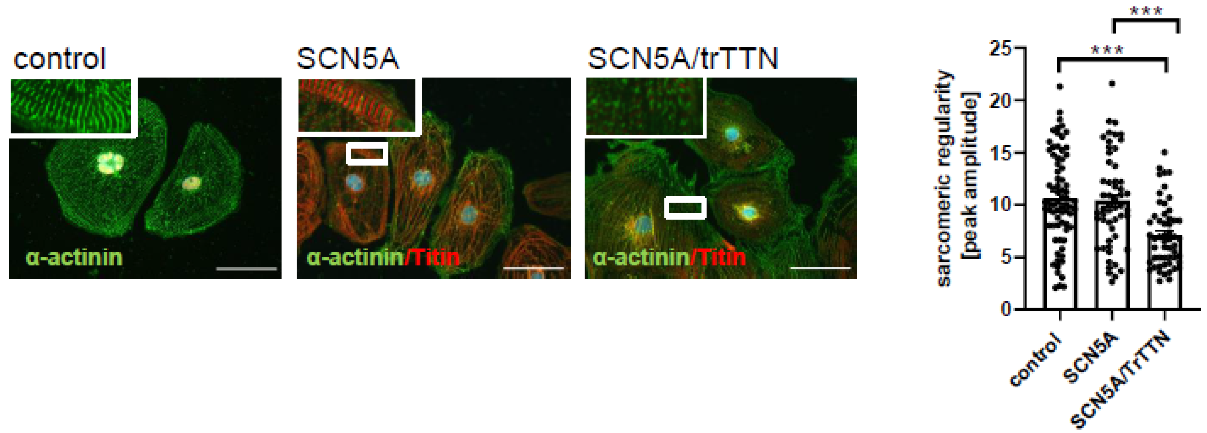
p.C335R is located in a highly conserved P-loop (pore-forming region) between S5 and S6 of Domain I of SCN5a (Figure 1C). Electrophysiological analysis in Xenopus oocytes demonstrated that the SCN5a p.C335R variant completely abolished the activity of the human Nav1.5 channel indicating a loss of function of the channel due to the mutated variant (Figure 3A). The SCN5a expression in the p.335R variant was assessed at similar levels in comparison to the wild-type by using Western blot analysis, supporting the findings that the mutant caused a loss of function of the channel rather than affecting the expression of the protein (Figure 3A). For further specific analyses, iPSC-CMs were generated from a patient with only SCN5a p.C335R (IV.14), another patient with both SCN5a p.C335R and TTNtv (IV.1), as well as a healthy family member with none of these variants (IV.12). All generated iPSCs were tested regarding pluripotency (Supplemental Figure S2) and differentiated into functional beating cardiomyocytes with a purity of over 95% cardiac cTNT positive cells. Similar to sodium current traces from Nav1.5 in transfected Xenopus oocytes, peak Na+ channel currents were reduced in iPSC-CMs from DCM patients with the SCN5a p.C335R variant (Figure 3B), showing that this mutation led to loss-of-function and decreased peak INa in patients with cardiac conduction disease and DCM in this family. In contrast, only iPSC-CMs with multiple gene mutations (SCN5a p.C335R and TTNtv) showed a more significant dysregulation of sarcomere structures compared to iPSC-CMs with SCN5a p.C335R or healthy controls (Figure 4), which may explain the severity of the disease and earlier onset of DCM in patient IV.1.

4. Discussion

Precise diagnostic tools in cardiomyopathies are essential for accurate diagnosis and treatment. DCM develops due to numerous genetic and non-genetic factors. Family screening and precise genetic evaluation of DCM patients can help to identify these at an earlier stage and improve their survival [12]. Due to rapid and cost-effective DNA sequencing, multigene sequencing panels are now in routine clinical use to diagnose genetic disorders [13,14]. This leads to the identification of thousands of variants with unknown significance (VUS). As a result, interpretation of these VUSs or the consequence of genetic testing and genotype-phenotype relationships are the major challenges in clinical application [15]. False pathogenicity evaluation of variants can lead to negative consequences and psychosocial burden for DCM patients and their relatives [13]. This highlights the importance of functional analysis in these rare novel variants.
Familial DCM patients with SCN5a mutations present with a progressive disease with conduction abnormalities and fatal arrhythmias [1]. The cardiac Nav1.5 is encoded by the SCN5a gene and mediates the fast influx of Na+ (INa), which initiates depolarization of the cardiac cells [16]. Mutations in SCN5a cause a variety of cardiac phenotypes depending on gain or loss of function in SCN5a [7]. While gain of function mutations lead to an increase of INa and thus longQT syndrome, loss of function mutations cause conduction disease, Brugada syndrome, or DCM [17,18,19]. SCN5a p.C335R was reported previously in association with arrhythmia; however, its pathogenicity was not well investigated [10,11,20]. In this study, we identified the SCN5a p.C335R variant in a very large family with DCM and conduction disease. To support the pathogenicity of this variant, we performed functional analyses. Na+ current traces in Nav1.5 p.C335R transfected Xenopus oocytes and in iPSC-CMs with SCN5a p.C335R variant showed loss of function. The large Nav1.5 protein consisting of 2016 amino acids contains four domains (D1–D4). Each domain contains six trans-membrane segments (S1–S6) [16]. Between S5 and S6 of each domain is the highly-conserved pore-forming region (P-loop). The SCN5a p.C335R variant is located in the P-loop between S5 and S6 of Domain-I. The P-loop of each domain provides a 3D-structure with a hole through which Na+ can cross the cell membrane [16]. Any amino acid change in the P-loop can disrupt the Nav1.5 function, as C335R leads to its loss of function. The decrease of INa affects the intracellular sodium homeostasis, which interrupts Ca2+ and H+ homeostasis due to Na+/Ca2+ and Na+/H+ exchanger. These changes can negatively affect the contractile properties of the cardiomyocyte and cause DCM [21]. Furthermore, DCM could develop secondary to long-time conduction disease at a late stage [7]. There are still no options to activate Nav1.5 in case of its loss of function. CRT-D is recommended in symptomatic DCM patients with LV-EF ≤ 35% and LBBB (class IA and IB) [22]. Based on this recommendation, all patients with the SCN5a C335R mutation fulfilling these criteria received CRTs; however, none of them were responders. This finding brings up a new hypothesis that CRT-response may be associated with patients’ genetic background. More studies should be performed to evaluate the genetic background in CRT-non-responder DCM patients.
The SCN5a p.C335R variant was the main cause of DCM with conduction disease in this family. However, one patient with both SCN5a p.C335R and TTNtv showed a more severe phenotype. This example demonstrates that the co-existence of more genetic variants can affect the severity of the phenotype. Nav1.5 interaction partner Telethonin could be one reason for the aggregation of phenotype in these patients. Telethonin is known to interact with titin and has been shown to co-precipitate with SCN5a and co-localize in cardiomyocytes [23]. TTNtvs occur in 14 to 25% of DCM cases, but the clinical presentation varies from benign to severe [24,25,26]. While Herman et al. reported similar outcomes in DCM patients with and without TTNtvs, Corden et al. described TTNtv as an important risk factor for severe arrhythmia in patients with DCM [25,27]. Recently, male sex and left ventricular systolic dysfunction were identified as two independent predictors for worse outcomes in TTNtv carriers [28]. The reason for this phenotype variability is still unclear; however, the co-existence of pathogenic variants in two different genes could be one of the reasons for wide phenotypic and variable clinical expression of the disease by SCN5a or TTNtv mutation.

5. Conclusions

Functional analysis of rare variants with unknown significance is essential. In this study, we showed that SCN5a p.C335R leads to loss of function and DCM with conduction disease. DCM patients, who carried this variant and received CRT-D, were non-responders. Patients’ genetic background, especially mutations in sodium channels, may be one reason for this nonresponse. Furthermore, we showed that the co-existence of multiple mutations could induce a more severe phenotype. These all highlight the importance of precise diagnostics for precise therapy.

Supplementary Materials

The following are available online at https://www.mdpi.com/article/10.3390/ijms222312990/s1.

Author Contributions

Conceptualization, E.K. and F.S.-H.; methodology, F.S.-H., E.K., S.R., S.C., T.K., R.Z., Z.L., Q.X. and I.A.; formal analysis, J.H., I.E.-B., S.R.; investigation F.S.-H., E.K., K.S.-B.; resources N.F., E.Z., K.S.-B., X.Z., I.E.-B.; data curation F.S.-H., E.K., K.S.-B., S.C.; writing—original draft preparation, F.S.-H., E.K.; writing—review and editing, K.S.-B., E.K., I.E.-B., J.H., S.C.; S.R. and F.S.-H.; supervision, N.F. and K.S.-B.; funding acquisition, F.S.-H., E.K. and K.S.-B. All authors have read and agreed to the published version of the manuscript.

Funding

This work was supported by Olympia Morata Programme of Heidelberg university, the German Center for Cardiovascular Research (DZHK), the Bundesministerium für Bildung und Forschung (BMBF) and German Cardiac Society (DGK).

Institutional Review Board Statement

The study was conducted according to the guidelines of the Declaration of Helsinki, and approved by the Ethics Committee university hospital Heidelberg.

Informed Consent Statement

Informed consent was obtained from all subjects involved in the study.

Conflicts of Interest

The authors declare no conflict of interest.

References

  1. Kayvanpour, E.; Sedaghat-Hamedani, F.; Amr, A.; Lai, A.; Haas, J.; Holzer, D.B.; Frese, K.S.; Keller, A.; Jensen, K.; Katus, H.A.; et al. Genotype-phenotype associations in dilated cardiomyopathy: Metaanalysis on more than 8000 individuals. Clin. Res. Cardiol. 2017, 106, 127–139. [Google Scholar] [CrossRef]
  2. Kayvanpour, E.; Katus, H.A.; Meder, B. Determined to Fail—The Role of Genetic Mechanisms in Heart Failure. Curr. Heart Fail Rep. 2015, 12, 333–338. [Google Scholar] [CrossRef] [PubMed]
  3. Haas, J.; Frese, K.S.; Peil, B.; Kloos, W.; Keller, A.; Nietsch, R.; Feng, Z.; Müller, S.; Kayvanpour, E.; Vogel, B.; et al. Atlas of the clinical genetics of human dilated cardiomyopathy. Eur. Heart J. 2015, 36, 1123–1135. [Google Scholar] [CrossRef]
  4. Hershberger, R.E.; Hedges, D.J.; Morales, A. Dilated cardiomyopathy: The complexity of a diverse genetic architecture. Nat. Rev. Cardiol. 2013, 10, 531–547. [Google Scholar] [CrossRef] [PubMed]
  5. Golbus, J.; Puckelwartz, M.J.; Dellefave-Castillo, L.; Fahrenbach, J.; Nelakuditi, V.; Pesce, L.L.; Pytel, P.; McNally, E.M. Targeted analysis of whole genome sequence data to diagnose genetic cardiomyopathy. Circ. Cardiovasc. Genet. 2014, 7, 751–759. [Google Scholar] [CrossRef] [PubMed]
  6. Gosselin-Badaroudine, P.; Keller, D.I.; Huang, H.; Pouliot, V.; Chatelier, A.; Osswald, S.; Brink, M.; Chahine, M. A proton leak current through the cardiac sodium channel is linked to mixed arrhythmia and the dilated cardiomyopathy phenotype. PLoS ONE 2012, 7, e38331. [Google Scholar]
  7. Liu, M.; Yang, K.C.; Dudley, S.C., Jr. Cardiac sodium channel mutations: Why so many phenotypes? Nat. Rev. Cardiol. 2014, 11, 607–615. [Google Scholar] [CrossRef] [Green Version]
  8. Moreau, A.; Gosselin-Badaroudine, P.; Mercier, A.; Burger, B.; Keller, D.I.; Chahine, M. A leaky voltage sensor domain of cardiac sodium channels causes arrhythmias associated with dilated cardiomyopathy. Sci. Rep. 2018, 8, 13804. [Google Scholar] [CrossRef]
  9. Borchert, T.; Hübscher, D.; Guessoum, C.I.; Lam, T.D.; Ghadri, J.R.; Schellinger, I.N.; Tiburcy, M.; Liaw, N.Y.; Li, Y.; Haas, J.; et al. Catecholamine-Dependent beta-Adrenergic Signaling in a Pluripotent Stem Cell Model of Takotsubo Cardiomyopathy. J. Am. Coll. Cardiol. 2017, 70, 975–991. [Google Scholar] [CrossRef]
  10. Huang, H.; Ding, D.-B.; Fan, L.-L.; Jin, J.-Y.; Li, J.-J.; Guo, S.; Chen, Y.-Q.; Xiang, R. Whole-exome sequencing identifies a Novel SCN5A mutation (C335R) in a Chinese family with arrhythmia. Cardiol. Young 2018, 28, 688–691. [Google Scholar] [CrossRef]
  11. Van Malderen, S.C.; Daneels, D.; Kerkhove, D.; Peeters, U.; Theuns, D.A.; Droogmans, S.; Van Camp, G.; Weytjens, C.; Biervliet, M.; Bonduelle, M.-L.; et al. Prolonged Right Ventricular Ejection Delay in Brugada Syndrome Depends on the Type of SCN5A Variant- Electromechanical Coupling Through Tissue Velocity Imaging as a Bridge Between Genotyping and Phenotyping. Circ. J. 2017, 82, 53–61. [Google Scholar] [CrossRef] [Green Version]
  12. Moretti, M.; Merlo, M.; Barbati, G.; Di Lenarda, A.; Brun, F.; Pinamonti, B.; Gregori, D.; Mestroni, L.; Sinagra, G. Prognostic impact of familial screening in dilated cardiomyopathy. Eur. J. Heart Fail. 2010, 12, 922–927. [Google Scholar] [CrossRef]
  13. Leung, G.K.C.; Luk, H.M.; Tang, V.H.M.; Gao, W.W.; Mak, C.C.Y.; Yu, M.H.C.; Wong, H.S.W.; Chu, Y.W.Y.; Yang, W.L.; Ma, A.C.H.; et al. Integrating Functional Analysis in the Next-Generation Sequencing Diagnostic Pipeline of RASopathies. Sci. Rep. 2018, 8, 2421. [Google Scholar] [CrossRef] [Green Version]
  14. Sedaghat-Hamedani, F.; Katus, H.A.; Meder, B. Precision medicine for cardiovascular disease: Learning lessons from cardiomyopathies. Herz 2018, 43, 123–130. [Google Scholar] [CrossRef] [PubMed]
  15. Jordan, E.; Peterson, L.; Ai, T.; Asatryan, B.; Bronicki, L.; Brown, E.; Celeghin, R.; Edwards, M.; Fan, J.; Ingles, J.; et al. Evidence-Based Assessment of Genes in Dilated Cardiomyopathy. Circultion 2021, 144, 7–19. [Google Scholar] [CrossRef]
  16. Detta, N.; Frisso, G.; Salvatore, F. The multi-faceted aspects of the complex cardiac Nav1.5 protein in membrane function and pathophysiology. Biochim. Biophys. Acta 2015, 1854, 1502–1509. [Google Scholar] [CrossRef] [Green Version]
  17. Swan, H.; Amarouch, M.Y.; Leinonen, J.; Marjamaa, A.; Kucera, J.P.; Laitinen-Forsblom, P.J.; Lahtinen, A.M.; Palotie, A.; Kontula, K.; Toivonen, L.; et al. Gain-of-function mutation of the SCN5A gene causes exercise-induced polymorphic ventricular arrhythmias. Circ. Cardiovasc. Genet. 2014, 7, 771–781. [Google Scholar] [CrossRef] [Green Version]
  18. Cheng, J.; Morales, A.; Siegfried, J.D.; Li, D.; Norton, N.; Song, J.; Gonzalez-Quintana, J.; Makielski, J.C.; Hershberger, R.E. SCN5A rare variants in familial dilated cardiomyopathy decrease peak sodium current depending on the common polymorphism H558R and common splice variant Q1077del. Clin. Transl. Sci. 2010, 3, 287–294. [Google Scholar] [CrossRef] [PubMed]
  19. Olson, T.M.; Michels, V.V.; Ballew, J.D.; Reyna, S.P.; Karst, M.L.; Herron, K.J.; Horton, S.C.; Rodeheffer, R.J.; Anderson, J.L. Sodium channel mutations and susceptibility to heart failure and atrial fibrillation. JAMA 2005, 293, 447–454. [Google Scholar] [CrossRef] [PubMed]
  20. Glazer, A.M.; Wada, Y.; Li, B.; Muhammad, A.; Kalash, O.R.; O’Neill, M.J.; Shields, T.; Hall, L.; Short, L.; Blair, M.A.; et al. High-Throughput Reclassification of SCN5A Variants. Am. J. Hum. Genet. 2020, 107, 111–123. [Google Scholar] [CrossRef]
  21. Bezzina, C.R.; Remme, C.A. Dilated cardiomyopathy due to sodium channel dysfunction: What is the connection? Circ. Arrhythm. Electrophysiol. 2008, 1, 80–82. [Google Scholar] [CrossRef] [Green Version]
  22. Brignole, M.; Auricchio, A.; Baron-Esquivias, G.; Bordachar, P.; Boriani, G.; Breithardt, O.; Cleland, J.; Deharo, J.; Delgado, V.; Elliott, P.M.; et al. 2013 ESC Guidelines on cardiac pacing and cardiac resynchronization therapy: The Task Force on cardiac pacing and resynchronization therapy of the European Society of Cardiology (ESC). Developed in collaboration with the European Heart Rhythm Association (EHRA). Eur. Heart J. 2013, 34, 2281–2329. [Google Scholar] [PubMed] [Green Version]
  23. Shy, D.; Gillet, L.; Abriel, H. Cardiac sodium channel NaV1.5 distribution in myocytes via interacting proteins: The multiple pool model. Biochim. Biophys. Acta 2013, 1833, 886–894. [Google Scholar] [CrossRef] [PubMed] [Green Version]
  24. Jansweijer, J.A.; Nieuwhof, K.; Russo, F.; Hoorntje, E.T.; Jongbloed, J.D.H.; Deprez, R.H.L.; Postma, A.; Bronk, M.; Van Rijsingen, I.A.W.; De Haij, S.; et al. Truncating titin mutations are associated with a mild and treatable form of dilated cardiomyopathy. Eur. J. Heart Fail. 2017, 19, 512–521. [Google Scholar] [CrossRef] [PubMed]
  25. Herman, D.S.; Lam, L.; Taylor, M.R.; Wang, L.; Teekakirikul, P.; Christodoulou, D.; Conner, L.; DePalma, S.R.; McDonough, B.; Sparks, E.; et al. Truncations of titin causing dilated cardiomyopathy. N. Engl. J. Med. 2012, 366, 619–628. [Google Scholar] [CrossRef] [PubMed] [Green Version]
  26. Jansen, M.; Baas, A.F.; van Spaendonck-Zwarts, K.Y.; Ummels, A.S.; Wijngaard, A.V.D.; Jongbloed, J.D.; van Slegtenhorst, M.A.; Deprez, R.H.L.; Wessels, M.W.; Michels, M.; et al. Mortality Risk Associated With Truncating Founder Mutations in Titin. Circ. Genom. Precis. Med. 2019, 12, e002436. [Google Scholar] [CrossRef] [Green Version]
  27. Corden, B.; Jarman, J.; Whiffin, N.; Tayal, U.; Buchan, R.; Sehmi, J.; Harper, A.; Midwinter, W.; Lascelles, K.; Markides, V.; et al. Association of Titin-Truncating Genetic Variants With Life-threatening Cardiac Arrhythmias in Patients With Dilated Cardiomyopathy and Implanted Defibrillators. JAMA Netw. Open 2019, 2, e196520. [Google Scholar] [CrossRef] [PubMed] [Green Version]
  28. Akhtar, M.M.; Lorenzini, M.; Cicerchia, M.; Ochoa, J.P.; Hey, T.M.; Molina, M.S.; Restrepo-Cordoba, M.A.; Ferro, M.D.; Stolfo, D.; Johnson, R.; et al. Clinical Phenotypes and Prognosis of Dilated Cardiomyopathy Caused by Truncating Variants in the TTN Gene. Circ. Heart Fail. 2020, 13, e006832. [Google Scholar] [CrossRef]
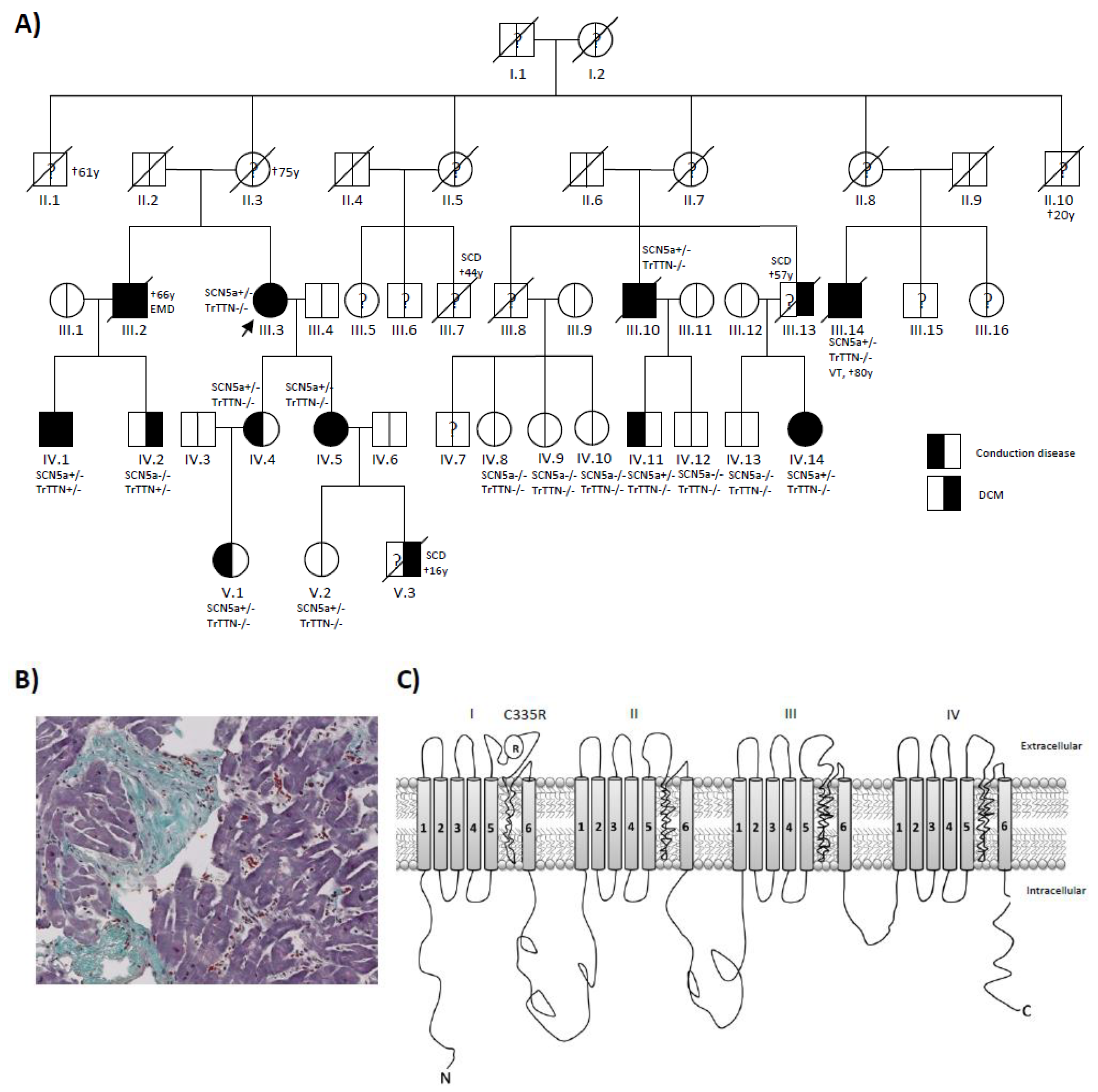
Figure 1. Identifying SCN5a p.C335R and TTN truncating variants by using precision diagnostic methods. (A) Patient’s pedigree showing co-segregation of an SCN5a C335R variant. The index patient (III. 3) was diagnosed with DCM and sick sinus syndrome at age 59 and underwent CRT-D implantation. She only carried the SCN5a C335R variant. Patient IV.1 was diagnosed with DCM and conduction disease at age 24. He carried both SCN5a and TTN truncating variants. (B) Masson Trichrome stained left ventricular endomyocardial biopsy of patient IV.1 with SCN5a and TTN truncating variants. Histopathological examination demonstrated extensive cardiac fibrosis in this patient with DCM and conduction disease. (C) Schematic representation of the Nav1.5 cardiac sodium channel. The variant is located in Ploop between S5 and S6 of Domain-I.
Ijms 22 12990 g001
Figure 2. SCN5a p.C335R carriers show conduction disease and reduced left ventricular ejection fraction (LV-EF). All family members with SCN5a p.C335R variant showed significantly longer PQ (A) and QRS intervals (B), lower left ventricular EF (C), and higher atrial fibrillation (AF) rates (D) than other family members without this variant. All 4 family members carrying SCN5a p.C335R who received CRT-D were non-responders (E,F). T-Test; * = p ≤ 0.05, ** = p ≤ 0.01. Data are presented as mean ± SD.
Ijms 22 12990 g002
Figure 3. Functional analysis of SCN5a p.C335R variant. (A) Na+ current traces from Nav1.5 wild-type and p.C335R transfected Xenopus oocytes. This showed a loss of function in Nav1.5 p. C335R. Sodium channel currents (INa) were evoked from −80 mV to 70 mV. Western blot analysis confirmed that the wild-type and mutated Nav1.5 were expressed and that loss of Nav1.5 function was not due to absent expression of the protein. Anti-Rad50 used as a reference. (B,C) Representative traces of INa in iPSC-CMs with wild-type and SCN5a p.C335R variant. Sodium channel currents (n = 9) were reduced in iPSC-CMs of the DCM patients.
Ijms 22 12990 g003
Figure 4. Disturbed sarcomeric z-disc regularity in iPSC-CMs with TTNtv. Sarcomeric structure is visualized by immunofluorescence staining against α-actinin (green) and TitinM8/M9 (red). Scale bar = 50 μm. Using fast Fourier transformation (FFT), the sarcomeric pattern regularity of the α-actinin channel was analyzed. Four cardiac differentiations for control (n = 72) and 3 cardiac differentiations for each patient-specific iPSC line (each n = 54) were analyzed. Data are presented as mean ± SEM. p < 0.001 by one-way ANOVA with Tukey’s correction. *** = p ≤ 0.001.
Ijms 22 12990 g004
Table 1. Patient characteristics of the family with SCN5a p.C335R variant.
IDAgeGenderDCMConduction DiseaseSCN5a: C335RTTNtvAge at OnsetNYHAEFRhythmOutcome
III.266MYesRBBBNANA60II15AF, PMCRT-D, †66y:electromechanical dissociation
III. 378FYesLBBB, SSS+/−−/−59III35AF, PMCRT-D
III.1079MYesLBBB, SSS+/−−/−63III30AF, PMCRT-D, †79y
III.1480MYesLBBB, SSS, AVB-III°+/−−/−64III20AF, PMVT 70y, CRT-D, ICD therapy 72 and 78y, †80y
IV.135MYesLBBB, AVB-III°+/−+/−24I48SRCPR 34y AVB-III°, PM
IV.233MYesNo−/−+/−33I45SR
IV.455FNoAV-I°+/−−/−49I55SR
IV.544FYesLAH+/−−/−44I40SR
IV.836FNoNo−/−−/−-I60SR
IV.945FNoNo−/−−/−-I59SR
IV.1044FNoNo−/−−/−-I60SR
IV.1145MNoRBBB+/−−/−45I56SR
IV.1249MNoNo−/−−/−-I60SR
IV.1364MNoNo−/−−/−-I62SR
IV.1461FYesLBBB+/−−/−57I50SR
V.119FNoAV-I°+/−−/−19I59SRx2 Syncope 22y
V.218FNoNo−/−−/−-Î60SR
AF: atrial fibrillation; AVB: atrioventricular block; CRT-D: cardiac resynchronization therapy-defibrillator; DCM: dilated cardiomyopathy; F: female; ICD: implantable cardioverter-defibrillator; LAH: left anterior hemiblock; LBBB: left bundle branch block; M: male; NA: not available; NYHA: New York Heart Association; SR: sinus rhythm; PM: pacemaker; RBBB: right bundle branch block; SSS: sick sinus syndrome; y: year; †: death.
Publisher’s Note: MDPI stays neutral with regard to jurisdictional claims in published maps and institutional affiliations.