Article

H2O2 Functions as a Downstream Signal of IAA to Mediate H2S-Induced Chilling Tolerance in Cucumber

State Key Laboratory of Crop Biology, Key Laboratory of Crop Biology and Genetic Improvement of Horticultural Crops in Huanghuai Region, College of Horticulture Science and Engineering, Shandong Agricultural University, Tai’an 271018, China
*
Author to whom correspondence should be addressed.
Int. J. Mol. Sci. 2021, 22(23), 12910; https://doi.org/10.3390/ijms222312910
Submission received: 12 October 2021 / Revised: 25 November 2021 / Accepted: 26 November 2021 / Published: 29 November 2021

Abstract

:
Hydrogen sulfide (H2S) plays a crucial role in regulating chilling tolerance. However, the role of hydrogen peroxide (H2O2) and auxin in H2S-induced signal transduction in the chilling stress response of plants was unclear. In this study, 1.0 mM exogenous H2O2 and 75 μM indole-3-acetic acid (IAA) significantly improved the chilling tolerance of cucumber seedlings, as demonstrated by the mild plant chilling injury symptoms, lower chilling injury index (CI), electrolyte leakage (EL), and malondialdehyde content (MDA) as well as higher levels of photosynthesis and cold-responsive genes under chilling stress. IAA-induced chilling tolerance was weakened by N, N′-dimethylthiourea (DMTU, a scavenger of H2O2), but the polar transport inhibitor of IAA (1-naphthylphthalamic acid, NPA) did not affect H2O2-induced mitigation of chilling stress. IAA significantly enhanced endogenous H2O2 synthesis, but H2O2 had minimal effects on endogenous IAA content in cucumber seedlings. In addition, the H2O2 scavenger DMTU, inhibitor of H2O2 synthesis (diphenyleneiodonium chloride, DPI), and IAA polar transport inhibitor NPA reduced H2S-induced chilling tolerance. Sodium hydrosulfide (NaHS) increased H2O2 and IAA levels, flavin monooxygenase (FMO) activity, and respiratory burst oxidase homolog (RBOH1) and FMO-like protein (YUCCA2) mRNA levels in cucumber seedlings. DMTU, DPI, and NPA diminished NaHS-induced H2O2 production, but DMTU and DPI did not affect IAA levels induced by NaHS during chilling stress. Taken together, the present data indicate that H2O2 as a downstream signal of IAA mediates H2S-induced chilling tolerance in cucumber seedlings.

1. Introduction

Cucumbers (Cucumis sativus L.) are typical light-loving and cold-sensitive plants, but they are mainly cultivated in solar greenhouses in northern China. When exposed to temperatures below 10 °C, cucumber plants generally suffer chilling injury (Ai et al.) [1]. Therefore, chilling is considered as a crucial limitation to growth and yield in cucumber production. Hydrogen sulfide (H2S) is a novel gaseous signaling molecule that plays an important role in regulating plant growth and development and defense responses to various abiotic stresses. Previous studies revealed that H2S upregulated the expression levels of mitogen-activated protein kinase (MAPK) and was involved in the upregulation of MAPK gene expression caused by cold stress [2]. The exogenous fumigation of H2S or application of sodium hydrosulfide (NaHS, the H2S donor), can relieve multiple abiotic stresses, such as chilling, heat, salinity, drought, hypoxia, and heavy metal toxicity [3]. We recently found that NaHS enhances the chilling tolerance of cucumber by scavenging reactive oxygen species (ROS), increasing CO2 assimilation, and upregulating the expression of cold-responsive genes [4]. Some signaling molecules, such as nitrogen monoxide (NO), Ca2+, abscisic acid (ABA), and indol-3-acetic acid (IAA) are involved in H2S-induced response to chilling stress in cucumber [4,5,6,7]. However, whether any other signaling molecules are involved in the process of H2S-induced chilling tolerance, the relationship between these signaling molecules remains unclear.
Studies over the last decades have indicated that endogenous hydrogen peroxide (H2O2) is induced in plants after exposure to abiotic stress, such as low or high temperature, heavy metals, water stress, etc. [8,9,10]. H2O2 interacts with other plant growth regulators, such as auxins, gibberellins, cytokinins, etc. (as signaling molecules) synergistically or antagonistically, and it mediates plant growth and development and responses to abiotic stresses [10]. Pasternak et al. [11] suggested that the variation of PINOID gene expression triggered by H2O2 influenced the polar transport of auxin and might alter auxin homeostasis. The application of H2O2 induced the formation of adventitious roots in Linum usitatissimum by regulating endogenous auxin levels [12]. Zhu et al. [13] demonstrated that ethylene and H2O2 play an important role in triggering brassinosteroid-induced salt tolerance in tomato plants. Our recent study suggests that H2O2 is involved in H2S-induced photoprotection in cucumber seedlings after exposure to chilling [14].
Auxin plays an essential role in the regulation of plant growth and development, but information about its role under chilling stress remains limited. Previous studies have revealed that changes in plant growth and development caused by cold stress are closely related to the intracellular auxin gradient, which is regulated by the polar deployment and intracellular trafficking of auxin transporters [15]. Recently, we found that IAA, a main auxin, could increase chilling tolerance by decreasing ROS accumulation, increasing the enzyme activities of photosynthesis and upregulating the expression of cold-responsive genes [4]. IAA also participates in the H2S-mediated response to chilling stress in cucumber, and it controls the H2O2 in the growing part of the root [16]. Therefore, we speculate that crosstalk may exist among H2O2, IAA, and H2S in response to chilling stress. To test this assumption, we investigated the effect of H2O2 and IAA on the ROS accumulation, photosynthesis, and relative expression of cold-responsive genes and the role of H2O2 and IAA in H2S-induced chilling tolerance in cucumber seedlings.

2. Results

2.1. H2O2 Is Involved in H2S-Induced Chilling Tolerance in Cucumber

To explore the effect of exogenous H2O2 on chilling tolerance in cucumber, we determined the maximum photochemical efficiency of PSII (Fv/Fm), actual photochemical efficiency of PSII (ΦPSII), chilling injury index (CI), electrolyte leakage (EL), and photosynthetic rate (Pn) of cucumber seedlings, which were pretreated with different concentrations of H2O2 after exposure to 8/5 °C (day/night) for 24–72 h. As shown in Figure 1, H2O2 alleviated chilling injury symptoms in cucumber seedlings, and this alleviation effect was increased at low concentrations of H2O2 but was suppressed when the concentration exceeded 1.0 mM. The Fv/Fm, ΦPSII, and Pn of H2O2-treated seedlings were much higher, and the CI and EL were much lower than 0 mM H2O2 (H2O) treatments. These results reveal that H2O2 improves the chilling tolerance of cucumber seedlings, and its effect is concentration dependent. Thus, we use 1.0 mM H2O2 in further experiments.
We previously demonstrated that 1.0 mM NaHS markedly increased endogenous H2O2 accumulation, and H2S-induced H2O2 plays an important role in CO2 assimilation and photoprotection in cucumber [14,17]. Consistent with previous results, we found that NaHS induced endogenous H2O2 production. However, both N, N′-dimethylthiourea (DMTU, a H2O2 scavenger) and diphenyleneiodonium chloride (DPI, a H2O2 synthesis inhibitor) markedly inhibited the H2S-induced increase in H2O2 biosynthesis and Respiratory burst oxidase homolog (RBOH1) mRNA abundance in seedlings under chilling stress (Figure 2a,b). NaHS obviously decreased the CI and EL and increased Fv/Fm and ΦPSII, but the NaHS-induced decrease in CI and EL or increase in Fv/Fm and ΦPSII in stressed seedlings were weakened by DMTU and DPI (Figure 2c–e). Therefore, we speculate that H2O2 is involved in the H2S-induced response to chilling stress.

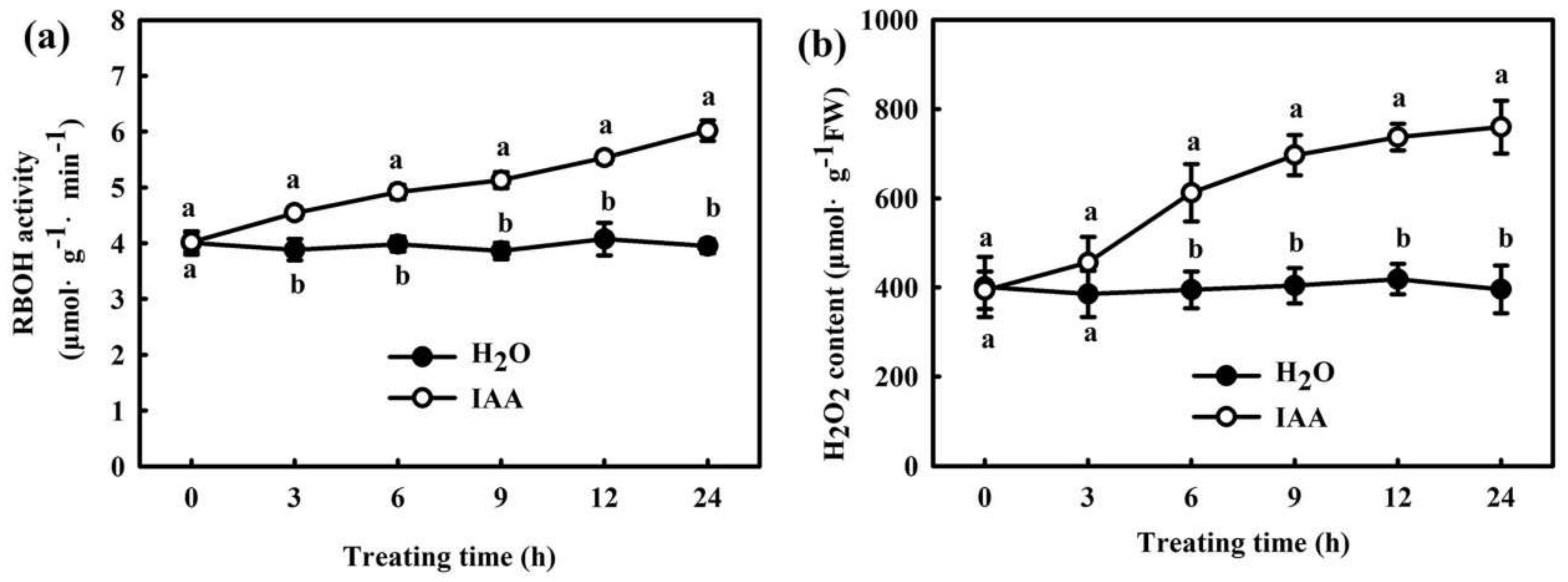
2.2. H2O2 Participates in IAA-Induced Chilling Tolerance in Cucumber

Our previous study demonstrated that IAA acts as a downstream signaling molecule and is involved in H2S-induced chilling tolerance in cucumber seedlings [4]. To explore the interactions of H2S, IAA, and H2O2 in response to chilling stress, we studied the interaction between H2O2 and IAA in the chilling stress response in cucumber. We first measured the EL, CI, and malondialdehyde (MDA) content in cucumber seedlings pretreated with 75 μM IAA, 1.0 mM H2O2, 5.0 mM DMTU + 75 μM IAA, 50 μM 1-naphthylphthalamic acid (NPA, a polar transport inhibitor of IAA) + 1.0 mM H2O2, or deionized water, after exposure to 8/5 °C for 48–72 h. Seedlings pretreated with IAA and H2O2 showed remarkably lower EL, CI, and MDA content than H2O-pretreated seedlings during chilling stress (Figure 3a–c). The decrease in EL, CI, and MDA content in IAA treatment was blocked by DMTU, but the values in H2O2 pretreated seedlings were not significantly affected by the IAA polar transport inhibitor NPA. The IAA- and H2O2 pretreated seedlings exhibited distinctly less damage caused by chilling. The effects of IAA in mitigating in chilling damage in cucumber seedlings was weakened by DMTU, but NPA had minimal effect on H2O2-induced remission of chilling damage (Figure 3d).
Then, we detected the interactive effects of IAA and H2O2 on the mRNA levels of large and small subunits (rbcL, rbcS) of ribulose 1, 5-bisphosphate carboxylase/oxygenase (rubisco) and rubisco activase (RCA), as well as the Pn, Fv/Fm and ΦPSII under chilling stress. Both IAA and H2O2 treatments revealed a marked increase in mRNA levels of rbcL, rbcS, and RCA (Figure 4a–c), and rbcL and RCA protein levels (Figure 4e), compared with H2O treatment (p < 0.05). The application of DMTU distinctly repressed IAA-induced expression of rbcL, rbcS, and RCA, but NPA did not inhibit the effect of H2O2 on rbcL, rbcS, and RCA mRNA levels. Chilling stress significantly reduced the Pn of cucumber seedlings. After chilling treatment for 24 h, the decrease in Pn in cucumber seedlings was 77.6%, 53.9%, 58.6%, 74.2%, and 61.8% in the H2O, IAA, H2O2, DMTU + IAA, and NPA+ H2O2 treatments respectively, compared to the control (Figure 4d). Figure 4f shows that Fv/Fm and ΦPSII were markedly higher in IAA- and H2O2-treated than in H2O-treated seedlings during chilling stress. The application of DMTU significantly weakened the IAA-induced increase in Fv/Fm and ΦPSII, but NPA showed a minimal influence on the H2O2-induced variation of Fv/Fm and ΦPSII. These data suggest that IAA and H2O2 mitigate the negative effect of chilling stress on the photosynthetic function by upregulating the mRNA and protein levels of the key photosynthetic enzymes and activating the photoprotective mechanism.
We also analyzed the effect of IAA and H2O2 on the relative expression of the cold responsive genes after seedlings were exposed to chilling stress for 24 h. IAA and H2O2 notably increased the mRNA levels of C-repeat-binding factor (CBF1), inducer of CBF expression (ICE1) and cold responsive (COR47) genes (Figure 5a–c) as well as CBF1 protein levels (Figure 5d) in cucumber seedlings under chilling stress. The increases in the mRNA and protein levels of the cold responsive genes in IAA-treated seedlings were dramatically weakened by DMTU, whereas those in H2O2-treated seedlings were minimally affected by NPA.
We found that 75 μM IAA remarkably increased the RBOH activity (Figure 6a) and H2O2 content (Figure 6b) in cucumber seedlings, and the increase was remarkable after treatment for 6 h. However, no remarkable differences in flavin monooxygenase (FMO) activity and IAA content were observed between H2O2- and H2O-treated seedlings (Figure 7). At normal temperature, the H2O2-treated seedlings showed similar mRNA expressions of PIN1 and AUX2 to the H2O-treated seedlings. After 9 h or 24 h of chilling stress, PIN1 and AUX2 mRNA levels markedly increased in both H2O2 and H2O treatment, but the extent of the increase did not vary and showed no significant differences between H2O2 and H2O-treated seedlings (Supplemental Figure S1). All the above results indicate that IAA affects H2O2 signaling in cucumber seedlings under chilling stress. H2O2 might play a critical role in the IAA-induced positive response to chilling stress in cucumber seedlings.

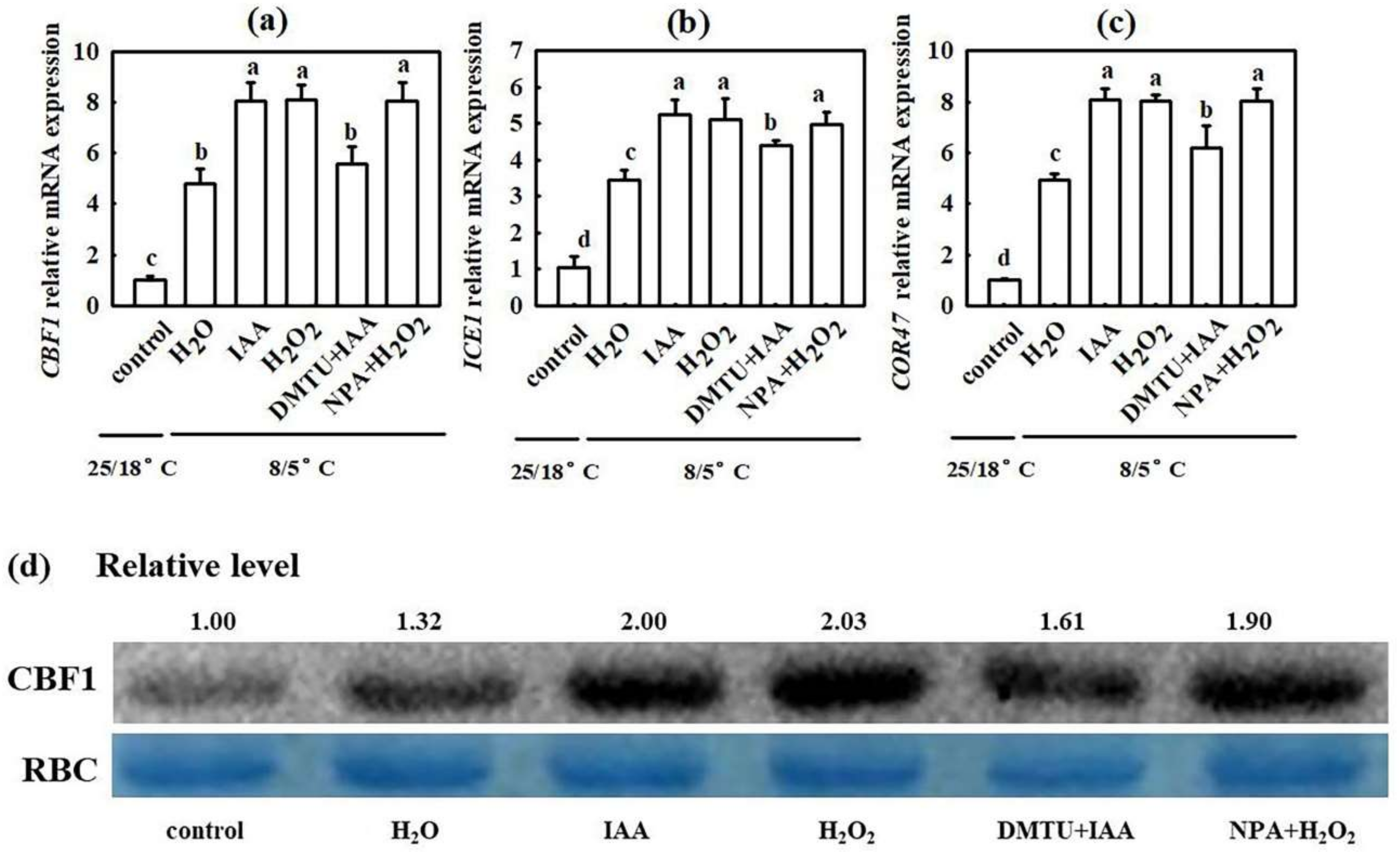
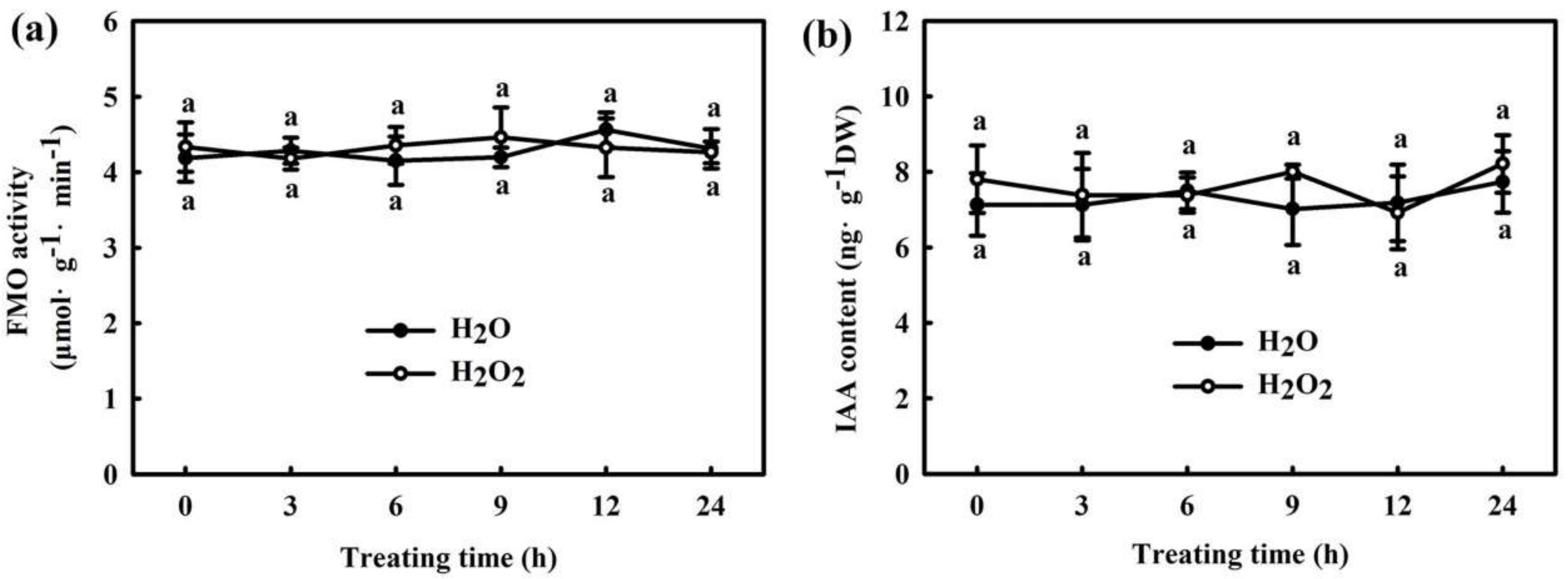
2.3. Interaction of IAA and H2O2 in H2S-Induced Chilling Tolerance in Cucumber
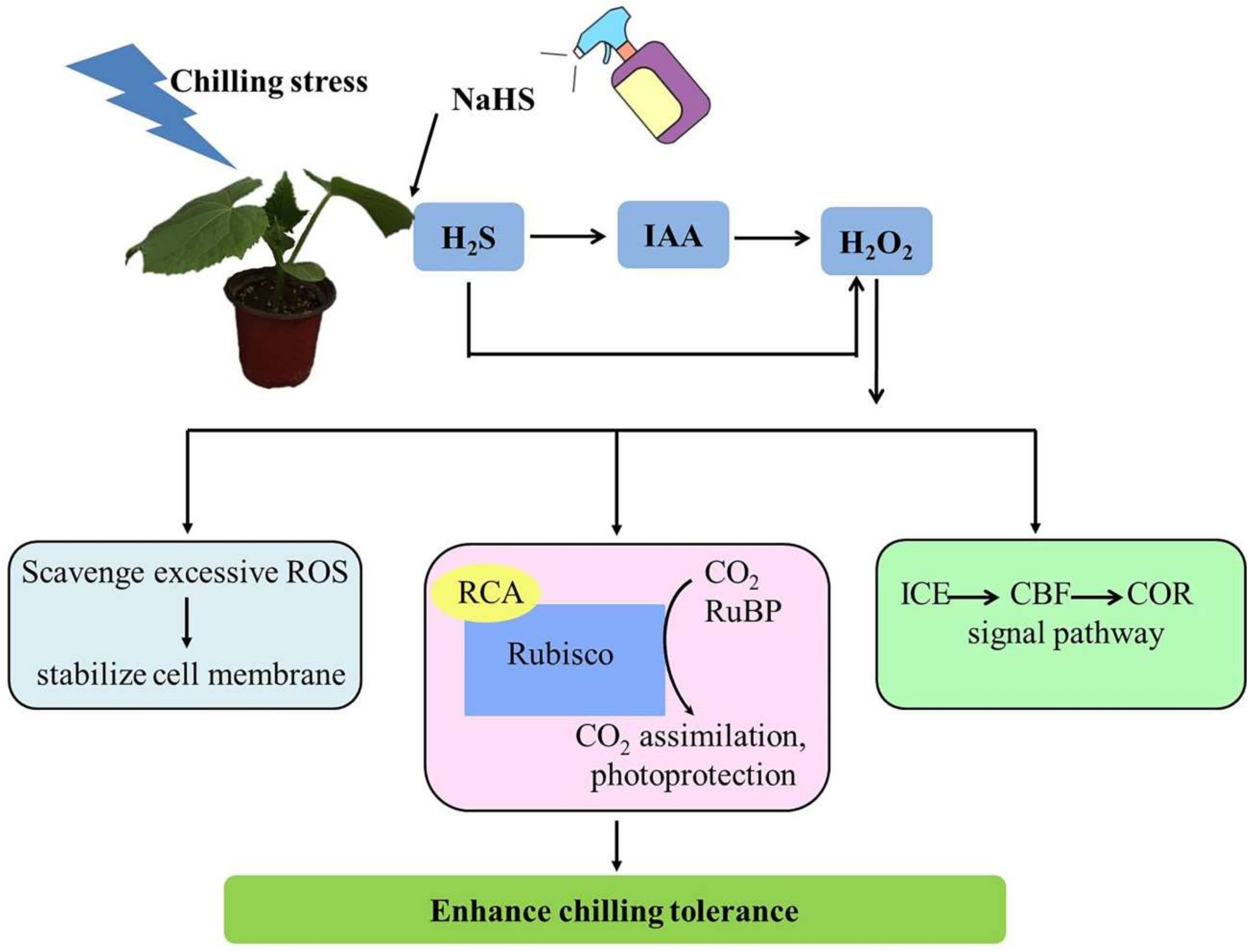
To further analyze the upstream and downstream relationship between IAA and H2O2 in H2S-mediated plant stress response, we determined the effect of NPA on H2S-induced H2O2 production and that of the H2O2 scavenger DMTU and H2O2 synthetic inhibitor DPI on H2S-induced IAA biosynthesis. As shown in Figure 8, 1.0 mM NaHS markedly increased RBOH1 mRNA abundance and H2O2 accumulation. NPA significantly inhibited the increase in RBOH1 mRNA abundance and H2O2 content induced by NaHS, suggesting that IAA is involved in H2S-induced H2O2 production. NaHS also upregulated FMO-like protein (YUCCA2) mRNA abundance, FMO activity, and IAA levels, but DMTU and DPI had minimal effects on H2S-induced IAA biosynthesis in cucumber leaves (Figure 9). Combining the results of Figure 2, it is further inferred that H2O2, a downstream component of IAA, is involved in H2S-induced chilling tolerance in cucumber seedlings.

3. Discussion

Chilling is a major abiotic stress that affects the growth, development, and geographical distribution of plants [18,19]. Low-temperature stress mainly affects light energy utilization and photosynthetic efficiency by destroying electron transport chains in chloroplasts and mitochondria, leading to ROS accumulation, and eventually inducing cell membrane damage in plants [20]. H2S, as a major gaseous transmitter, plays a critical role in plant resistance to various stress conditions, such as low temperature, salt, drought, and heavy metals [21,22]. The application of exogenous H2S can enhance chilling tolerance in Arabidopsis thaliana [1], hawthorns [23], and cucumbers [3]. ABA, NO, Ca2+, and SA are involved in H2S-induced resistance to abiotic stresses in plants [24,25,26]. Recently, we verified that H2S interacts with NO, ABA, Ca2+, IAA, and SA to enhance the chilling tolerance in cucumbers [4,5,6,7,27]. However, whether H2O2 and IAA exhibit synergistic effects on the H2S-mediated plant stress response remains unclear.
Previous studies have revealed that H2O2 is a key molecule of signal transduction and regulates various physiological metabolic processes. For example, H2O2 recruited the promoter of the senescence-related transcription factor WRKY53, which in turn activated WRKY53 transcription and led to a senescence of Arabidopsis [28]. The H2O2 response gene (HRG1/2) could quickly respond to exogenous or endogenous H2O2 and further regulated Arabidopsis seed germination [29]. Islam et al. proved that by inducing production of the reactive carbonyl species (RCS), H2O2 could induce stomatal closure of guard cells in Arabidopsis [30]. Moreover, H2O2 responds to many abiotic stress of plants. Sun et al. showed that as a signal, on the one hand, Respiratory burst oxidase homologue-dependent H2O2 (RBOH-H2O2) enhanced the heat tolerance of heat sensitive tomato. On the other hand, RBOH-H2O2 regulated the activities of antioxidant enzymes to control the total H2O2 at a level conducive to heat stress memory, which in turn maintained a lower level of total H2O2 during the future heat stress challenge [8]. H2O2 also could interact synergistically with other hormones or regulators, such as IAA, ABA, SA, MeJA, etc., mediating the response to abiotic stress in plants [10,31]. Recently, our results showed that H2O2 induced CO2 assimilation and photoprotection in cucumber seedlings during chilling stress [14]. In this study, we found that H2O2 increased the chilling tolerance of cucumber seedlings (Figure 1), suggesting that the response of H2O2 to chilling stress is consistent with previous studies. NaHS significantly enhanced H2O2 levels, RBOH1 mRNA abundance, and chilling tolerance in cucumber seedlings. The H2O2 scavenger DMTU and inhibitor of H2O2 synthesis DPI decreased H2S-induced H2O2 accumulation and chilling tolerance (Figure 2). These results indicate that H2O2 may crosstalk with H2S to improve the chilling stress response in cucumber seedlings.
Auxin is a major phytohormone that controls various aspects of plant growth and development, including cell division and elongation, tissue patterning, and the response to environmental stimuli [32,33], but knowledge about its role and interaction with other signals under chilling stress is limited. Previous investigations have indicated that chilling-induced variation in plant growth and development is closely related to the intracellular auxin gradient. Chilling stress promotes auxin biosynthesis or changes auxin gradient distribution, thus affecting the root gravity response in Arabidopsis, rice, and poplar [15,34,35,36]. Recently, we learned that NaHS increased endogenous IAA accumulation and improved chilling tolerance. The IAA polar transport inhibitor NPA suppressed H2S-induced chilling tolerance. IAA reduced the negative effects of chilling stress on growth and photosynthesis, but it showed minimal effects on endogenous H2S levels. H2S scavengers did not influence the chilling tolerance induced by IAA [4]. Here, we observed that IAA-induced chilling tolerance was repressed by the H2O2 scavenger DMTU, but the IAA inhibitor NPA did not affect H2O2–induced tolerance to chilling stress (Figure 3, Figure 4 and Figure 5). IAA significantly enhanced endogenous H2O2 synthesis, but H2O2 showed minimal effects on endogenous IAA level in cucumber seedlings (Figure 6 and Figure 7). Thus, we speculate that IAA depends on the H2O2 signaling pathway in the regulation to chilling stress response. In addition, NPA significantly decreased H2S-induced RBOH1 and H2O2 levels (Figure 8), whereas DMTU and DPI showed no marked effect on H2S-induced YUCCA2 mRNA abundance, FMO activity, or IAA levels (Figure 9). These results suggest that H2O2 lies downstream of IAA in the regulation of H2S to the chilling stress response.
Based on previous studies and the above results, we proposed a model of H2O2 and IAA regulating the H2S-mediated chilling stress response in cucumber seedlings. Figure 10 shows that endogenous H2S induced by chilling stress or the application of exogenous NaHS, H2O2, and IAA all enhanced chilling tolerance in cucumber seedlings by scavenging excessive ROS, improving photosynthetic capacity, and upregulating the mRNA and protein levels of cold responsive genes. Chilling stress-induced or exogenous H2S promotes IAA generation, and IAA further triggers H2O2 accumulation and subsequently increases chilling tolerance. Thus, H2O2 may act as a downstream signal of IAA and play a significant role in H2S-mediated chilling stress tolerance in cucumber seedlings. Further studies using advanced molecular techniques and mutants are required to better reveal the mechanisms and interactions of H2S-, IAA-, and H2O2-induced chilling tolerance in plants.
In summary, H2O2 and IAA markedly improved the chilling tolerance of cucumber seedlings, as illustrated by the decrease in stress-induced CI and EL, the increase in CO2 assimilation, and the upregulation in the level of cold-responsive genes. Even more importantly, our results first confirmed that H2O2 interacts with IAA signaling and is jointly involved in H2S-induced chilling tolerance in cucumber. Moreover, H2O2 may act as a downstream signaling molecule of IAA, and it plays a critical role in H2S-mediated chilling stress response in cucumber.

4. Materials and Methods

4.1. Plant Materials and Growth Condition

“Jinyou 35” cucumber (Cucumis sativus L.) seedlings were used in the current study. After soaking and germinating, the seeds were sown in nutrition bowls filled with seedling substrate, which consisted of peat, vermiculite, and perlite (5:3:1, v/v), and then transferred to a climate chamber with a photon flux density (PFD) of 600 μmol m−2·s−1, a 26/17 °C thermoperiod, an 11 h photoperiod, and 80% relative humidity.

4.2. Experimental Design

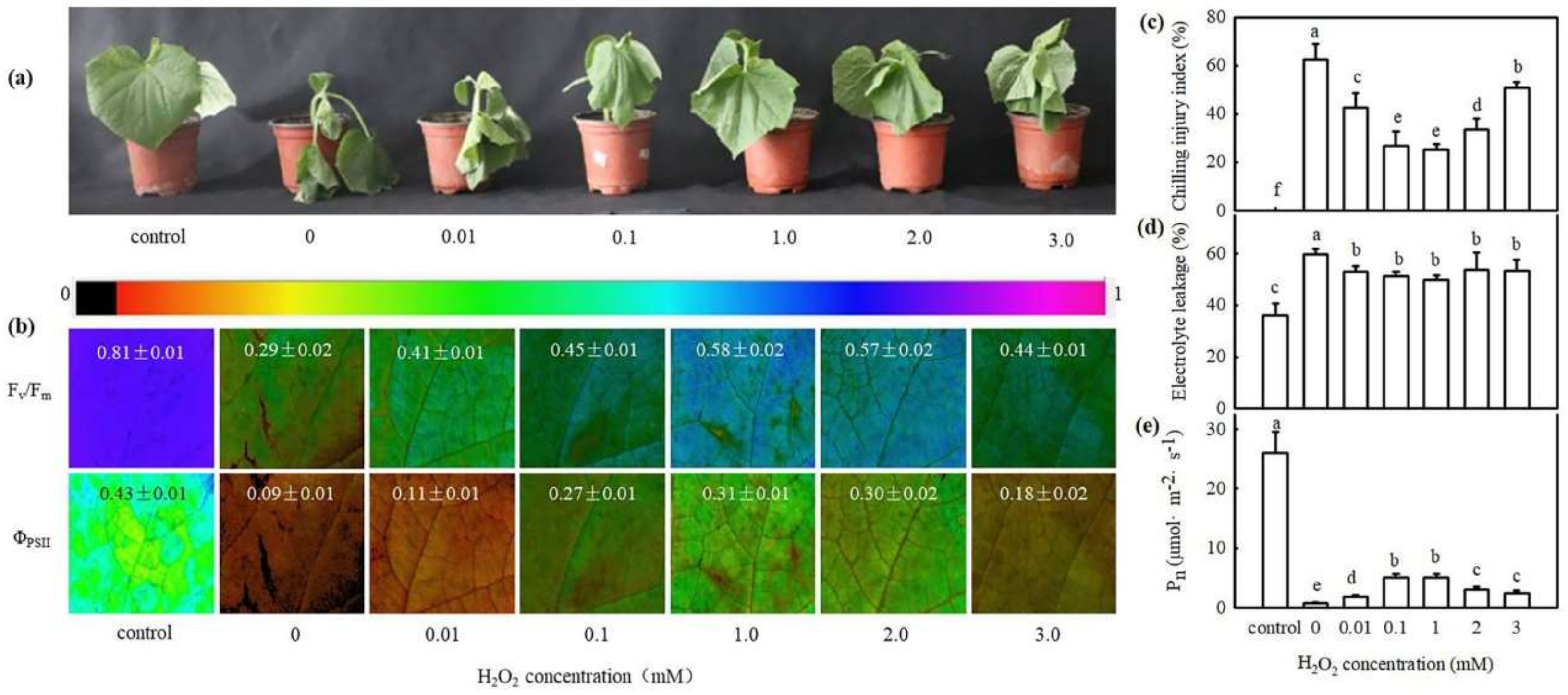
4.2.1. Effect of H2O2 on the Chilling Tolerance of Cucumber Seedlings

The seedlings with two leaves were foliar sprayed with 0 (control), 0.01, 0.1, 1.0, 2.0, and 3.0 mM H2O2, respectively. Twenty-four hours later, the pretreated seedlings were exposed to 8/5 °C to analyze the CI, EL, Pn, Fv/Fm, and ΦPSII.

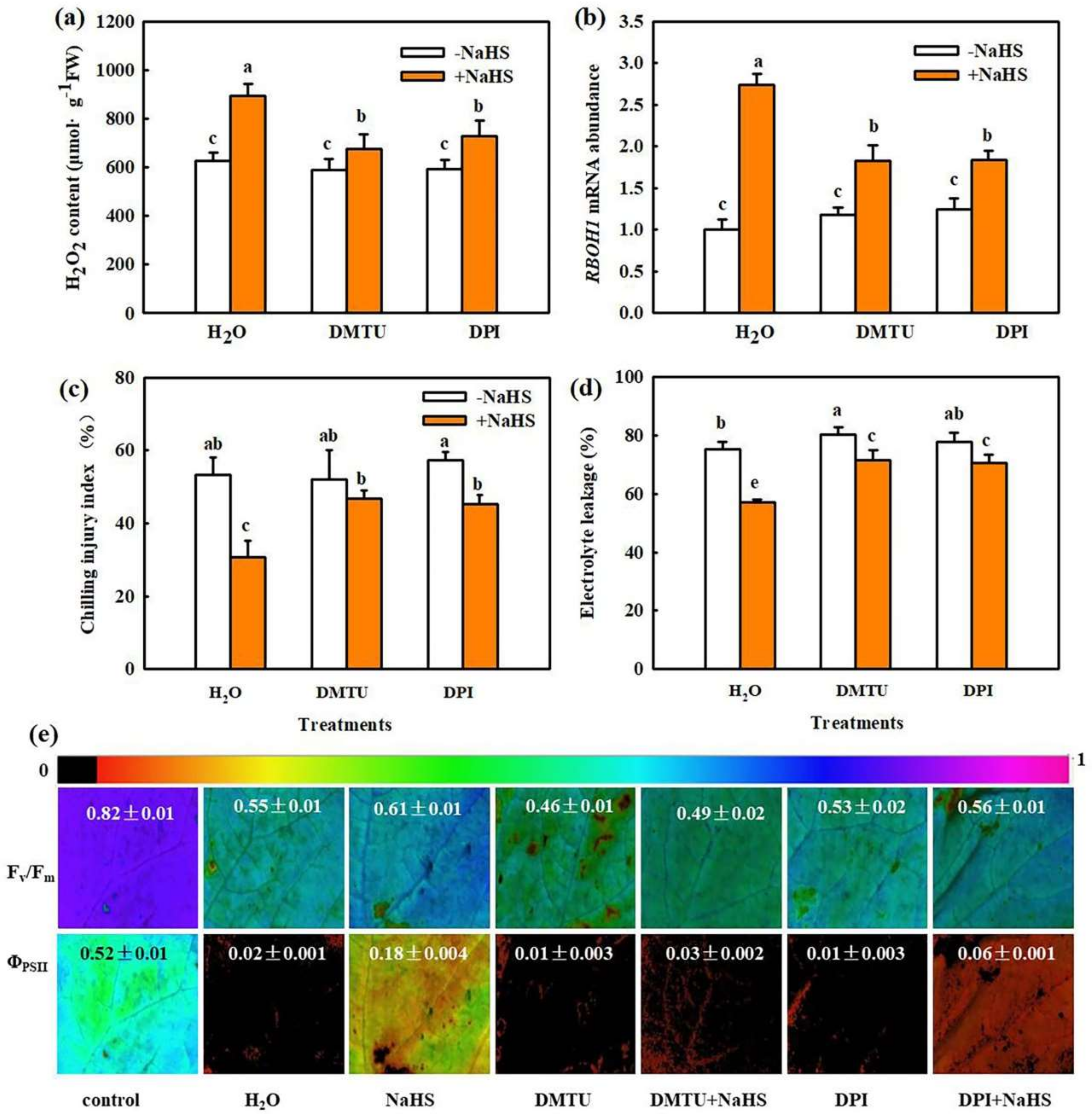
4.2.2. Effect of H2O2 Scavenger or Inhibitor on H2S-Induced H2O2 Biosynthesis and Chilling Tolerance

The seedlings were pretreated with 1.0 mM NaHS, (a H2S donor), 5.0 mM DMTU (a H2O2 scavenger), 100 μM DPI (a H2O2 synthesis inhibitor), 5.0 mM DMTU + 1.0 mM NaHS, 100 μM DPI + 1.0 mM NaHS, or deionized water (H2O). Twenty-four hours later, the pretreated seedlings were subjected to 8/5 °C for 9–72 h to assay the biosynthesis of H2O2, EL, CI, Fv/Fm, ΦPSII and relative expression of cold-responsive genes. The H2O treatment at normal temperature served as the control.

4.2.3. Interaction between IAA and H2O2 in Response to Chilling Stress

The seedlings were pretreated with 75 μM IAA, 1.0 mM H2O2, 5.0 mM DMTU + 75 μM IAA, 50 μM NPA (a polar transport inhibitor of IAA) +1.0 mM H2O2, or deionized water (H2O). At 24 h after pretreatment, the seedlings were exposed to 8/5 °C to assay the Pn, fluorescence parameters, gene expression and protein expression of key photosynthesis enzymes, and relative expression of cold-responsive genes. H2O treatment under normal conditions served as the control.

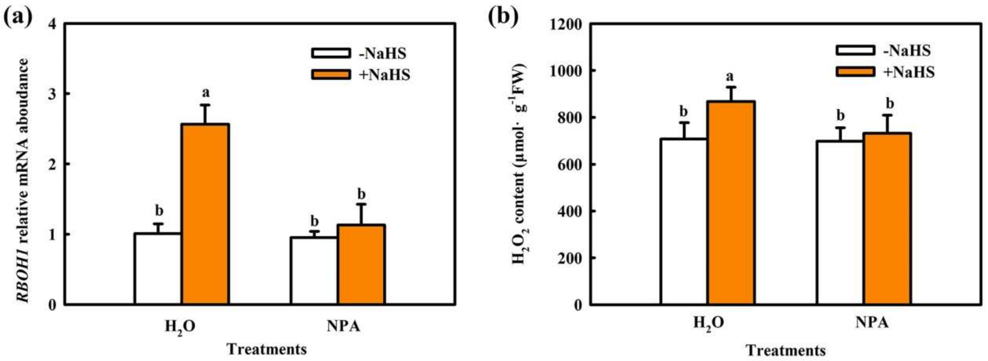
4.2.4. Effect of IAA Inhibitor on H2S-Induced H2O2 Biosynthesis in Cucumber Seedlings

The seedlings were pretreated with 1.0 mM NaHS, 50 μM NPA, 50 μM NPA + 1.0 mM NaHS, or deionized water (H2O). Twenty-four hours later, the pretreated seedlings were subjected to 5 °C for 9 h to assay the relative expression of RBOH1 and H2O2 content.

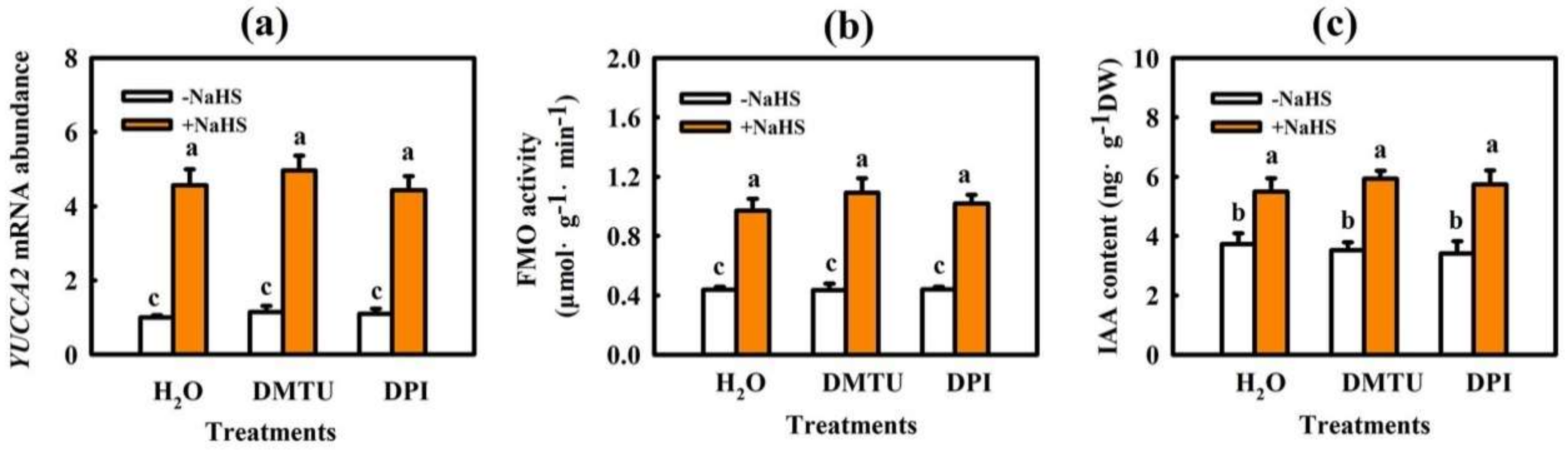
4.2.5. Effect of Scavengers and Synthetic Inhibitors of H2O2 on H2S-Induced IAA Biosynthesis in Cucumber Seedlings

The seedlings were pretreated with 1.0 mM NaHS, 5.0 mM DMTU, 100 μM DPI, 5.0 mM DMTU + 1.0 mM NaHS, 100 μM DPI + 1.0 mM NaHS, or deionized water (H2O). Twenty-four hours later, the pretreated seedlings were subjected to 5 °C for 9 h to assay the FMO activity, YUCCA2 mRNA abundance, and IAA content.

4.3. CI, EL, and MDA Measurements

The chilling stressed cucumber seedlings were graded based on the Semeniuk et al. [37] standard, and the CI was calculated according to the following formulas: CI = Σ (plants of different grade × grade)/[total plants × 5 (the maximum grade)].
EL was measured as described by Dong et al. [38]. Leaf discs (0.2 g) were incubated at 25 °C in 20 mL deionized water for 3 h, and the electrical conductivity (EC1) was estimated using a conductivity meter (DDB-303A, Shanghai, China). Afterwards, the leaf discs were boiled for 30 min and then cooled to detect EC2. EL was calculated according to the following formula: EL= EC1/EC2 × 100.
MDA content was determined using the thiobarbituric acid (TBA) colorimetric method as described by Heath and Packer [39].

4.4. Detection of Pn and Chlorophyll Fluorescence

The Pn was determined using a portable photosynthetic system (Ciras-3, PP-systems International, Hitchin, Hertfordshire, UK). Constant PFD (600 μmol·m−2·s−1), CO2 concentration (380 mg·L−1), and leaf temperature (25 ± 1 °C) were maintained during the assessment. Fv/Fm was measured after seedlings were dark-adapted for 45 min, and the ΦPSII was determined after leaves were light-adapted for 30 min using a portable pulse-modulated fluorometer (FMS-2, Hansatech, King’s Lynn, Norfolk, UK). The chlorophyll fluorescence parameters were calculated according to Demmig-Adams and Adams [40] and Maxwell [41] as follows: Fv/Fm = (Fm−F0)/Fm′; ΦPSII = (Fm′−Fs)/Fm′. Chlorophyll fluorescence imaging was visualized using a chlorophyll fluorescence imaging system (Imaging PAM, Walz, Wurzburg, Germany) with a computer-operated PAM-control system [42].

4.5. Detection of H2O2 Content and RBOH Activity

H2O2 content was determined using an H2O2 kit (Nanjing Jiancheng Bioengineering Institute, Nanjing, China). RBOH activity was detected with an ELISA kit (Jiangsu Meimian Industrial Co. Ltd., Yancheng, China) according to the instructions.

4.6. IAA Content and FMO Activity Assay

IAA content was determined by the method of Zhang et al. [4]. In brief, 0.3 g sample ground with liquid nitrogen and extracted thrice with 80% methanol (containing 30 μg·mL−1 sodium diethyldithiocarbamate). Samples were centrifuged (7155 g, 10 min, 4 °C) to obtain the supernatant by rotary evaporation (Shanghai EYELA, N-1210B, Shanghai, China) at 38 °C. The residue was washed with 5 ml of PBS (pH = 8, 0.2 M) and 4 mL of trichloromethane, shaken for 20 min, and allowed to stand for 30 min to remove pigment present in the trichloromethane. The resulting residues were added to 0.15 g of polyvinylpolypyrrolidone (PVPP) to remove phenols. Then, the samples were centrifuged at 7155 g for 10 min, and the resulting supernatant was re-extracted with ethyl acetate thrice and dried with a rotary evaporator in vacuo at 36 °C. The dried material was dissolved in 1.0 mL mobile phases (methanol: 0.04% acetic acid = 45:55, v/v), and the filtrate was used for HPLC–MS (Thermo Fisher Scientific, TSQ Quantum Access, San Jose, CA, USA) analysis followed by the method of Zhang et al. [4].
Flavin monooxygenase (FMO) activity was estimated using an ELISA kit (Jiangsu Meimian Industrial Co. Ltd., Yancheng, China). In brief, the FMO1 antibody was conjugated with standard, sample, and horseradish peroxidase (HRP)-labeled detection antibody and incubated, aspirated, and washed. Then, chromogen solution was added, and the reaction was terminated with sulfuric acid solution. The absorbance was detected at 450 nm with a microplate reader, and FMO activity was calculated using the standard curve [4].

4.7. Quantitative Real-Time PCR Analysis

Total RNA was extracted from cucumber leaves using an RNA extraction kit (TRIzol; Tiangen, Beijing, China). The isolated RNA was reverse transcribed with the PrimeScript® RT Master Mix Perfect Real Time (TaKaRa, Dalian, China). qRT-PCR was performed using the TransStart® TipTop Green q-PCR SuperMix (Cwbio, Beijing, China). The relative expression levels were standardized to those of cucumber β-actin gene (Solyc11g005330). The qRT-PCR primers are shown in Supplemental Table S1.

4.8. SDS-PAGE and Immunoblot Analysis

The extracted total protein of samples was separated using a 10% SDS-PAGE gel, and the resulting proteins were transferred to polyvinylidene difluoride (PVDF) membranes. The PVDF membrane was blocked for 2 h with 5% (w/w) skimmed milk and then incubated with the primary antibody followed by a horseradish peroxidase-conjugated anti-rabbit IgG antibody (ComWin Biotech Co., Ltd., Beijing, China) for 2 h. Finally, the immunoreaction was tested using the eECL Western blot Kit (CW00495, ComWin Biotech Co., Ltd., Beijing, China) and the ChemiDoc™ XRS imaging system (Bio-Rad Laboratories, Inc., Hercules, CA, USA). Primary antibodies against RbcL and RCA (ATCG00490, AT2G39730) were obtained from PhytoAB Co. Ltd. (San Francisco, CA, USA), and the CBF1 antibody was obtained from GenScript Co., Ltd. (Nanjing, China).

4.9. Statistical Analysis

The whole experiment was performed in triplicate, and the results shown are the mean ± standard deviation (SD). Statistical analysis was performed with DPS software, and the comparison of treatments was based on the analysis of variance by Duncan’s multiple range test (DMRT) at a significance level of 5% (p < 0.05).

Supplementary Materials

The following are available online at https://www.mdpi.com/article/10.3390/ijms222312910/s1.

Author Contributions

X.A. and X.Z. designed the experiment and wrote the paper. X.Z. performed the research and analyzed the data. Y.Z., C.X., K.L. and H.B. worked together with X.Z. to accomplish the experiment. All authors have read and agreed to the published version of the manuscript.

Funding

This research was funded by the National Science Foundation of China (31572170); the Major Science and Technology Innovation of Shandong Province in China (2019JZZY010715); the Special Fund of Modern Agriculture Industrial Technology System of Shandong Province in China (SDAIT-05–10); and the Funds of Shandong “Double Tops” Program (SYL2017YSTD06).

Institutional Review Board Statement

Not applicable.

Informed Consent Statement

Not applicable.

Data Availability Statement

The original contributions presented in the study are included in the article, further inquiries can be directed to the corresponding author.

Conflicts of Interest

The authors agreed with the final manuscript and have no conflicts of interest.

References

  1. Goldstein, I.; Chastre, J.; Rouby, J.-J. Novel and Innovative Strategies to Treat Ventilator-Associated Pneumonia: Optimizing the Duration of Therapy and Nebulizing Antimicrobial Agents. Semin. Respir. Crit. Care Med. 2006, 27, 082–091. [Google Scholar] [CrossRef]
  2. Du, X.; Jin, Z.; Liu, D.; Yang, G.; Pei, Y. Hydrogen sulfide alleviates the cold stress through MPK4 in Arabidopsis thaliana. Plant Physiol. Biochem. 2017, 120, 112–119. [Google Scholar] [CrossRef] [PubMed]
  3. Banerjee, A.; Tripathi, D.K.; Roychoudhury, A. Hydrogen sulphide trapeze: Environmental stress amelioration and phytohormone crosstalk. Plant Physiol. Biochem. 2018, 132, 46–53. [Google Scholar] [CrossRef]
  4. Zhang, X.-W.; Liu, F.-J.; Zhai, J.; Li, F.-D.; Bi, H.-G.; Ai, X.-Z. Auxin acts as a downstream signaling molecule involved in hydrogen sulfide-induced chilling tolerance in cucumber. Planta 2020, 251, 69. [Google Scholar] [CrossRef] [PubMed]
  5. Wu, G.X.; Cai, B.B.; Zhou, C.F.; Li, D.D.; Bi, H.G.; Ai, X.Z. Hydrogen sulfide-induced chilling tolerance of cucumber and involvement of nitric oxide. J. Plant Biol. Res. 2016, 5, 58–69. [Google Scholar]
  6. Wu, G.X.; Li, D.D.; Sun, C.C.; Sun, S.N.; Liu, F.J.; Bi, H.G.; Ai, X.Z. Hydrogen sulfide interacts with Ca2+ to enhance chilling tolerance of cucumber seedlings. Chin. J. Biochem. Mol. Biol. 2017, 33, 1037–1046. [Google Scholar] [CrossRef]
  7. Li, D.D.; Zhang, X.W.; Liu, F.J.; Pan, D.Y.; Ai, X.Z. Hydrogen sulfide interacting with abscisic acid counteracts oxidative damages against chilling stress in cucumber seedlings. Acta Hortic. Sin. 2018, 45, 2395–2406. [Google Scholar] [CrossRef]
  8. Sun, M.; Jiang, F.; Zhou, R.; Wen, J.; Cui, S.; Wang, W.; Wu, Z. Respiratory burst oxidase homologue-dependent H2O2 is essential during heat stress memory in heat sensitive tomato. Sci. Hortic. 2019, 258, 108777. [Google Scholar] [CrossRef]
  9. Nobakht, P.; Ebadi, A.; Parmoon, G.; Bahrami, R.N. Study role H2O2 in Photosynthetic pigments Peppermint (Mentha piperita L) on Water stress conditions. J. Plant Proc. Func. 2019, 7, 19–30. [Google Scholar]
  10. Nazir, F.; Fariduddin, Q.; Alam Khan, T. Hydrogen peroxide as a signalling molecule in plants and its crosstalk with other plant growth regulators under heavy metal stress. Chemosphere 2020, 252, 126486. [Google Scholar] [CrossRef] [PubMed]
  11. Pasternak, T.; Potters, G.; Caubergs, R.; Jansen, M. Complementary interactions between oxidative stress and auxins control plant growth responses at plant, organ, and cellular level. J. Exp. Bot. 2005, 56, 1991–2001. [Google Scholar] [CrossRef]
  12. Takáč, T.; Obert, B.; Rolčík, J.; Šamaj, J. Improvement of adventitious root formation in flax using hydrogen peroxide. New Biotechnol. 2016, 33, 728–734. [Google Scholar] [CrossRef] [PubMed]
  13. Zhu, T.; Deng, X.; Zhou, X.; Zhu, L.; Zou, L.; Li, P.; Zhang, D.; Lin, H. Ethylene and hydrogen peroxide are involved in brassinosteroid-induced salt tolerance in tomato. Sci. Rep. 2016, 6, 35392. [Google Scholar] [CrossRef] [Green Version]
  14. Liu, F.; Fu, X.; Wu, G.; Feng, Y.; Li, F.; Bi, H.; Ai, X. Hydrogen peroxide is involved in hydrogen sulfide-induced carbon assimilation and photoprotection in cucumber seedlings. Environ. Exp. Bot. 2020, 175, 104052. [Google Scholar] [CrossRef]
  15. Rahman, A. Auxin: A regulator of cold stress response. Physiol. Plant 2013, 147, 28–35. [Google Scholar] [CrossRef]
  16. Ivanchenko, M.G.; Os, D.D.; Monshausen, G.B.; Dubrovsky, J.G.; Bednarova, A.; Krishnan, N. Auxin increases the hydrogen peroxide (H2O2) concentration in tomato (Solanum lycopersicum) root tips while inhibiting root growth. Ann. Bot. 2013, 112, 1107–1116. [Google Scholar] [CrossRef] [Green Version]
  17. Liu, F.; Zhang, X.; Cai, B.; Pan, D.; Fu, X.; Bi, H.; Ai, X. Physiological response and transcription profiling analysis reveal the role of glutathione in H2S-induced chilling stress tolerance of cucumber seedlings. Plant Sci. 2020, 291, 110363. [Google Scholar] [CrossRef]
  18. Zhou, J.; Wang, J.; Shi, K.; Xia, X.J.; Zhou, Y.H.; Yu, J.Q. Hydrogen peroxide is involved in the cold acclimation-induced chilling tolerance of tomato plants. Plant Physiol. Biochem. 2012, 60, 141–149. [Google Scholar] [CrossRef] [PubMed]
  19. Ma, X.; Chen, C.; Yang, M.; Dong, X.; Lv, W.; Meng, Q. Cold-regulated protein (SlCOR413IM1) confers chilling stress tolerance in tomato plants. Plant Physiol. Biochem. 2018, 124, 29–39. [Google Scholar] [CrossRef]
  20. Fan, J.; Hu, Z.; Xie, Y.; Chan, Z.; Chen, K.; Amombo, E.; Chen, L.; Fu, J. Alleviation of cold damage to photosystem II and metabolisms by melatonin in Bermudagrass. Front. Plant Sci. 2015, 6, 925. [Google Scholar] [CrossRef] [Green Version]
  21. Huang, D.; Huo, J.; Liao, W. Hydrogen sulfide: Roles in plant abiotic stress response and crosstalk with other signals. Plant Sci. 2021, 302, 110733. [Google Scholar] [CrossRef]
  22. Arif, Y.; Hayat, S.; Yusuf, M.; Bajguz, A. Hydrogen sulfide: A versatile gaseous molecule in plants. Plant Physiol. Biochem. 2021, 158, 372–384. [Google Scholar] [CrossRef] [PubMed]
  23. Aghdam, M.S.; Mahmoudi, R.; Razavi, F.; Rabiei, V.; Soleimani, A. Hydrogen sulfide treatment confers chilling tolerance in hawthorn fruit during cold storage by triggering endogenous H 2 S accumulation, enhancing antioxidant enzymes activity and promoting phenols accumulation. Sci. Hortic. 2018, 238, 264–271. [Google Scholar] [CrossRef]
  24. Khan, M.N.; Siddiqui, M.H.; AlSolami, M.A.; Alamri, S.; Hu, Y.; Ali, H.M.; Al-Amri, A.A.; Alsubaie, Q.D.; Al-Munqedhi, B.M.; Al-Ghamdi, A. Crosstalk of hydrogen sulfide and nitric oxide requires calcium to mitigate impaired photosynthesis under cadmium stress by activating defense mechanisms in Vigna radiata. Plant Physiol. Biochem. 2020, 156, 278–290. [Google Scholar] [CrossRef] [PubMed]
  25. Kour, J.; Khanna, K.; Sharma, P.; Singh, A.D.; Sharma, I.; Arora, P.; Kumar, P.; Devi, K.; Ibrahim, M.; Ohri, P.; et al. Hydrogen sulfide and phytohormones crosstalk in plant defense against abiotic stress. In Hydrogen Sulfide in Plant Biology; Singh, S., Singh, V.P., Tripathi, D.K., Prasad, S.M., Chauhan, D.K., Dubey, N.K., Eds.; Elsevier: Amsterdam, The Netherlands, 2021; pp. 267–302. [Google Scholar] [CrossRef]
  26. Chen, J.; Zhou, H.; Xie, Y. SnRK2.6 phosphorylation/persulfidation: Where ABA and H2S signaling meet. Trends Plant Sci. 2021, 26, 1207–1209. [Google Scholar] [CrossRef] [PubMed]
  27. Pan, D.-Y.; Fu, X.; Zhang, X.-W.; Liu, F.-J.; Bi, H.-G.; Ai, X.-Z. Hydrogen sulfide is required for salicylic acid–induced chilling tolerance of cucumber seedlings. Protoplasma 2020, 257, 1543–1557. [Google Scholar] [CrossRef] [PubMed]
  28. Lin, W.; Huang, D.; Shi, X.; Deng, B.; Ren, Y.; Lin, W.; Miao, Y. H2O2 as a Feedback Signal on Dual-Located WHIRLY1 Associates with Leaf Senescence in Arabidopsis. Cells 2019, 8, 1585. [Google Scholar] [CrossRef] [PubMed] [Green Version]
  29. Gong, F.; Yao, Z.; Liu, Y.; Sun, M.; Peng, X. H2O2 response gene 1/2 are novel sensors or responders of H2O2 and involve in maintaining embryonic root meristem activity in Arabidopsis thaliana. Plant Sci. 2021, 310, 110981. [Google Scholar] [CrossRef]
  30. Islam, M.; Ye, W.; Matsushima, D.; Rhaman, M.S.; Munemasa, S.; Okuma, E.; Nakamura, Y.; Biswas, S.; Mano, J.; Murata, Y. Reactive Carbonyl Species Function as Signal Mediators Downstream of H2O2 Production and Regulate [Ca2+]cyt Elevation in ABA Signal Pathway in Arabidopsis Guard Cells. Plant Cell Physiol. 2019, 60, 1146–1159. [Google Scholar] [CrossRef] [PubMed]
  31. Li, H.; Guo, Y.; Lan, Z.; Xu, K.; Chang, J.; Ahammed, G.J.; Ma, J.; Wei, C.; Zhang, X. Methyl jasmonate mediates melatonin-induced cold tolerance of grafted watermelon plants. Hortic. Res. 2021, 8, 57. [Google Scholar] [CrossRef]
  32. Brumos, J.; Robles, L.M.; Yun, J.; Vu, T.C.; Jackson, S.; Alonso, J.; Stepanova, A.N. Local Auxin Biosynthesis Is a Key Regulator of Plant Development. Dev. Cell 2018, 47, 306–318.e5. [Google Scholar] [CrossRef] [PubMed] [Green Version]
  33. Lv, B.; Yan, Z.; Tian, H.; Zhang, X.; Ding, Z. Local Auxin Biosynthesis Mediates Plant Growth and Development. Trends Plant Sci. 2019, 24, 6–9. [Google Scholar] [CrossRef] [PubMed]
  34. Shibasaki, K.; Uemura, M.; Tsurumi, S.; Rahman, A. Auxin Response inArabidopsisunder Cold Stress: Underlying Molecular Mechanisms. Plant Cell 2009, 21, 3823–3838. [Google Scholar] [CrossRef] [PubMed] [Green Version]
  35. Popko, J.; Hänsch, R.; Mendel, R.-R.; Polle, A.; Teichmann, T. The role of abscisic acid and auxin in the response of poplar to abiotic stress. Plant Biol. 2010, 12, 242–258. [Google Scholar] [CrossRef] [PubMed]
  36. Du, H.; Liu, H.; Xiong, L. Endogenous auxin and jasmonic acid levels are differentially modulated by abiotic stresses in rice. Front. Plant Sci. 2013, 4, 397. [Google Scholar] [CrossRef] [Green Version]
  37. Chen, B.; Wu, J. Coupling and Decoupling between Range and Angle. Synth. Impulse Aperture Radar (SIAR) 2014, 111, 241–257. [Google Scholar] [CrossRef]
  38. Dong, X.; Bi, H.; Wu, G.; Ai, X. Drought-induced chilling tolerance in cucumber involves membrane stabilisation improved by antioxidant system. Int. J. Plant Prod. 2013, 7, 67–80. [Google Scholar]
  39. Heath, R.L.; Packer, L.J. Photoperoxidation in isolated chloroplasts. i. kinetics and stoichiometry of fatty acid peroxidation. Arch. Biochem. Biophys. 1968, 125, 189–198. [Google Scholar] [CrossRef]
  40. Demmig-Adams, B.; Adams, W.W. Xanthophyll cycle and light stress in nature: Uniform response to excess direct sunlight among higher plant species. Planta 1996, 198, 460–470. [Google Scholar] [CrossRef]
  41. Maxwell, K.; Johnson, G.N. Chlorophyll fluorescence—A practical guide. J. Exp. Bot. 2000, 51, 659–668. [Google Scholar] [CrossRef]
  42. Tian, Y.; Ungerer, P.; Zhang, H.; Ruban, A.V. Direct impact of the sustained decline in the photosystem II efficiency upon plant productivity at different developmental stages. J. Plant Physiol. 2017, 212, 45–53. [Google Scholar] [CrossRef] [PubMed]
Figure 1. Effect of H2O2 on the chilling tolerance of cucumber seedlings. (a) Phenotype characterization of cucumber seedlings pretreated with H2O2 or deionized water under chilling stress (8/5 °C) for 48 h. Deionized water-treated seedlings before chilling stress were used as the control. The experiments were repeated three times with similar results. A typical picture is shown here. (b) Image of Fv/Fm and ΦPSII in seedlings before (control) and after chilling stress for 24 h. The false color code depicted at top of the image represents the degree of photoinhibition at PSII. (c) CI of seedlings before (control) and after chilling stress for 72 h. (d) EL of seedlings before (control) and after chilling stress for 48 h. (e) Pn of seedlings before (control) and after chilling stress for 24 h. Two-leaf stage cucumber seedlings were foliage sprayed with 0, 0.01, 0.1, 1.0, 2.0 and 3.0 mM H2O2 solution for 24 h and subsequently were exposed to 8/5 °C (day/night). The data represent the mean ± SD of three biological replicates. Different letters indicate significant differences (p < 0.05), according to Duncan’s new multiple range test.
Ijms 22 12910 g001
Figure 2. Effects of DMTU and DPI on H2S-induced H2O2 content, RBOH1 mRNA abundance, and chilling tolerance in cucumber. (a) H2O2 content in seedlings before (control) and after chilling stress for 9 h; (b) mRNA abundance of RBOH1 in seedlings before (control) and after chilling stress for 9 h. (c) CI of seedlings before (control) and after chilling stress for 72 h; (d) EL of seedlings before (control) and after chilling stress for 48 h; (e) Image of Fv/Fm and ΦPSII of seedlings before (control) and after chilling stress for 24 h. The false color code depicted at top of the image represents the degree of photoinhibition at PSII. Two-leaf stage cucumber seedlings were pretreated with DMTU, DPI, or deionized water and then sprayed with NaHS after 6 h. Twelve hours later, the seedlings were exposed to chilling stress. The data represent mean ± SD of three biological replicates. Different letters indicate significant differences (p < 0.05), according to Duncan’s new multiple range test.
Ijms 22 12910 g002
Figure 3. Interactive effects of IAA and H2O2 on the chilling tolerance of cucumber seedlings. Cucumber seedlings were pretreated with 75 μM IAA, 1.0 mM H2O2, 5.0 mM DMTU + 75 μM IAA, 50 μM NPA +1.0 mM H2O2, or deionized water (control) for 24 h and subsequently were exposed to chilling (8/5 °C, day/night). (a) EL of seedlings before (control) and after chilling stress for 48 h. (b) CI of seedlings before (control) and after chilling stress for 72 h. (c) MDA content of seedlings before (control) and after chilling stress for 48 h; (d) Phenotype characterization of different treatments before (control) and after chilling stress for 48 h. Deionized water-treated seedlings before chilling stress were used as the control. The experiments were repeated three times with similar results. A typical picture is shown here. The data represent the mean ± SD of three biological replicates. Different letters indicate significant differences (p < 0.05), according to Duncan’s new multiple range test.
Ijms 22 12910 g003
Figure 4. Interactive effects of IAA and H2O2 on mRNA abundances of rbsL, rbcS, and RCA, and protein levels of rbsL and RCA in cucumber seedlings under chilling stress. (ac) mRNA abundances of rbcL, rbcS, and RCA; (d) Pn; (e) Protein levels of rbcL and RCA; (f) Image of Fv/Fm and ΦPSII. The false color code depicted at top of the image represents the degree of photoinhibition at PSII. Cucumber seedlings were pretreated with 75 μM IAA, 1.0 mM H2O2, 5.0 mM DMTU +75 μM IAA, 50 μM NPA +1.0 mM H2O2, or deionized water (control) for 24 h, and subsequently exposed to 5 °C for 24 h. The data represent the mean ± SD of three biological replicates. Different letters indicate significant differences (p < 0.05), according to Duncan’s new multiple range test.
Ijms 22 12910 g004
Figure 5. Interactive effects of IAA and H2O2 on the level of cold responsive genes in cucumber seedlings under chilling stress. (ac) mRNA abundances of CBF1, ICE1, and COR47, respectively; (d) CBF1 protein level. Cucumber seedlings were pretreated with 75 μM IAA, 1.0 mM H2O2, 5.0 mM DMTU +75 μM IAA, 50 μM NPA +1.0 mM H2O2, or deionized water (control) for 24 h and subsequently exposed to 5 °C for 24 h. The data represent the mean ± SD of three biological replicates. Different letters indicate significant differences (p < 0.05), according to Duncan’s new multiple range test.
Ijms 22 12910 g005
Figure 6. Effect of IAA on the RBOH activity (a) and H2O2 accumulation (b) in cucumber seedlings. Cucumber seedlings were foliar sprayed with 75 μM IAA or deionized water (control), and then, we measured the changes of RBOH activity and H2O2 content within 24 h. The data represent the mean ± SD of three biological replicates. Different letters indicate significant differences (p < 0.05), according to Duncan’s new multiple range test.
Ijms 22 12910 g006
Figure 7. Effects of H2O2 on the FMO activity (a) and IAA content (b) in cucumber seedlings. Cucumber seedlings were foliar sprayed with 1.0 mM H2O2 or deionized water (control), and then, we measured the changes of FMO activity and IAA content within 24 h. The data represent the mean ± SD of three biological replicates. Different letters indicate significant differences (p < 0.05), according to Duncan’s new multiple range test.
Ijms 22 12910 g007
Figure 8. Effect of NPA on H2S-induced RBOH1 mRNA abundance (a) and H2O2 accumulation (b) in cucumber seedlings. Cucumber seedlings were pretreated with 50 μM NPA or deionized water and then sprayed with 1.0 mM NaHS after 6 h. Twelve hours later, the seedlings were exposed to 5 °C for 9 h. The data represent the mean ± SD of three biological replicates. Different letters indicate significant differences (p < 0.05), according to Duncan’s new multiple range test.
Ijms 22 12910 g008
Figure 9. Effect of DMTU and DPI on H2S-induced IAA production in cucumber seedlings. (a) YUCCA2 mRNA abundance; (b) FMO activity; (c) IAA accumulation. Cucumber seedlings were pretreated with 5.0 mM DMTU, 100 μM DPI, or deionized water and then sprayed with NaHS after 6 h. Twelve hours later, the seedlings were exposed to 5 °C for 9 h. The data represent the mean ± SD of three biological replicates. Different letters indicate significant differences (p < 0.05), according to Duncan’s new multiple range test.
Ijms 22 12910 g009
Figure 10. A proposed model for the role of IAA and H2O2 in H2S-induced chilling tolerance in cucumber. Chilling induces the accumulation of H2S in the plants. H2S induced by chilling promotes IAA generation, triggers H2O2 accumulation, and subsequently increases chilling tolerance by scavenging excessive ROS, improving CO2 assimilation and photoprotection, and upregulating the levels of cold-responsive genes.
Ijms 22 12910 g010
Publisher’s Note: MDPI stays neutral with regard to jurisdictional claims in published maps and institutional affiliations.

