Review

Functional Studies of Plant Latex as a Rich Source of Bioactive Compounds: Focus on Proteins and Alkaloids

Molecular Virology Research Unit, Institute of Experimental Biology, Faculty of Biology, Adam Mickiewicz University, Poznań, Uniwersytetu Poznańskiego 6, 61-614 Poznań, Poland
*
Author to whom correspondence should be addressed.
Int. J. Mol. Sci. 2021, 22(22), 12427; https://doi.org/10.3390/ijms222212427
Submission received: 15 October 2021 / Revised: 11 November 2021 / Accepted: 13 November 2021 / Published: 17 November 2021

Abstract

:
Latex, a sticky emulsion produced by specialized cells called laticifers, is a crucial part of a plant’s defense system against herbivory and pathogens. It consists of a broad spectrum of active compounds, which are beneficial not only for plants, but for human health as well, enough to mention the use of morphine or codeine from poppy latex. Here, we reviewed latex’s general role in plant physiology and the significance of particular compounds (alkaloids and proteins) to its defense system with the example of Chelidonium majus L. from the poppy family. We further attempt to present latex chemicals used so far in medicine and then focus on functional studies of proteins and other compounds with potential pharmacological activities using modern techniques such as CRISPR/Cas9 gene editing. Despite the centuries-old tradition of using latex-bearing plants in therapies, there are still a lot of promising molecules waiting to be explored.

1. Introduction

Latex-bearing plants have a long history of benefiting human health and medicinal use in many different regions and cultures all over the world. Recent research suggests that the opium poppy (Papaver somniferum L.) was already in the process of domestication at the end of 4th millennium BC [1] and early domesticated ancestors of Cannabis sativa L. diverged ~10,000 years BC [2]. Those are two leading examples of laticiferous plant species used for therapies and together with Hevea brasiliensis Muli. Arg., which is the main and irreplaceable natural rubber source, have the best known and described latex composition. These complex fluids consist of different secondary metabolites, like terpenes, alkaloids, or phenolics, and jointly with a broad range of proteins are the first line of plant herbivore defense system. Another extensively studied laticiferous medicinal plant is Greater Celandine (Chelidonium majus L.), a relative of the opium poppy, which is a rich source of numerous biologically active compounds, used in traditional folk medicine as antiviral, antibacterial, antifungal, choleretic, and anticancer agents [3,4,5,6]. Many compounds of latex are active in both eukaryotic and prokaryotic organisms [7]. At present, when mankind is running out of antibiotics and other antimicrobial compounds, new cancer therapies are still needed, and to make matters worse, the scale of pest and microbial resistance is increasing. Thus, the exploration of such rich natural deposits of active molecules is a very promising research direction.
This review paper characterizes the main components of latex and summarizes their known potentially therapeutic activities. We focus on two classes of compounds, proteins and alkaloids, which represent complex macromolecules and low-molecular compounds. Both types of molecules co-exist in plant latex and possibly actively cooperate in a synergistic manner to enable and boost their biological activities. We propose a model of antiviral latex activity and present examples of CRISPR/Cas9 editing genomes, which can shed light on a complex network of specialized metabolites synthesis and interactions or complementation. Despite its long history of use, there is still room for improvement of agronomic traits in domesticated latex-bearing plants and for the exploitation of wild species latex compounds to prepare a range of novel compounds of therapeutic potential, as well as novel drugs and drug carriers.

2. Diversity and Role of Latex in Plant Physiology

Latex is a milky emulsion produced by complex secretory structures called laticifers. It is defined as a suspension of various particles (organic and inorganic) dispersed in a liquid with different refractive index. Depending on prevalent content and plant species studied, it can be milky white or yellowish, orange to brown or even colorless. However, it is more than a liquid. It is identified as a laticifer’s protoplast with mitochondria, plastids, endoplasmic reticulum, Golgi bodies, polyribosomes, and vacuoles [8]. Laticifers are latex-producing, highly specialized plant cells or connected cells, which are spread through the whole plant body in the form of linear tubes which can grow and elongate with plant organs. They can be found in almost every part of latescent plants, namely in the root, stem, leaf, sepal, petal, stamen, ovary, and stigma, or they can occur only in some particular organs. Latex occupies the whole volume of the laticifer system [9].
Latex was identified in at least 20,000 plant species belonging to 43 families of vascular plants. Most of them are Angiosperms (41 families), one family belongs to ferns, and one to gymnosperms [8,9,10]. In a great example of convergent evolution events in the plant kingdom, latex occurrences take place several times in phylogenetically unrelated orders. Laticifers developed in both monocotyledonous and dicotyledonous, in the basal clades (Ana-grade), magnoliids, monocots, basal eudicots, rosids, and asterides [11,12]. One morphotype of laticifers, articulated, which are fused chains of cells with intact, porous, or even absent terminal walls, form laticiferous vessels and were recorded in 27 families. The other main morphotype is called non-articulated, which occurs more rarely and is formed by a single plant cell with almost infinite growth potential. Both types of laticifers can extend not only longitudinally with the growth of organs, but also radically create branched networks of tubes. For the proper classification of the laticifer system, it is essential to use plant material with embryos or meristems. Only based on ontology can articulated and non-articulated morphotypes be distinguished (analysis of number of precursor cells and phase of laticifers development often coupled with analysis of laticifers enzymatic activity of pectinases and chitinases, with the latter active only in articulated laticifers [13]). Previously, cases of incorrect assignment to the appropriate laticifer types were described, e.g., for mulberry [14,15] or for Ficus montana Burm.f. and Maclura tinctoria L. [13]. Attempts were made to use types of laticifers as a diagnostic tool for some taxa, but it is more likely that different morphotypes will be found in different species within the same family [11].
Nowadays, it is well established that the biological role of latex is plant defense against herbivores and pathogens [10], but in 1989 Webster and Baulk concluded that the function of latex was unknown [16]. Many latex metabolites are stored within large vacuoles and are released after being physically damaged at the site of injury. Some of them act as toxic and dissuasive components. After mechanical disruption of plant tissue, latex is immediately released and is the first line of plant defense. Thanks to its inherent stickiness and coagulation properties, latex forms a barrier against pathogen invasion. Moreover, latex’s rapid coagulation and high viscosity can restrict herbivore movements, as well as immobilize mouthparts and other sense organs [17]. This strategy gives an advantage to latescent plants, especially in environments with high a herbivory rate, like tropical or subtropical forests [18].

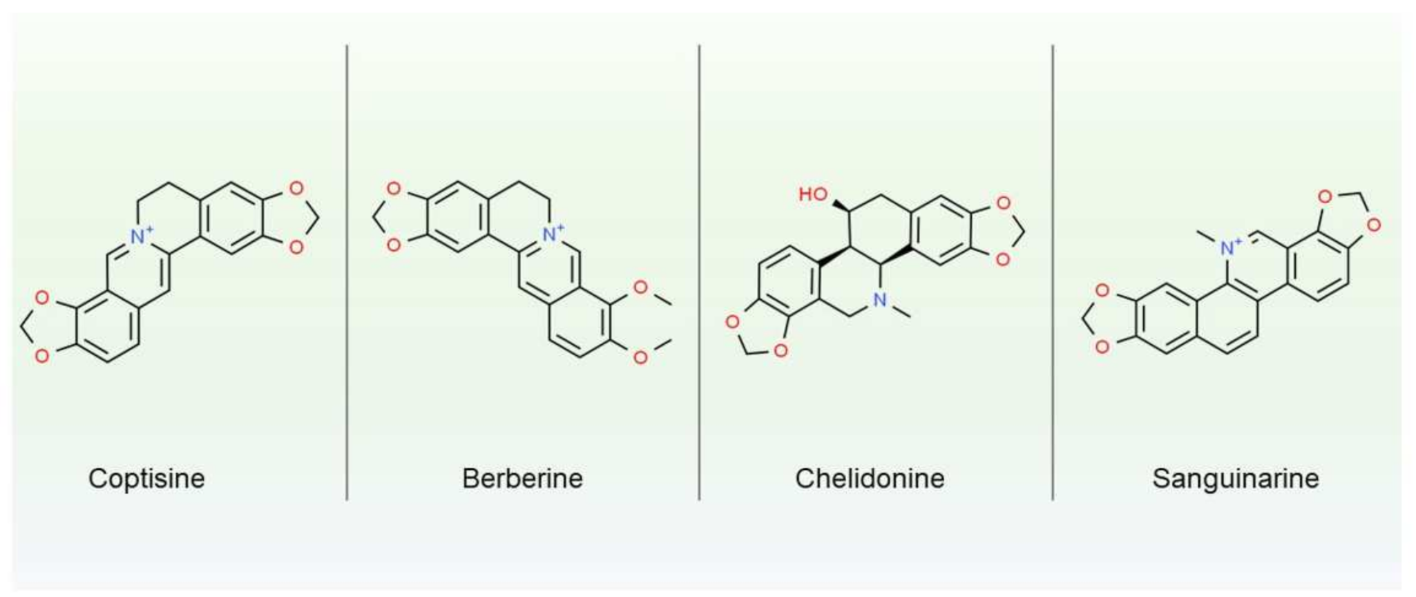
3. Main Components of Latex-Secondary Metabolites

Taking into consideration the defense role of latex in plant development, it should not be surprising how complex and diverse the latex composition can be. Despite tremendous variability in latex components, which is dependent on the species, phase of development, external and internal stimuli, and stresses [19], two major groups of biologically active compounds can be distinguished, namely secondary metabolites and proteins. Many of those products are cytotoxic and it was suggested that laticifers evolved as sequestering compartments, which ensure the storage of such substances regardless of the vascular system. This solution provides a unique and preformed defense mechanism with almost immediate response to herbivory attack. The internal pressure of latex causes the secretion of concentrated active substances at the point of damage in a few seconds. In contrast, an inducible defense system needs hours or even days to synthesize and collect sufficient amounts of active substances to act against pathogens [20]. In the context of latex composition, it is worth noticing that a synergetic mode of action was established for some of its constituents, like terpenes associated with phenolic compounds [20,21] or different proteins exhibiting defense functions against insects or fungi [22,23]. Therapeutic properties of selected latex compounds are described in detail in Section 5. Below, we present a short characterization of secondary metabolites common for latex-bearing plants.
Secondary metabolites are a heterogeneous group of chemical compounds not essential to vegetative growth, but for plant adaptation to changes in the external as well as internal environment. As mentioned before, the presence of specific metabolites in latex is a highly species-specific trait, but in general terpenes, phenolics, alkaloids, and cardenolides are present in most of the laticifer types.
One of the most abundant groups of secondary metabolites in plant latex are isoprene-derived compounds, terpenes. Within this group, the most economically important member is rubber (cis-1,4-polyisoprene), which is found in 2500 plant species (300 plant genera from eight families) [7,24], but harvested on global scale from one, Hevea brasiliensis species. Rubber particles may constitute up to 50% of H. brasiliensis latex volume [25]. It is proposed that the main function of rubber is related to the coagulation process [20], but it was also suggested that rubber biosynthesis accumulate excess of photosynthate and prevent damage to photosynthesis apparatus under stress conditions [26].
Apart from rubber, in latex of Euphorbia genus, some triterpenoids (i.e., cycloartenol, 24-methylenecycloartenol, lupeol, lupeol acetate, lanosterol, and 24-methylenelanosterol) are always present in higher concentration than in other organs. Similarly, latex of Lactuca sativa L. contains high levels of several sesquiterpene lactones (concentration of lactucopicrin oxalate is 1000 higher than in leaves) [27]. Those terpenoids are often accompanied with steroids. It is presumed that together they can disrupt cell walls of insects or microorganisms, intercalate membranes, and form channels which enable the migration of small, toxic, hydrophilic molecules such as phenolics inside the cell [28].
Phenolics (e.g., tannins, lignans, coumarins, flavonoids), another broad group of secondary metabolites found in latex, have been known for their antioxidant properties and taking part in response to oxidative stress conditions [29]. They are mainly products of shikimate pathways and were found, among others, in latex of sweet potato Ipomoea batatas L. The overall concentration of p-coumarate esters exceeded 3% fresh vine latex and 10% root latex of the variety “Jewel”. The presence of those phenolics is inversely correlated with the acceptability of sweet potato by weevils [30], which supports the anti-herbivore latex function. Different polyphenol compounds were identified in latex of H. brasiliensis, namely gallic acid, naphthoic acid, quercetin, chlorogenic acid, and rutin, which are also postulated to play a role in plant defense system [31]. In common dandelion (Taraxacum officinale F.H. Wigg.), phenolic esters, next to sesquiterpene lactones and triterpene acetates, were found in high concentration in the main root. Those active compounds showed a repelling effect in experiments with Diabrotica balteata larvae [32].
Another group of secondary metabolites sequestered in laticifers are alkaloids. Those amino acid derivatives, which are highly bioactive and often toxic, serve eco-physiological functions in plants, providing better fitness to specific environmental niches [33]. Alkaloids for thousands of years have been used and abused by humans, even leading to military conflicts (like opium wars in the 19th century or ongoing drug wars in many countries). Those low molecular compounds were found amongst 35 families, mostly angiosperms, including Apocynaceae, Papaveraceae, and Moracea [8]. The best known and described example of laticifers rich in alkaloids is opium, namely the dried latex of P. somniferum used in folk and traditional medicine, as well as psychedelic drugs. Opium contains at least 20 alkaloids, such as morphine, papaverine, and codeine. Morphine may constitute up to 5% of fresh latex, and codeine up to 1% [34]. Latex of Chelidonium majus, a species closely related to P. somniferum, is also a rich source of bioactive alkaloids, e.g., chelidonine, sanguinarine, berberine or coptisine (chemical structures are depicted on Figure 1). Those isoquinoline alkaloids can reach up to 20% of fresh latex mass [35] and are known for their multiple pharmacological effects (antioxidant, anti-inflammatory, anticancer, anti-neurodegenerative, and antimicrobial). Although C. majus is related to P. somniferum, its latex does not contain morphine-like alkaloids, such as morphine or codeine, and therefore does not have sedative effects. Yet, some of the C. majus secondary metabolites, like berberine and chelidonine, as well as protein enriched extracts can have analgesic effects, similar to morphine [36]
Cardenolides, a specific type of steroids (cardiac glycoside) consisting of sugar, steroid, and lactone, are the next group of metabolites commonly occurring in latex. There is no known function of those compounds, other than defense. Cardenolides are inhibitors of Na+/K+-ATPases, which are essential to maintain cell electrical potential, regulate cellular volume, and take part in transmembrane transport. As inhibitors cardiac glycosides are remarkably toxic to most animals, therefore latex of Antiaris toxicaria Lesch. (Moraceae) rich in cardenolides, were used by South Asia tribes as a poison on darts during hunting [37]. It is worth mentioning the widely known interaction between cardenolides from Asclepias spp. and monarch butterflies (Danaus plexippus). Monarch larvae, which feed on plants containing cardenolides, have developed the ability to sequestrate those compounds, which then cumulate in wings and protect them from bird predators [38,39].

4. Spectrum of Latex Proteins

Laticifers are not only a reservoir system for low-molecular weight defense compounds, but as mentioned earlier, constitute a living cell, and as one, has a distinct proteome. A wide range of both constitutive and inducible proteins are present in latex, with huge diversity between different plant species. Comprehensive studies of 1208 latex proteins from 20 various latex-bearing plants led to the identification of 887 non-redundant proteins from three main species, i.e., L. sativa, H. brasiliensis, and P. somniferum. Only 11 proteins were found in all three species [40], which represents well-illustrated variability within latex proteomes. In a set of 887 proteins, GO enrichment analysis showed that response to chemical and abiotic stimuli categories were highly overrepresented. Within this category, at least 21 proteins are involved in response to cadmium ion stress, while another 13 proteins were associated with response to high salt conditions. Proteins with function in defense were also identified. Amongst them two proteins, glycosyl hydrolase superfamily proteins (AT4G16260) and the NAD(P)H subunit NDH-N (AT5G58260), were pinpointed for their response to fungi infections. In the category of cellular components, most proteins were located in cytoplasm, but terms related to intracellular membrane-bounded organelles (including plastids and mitochondria) were highly enriched as well. Cho and colleagues also compared latex and phloem proteomes. GO terms common for those two conductive systems were related to metabolic pathways like the metabolism of nitrogen compounds, amine, alcohol, hexose, carboxylic acid, and carbohydrate catabolism. Stress response related terms were shared in laticifers and phloem systems, but for example, osmotic stress GO terms were identified only in latex. On the other hand, GO terms connected with response to zinc were found only in phloem [40]. Despite some similarities with functionally related conduit systems, laticifers present a unique and distinct set of proteins. Nevertheless, we can distinguish some common protein functional groups prevalent in different laticiferous plant species, like proteases, protease inhibitors, lectins, oxidases, chitinases, or defense-related proteins. Below, we survey those common for latex protein’s groups.
The most frequently reported latex proteins are proteases. One of the best described examples of protease in laticiferous plants is papain, a cysteine protease in latex of the Papaya tree (Carica papaya L.). Papain is an enzyme practically used as a meat tenderizer and in the cosmetic industry. In papaya latex, its concentration is 200 times higher than in leaf tissue, which leads to 20 times higher papain activity [41]. Experiments with protease inhibitors, E-64 and larvae of Eri silkworm, give clear evidence of papain involvement in plant resistance against herbivory (leaf toxicity to Eri silkworm was lost after covering its surface with E-64). Similarly, ficin (cysteine protease from fig) inhibition by E-64 makes fig leaf edible for insects [41]. Nonetheless, the exact site of action and mechanism of protease toxicity remains unclear. Other groups of proteases-serine proteases-were found in Moraceae, Euphorbiaceae, Apocynaceae, and Convolvulaceae families. Rarely are those two protease classes (cysteine and serine) identified in the latex of the same species. More often, one type of peptidases is described for a particular plant. Known exceptions are species from the Plumeria genus, where both proteolytic mechanisms have been reported [42].
In the latex of many plants, not only were proteases identified, but protease inhibitors as well. Those compounds bind to proteases and prevent the digestion of proteins, thus causing a shortage of amino acids and impairing the growth and development of aggressors [8]. Protease inhibitors belong to group 6 of Pathogenesis-Related (PR) proteins and their role in plant defense in species without latex is well established [43]. In laticiferous species Ficus carica L., the expression level of trypsin inhibitor increases significantly after wounding and jasmonic acid treatment [43,44]. Moreover, in papaya latex, trypsin inhibitor (together with class-II chitinase and a glutaminyl cyclase) was one of the compounds accumulated after mechanical damage [45].
Lectins are proteins with one or more domains which enable them to recognize and bind to specific sugar structure (in free form or as a part of glycoproteins and glycolipids). They are compounds necessary for the perception of possible invasion by recognizing specific glycans at damage sites and from pathogens. Several types of lectins were identified in the families Euphorbiaceae, Moraceae and Apocynaceae. Hevein, the major latex protein from H. brasiliensis, is responsible for rubber agglutination, by bridging rubber particles after the recognition of 22 kDa glycoprotein receptor [46]. This small 43 amino acid protein with lectin domain turned out to be the main contact allergen from natural rubber [46,47]. In contrast to indirect mechanisms of latex coagulation, there are studies reporting a straight negative impact of lectins on pests from different families, e.g., Lepidoptera, Coleoptera, Diptera and Hemiptera [18]. It was shown that the expression of exogenous lectins in genetically engineered plants led to various detrimental effects in invading insects, ranging from a severe delay in development to high mortality rates. Moreover, the introduction of specific lectins to the insect diet negatively affects pest performance, as reviewed in [48]. The toxicity of lectins depends on the presence of specific carbohydrates in the insect body, which, in turn, is correlated with insects’ developmental stage [49,50].
Latex from plants belonging to families Euphorbiaceae, Moraceae, and Anacardiaceae shows oxidase activity [51]. Polyphenol oxidase (PPO) and peroxidase (POD) are the most commonly occurring peptides. Both enzymes are known for their role in plant defense, not only in lactiferous plant species. Polyphenol oxidase catalyzes the oxidation of phenols to o-quinones, which are highly reactive and secondary non-enzymatic reactions lead to formation of polymers with protein functional groups. At least three mechanisms of PPO toxicity to insects were proposed: decreased nutritive value of leaf proteins, oxidative burst in insect gut, and direct toxicity of PPO catalyzed products [52]. Products of PPO activity and polymerization are black, brown, or red in color and are responsible for the darkening of latex after contact with air. Peroxidases on the other hand are H2O2 scavengers–they catalyze oxidation of phenolic substrates using H2O2 as an electron acceptor. Subsequent reactions lead to cross-linking products of phenolic compounds, such as lignin or suberin [52,53]. The ability to reduce ROS makes those enzymes a crucial part of the antioxidant system, which is often triggered in response to both biotic and abiotic stress conditions. As a downstream result of peroxidase activity, the lignification of the plant cell walls occurs, which inhibits, for example, heavy metal entry [54].
Chitinases hydrolyze the β-1,4-glycosidic bonds of chitin, which builds fungi cell walls and insects exoskeletons and peritrophic matrix. Chitinases are classified as pathogenesis related proteins (PR proteins) and are expressed inducible or constitutively in tissues vulnerable to pathogen attack, like laticifers. Several ways of direct chitinase involvement in antifungal defense were established. Plant chitinases can inhibit hyphal growth. The overexpression of those enzymes in transgenic plants increased pathogen resistance in vivo. Meanwhile, products of chitin breakdown, via phytoalexins action, to induce systemic defense response [55]. Insecticidal effects constitute another well documented chitinase activity. For example, after the addition of two chitinases from mulberry (Morus sp.) to the Drosophila melanogaster larvae diet, 80% were found to be dead [56]. Chitinases identified from Calotropis procera (Aiton) W.T.Aiton affected larval survival and weight, mean developmental time and emergence of adults [23]. The mechanism of chitinase’s action on insects is not well established. It is postulated that it may involve the hydrolysis of chitin present in their peritrophic membranes, which are responsible for protection against mechanical damage and invasion by microorganisms or parasites. After its destruction, fatal infection can develop [57,58]. Moreover, in latex, chitinases present multiple isoforms. Three isoforms were purified and characterized from Ficus microcarpa latex [59], six basic chitinases were identified in H. brasiliensis latex [60] and at least 15 isoforms in latex of C. procera [61]. Different isoforms exhibit differences in sequences as well as in post-translational modification. Particular isoforms may in consequence differ in structural and biological properties, which need to be included in further studies regarding plant chitinases [61].
As mentioned before, chitinases belong to the group of pathogenesis related proteins. All PR proteins (as the name suggests) take part in plant defense mechanisms, but they are a very diverse group in the context of structures and activities. They were categorized into 17 distinct families (summarized in Table 1). For instance in C. majus latex representatives of 12 families were identified [62,63]. Two quantitatively predominant families were PR-9 (peroxidases described previously) and major latex proteins (MLPs), which are homologs of PR-10 proteins (ribonuclease-like/Bet v1 protein family). Similarly, in latex of P. somniferum, those two families were also overrepresented. In another well studied laticiferous plant, H. brasiliensis, members of 9 PR families were described [8]. PR proteins are low molecular weight and are induced by phytopathogens as well as defense-related signaling molecules, like salicylic and jasmonic acid. Due to their mode of action in biotic and abiotic stress conditions, they are one of the most promising targets for engineering multiple stress tolerant varieties [8,63].
One of the most interesting latex proteins is a major latex protein (MLP). It was discovered for the first time in the latex of the opium poppy (P. somniferum) in 1980′s by Nessler et al. Despite its abundance (up to 50% of soluble P. somniferum latex sub proteome) shown using SDS-PAGE and the presence of laticifer-specific peptides, its function remained unknown [64,65,66]. MLPs, being homologs of opium poppy major latex protein, have also been found in other non-latex-bearing plants, namely peach fruits (Prunus persica L. Batsch, cv Springcrest) [67], Arabidopsis thaliana L. [68], ripening kiwi fruit [69], Panax ginseng C.A. Meyer [70], tobacco (Nicotiana tabacum L.) [71], pepper (Capsicum annuum L.) [72], and cotton [73]. Recent data have shown the functional role of MLPs resembling the function of PR-10 proteins family [67], member of which are involved in the defense of plants against different pathogens and participate in plant metabolism [74,75,76]. The first proposed ribonuclease activity was demonstrated in major birch pollen allergen, Bet v1 which expresses homology to PR-10-like protein from white lupin (Lupinus albus L.) [77]. The most distinctive feature of Bet v1 protein superfamily (Pfam: PF00407) is a large solvent accessible hydrophobic cavity which might potentially function as a site to bind ligands [78]. Both MLPs and PR-10 families are members of Bet v1 superfamily although their sequence similarity is low and is below 25% [68]. Major latex protein/ripening-related proteins (MLP/RRP) subfamily is the second largest subfamily within the plant kingdom. It has 60 members, from which 31 are present in A. thaliana. The biological function of MLPs is still unknown, but there are assumptions that they are associated with development of fruit and flower along with response to stress and defense [78]. One of the recent studies has investigated the potential antiviral activity of MLP proteins against potato virus Y (PVY). It is transmitted by aphids and can cause mosaic, dwarfism, mottle, deformities, and even lead to necrosis in tobacco plants. MLP-like protein 28 (NbMLP28) from Nicotiana benthamiana Domin. was identified and cloned. Its expression profile has shown responsiveness towards PVY infection and defense-related signaling molecules, such as JA, SA, and ET. Virus-induced silencing of NbMLP28 made plants more susceptible to infection by PVY, though the transient overexpression of NbMLP28 gene improved resistance towards PVY in tobacco plants. The pathway responsible for modulation of the expression of NbMLP28 gene in N. benthamiana has also been identified. It showed the cis-acting elements in response to JA, light, auxin, drought, and endosperm expression to be present in the promoter sequence of NbMLP28 [79]. Antiviral properties towards TMV-P0 virus have been proven for CaPR10 proteins from hot pepper which have inhibited the viral penetration and/or replication. This study showed that CaPR10 after inoculation with the virus is phosphorylated and functions as RNase cleaving viral RNA [71]. During the studies on melon phloem-sap proteome, it was also found that major latex protein was present in the sap collected from the cucumber mosaic virus (CMV)-MP-expressing plants and CMV-infected plants [80]. Proteomic studies of C. majus latex have shown that MLP is highly overrepresented in C. majus latex [78] and can be seen at different stages of plant development till the fruit ripening. It is accumulated early in laticifer development and persists till maturity [78]. C. majus MLP (CmMLP) is composed of 147 amino acids, has a molecular weight of 16.77 kDa and theoretical pI 5.88, which corresponds to the typical sizes of MLPs (17 kDa) in other plants [74]. MLPs bind hormones and other metabolites with their conserved hydrophobic cavity during plant growth and development [81]. MLPs from other species, including A. thaliana, N. benthamiana, or Cucumis melo L., as mentioned previously, have also been proved to be involved in antiviral response. This can help to explain the potential antiviral activity of C. majus among its other biomedical properties [82] and could serve as a potential molecular target to be used in pharmacology or medicine and to improve the defense potential of agriculturally important crops against viral and non-viral pathogens.
Table 1. Summary of PR proteins previously identified in latex-bearing plants.
PR ProteinsFunctionLatex-Bearing Plant SpeciesReference
PR 2β-1,3-glucanasesChelidonium majus
Hevea brasiliensis
[78,83]
PR 3Class I, II, IV, V, VI, VII ChitinasesChelidonium majus
Hevea brasiliensis
[78,83]
PR 4Class I, II ChitinasesChelidonium majus
Hevea brasiliensis
Carica papaya
[45,78,83]
PR 5Thaumatin-like proteinsChelidonium majus
Hevea brasiliensis
[78,83]
PR 6Proteinase inhibitorHevea brasiliensis
Ficus carica
Carica papaya
[44,45,83]
PR 7EndoproteinaseChelidonium majus
Hevea brasiliensis
[78,83]
PR 8Class III ChitinaseHevea brasiliensis[83]
PR 9PeroxidaseChelidonium majus
Hevea brasiliensis
Papaver somniferum
[78,83,84]
PR 10Ribonuclease-like proteinsChelidonium majus
Papaver somniferum
[78,84]
PR 11Class I ChitinaseChelidonium majus[78]
PR 12DefensinChelidonium majus
Hevea brasiliensis
[78,83]
PR 14Lipid-transfer proteinChelidonium majus
Hevea brasiliensis
[78,83]
PR 15Oxalate oxidaseChelidonium majus[78]
PR 16Oxidase-likeChelidonium majus[78]
PR 17Antifungal and antiviralChelidonium majus[78]

5. Biomedical Properties of Latex from Selected Plants with the Focus on Chelidonium majus L.

Nowadays, the demand for complementary therapeutics with herbal medicine including plant latex extracts, is constantly rising. Approximately 40% of drugs available at the market contain herbal active ingredients and the number is still growing [85]. Herbal medicines can act synergistically with currently used therapeutics and have fewer side effects compared to synthetic drugs. Both wild medical plants such as C. majus and domesticated species can serve as a source of raw materials (extracts) from which effective remedies can be obtained. C. majus is a plant that has been used for centuries to treat warts, papillae, and condylomas which are epidermal symptoms of human papillomavirus (HPV) infection [78]. In North America and Great Britain, it was used as a cure for infantile jaundice and ulcers of the eye. In traditional Chinese medicine, it was also used to fight fever, diminish the cough, promote diuresis in edema and ascites, alleviate the pain and treat blood stasis [86]. To date, it is still exploited in homeopathy and according to Foster et al. it can be used for liver and gallbladder disorders along with rheumatism and respiratory inflammations [87]. Although the molecular mechanism of C. majus latex compounds action is largely unknown, its antiparasitic, insecticidal, anti-neoplastic, antiproliferative, antimycotic, immunomodulating, and antiviral properties are well established [4,20,85,88,89,90].
C. majus belongs to the Papaveraceae family which also includes plants like bloodroot (Sanguinaria canadensis L.), persian poppy (Papaver bracteatum Lindl.), or opium poppy (P. somniferum). As mentioned before, P. somniferum serves as a source of several pharmacologically active substances, such as papaverine with vasodilator activity, noscapine, and sanguinarine with antimicrobial properties [85,91]. So far, it remains the only commercial source for benzylisoquinoline alkaloids, e.g., codeine, morphine, and its semi-synthetic forms like naltrexone or oxycodone, which are broadly used in medicine worldwide. C. majus is also a very important plant concerning its latex with a broad range of different components, connected with its multiple biological activities [3,4,5]. California poppy (Eschscholzia californica Cham.) is used for the production of bioactive compounds in vitro. Similarly to P. somniferum, it is involved in synthesis of benzylisoquinoline alkaloids like chelerythrine or sanguinarine [85]. Biomedical properties of different poppy species with the focus on P. somniferum have been already broadly studied and well described in the recent review article of Labanca et al. [92], and therefore it is not necessary to cover in the scope of this article. Another plant whose latex is rich in different molecules with biomedical properties including psychoactive ones is Cannabis sativa. It contains phytocannabinoids such as tetrahydrocannabinol (THC) and cannabidiol (CBD). THC has been shown to exhibit anti-cancer, analgesic, muscle relaxing, neuro-antioxidative, anti-inflammatory, and antispasmodic activity [93,94]. CBD also has numerous pharmacological properties and can be used in the case of metabolic syndrome, type I diabetic cardiomyopathy, or inflammatory lung diseases along with many others described in the review article of Burstein S. [95].
Nevertheless, the exact molecular mechanism responsible for many biomedical properties of laticiferous plant species is still not well established. Yet, the research community managed to define some interesting therapeutic activities and latex active compounds responsible for their occurrence. Those examples are presented below.

5.1. Antiviral Activity

In several assays, the ability of latex extracts was confirmed to act not only against plant viruses, but animal and human as well. As previously mentioned, C. majus has been effective against skin symptoms of HPV infections and different human viruses (HPV, HSV-1, HIV), although the mechanism of this activity is still undiscovered [96,97,98]. It is assumed that two groups of proteins are responsible for antiviral properties-peroxidases and nucleic acid binding proteins (see Table 2). Mechanical damage of the plant cell is accompanied by oxidative burst, which prevents any virus from the possible entry [99,100]. Second lines of defense include proteins such as lipoxygenases (LOX) and peroxidases (POX) which generate the H2O2 after pathogen attack [78,101]. The third line includes nucleic acid binding proteins, such as MLPs and/or GRPs, which have deoxyribonucleic and ribonucleic activities that allow them to potentially digest the viral RNA and DNA or act in yet not known mechanisms [74,78,102,103]. Another proposed mechanism is based on satellite RNA (satRNA) encapsulation and has been found in plants without significant symptoms of cucumber mosaic virus (CMV) infection. Primary plant defense is based on the silencing of RNA but PTI-based (pattern-triggered immunity-based) innate immune response is connected to antiviral defense. PTI is enabled by conserved pathogen-associated molecular patterns (PAMPs) detected by transmembrane pattern recognition receptors. On the other hand, effector-triggered immunity (ETI) is an “amplified” version of PTI, often associated with hypersensitive response (HR) and programmed cell death (PCD). In this mechanism, proteins from the NBS-LRR family (ETI-based R proteins) could recognize the avirulence (Avr) proteins of RNA viruses, effectors of non-viral origin, and trigger apoptosis in virus-resistant hosts. Avr proteins can function as silencing suppressors, which leads to the statement that innate immunity (both PTI and ETI) can be involved in the mechanism of the fight against plant viruses and can serve as a hint to understanding the activity against animal and human viruses [99,104]. Not only are proteins, but also certain alkaloids crucial to latex antiviral activity. Different secondary metabolites have already shown antiviral properties against herpes simplex virus and human adenoviruses (type 5 and 12) [105,106]. The antiviral activity of C. majus against retroviruses has been proven through isolation of the anti-HIV-1 latex compound named ChM-P2 from its aqueous extract [99]. Analysis has shown the low-sulfated poly-glycosaminoglycan character of the isolated molecule. It prevents infection of human CD4+ T-cell lines (AA2, H9) with HIV-1 and subsequent cell death. Virus-induced syncytium formation as well as lower cell-to-cell virus spread were also observed in H9 cells. The anti-retroviral activity of the ChM-P2 substance was also confirmed in vivo in mouse AIDS (MAIDS) model C57Bl/6 [99]. The activity of C. majus against herpesvirus, influenza virus, and poxviruses has also been tested on albino mice by Lozjuk et al. The effectiveness of C. majus alkaloids was estimated on the median duration of mouse life, degree of pulmonary tissue changes and the differences in median HR titres. All of the used alkaloids have shown an inhibiting effect on the infection [107]. C. majus latex was also used in oral form to treat a group of 20 patients suffering from SARS-CoV-2 infection with significant clinical improvement after three days of drug administration [86].
Antiviral properties of latex ingredients have been also presented in different plant species. A study conducted by Camero et al. showed the in vitro antiviral activity of fig fruit F. carica latex against caprine herpesvirus-1 (Cp-HV1) through a reduction of viral titers produced by MDBK cells [108]. Although the Cp-HV1 affects goats, it shares significant similarities with human genital herpes virus (HHV-2), and therefore it can serve as a model in further research. In another study of F. carica latex, presented by Houda Lazreg Aref et al., the hexane and ethyl acetate–hexane latex extracts were proven to be active against herpes simplex virus (HSV-1), european catfish virus (ECV-11), and adenovirus [109]. Such activity can be caused by the presence of ferulic acid being the major phenolic compound in the F. carica latex extracts. Lyophilized extracts from Momordica charantia L. were also shown to be active against HSV-1 virus, along with their antiviral activity towards sindbis virus (SINV) [110]. Other antiviral agents that have been found in latex of medical plants include inophyllum, coumarins, and calanolide A from Calophyllum teysmannii Miq., which serves as a unique non-nucleoside reverse transcriptase inhibitor and can be effective against HIV-1 [111]. Oligomeric proanthocyanidin (SP-303) from Croton lechleri latex was shown to inhibit respiratory syncytial virus (RSV) and HSV viral absorption and penetration through the plasma membrane [112]. In turn, (+)-pinoresinol-4-O-β-D-glucopyranoside from Calotropis gigantea L. latex presented anti-influenza activity towards a panel of human viruses (A/PR/8/34 (H1N1), A/FM/1/47 (H1N1) and A/Aichi/2/68 (H3N2)) [113].

5.2. Cytotoxicity

C. majus along with other medical plants are often studied in the context of cancer treatment and cytotoxicity. In numerous studies, cytotoxicity has already been confirmed towards keratinocytes, cells tightly linked to HPV life cycle [35,114,115,116]. Sanguinarine, protopine, and less noticeably chelidonine were able to inhibit the growth of keratinocytes as well as apoptosis of MT-4 cells present in acute T lymphoblastic leukemia. Chelidonine from C. majus was found to block the cell cycle of MT-4 cells at G2/M phase. Although it does not bind to DNA directly, it was more successful at apoptosis induction than sanguinarine [117]. Chelidonine was also involved in the apoptosis in HeLA cells by activation of signaling pathways connected to p38–p53 proteins and AKT/PI3 kinase. Morphological analysis has shown the cell shrinkage and blebbing which are typical for apoptosis. Levels of MAPK enzyme p83 responsible for ROS-induced apoptosis in cells were also up-regulated. Chelidonine caused the increase in sub-G1 and G0/G1 cell populations, demonstrating an inhibitory effect. It also led to an increase of mitochondria membrane permeability, thereby allowing for the release of cytochrome c and activation of Apaf-1 involved in apoptosis. The treatment of HeLa cells with C. majus extracts also resulted in a decrease of the expression of PI3K, JAK3, AKT, and STAT3 pathways involved in numerous cellular processes, such as apoptosis, survival, proliferation, and cell growth. The down-regulation of oncogenic E6 and E7 HPV proteins was also noticed, further showing its anticancer potential [118]. It was also found to induce expression of telomerase reverse transcriptase (hTERT) and accelerate the senescence of the cells through activation of telomerase in HepG2 cells [117]. In other studies, five alkaloids (sanguinarine, chelidonine, protopine, stylopines) from C. majus latex have been tested against melanoma cells, which led to apoptosis of the cancer cells with only a mild effect on normal cells [119]. The effectiveness of C. majus milky sap on dermal tumors has been also tested by Isolde Riede’s team. Clinical data have proven the destruction of pathologically altered tissues and eradication of preneoplastic lesions after regular application of the latex [120]. A study conducted by Nawrot et al. has shown the ability of CMN1 and CMN2 nucleases from the milky sap to exert an apoptotic effect after 48 h on neoplastic cell line HeLa with no significant effect on ovarian fibroblast cells of Chinese hamsters. The activity depended on the concentration of nucleases as well as the season time of C. majus latex collection. In the case of CMN2, lower pro-apoptotic activity was noticed in October, compared to May. This can be explained by differences in the post-translational modification of proteins as well as by the amount of certain enzyme cofactors [19].
Different plants from the Ficus genus have also exhibited cytotoxic activity towards cancer cells. Study conducted by Azza M. Abdel-Aty et al. has shown the cytotoxic activity of phenolic latex extracts from Ficus sycomorus L., F. carica, and Euphorbia tirucalli L. towards the acute myeloid leukemia HL-60, liver HepG2, and breast MCF-7 cancer cell lines. F. carica extract exhibited moderate cytotoxic activity towards colon HCT116 cancer cell line and latex extract from E. tirucalli had moderate cytotoxic effect on lung A549 cancer cell line. The cytotoxic effect was similar to doxorubicin, which is already used as an anticancer drug. Additional HPLC analysis of latex extracts used in the study helped to distinguish a few bioactive compounds, which include, among others, psoralen, xanthotoxin, phthalic acid, and lanosterol. F. carica latex ethanol, dichloromethane, and ethyl acetate extracts were also found moderately cytotoxic towards the HeLa cell line with no significant differences between extracts and crude latex [121]. In a different study conducted by Tulasi et al., the solvent extracts of Ficus benghalensis L. and Ficus religiosa L. showed an anti-proliferative effect on breast MCF-7 cancer cell line with a 90% inhibition rate at the highest concentration of extract used (200 µg/mL) [122].
Cytotoxic activity towards cancer cell lines has also been proven for other plants from the Euphorbia genus. In the study carried out by Lívia E.C.Luz et al., the cytotoxic effect of Euphorbia umbellata (Pax) Bruyns latex extract was tested on HeLa and HRT-18 cells. Both cell lines were responsive to treatment and morphological analysis demonstrated the presence of apoptotic events and signs of severe toxicity [123]. E. umbellata latex was also used on melanoma cells (B16F10) and cytotoxicity in vitro, as well as in vivo were confirmed. Latex application was responsible for lowering the tumor mass in mice. Such activity was linked to the presence of triterpenes in the plants latex [123]. Cytotoxic activity of Euphoria genus latex was also proven by using Euphorbia helioscopia L. latex to treat hepatocellular carcinoma in nude mice xenograft. A higher concentration of latex caused the downregulation of cyclin D1 expression, a protein associated with cell cycle regulation (G1 phase), which resulted in cell proliferation inhibition, as well as apoptosis induction. Another protein whose expression was downregulated was bcl-2 protein, known as an antiapoptotic factor. Latex administration, on the other hand, increased the expression of two proapoptotic factors, bax and caspase-3, which induced cell apoptosis in xenografts. A significant decrease of MMP-9 protein, responsible for degradation of the extracellular matrix, was also confirmed. Together with overexpression of nm23-H1 protein, involved in metastasis inhibition processes, it allowed the suppression of cancer cell invasion and migration [124]. Another species from the Euphorbia genus, Euphorbia macroclada Boiss. was used against breast cancer cell lines (MDA-MB-468 cell line) with Taxol, an anticancer drug, as a positive control. Dichloromethane and ethyl acetate extracts have shown cytotoxic activity, which resulted in at least 50% growth inhibition of cancerous cells [125]. Another in vivo study has been conducted using mice animal model to present C. procera dried latex cytotoxic effect on hepatocellular carcinoma cells [126]. For 15 weeks, mice were orally administered with dried latex which resulted in significant decrease of vascular endothelial growth factor (VEGF) levels in the serum. Since VEGF serves as a marker of angiogenesis, the chemopreventive effect of oral administration of C. procera latex in vivo can be assumed. An in vitro assay also confirmed the cytotoxic activity via the increased activity of cellular nucleases, which led to DNA fragmentation. Cytotoxic activity in vitro was associated with polar fractions and studies on different cell lines: non-hepatoma (COS-1), hepatoma (Huh-7), and non-cancerous line (AML12). Cytotoxicity was strongly selective for transformed cells.

5.3. Antimicrobial Activity

Because of the rising concern related to antibiotic resistant bacteria, it is crucial to look for alternative antimicrobial compounds. Examples of the use of different plant species latex along with treated bacteria and fungi species are presented in Table 3. Studies by Colombo et. al. have shown the quaternary ammonium groups of isoquinoline alkaloids to be responsible for antibacterial activity since both natural and synthetic compounds lacking those groups had no antimicrobial activity [127]. C. majus antimicrobial activity has been confirmed in a few studies, for separated compounds which included different alkaloids and glycoproteins summarized in Table 3 [128,129,130,131]. For example, studies conducted by Pavão and Pinto have shown that berberine, coptisine, and sanguinarine had antibacterial effects on Bacillus subtilis [132].
The presence of antimicrobial peptides (AMP) has been noted in latex-bearing plants and is also responsible for antimicrobial characteristics. The majority of AMPs consist of short peptide sequences (between 12 and 50 amino acids) with about 50% of amino acids present being hydrophobic (Val, Ile, Phe, Trp, Leu), therefore allowing AMPs to penetrate cell membranes [146]. They have been proven to act against bacteria and exhibit immunomodulatory properties through suppression of the inflammatory response and stimulation of the host’s immune response [147]. They show activity against bacteria, viruses, fungi, and parasites [148,149]. In the study of J. Siritapetawee et al., the activity of 48-kDa protease (AMP48) from Artocarpus heterophyllus Lam. latex has been proven to act against Pseudomonas aeruginosa and Candida albicans (Table 3) [135]. In the case of bacteria, AMP48 was able to alter the morphology of the bacteria cell and therefore significantly reduce its size after the treatment. Other studies have presented the activity of hevein, a small cysteine-rich peptide found in H. brasiliensis latex. The inhibition of hyphal growth of fungi by interacting with chitin present in the fungi cell wall was demonstrated [150].
Other examples of antimicrobial latex activity are related to studies on Aloe harlana, specifically two latex proteins anthrone (aloin) and chromone (7-O-methylaloeresin A), which were tested against 23 bacterial and four fungi strains (Table 3). The antimicrobial activity of those proteins was comparable to the reference drugs. Additional screenings also showed in vitro antioxidant activity (in 2-deoxyribose and DPPH degradation assays), which is probably caused by the phenolic nature of aloin and chromone [133].
It is also crucial to note that plant latex can serve as a support to traditionally used antibiotics or antimicrobial medicines. For example, C. procera latex can act synergistically when used with reference drugs. Both Ciprofloxacin and Clotrimazole had better efficacy when used together with crude latex extract of C. procera. The modification of therapeutic doses can help with potential side effects, drug–drug interactions, and the amount of medicine and latex used [137].

5.4. Immunomodulatory Properties

Complexes of proteins and polysaccharides from C. majus (CN-Ala) have been shown to have immunomodulatory potential. Such complexes had a mitogenic activity on the bone marrow and spleen cells, along with increasing the levels of granulocytes and macrophage colony stimulating factors (GM-CSF) [151]. Another study has demonstrated the immunomodulatory effect of C. majus latex using mouse peritoneal macrophages. The combination of latex and recombinant interferon (rIFN-y) led to a significant increase in nitric oxide (NO) production, expression of inducible nitric oxide synthase (iNOS), and increase in TNF-alpha production. It is important to note that the C. majus latex acted synergistically with rIFN-y as the combination of two had better effect on NO, iNOS and TNF-alpha production than the use of separate compounds [152]. Extracts isolated from C. majus were also able to increase humoral and cell-mediated immunity and reduce the frequency of relapses in pharyngitis in children with chronic tonsillitis [153]. Such immunomodulatory effects are thought to be connected with cytoprotective effects through the alleviation of oxidative stress and reduction of the proinflammatory cytokines levels (such as TNF-α, IL-6) [6]. The study conducted by Danielle Cristina de Oliveira Nasciment et al. examined the immunomodulatory properties of latex from the C. procera plant against the experimental infection of Listeria monocytogenes. C. procera latex up-regulated the pro-inflammatory cytokines, involved in leukocyte recruitment and activation. It also prompted TNF-α and IL-6 mRNA transcripts after infection which resulted in higher efficacy of intracellular bacterial killing. Additional in vivo studies on Swiss mice have shown that even one administration of C. procera latex resulted in higher survival rate, ease of symptoms of infection, and accumulation of leukocytes in bloodstream along with its increased migration into the peritoneal cavity [154].
Other immunomodulatory activities include immunosuppression effect through reduction of antibody titer (delayed type hypersensitivity), which can be used in the treatment of liver disorders, enhancement of activity of Th1 and Th2 helper T cells as well as natural killer (NK) cells [155], enhancement of production of serum lysozyme, total serum proteins, as well as tissue superoxide dismutase (SOD) and immunoglobulins [156]. There are also reports of phagocytosis activity and stimulation of humoral immunity [157,158,159,160,161].
Although medicinal plants carry numerous biomedical benefits to human health, it is also worth noting the potential toxicity of plant extracts. The most important is the hepatotoxic activity which can bring undesirable effects when administering the drug orally. Liver damage after consumption of C. majus herb extracts have been noticed. After discontinuation of treatment symptoms resolved and the liver recovered within 2 months [97,162,163]. Due to the hepatotoxic effects, which are assumed to be connected to pharmacological interaction with non-steroidal anti-inflammatory drugs or hormones, caution is needed [6]. Another safety issue is related to phototoxicity. When administering the plant-based drug on skin, patients should be alerted to the potential danger of sun exposure. The efficacy of the treatment is dependent on several factors, which include the need for regular application or oral administration of the drugs, time of plant harvest since the concentration of secondary metabolites, and protein content variability between seasons, as well as the individual profile of the patients since some subsequent hypotheses suggest that there is always a certain percentage of patients resistant to treatment, no matter what kind of drug is tested [164]. Because of still unknown mechanisms underlying the mode of action of certain latex biomedical compounds, the possibility of side effects such as liver issues, and the small number of clinical studies, further experiments must take place in order to successfully introduce more medicines from plant latexes to the drug market.

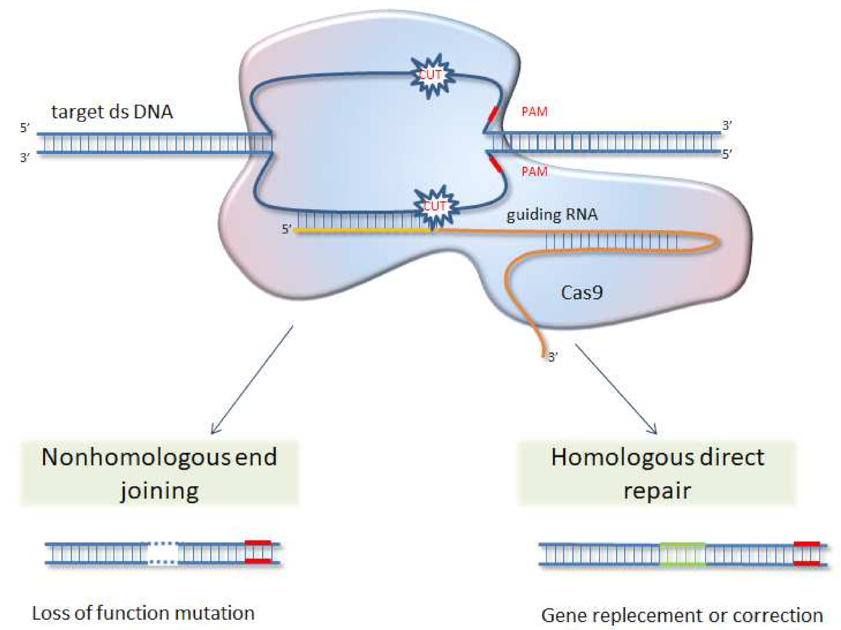
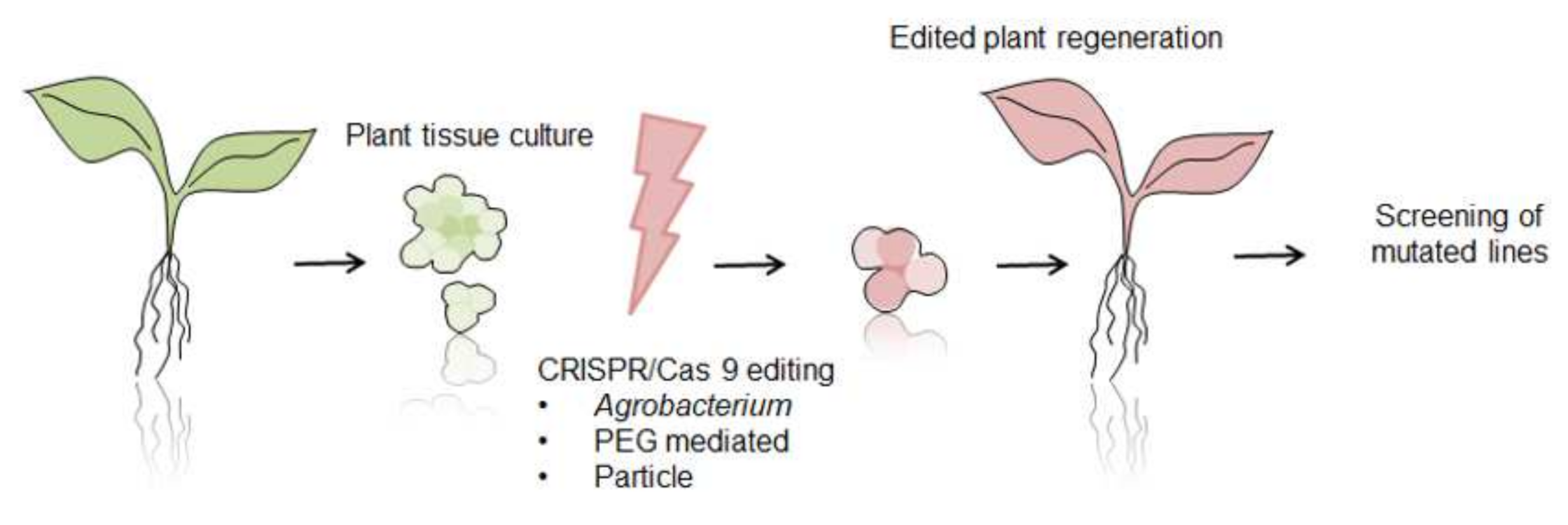
6. CRISPR/Cas 9 System as Future Direction for Functional Analysis of Proteins

Since 2013, the CRISPR/Cas9 (clustered regularly interspaced short palindromic repeats/CRISPR associated protein 9) genome editing system, with its Cas9 nuclease directed by target-specifying single guide RNA (sgRNA), has emerged as a practical method for the functional analysis of proteins and then as promising tool for the breeding of new varieties [165]. The CRISPR/Cas9 is a naturally occurring mechanism in bacteria and a few Archaea species. It works similarly to human immune systems and enables bacteria to acquire resistance to viruses after infection [166]. Genome editing with the use of CRISPR/Cas9 system is achieved by the induction of site-specific double-strand breaks, which are in turn repaired by either non-homologous end-joining (NHEJ) or homology-directed repair (HDR) machinery of the cell (see Figure 2 and Figure 3). Guide RNAs are designed to identify a three-base-pair protospacer adjacent motif (PAM) sequence occurring downstream of the target DNA. Then DNA is cut by Cas9 nuclease leading to gene knock-out [167]. The CRISPR/Cas9 tool has advantages over previously used methods, like zinc-finger nucleases (ZFNs) and transcription activator-like endonucleases (TALENs), in being easier with simple cloning steps needed, high ease of multiplexing (knockout multiple genes), and large-scale library preparation capacity. This method was successfully used in studies of different gene function, regulatory elements, and genetic mechanisms underlying quantitative trait loci (QTLs) in model organisms, as well as major crop species [168,169,170,171,172,173,174]. Several attempts of laticiferous plant modifications with CRISPR/Cas9 were also taken and are surveyed below.
Due to the high importance of natural rubber used to manufacture about 50,000 products, from tires to medical gloves, the Brazilian rubber tree (H. brasiliensis), a main source of rubber on a global scale, is subject to modification with CRISPR/Cas9 technology. Five sgRNAs were introduced to H. brasiliensis protoplast culture and targeted five different genes involved in flowering regulation-two genes form FLOWERING LOCUS T (FT) subfamily and three genes form TERMINAL FLOWER1 (TFL1) subfamily. Using the RNP-based genome editing system, modifications were introduced in all five genes, from which majority were deletions. The −1 nt deletion of the fourth nucleotide upstream of the PAM sites was the most frequently observed in all cases [175]. The rubber tree is a perennial tree species with a long juvenile phase, so conventional breeding for agronomic trait improvement is time consuming. The creation of early-flowering or delayed-flowering rubber tree plants will undoubtedly push forward studies related to the improvement of yield and quality, along with disease and stress resistance traits of this plant species.
Another example of laticiferous plant modification is the genetic engineering of Taraxacum kok-saghyz L.E.Rodin, commonly named Russian dandelion. It is an undomesticated dandelion species which can be a natural rubber source, alternative to H. brasiliensis. CRISPR/Cas9 was deployed to knockout gene encoding fructan:fructan 1-fructosyltransferase (1-FFT), a key enzyme in inulin biosynthesis [176]. Inulin is considered the main antagonist of rubber production, so the reduction of its synthesis should boost rubber particles formation. Such a modification not only shed lights on rubber biosynthesis mechanisms, but accelerates the domestication of dandelion as a rubber producing crop [177]. The application of A. rhizogenes-mediated hairy root induction allows to quickly obtain plants with a mutation rate as high as 80.0% (in regenerated plants).
For the enhancement of T. kok-saghyz agronomic performance, the CRISPR/Cas9 system was also used to induce a mutation in a gene called Rapid Alkalinisation Factor 1 (RALF1). In A. thaliana, RALF1 has been shown to suppress root growth [178]. As rubber is extracted from dandelion roots, it should be beneficial to change its morphology from branched to taproots, which are easier to harvest, and the wasted yield of lateral roots is minimalized. Knockout of gene TkRALFL1, achieved using Agrobacterium tumefaciens, resulted in introduction of premature stop codon or shortened sequence, which caused the removal of the functionally critical cysteine residues on the protein level. The root volume was 35% higher on average in the heterozygous knockout plants and 60% higher on average in homozygous knockout plants. Moreover, the inulin levels were higher in the knockouts whereas rubber levels were lower. Lower rubber content was compensated for with a higher dry weight of modified roots, such as the total yield per plant of inulin and rubber, which were much higher in knockout plants than in control [179]. It was shown that modification of gene TkRALFL1 can be used in subsequent breeding of profitable new dandelion varieties.
P. somniferum, as mentioned before, is a rich source of clinically important metabolites, which belongs to a group of benzylisoquinoline alkaloids (BIAs). For a better understanding of the gene regulation of those compounds synthesis, the CRISPR/Cas9-based gene knockout system was used to alter BIAs biosynthesis pathway. Enzyme 3′-hydroxy-N-methylcoclaurine 4′-O-methyltransferase (4′OMT), which catalyzes the conversion of central intermediate in BIAs synthesis, was targeted in Agrobacterium-mediated transformation. As a result, decreased total alkaloid content was confirmed. Most dramatic reductions were found in S-reticuline and laudanosine, direct products of 4′OMT [180].
For Cannabis sativa, a laticiferous plant species with growing importance in human therapies, a stable transformation protocol for modification with CRISPR/Cas9 strategy has been established recently. In the first step, five genes previously recognized as plant development regulators, were cloned to hypocotyls isolated from the DMG278 variety. Agrobacterium-mediated transformation led to the stimulation of shoot induction, most prominent for the combination of two genes (CsGRF3-C., sativa GROWTH-REGULATING FACTOR and CsGIF1-C., sativa GRF-INTERACTING FACTOR). After optimization of variety and explant, the constructs expressing sgRNA targeting the CsPDS1 gene were co-transformed into protoplasts from modified shoot culture. CsPDS1 encode phytoene desaturase (an enzyme essential for plant carotenoid biosynthesis) and is a common marker gene that can test genetic manipulation tools. Successful knockout of phytoene desaturase results in an easily recognized albino phenotype. Finally, four calli generated white seedlings with the edited CsPDS1 gene were obtained (account for 2.48% of the generated shoot) [181]. This is the first report of successful gene editing as well as stable transformation in C. sativa, which opens new possibilities to improved cannabis varieties production.

7. Conclusions

In the last decades, we have witnessed a huge expansion of demand for herbal medicines, phytonutrients, or nutraceuticals in developing as well as developed countries. They have become a substantial proportion of the global drug market. Moreover, it is estimated that at least four million people rely on herbal medicines as primary sources of healthcare [182]. Therefore, the need for new therapies, but also for plant-derived drugs with a long history of medicinal use (e.g., morphine), is still growing. Laticiferous plant species are a rich source of bioactive compounds (secondary metabolites and proteins), with only partially known medicinal use. The advent of new high throughput sequencing technologies and the fast, efficient genome editing system of CRISPR/Cas9 gives the research community the tools necessary to fulfill the broadened gap between supply and demand for medicines of plant origin. One of the greatest constraints to working with latex-bearing plants is insufficient genome information. Exploration of the genome of the plant of interest enables the precise modification and avoidance of off-target mutations. Nevertheless, the application of CRISPR/Cas9 modification sheds a new light on the function of some genes in laticiferous plant species, as well as sets directions for the improvement of agronomically important traits. Moreover, it provides a modern basis for further exploration and pharmacological utilization of latex compounds.

Author Contributions

Writing—original draft preparation, review and editing, J.G.-B., O.M. and R.N.; visualization, J.G.-B.; supervision, R.N. All authors have read and agreed to the published version of the manuscript.

Funding

This research was funded by the National Science Centre, Poland (grant number 2019/35/B/NZ9/03851).

Institutional Review Board Statement

Not applicable.

Informed Consent Statement

Not applicable.

Data Availability Statement

Data is contained within the article.

Conflicts of Interest

The authors declare no conflict of interest.

References

  1. Jesus, A.; Bonhomme, V.; Evin, A.; Ivorra, S.; Soteras, R.; Salavert, A.; Antolín, F.; Bouby, L. A Morphometric Approach to Track Opium Poppy Domestication. Sci. Rep. 2021, 11, 9778. [Google Scholar] [CrossRef]
  2. Ren, G.; Zhang, X.; Li, Y.; Ridout, K.; Serrano-Serrano, M.L.; Yang, Y.; Liu, A.; Ravikanth, G.; Nawaz, M.A.; Mumtaz, A.S.; et al. Large-Scale Whole-Genome Resequencing Unravels the Domestication History of Cannabis sativa. Sci. Adv. 2021, 7, eabg2286. [Google Scholar] [CrossRef]
  3. Zielińska, S.; Jezierska-Domaradzka, A.; Wójciak-Kosior, M.; Sowa, I.; Junka, A.; Matkowski, A.M. Greater Celandine’s Ups and Downs—21 Centuries of Medicinal Uses of Chelidonium majus from the Viewpoint of Today’s Pharmacology. Front. Pharmacol. 2018, 9, 299. [Google Scholar] [CrossRef] [Green Version]
  4. Gilca, M.; Gaman, L.; Panait, E.; Stoian, I.; Atanasiu, V. Chelidonium maju—An Integrative Review: Traditional Knowledge versus Modern Findings. Complementary Med. Res. 2010, 17, 241–248. [Google Scholar] [CrossRef] [PubMed]
  5. Maji, A.K.; Banerji, P. Chelidonium majus L. (Greater celandine—A Review on Its Phytochemical and Therapeutic Perspectives. Int. J. Herb. Med. 2015, 3, 10–27. [Google Scholar] [CrossRef]
  6. Nawrot, J.; Wilk-Jędrusik, M.; Nawrot, S.; Nawrot, K.; Wilk, B.; Dawid-Pać, R.; Urbańska, M.; Micek, I.; Nowak, G.; Gornowicz-Porowska, J. Milky Sap of Greater Celandine (Chelidonium majus L.) and Anti-Viral Properties. Int. J. Environ. Res. Public Health 2020, 17, 1540. [Google Scholar] [CrossRef] [PubMed] [Green Version]
  7. Salomé Abarca, L.F.; Klinkhamer, P.G.L.; Choi, Y.H. Plant Latex, from Ecological Interests to Bioactive Chemical Resources. Planta Med. 2019, 85, 856–868. [Google Scholar] [CrossRef] [Green Version]
  8. Nawrot, R. (Ed.) Latex, Laticifers and Their Molecular Components—From Functions to Possible Applications; Advances in Botanical Research Series; Academic Press: Cambridge, MA, USA, 2020. [Google Scholar]
  9. Kekwick, R.G.O. Latex and Laticifers. In Encyclopedia of Life Sciences 2002; John Wiley & Sons: London, UK; New York, NY, USA, 2002. [Google Scholar]
  10. Ramos, M.V.; Demarco, D.; da Costa Souza, I.C.; de Freitas, C.D.T. Laticifers, Latex, and Their Role in Plant Defense. Trends Plant Sci. 2019, 24, 553–567. [Google Scholar] [CrossRef]
  11. Hagel, J.M.; Yeung, E.C.; Facchini, P.J. Got Milk? The Secret Life of Laticifers. Trends Plant Sci. 2008, 13, 631–639. [Google Scholar] [CrossRef]
  12. Prado, E.; Demarco, D. Laticifers and secretory ducts: Similarities and differences. In Ecosystem Services and Global Ecology; InTech: London, UK, 2018; ISBN 9781789237382. [Google Scholar]
  13. Marinho, C.R.; Teixeira, S.P. Cellulases and Pectinases Act Together on the Development of Articulated Laticifers in Ficus montana and Maclura tinctoria (Moraceae). Protoplasma 2019, 256, 1093–1107. [Google Scholar] [CrossRef] [PubMed]
  14. Kitajima, S.; Taira, T.; Oda, K.; Yamato, K.T.; Inukai, Y.; Hori, Y. Comparative Study of Gene Expression and Major Proteins’ Function of Laticifers in Lignified and Unlignified Organs of Mulberry. Planta 2012, 235, 589–601. [Google Scholar] [CrossRef]
  15. Van Veenendaal, W.L.H.; Den Outer, R.W. Distribution and Development of the Non-Articulated Branched Laticifers of Morus nigra L. (Moraceae). Acta Bot. Neerl. 1990, 39, 285–296. [Google Scholar] [CrossRef]
  16. Webster, C.C. Natural Rubber: Biology, Cultivation and Technology. Agric. Syst. 1994, 45, 233–235. [Google Scholar] [CrossRef]
  17. Dussourd, D.E. Entrapment of Aphids and Whiteflies in Lettuce Latex. Ann. Entomol. Soc. Am. 1995, 88, 163–172. [Google Scholar] [CrossRef]
  18. Agrawal, A.A.; Konno, K. Latex: A Model for Understanding Mechanisms, Ecology, and Evolution of Plant Defense against Herbivory. Annu. Rev. Ecol. Evol. Syst. 2009, 40, 311–331. [Google Scholar] [CrossRef] [Green Version]
  19. Nawrot, R.; Wołuń-Cholewa, M.; Goździcka-Józefiak, A. Nucleases Isolated from Chelidonium majus L. Milky Sap Can Induce Apoptosis in Human Cervical Carcinoma HeLa Cells but Not in Chinese Hamster Ovary CHO Cells. Folia Histochem. Cytobiol. 2008, 46, 79–83. [Google Scholar] [CrossRef] [Green Version]
  20. Konno, K. Plant Latex and Other Exudates as Plant Defense Systems: Roles of Various Defense Chemicals and Proteins Contained Therein. Phytochemistry 2011, 72, 1510–1530. [Google Scholar] [CrossRef] [PubMed]
  21. Wink, M. Plant Secondary Metabolism: Diversity, Function and Its Evolution. Nat. Prod. Commun. 2008, 3, 1205–1216. [Google Scholar] [CrossRef] [Green Version]
  22. Souza, D.P.; Freitas, C.D.T.; Pereira, D.A.; Nogueira, F.C.; Silva, F.D.A.; Salas, C.E.; Ramos, M.V. Laticifer Proteins Play a Defensive Role against Hemibiotrophic and Necrotrophic Phytopathogens. Planta 2011, 234, 183–193. [Google Scholar] [CrossRef]
  23. Ramos, M.V.; Grangeiro, T.B.; Freire, E.A.; Sales, M.P.; Souza, D.P.; Araújo, E.S.; Freitas, C.D.T. The Defensive Role of Latex in Plants: Detrimental Effects on Insects. Arthropod-Plant Interact. 2010, 4, 57–67. [Google Scholar] [CrossRef]
  24. Men, X.; Wang, F.; Chen, G.-Q.; Zhang, H.-B.; Xian, M. Biosynthesis of Natural Rubber: Current State and Perspectives. Int. J. Mol. Sci. 2018, 20, 50. [Google Scholar] [CrossRef] [Green Version]
  25. Tang, C.; Qi, J.; Li, H.; Zhang, C.; Wang, Y. A Convenient and Efficient Protocol for Isolating High-Quality RNA from Latex of Hevea brasiliensis (para Rubber Tree). J. Biochem. Biophys. Methods 2007, 70, 749–754. [Google Scholar] [CrossRef]
  26. Salvucci, M.E.; Barta, C.; Byers, J.A.; Canarini, A. Photosynthesis and Assimilate Partitioning between Carbohydrates and Isoprenoid Products in Vegetatively Active and Dormant Guayule: Physiological and Environmental Constraints on Rubber Accumulation in a Semiarid Shrub. Physiol. Plant. 2010, 140, 368–379. [Google Scholar] [CrossRef]
  27. Sessa, R.A.; Bennett, M.H.; Lewis, M.J.; Mansfield, J.W.; Beale, M.H. Metabolite Profiling of Sesquiterpene Lactones from Lactuca Species. Major Latex Components Are Novel Oxalate and Sulfate Conjugates of Lactucin and Its Derivatives. J. Biol. Chem. 2000, 275, 26877–26884. [Google Scholar] [CrossRef]
  28. Valitova, J.N.; Sulkarnayeva, A.G.; Minibayeva, F.V. Plant Sterols: Diversity, Biosynthesis, and Physiological Functions. Biochemistry 2016, 81, 819–834. [Google Scholar] [CrossRef] [PubMed]
  29. Che Sulaiman, I.S.; Basri, M.; Fard Masoumi, H.R.; Chee, W.J.; Ashari, S.E.; Ismail, M. Effects of Temperature, Time, and Solvent Ratio on the Extraction of Phenolic Compounds and the Anti-Radical Activity of Clinacanthus Nutans Lindau Leaves by Response Surface Methodology. Chem. Cent. J. 2017, 11, 54. [Google Scholar] [CrossRef]
  30. Snook, M.E.; Data, E.S.; Kays, S.J. Characterization and Quantitation of Hexadecyl, Octadecyl, and Eicosyl Esters of P-Coumaric Acid in the Vine and Root Latex of Sweetpotato [Ipomoea batatas (L.) Lam.]. J. Agric. Food Chem. 1994, 42, 2589–2595. [Google Scholar] [CrossRef]
  31. Ismun, A.; Ariffin, M.M.; Razak, S.B.A.; Wei, O.C.; Ahmad, F.T.; Mubarak, A. determination of polyphenol contents in Hevea brasiliensis and rubber-processing effluent. Malays. J. Anal. Sci. 2018, 22, 185–196. [Google Scholar] [CrossRef]
  32. Huber, M.; Triebwasser-Freese, D.; Reichelt, M.; Heiling, S.; Paetz, C.; Chandran, J.N.; Bartram, S.; Schneider, B.; Gershenzon, J.; Erb, M. Identification, Quantification, Spatiotemporal Distribution and Genetic Variation of Major Latex Secondary Metabolites in the Common Dandelion (Taraxacum officinale Agg.). Phytochemistry 2015, 115, 89–98. [Google Scholar] [CrossRef]
  33. Nguyen, T.-D.; Dang, T.-T.T. Cytochrome P450 Enzymes as Key Drivers of Alkaloid Chemical Diversification in Plants. Front. Plant Sci. 2021, 12, 682181. [Google Scholar] [CrossRef]
  34. Itenov, K.; Mølgaard, P.; Nyman, U. Diurnal Fluctuations of the Alkaloid Concentration in Latex of Poppy Papaver Somniferum Is due to Day–night Fluctuations of the Latex Water Content. Phytochemistry 1999, 52, 1229–1234. [Google Scholar] [CrossRef]
  35. Tomè, F.; Colombo, M.L. Distribution of Alkaloids in Chelidonium majus and Factors Affecting Their Accumulation. Phytochemistry 1995, 40, 37–39. [Google Scholar] [CrossRef]
  36. Mikołajczak, P.Ł.; Kędzia, B.; Ożarowski, M.; Kujawski, R.; Bogacz, A.; Bartkowiak-Wieczorek, J.; Białas, W.; Gryszczyńska, A.; Buchwald, W.; Szulc, M.; et al. Evaluation of Anti-Inflammatory and Analgesic Activities of Extracts from Herb of Chelidonium majus L. Cent. Eur. J. Immunol. 2015, 40, 400–410. [Google Scholar] [CrossRef] [Green Version]
  37. Kopp, B.; Bauer, W.P.; Bernkop-Schnürch, A. Analysis of Some Malaysian Dart Poisons. J. Ethnopharmacol. 1992, 36, 57–62. [Google Scholar] [CrossRef]
  38. Agrawal, A.A.; Petschenka, G.; Bingham, R.A.; Weber, M.G.; Rasmann, S. Toxic Cardenolides: Chemical Ecology and Coevolution of Specialized Plant-Herbivore Interactions. New Phytol. 2012, 194, 28–45. [Google Scholar] [CrossRef]
  39. Malcolm, S.B. Milkweeds, Monarch Butterflies and the Ecological Significance of Cardenolides. Chemoecology 1994, 5–6, 101–117. [Google Scholar] [CrossRef]
  40. Cho, W.K.; Jo, Y.; Chu, H.; Park, S.-H.; Kim, K.-H. Integration of Latex Protein Sequence Data Provides Comprehensive Functional Overview of Latex Proteins. Mol. Biol. Rep. 2014, 41, 1469–1481. [Google Scholar] [CrossRef]
  41. Balakireva, A.V.; Zamyatnin, A.A. Indispensable Role of Proteases in Plant Innate Immunity. Int. J. Mol. Sci. 2018, 19, 629. [Google Scholar] [CrossRef] [PubMed] [Green Version]
  42. Konno, K.; Hirayama, C.; Nakamura, M.; Tateishi, K.; Tamura, Y.; Hattori, M.; Kohno, K. Papain Protects Papaya Trees from Herbivorous Insects: Role of Cysteine Proteases in Latex. Plant J. 2004, 37, 370–378. [Google Scholar] [CrossRef]
  43. de Freitas, C.D.T.; de Freitas, C.D.T.; de Souza, D.P.; Araújo, E.S.; Cavalheiro, M.G.; Oliveira, L.S.; Ramos, M.V. Anti-Oxidative and Proteolytic Activities and Protein Profile of Laticifer Cells of Cryptostegia Grandiflora, Plumeria Rubra and Euphorbia Tirucalli. Braz. J. Plant Physiol. 2010, 22, 11–22. [Google Scholar] [CrossRef] [Green Version]
  44. Kim, J.-Y.; Park, S.-C.; Hwang, I.; Cheong, H.; Nah, J.-W.; Hahm, K.-S.; Park, Y. Protease Inhibitors from Plants with Antimicrobial Activity. Int. J. Mol. Sci. 2009, 10, 2860–2872. [Google Scholar] [CrossRef] [PubMed] [Green Version]
  45. Kim, J.S.; Kim, Y.O.; Ryu, H.J.; Kwak, Y.S.; Lee, J.Y.; Kang, H. Isolation of Stress-Related Genes of Rubber Particles and Latex in Fig Tree (Ficus Carica) and Their Expressions by Abiotic Stress or Plant Hormone Treatments. Plant Cell Physiol. 2003, 44, 412–414. [Google Scholar] [CrossRef] [PubMed] [Green Version]
  46. Azarkan, M.; Wintjens, R.; Looze, Y.; Baeyens-Volant, D. Detection of Three Wound-Induced Proteins in Papaya Latex. Phytochemistry 2004, 65, 525–534. [Google Scholar] [CrossRef] [PubMed]
  47. Gidrol, X.; Chrestin, H.; Tan, H.L.; Kush, A. Hevein, a Lectin-like Protein from Hevea Brasiliensis (rubber Tree) Is Involved in the Coagulation of Latex. J. Biol. Chem. 1994, 269, 9278–9283. [Google Scholar] [CrossRef]
  48. Barre, A.; Van Damme, E.J.M.; Simplicien, M.; Benoist, H.; Rougé, P. Are Dietary Lectins Relevant Allergens in Plant Food Allergy? Foods 2020, 9, 1724. [Google Scholar] [CrossRef]
  49. Vandenborre, G.; Smagghe, G.; Van Damme, E.J.M. Plant Lectins as Defense Proteins against Phytophagous Insects. Phytochemistry 2011, 72, 1538–1550. [Google Scholar] [CrossRef]
  50. Lehrman, A. Does Pea Lectin Expressed Transgenically in Oilseed Rape (Brassica napus) Influence Honey Bee (Apis mellifera) Larvae? Environ. Biosafety Res. 2007, 6, 271–278. [Google Scholar] [CrossRef] [Green Version]
  51. John, K.S.; Bhat, S.G.; Prasada Rao, U.J.S. Biochemical Characterization of Sap (latex) of a Few Indian Mango Varieties. Phytochemistry 2003, 62, 13–19. [Google Scholar] [CrossRef]
  52. Wititsuwannakul, D.; Chareonthiphakorn, N.; Pace, M.; Wititsuwannakul, R. Polyphenol Oxidases from Latex of Hevea Brasiliensis: Purification and Characterization. Phytochemistry 2002, 61, 115–121. [Google Scholar] [CrossRef]
  53. Zhang, J.; Sun, X. Recent Advances in Polyphenol Oxidase-Mediated Plant Stress Responses. Phytochemistry 2021, 181, 112588. [Google Scholar] [CrossRef]
  54. Rajput, V.D.; Harish; Singh, R.K.; Verma, K.K.; Sharma, L.; Quiroz-Figueroa, F.R.; Meena, M.; Gour, V.S.; Minkina, T.; Sushkova, S.; et al. Recent Developments in Enzymatic Antioxidant Defence Mechanism in Plants with Special Reference to Abiotic Stress. Biology 2021, 10, 267. [Google Scholar] [CrossRef]
  55. Kidwai, M.; Ahmad, I.Z.; Chakrabarty, D. Class III Peroxidase: An Indispensable Enzyme for Biotic/abiotic Stress Tolerance and a Potent Candidate for Crop Improvement. Plant Cell Rep. 2020, 39, 1381–1393. [Google Scholar] [CrossRef]
  56. Bishop, J.G.; Dean, A.M.; Mitchell-Olds, T. Rapid Evolution in Plant Chitinases: Molecular Targets of Selection in Plant-Pathogen Coevolution. Proc. Natl. Acad. Sci. USA 2000, 97, 5322–5327. [Google Scholar] [CrossRef] [Green Version]
  57. Kitajima, S.; Kamei, K.; Taketani, S.; Yamaguchi, M.; Kawai, F.; Komatsu, A.; Inukai, Y. Two Chitinase-like Proteins Abundantly Accumulated in Latex of Mulberry Show Insecticidal Activity. BMC Biochem. 2010, 11, 6. [Google Scholar] [CrossRef] [Green Version]
  58. Gursharan, S.; Shailendra Kumar, A. Antifungal and Insecticidal Potential of Chitinases: A Credible Choice for the Eco-Friendly Farming. Biocatal. Agric. Biotechnol. 2019, 20, 101289. [Google Scholar]
  59. Taira, T.; Ohdomari, A.; Nakama, N.; Shimoji, M.; Ishihara, M. Characterization and Antifungal Activity of Gazyumaru (Ficus Microcarpa) Latex Chitinases: Both the Chitin-Binding and the Antifungal Activities of Class I Chitinase Are Reinforced with Increasing Ionic Strength. Biosci. Biotechnol. Biochem. 2005, 69, 811–818. [Google Scholar] [CrossRef] [Green Version]
  60. Martin, M.N. The Latex of Hevea Brasiliensis Contains High Levels of Both Chitinases and Chitinases/Lysozymes. Plant Physiol. 1991, 95, 469–476. [Google Scholar] [CrossRef] [Green Version]
  61. Freitas, C.D.T.; Viana, C.A.; Vasconcelos, I.M.; Moreno, F.B.B.; Lima-Filho, J.V.; Oliveira, H.D.; Moreira, R.A.; Monteiro-Moreira, A.C.O.; Ramos, M.V. First Insights into the Diversity and Functional Properties of Chitinases of the Latex of Calotropis procera. Plant Physiol. Biochem. 2016, 108, 361–371. [Google Scholar] [CrossRef]
  62. Musidlak, O.; Nawrot, R.; Goździcka-Józefiak, A. Which Plant Proteins Are Involved in Antiviral Defense? Review on In Vivo and In Vitro Activities of Selected Plant Proteins against Viruses. Int. J. Mol. Sci. 2017, 18, 2300. [Google Scholar] [CrossRef] [Green Version]
  63. Ali, S.; Ganai, B.A.; Kamili, A.N.; Bhat, A.A.; Mir, Z.A.; Bhat, J.A.; Tyagi, A.; Islam, S.T.; Mushtaq, M.; Yadav, P.; et al. Pathogenesis-Related Proteins and Peptides as Promising Tools for Engineering Plants with Multiple Stress Tolerance. Microbiol. Res. 2018, 212–213, 29–37. [Google Scholar] [CrossRef]
  64. Nessler, C.L. Sequence Analysis of Two New Members of the Major Latex Protein Gene Family Supports the Triploid-Hybrid Origin of the Opium Poppy. Gene 1994, 139, 207–209. [Google Scholar] [CrossRef]
  65. Nessler, C.L.; Allen, R.D.; Galewsky, S. Identification and Characterization of Latex-Specific Proteins in Opium Poppy. Plant Physiol. 1985, 79, 499–504. [Google Scholar] [CrossRef] [PubMed] [Green Version]
  66. Nessler, C.L.; Vonder Haar, R.A. Cloning and Expression Analysis of DNA Sequences for the Major Latex Protein of Opium Poppy. Planta 1990, 180, 487–491. [Google Scholar] [CrossRef] [PubMed]
  67. Ruperti, B.; Bonghi, C.; Ziliotto, F.; Pagni, S.; Rasori, A.; Varotto, S.; Tonutti, P.; Giovannoni, J.J.; Ramina, A. Characterization of a Major Latex Protein (MLP) Gene down-Regulated by Ethylene during Peach Fruitlet Abscission. Plant Sci. 2002, 163, 265–272. [Google Scholar] [CrossRef]
  68. Lytle, B.L.; Song, J.; de la Cruz, N.B.; Peterson, F.C.; Johnson, K.A.; Bingman, C.A.; Phillips, G.N., Jr.; Volkman, B.F. Structures of Two Arabidopsis Thaliana Major Latex Proteins Represent Novel Helix-Grip Folds. Proteins 2009, 76, 237–243. [Google Scholar] [CrossRef] [PubMed] [Green Version]
  69. Chruszcz, M.; Ciardiello, M.A.; Osinski, T.; Majorek, K.A.; Giangrieco, I.; Font, J.; Breiteneder, H.; Thalassinos, K.; Minor, W. Structural and Bioinformatic Analysis of the Kiwifruit Allergen Act D 11, a Member of the Family of Ripening-Related Proteins. Mol. Immunol. 2013, 56, 794–803. [Google Scholar] [CrossRef] [Green Version]
  70. Sun, H.; Kim, M.-K.; Pulla, R.K.; Kim, Y.-J.; Yang, D.-C. Isolation and Expression Analysis of a Novel Major Latex-like Protein (MLP151) Gene from Panax Ginseng. Mol. Biol. Rep. 2010, 37, 2215–2222. [Google Scholar] [CrossRef] [PubMed]
  71. Osmark, P.; Boyle, B.; Brisson, N. Sequential and Structural Homology between Intracellular Pathogenesis-Related Proteins and a Group of Latex Proteins. Plant Mol. Biol. 1998, 38, 1243–1246. [Google Scholar] [CrossRef]
  72. Park, C.-J.; Kim, K.-J.; Shin, R.; Park, J.M.; Shin, Y.-C.; Paek, K.-H. Pathogenesis-Related Protein 10 Isolated from Hot Pepper Functions as a Ribonuclease in an Antiviral Pathway. Plant J. 2004, 37, 186–198. [Google Scholar] [CrossRef]
  73. Yang, C.-L.; Liang, S.; Wang, H.-Y.; Han, L.-B.; Wang, F.-X.; Cheng, H.-Q.; Wu, X.-M.; Qu, Z.-L.; Wu, J.-H.; Xia, G.-X. Cotton Major Latex Protein 28 Functions as a Positive Regulator of the Ethylene Responsive Factor 6 in Defense against Verticillium dahliae. Mol. Plant 2015, 8, 399–411. [Google Scholar] [CrossRef] [Green Version]
  74. Nawrot, R. Defense-Related Proteins from Chelidonium majus L. as Important Components of Its Latex. Curr. Protein Pept. Sci. 2017, 18, 864–880. [Google Scholar] [CrossRef] [PubMed]
  75. Chadha, P.; Das, R.H. A Pathogenesis Related Protein, AhPR10 from Peanut: An Insight of Its Mode of Antifungal Activity. Planta 2006, 225, 213–222. [Google Scholar] [CrossRef]
  76. Michalska, K.; Fernandes, H.; Sikorski, M.; Jaskolski, M. Crystal Structure of Hyp-1, a St. John’s Wort Protein Implicated in the Biosynthesis of Hypericin. J. Struct. Biol. 2010, 169, 161–171. [Google Scholar] [CrossRef] [PubMed]
  77. Bantignies, B.; Séguin, J.; Muzac, I.; Dédaldéchamp, F.; Gulick, P.; Ibrahim, R. Direct Evidence for Ribonucleolytic Activity of a PR-10-like Protein from White Lupin Roots. Plant Mol. Biol. 2000, 42, 871–881. [Google Scholar] [CrossRef] [PubMed]
  78. Nawrot, R.; Barylski, J.; Lippmann, R.; Altschmied, L.; Mock, H.-P. Combination of Transcriptomic and Proteomic Approaches Helps to Unravel the Protein Composition of Chelidonium majus L. Milky Sap. Planta 2016, 244, 1055–1064. [Google Scholar] [CrossRef] [Green Version]
  79. Song, L.; Wang, J.; Jia, H.; Kamran, A.; Qin, Y.; Liu, Y.; Hao, K.; Han, F.; Zhang, C.; Li, B.; et al. Identification and Functional Characterization of NbMLP28, a Novel MLP-like Protein 28 Enhancing Potato Virus Y Resistance in Nicotiana Benthamiana. BMC Microbiol. 2020, 20, 55. [Google Scholar] [CrossRef] [PubMed]
  80. Malter, D.; Wolf, S. Melon Phloem-Sap Proteome: Developmental Control and Response to Viral Infection. Protoplasma 2011, 248, 217–224. [Google Scholar] [CrossRef] [PubMed]
  81. Onoyovwe, A.; Hagel, J.M.; Chen, X.; Khan, M.F.; Schriemer, D.C.; Facchini, P.J. Morphine Biosynthesis in Opium Poppy Involves Two Cell Types: Sieve Elements and Laticifers. Plant Cell 2013, 25, 4110–4122. [Google Scholar] [CrossRef] [PubMed] [Green Version]
  82. Nawrot, R.; Lippmann, R.; Matros, A.; Musidlak, O.; Nowicki, G.; Mock, H.-P. Proteomic Comparison of Chelidonium majus L. Latex in Different Phases of Plant Development. Plant Physiol. Biochem. 2017, 112, 312–325. [Google Scholar] [CrossRef]
  83. D’Amato, A.; Bachi, A.; Fasoli, E.; Boschetti, E.; Peltre, G.; Sénéchal, H.; Sutra, J.P.; Citterio, A.; Righetti, P.G. In-Depth Exploration of Hevea Brasiliensis Latex Proteome and “Hidden Allergens” via Combinatorial Peptide Ligand Libraries. J. Proteom. 2010, 73, 1368–1380. [Google Scholar] [CrossRef] [PubMed]
  84. Zulak, K.G.; Khan, M.F.; Alcantara, J.; Schriemer, D.C.; Facchini, P.J. Plant Defense Responses in Opium Poppy Cell Cultures Revealed by Liquid Chromatography-Tandem Mass Spectrometry Proteomics. Mol. Cell. Proteom. 2009, 8, 86–98. [Google Scholar] [CrossRef] [Green Version]
  85. Samatadze, T.E.; Zoshchuk, S.A.; Khomik, A.S.; Amosova, A.V.; Svistunova, N.Y.; Suslina, S.N.; Hazieva, F.M.; Yurkevich, O.Y.; Muravenko, O.V. Molecular Cytogenetic Characterization, Leaf Anatomy and Ultrastructure of the Medicinal Plant Potentilla alba L. Genet. Resour. Crop. Evol. 2018, 65, 1637–1647. [Google Scholar] [CrossRef]
  86. Gardin, N.E.; Braga, A.J. Greater Celandine (Chelidonium majus L.) for COVID-19: A Twenty-Case Series. Phytother. Res. 2021, 35, 3792–3798. [Google Scholar] [CrossRef]
  87. Foster, S.; Bradshaw, R.H.; McLanahan, S.; Redwood, D.; Upton, R. Book Reviews: The Complete German Commission E Monographs: Therapeutic Guide to Herbal Medicines. J. Altern. Complementary Med. 1998, 4, 479–486. [Google Scholar] [CrossRef]
  88. Lee, E.-J.; Hagel, J.M.; Facchini, P.J. Role of the Phloem in the Biochemistry and Ecophysiology of Benzylisoquinoline Alkaloid Metabolism. Front. Plant Sci. 2013, 4, 182. [Google Scholar] [CrossRef] [PubMed] [Green Version]
  89. Mithöfer, A.; Boland, W. Plant Defense against Herbivores: Chemical Aspects. Annu. Rev. Plant Biol. 2012, 63, 431–450. [Google Scholar] [CrossRef] [Green Version]
  90. Upadhyay, R.K. Plant Latex: A Natural Source of Pharmaceuticals and Pesticides. Int. J. Green Pharm. (IJGP) 2011, 5, 169–180. [Google Scholar] [CrossRef]
  91. Gurkok, T.; Turktas, M.; Parmaksiz, I.; Unver, T. Transcriptome Profiling of Alkaloid Biosynthesis in Elicitor Induced Opium Poppy. Plant Mol. Biol. Rep. 2015, 33, 673–688. [Google Scholar] [CrossRef]
  92. Labanca, F.; Ovesnà, J.; Milella, L. Papaver Somniferum L. Taxonomy, Uses and New Insight in Poppy Alkaloid Pathways. Phytochem. Rev. 2018, 17, 853–871. [Google Scholar] [CrossRef]
  93. De Petrocellis, L.; Ligresti, A.; Moriello, A.S.; Allarà, M.; Bisogno, T.; Petrosino, S.; Stott, C.G.; Di Marzo, V. Effects of Cannabinoids and Cannabinoid-Enriched Cannabis Extracts on TRP Channels and Endocannabinoid Metabolic Enzymes. Br. J. Pharmacol. 2011, 163, 1479–1494. [Google Scholar] [CrossRef] [Green Version]
  94. Pacher, P.; Bátkai, S.; Kunos, G. The Endocannabinoid System as an Emerging Target of Pharmacotherapy. Pharmacol. Rev. 2006, 58, 389–462. [Google Scholar] [CrossRef] [PubMed] [Green Version]
  95. Burstein, S. Cannabidiol (CBD) and Its Analogs: A Review of Their Effects on Inflammation. Bioorg. Med. Chem. 2015, 23, 1377–1385. [Google Scholar] [CrossRef]
  96. Etxenagusia, M.A.; Anda, M.; González-Mahave, I.; Fernández, E.; Fernández de Corrès, L. Contact Dermatitis from Chelidonium majus (greater Celandine). Contact Dermat. 2000, 43, 47. [Google Scholar]
  97. Monavari, S.H.; Shahrabadi, M.S.; Keyvani, H.; Bokharaei-Salim, F. Evaluation of In Vitro Antiviral Activity of Chelidonium majus L. against Herpes Simplex Virus Type-1. Afr. J. Microbiol. Res. 2012, 6, 4360–4364. [Google Scholar]
  98. Horvath, J.; Kery, A.; Kulcsar, G.; Dan, P.; Nasz, J. Antiviral Effect of Chelidonium Extracts. In Spitzy ILH, Karrev K (eds) 13–14 Proc Int Congr Chemother; Egermann: Vienna, Austria, 1983; Volume 9, pp. 124/106–124/110. [Google Scholar]
  99. Gerencer, M.; Turecek, P.L.; Kistner, O.; Mitterer, A.; Savidis-Dacho, H.; Barrett, N.P. In Vitro and in Vivo Anti-Retroviral Activity of the Substance Purified from the Aqueous Extract of Chelidonium majus L. Antivir. Res. 2006, 72, 153–156. [Google Scholar] [CrossRef]
  100. Mandadi, K.K.; Scholthof, K.-B.G. Plant Immune Responses against Viruses: How Does a Virus Cause Disease? Plant Cell 2013, 25, 1489–1505. [Google Scholar] [CrossRef] [PubMed] [Green Version]
  101. Wojtaszek, P. Oxidative Burst: An Early Plant Response to Pathogen Infection. Biochem. J. 1997, 322 Pt 3, 681–692. [Google Scholar] [CrossRef] [PubMed] [Green Version]
  102. Nawrot, R.; Kalinowski, A.; Gozdzicka-Jozefiak, A. Proteomic Analysis of Chelidonium majus Milky Sap Using Two-Dimensional Gel Electrophoresis and Tandem Mass Spectrometry. Phytochemistry 2007, 68, 1612–1622. [Google Scholar] [CrossRef]
  103. Nawrot, R.; Tomaszewski, Ł.; Czerwoniec, A.; Goździcka-Józefiak, A. Identification of a Coding Sequence and Structure Modeling of a Glycine-Rich RNA-Binding Protein (CmGRP1) from Chelidonium majus L. Plant Mol. Biol. Report. 2013, 31, 470–476. [Google Scholar] [CrossRef] [Green Version]
  104. Huh, S.U.; Paek, K.-H. Plant RNA Binding Proteins for Control of RNA Virus Infection. Front. Physiol. 2013, 4, 397. [Google Scholar] [CrossRef] [Green Version]
  105. Zvereva, A.S.; Pooggin, M.M. Silencing and Innate Immunity in Plant Defense against Viral and Non-Viral Pathogens. Viruses 2012, 4, 2578–2597. [Google Scholar] [CrossRef] [Green Version]
  106. Kéry, A.; Horváth, J.; Nász, I.; Verzár-Petri, G.; Kulcsár, G.; Dán, P. Antiviral Alkaloid in Chelidonium majus L. Acta Pharm. Hung. 1987, 57, 19–25. [Google Scholar]
  107. Lozjuk, R.M.; Lisnyak, O.I.; Lozjuk, L.V. Theoretical Grounds and Experimental Confirmation of the Antiviral Effect of the Preparation Ukrain. Drugs Exp. Clin. Res. 1996, 22, 213–217. [Google Scholar] [PubMed]
  108. Camero, M.; Marinaro, M.; Lovero, A.; Elia, G.; Losurdo, M.; Buonavoglia, C.; Tempesta, M. In Vitroantiviral Activity ofFicus Caricalatex against Caprine Herpesvirus-1. Nat. Prod. Res. 2014, 28, 2031–2035. [Google Scholar] [CrossRef] [PubMed]
  109. Aref, H.L.; Gaaliche, B.; Fekih, A.; Mars, M.; Aouni, M.; Chaumon, J.P.; Said, K. In Vitrocytotoxic and Antiviral Activities ofFicus Caricalatex Extracts. Nat. Prod. Res. 2011, 25, 310–319. [Google Scholar] [CrossRef]
  110. Beloin, N.; Gbeassor, M.; Akpagana, K.; Hudson, J.; de Soussa, K.; Koumaglo, K.; Thor Arnason, J. Ethnomedicinal Uses of Momordica Charantia (Cucurbitaceae) in Togo and Relation to Its Phytochemistry and Biological Activity. J. Ethnopharmacol. 2005, 96, 49–55. [Google Scholar] [CrossRef]
  111. Pengsuparp, T.; Serit, M.; Hughes, S.H.; Soejarto, D.D.; Pezzuto, J.M. Specific Inhibition of Human Immunodeficiency Virus Type 1 Reverse Transcriptase Mediated by Soulattrolide, a Coumarin Isolated from the Latex of Calophyllum Teysmannii. J. Nat. Prod. 1996, 59, 839–842. [Google Scholar] [CrossRef]
  112. Ubillas, R.; Jolad, S.D.; Bruening, R.C.; Kernan, M.R.; King, S.R.; Sesin, D.F.; Barrett, M.; Stoddart, C.A.; Flaster, T.; Kuo, J.; et al. SP-303, an Antiviral Oligomeric Proanthocyanidin from the Latex of Croton Lechleri (Sangre de Drago). Phytomedicine 1994, 1, 77–106. [Google Scholar] [CrossRef]
  113. Parhira, S.; Yang, Z.-F.; Zhu, G.-Y.; Chen, Q.-L.; Zhou, B.-X.; Wang, Y.-T.; Liu, L.; Bai, L.-P.; Jiang, Z.-H. In Vitro Anti-Influenza Virus Activities of a New Lignan Glycoside from the Latex of Calotropis Gigantea. PLoS ONE 2014, 9, e104544. [Google Scholar] [CrossRef]
  114. Kim, H.K.; Farnsworth, N.R.; Blomster, R.N.; Fong, H.H. Biological and Phytochemical Evaluation of Plants. V. Isolation of Two Cytotoxic Alkaloids from Chelidonium majus. J. Pharm. Sci. 1969, 58, 372–374. [Google Scholar] [CrossRef]
  115. Vavrečková, C.; Gawlik, I.; Müller, K. Benzophenanthridine Alkaloids of Chelidonium majus; II. Potent Inhibitory Action Against the Growth of Human Keratinocytes. Planta Med. 1996, 62, 491–494. [Google Scholar] [CrossRef]
  116. Rogelj, B.; Popovic, T.; Ritonja, A.; Strukelj, B.; Brzin, J. Chelidocystatin, a Novel Phytocystatin from Chelidonium majus. Phytochemistry 1998, 49, 1645–1649. [Google Scholar] [CrossRef]
  117. Philchenkov, A.; Kaminskyy, V.; Zavelevich, M.; Stoika, R. Apoptogenic Activity of Two Benzophenanthridine Alkaloids from Chelidonium majus L. Does Not Correlate with Their DNA Damaging Effects. Toxicol. In Vitro 2008, 22, 287–295. [Google Scholar] [CrossRef]
  118. Paul, A.; Bishayee, K.; Ghosh, S.; Mukherjee, A.; Sikdar, S.; Chakraborty, D.; Boujedaini, N.; Khuda-Bukhsh, A.R. Chelidonine Isolated from Ethanolic Extract of Chelidonium majus Promotes Apoptosis in HeLa Cells through p38-p53 and PI3K/AKT Signalling Pathways. Zhong Xi Yi Jie He Xue Bao 2012, 10, 1025–1038. [Google Scholar] [CrossRef]
  119. Kulp, M.; Bragina, O. Capillary Electrophoretic Study of the Synergistic Biological Effects of Alkaloids from Chelidonium majus L. in Normal and Cancer Cells. Anal. Bioanal. Chem. 2013, 405, 3391–3397. [Google Scholar] [CrossRef]
  120. Riede, F.v.D.I. Chelidonium majus in der Therapie Prämaligner Hautveränderungen. Available online: https://www.researchgate.net/publication/260197370_Chelidonium_majus_in_der_Therapie_pramaligner_Hautveranderungen (accessed on 15 November 2021).
  121. Abdel-Aty, A.M.; Hamed, M.B.; Salama, W.H.; Ali, M.M.; Fahmy, A.S.; Mohamed, S.A. Ficus Carica, Ficus Sycomorus and Euphorbia Tirucalli Latex Extracts: Phytochemical Screening, Antioxidant and Cytotoxic Properties. Biocatal. Agric. Biotechnol. 2019, 20, 101199. [Google Scholar] [CrossRef]
  122. Tulasi, C.; Lakshmi Narasu, M.; Saida, L. Cytotoxic Effect of Ficus Religiosa and Ficus Benghalensis Latex Extracts on MCF-7 Cell Line. Int. J. Sci. Res. Biol. Sci. 2019, 5, 96–100. [Google Scholar] [CrossRef] [Green Version]
  123. Available online: https://www.researchgate.net/profile/Flavio-Beltrame/publication/350549554_TERPENES_OF_EUPHORBIA_UMBELLATA_LATEX_ARE_INVOLVED_IN_CYTOTOXIC_EFFECT_AGAINST_MELANOMA_CELLS/links/60b52790a6fdcc476bda6c8d/TERPENES-OF-EUPHORBIA-UMBELLATA-LATEX-ARE-INVOLVED-IN-CYTOTOXIC-EFFECT-AGAINST-MELANOMA-CELLS.pdf (accessed on 30 September 2021).
  124. Cheng, J.; Han, W.; Wang, Z.; Shao, Y.; Wang, Y.; Zhang, Y.; Li, Z.; Xu, X.; Zhang, Y. Hepatocellular Carcinoma Growth Is Inhibited by Euphorbia Helioscopia L. Extract in Nude Mice Xenografts. Biomed Res. Int. 2015, 2015, 601015. [Google Scholar] [CrossRef]
  125. Sadeghi-Aliabadi, H.; Sajjadi, S.E.; Khodamoradi, M. Cytotoxicity of Euphorbia Macroclada on MDA-MB-468 Breast Cancer Cell Line. Iran. J. Pharm. Sci. 2009, 5, 103–108. [Google Scholar]
  126. Choedon, T.; Mathan, G.; Arya, S.; Kumar, V.L.; Kumar, V. Anticancer and Cytotoxic Properties of the Latex of Calotropis Procera in a Transgenic Mouse Model of Hepatocellular Carcinoma. World J. Gastroenterol. 2006, 12, 2517–2522. [Google Scholar] [CrossRef]
  127. Colombo, M.L.; Bosisio, E. Pharmacological Activities of Chelidonium Majus L. (Papaveraceae). Pharmacol. Res. 1996, 33, 127–134. [Google Scholar] [CrossRef]
  128. Ciric, A.; Vinterhalter, B.; Savikin-Fodulovic, K.; Sokovic, M.; Vinterhalter, D. Chemical Analysis and Antimicrobial Activity of Methanol Extracts of Celandine (Chelidonium majus L.) Plants Growing in Nature and Cultured in Vitro. Arch. Biol. Sci. 2008, 60, 7–8. [Google Scholar] [CrossRef]
  129. Kokoska, L.; Polesny, Z.; Rada, V.; Nepovim, A.; Vanek, T. Screening of Some Siberian Medicinal Plants for Antimicrobial Activity. J. Ethnopharmacol. 2002, 82, 51–53. [Google Scholar] [CrossRef]
  130. Recio, M.C.; Rios, J.L.; Villar, A. Antimicrobial Activity of Selected Plants Employed in the Spanish Mediterranean Area. Part II. Phytother. Res. 1989, 3, 77–80. [Google Scholar] [CrossRef]
  131. Miao, F.; Yang, X.-J.; Zhou, L.; Hu, H.-J.; Zheng, F.; Ding, X.-D.; Sun, D.-M.; Zhou, C.-D.; Sun, W. Structural Modification of Sanguinarine and Chelerythrine and Their Antibacterial Activity. Nat. Prod. Res. 2011, 25, 863–875. [Google Scholar] [CrossRef] [PubMed]
  132. Pavão, M.L.; Pinto, R.E. Sensivity of Bacillus Subtilis to Water Soluble Alkaloid Extracts of Chelidonium majus L. (Papaveracea) Roots from Azores. ARQUIPÉLAGO. Ciências Biológicas e Marinhas (Life Mar. Sci.) 1995, 13, 93–97. [Google Scholar]
  133. Asamenew, G.; Bisrat, D.; Mazumder, A.; Asres, K. In Vitro Antimicrobial and Antioxidant Activities of Anthrone and Chromone from the Latex of Aloe Harlana Reynolds. Phytother. Res. 2011, 25, 1756–1760. [Google Scholar] [CrossRef]
  134. Emiru, Y.K.; Siraj, E.A.; Teklehaimanot, T.T.; Amare, G.G. Antibacterial Potential of Aloe weloensis (Aloeacea) Leaf Latex against Gram-Positive and Gram-Negative Bacteria Strains. Int. J. Microbiol. 2019, 2019, 5328238. [Google Scholar] [CrossRef] [PubMed] [Green Version]
  135. Siritapetawee, J.; Thammasirirak, S.; Samosornsuk, W. Antimicrobial Activity of a 48-kDa Protease (AMP48) from Artocarpus heterophyllus Latex. Eur. Rev. Med. Pharmacol. Sci. 2012, 16, 132–137. [Google Scholar] [PubMed]
  136. Raghavendra, R.; Mahadevan, G.D. In Vitro Antimicrobial Activity of Various Plant Latex against Resistant Human Pathogens. Int. J. Pharm. Pharm. Sci. 2011, 3, 70–72. [Google Scholar]
  137. Nenaah, E.G.; Ahmed, M.E. Antimicrobial Activity of Extracts and Latex of Calotropis procera (Ait.) and Synergistic Effect with Reference Antimicrobials. J. Med. Plant Res. 2011, 5, 706–716. [Google Scholar] [CrossRef] [Green Version]
  138. Ishnava, K.B.; Chauhan, J.B.; Garg, A.A.; Thakkar, A.M. Antibacterial and Phytochemical Studies on Calotropis gigantia (L.) R. Br. Latex against Selected Cariogenic Bacteria. Saudi J. Biol. Sci. 2012, 19, 87–91. [Google Scholar] [CrossRef] [Green Version]
  139. Kori, P.; Alawa, P. Antimicrobial Activity and Phytochemical Analysis of Calotropis Gigantea Root, Latex Extracts. IOSR J. Pharm. (IOSRPHR) 2014, 4, 7–11. [Google Scholar] [CrossRef]
  140. Ml, P.; Mk, M.; Mary, R.S. Efficacy of Euphorbia Heterophylla Latex against Pathogenic Bacteria and Fungi. Asian J. Pharm. Clin. Res. 2020, 13, 141–145. [Google Scholar] [CrossRef]
  141. Lazreg-Aref, H.; Mars, M.; Fekih, A.; Aouni, M.; Said, K. Chemical Composition and Antibacterial Activity of a Hexane Extract of Tunisian Caprifig Latex from the Unripe Fruit of Ficus Carica. Pharm. Biol. 2012, 50, 407–412. [Google Scholar] [CrossRef] [Green Version]
  142. Prastiyanto, M.E.; Tama, P.D.; Ananda, N.; Wilson, W.; Mukaromah, A.H. Antibacterial Potential of Jatropha Sp. Latex against Multidrug-Resistant Bacteria. Int. J. Microbiol. 2020, 2020, 8509650. [Google Scholar] [CrossRef]
  143. Arun, K.; Anu, B.; Ruchika, G. A Comparative Study of Antibacterial Activity of Leaves and Latex of Jatropha Curcas L. Int. J. Pharmacogn. Phytochem. Res. 2012, 4, 190–194. [Google Scholar]
  144. Arekemase, M.O.; Kayode, R.M.O.; Ajiboye, A.E. Antimicrobial Activity and Phytochemical Analysis of Jatropha Curcas Plant against Some Selected Microorganisms. Int. J. Biol. 2011, 3, 52. [Google Scholar] [CrossRef]
  145. Hoekou, P.Y.; Tchacondo, T.; Gbogbo, K.A.; Tchelougou, D.; Pissang, P.; Karou, S.D.; Améyapohm, Y.A.; Batawila, K.; Annigoni, P.; Faso, B. Antibacterial Activities of Three Latex Plants of Asclepiadaceae Family Used in Traditional Medicine in South Togo. Int. J. Curr. Microbiol. App. Sci. 2015, 4, 882–891. [Google Scholar]
  146. Liu, C.-B.; Shan, B.; Bai, H.-M.; Tang, J.; Yan, L.-Z.; Ma, Y.-B. Hydrophilic/hydrophobic Characters of Antimicrobial Peptides Derived from Animals and Their Effects on Multidrug Resistant Clinical Isolates. Dongwuxue Yanjiu 2015, 36, 41–47. [Google Scholar]
  147. Fox, J.L. Antimicrobial Peptides Stage a Comeback: Better Understanding of the Mechanisms of Action, Modification and Synthesis of Antimicrobial Peptides Is Reigniting Commercial Development. Nat. Biotechnol. 2013, 31, 379–382. [Google Scholar] [CrossRef]
  148. Bahar, A.A.; Ren, D. Antimicrobial Peptides. Pharmaceuticals 2013, 6, 1543–1575. [Google Scholar] [CrossRef] [PubMed] [Green Version]
  149. Silva, O.N.; de la Fuente-Núñez, C.; Haney, E.F.; Fensterseifer, I.C.M.; Ribeiro, S.M.; Porto, W.F.; Brown, P.; Faria-Junior, C.; Rezende, T.M.B.; Moreno, S.E.; et al. An Anti-Infective Synthetic Peptide with Dual Antimicrobial and Immunomodulatory Activities. Sci. Rep. 2016, 6, 35465. [Google Scholar] [CrossRef] [PubMed] [Green Version]
  150. Van Parijs, J.; Broekaert, W.F.; Goldstein, I.J.; Peumans, W.J. Hevein: An Antifungal Protein from Rubber-Tree (Hevea Brasiliensis) Latex. Planta 1991, 183, 258–264. [Google Scholar] [CrossRef] [PubMed] [Green Version]
  151. Song, J.-Y.; Yang, H.-O.; Pyo, S.-N.; Jung, I.-S.; Yi, S.-Y.; Yun, Y.-S. Immunomodulatory Activity of Protein-Bound Polysaccharide Extracted from Chelidonium majus. Arch. Pharm. Res. 2002, 25, 158–164. [Google Scholar] [CrossRef] [PubMed]
  152. Chung, H.-S.; An, H.-J.; Jeong, H.-J.; Won, J.-H.; Hong, S.-H.; Kim, H.-M. Water Extract Isolated from Chelidonium majus Enhances Nitric Oxide and Tumour Necrosis Factor-α Production via Nuclear Factor-κB Activation in Mouse Peritoneal Macrophages. J. Pharm. Pharmacol. 2010, 56, 129–134. [Google Scholar] [CrossRef] [PubMed]
  153. Khmel’nitskaia, N.M.; Vorob’ev, K.V.; Kliachko, L.L.; Ankhimova, E.S.; Kosenko, V.A.; Tyrnova, E.V.; Mal’tseva, G.S.; Medvedev, E.A. A comparative study of conservative treatment schemes in chronic tonsillitis in children. Vestn. Otorinolaringol. 1998, 4, 39–42. [Google Scholar]
  154. Nascimento, D.C.d.O.; de Oliveira Nascimento, D.C.; Ralph, M.T.; Batista, J.E.C.; Silva, D.M.F.; Gomes-Filho, M.A.; Alencar, N.M.; Leal, N.C.; Ramos, M.V.; Lima-Filho, J.V. Latex Protein Extracts from Calotropis Procera with Immunomodulatory Properties Protect against Experimental Infections with Listeria Monocytogenes. Phytomedicine 2016, 23, 745–753. [Google Scholar] [CrossRef] [PubMed]
  155. Nakano, K.; Kim, D.; Jiang, Z.; Ueno, M.; Okimura, T.; Yamaguchi, K.; Oda, T. Immunostimulatory Activities of the Sulfated Polysaccharide Ascophyllan from Ascophyllum Nodosum in in Vivo and in Vitro Systems. Biosci. Biotechnol. Biochem. 2012, 76, 1573–1576. [Google Scholar] [CrossRef] [PubMed] [Green Version]
  156. Verma, V.K.; Rani, K.V.; Sehgal, N.; Prakash, O. Immunostimulatory Response Induced by Supplementation of Ficus Benghalensis Root Powder, in the Artificial Feed the Indian Freshwater Murrel, Channa Punctatus. Fish Shellfish Immunol. 2012, 33, 590–596. [Google Scholar] [CrossRef]
  157. Akram, M.; Hamid, A.; Khalil, A.; Ghaffar, A.; Tayyaba, N.; Saeed, A.; Ali, M.; Naveed, A. Review on Medicinal Uses, Pharmacological, Phytochemistry and Immunomodulatory Activity of Plants. Int. J. Immunopathol. Pharmacol. 2014, 27, 313–319. [Google Scholar] [CrossRef] [PubMed]
  158. Kukhetpitakwong, R.; Hahnvajanawong, C.; Homchampa, P.; Leelavatcharamas, V.; Satra, J.; Khunkitti, W. Immunological Adjuvant Activities of Saponin Extracts from the Pods of Acacia Concinna. Int. Immunopharmacol. 2006, 6, 1729–1735. [Google Scholar] [CrossRef] [PubMed]
  159. Hueza, I.M.; Gorniak, S.L. The Immunomodulatory Effects of Ipomoea Carnea in Rats Vary Depending on Life Stage. Hum. Exp. Toxicol. 2011, 30, 1690–1700. [Google Scholar] [CrossRef] [PubMed]
  160. Ramesh, K.V.; Padmavathi, K. Assessment of Immunomodulatory Activity of Euphorbia hirta L. Indian J. Pharm. Sci. 2010, 72, 621–625. [Google Scholar]
  161. Shah, A.S.; Wakade, A.S.; Juvekar, A.R. Immunomodulatory Activity of Methanolic Extract of Murraya koenigii (L) Spreng. Leaves. Indian J. Exp. Biol. 2008, 46, 505–509. [Google Scholar]
  162. Benninger, J.; Thomas Schneider, H.; Schuppan, D.; Kirchner, T.; Hahn, E.G. Acute Hepatitis Induced by Greater Celandine (Chelidonium majus). Gastroenterology 1999, 117, 1234–1237. [Google Scholar] [CrossRef]
  163. Moro, P.A.; Cassetti, F.; Giugliano, G.; Falce, M.T.; Mazzanti, G.; Menniti-Ippolito, F.; Raschetti, R.; Santuccio, C. Hepatitis from Greater Celandine (Chelidonium majus L.): Review of Literature and Report of a New Case. J. Ethnopharmacol. 2009, 124, 328–332. [Google Scholar] [CrossRef]
  164. Jakovljevic, Z.D.; Stankovic, S.M.; Topuzovic, D.M. Seasonal Variability of Chelidonium majus L. Secondary Metabolites Content and Antioxidant Activity. EXCLI J. 2013, 12, 260–268. [Google Scholar] [PubMed]
  165. Rao, M.J.; Wang, L. CRISPR/Cas9 Technology for Improving Agronomic Traits and Future Prospective in Agriculture. Planta 2021, 254, 68. [Google Scholar] [CrossRef] [PubMed]
  166. Alok, A.; Jain, P.; Kumar, J.; Yajnik, K.; Bhalothia, P. Genome Engineering in Medicinally Important Plants Using CRISPR/Cas9 Tool. In Genome Engineering via CRISPR-Cas9 System; Academic Press: Cambridge, MA, USA, 2020; pp. 155–161. [Google Scholar]
  167. Dey, A. CRISPR/Cas Genome Editing to Optimize Pharmacologically Active Plant Natural Products. Pharmacol. Res. 2021, 164, 105359. [Google Scholar] [CrossRef]
  168. Shen, C.; Que, Z.; Xia, Y.; Tang, N.; Li, D.; He, R.; Cao, M. Knock out of the Annexin Gene OsAnn3 via CRISPR/Cas9-Mediated Genome Editing Decreased Cold Tolerance in Rice. J. Plant Biol. 2017, 60, 539–547. [Google Scholar] [CrossRef]
  169. Li, J.-F.; Norville, J.E.; Aach, J.; McCormack, M.; Zhang, D.; Bush, J.; Church, G.M.; Sheen, J. Multiplex and Homologous Recombination-Mediated Genome Editing in Arabidopsis and Nicotiana Benthamiana Using Guide RNA and Cas9. Nat. Biotechnol. 2013, 31, 688–691. [Google Scholar] [CrossRef] [PubMed]
  170. Xu, R.; Wei, P.; Yang, J. Use of CRISPR/Cas Genome Editing Technology for Targeted Mutagenesis in Rice. Methods Mol. Biol. 2017, 1498, 33–40. [Google Scholar]
  171. Liang, Z.; Zhang, K.; Chen, K.; Gao, C. Targeted Mutagenesis in Zea Mays Using TALENs and the CRISPR/Cas System. J. Genet. Genom. 2014, 41, 63–68. [Google Scholar] [CrossRef]
  172. Michno, J.-M.; Wang, X.; Liu, J.; Curtin, S.J.; Kono, T.J.; Stupar, R.M. CRISPR/Cas Mutagenesis of Soybean and Medicago Truncatula Using a New Web-Tool and a Modified Cas9 Enzyme. GM Crops Food 2015, 6, 243–252. [Google Scholar] [CrossRef] [PubMed] [Green Version]
  173. Nekrasov, V.; Staskawicz, B.; Weigel, D.; Jones, J.D.G.; Kamoun, S. Targeted Mutagenesis in the Model Plant Nicotiana Benthamiana Using Cas9 RNA-Guided Endonuclease. Nat. Biotechnol. 2013, 31, 691–693. [Google Scholar] [CrossRef]
  174. Wang, S.; Zhang, S.; Wang, W.; Xiong, X.; Meng, F.; Cui, X. Efficient Targeted Mutagenesis in Potato by the CRISPR/Cas9 System. Plant Cell Rep. 2015, 34, 1473–1476. [Google Scholar] [CrossRef] [Green Version]
  175. Fan, Y.; Xin, S.; Dai, X.; Yang, X.; Huang, H.; Hua, Y. Efficient Genome Editing of Rubber Tree (Hevea brasiliensis) Protoplasts Using CRISPR/Cas9 Ribonucleoproteins. Ind. Crops Prod. 2020, 146, 112146. [Google Scholar] [CrossRef]
  176. Van Laere, A.; Van Den Ende, W. Inulin Metabolism in Dicots: Chicory as a Model System. Plant Cell Environ. 2002, 25, 803–813. [Google Scholar] [CrossRef]
  177. Iaffaldano, B.; Zhang, Y.; Cornish, K. CRISPR/Cas9 Genome Editing of Rubber Producing Dandelion Taraxacum Kok-Saghyz Using Agrobacterium Rhizogenes without Selection. Ind. Crops Prod. 2016, 89, 356–362. [Google Scholar] [CrossRef] [Green Version]
  178. Bergonci, T.; Ribeiro, B.; Ceciliato, P.H.O.; Guerrero-Abad, J.C.; Silva-Filho, M.C.; Moura, D.S. Arabidopsis Thaliana RALF1 Opposes Brassinosteroid Effects on Root Cell Elongation and Lateral Root Formation. J. Exp. Bot. 2014, 65, 2219–2230. [Google Scholar] [CrossRef] [PubMed] [Green Version]
  179. Wieghaus, A.; Prüfer, D.; Schulze Gronover, C. Loss of Function Mutation of the Rapid Alkalinization Factor (RALF1)-like Peptide in the Dandelion Taraxacum Koksaghyz Entails a High-Biomass Taproot Phenotype. PLoS ONE 2019, 14, e0217454. [Google Scholar] [CrossRef] [Green Version]
  180. Alagoz, Y.; Gurkok, T.; Zhang, B.; Unver, T. Manipulating the Biosynthesis of Bioactive Compound Alkaloids for Next-Generation Metabolic Engineering in Opium Poppy Using CRISPR-Cas 9 Genome Editing Technology. Sci. Rep. 2016, 6, 30910. [Google Scholar] [CrossRef] [PubMed] [Green Version]
  181. Zhang, X.; Xu, G.; Cheng, C.; Lei, L.; Sun, J.; Xu, Y.; Deng, C.; Dai, Z.; Yang, Z.; Chen, X.; et al. Establishment of an Agrobacterium-Mediated Genetic Transformation and CRISPR/Cas9-Mediated Targeted Mutagenesis in Hemp (Cannabis sativa L.). Plant Biotechnol. J. 2021, 19, 1979–1987. [Google Scholar] [CrossRef] [PubMed]
  182. Ekor, M. The Growing Use of Herbal Medicines: Issues Relating to Adverse Reactions and Challenges in Monitoring Safety. Front. Pharmacol. 2014, 4, 177. [Google Scholar] [CrossRef] [PubMed] [Green Version]
Figure 1. Chemical structure of four most studied alkaloids from Chelidonium majus.
Ijms 22 12427 g001
Figure 2. Scheme of CRISPR/Cas9 genome editing technology. Guide RNA, directed by PAM sequences near the targeted gene, lead Cas9 nuclease to altered DNA in desired location. Double strand breaks are repaired either by NHEJ or HDR mechanisms upon the existence of a donor template, which in result lead to deletion or insertion and gene knockout. NHEJ is more efficient than HDR, but may produce indel mutations, whereas HDR can provide a precise gene modification.
Ijms 22 12427 g002
Figure 3. Schematic overview of plant genome editing with CRISPR/Cas9 tool. After obtaining tissue culture from a plant of interest, compounds of CRISPR/Cas9 construct are delivered through Agrobacterium, PEG or particle bombardment. In the next step selection of mutated lines, regeneration and screening of mutated plants are performed.
Ijms 22 12427 g003
Table 2. Model of antiviral response of Chelidonium majus latex based on in vitro studies, which represents preformed immediate defense response with exuding latex. 1st line of defense-after mechanical damage (e.g., herbivore bite) the latex exudes and due its stickiness clots and stops or kills the herbivore. 2nd line of defense-cell wall damage is the prerequisite for the possibility of viral infection. Thereafter it can be stopped by oxidative burst and antiviral response (3rd line of defense). Abbreviations: PPO–polyphenol oxidase; LOX-lipoxygenase; POX-peroxidase; MLP-major latex protein; GRP-glycine-rich protein. According to [74].
Line of DefenseType of ActionPredominant Proteins and Compounds
1stMechanical damagePPO, LOX
(latex stickiness, different chemicals)
2ndOxidative burstPOX, LOX et al.
3rdAntiviral activityMLP, GRP (RNase/DNase activity, nucleic acid binding)
Table 3. Examples of antimicrobial activity of latex bearing plants.
Latex-Bearing Plant SpeciesExamined BacteriaExamined FungiBioactive CompoundsReference
Aloe harlana ReynoldsBacillus pumilus (82)
Bacillus subtilis (ATCC 6633)
Escherichia coli (CD/99/1, K88, K99, LT37, ROW 7/12, 3:37C, 306, 872)
Salmonella typhi (Ty2)
Shigella boydii (D13629)
Shigella dysentery 1
Shigella dysentery 8
Shigella flexneri (Type 6)
Shigella sonnei 1
Staphylococcus aureus (ML267)
Vibrio cholerae (85, 293, 1313, 1315)
Aspergillus niger (ATCC 6275)
Candida albicans (ATCC 10231) Penicillium funiculosum (NCTC 287)
Penicillium notatum (ATCC 11625)
Anthrone (aloin)
Chromone (7-O-methylaloeresin A)
[133]
Aloe weloensis SebsebeEnterococcus faecalis
Escherichia coli
Pseudomonas aeruginosa
Staphylococcus aureus
- Alkaloids
Anthraquinone
Flavonoids
Glycosides
Tannins
Terpenoids
[134]
Artocarpus
heterophyllus Lam.
Bacillus subtilis
Klebsiella Pneumoniae
Pseudomonas aeruginosa (ATCC 27853)
Streptococcus haemolyticus
Salmonella typhi
Aspergillus niger
Candida albicans
48-kDa protease (AMP48)[135,136]
Calotropis procera (Aiton) W.T.AitonBacillus cereus
Bacillus subtilis
Escherichia coli
Klebsiella Pneumoniae
Pseudomonas aeruginosa
Salmonella typhi
Staphylococcus aureus
Staphylococcus epidermidis
Streptococcus haemolyticus
Streptococcus pneumoniae
Aspergillus flavus
Aspergillus niger
Candida albicans
Candida tropicalis
Penicillium chrysogenum
Saccharomyces cereviciae
-[137]
Calotropis gigantea L.Bacillus cereus
Escherichia coli
Lactobacillus acidophilus
Micrococcus luteus
Staphylococcus aureus
Streptococcus mutans
Candida kruseiAlkaloids
Phenolic
Steroids
Terpenes
Cardiac glycoside
[138,139]
Carica papaya L.Bacillus subtilis
Klebsiella Pneumoniae
Streptococcus haemolyticus
Salmonella typhi
Aspergillus niger
Candida albicans
-[136]
Chelidonium majus L.Aeromonas hydrophila
Agrobacterium tumefacians
Bacillus cereus
Bacillus subtilis
Candida albicans
Escherichia coli
Micrococcus luteus
Mycobacterium phlei
Salmonella enteritidis
Sarcina lutea
Staphylococcus aureus
Candida albicans [127,128,129,130]
Euphorbia heterophylla L.Bacillus subtilis
Proteus vulgaris
Pseudomonas aeruginosa
Staphylococcus aureus
Aspergillus niger
Fusarium oxysporum
Penicillium sp.
Alkaloids
Flavonoids
Phenols
Saponins
Steroids
Tannins
[140]
Ficus carica L.Enterobacter cloacae
Enterococcus faecalis (ATCC 29212)
Escherichia coli
Escherichia coli ATCC 25922
Pseudomonas aeruginosa (ATCC 2783, ATCC 27950)
Staphylococcus aureus
Staphylococcus aureus (ATCC 25923)
Staphylococcus epidermidis
Staphylococcus saprophyticus
- alpha-Amyrenyl acetate
Aristolone
Bornanone-3
Lanosta-8
Olean-12-en-3-ol, acetate
Urs-12-en-24-oic acid
[141]
Jatropha gossypifolia L.Pseudomonas aeruginosa (CRPA)
Staphylococcus aureus (MRSA)
- Flavonoids[142]
Jatropha multifida L.Pseudomonas aeruginosa (CRPA)
Staphylococcus aureus (MRSA)
- Flavonoids[142]
Jatropa carcass L.Bacillus subtilis
Escherichia coli
Klebsiella Pneumoniae
Neisseria gonorrhoea
Pseudomonas aeruginosa
Salmonella typhi
Staphylococcus aureus
Streptococcus haemolyticus
Aspergillus niger
Candida albicans
Alkaloid
Glycoside
Saponin
Steroid
Tannin
[136,143,144]
Leptadenia hastata (Pers.) Decne.Klebsiella Pneumoniae
Staphylococcus aureus
Staphylococcus aureus (ATCC 29213)
Salmonella typhi
- Alkaloids
Flavonoids
Glycosides
Phenolic
Proanthocyanidins
Saponins
Tannins
Triterpene
[145]
Pergularia daemia (Forssk.) Chiov.Escherichia coli (ATCC 25922)
Klebsiella pneumoniae
Pseudomonas aeruginosa (ATCC 27853)
Staphylococcus aureus
Staphylococcus aureus (ATCC 29213)
Salmonella typhi
- Alkaloids
Flavonoids
Phenols
Saponins
Steroids
Tannins
Terpenoids
[145]
Secamone afzelii
(Schult.) K.Schum.
Escherichia coli ATCC 25922
Klebsiella pneumoniae
Pseudomonas aeruginosa ATCC 27853
Secamone afzelii
Staphylococcus aureus
Staphylococcus aureus ATCC 29213
Salmonella typhi
- Alkaloids
Cardiac
glycosides
Saponins
Tannins
[145]
Thevetia peruviana L.Bacillus subtilis
Klebsiella Pneumoniae
Streptococcus haemolyticus
Salmonella typhi
Aspergillus niger
Candida albicans
-[136]
Publisher’s Note: MDPI stays neutral with regard to jurisdictional claims in published maps and institutional affiliations.

