Article

Expression of Human Mutant Preproinsulins Induced Unfolded Protein Response, Gadd45 Expression, JAK-STAT Activation, and Growth Inhibition in Drosophila

Department of Insect Biomedical Research, Kyoto Institute of Technology, Matsugasaki, Kyoto 606-0962, Sakyo, Japan
*
Author to whom correspondence should be addressed.
Int. J. Mol. Sci. 2021, 22(21), 12038; https://doi.org/10.3390/ijms222112038
Submission received: 27 September 2021 / Revised: 25 October 2021 / Accepted: 28 October 2021 / Published: 7 November 2021
(This article belongs to the Special Issue Endoplasmic Reticulum Stress and Unfolded Protein Response 2021)

Abstract

:
Mutations in the insulin gene (INS) are frequently associated with human permanent neonatal diabetes mellitus. However, the mechanisms underlying the onset of this genetic disease is not sufficiently decoded. We induced expression of two types of human mutant INSs in Drosophila using its ectopic expression system and investigated the resultant responses in development. Expression of the wild-type preproinsulin in the insulin-producing cells (IPCs) throughout the larval stage led to a stimulation of the overall and wing growth. However, ectopic expression of human mutant preproinsulins, hINSC96Y and hINSLB15YB16delinsH, neither of which secreted from the β-cells, could not stimulate the Drosophila growth. Furthermore, neither of the mutant polypeptides induced caspase activation leading to apoptosis. Instead, they induced expression of several markers indicating the activation of unfolded protein response, such as ER stress-dependent Xbp1 mRNA splicing and ER chaperone induction. We newly found that the mutant polypeptides induced the expression of Growth arrest and DNA-damage-inducible 45 (Gadd45) in imaginal disc cells. ER stress induced by hINSC96Y also activated the JAK-STAT signaling, involved in inflammatory responses. Collectively, we speculate that the diabetes-like growth defects appeared as a consequence of the human mutant preproinsulin expression was involved in dysfunction of the IPCs, rather than apoptosis.

1. Introduction

Diabetes is a group of metabolic diseases wherein patients show a condition of elevated blood sugar levels called hyperglycemia. This disease is roughly classified into three principal types: type 1 diabetes, type 2 diabetes, and gestational diabetes [1]. Among them, type 1 diabetes is considered to be an autoimmune disease characterized by inflammatory responses that leads to the gradual destruction of pancreatic β-cells. This type of cell damage causes insulin deficiency, and results in deregulation of glucose metabolism [2]. Neonatal diabetes mellitus (NDM) is a rare disorder associated with defects in mass and function of pancreatic β-cells. NDM is not an autoimmune disease but a monogenic form of diabetes resulting from mutations in several genes encoding proteins crucial for the normal activity of the pancreatic β-cells. Mutations in the human insulin gene (INS) are a common cause of permanent neonatal diabetes mellitus (PNDM). When some of these mutant proinsulin genes are expressed in human cultured cells, folding and/or secretion of the mutant polypeptides are disrupted [3,4,5]. Exogenous insulin therapy is the only treatment for PNDM patients with the INS mutations. Hence, it is crucial to identify factors involved in the pathogenesis and understand the mechanisms by which the onset of PNDM using animal models for development of more effective therapeutic agents.
It has been considered that the onset of NDM is caused by the dysfunction or destruction of pancreatic β-cells [6,7,8,9]. It was recently suggested that various types of cellular stresses other than autoimmune responses, especially that of the endoplasmic reticulum (ER), are associated with the destruction or the malfunctioning of β-cells [10,11,12]. However, the mechanism by which ER stress triggers dysfunction and/or destruction of β-cells remains unidentified.
Excessive accumulation of unfolded or misfolded proteins, produced by exceeding the folding capacity of chaperones, causes a stress condition called ER stress, and activates the Unfolded Protein Response (UPR). Three principal branch cascades involved in the UPR were identified previously [13], and each branch is controlled by a different sensor protein, namely inositol-requiring enzyme 1 (IRE1), double-stranded RNA-activated protein kinase-like ER kinase (PERK), and activating transcription factor 6 (ATF6). These proteins sense the accumulation of unfolded proteins in ER, and stimulate the transcription of genes encoding ER chaperones, such as GRP78. Furthermore, cell death is induced in cases of continuous stress accumulation that failed to be managed by the UPR. ER stress-induced apoptosis is associated with many human diseases, including neurodegenerative diseases and diabetes [14]. Moreover, it was reported that ER stress-induced UPR triggers the induction of several stress-associated genes. Among them, Gadd45 (Growth Arrest and DNA-damage-inducible 45) plays a crucial role in DNA repair, cell cycle arrest, and apoptosis. It is induced by various genotoxic stresses through the tumor suppressor protein, p53 in mammalian cells [15]. Recent studies presented evidence suggesting that one of the Gadd45 family proteins, Gadd45α, was induced by ER stress through the PERK-eIF2α pathway in mouse embryonic fibroblasts [16]. However, the relationship between UPR and this Gadd45 gene in living organisms remains largely unexplored.
Tissues in which many secreted proteins are synthesized (like pancreas) are susceptible to the induction of ER stress [17]. Therefore, excessive ER stress or attenuation of the UPR in IPCs could cause functional inhibition of the cell functions, resulting in reduced secretion of insulin, eventually leading to onset of diabetes. It, however, is uncertain whether the ER stress and attenuation of the UPR promote the development of the disease. Extensive studies using animal models are thus required to investigate this causal relationship.
Drosophila melanogaster has emerged a powerful model organism for many human diseases for the past decade, such as metabolic diseases including diabetes [18,19]. Insulin, its receptor, and its downstream signaling pathway are highly conserved between Drosophila and mammals [18,20]. 8 types of insulin-like peptides (Dilp1–8) were identified in Drosophila [21,22,23,24]. Of these, all Dilps except Dilp8 bind to a unique insulin receptor, termed InR, and trigger the conserved signaling cascade. Further, three types of Dilps are found to be expressed in 7 insulin-producing neurons in a hemisphere of the brain. The Drosophila insulin-producing cells (IPCs) work as counterparts of pancreatic β-cells in mammals [25,26]. Additionally, the experimental techniques to identify the IPCs and to induce a specific gene expression in the cells are established in this organism [18]. Therefore, we selected this model organism to address the issue of whether ER stress in the insulin-producing cells contributed to the pathogenesis of diabetes. Thus, investigation of these conserved mechanisms can further improve our understanding of the connections between ER stress and IPC dysfunction, and the factors that lead to diabetes.
Previously, Katsube and colleagues demonstrated that the expression of a dominant-negative mutant of Hsc70-3 (Hsc70-3DN), a Drosophila ER chaperone, induces ER stress in Drosophila tissues [27]. Using this Hsc70-3DN-induced ER stress model, the authors demonstrated that ER-stress resulted in the apoptosis of IPCs leading to the onset of the diseases. However, further studies are required to decide whether apoptosis in the IPCs is involved in the pathogenesis of PNDM under physiological condition.
In this study, to understand the mechanisms underlying the onset of PNDM at the organism level, we induced the expression of two types of human mutant preproinsulin identified from PNDM patients, hINSC96Y (Akita mutation identified from PNDM mice introduced into human preproinsulin) and hINSLB15YB16delinsH (identified from human PNDM patients) in Drosophila tissues during development. Neither of the human mutant peptides could be secreted from cultured cells derived from pancreatic β-cells, when they were ectopically induced [3]. We investigated whether expression of the human mutant preproinsulins induced the UPR in the Drosophila tissues. Moreover, as the relationship between UPR and the Gadd45 had been unexplored in Drosophila, we first examined whether the Gadd45 gene expression depends on p53 in the tissues, similar to that in mammalian cultured cells. Then, we investigated whether the Gadd45 transcription was induced in tissues that have an accumulation of ER stress. Here, we demonstrated that the Gadd45-GFP reporter was an excellent tool that monitors ER-stress response. Furthermore, we investigated whether the human mutant preproinsulins induced activation of UPR, and the JAK-STAT signaling that potentially regulates inflammatory responses. The c-Jun NH2-terminal kinases (JNK), known as one of the stress-activated MAP kinases (SAPK), is also related to UPR. The results from our investigation collectively suggested the crucial role of Gadd45 or JAK-STAT pathway in tissue dysfunction or destruction in organisms as a result of ER stress.

2. Results

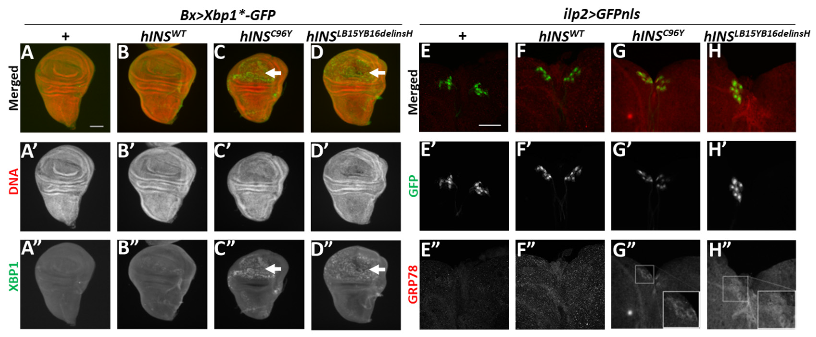
2.1. Ectopic Expression of Human Mutant Preproinsulins Induced the Expression of Two Types of ER Stress Marker in Drosophila Tissues

To clarify whether the expression of human mutant preproinsulin was responsible for causing the PNDM, we induced the expression of the mutant proteins hINSC96Y and hINSLB15YB16delinsH (Figure S1) in Drosophila melanogaster. We investigated whether the ectopic expression of these polypeptides resulted in tissue dysfunction and developmental abnormality, which are reminiscent of diabetes. To achieve this, first, we induced targeted expression of Drosophila insulin-like peptide 2 and human preproinsulins in Insulin Producing Cells (IPCs) in Drosophila adults using the Gal4/UAS system. We then investigated whether normal and mutant preproinsulins were successfully synthesized and secreted from the cells in Drosophila adults. Upon overexpression of Dilp2 (Drosophila insulin-like peptide 2) in IPCs using an IPC-specific Gal4 driver, ilp2-Gal4, a stronger anti-Dilp2 immunostaining signal in the cells was observed after 12 h starvation (Figure S2A,A”). Compared with the Dilp2 signal in the IPCs, the signal became weaker due to its release from the cells in adults re-fed for 2 h after the starvation (n > 7, p < 0.05, Student’s t-test) (Figure S2B,B”,G). Consistently, immunostaining signal with a specific antibody against the human polypeptide was detected in the IPCs of Drosophila adults starved in the same way, harboring the cell-specific expression of wild-type hINS (ilp2 > hINSWT) (n > 11, p < 0.01, Student’s t-test) (Figure S2C,C”). Here also, the signal consequently became weaker after re-feeding the adults with the diet (Figure S2D,D”,G). These observations suggested that it was possible to express the human wild-type insulin in Drosophila IPCs, and its release could be controlled by modulation of the diet. Then, we examined whether a human mutant polypeptide hINSC96Y having the Akita mutation identified in PNDM model mice [3]. This mutant polypeptide has a Cys to Tyr substitution at proinsulin position 96 (C96Y; encoding residue 7 of the insulin A-chain), which disrupts a disulfide bond connecting the insulin B and A-chains (Figure S1). It was accumulated in the IPCs and released from the cells depending on nutrient condition. Upon expressing the human mutant insulin, in the IPCs, we detected the immunostaining signal over background level in the cells from the starved adults (ilp2 > hINSC96Y) (Figure S2E,E”). However, a significant reduction of the signal was not observed in the IPCs expressing the human mutant insulin after re-feeding the diet (n > 14, p = 0.17, Student’s t-test) (Figure S2F,F”,G). This observation corroborated the published result that the mutant insulin failed to be secreted from cultured cells derived from pancreatic β-cells [3].
Next, we investigated whether the ectopic expression of the human polypeptides induced ER stress in the imaginal disc cells using an ER stress reporter Xbp1*-GFP. We used wing imaginal discs because they were easier to observe GFP expression than IPCs. The ER stress triggers splicing of Xbp1 pre-mRNA (Xbp1*) encoding a truncated Xbp1 precursor to remove an extra exon harboring a stop codon. If the full-length Xbp1-GFP protein is synthesized from a mature Xbp1 mRNA in response to the stress, GFP fluorescence can be seen. In the control wing discs (Bx-Gal4/+), GFP fluorescence was not detected due to the absence of ER stress-induced splicing (Figure 1A). Similarly, Bx-Gal4-dependent expression of human wild-type preproinsulin (hINSWT) did not induce Xbp1*-GFP expression in the discs, suggesting the lack of ER stress (Figure 1B). In contrast, intense GFP fluorescence was observed specifically in the wing pouch region of wing imaginal discs, in which the ectopic expression of the human mutant polypeptide, hINSC96Y or hINSLB15YB16delinsH, both of which failed to be secreted from β-cells, was induced (Figure S1). These human polypeptides have different amino acid substitutions identified from NDM model mouse and patients [3]. In addition to the hINSC96Y described before, another human mutant insulin, hINSLB15YB16delinsH carrying an in-frame substitution, in which B-chain Leu15 and Tyr16 are replaced by a single His residue. Both mutant polypeptides failed to secrete from β-cells. These observations indicate the induction of the ER stress (in Bx > hINSC96Y, Xbp1*-GFP or Bx > hINSLB15YB16delinsH, Xbp1*-GFP) (Figure 1C,D). We consistently observed distinctive GFP fluorescence in the wing disc region expressing the mutant peptides, although hINSLB15YB16delinsH produced less intense fluorescence than hINSC96Y. Thus, both human mutant preproinsulins hINSC96Y and hINSLB15YB16delinsH can induce the UPR in Drosophila tissues. Especially, the former polypeptide causes a stronger response.
Next, we questioned whether the expression of the mutant polypeptides in IPCs resulted in the pathogenesis of diabetes-like phenotype via ER stress accumulation in the cells. Previously studies reported that Drosophila ER chaperone(s) and Hsc70 family proteins recognized by anti-GRP78 antibody are up-regulated under these stress conditions [27,28]. We induced these mutant polypeptides in IPCs of Drosophila larvae using ilp2-Gal4. We performed immunostaining of whole larval brains from control larvae (ilp2-Gal4/+) (Figure 1E), or brains expressing normal human preproinsulin or the mutant polypeptides (Figure 1F–H). The anti-GRP78 immunostaining signal was not observed in control IPCs without expression of the human polypeptides (ilp2-Gal4/+) (Figure 1E”). Similarly, in the larval IPCs expressing hINSWT (ilp2 > hINSWT), the immunostaining signal was similar to the background levels (Figure 1F”). In contrast, a relatively higher signal was seen in IPCs expressing hINSC96Y and the cells expressing hINSLB15YB16delinsH over the background levels (insets in Figure 1G”,H”). However, we failed to identify the extent of the differences in the signal intensity elicited by these two mutant polypeptides. These results indicated that the ectopic expression of the human mutant preproinsulins induced ER stress; thereby, activating the UPR that induces ER chaperons in Drosophila tissues.

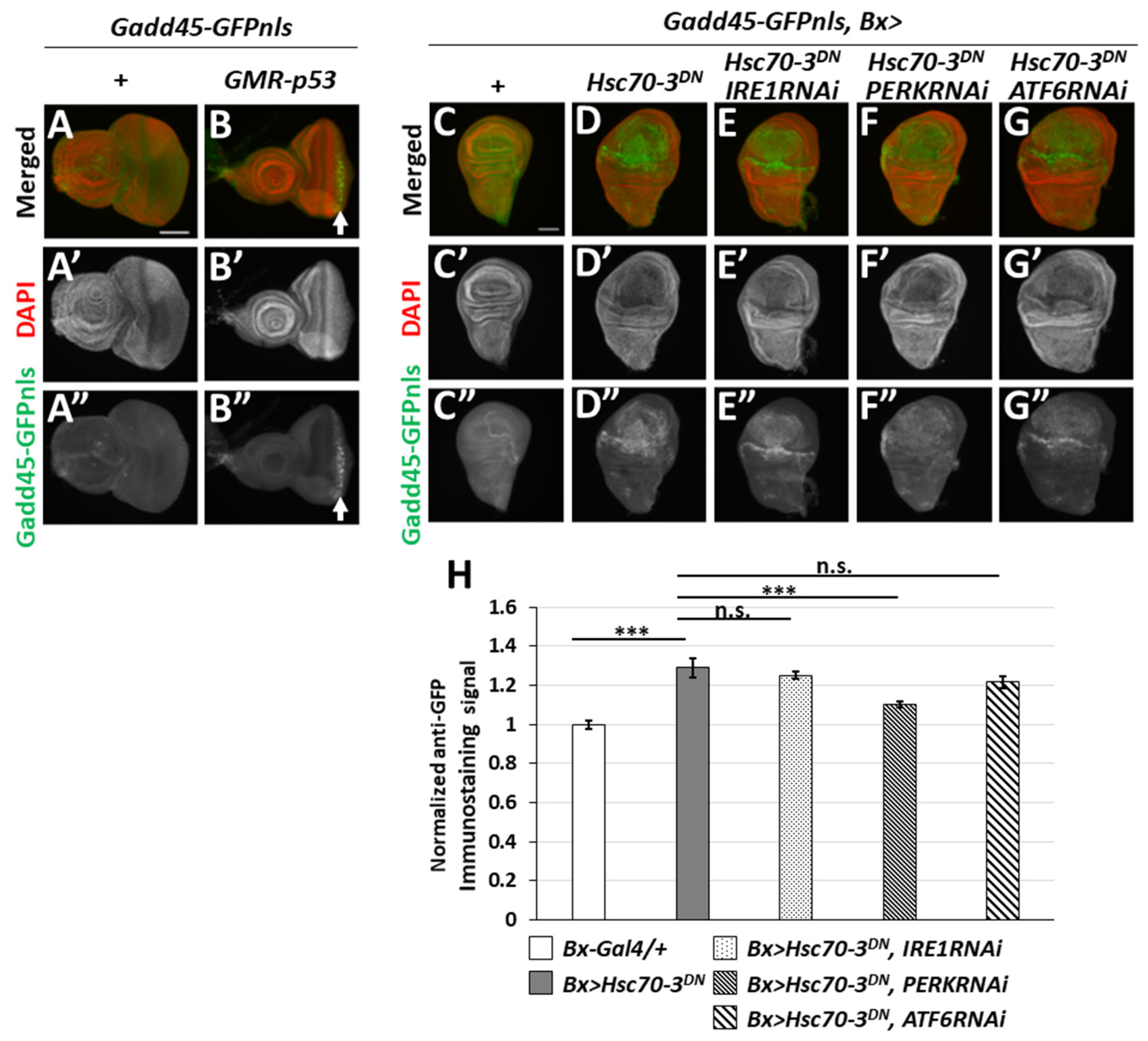
2.2. ER-Stress Induced the Expression of Gadd45 via UPR Pathway Mediated by PERK

We then generated a transgenic line harboring Gadd45-GFP reporter in which the coding frame of GFP possessed a nuclear localization sequence (NLS) under a transcriptional regulatory region of the Gadd45 gene (see Materials and Methods) to visualize the expression of Gadd45, a Drosophila orthologue of a known stress-responsible genes in mammals. Next, to confirm previous evidence that the Gadd45 gene expression is induced via p53 in a response to DNA damage in mammalian cells [29], we induced expression of p53 in the eye imaginal discs under the control of glass multiple reporter (GMR) promoter (GMR-p53, Gadd45-GFP). We investigated whether the GFP reporter could successfully monitor the expression of Gadd45. Our investigations revealed that p53 could drive the transcription of Gadd45, resulting in GFP fluorescence in cell nuclei of eye imaginal discs, but none in the control (Gadd45-GFP) discs (Figure 2A,A” and an arrow in Figure 2B,B”). These results demonstrated how the Gadd45-GFP reporter helped to monitor Gadd45 transcription under stress responses.
Thus, we next examined whether the ER stress-induced expression of the GFP reporter was observed. As previously described, the ectopic expression of a dominant-negative mutant of Drosophila ER chaperone (Hsc70-3DN) is a known ER stress inducer [27]. We observed a strong GFP fluorescence in the wing pouch region expressing Hsc70-3DN in wing discs using Bx-Gal4 driver (Gadd45-GFP, Bx > Hsc70-3DN), indicating that Gadd45 expression was induced. By contrast, no GFP signals in the control wing discs (Bx-Gal4/+; Gadd45-GFP/+) (Figure 2C,D,H). These results suggested that Gadd45 is an UPR target gene whose expression is also induced in response to ER stress, other than DNA damage.
It is known that ER stress activates three independent UPR signaling pathways mediated by IRE, PERK, and ATF6 (see Introduction). Hence, we induced simultaneous expression of dsRNA against either IRE1, PERK, or ATF6 mRNA, encoding principal ER stress sensors, in wing discs expressing Hsc70-3DN to determine which of these mediators are essential for Gadd45 induction under ER stress. We observed a distinctive GFP fluorescence representing Gadd45-GFP expression in the disc cells, whereas no GFP signals higher than the background were observed in the control wing imaginal discs (Bx-Gal4/+; Gadd45-GFP/+) (Figure 2C,C”,D,D”,H). Further, neither the dsRNA-derived depletion of IRE1 nor that of ATF6 suppressed the GFP signals in the wing discs expressing Hsc70-3DN (Gadd45-GFP, Bx > Hsc70-3DN, IRE1RNAi, or Gadd45-GFP, Bx > Hsc70-3DN, ATF6RNAi, respectively) (Figure 2E,G,H). In contrast, GFP fluorescence indicating Gadd45 gene expression was significantly suppressed by dsRNA-derived depletion of PERK (Gadd45-GFP, Bx > Hsc70-3DN, PERKRNAi) (Figure 2F,H, p < 0.001, Student’s t-test). These observations suggested that ER stress induced Gadd45 transcription via the PERK pathway in Drosophila imaginal discs. Thus, it can be further concluded that Gadd45-GFP is a useful reporter to monitor cellular response to ER stress.

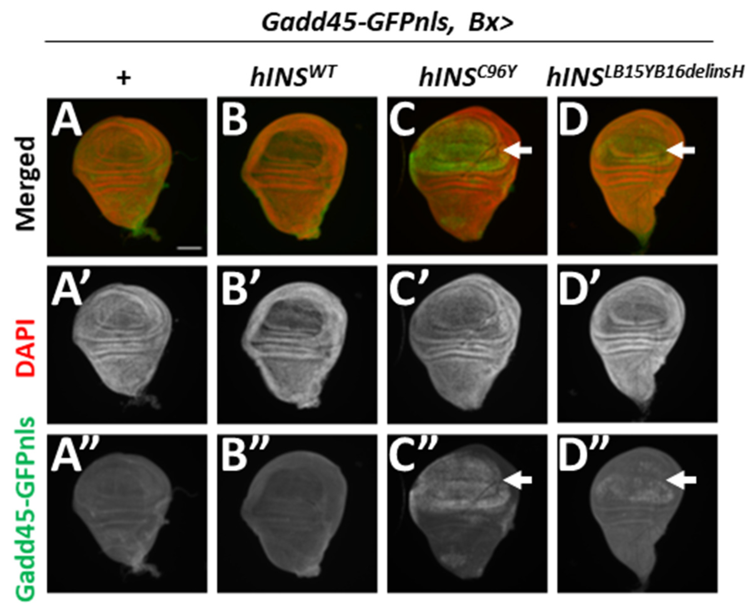
2.3. Human Mutant Preproinsulin-Induced ER Stress Also Induced Gadd45 Expression

To assess whether the human mutant preproinsulins induce the UPR in a response to the ER stress, we next investigated whether the ectopic expression of hINSC96Y and hINSLB15YB16delinsH induced transcription of the Gadd45 gene using the GFP reporter. No GFP signal was observed in the control wing imaginal discs (Gadd45-GFP, Bx-Gal4/+) (Figure 3A,A”). Similarly, the expression of hINSWT failed to induce GFP fluorescence, indicating induction of Gadd45 transcription in the wing pouch region of the imaginal discs (Figure 3B,B”). In contrast, the GFP signal was detected in the pouch region expressing hINSC96Y and hINSLB15YB16delinsH (Gadd45-GFP, Bx > hINSC96Y and Gadd45-GFP, Bx > hINSL39Y40delinH, respectively) (Figure 3C,C”,D,D”). The GFP signal in the discs expressing hINSC96Y was more intense than that in hINSLB15YB16delinsH. These results indicate that ER stress associated with the expression of human mutant preproinsulin induced Gadd45 transcription.

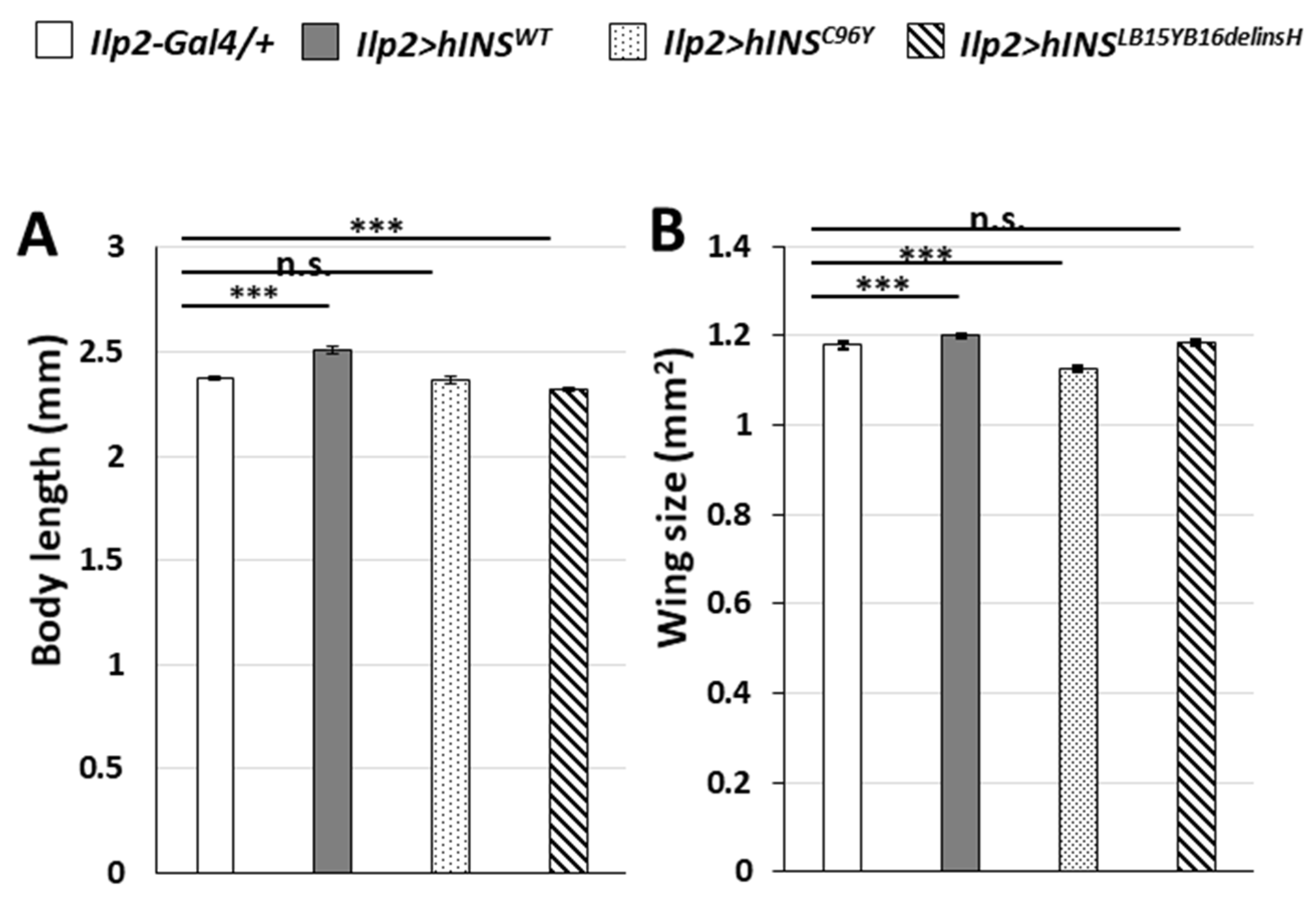
2.4. Induction of ER Stress by a Constitutive Expression of Human Mutant Preproinsulins in IPCs Resulted in Growth Inhibition of Drosophila

Previous studies using mammalian type 1 diabetes models that chronic ER stress in insulin-producing cells resulted in the onset and pathogenesis of diabetes [30]. Similarly, our group had previously reported that the accumulation of ER stress by the continuous expression of Hsc70-3DN in IPCs resulted in the diabetes-like phenotypes such as growth inhibition, decreased mRNA level of Dilps, and higher glucose level in larval hemolymph [27]. Thus, we were interested in finding if a similar diabetes-like phenotype could be observed upon the ectopic expression of human mutant preproinsulins in IPCs. As mentioned earlier, the expression of human mutant preproinsulins in IPCs activates the UPR pathway. First, we observed the adult phenotypes in which hINSWT was continuously expressed in IPCs throughout their development (ilp2 > hINSWT), which resulted in significantly larger whole bodies in these flies compared with the controls (ilp2-Gal4/+). The average body length of male flies raised under this condition was 5.9% higher on an average than that of the controls (n = 94, Figure 4A). Similarly, the average area of whole wings of ilp2 > hINSWT adult males increased by 2.0% compared with that of control (n = 175) (Figure 4B). These results demonstrated how the constitutive expression of the human wild-type preproinsulin in IPCs throughout development partially stimulated Drosophila growth.
Next, we assessed the effect of human mutant preproinsulin-overexpression in IPCs on the overall Drosophila growth. The average body length of the male flies with continuous hINSLB15YB16delinsH expression in IPCs significantly decreased by 2.2% compared with that of the controls (n = 69). In contrast, the differences in body lengths between the hINSC96Y group and the controls were not significant (n = 79) (p > 0.05, Student’s t-test, Figure 4A). Furthermore, the whole wing size of ilp2 > hINSC96Y adult males decreased by 4.4%, compared with the controls (n = 125). However, the differences between the wing of hINSLB15YB16delinsH males and the controls were not statistically significant (n = 113) (p > 0.05, Student’s t-test, Figure 4B). These data collectively suggested that the expression of the human mutant preproinsulin in IPCs resulted in partial growth inhibition of the organisms.

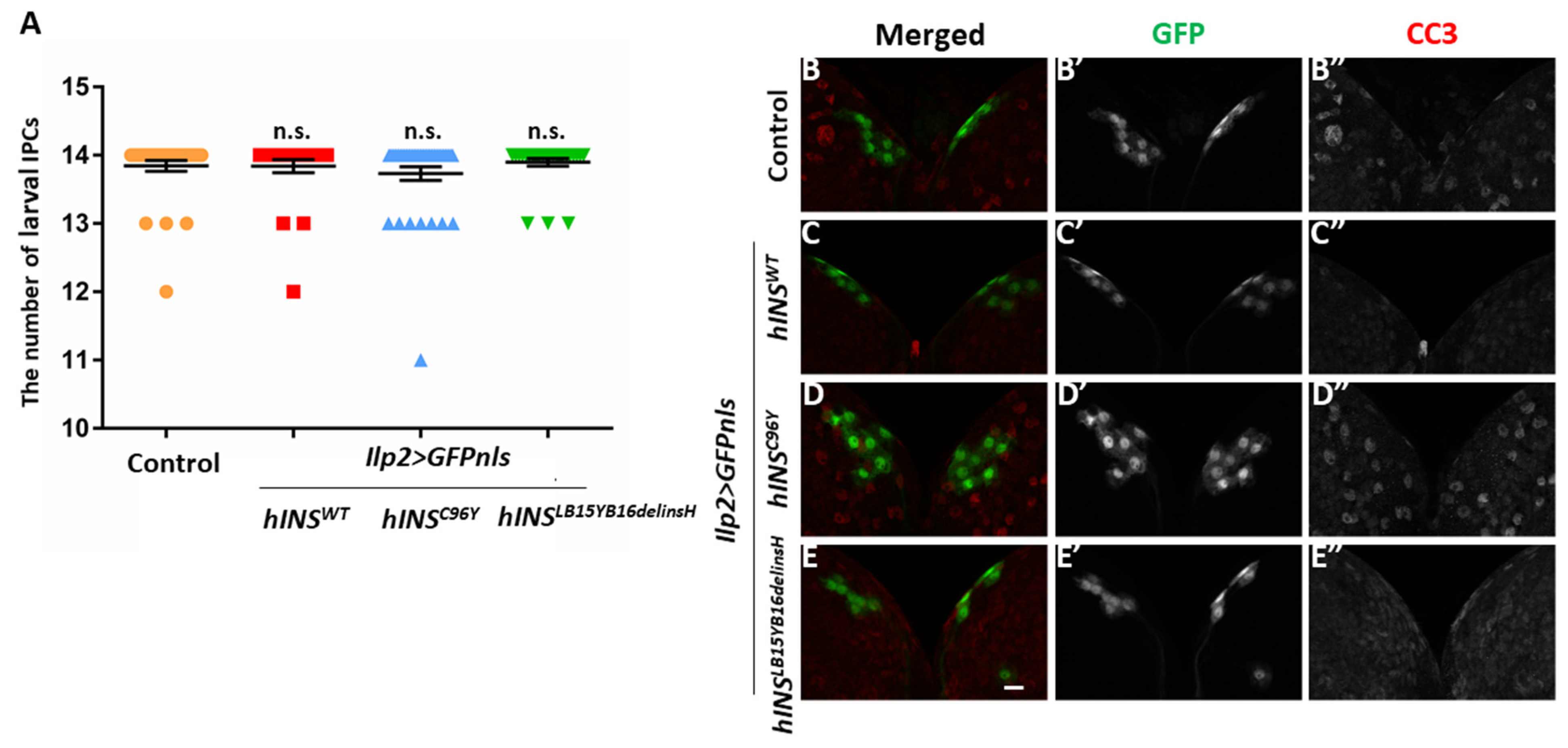
2.5. The Ectopic Expression of Human Mutant Preproinsulins Failed to Induce Apoptosis in IPCs

It has been speculated that dysfunction and/or a loss of insulin-producing cells eventually leads to human type 1 diabetes [2]. Hence, we first induced GFP with a nuclear localization sequence (NLS) exclusively in the larval IPCs to count the cell numbers to examine whether the expression of human mutant preproinsulin leads to the accumulation of ER stress, resulting in the cell death of IPCs. It was found that none of the types of human preproinsulin in IPCs reduced the number of these cells, suggesting that apoptosis was not induced (Figure 5A).
To test whether pro-apoptotic caspases are activated in IPCs expressing the human mutant polypeptides, whole-mount staining of the larval brain was carried out with an anti-cleaved caspase-3 (CC3) antibody, which recognizes active caspase-3. Consistently, we observed no anti-CC3 immunofluorescence signal over the background levels in IPCs expressing either human wild-type or the two mutant types of preproinsulin (Figure 5B–E,B”–E”). These results collectively led us to conclude that ER stress accumulation induced by targeted expression of the human mutant preproinsulins can activate UPR, but not the initiator caspase. These results could explain why the expression of the mutant peptides did not lead to cell death in the IPC.

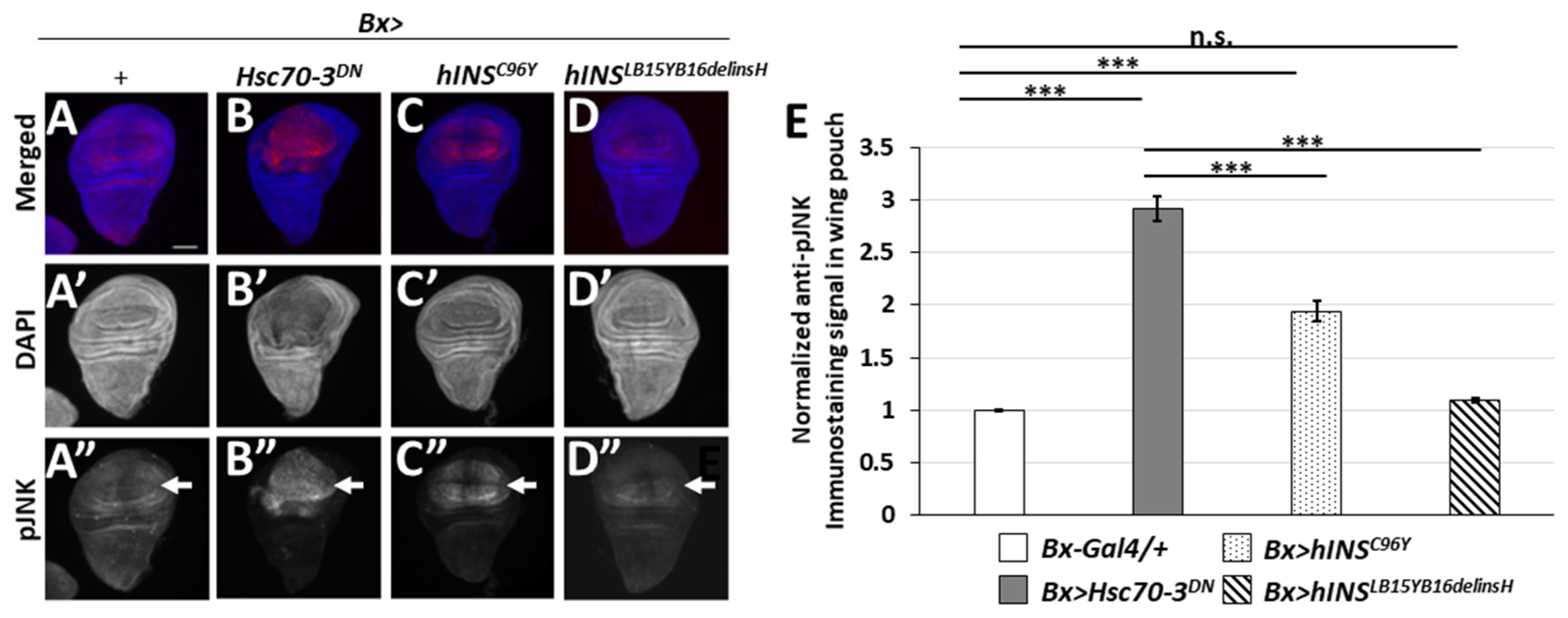
2.6. The Ectopic Expression of the Human Mutant Preproinsulins Activated the JNK Pathway Less Efficiently Than That of the Stronger ER Stress Inducer, Hsc70-3DN

The JNK pathway is a widely known key mediator for the transduction of stress-related signals, and thereby for apoptosis induction. We had previously reported that the ER stress by Hsc70-3DN expression induced apoptosis dependent on this pathway [27]. To further understand the mechanisms by which the ER stress led to apoptosis induction, we examined whether expression of the human mutant polypeptides activated the JNK pathway. First, we performed immunostaining of wing discs expressing Hsc70-3DN with an antibody recognizing a phosphorylated JNK (pJNK). We observed a weak striped pattern signal of pJNK, which corresponded to a known developmental signal in the control wing discs (Bx-Gal4/+) (arrows in Figure 6A,A”,E) [31]. By contrast, a much more intense pJNK signal was observed in wing discs expressing Hsc70-3DN (Bx > Hsc70-3DN) (arrow in Figure 6B”,E). Moreover, we examined whether the pJNK immunostaining signal was observed in the wing discs expressing normal or the mutant types of human preproinsulin; in those expressing the normal human peptide (Bx > hINSWT), an enhanced signal over the basal levels was not observed (data not shown). In contrast, we found relatively weak pJNK signal in the wing pouch region expressing hINSC96Y (Bx > hINSC96Y). The pJNK signal was less robust than the signal in the Hsc70-3DN-expressing region, but it was significantly higher than that in the discs of the control flies (arrows in Figure 6A”,C”,E). Similarly, the signal in the wing discs expressing hINSLB15YB16delinsH (Bx > hINSLB15YB16delinsH) was faint, but slightly higher than that in the control discs (arrows in Figure 6A”,D”,E). These results suggested that the ER stress induced by two human mutant preproinsulins could result in the weak activation of the JNK pathway, but the level was not strong enough to induce apoptosis in Drosophila tissues.

2.7. ER Stress Accumulated in the Wing Disc Cells Resulted in the Activation of the JAK/STAT Pathway

It is hypothesized that ER stress drives the pathogenesis of human diseases due to inflammatory signaling, thus facilitating cell death [32]. In this study, we examined whether ER stress accumulation activated the JAK-STAT pathway in Drosophila tissues. We used a 10XSTAT92E-GFP reporter, in which the GFP gene is under tandem repeats of STAT92E-binding sequences to visualize this activation of the signaling pathway [33]. Thus, the activation of the JAK-STAT pathway would lead to GFP fluorescence in our system. We then induced targeted expression of Hsc70-3DN, human normal or mutant type preproinsulin in the wing pouch region of the discs. As a result, a relatively intense GFP fluorescence was observed in control wing discs (Bx-Gal4/+; 10XSTAT92E-GFP) (arrowhead in Figure 7A”), but not in the wing pouch region (arrow in Figure 7A”). These data support the known observation that this signaling pathway is indispensable for the normal development in this hinge region of normal wing discs [34]. Moreover, we found a more intense GFP signal in Hsc70-3DN-expressing regions of all the wing discs examined (10XSTAT92E-GFP, Bx > Hsc70-3DN) (n = 29) (arrow in Figure 7B”, but not in Figure 7A”), which suggested that the JAK-STAT pathway was activated in response to ER stress. Similarly, the ectopic expression of hINSC96Y (10XSTAT92E-GFP, Bx > hINSC96Y) in the wing pouch region resulted in the induction of GFP fluorescence less frequently (33.3% of wing discs examined) (n = 33) (Figure 7C). In contrast, the expression of hINSLB15YB16delinsH in the wing imaginal discs failed to induce GFP fluorescence over the background level (Figure 7D). These results indicated that a relatively stronger ER stress induced by hINSC96Y triggered the activation of the JAK-STAT signaling pathway, which could induce multiple stress responses.

3. Discussion

3.1. Expression of Human Mutant Preproinsulin in IPCs Gives Rise to the Drosophila Growth Inhibition Reminiscent of Undernutrition in Diabetes

It has been argued that ER stress is related to the loss and/or dysfunction of pancreatic β-cells in diabetes [10,12,35]. To verify this hypothesis, Katsube and colleagues induced ER stress in IPCs of Drosophila larvae by ectopically expressing the dominant negative mutant ER chaperone, Hsc70-3DN. They observed a loss of the IPCs, accompanied by reduced Dilps production and higher glucose level in the larval hemolymph, which eventually led to growth inhibition reminiscent of undernutrition in diabetes [27]. In this study, we found that the ectopic expression of the human mutant preproinsulin, hINSC96Y, and hINSLB15YB16delinsH resulted in a weaker but similar growth inhibition phenotype as observed in the adults having the Hsc70-3DN– expressing IPCs. Either of the mutant human insulins failed to be secreted from cultured pancreatic β cells [3] or from Drosophila IPCs (this study). Therefore, we interpret that the mutant polypeptides were accumulated in ER of the IPC, and thereby induced ER stress. Nevertheless, the caspase activation or the loss of IPCs was not observed. Thus, we speculate that the dysfunction of IPCs, rather than affecting their viability is involved in this growth inhibition. Ectopic expression of human mutant preproinsulin induced a less severe phenotype than Hsc70-3DN [27]. In contrast to the apoptosis induced by direct inhibition of the ER chaperones, the accumulation of misfolded polypeptides is more similar to the physiological condition that occurs in pancreatic β-cells in diabetes patients.
In mammals, C/EBP-homologous protein (CHOP), an essential target of the PERK-ATF4 pathway, is implicated in mediating ER stress-triggered apoptosis [36]. However, no apparent homologs of CHOP genes are conserved in the Drosophila genome [37]. Instead, ER stress caused by mutant Rhodopsin (Rh1G69D) triggered apoptosis via a signaling cascade mediated by CDK5, MEKK1, and JNK [37,38]). Among these, JNK is a well-known key mediator for the transduction of a stress signal and induction of apoptosis in response to activation of several types of stress [39]. Katsube and colleagues reported that the ectopic expression of Hsc70-3DN induced apoptosis via activation of the JNK pathway [27]. Furthermore, here we demonstrated that a relatively stronger ER stress by hINSC96Y expression triggered activation of the JNK signaling pathway. By contrast, a lesser ER stress by hINSLB15YB16delinsH failed to do so. Although a relationship between the growth inhibition due to hINSC96Y expression in IPCs and the subsequent JNK activation is uncertain, it is possible to consider that this activation can occur because of various cell stress stimuli, including protein synthesis inhibition in human IPCs [40]. Hence, it would also be interesting to investigate whether expression of endogenous Drosophila insulin-like peptides (Dilps) is inhibited in the IPCs expressing hINSC96Y.

3.2. Gadd45 Transcription Is Induced in Response to ER Stress in Drosophila Tissues

Gadd45 (Growth Arrest and DNA Damage-inducible 45) is a stress-responsive gene that plays a crucial role in DNA repair, cell cycle arrest, and apoptosis in mammalian cells [41,42]. Recently, a line of studies suggested a correlation between Gadd45 expression and activation of UPR caused by ER stress. In a previous study using the human lung cancer cell line PC-9, it was shown that CHOP which mediates ER stress-induced apoptosis promotes the expression of Gadd45 [43]. In addition, one of the Gadd45 family proteins, Gadd45α, is induced by ER stress through the PERK-eIF2α pathway in mouse embryonic fibroblasts (MEFs) [16]. Here, we demonstrated that these findings in cultured cells were able to apply to organisms using Drosophila. Several studies have linked Gadd45 family proteins with a pro-apoptotic function [41,44]. In contrast, there is another evidence suggesting that Gadd45 plays a role in cell survival. Gadd45α and Gadd45β cooperate to keep hematopoietic cells alive longer after UV irradiation [45]. Although Gadd45 was shown to be one of the ER stress-inducible genes, and to plays a vital role in stress signaling, its function in ER stress response remains uninvestigated. Thus, it is necessary to examine whether the induction of Gadd45 expression results in apoptosis or cell survival in Drosophila tissues.
It was also reported that Gadd45β overexpression inhibited JNK activation and suppressed IL-1β-induced apoptosis in the insulinoma cells [46,47]. Thus, these findings suggested that Gadd45 may have a cell survival function in insulin-producing cells under diabetic conditions. Consistently, in a previous study using Drosophila, the flies with Gadd45 overexpression in the nervous system showed a higher level of stress resistance than flies without overexpression [15,48,49]. As Drosophila IPCs are neurosecretory cells, it is possible to consider that Gadd45 prevents these cell types from accumulating ER stress and minimizes the deleterious effect of the stress. This possibility leads to the scope of further investigations in future studies.

3.3. Gadd45-GFP Is a Useful Reporter for the Detection of ER Stress Accumulation and UPR Induction

A previous study had reported that Gadd45α expression is induced by ER stress through the PERK-eIF2α pathway in mammalian culture cells [16]. Consistent with this result, our experiments using Gadd45-GFP reporter demonstrated that ER stress led to the induction of Gadd45 transcription via the PERK-mediated branch of UPR in Drosophila tissues. These results also indicate that the Gadd45-GFP reporter is useful as a novel reporter that detects ER stress. Besides, previous studies have reported that reactive oxygen species (ROS) trigger the activation of the PERK-eIF2α-CHOP pathway in mammalian cells [50,51]. Similarly, we have shown a significant accumulation of ROS in Drosophila tissues under ER stress conditions [52]. ER stress stimulates p53 expression through NF-κB activation [53]. Thus, Gadd45 transcription is precisely regulated by p53 in mammalian cells. Consistently, we demonstrated using the Gadd45-GFP reporter that Gadd45 transcription is also induced by p53 in Drosophila tissues. Taken together our current findings with previous results collectively, Gadd45 could be induced potentially as a consequence of oxidative stress response and inflammatory response possibly mediated by p53, associated with ER stress. Therefore, it is important to examine whether Gadd45-GFP expression is affected in tissues having ER stress accumulation in p53 mutant background to verify this possibility.

3.4. Accumulation of ER Stress by hINSC96Y Expression Triggers Activation of JNK and JAK-STAT Signaling Pathways

Recent studies argued that ER stress-induced UPR signaling also participates in the induction of inflammation, which exacerbates conditions of several diseases such as diabetes, obesity, atherosclerosis, and cancer [54]. In this study, we found that the JAK-STAT pathway, which is also related to an inflammatory response [55], was activated in Drosophila tissues having ER stress induced by either Hsc70-3DN or hINSC96Y expression. This result corroborates the previous findings that JAK1-STAT3 signaling is activated in mouse astrocytes having ER stress is accumulation. This phenomenon is reported to be dependent on PERK-mediated UPR signaling [56]. However, it is uncertain whether UPR can directly induce activation of the JAK-STAT pathway. Alternatively, the activation of the JAK-STAT pathway occurs to regenerate tissues lost by apoptosis. During the regeneration of larval imaginal discs mechanically wounded at larval to pupal stages, JAK-STAT signaling is also activated via a stress-responsible kinase, JNK [57,58,59]. Consistently, the ectopic expression of Hsc70-3DN can induce apoptosis via strong activation of JNK pathway and pro-apoptotic caspases [27]. Therefore, to verify these two possibilities, it is important to examine whether activation of the JAK-STAT pathway occurs dependent on JNK signaling, by depletion of the JNK pathway components in tissues having ER stress accumulation.

4. Materials and Methods

4.1. Drosophila Stocks and Husbandry

Flies were maintained on normal fly food at 25 °C. Normal fly food is composed of 7.2 g agar, 40 g cornmeal, 40 g dry yeast, and 100 g glucose in 1 L water, as described previously [60]. Experiments were performed using third instar larvae raised on 28 ºC for Gal4 depending-expression. P{GMR-p53.Ex}3 (GMR-p53) (#8417) for induced expression of wild type p53 in the eye imaginal discs [61] was distributed from Bloomington Drosophila Stock Center (BDSC, Indiana University, Bloomington, IN, USA). The following Gal4 lines were observed from BDSC and used for the ectopic gene expression: P{ilp2-GAL4.R}2 (ilp2-Gal4) (#37516) for IPC-specific expression, and P{GawB}BxMS1096-KE(Bx-Gal4) (#8860) for the pouch region-specific expression in wing discs. w1118 from BDSC was used as a normal control stock. Bx-Gal4 and ilp2-Gal4 lines were crossed with w1118 flies to generate control larvae or adults (Bx-Gal4/+, ilp2-Gal4/+, respectively).
The following UAS lines were distributed from BDSC: P{UAS-GFPnls}8 (UAS-GFPnls) (#4776), and P{UAS-Hsc70-3.D231S}D (UAS-Hsc70-3DN) (#5841) [27]. P{USA-Xbp1-EGFP. LG} (UAS-Xbp1-GFP) was a gift from Dr. H. Steller (Rockefeller University) [37]. The following UAS-RNAi lines were distributed from BDSC: UAS-PERKRNAi [TRiP.GL00030] (#35162) [62], UAS-ATF6RNAi [TRiP.JF02109] (#26211), Another UAS-RNAi line, P{GD3071} (UAS-IRE1RNAi) was obtained from Vienna Drosophila Resource Center (VDRC, Vienna, Austria). P{10XSTAT92E-GFP}1 (10xSTAT92E-GFP) (BDSC, #26197) was used for monitoring the activation of Drosophila JAK/STAT pathway [33].
For induced expression of either human wild-type (hINSWT) or mutant preproinsulins (hINSC96Y and hINSL39Y40delindH) in Drosophila tissues, we established transgenic lines harboring cDNA for the preproinsulins under UAS. These human preproinsulin cDNAs [3] were kindly provided by Prof. F. Barbetti (Rome Tor Vergata University). We amplified each cDNA fragment by PCR using the following set of primers:
human insulinFW 5′-CTTGAATTCGTCCTTCTGCCATGGCCCTG-3′,
human insulinRV 5′-GCATCTAGATCCATCTCTCTCGGTGCAGG-3′.
After the PCR fragment was digested with EcoRI and XbaI, the purified cDNA fragment was inserted into pUASt vector digested by same enzymes [63]. For a germline transformation, the resultant plasmid DNA was provided for micro-injection into w1118 early embryos before cellular blastoderm stage in BestGene Co. (Chino hills, CA, USA)
For monitoring the Gadd45 expression, we generated Gadd45-GFP line. We amplified genomic DNA fragment containing 2 to 2631 bp upstream of a tentative transcription initiation site (corresponding to 5′ end of the longest cDNA clone) by PCR using the following set of primers:
Gadd45FW 5′-TGAGGTACCGACTCATTTGG-3′,
Gadd45RV 5′-TTGCTCGAGTTATCCGTTTG-3′.
After the PCR fragment was digested with XhoI and KpnI, the purified DNA fragment encompassing tentative upstream sequences of Gadd45 gene was inserted into pH-stinger vector harboring cDNA encoding EGFPnls after the multiple cloning sites, digested by XhoI and KpnI [64]. For a germline transformation, the resultant plasmid DNA was provided for micro-injection into w1118 early embryos before cellular blastoderm stage in BestGene Co. (Chino Hills, CA, USA). To monitor the Gadd45 expression in wing imaginal discs, we prepared the disc samples as described below.

4.2. GFP Reporter Assay

To detect activation of Drosophila JAK/STAT pathway in wing imaginal discs, we observed expression of GFP regulated by tandem repeats of 441bp genomic region from the first intron of Socs36E gene, which contains two potential STAT92E binding sites [33]. Wing discs were collected from mature third instar larvae in 1xPBS. The discs were fixed in 3.7% formaldehyde for 15 min and washed in 1xPBS thoroughly. To detect GFP fluorescence, samples were observed under a fluorescent microscopy (Olympus, Tokyo, Japan, model: IX81). To detect the ER stress-induced UPR, we used the UAS-Xbp1-GFP stock, in which cDNA encoding Xbp1 pre-mRNA encoding a truncated Xbp1 precursor having an extra exon harboring a stop codon were placed under the UAS sequences [37].

4.3. Immunostaining Procedures

Immunostaining of imaginal discs and larval brains were carried out according to the method previously described in [27]. In this study, the following antibodies were used as primary antibodies: anti-GRP78 (StressMarq Biosciences, Cadboro Bay, Victoria, BC, Canada), rabbit anti-Cleaved Caspase-3 (Asp175) (9661, Cell Signaling, Danvers, MA, USA), and anti-phospho-JNK (pThr183, pTyr185) (Calbiochem Co., La Jolla, CA, USA). The samples were observed under a fluorescent microscope model IX81 (Olympus Co., Tokyo, Japan). Image acquisition was performed using the Metamorph software version 7.6 (Molecular Devices, San Jose, CA, USA), and processed with ImageJ or Adobe Photoshop CS3 (Adobe Japan, Tokyo, Japan). Quantification of immunofluorescence intensities in the wing discs, in which Bx-Gal4-dependent gene expression occurred, were performed using image-J software (NIH Image, Bethesda, MD, USA). The intensity values calculated were normalized as 1.0 [27]. Comparisons of the two groups were carried out using the Student’s t-test. A p value of less than 0.05 was considered to be significant.

4.4. Counting of IPCs

IPC nuclei were labelled by IPC-specific expression of GFP carrying NLS and counted the number of GFP-positive nuclei in whole brains as described previously [27]. Larval brains of third instar larvae were prepared and fixed in formaldehyde, and subsequently mounted in Vectashield (Vector Laboratories, Burlingame, CA, USA). The samples were observed with a fluorescent microscope IX81 (Olympus, Tokyo, Japan). More than 20 larvae of each genotype were examined for IPC counting. Bar graphs were created using GraphPad Prism 6 (GraphPad Software, San Diego, CA, USA). Each dataset was assessed by Mann-Whitney’s U-test, as previously described [27]. When the p-value was less than 0.05, it was calculated using Mann–Whitney’s U-test of unequal variance.

4.5. Measurement of Adult Body Length and Wing Area

Images of adult whole bodies and gross area of wings were captured using a Nikon DIGITAL SIGHT camera (Nikon Co., Tokyo, Japan). The adult body length from the anterior end of the head to the posterior end of the abdomen was measured using the manual measurement system of Nikon DIGITAL SIGHT, according to Katsube et al. [27]. Wing area of each image was measured using Image-J software (NIH Image, Bethesda, MD, USA). The two groups were statistically compared with each other using the Student’s t-test. Data were considered significant at p-value < 0.05.

5. Conclusions

To understand the mechanism how the ER stress by abnormal preproinsulins that failed to be secreted from β-cells leads to onset of diabetes, we induced expression of human insulin genes carrying mutations responsible for PNDM in Drosophila. Normal human preproinsulin stimulated growth of whole fly bodies and tissues, when it was induced in the IPCs. By contrast, the mutant peptides which failed to be secreted from the β-cells did not stimulate the growth. Instead, they induced expression of known UPR markers, while neither of them induced apoptosis in the tissues. We newly found that the ER stress induced the expression of Gadd45, known as a DNA-damage responsible gene, and activated the JAK-STAT signaling. Collectively, we speculate that the growth inhibition by the mutant preproinsulins resulted from the dysfunction of IPCs, rather than apoptosis.

Supplementary Materials

The following are available online at https://www.mdpi.com/article/10.3390/ijms222112038/s1.

Author Contributions

T.Y. carried out observations of IPC phenotypes, immunostaining experiments, and qRT-PCR experiments with a support by H.K.; Y.N. established the Gadd45-GFP stock. Y.H.I. planned, organized the project, and interpreted the data; T.Y. and Y.H.I. wrote the manuscript. Y.H.I. revised the manuscript. All authors have read and agreed to the published version of the manuscript.

Funding

This study was partially supported by JSPS KAKENHI Grant-in-Aid for Scientific Research C (17K06277) and to YHI.

Institutional Review Board Statement

Ethical review and approval were waived for this study because the ethical approval for Drosophila melanogaster used in this study is not required by national laws in Japan. The study was conducted according to the guidelines of the Declaration of Helsinki, and approved by the Animal Care and Use Committee for Kyoto Institute of Technology.

Informed Consent Statement

Not applicable.

Data Availability Statement

Not applicable.

Acknowledgments

We acknowledge F. Barbetti (Roma Tor Vergata Univ) for providing us the human cDNAs encoding human mutant preproinsulins. We acknowledge H. Steller (Rockefeller Univ.) for providing us the Xbp1-GFP stock, and Bloomington Drosophila Stock Center and Vienna Drosophila RNAi Center for providing fly stocks.

Conflicts of Interest

The authors declare no competing interests.

References

  1. Collares, C.V.; Evangelista, A.F.; Xavier, D.J.; Rassi, D.M.; Arns, T.; Foss-Freitas, M.C.; Foss, M.C.; Puthier, D.; Sakamoto-Hojo, E.T.; A Passos, G.; et al. Identifying common and specific microRNAs expressed in peripheral blood mononuclear cell of type 1, type 2, and gestational diabetes mellitus patients. BMC Res. Notes 2013, 6, 491. [Google Scholar] [CrossRef] [Green Version]
  2. Cerf, M.E. Beta Cell Dysfunction and Insulin Resistance. Front. Endocrinol. 2013, 4, 37. [Google Scholar] [CrossRef] [PubMed] [Green Version]
  3. Colombo, C.; Porzio, O.; Liu, M.; Massa, O.; Vasta, M.; Salardi, S.; Beccaria, L.; Monciotti, C.; Toni, S.; Pedersen, O.; et al. Seven mutations in the human insulin gene linked to permanent neonatal/infancy-onset diabetes mellitus. J. Clin. Investig. 2008, 118, 2148–2156. [Google Scholar] [CrossRef] [PubMed] [Green Version]
  4. Støy, J.; Edghill, E.L.; Flanagan, S.E.; Ye, H.; Paz, V.P.; Pluzhnikov, A.; Below, J.E.; Hayes, M.G.; Cox, N.J.; Lipkind, G.M.; et al. Insulin gene mutations as a cause of permanent neonatal diabetes. Proc. Natl. Acad. Sci. USA 2007, 104, 15040–15044. [Google Scholar] [CrossRef] [Green Version]
  5. Støy, J.; Steiner, D.F.; Park, S.-Y.; Ye, H.; Philipson, L.H.; Bell, G.I. Clinical and molecular genetics of neonatal diabetes due to mutations in the insulin gene. Rev. Endocr. Metab. Disord. 2010, 11, 205–215. [Google Scholar] [CrossRef] [PubMed] [Green Version]
  6. Ferrannini, E.; Mari, A.; Nofrate, V.; Sosenko, J.M.; Skyler, J.S.; Group, for the D.-1 S. Progression to diabetes in relatives of type 1 diabetic patients: Mechanisms and Mode of Onset. Diabetes 2010, 59, 679–685. [Google Scholar] [CrossRef] [Green Version]
  7. Ize-Ludlow, D.; Lightfoot, Y.L.; Parker, M.; Xue, S.; Wasserfall, C.; Haller, M.J.; Schatz, D.; Becker, D.J.; Atkinson, M.A.; Mathews, C.E. Progressive erosion of β-cell function precedes the onset of hyperglycemia in the NOD mouse model of type 1 diabetes. Diabetes 2011, 60, 2086–2091. [Google Scholar] [CrossRef] [PubMed] [Green Version]
  8. Keskinen, P.; Korhonen, S.; Kupila, A.; Veijola, R.; Erkkilä, S.; Savolainen, H.; Arvilommi, P.; Simell, T.; Ilonen, J.; Knip, M.; et al. First-phase insulin response in young healthy children at genetic and immunological risk for Type I diabetes. Diabetologia 2002, 45, 1639–1648. [Google Scholar] [PubMed] [Green Version]
  9. Sreenan, S.; Pick, A.J.; Levisetti, M.; Baldwin, A.C.; Pugh, W.; Polonsky, K.S. Increased beta-cell proliferation and reduced mass before diabetes onset in the nonobese diabetic mouse. Diabetes 1999, 48, 989–996. [Google Scholar] [CrossRef]
  10. Lombardi, A.; Tomer, Y. Interferon alpha impairs insulin production in human beta cells via endoplasmic reticulum stress. J. Autoimmun. 2017, 80, 48–55. [Google Scholar] [CrossRef]
  11. O’Sullivan-Murphy, B.; Urano, F. ER stress as a trigger for β-cell dysfunction and autoimmunity in type 1 diabetes. Diabetes 2012, 61, 780–781. [Google Scholar] [CrossRef] [PubMed] [Green Version]
  12. Tersey, S.A.; Nishiki, Y.; Templin, A.T.; Cabrera, S.M.; Stull, N.D.; Colvin, S.C.; Evans-Molina, C.; Rickus, J.L.; Maier, B.; Mirmira, R.G. Islet β-cell endoplasmic reticulum stress precedes the onset of type 1 diabetes in the nonobese diabetic mouse model. Diabetes 2012, 61, 818–827. [Google Scholar] [CrossRef] [PubMed] [Green Version]
  13. Walter, P.; Ron, D. The unfolded protein response: From stress pathway to homeostatic regulation. Science 2011, 334, 1081–1086. [Google Scholar] [CrossRef] [PubMed] [Green Version]
  14. Kaufman, R.J. Orchestrating the unfolded protein response in health and disease. J. Clin. Investig. 2002, 110, 1389–1398. [Google Scholar] [CrossRef]
  15. Moskalev, A.A.; Smit-McBride, Z.; Shaposhnikov, M.V.; Plyusnina, E.N.; Zhavoronkov, A.; Budovsky, A.; Tacutu, R.; Fraifeld, V.E. Gadd45 proteins: Relevance to aging, longevity and age-related pathologies. Ageing Res. Rev. 2012, 11, 51–66. [Google Scholar] [CrossRef] [PubMed] [Green Version]
  16. Lee, D.; Hokinson, D.; Park, S.; Elvira, R.; Kusuma, F.; Lee, J.-M.; Yun, M.; Lee, S.-G.; Han, J. ER stress induces cell cycle arrest at the G2/M phase through eIF2α phosphorylation and GADD45α. Int. J. Mol. Sci. 2019, 20, 6309. [Google Scholar] [CrossRef] [Green Version]
  17. Boot-Handford, R.P.; Briggs, M.D. The unfolded protein response and its relevance to connective tissue diseases. Cell Tissue Res. 2010, 339, 197–211. [Google Scholar] [CrossRef] [Green Version]
  18. Inoue, Y.H.; Katsube, H.; Hinami, Y. Drosophila models to investigate Insulin action and mechanisms underlying human diabetes mellitus. In Drosophila Models for Human Diseases; Yamaguchi, M., Ed.; Springer: Singapore, 2018; pp. 235–256. [Google Scholar]
  19. Ugur, B.; Chen, K.; Bellen, H.J. Drosophila tools and assays for the study of human diseases. Dis. Models Mech. 2016, 9, 235–244. [Google Scholar] [CrossRef] [Green Version]
  20. Teleman, A.A. Molecular mechanisms of metabolic regulation by insulin in Drosophila. Biochem. J. 2009, 425, 13–26. [Google Scholar] [CrossRef] [Green Version]
  21. Brogiolo, W.; Stocker, H.; Ikeya, T.; Rintelen, F.; Fernandez, R.; Hafen, E. An evolutionarily conserved function of the Drosophila insulin receptor and insulin-like peptides in growth control. Curr. Biol. 2001, 11, 213–221. [Google Scholar] [CrossRef] [Green Version]
  22. Broughton, S.J.; Piper, M.D.W.; Ikeya, T.; Bass, T.M.; Jacobson, J.; Driege, Y.; Martinez, P.; Hafen, E.; Withers, D.J.; Leevers, S.J.; et al. Longer lifespan, altered metabolism, and stress resistance in Drosophila from ablation of cells making insulin-like ligands. Proc. Natl. Acad. Sci. USA 2005, 102, 3105–3110. [Google Scholar] [CrossRef] [Green Version]
  23. Colombani, J.; Andersen, D.S.; Léopold, P. Secreted Peptide Dilp8 Coordinates Drosophila Tissue Growth with Developmental Timing. Science 2012, 336, 582–585. [Google Scholar] [CrossRef]
  24. Garelli, A.; Gontijo, A.M.; Miguela, V.; Caparros, E.; Dominguez, M. Imaginal Discs Secrete Insulin-Like Peptide 8 to Mediate Plasticity of Growth and Maturation. Science 2012, 336, 579–582. [Google Scholar] [CrossRef]
  25. Ikeya, T.; Galic, M.; Belawat, P.; Nairz, K.; Hafen, E. Nutrient-Dependent Expression of Insulin-like Peptides from Neuroendocrine Cells in the CNS Contributes to Growth Regulation in Drosophila. Curr. Biol. 2002, 12, 1293–1300. [Google Scholar] [CrossRef] [Green Version]
  26. Rulifson, E.J.; Kim, S.K.; Nusse, R. Ablation of insulin-producing neurons in flies: Growth and diabetic phenotypes. Science 2002, 296, 1118–1120. [Google Scholar] [CrossRef] [PubMed]
  27. Katsube, H.; Hinami, Y.; Yamazoe, T.; Inoue, Y.H. Endoplasmic reticulum stress-induced cellular dysfunction and cell death in insulin-producing cells results in diabetes-like phenotypes in Drosophila. Biol. Open 2019, 8, bio046524. [Google Scholar] [CrossRef] [Green Version]
  28. Ham, H.; Woolery, A.R.; Tracy, C.; Stenesen, D.; Krämer, H.; Orth, K. Unfolded protein response-regulated Drosophila fic (dFic) protein reversibly AMPylates BiP chaperone during endoplasmic reticulum homeostasis. J. Biol. Chem. 2014, 289, 36059–36069. [Google Scholar] [CrossRef] [Green Version]
  29. Carrier, F.; Georgel, P.T.; Pourquier, P.; Blake, M.; Kontny, H.U.; Antinore, M.J.; Gariboldi, M.; Myers, T.G.; Weinstein, J.N.; Pommier, Y.; et al. Gadd45, a p53-responsive stress protein, modifies DNA accessibility on damaged chromatin. Mol. Cell Biol. 1999, 19, 1673–1685. [Google Scholar] [CrossRef] [Green Version]
  30. Papa, F.R. Endoplasmic reticulum stress, pancreatic β-cell degeneration, and diabetes. Cold Spring Harb. Perspect. Med. 2012, 2, A007666. [Google Scholar] [CrossRef] [Green Version]
  31. Willsey, H.R.; Zheng, X.; Carlos Pastor-Pareja, J.; Willsey, A.J.; Beachy, P.A.; Xu, T. Localized JNK signaling regulates organ size during development. eLife 2016, 5, e11491. [Google Scholar] [CrossRef] [PubMed]
  32. Cao, S.S.; Luo, K.L.; Shi, L. Endoplasmic Reticulum Stress Interacts with Inflammation in Human Diseases. J. Cell. Physiol. 2016, 231, 288–294. [Google Scholar] [CrossRef] [PubMed]
  33. Bach, E.; Ekas, L.A.; Ayala-Camargo, A.; Flaherty, M.S.; Lee, H.; Perrimon, N.; Baeg, G.-H. GFP reporters detect the activation of the Drosophila JAK/STAT pathway in vivo. Gene Expr. Patterns 2007, 7, 323–331. [Google Scholar] [CrossRef]
  34. Johnstone, K.; Wells, R.E.; Strutt, D.; Zeidler, M.P. Localised JAK/STAT pathway activation is required for Drosophila wing hinge development. PLoS ONE 2013, 8, e65076. [Google Scholar] [CrossRef]
  35. Oyadomari, S.; Araki, E.; Mori, M. Endoplasmic reticulum stress-mediated apoptosis in pancreatic β-cells. Apoptosis 2002, 7, 335–345. [Google Scholar] [CrossRef]
  36. Zinszner, H.; Kuroda, M.; Wang, X.; Batchvarova, N.; Lightfoot, R.T.; Remotti, H.; Stevens, J.L.; Ron, D. CHOP is implicated in programmed cell death in response to impaired function of the endoplasmic reticulum. Genes Dev. 1998, 12, 982–995. [Google Scholar] [CrossRef]
  37. Ryoo, H.D.; Domingos, P.M.; Kang, M.-J.; Steller, H. Unfolded protein response in a Drosophila model for retinal degeneration. EMBO J. 2007, 26, 242–252. [Google Scholar] [CrossRef] [Green Version]
  38. Kang, M.-J.; Chung, J.; Ryoo, H.D. CDK5 and MEKK1 mediate pro-apoptotic signalling following endoplasmic reticulum stress in an autosomal dominant retinitis pigmentosa model. Nat. Cell Biol. 2012, 14, 409–415. [Google Scholar] [CrossRef] [Green Version]
  39. Davis, R.J. Signal Transduction by the JNK Group of MAP Kinases. Cell 2000, 103, 239–252. [Google Scholar] [CrossRef] [Green Version]
  40. Ouyang, D.Y.; Wang, Y.Y.; Zheng, Y.T. Activation of c-Jun N-terminal kinases by ribotoxic stresses. Cell. Mol. Immunol. 2005, 2, 419–425. [Google Scholar] [PubMed]
  41. Salvador, J.M.; Brown-Clay, J.D.; Fornace, A.J., Jr. Gadd45 in stress signaling, cell cycle control, and apoptosis. Adv. Exp. Med Biol. 2013, 793, 1–19. [Google Scholar]
  42. Schäfer, A. Gadd45 proteins: Key players of repair-mediated DNA demethylation. Adv. Exp. Med. Biolol. 2013, 793, 35–50. [Google Scholar]
  43. Saha, A.; Kuzuhara, T.; Echigo, N.; Fujii, A.; Suganuma, M.; Fujiki, H. Apoptosis of human lung cancer cells by curcumin mediated through up-regulation of Growth arrest and DNA damage inducible genes 45 and 153. Biol. Pharm. Bull. 2010, 33, 1291–1299. [Google Scholar] [CrossRef] [PubMed] [Green Version]
  44. Takekawa, M.; Saito, H. A family of stress-inducible GADD45-like proteins mediate activation of the stress-responsive MTK1/MEKK4 MAPKKK. Cell 1998, 95, 521–530. [Google Scholar] [CrossRef] [Green Version]
  45. Gupta, M.; Gupta, S.K.; Hoffman, B.; Liebermann, D.A. Gadd45a and Gadd45b Protect Hematopoietic Cells from UV-induced Apoptosis via Distinct Signaling Pathways, including p38 Activation and JNK Inhibition. J. Biol. Chem. 2006, 281, 17552–17558. [Google Scholar] [CrossRef] [Green Version]
  46. Ammendrup, A.; Maillard, A.; Nielsen, K.; Andersen, N.A.; Serup, P.; Madsen, O.D.; Mandrup-Poulsen, T.; Bonny, C. The c-Jun amino-terminal kinase pathway is preferentially activated by interleukin-1 and controls apoptosis in differentiating pancreatic beta-cells. Diabetes 2000, 49, 1468–1476. [Google Scholar] [CrossRef] [Green Version]
  47. Larsen, C.M.; Døssing, M.G.; Papa, S.; Franzoso, G.; Billestrup, N.; Mandrup-Poulsen, T. Growth arrest- and DNA-damage-inducible 45β gene inhibits c-Jun N-terminal kinase and extracellular signal-regulated kinase and decreases IL-1β-induced apoptosis in insulin-producing INS-1E cells. Diabetologia 2006, 49, 980–989. [Google Scholar] [CrossRef] [PubMed] [Green Version]
  48. Bgatova, N.; Dubatolova, T.; Omelyanchuk, L.; Plyusnina, E.; Shaposhnikov, M.; Moskalev, A. Gadd45 expression correlates with age dependent neurodegeneration in Drosophila melanogaster. Biogerontology 2014, 16, 53–61. [Google Scholar] [CrossRef]
  49. Plyusnina, E.N.; Shaposhnikov, M.V.; Moskalev, A.A. Increase of Drosophila melanogaster lifespan due to D-GADD45 overexpression in the nervous system. Biogerontology 2011, 12, 211–226. [Google Scholar] [CrossRef]
  50. Zeeshan, H.M.A.; Lee, G.H.; Kim, H.-R.; Chae, H.-J. Endoplasmic reticulum stress and associated ROS. Int. J. Mol. Sci. 2016, 17, 327. [Google Scholar] [CrossRef] [PubMed] [Green Version]
  51. Zhong, F.; Xie, J.; Zhang, D.; Han, Y.; Wang, C. Polypeptide from Chlamys farreri suppresses ultraviolet-B irradiation-induced apoptosis through restoring ER redox homeostasis, scavenging ROS generation, and suppressing the PERK-eIF2a-CHOP pathway in HaCaT cells. J. Photochem. Photobiol. B 2015, 151, 10–16. [Google Scholar] [CrossRef]
  52. Katsube, H.; Yamazoe, T.; Inoue, Y.H. Association of oxidative stress with ER stress are involved in a loss of IPCs and their dysfunction in Drosophila diabetes model. Dis. Model Mech. 2021. submitted for publication. [Google Scholar]
  53. Lin, W.-C.; Chuang, Y.-C.; Chang, Y.-S.; Lai, M.-D.; Teng, Y.-N.; Su, I.-J.; Wang, C.C.C.; Lee, K.-H.; Hung, J.-H. Endoplasmic reticulum stress stimulates p53 expression through NF-κB activation. PLoS ONE 2012, 7, e39120. [Google Scholar] [CrossRef] [Green Version]
  54. Garg, A.D.; Kaczmarek, A.; Krysko, O.; Vandenabeele, P.; Krysko, D.V.; Agostinis, P. ER stress-induced inflammation: Does it aid or impede disease progression? Trends Mol. Med. 2012, 18, 589–598. [Google Scholar] [CrossRef] [PubMed]
  55. Banerjee, S.; Biehl, A.; Gadina, M.; Hasni, S.; Schwartz, D. JAK–STAT Signaling as a Target for Inflammatory and Autoimmune Diseases: Current and Future Prospects. Drugs 2017, 77, 521–546. [Google Scholar] [CrossRef]
  56. Meares, G.P.; Liu, Y.; Rajbhandari, R.; Qin, H.; Nozell, S.E.; Mobley, J.A.; Corbett, J.A.; Benveniste, E.N. PERK-dependent activation of JAK1 and STAT3 contributes to endoplasmic reticulum stress-induced inflammation. Mol. Cell. Biol. 2014, 34, 3911–3925. [Google Scholar] [CrossRef] [Green Version]
  57. Ahmed-De-Prado, S.; Diaz-Garcia, S.; Baonza, A. JNK and JAK/STAT signalling are required for inducing loss of cell fate specification during imaginal wing discs regeneration in Drosophila melanogaster. Dev. Biol. 2018, 441, 31–41. [Google Scholar] [CrossRef]
  58. Bosch, M.; Serras, F.; Martín-Blanco, E.; Baguñà, J. JNK signaling pathway required for wound healing in regenerating Drosophila wing imaginal discs. Dev. Biol. 2005, 280, 73–86. [Google Scholar] [CrossRef] [Green Version]
  59. Katsuyama, T.; Comoglio, F.; Seimiya, M.; Cabuy, E.; Paro, R. During Drosophila disc regeneration, JAK/STAT coordinates cell proliferation with Dilp8-mediated developmental delay. Proc. Natl. Acad. Sci. USA 2015, 112, E2327–E2336. [Google Scholar] [CrossRef] [Green Version]
  60. Oka, S.; Hirai, J.; Yasukawa, T.; Nakahara, Y.; Inoue, Y.H. A correlation of reactive oxygen species accumulation by depletion of superoxide dismutases with age-dependent impairment in the nervous system and muscles of Drosophila adults. Biogerontology 2015, 16, 485–501. [Google Scholar] [CrossRef] [PubMed]
  61. Simmonds, A.J.; Brook, W.J.; Cohen, S.M.; Bell, J.B. Distinguishable functions for engrailed and invected in anterior–posterior patterning in the Drosophila wing. Nature 1995, 376, 424–427. [Google Scholar] [CrossRef]
  62. Nagy, P.; Varga, A.; Pircs, K.; Hegedűs, K.; Juhász, G. Myc-driven overgrowth requires unfolded protein response-mediated induction of autophagy and antioxidant responses in Drosophila melanogaster. PLoS Genet. 2013, 9, e1003664. [Google Scholar] [CrossRef] [PubMed]
  63. Brand, A.; Perrimon, N. Targeted gene expression as a means of altering cell fates and generating dominant phenotypes. Development 1993, 118, 401–415. [Google Scholar] [CrossRef] [PubMed]
  64. Barolo, S.; Carver, L.A.; Posakony, J.W. GFP and beta-galactosidase transformation vectors for promoter/enhancer analysis in Drosophila. BioTechniques 2000, 29, 726–732. [Google Scholar] [CrossRef] [PubMed] [Green Version]
Figure 1. Induction of UPR observed in wing imaginal discs and larval IPCs harboring the ectopic expressing human mutant preproinsulin. (AD) Xbp1-GFP fluorescence (green in (AD), white in (A”D”), arrows in (C,C”) and (D,D”)) produced because of ER stress-dependent splicing of Xbp1*-GFP mRNA in wing imaginal discs. Red in (AD) (white in (A’D’)): DNA staining. (AA”) A control wing imaginal disc (Bx-Gal4/+; UAS-Xbp1*-GFP). (BB”). A wing imaginal disc expressing hINSWT in the wing pouch region (Bx > hINSWT, Xbp1*-GFP). (CC”) A wing imaginal disc expressing hINSC96Y (Bx > hINSC96Y, Xbp1*-GFP). (DD”) A wing imaginal disc expressing hINSLB15YB16delinsH (Bx > hINSLB15YB16delinsH, Xbp1*-GFP). Scale bar: 100 μm. (EH) Anti-GRP78 immunostaining (red in (EH), white in (E”H”)) of brains the ectopically expressing GFPnls in IPCs from third-instar larvae. IPC nuclei are visualized by GFP expression (green in (EH), white in (E’H’)). (EE”) A control larval brain (ilp2 > GFPnls). (FF”) A Larval brain with IPC-specific expression of hINSWT (ilp2 > hINSWT, GFPnls). (GG”) A Larval brain expressing hINSC96Y in IPCs (ilp2 > hINSC96Y, GFPnls). (HH”) A Larval brain expressing hINSLB15YB16delinsH in IPCs (ilp2 > hINSLB15YB16delinsH, GFPnls). Insets in (G”) and (H”) present a magnified view of the IPC cluster. Scale bar: 50 μm.
Ijms 22 12038 g001
Figure 2. p53-Dependent Gadd45 expression and a requirement of three ER stress sensors for ER-stress induced Gadd45 expression in wing imaginal discs. (A,B) Induction of Gadd45-GFP (green in (A,B), white in (A”,B”), arrow in (B,B”)) generated by p53-dependent cellular stress response. Red in (A,B) (white in (A’,B’)): DNA staining. (AA”) A control eye imaginal disc (Gadd45-GFP). (BB”) An eye imaginal disc expressing p53 in after a morphogenic furrow of eye imaginal discs under the GMR promoter. Scale bar: 100 μm (CG). Fluorescent micrographs of wing imaginal discs harboring Gadd45-GFP reporter. DAPI staining (red). (CC”) A Control wing disc (Bx-Gal4/+; Gadd45-GFP/+). (DD”) A wing disc having Hsc70-3DN expression in the wing pouch region (Gadd45-GFP, Bx > Hsc70-3DN). (EE”) A wing disc having Hsc70-3DN expression and IRE1 depletion (Gadd45-GFP, Bx > Hsc70-3DN, IRE1RNAi). (FF”) A wing disc having Hsc70-3DN expression and PERK depletion (Gadd45-GFP, Bx > Hsc70-3DN, PERKRNAi). (GG”) A wing disc having Hsc70-3DN expression, ATF6 depletion (Gadd45-GFP, Bx > Hsc70-3DN, ATF6RNAi). Scale bar: 100 μm. (H) Quantification of GFP levels in wing discs. The intensity of Gadd45-GFP fluorescence in each wing imaginal disc expressing Hsc70-3DN and/or dsRNA against each of three ER stress sensors as calculated and normalized to that of the control, set to 1.0 (Bx-Gal4/+) (n > 21, *** p < 0.001, n.s.: not significant, Student’s t-test). Error bars represent SEMs.
Ijms 22 12038 g002
Figure 3. The ectopic expression of human mutant preproinsulin, hINSC96Y, and hINSLB15YB16delinsH induced Gadd45 transcription in wing imaginal discs. (A,B) GFP fluorescence (green in (A,B), white in (A”D”), arrow in (C,C”) and (D,D”)) indicating Gadd45 expression in wing imaginal discs. Red in AD: DNA staining (white in A’D’). (AA”) A control wing disc (Bx-Gal4/+; Gadd45-GFP/+). (BB”) A wing disc expressing hINSWT in the wing pouch region (Gadd45-GFP, Bx > hINSWT). (CC”) A wing disc expressing hINSC96Y (Gadd45-GFP, Bx > hINSC96Y). (DD”) A wing disc expressing hINSLB15YB16delinsH (Gadd45-GFP, Bx > hINSLB15YB16delinsH). Scale bar: 100 μm.
Ijms 22 12038 g003
Figure 4. The ectopic expression of human mutant preproinsulin in IPCs resulted in the production of smaller adults indicating growth inhibition. (A,B) Adult phenotypes in male flies derived from individuals harboring targeted expression of human preproinsulin in IPCs throughout development. Note that the expression of hINSWT in IPCs stimulated Drosophila growth, while either of hINSC96Y or hINSLB15YB16delinsH failed. (A) Quantification of body length of male adults (n > 69, n.s.: not significant, *** p < 0.001, Student’s t-test). Error bars represent SEMs. (B) Quantification of wing size of the male adults (n > 113, n.s.: not significant, *** p < 0.001, Student’s t-test). Error bars represent SEMs.
Ijms 22 12038 g004
Figure 5. The ectopic expression of neither of hINSC96Y or hINSLB15YB16delinsH induced caspase activation or the loss of larval IPCs. (A) The number of larval IPCs in control larvae and larvae expressing human preproinsulin at the third-instar stage. Note that the differences in the IPC number between larvae expressing every three types of preproinsulin and control larvae (ilp2 > GFPnls) were not significant (n > 25, n.s.: not significant, Mann–Whitney’s U-test). (BE) Immunostaining of third-instar larval brains having IPCs labeled with GFPnls with anti-Cleaved Caspase-3 (CC3) antibody. Red in B–E (white in (B”E”)): Anti-CC3 immunostaining signal. Green I n (BE) (white in (B’E’)): IPCs visualized by expression of GFPnls. (BB”) Control larval brain (ilp2 > GFPnls). (CC”) Larval brain the ectopic expressing hINSWT in IPCs (ilp2 > hINSWT, GFPnls). (DD”) Larval brain with IPCs-specific expression of hINSC96Y (ilp2 > hINSC96Y, GFPnls). (EE”) Larval brain with IPCs-specific expression of hINSLB15YB16delinsH (ilp2 > hINSLB15YB16delinsH, GFPnls). Scale bar: 10 μm.
Ijms 22 12038 g005
Figure 6. The ectopic expression of hINSC96Y can activate JNK pathway in wing imaginal discs. (AD) Anti-pJNK immunostaining of wing imaginal discs having an accumulation of ER stress in third-instar larvae. (AA”) A control wing disc (Bx-Gal4/+). (BB”) A wing discs expressing Hsc70-3DN (Bx > Hsc70-3DN). (CC”) A wing disc expressing hINSC96Y (Bx > hINSC96Y). (DD”) A wing disc expressing hINSLB15YB16delinsH (Bx > hINSLB15YB16delinsH). Anti-pJNK immunostaining signal and DNA staining are colored in red (white in (A”D”)) and blue (white in (A’D’)), respectively. Scale bar: 100 μm. (E) Quantification of pJNK signals in wing imaginal discs. The intensity of anti-pJNK immunofluorescence was calculated and normalized to the control value, which was set as 1.0 (Bx-Gal4/+) (n > 15, *** p < 0.001, n.s.: not significant, Student’s t-test) Error bars represent SEMs.
Ijms 22 12038 g006
Figure 7. Activation of JAK/STAT signaling pathway observed in wing imaginal discs with the ectopic expression of Hsc70-3DN or hINSC96Y. (AD) GFP fluorescence (Green in (AD), white in (AB”)) indicating activation of JAK/STAT signaling pathway in wing imaginal discs. Red in (AD) (white in (A’D’)): DNA staining. (AA”) A control wing imaginal disc (Bx-Gal4/+; 10XSTAT92E-GFP). (BB”) A wing disc expressing Hsc70-3DN (10XSTAT92E-GFP, Bx > Hsc70-3DN). (CC”) A wing disc expressing hINSC96Y (10XSTAT92E-GFP, Bx > hINSC96Y). (DD”) A wing disc expressing hINSLB15YB16delinsH (10XSTAT92E-GFP, Bx > hINSLB15YB16delinsH).
Ijms 22 12038 g007
Publisher’s Note: MDPI stays neutral with regard to jurisdictional claims in published maps and institutional affiliations.

