Review

The Pathogenesis, Molecular Mechanisms, and Therapeutic Potential of the Interferon Pathway in Systemic Lupus Erythematosus and Other Autoimmune Diseases

1
Translational Science and Experimental Medicine, BioPharmaceuticals R&D, AstraZeneca, Gaithersburg, MD 20878, USA
2
Respiratory, Inflammation & Autoimmunity, BioPharmaceuticals R&D, AstraZeneca, Gaithersburg, MD 20878, USA
3
Translational Medicine, BioPharmaceuticals R&D, AstraZeneca, Gaithersburg, MD 20878, USA
*
Author to whom correspondence should be addressed.
Int. J. Mol. Sci. 2021, 22(20), 11286; https://doi.org/10.3390/ijms222011286
Submission received: 27 August 2021 / Revised: 1 October 2021 / Accepted: 4 October 2021 / Published: 19 October 2021
(This article belongs to the Special Issue Interferons in Lupus)

Abstract

:
Therapeutic success in treating patients with systemic lupus erythematosus (SLE) is limited by the multivariate disease etiology, multi-organ presentation, systemic involvement, and complex immunopathogenesis. Agents targeting B-cell differentiation and survival are not efficacious for all patients, indicating a need to target other inflammatory mediators. One such target is the type I interferon pathway. Type I interferons upregulate interferon gene signatures and mediate critical antiviral responses. Dysregulated type I interferon signaling is detectable in many patients with SLE and other autoimmune diseases, and the extent of this dysregulation is associated with disease severity, making type I interferons therapeutically tangible targets. The recent approval of the type I interferon-blocking antibody, anifrolumab, by the US Food and Drug Administration for the treatment of patients with SLE demonstrates the value of targeting this pathway. Nevertheless, the interferon pathway has pleiotropic biology, with multiple cellular targets and signaling components that are incompletely understood. Deconvoluting the complexity of the type I interferon pathway and its intersection with lupus disease pathology will be valuable for further development of targeted SLE therapeutics. This review summarizes the immune mediators of the interferon pathway, its association with disease pathogenesis, and therapeutic modalities targeting the dysregulated interferon pathway.

1. The Interferon Pathway and Its Role in Autoimmunity: Systemic Lupus Erythematosus and Beyond

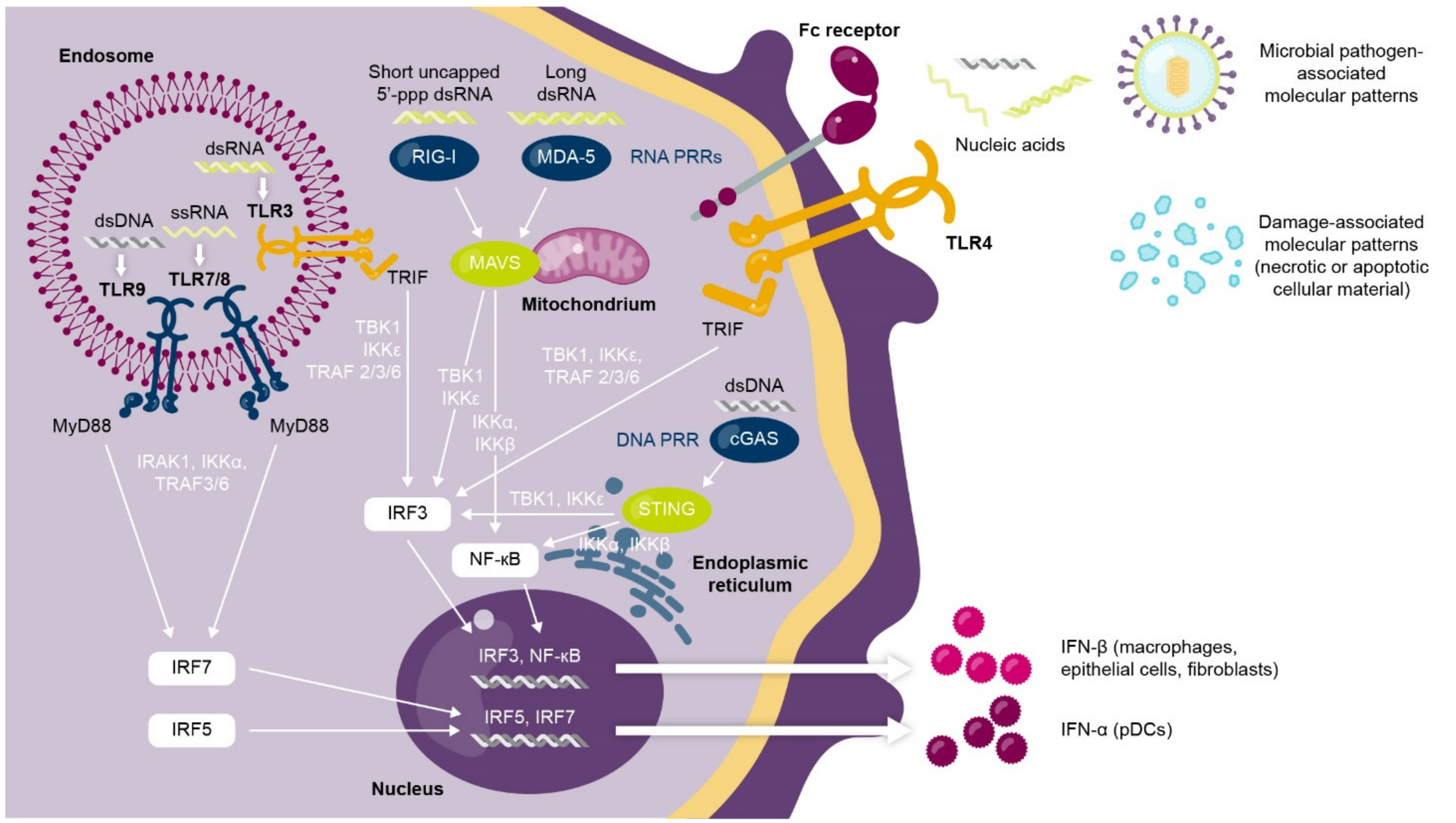
1.1. Interferons Are a Complex Family of Cytokines with Antiviral and Immunomodulatory Functions

Interferons are cytokines with pleiotropic roles in immune regulation that can be categorized into three families (type I, II, or III) based on sequence homology [1]. There is one member of the type II interferon family (interferon [IFN]-γ) and four members of the type III family (IFN-λ1–4). The type I interferon family is the largest, comprising five classes (IFN-α, IFN-β, IFN-ω, IFN-κ, and IFN-ε), of which there are 12 further subgroups of IFN-α proteins in humans [2].
Type I interferons mediate critical antiviral responses and play an immunomodulatory “bridging” role that links both the innate and adaptive immune systems [2]. The innate immune response to viral infection transiently induces high levels of type I interferon expression [3,4]. In brief, the type I interferon response is activated when nucleic acids (RNA, DNA) derived from viruses trigger intracellular pathways mediated by pattern recognition receptors (including Toll-like receptors [TLRs]) to converge on the expression of a type I interferon [3,4] (Figure 1).
The type I interferon then binds to type I interferon α/β receptors (IFNAR) on effector immune cells of the innate and adaptive systems [2,5,6]. The interferon-IFNAR1 complex activates an intracellular signaling cascade, mediated in part by janus kinase (JAK) and signal transducer and activator of transcription (STAT) phosphorylation cascades, which trigger expression/modulation of interferon-regulated antiviral genes [2]. These interferon-stimulated gene products activate the antiviral innate and adaptive immune systems and trigger antigen-specific antiviral T-cell and B-cell responses [7,8,9].

1.2. Dysregulated Type I Interferon Signaling Is Associated with Loss of Immune Tolerance and Autoimmunity

While the transient induction of type I interferon genes in response to viral stimuli is key for host immunity, chronic or dysregulated activation of the type I interferon pathway can contribute to the development of systemic lupus erythematosus (SLE) [2,10,11,12]. In patients with SLE and other autoimmune diseases, this aberrant type I interferon production is triggered by self-nucleic acids in autoimmune complexes, which are often generated as a by-product of a deficiency in apoptotic cell clearance or from dysregulated neutrophil functions [13,14,15,16]. Combined with a loss of the negative feedback signal usually required to “switch off” type I interferon signaling, as well as variants in interferon-related genes [2,17], this chronic type I interferon production stimulates the innate and adaptive immune systems in response to a self-derived trigger, enhancing the original stimulus and contributing to the loss of immune tolerance that is characteristic of autoimmunity [7,17]. This process induces the production of further autoantibodies [18] and type I interferon in a positive autoamplification loop, or vicious cycle [7,14,19].
A build-up of autoimmune complexes can induce systemic and localized inflammation. In patients with SLE, deposits of autoimmune complexes and subsequent inflammation in the tissues contribute to irreversible organ damage and substantial disease burden [20] (Figure 2). Patients with SLE can have a heterogeneous array of organ manifestations, with the most commonly affected organs being the skin, joints, and kidneys [21,22,23]. Of note, non-European patients with SLE often have increased autoantibody reactivity compared with European patients, which may explain why patients who are Black, Asian, or Hispanic present with more severe lupus phenotypes, a greater number of organ manifestations, and a more rapid accumulation of organ damage than patients who are White [24].
While the cellular source of chronic type I interferon production in SLE and other autoimmune diseases is incompletely understood, the predominant source in SLE is likely the plasmacytoid dendritic cell (pDC) [19,25]. pDCs play a predominant role in the aforementioned autoamplification loop that drives type I interferon production and downstream autoantibody generation, as antigen-presentation and type I interferon production take place simultaneously in pDCs and can recruit new pDCs to tissues [19]. Other cell types are also known to contribute, including non-hematopoietic cells, which are thought to play a key role early in disease pathogenesis before clinical autoimmunity [26].

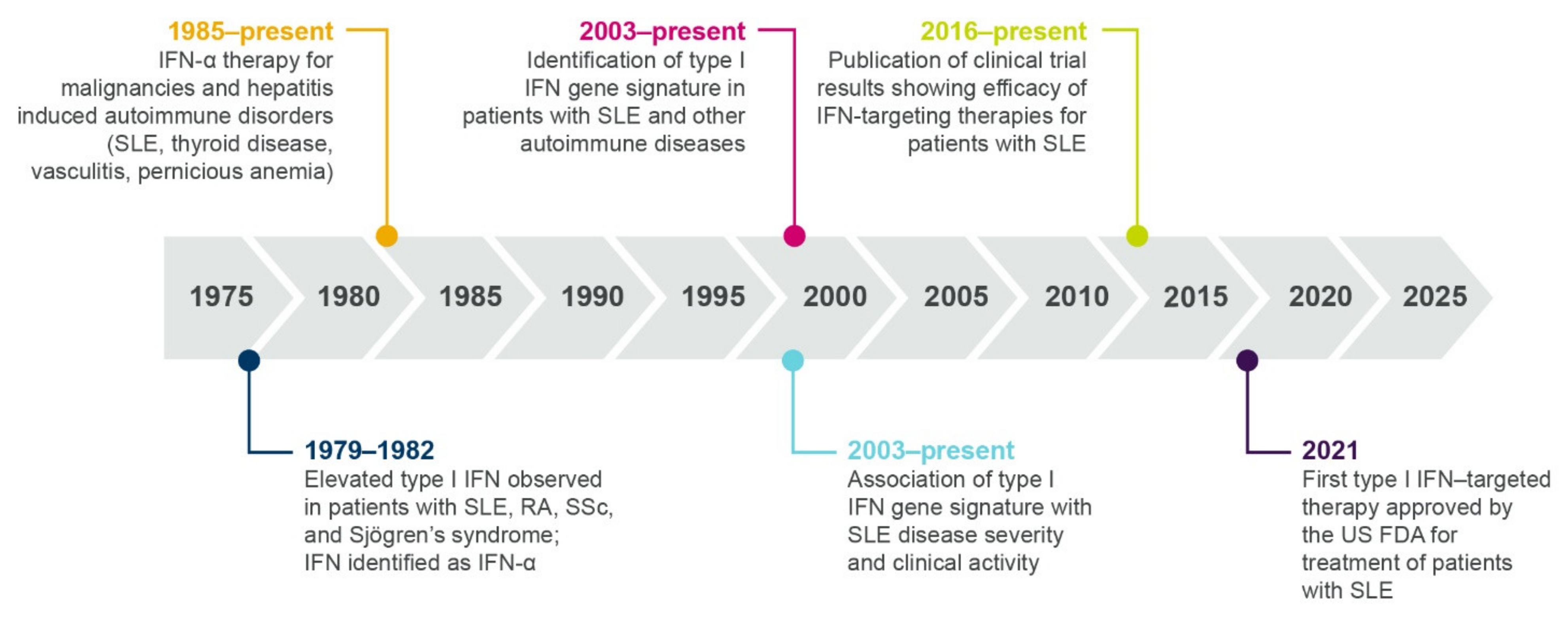
1.3. Multiple Lines of Evidence Support the Role of Interferons in SLE and Autoimmunity

Over the past 40 years, progressive research has contributed to our understanding of the role of interferons in the immunopathogenesis of SLE and other autoimmune diseases (summarized in Figure 3).

1.3.1. Elevated Interferon Protein Is Detected in Patients with Autoimmune Diseases

A role for type I interferons in autoimmunity was first proposed in 1979, when Hooks et al. identified elevated levels of interferon in the serum of patients with SLE, rheumatoid arthritis, systemic sclerosis (SSc), and Sjögren’s syndrome [27]. In 1982, the serum interferon in patients with SLE was identified as IFN-α [28]. More recently, type I interferon protein or transcript signatures have been identified in patients with SLE, myositis (particularly dermatomyositis (DM)), lupus nephritis (LN), SSc, and Sjogren’s syndrome [29,30,31,32,33,34], and low levels of type III interferons have also been identified in patients with SLE [35].
Studies have identified variation in interferon protein expression across SLE racial groups, with greater IFN-α expression in patients with non-European versus European ancestry [36,37]. Furthermore, the autoantibody profile differs across ancestral groups, suggesting alternative mechanisms of interferon induction across different racial groups of patients with SLE [37].

1.3.2. Therapeutic Interferon Can Induce/Exacerbate Autoimmunity

Following initial observations in the 1970s of elevated interferon proteins in patients with autoimmune disease, the next line of evidence for a role of interferons came from observational studies and case reports beginning in 1985. These studies described individuals who developed autoantibodies and autoimmunity (including SLE and SSc) following treatment with IFN-α for malignancies or hepatitis [38,39,40,41]. A randomized, placebo-controlled trial revealed that treatment with IFN-α exacerbated skin and lung manifestations compared with standard therapy in patients with SSc, further supporting a role for type I interferon in disease pathogenesis [42]. As well as inducing the development of SLE and SSc, IFN-α treatment has been rarely associated with the induction of myositis, an autoimmune disease causing muscle weakness [38,43]. A case study described a patient developing inclusion body myositis following treatment with IFN-α, with symptoms exacerbated after repeat dosing [43]. Furthermore, electron microscopy following intramuscular IFN-α administration identified the formation of “lupus inclusions”, a common manifestation of DM, in muscle endothelium [38,43,44].

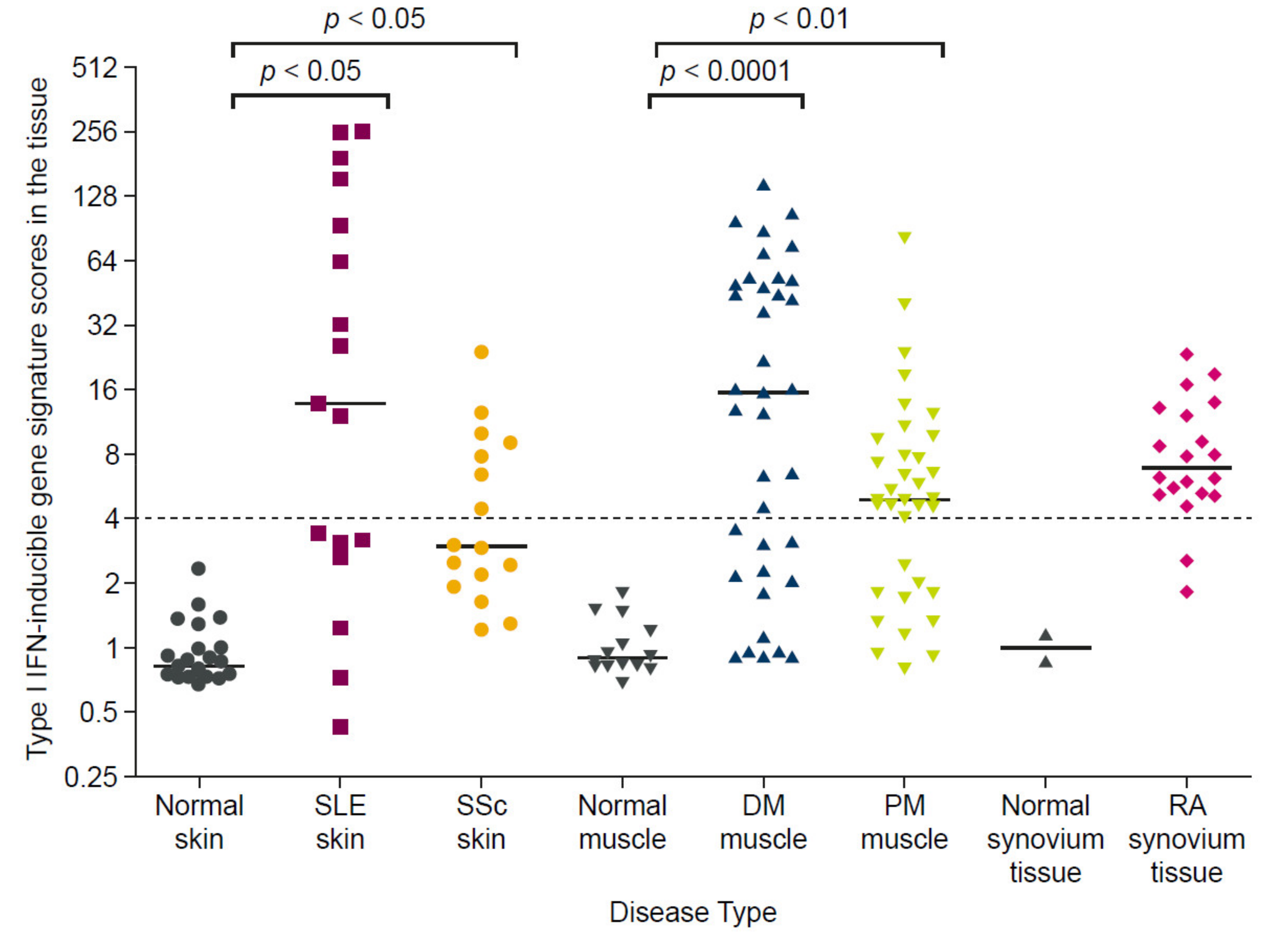
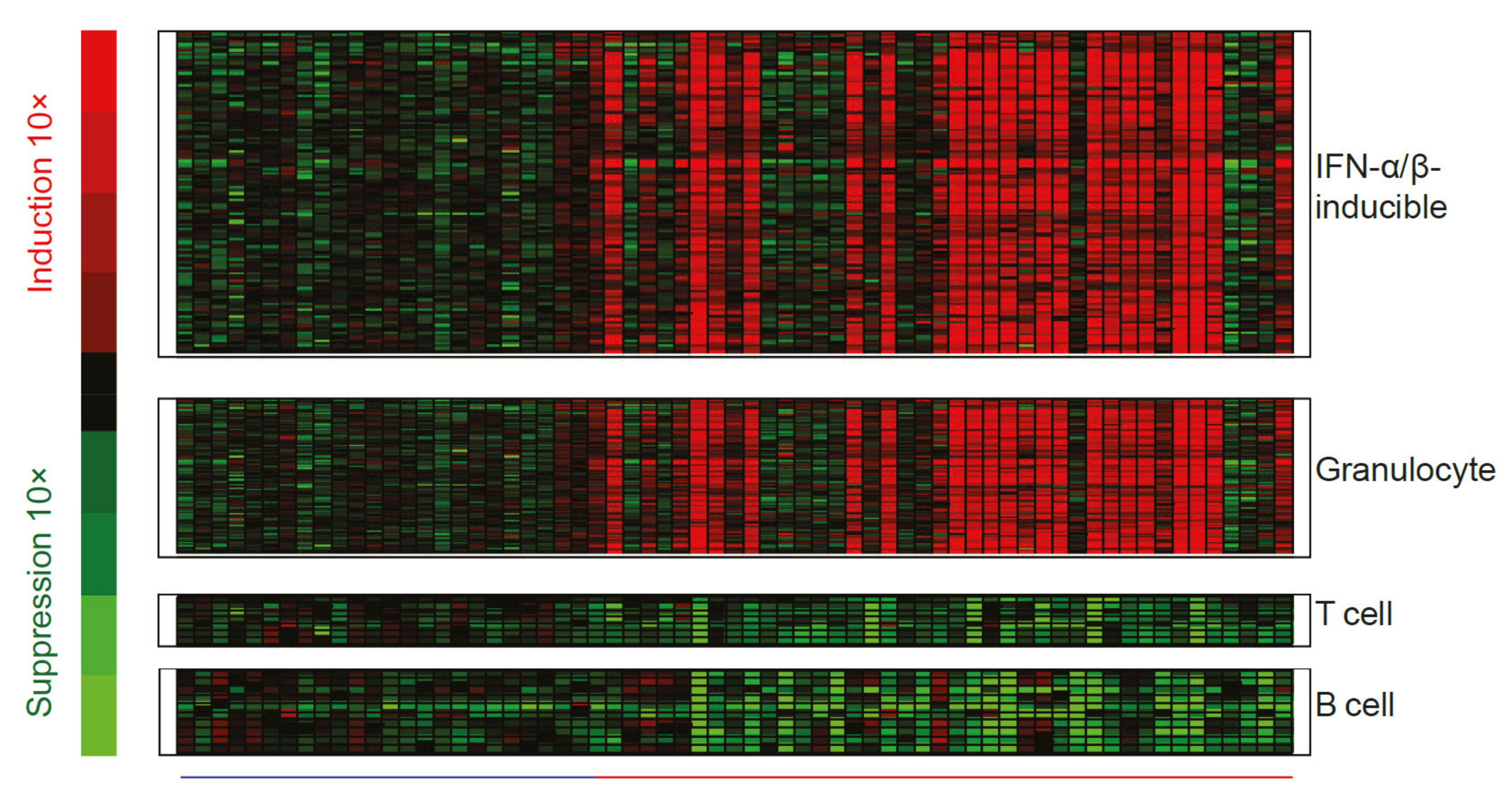
1.3.3. Interferon Gene Signatures Are Upregulated in Patients with Autoimmune Diseases

While measurement of interferon chemokines and reports of unwanted side effects of IFN-α therapy were fundamental in establishing a role for type I interferons in patients with SLE, there are practical challenges associated with quantifying IFN-α protein, which is often present at very low concentrations. These challenges necessitated the development of an interferon gene signature (IFNGS) that could be accurately and accessibly measured as an alternative output for type I interferon pathway activity [45]. In the past 20 years, dysregulated IFNGS expression in blood and affected tissues has been detected in 52–87% of adult patients with SLE [30,45,46,47,48,49,50,51,52,53]. Studies have characterized the key IFN-α/β-inducible transcripts that are overexpressed in whole blood or tissue of patients with SLE compared with healthy controls (Figure 4) [54,55], the dysregulation of which may contribute to SLE immunopathogenesis by way of mechanisms yet to be fully understood [10].
The proportion of patients with SLE who have an elevated IFNGS expression varies with age at diagnosis, disease manifestations, and race; for example, more than 90% of pediatric patients with SLE [56] and 67–83% of adult patients with renal manifestations (LN) [47,57] have an elevated IFNGS expression. An elevated IFNGS expression is also more prevalent among Black and Asian patients than White patients with SLE [58].
Dysregulated type I interferon signaling is also extensively reported for autoimmune diseases other than SLE [59], with an elevated IFNGS expression broadly detected in 68% of patients with SSc, 66% of patients with DM, 61% of patients with polymyositis (PM), and 33% of patients with rheumatoid arthritis, as shown in Figure 5 [30]. Further support for the centrality of interferon axis dysregulation in these autoimmune diseases can be gathered from the finding that type I IFNGS expression could be identified from the earliest SSc disease phases in one patient cohort, even before the emergence of characteristic markers of SSc skin fibrosis that signify the start of organ damage [33]. In the same study, the presence of the IFNGS in monocytes correlated with markers of disease progression, leading the authors to conclude that the type I interferon pathway contributes to both the pathogenesis and progression of SSc [33]. Similarly, in two independent studies, an elevated IFNGS expression was associated with subsequent SLE diagnosis among individuals deemed at high risk of developing an autoimmune condition, supporting a role for interferons in early SLE pathogenesis and progression [60,61].
An elevated type I IFNGS expression in patients with SLE is reported more frequently than an elevated type II or III IFNGS. However, the downstream effects of type I, II, and III interferon signaling overlap considerably, with a number of common genes activated by all three interferon families [62,63,64]. As such, the type I IFNGS expression observed in patients with SLE may also reflect type II and III interferon activity, and assessment of IFN cytokine levels in future studies could help to pinpoint the precise contributing interferon mediators.

1.3.4. Many Immune Cell Types Express Interferon Gene Signatures in Autoimmune Disease

Recently, single-cell analyses have been utilized to investigate the cellular source of the elevated type I interferon gene signature detected in serum and tissue of patients with SLE or LN (i.e., the cells responding to the aberrantly produced pDC-derived type I interferon). These studies identified elevated interferon-regulated gene expression in a broad range of immune cell types (e.g., B cells, natural killer cells, monocytes, T cells, conventional dendritic cells, pDCs, neutrophils, and low-density granulocytes) [65,66,67,68]. Furthermore, single-cell RNA sequencing of renal biopsies from patients with LN identified type I interferon responses in keratinocytes and tubular cells that distinguished patients with LN from healthy controls [69]. The broad range of cell types expressing the IFNGS likely reflects the ubiquity of IFNAR1 expression across many immune cell types [2,6]. Signal blockade via the IFNAR1 thereby has the potential to affect disease biology across a range of immune cell types, including B cells, T cells, pDCs, and natural killer cells [6]; these studies also highlight the potential for targeting specific IFN-producing cell types (such as pDCs, discussed in a later section).

1.3.5. Polymorphisms in Interferon-Related Genes Associate with Increased Risk of Autoimmune Disease

Within the past 13 years, genome-wide association and pathway-centered sequencing studies have identified many polymorphisms in interferon pathway genes that predispose an individual to developing SLE, including polymorphisms affecting the STAT4, IRF3, and IFNA1 genes [70,71,72,73,74]. While single polymorphisms may not be sufficient to cause autoimmunity alone, accumulating numbers of risk variants may cause the immune system to become overwhelmed, leading to immune dysregulation [24,75]. Furthermore, interferon signaling upregulation inpatients with LN may trigger nephropathy associated with apolipoprotein L1 (APOL1) risk-alleles (which are prevalent in individuals with Sub-Saharan African ancestry) by upregulating APOL1 mRNA expression and increasing genotype-associated disease severity [76]. Risk-associated variants in interferon-related genes have also been identified for other autoimmune diseases, including SSc, for which family history remains the strongest known risk factor [32].

1.3.6. Type I Interferon Gene Signature Expression Occurs within a Broader Context of Immune Dysregulation

Decades of profiling gene and protein expression have confirmed the molecular heterogeneity of SLE, and recent studies have attempted to contextualize interferon upregulation within broader immune dysregulation. In a transcriptomics study of pediatric patients with SLE, “personalized immunomonitoring” over time revealed gene expression modules, supported by genotypes, that enabled patient stratification by disease activity. These modules included interferon response genes as well as genes categorized by plasmablast, cell cycle, neutrophil, histone, and B-cell functions [77]. A separate investigation of longitudinal gene expression identified multiple gene modules associated with neutrophil and lymphocyte activity, and differentially associated with type I interferon activity, that allowed adult and pediatric patients with SLE to be stratified by disease activity [78]. Another study assessing gene co-expression across different immune cell types in patients with SLE identified coordinated interferon responses and cross-cell type correlations linking T-helper cell markers and B-cell responses that, in turn, correlated with disease severity [79]. These findings are relevant because stratification by disease activity may provide rationale for clinical trial failures/successes and may help to identify subpopulations suited for targeted therapies.

1.4. Interferon Dysregulation Associates with Clinical Disease Activity and Response to Standard Therapy in Patients with SLE

A wealth of literature supports an association between the presence/extent of type I interferon signaling dysregulation and lupus disease activity. Multiple studies assessing independent cohorts of patients with SLE identified higher global disease activity scores for patients with an elevated IFNGS expression/activity, compared with patients who had a normal IFNGS expression [30,45,47,65]. Greater disease activity was also identified in patients with SLE who had type I interferon protein signatures [29,31], and IFN-λ1 protein levels also correlated with global disease activity in patients with LN and SLE [80].
Interferon pathway dysregulation also associates with the presence of organ-specific disease manifestations in patients with SLE and LN, including kidney, skin, and cardiovascular disease (reviewed in further detail in [2]). pDC infiltrates were detected in the affected kidneys of patients with LN [81], and multiple studies identified an association between serum or kidney-specific type I IFNGS expression and the presence and extent of LN disease activity [66,80,82]. In addition to type I interferons, the detection of IFN-γ-producing autoreactive T cells in the urine of patients with LN also suggests infiltration in inflamed kidneys [83]. The IFNGS expression in blood also correlated with skin disease activity in patients with subacute cutaneous lupus erythematosus (CLE) [84], and multiple studies identified an elevated IFNGS expression in the affected skin of patients with SLE, using both traditional biopsy approaches and, more recently, using non-invasive tape strip sampling [30,85,86]. Notably, IFN-κ production by keratinocytes may be an early step in driving lupus skin disease, where IFN- κ is proposed to activate dendritic cells, amplify IFN-α responses, and contribute to skin photosensitivity [26,87]. Type I interferon has also been implicated in cardiovascular disease in patients with SLE, with multiple studies identifying an association between type I interferon dysregulation and subclinical and clinical cardiovascular disease markers, independent of known cardiovascular risk factors [88,89,90].
While the role of type I interferon in kidney, skin, and cardiovascular disease is established, the immunopathogenesis of lupus joint manifestations is likely more complex, as multiple studies have detected no significant correlation between type I interferon pathway activity (assessed using serum IFN-α activity or interferon gene module expression) and SLE joint involvement using independent patient cohorts [36,48]. Furthermore, local IFN-β production in joints may even have a protective effect in patients with rheumatoid arthritis [91]. However, joint improvement in patients with SLE who received a therapeutic antibody that targeted the type I interferon pathway (discussed later) supports a role for type I interferon in lupus arthritis [50,51,52].
SLE is characterized by unpredictable longitudinal fluctuations in organ-specific and global disease activity, with periods of high disease activity known as flares [92,93]. While the relationship between type I interferon signaling and lupus flares is complex, studies have identified associations between type I interferon activity and longitudinal disease activity changes. For example, high levels of the IFN-α protein were associated with symptom relapse in one cohort of patients with SLE [94], while an elevated IFNGS expression predicted SLE flares in a separate cohort [95]. Despite the sporadic increases in disease activity observed in patients with SLE, the IFNGS expression tends to be stable over time in most patients receiving standard therapies. For example, in phase two and three trials of the anti-IFNAR1 antibody, anifrolumab, the IFNGS expression remained stable throughout the treatment period among patients in the control group who received background standard therapy with oral glucocorticoids and/or immunosuppressants (Figure 6) [50,51,52].
Although an IFNGS generally remains stable over time in patients receiving standard therapies, the extent of interferon dysregulation can affect the standard therapy dosage and response. Pooled data from two randomized controlled trials showed that more patients with SLE who had an elevated IFNGS expression were taking high doses of glucocorticoids (≥10 mg/day) than patients with low levels of IFNGS expression [96]. In a separate single-cell transcriptomic analysis of renal tissue from patients with LN, the patients who did not respond to standard therapy had a greater expression of type I interferon response genes in tubular cells than those who achieved at least a partial response [69]. Similarly, levels of IFN-α but not levels of IFN-λ1 protein correlated with poor response to immunosuppressive therapy in patients with SLE [80], while a reduction in serum IFN-γ protein levels was associated with response to ustekinumab, an anti-interleukin-12/-23 monoclonal antibody (mAb), in a phase two SLE trial [97].
Together, multiple lines of evidence support the fundamental role of interferons in autoimmunity and mediating response to standard therapies and highlight the therapeutic potential of targeting the type I interferon pathway in autoimmune diseases.

2. Therapeutic Highlights: Past, Present, and Future

2.1. Current Standard Therapies, Approved Biologics, and Impact on Interferon Signaling

Currently, the approved standard therapies for patients with SLE include glucocorticoids, antimalarials, and immunosuppressants, for which usage recommendations are based on expert opinion and the literature supporting their effectiveness in treating these patients [92,93]. Some evidence shows that antimalarial and immunosuppressant agents modulate interferon pathway activity as part of their broad mechanism of action, whereas data assessing the effect of glucocorticoid use on interferons are limited (summarized in Table 1).
The largest body of evidence available for the modulation of type I interferons by standard therapies is for the antimalarial agent hydroxychloroquine (HCQ), which prevents the maturation of endosomes and disrupts the activity of endosome-resident TLRs, key components in type I interferon production [25]. Multiple studies have reported the inhibition of IFN-α production or IFNGS expression in patients with SLE or Sjögren’s syndrome following HCQ treatment [25,99,100]. However, the extent to which interferon inhibition by HCQ relates to its clinical effectiveness remains to be investigated.
The immunosuppressant drug mycophenolate mofetil (MMF) was also shown in one recent study to non-specifically inhibit the production of IFN-α by pDCs in a dose-dependent manner, and this was confirmed by transcript levels [98]. However, this study assessed pDCs derived from healthy volunteers only; therefore, future research is required to investigate the effect in an autoimmune environment (where patients may have dysfunctional pDCs [120]), and to investigate the downstream effect of IFNGS expression on MMF effectiveness.
Although use of standard therapies is associated with short-term benefits and these therapies are recommended by current treatment guidelines, not all patients will respond to them, and the use of these agents is often accompanied by adverse effects [92,93]. Glucocorticoid use, in particular, is associated with a significant burden, as high dosages (prednisone ≥ 7.5 mg/day or equivalent) are associated with an increased risk of organ damage compared with lower dosages [121,122,123].
Due to the unpredictable response and safety issues with standard therapies, there remains a need for additional therapeutic options to treat patients with SLE. Targeted therapeutics and precision medicine may be particularly valuable for a disease such as SLE, for which disease course, therapeutic response, and underlying immune mechanisms are both complex and heterogeneous, suggesting that a “one size fits all” approach may not be appropriate [124]. The need for precision medicine is further illustrated by the low response rates to standard therapies in patients with SLE [125].
Approval of belimumab, the first targeted biologic approved for use in patients with active SLE or LN despite standard therapy, represented a therapeutic milestone [126,127]. Belimumab is a mAb that inhibits the soluble counterpart of the B-lymphocyte stimulator (BLyS), which is a key mediator of B-cell differentiation and survival, and thus dampens B-cell mediated autoimmunity [126,127]. A recent post hoc analysis of phase three trial data indicated that baseline BLyS and IFN-1 mRNA levels were highly correlated and that the Systemic Lupus Erythematosus Responder Index ≥ 4 (SRI(4)) response to belimumab for IFNGS-high patients was consistent with the response overall [128], suggesting an indirect interaction between BLyS inhibition and interferon modulation.

2.2. An Interferon-Targeting Therapeutic Is Now Approved for the Treatment of SLE and Others Are in Clinical Development

The role of type I interferons in autoimmunity highlights the therapeutic potential of targeting the type I interferon pathway in autoimmune diseases. While standard therapies and belimumab may have indirect effects on type I interferon signaling as part of their broad mechanism of action, a number of targeted therapeutics that inhibit type I interferon signaling, either directly or indirectly, are at various stages of clinical development (summarized in Table 1). A timeline showing the clinical development of interferon-targeted biologics is presented in Figure 7. These molecules include biologics that directly target IFN-α chemokine or the IFNAR, and biologics that block the production of type I interferon by inhibiting or depleting the pDC function. Small molecule inhibitors, specifically JAK inhibitors, also indirectly target intracellular mediators of the type I interferon/IFNAR1 axis.
As of 2021, four direct type I interferon-targeting therapies have been developed for the treatment of patients with SLE, myositis, or LN, and have demonstrated variable clinical benefits. IFN-α kinoid (IFN-K) is an immunotherapeutic vaccine that induces the production of polyclonal anti-IFN-α antibodies to subsequently block type I interferon activity and normalize the IFNGS [107,108]. IFN-K was recently assessed in a phase two trial of patients with active SLE despite standard therapy and a positive IFNGS. Although more patients attained a lupus low disease activity state (LLDAS) or glucocorticoid tapering with IFN-K than with the placebo, the trial did not meet its clinical primary endpoint (modified British Isles Lupus Assessment Group (BILAG)-based Composite Lupus Assessment (BICLA) response) [109]. The development of IFN-K has since been discontinued and no further trials are planned.
Rontalizumab is a humanized mAb and sifalimumab is a fully human mAb that inhibit IFN-α and suppress IFNGS expression [53,110,111,129]. Rontalizumab treatment was associated with moderate clinical benefit in a phase two trial of patients with SLE with low IFNGS scores [111]; however, neither primary nor secondary endpoints (BILAG-2004 reduction, SLE Responder Index ≥ 4 (SRI(4)) response at Week 24) were met for all patients or for IFNGS-high patients [111], and development was discontinued. The study design may have affected the outcomes. Patients received the study drug intravenously for 24 weeks during stage one, followed by re-randomization and subcutaneous administration of study drug for a further 24 weeks in stage two [111,130]. The change from intravenous to subcutaneous administration halfway through the trial may have led to inadequate dosing, which could explain the lack of response for IFNGS-high patients [111,130]. Additionally, all patients taking immunosuppressive agents at baseline were required per protocol to discontinue, which could have confounded efficacy estimates, especially accounting for the 24-week duration of treatment prior to efficacy assessment, which is shorter than most other SLE trials [130]. This, combined with a small sample size, may have contributed to the failed primary outcome [111,130].
More promising results were observed with sifalimumab, which was associated with clinical benefit for patients with SLE and those with myositis [53,129]. In a phase 2b trial of patients with moderate to severe SLE, sifalimumab suppressed the IFNGS, and primary and secondary endpoints were met, with a greater proportion of patients achieving an SRI(4) response at Week 52 with sifalimumab versus placebo, accompanied by other improvements in skin disease activity and a reduction in swollen and tender joint counts [53]. In a phase one trial of patients with DM and PM, sifalimumab suppressed the IFNGS, and the extent of IFNGS suppression in muscle and blood was associated with an improvement in manual muscle test scores [129]. Despite the positive phase two results with sifalimumab, further development was paused in favor of the more substantial IFNGS neutralization observed with the anti-IFNAR1 antibody, anifrolumab.
In contrast to sifalimumab and rontalizumab, which target IFN-α, anifrolumab, the first US Food and Drug Administration (FDA)- and Japanese Ministry of Health, Labour and Welfare (MHLW)-approved therapeutic targeting the type I interferon pathway, is a fully human IgG1κ mAb that blocks all type I interferon/IFNAR1 signaling [103,104,105,106]. Anifrolumab rapidly internalizes the IFNAR1 and sterically inhibits the IFNAR1, preventing interferon/IFNAR1 complex formation, and ultimately suppressing the IFNGS [103,104]. Anifrolumab disrupts the type I interferon auto-amplification loop that can trigger the loss of immune tolerance and autoimmunity, as it blocks type I interferon signaling in pDCs and suppresses the induction of type I interferons following immune complex stimulation [103]. In addition, anifrolumab partially inhibits upregulation of costimulatory molecules and the production of pro-inflammatory cytokines by pDCs [103]. As well as inhibiting the type I IFN-pDC axis of the innate immune system, anifrolumab suppresses B-cell survival factors (e.g., BLyS, B lymphocyte chemoattractant (BLC)), inhibits B-cell differentiation, and suppresses T-cell activation, all of which are adaptive immune mediators of autoimmune pathogenesis [103,131,132].
Anifrolumab is indicated for the treatment of adult patients with moderate to severe SLE who are receiving standard therapy [104]. In phase two and phase three trials, anifrolumab treatment was associated with clinical benefit across multiple efficacy endpoints for patients with moderate to severe SLE despite standard therapy, consistently showing a greater improvement in overall disease activity as assessed by the BICLA response at Week 52 compared with a placebo [50,51,52]. While the BICLA response rates with anifrolumab were consistently higher than with a placebo, regardless of the extent of pharmacodynamic IFNGS expression, a positive relationship between greater IFNGS suppression and the magnitude of BICLA response rates was apparent [133]. Improvements were also seen for skin and joint disease activity with anifrolumab, and more patients were able to taper oral glucocorticoids to a low dosage threshold of ≤7.5 mg/day with anifrolumab than a placebo [50,51,52].
As well as showing improvements in clinical outcomes, anifrolumab treatment was associated with reduced levels of inflammatory and cardiometabolic disease markers in a phase two trial [90,131]. Patients with SLE were at a three- to four-fold greater risk of experiencing cardiovascular events than healthy controls (adjusting for known risk factors such as age, sex, and smoking history), as a result of immune-mediated vascular damage and premature atherosclerosis [134]. Anifrolumab treatment significantly modulated levels of multiple subclinical markers of cardiometabolic disease that contribute to SLE vasculopathy, suggesting that inhibiting type I IFN signaling could reduce atherosclerosis and cardiovascular markers in patients with SLE [90].
As well as a direct blockade of interferon/IFNAR1, strategies to inhibit type I interferon production from pDCs are in development for the treatment of patients with SLE and CLE. VIB7734 is a pDC-depleting mAb engineered for enhanced effector function that targets the pDC-specific marker immunoglobulin-like transcript 7 (ILT7) [112]. In a phase one trial that enrolled patients with CLE, VIB7734 depleted levels of circulating and tissue-resident pDCs, decreased local type I interferon production, and improved clinical disease activity [112].
Another mAb under development, BIIB059, binds the pDC-specific blood dendritic cell antigen 2 (BDCA2) receptor [114,115]. Unlike VIB7734, BIIB059 induces rapid BDCA2 internalization and inhibits type I interferon production without depleting pDCs [114]. In a phase one trial that enrolled patients with SLE, BIIB059 decreased the IFNGS expression in blood and normalized levels of interferon response proteins in the affected skin tissue [114]. In a phase two trial, BIIB059 treatment was associated with a reduction in the number of active joints and higher SRI(4) response rates compared with a placebo in patients with SLE [116]. In the same trial, BIIB059 treatment reduced skin disease activity scores compared with a placebo in patients with CLE [135]. These positive phase two results support continued clinical development of BIIB059 in patients with SLE and CLE [116,135].
In an alternative approach to targeting interferons, multiple JAK inhibitors (JAKi), which act downstream in the interferon signaling pathway, are in development. In a phase two trial of baricitinib (a JAK1- and JAK2-selective inhibitor), gene expression profiling revealed the suppression of interferon-responsive genes following 24 weeks of study treatment, in addition to the suppression of JAK-STAT signaling and other genes involved in SLE immunopathogenesis [119]. Similarly, the treatment of patients with SLE with tofacitinib (which inhibits JAK3, as well as JAK1 and JAK2 to a lesser extent) decreased the IFNGS expression in a phase one trial [117], and multiple JAKis were shown to suppress the IFNGS in a study of off-label use in pediatric patients with SLE [118].
Although the aforementioned interferon-targeting therapies are in clinical development for the treatment of patients with autoimmune disease, an interferon blockade also has the potential for clinical benefit in indications beyond autoimmunity. Recently, sustained type I interferon signaling was shown to cause resistance to immunotherapies in patients with melanoma [136]. Further investigations are needed to establish whether an interferon blockade could be beneficial in patients with cancer who have interferon-mediated therapeutic resistance.

2.3. The Future of Interferon Blockade and Lupus Therapies

The range of different interferon-targeting molecules in clinical development is perhaps due to the unprecedented clinical trial activity in SLE and other autoimmune diseases over the past two decades, in which trials are targeting varied mechanisms with biologics, kinase inhibitors, and other small molecule compounds [137]. While drug discovery in SLE has historically lagged behind other diseases, predominantly owing to challenges associated with measuring lupus disease activity in a heterogeneous disease, the vast number of new trials and improvements in trial outcome measures has led to a series of successful lupus trials with anifrolumab, voclosporin, and belimumab [137,138]. These recent successes, together with the sheer volume of ongoing clinical trials, increases the chance of additional drugs becoming available to patients within the next decade, illustrated by the recent approvals of belimumab and voclosporin for the treatment of patients with LN [137].
Future drug approvals may include interferon-targeting agents, based on the numerous interferon-targeting molecules (direct or indirect) currently in development and the recent successes seen in both early- and late-phase trials [50,112,135]. However, not all patients respond to type I IFN-targeting agents, and there is a need to understand the role played by other pathways and how these interact with type I IFN signaling.
Broader changes in lupus standard therapy use will likely emerge as more targeted therapeutics become available. Consistent with current trends, glucocorticoid use will likely decrease further in the next decade among patients with SLE, based on the recommendations of updated treatment guidelines [92,93]. Although glucocorticoid use is common among patients with SLE with an elevated IFNGS expression [96], type I interferon inhibition with anifrolumab reduced the need for glucocorticoid use in patients with SLE [50,51,52], highlighting the glucocorticoid-sparing capability of targeted anti-IFN therapies for some patients.
As we continue to deepen our understanding of lupus immunopathogenesis, stratification by immunophenotype will become increasingly commonplace in future SLE trials. In phase two and three anifrolumab trials, 75–83% of patients were IFNGS-high at screening, reflecting the natural distribution in this patient population [50,51,52]. A direct comparison of the anifrolumab treatment response for IFNGS-high versus IFNGS-low patients was limited by the small sample size of the IFNGS-low subgroup in the phase three anifrolumab trials. However, a post hoc analysis of pooled data from two phase three trials indicated that the magnitude of the treatment difference for anifrolumab versus placebo was greater among the IFNGS-high patients than the IFNGS-low patients, mostly attributable to the low placebo response rates with standard therapy in the IFNGS-high patients [50,51,52]. The availability of additional targeted therapeutic options will represent a further milestone for patients with dysregulated type I interferon signaling, who tend to have severe disease and suboptimal responses to standard therapy [30,45,69,80,96]. As an increased prevalence of type I interferon dysregulation has been identified in patients who are non-White versus White and treatment response can vary by race [24], future trials of targeted therapies should also monitor treatment response across racial groups.

3. Conclusions

The type I interferon pathway plays a major role in the etiology of SLE and many other autoimmune diseases. Patients with SLE with dysregulated type I interferon activity often have more active disease, which is resistant to standard therapies, compared with patients without dysregulated interferon signaling [30,45,47,65,80]. Over the past 40 years, extensive research from the lupus community has made substantial progress in deconvoluting the immune complexity of SLE and identifying key molecular and cellular components of type I interferon signaling. These efforts, combined with the unprecedented SLE clinical trial activity in the past two decades, have culminated in the approval of the first agent targeting the IFN pathway for the treatment of patients with SLE [104], highlighting that type I interferon is a viable therapeutic target that reduces disease activity in patients with SLE and other autoimmune diseases.

Author Contributions

M.R., R.T., K.S., A.N.d.C. and P.Z.B. were responsible for conceptualization, reviewing and revising the work, and approved the final draft of the manuscript. All authors have read and agreed to the published version of the manuscript.

Funding

This work was funded by AstraZeneca.

Institutional Review Board Statement

Not applicable.

Informed Consent Statement

Not applicable.

Data Availability Statement

Not applicable.

Acknowledgments

Medical writing support was provided by Rosie Butler, JK Associates, Inc., part of Fishawack Health. This support was funded by AstraZeneca.

Conflicts of Interest

M.R. (Senior scientist/ Translational Science Lead), R.T. (Global Clinical Head), K.S. (Senior Director/ Head of Translational Medicine), A.N.d.C. (Head of Precision Medicine Discovery and Development Immunology), and P.Z.B. (Senior Director/ Head of Biomarker Discovery, Senior Translational Science Lead) are current employees of AstraZeneca.

References

  1. Lazear, H.M.; Schoggins, J.W.; Diamond, M.S. Shared and Distinct Functions of Type I and Type III Interferons. Immunity 2019, 50, 907–923. [Google Scholar] [CrossRef] [PubMed]
  2. Rönnblom, L.; Leonard, D. Interferon Pathway in SLE: One Key to Unlocking the Mystery of the Disease. Lupus Sci. Med. 2019, 6, e000270. [Google Scholar] [CrossRef] [PubMed] [Green Version]
  3. Quaglia, M.; Merlotti, G.; De Andrea, M.; Borgogna, C.; Cantaluppi, V. Viral Infections and Systemic Lupus Erythematosus: New Players in an Old Story. Viruses 2021, 13, 277. [Google Scholar] [CrossRef] [PubMed]
  4. Quan, T.E.; Roman, R.M.; Rudenga, B.J.; Holers, V.M.; Craft, J.E. Epstein-Barr Virus Promotes Interferon-Alpha Production by Plasmacytoid Dendritic Cells. Arthritis Rheum. 2010, 62, 1693–1701. [Google Scholar] [CrossRef] [Green Version]
  5. Liao, A.P.; Salajegheh, M.; Morehouse, C.; Nazareno, R.; Jubin, R.G.; Jallal, B.; Yao, Y.; Greenberg, S.A. Human Plasmacytoid Dendritic Cell Accumulation Amplifies Their Type 1 Interferon Production. Clin. Immunol. 2010, 136, 130–138. [Google Scholar] [CrossRef] [Green Version]
  6. De Weerd, N.A.; Nguyen, T. The Interferons and Their Receptors—Distribution and Regulation. Immunol. Cell Biol. 2012, 90, 483–491. [Google Scholar] [CrossRef]
  7. Greth, W.; Robbie, G.J.; Brohawn, P.; Hultquist, M.; Yao, B. Targeting the Interferon Pathway with Sifalimumab for the Treatment of Systemic Lupus Erythematosus. Immunotherapy 2017, 9, 57–70. [Google Scholar] [CrossRef]
  8. Stetson, D.B.; Medzhitov, R. Type I Interferons in Host Defense. Immunity 2006, 25, 373–381. [Google Scholar] [CrossRef] [PubMed] [Green Version]
  9. De Goër de Herve, M.G.; Durali, D.; Dembele, B.; Giuliani, M.; Tran, T.A.; Azzarone, B.; Eid, P.; Tardieu, M.; Delfraissy, J.F.; Taoufik, Y. Interferon-Alpha Triggers B Cell Effector 1 (Be1) Commitment. PLoS ONE 2011, 6, e19366. [Google Scholar] [CrossRef]
  10. Crow, M.K. Type I Interferon in the Pathogenesis of Lupus. J. Immunol. 2014, 192, 5459–5468. [Google Scholar] [CrossRef]
  11. Niewold, T.B. Interferon Alpha as a Primary Pathogenic Factor in Human Lupus. J. Interferon Cytokine Res. 2011, 31, 887–892. [Google Scholar] [CrossRef] [PubMed] [Green Version]
  12. Ivashkiv, L.B. Type I Interferon Modulation of Cellular Responses to Cytokines and Infectious Pathogens: Potential Role in SLE Pathogenesis. Autoimmunity 2003, 36, 473–479. [Google Scholar] [CrossRef] [PubMed]
  13. Lande, R.; Ganguly, D.; Facchinetti, V.; Frasca, L.; Conrad, C.; Gregorio, J.; Meller, S.; Chamilos, G.; Sebasigari, R.; Riccieri, V.; et al. Neutrophils Activate Plasmacytoid Dendritic Cells by Releasing Self-DNA-Peptide Complexes in Systemic Lupus Erythematosus. Sci. Transl. Med. 2011, 3, 73ra19. [Google Scholar] [CrossRef] [PubMed] [Green Version]
  14. Garcia-Romo, G.S.; Caielli, S.; Vega, B.; Connolly, J.; Allantaz, F.; Xu, Z.; Punaro, M.; Baisch, J.; Guiducci, C.; Coffman, R.L.; et al. Netting Neutrophils are Major Inducers of Type I IFN Production in Pediatric Systemic Lupus Erythematosus. Sci. Transl. Med. 2011, 3, 73ra20. [Google Scholar] [CrossRef] [PubMed] [Green Version]
  15. Ainola, M.; Porola, P.; Takakubo, Y.; Przybyla, B.; Kouri, V.P.; Tolvanen, T.A.; Hänninen, A.; Nordström, D.C. Activation of Plasmacytoid Dendritic Cells by Apoptotic Particles—Mechanism for the Loss of Immunological Tolerance in Sjögren’s Syndrome. Clin. Exp. Immunol. 2018, 191, 301–310. [Google Scholar] [CrossRef] [Green Version]
  16. Schiller, M.; Parcina, M.; Heyder, P.; Foermer, S.; Ostrop, J.; Leo, A.; Heeg, K.; Herrmann, M.; Lorenz, H.M.; Bekeredjian-Ding, I. Induction of Type I IFN is a Physiological Immune Reaction to Apoptotic Cell-Derived Membrane Microparticles. J. Immunol. 2012, 189, 1747–1756. [Google Scholar] [CrossRef] [PubMed] [Green Version]
  17. Eloranta, M.L.; Rönnblom, L. Cause and Consequences of the Activated Type I Interferon System in SLE. J. Mol. Med. 2016, 94, 1103–1110. [Google Scholar] [CrossRef]
  18. Arbuckle, M.R.; McClain, M.T.; Rubertone, M.V.; Scofield, R.H.; Dennis, G.J.; James, J.A.; Harley, J.B. Development of Autoantibodies Before the Clinical Onset of Systemic Lupus Erythematosus. N. Engl. J. Med. 2003, 349, 1526–1533. [Google Scholar] [CrossRef] [Green Version]
  19. Rönnblom, L.; Alm, G.V. A Pivotal Role for the Natural Interferon Alpha-Producing Cells (Plasmacytoid Dendritic Cells) in the Pathogenesis of Lupus. J. Exp. Med. 2001, 194, F59–F63. [Google Scholar] [CrossRef]
  20. Mayadas, T.N.; Tsokos, G.C.; Tsuboi, N. Mechanisms of Immune Complex-Mediated Neutrophil Recruitment and Tissue Injury. Circulation 2009, 120, 2012–2024. [Google Scholar] [CrossRef] [Green Version]
  21. Uva, L.; Miguel, D.; Pinheiro, C.; Freitas, J.P.; Marques Gomes, M.; Filipe, P. Cutaneous Manifestations of Systemic Lupus Erythematosus. Autoimmune Dis. 2012, 2012, 834291. [Google Scholar] [CrossRef] [PubMed]
  22. McCauliffe, D.P. Cutaneous Lupus Erythematosus. Semin. Cutan. Med. Surg. 2001, 20, 14–26. [Google Scholar] [CrossRef]
  23. Zayat, A.S.; Md Yusof, M.Y.; Wakefield, R.J.; Conaghan, P.G.; Emery, P.; Vital, E.M. The Role of Ultrasound in Assessing Musculoskeletal Symptoms of Systemic Lupus Erythematosus: A Systematic Literature Review. Rheumatology 2016, 55, 485–494. [Google Scholar] [CrossRef] [PubMed] [Green Version]
  24. Lewis, M.J.; Jawad, A.S. The effect of ethnicity and genetic ancestry on the epidemiology, clinical features and outcome of systemic lupus erythematosus. Rheumatology 2017, 56, i67–i77. [Google Scholar] [CrossRef] [PubMed] [Green Version]
  25. Sacre, K.; Criswell, L.A.; McCune, J.M. Hydroxychloroquine is Associated With Impaired Interferon-Alpha and Tumor Necrosis Factor-Alpha Production by Plasmacytoid Dendritic Cells in Systemic Lupus Erythematosus. Arthritis Res. Ther. 2012, 14, R155. [Google Scholar] [CrossRef] [Green Version]
  26. Psarras, A.; Alase, A.; Antanaviciute, A.; Carr, I.M.; Md Yusof, M.Y.; Wittmann, M.; Emery, P.; Tsokos, G.C.; Vital, E.M. Functionally Impaired Plasmacytoid Dendritic Cells and Non-Haematopoietic Sources of Type I Interferon Characterize Human Autoimmunity. Nat. Commun. 2020, 11, 6149. [Google Scholar] [CrossRef]
  27. Hooks, J.J.; Moutsopoulos, H.M.; Geis, S.A.; Stahl, N.I.; Decker, J.L.; Notkins, A.L. Immune Interferon in the Circulation of Patients With Autoimmune Disease. N. Engl. J. Med. 1979, 301, 5–8. [Google Scholar] [CrossRef]
  28. Ytterberg, S.R.; Schnitzer, T.J. Serum Interferon Levels in Patients With Systemic Lupus Erythematosus. Arthritis Rheum. 1982, 25, 401–406. [Google Scholar] [CrossRef]
  29. Smith, M.A.; Henault, J.; Karnell, J.L.; Parker, M.L.; Riggs, J.M.; Sinibaldi, D.; Taylor, D.K.; Ettinger, R.; Grant, E.P.; Sanjuan, M.A.; et al. SLE Plasma Profiling Identifies Unique Signatures of Lupus Nephritis and Discoid Lupus. Sci. Rep. 2019, 9, 14433. [Google Scholar] [CrossRef] [Green Version]
  30. Higgs, B.W.; Liu, Z.; White, B.; Zhu, W.; White, W.I.; Morehouse, C.; Brohawn, P.; Kiener, P.A.; Richman, L.; Fiorentino, D.; et al. Patients With Systemic Lupus Erythematosus, Myositis, Rheumatoid Arthritis and Scleroderma Share Activation of a Common Type I Interferon Pathway. Ann. Rheum. Dis. 2011, 70, 2029–2036. [Google Scholar] [CrossRef]
  31. Smith, M.A.; Chiang, C.-C.; Zerrouki, K.; Rahman, S.; White, W.I.; Streicher, K.; Rees, W.A.; Schiffenbauer, A.; Rider, L.G.; Miller, F.W.; et al. Using the Circulating Proteome to Assess Type I Interferon Activity in Systemic Lupus Erythematosus. Sci. Rep. 2020, 10, 4462. [Google Scholar] [CrossRef]
  32. Skaug, B.; Assassi, S. Type I Interferon Dysregulation in Systemic Sclerosis. Cytokine 2020, 132, 154635. [Google Scholar] [CrossRef]
  33. Brkic, Z.; van Bon, L.; Cossu, M.; van Helden-Meeuwsen, C.G.; Vonk, M.C.; Knaapen, H.; van den Berg, W.; Dalm, V.A.; Van Daele, P.L.; Severino, A.; et al. The Interferon Type I Signature is Present in Systemic Sclerosis Before Overt Fibrosis and Might Contribute to its Pathogenesis Through High BAFF Gene Expression and High Collagen Synthesis. Ann. Rheum. Dis. 2016, 75, 1567–1573. [Google Scholar] [CrossRef]
  34. Yao, Y.; Liu, Z.; Jallal, B.; Shen, N.; Rönnblom, L. Type I Interferons In Sjögren’s Syndrome. Autoimmun. Rev. 2013, 12, 558–566. [Google Scholar] [CrossRef] [Green Version]
  35. Hjorton, K.; Hagberg, N.; Pucholt, P.; Eloranta, M.L.; Rönnblom, L. The Regulation and Pharmacological Modulation of Immune Complex Induced Type III IFN Production by Plasmacytoid Dendritic Cells. Arthritis Res. Ther. 2020, 22, 130. [Google Scholar] [CrossRef]
  36. Weckerle, C.E.; Franek, B.S.; Kelly, J.A.; Kumabe, M.; Mikolaitis, R.A.; Green, S.L.; Utset, T.O.; Jolly, M.; James, J.A.; Harley, J.B.; et al. Network Analysis of Associations Between Serum Interferon-A Activity, Autoantibodies, and Clinical Features in Systemic Lupus Erythematosus. Arthritis Rheum. 2011, 63, 1044–1053. [Google Scholar] [CrossRef]
  37. Ko, K.; Koldobskaya, Y.; Rosenzweig, E.; Niewold, T.B. Activation of the Interferon Pathway is Dependent Upon Autoantibodies in African-American SLE Patients, but not in European-American SLE Patients. Front. Immunol. 2013, 4, 309. [Google Scholar] [CrossRef] [PubMed] [Green Version]
  38. Grimley, P.M.; Davis, G.L.; Kang, Y.H.; Dooley, J.S.; Strohmaier, J.; Hoofnagle, J.H. Tubuloreticular Inclusions in Peripheral Blood Mononuclear Cells Related to Systemic Therapy With Alpha-Interferon. Lab Invest 1985, 52, 638–649. [Google Scholar] [PubMed]
  39. Rönnblom, L.E.; Alm, G.V.; Oberg, K.E. Autoimmunity after alpha-interferon therapy for malignant carcinoid tumors. Ann. Intern. Med. 1991, 115, 178–183. [Google Scholar] [CrossRef] [PubMed]
  40. Tahara, H.; Kojima, A.; Hirokawa, T.; Oyama, T.; Naganuma, A.; Maruta, S.; Okada, K.; Ban, S.; Yoshida, K.; Takagi, H.; et al. Systemic Sclerosis After Interferon Alphacon-1 Therapy for Hepatitis C. Intern. Med. 2007, 46, 473–476. [Google Scholar] [CrossRef] [PubMed] [Green Version]
  41. Rönnblom, L.E.; Alm, G.V.; Oberg, K.E. Possible Induction of Systemic Lupus Erythematosus by Interferon-Alpha Treatment in a Patient With a Malignant Carcinoid Tumour. J. Intern. Med. 1990, 227, 207–210. [Google Scholar] [CrossRef] [PubMed]
  42. Black, C.M.; Silman, A.J.; Herrick, A.I.; Denton, C.P.; Wilson, H.; Newman, J.; Pompon, L.; Shi-Wen, X. Interferon-Alpha Does not Improve Outcome at One Year in Patients With Diffuse Cutaneous Scleroderma: Results of a Randomized, Double-Blind, Placebo-Controlled Trial. Arthritis Rheum. 1999, 42, 299–305. [Google Scholar] [CrossRef]
  43. Warabi, Y.; Matsubara, S.; Mizutani, T.; Hayashi, H. Inclusion Body Myositis After Interferon-Alpha Treatment in a Patient with HCV and HTLV-1 Infection. Rinsho Shinkeigaku 2004, 44, 609–614. [Google Scholar] [PubMed]
  44. Greenberg, S.A. Dermatomyositis and Type 1 Interferons. Curr. Rheumatol. Rep. 2010, 12, 198–203. [Google Scholar] [CrossRef] [Green Version]
  45. Baechler, E.C.; Batliwalla, F.M.; Karypis, G.; Gaffney, P.M.; Ortmann, W.A.; Espe, K.J.; Shark, K.B.; Grande, W.J.; Hughes, K.M.; Kapur, V.; et al. Interferon-Inducible Gene Expression Signature in Peripheral Blood Cells of Patients With Severe Lupus. Proc. Natl. Acad. Sci. USA 2003, 100, 2610–2615. [Google Scholar] [CrossRef] [PubMed] [Green Version]
  46. Petri, M.; Singh, S.; Tesfasyone, H.; Dedrick, R.; Fry, K.; Lal, P.; Williams, G.; Bauer, J.; Gregersen, P.; Behrens, T.; et al. Longitudinal Expression of Type I Interferon Responsive Genes in Systemic Lupus Erythematosus. Lupus 2009, 18, 980–989. [Google Scholar] [CrossRef] [PubMed] [Green Version]
  47. Mai, L.; Asaduzzaman, A.; Noamani, B.; Fortin, P.R.; Gladman, D.D.; Touma, Z.; Urowitz, M.B.; Wither, J. The Baseline Interferon Signature Predicts Disease Severity Over the Subsequent 5 Years in Systemic Lupus Erythematosus. Arthritis Res. Ther. 2021, 23, 29. [Google Scholar] [CrossRef]
  48. Chiche, L.; Jourde-Chiche, N.; Whalen, E.; Presnell, S.; Gersuk, V.; Dang, K.; Anguiano, E.; Quinn, C.; Burtey, S.; Berland, Y.; et al. Modular Transcriptional Repertoire Analyses of Adults With Systemic Lupus Erythematosus Reveal Distinct Type I and Type II Interferon Signatures. Arthritis Rheum. 2014, 66, 1583–1595. [Google Scholar] [CrossRef]
  49. Merrill, J.T.; Immermann, F.; Whitley, M.; Zhou, T.; Hill, A.; O’Toole, M.; Reddy, P.; Honczarenko, M.; Thanou, A.; Rawdon, J.; et al. The Biomarkers of Lupus Disease Study: A Bold Approach May Mitigate Interference of Background Immunosuppressants in Clinical Trials. Arthritis Rheum. 2017, 69, 1257–1266. [Google Scholar] [CrossRef]
  50. Morand, E.F.; Furie, R.; Tanaka, Y.; Bruce, I.N.; Askanase, A.D.; Richez, C.; Bae, S.C.; Brohawn, P.Z.; Pineda, L.; Berglind, A.; et al. Trial of Anifrolumab in Active Systemic Lupus Erythematosus. N. Engl. J. Med. 2020, 382, 211–221. [Google Scholar] [CrossRef]
  51. Furie, R.; Morand, E.; Bruce, I.N.; Manzi, S.; Kalunian, K.; Vital, E.M.; Lawrence Ford, T.; Gupta, R.; Hiepe, F.; Santiago, M.; et al. Type I Interferon Inhibitor Anifrolumab in Active Systemic Lupus Erythematosus (TULIP-1): A Randomised, Controlled, Phase 3 Trial. Lancet Rheumatol. 2019, 1, e208–e219. [Google Scholar] [CrossRef]
  52. Furie, R.; Khamashta, M.; Merrill, J.T.; Werth, V.P.; Kalunian, K.; Brohawn, P.; Illei, G.G.; Drappa, J.; Wang, L.; Yoo, S. Anifrolumab, an Anti-Interferon-A Receptor Monoclonal Antibody, in Moderate-To-Severe Systemic Lupus Erythematosus. Arthritis Rheum. 2017, 69, 376–386. [Google Scholar] [CrossRef] [Green Version]
  53. Khamashta, M.; Merrill, J.T.; Werth, V.P.; Furie, R.; Kalunian, K.; Illei, G.G.; Drappa, J.; Wang, L.; Greth, W. Sifalimumab, an Anti-Interferon-A Monoclonal Antibody, in Moderate to Severe Systemic Lupus Erythematosus: A Randomised, Double-Blind, Placebo-Controlled Study. Ann. Rheum. Dis. 2016, 75, 1909–1916. [Google Scholar] [CrossRef] [Green Version]
  54. Yao, Y.; Higgs, B.W.; Morehouse, C.; de Los Reyes, M.; Trigona, W.; Brohawn, P.; White, W.; Zhang, J.; White, B.; Coyle, A.J.; et al. Development of Potential Pharmacodynamic and Diagnostic Markers for Anti-IFN-α Monoclonal Antibody Trials in Systemic Lupus Erythematosus. Hum. Genomics Proteom. 2009, 2009, 374312. [Google Scholar] [CrossRef] [Green Version]
  55. Yao, M.; Gao, C.; Zhang, C.; Di, X.; Liang, W.; Sun, W.; Wang, Q.; Zheng, Z. Identification of Molecular Markers Associated with the Pathophysiology and Treatment of Lupus Nephritis Based on Integrated Transcriptome Analysis. Front. Genet. 2020, 11, 583629. [Google Scholar] [CrossRef]
  56. Bennett, L.; Palucka, A.K.; Arce, E.; Cantrell, V.; Borvak, J.; Banchereau, J.; Pascual, V. Interferon and Granulopoiesis Signatures in Systemic Lupus Erythematosus Blood. J. Exp. Med. 2003, 197, 711–723. [Google Scholar] [CrossRef] [PubMed] [Green Version]
  57. Arriens, C.; Raja, Q.; Husain, S.; George, B.; Abedi, M.; Jacobs, A.; Guyon, T.; Wijesuriya, H.; Aberle, T.; Thanou, A.; et al. Increased Risk of Progression to Lupus Nephritis for Lupus Patients with Elevated Interferon Signature. Arthritis Rheum. 2019, 71 (Suppl. 10). [Google Scholar]
  58. Abedi, M.; Borisov, L.; Doyle, A.; Flores, F.; Fujimoto, J.; Jacobs, A.; Naranatt, P.; Pan, L.; Ricketts, W.; Spangler, J.; et al. Type 1 Interferon Levels Correlates With Age of Diagnosis and Ethnicity in Systemic Lupus Erythematous. Arthritis Rheum. 2018, 70 (Suppl. 10). [Google Scholar] [CrossRef] [Green Version]
  59. Szymczak, F.; Colli, M.L.; Mamula, M.J.; Evans-Molina, C.; Eizirik, D.L. Gene Expression Signatures of Target Tissues in Type 1 Diabetes, Lupus Erythematosus, Multiple Sclerosis, and Rheumatoid Arthritis. Sci. Adv. 2021, 7, eabd7600. [Google Scholar] [CrossRef] [PubMed]
  60. Lambers, W.M.; de Leeuw, K.; Doornbos-van der Meer, B.; Diercks, G.F.H.; Bootsma, H.; Westra, J. Interferon score is increased in incomplete systemic lupus erythematosus and correlates with myxovirus-resistance protein A in blood and skin. Arthritis Res. Ther. 2019, 21, 260. [Google Scholar] [CrossRef] [PubMed] [Green Version]
  61. Md Yusof, M.Y.; Psarras, A.; El-Sherbiny, Y.M.; Hensor, E.M.A.; Dutton, K.; Ul-Hassan, S.; Zayat, A.S.; Shalbaf, M.; Alase, A.; Wittmann, M.; et al. Prediction of Autoimmune Connective Tissue Disease in an At-Risk Cohort: Prognostic Value of a Novel Two-Score System for Interferon Status. Ann. Rheum. Dis. 2018, 77, 1432–1439. [Google Scholar] [CrossRef]
  62. Chyuan, I.T.; Tzeng, H.T.; Chen, J.Y. Signaling Pathways of Type I and Type III Interferons and Targeted Therapies in Systemic Lupus Erythematosus. Cells 2019, 8, 963. [Google Scholar] [CrossRef] [Green Version]
  63. Oke, V.; Gunnarsson, I.; Dorschner, J.; Eketjäll, S.; Zickert, A.; Niewold, T.B.; Svenungsson, E. High Levels of Circulating Interferons Type I, Type II And Type III Associate with Distinct Clinical Features of Active Systemic Lupus Erythematosus. Arthritis Res. Ther. 2019, 21, 107. [Google Scholar] [CrossRef] [PubMed] [Green Version]
  64. Lee, A.J.; Ashkar, A.A. The Dual Nature of Type I and Type II Interferons. Front. Immunol. 2018, 9, 2061. [Google Scholar] [CrossRef] [Green Version]
  65. Nehar-Belaid, D.; Hong, S.; Marches, R.; Chen, G.; Bolisetty, M.; Baisch, J.; Walters, L.; Punaro, M.; Rossi, R.J.; Chung, C.-H.; et al. Mapping Systemic Lupus Erythematosus Heterogeneity at The Single-Cell Level. Nat. Immunol. 2020, 21, 1094–1106. [Google Scholar] [CrossRef]
  66. Chen, Z.; Zhang, T.; Mao, K.; Shao, X.; Xu, Y.; Zhu, M.; Zhou, H.; Wang, Q.; Li, Z.; Xie, Y.; et al. A Single-Cell Survey of The Human Glomerulonephritis. J. Cell. Mol. Med. 2021, 25, 4684–4695. [Google Scholar] [CrossRef] [PubMed]
  67. Jin, Z.; Fan, W.; Jensen, M.A.; Dorschner, J.M.; Bonadurer, G.F., 3rd; Vsetecka, D.M.; Amin, S.; Makol, A.; Ernste, F.; Osborn, T.; et al. Single-cell gene expression patterns in lupus monocytes independently indicate disease activity, interferon and therapy. Lupus Sci. Med. 2017, 4, e000202. [Google Scholar] [CrossRef] [Green Version]
  68. Deng, Y.; Zheng, Y.; Li, D.; Hong, Q.; Zhang, M.; Li, Q.; Fu, B.; Wu, L.; Wang, X.; Shen, W.; et al. Expression characteristics of interferon-stimulated genes and possible regulatory mechanisms in lupus patients using transcriptomics analyses. EBioMedicine 2021, 70, 103477. [Google Scholar] [CrossRef] [PubMed]
  69. Der, E.; Suryawanshi, H.; Morozov, P.; Kustagi, M.; Goilav, B.; Ranabothu, S.; Izmirly, P.; Clancy, R.; Belmont, H.M.; Koenigsberg, M.; et al. Tubular Cell and Keratinocyte Single-Cell Transcriptomics Applied to Lupus Nephritis Reveal Type I IFN and Fibrosis Relevant Pathways. Nat. Immunol. 2019, 20, 915–927. [Google Scholar] [CrossRef]
  70. Harley, J.B.; Alarcón-Riquelme, M.E.; Criswell, L.A.; Jacob, C.O.; Kimberly, R.P.; Moser, K.L.; Tsao, B.P.; Vyse, T.J.; Langefeld, C.D.; Nath, S.K.; et al. Genome-Wide Association Scan in Women with Systemic Lupus Erythematosus Identifies Susceptibility Variants in ITGAM, PXK, KIAA1542 and Other Loci. Nat. Genet. 2008, 40, 204–210. [Google Scholar] [CrossRef] [PubMed]
  71. Sandling, J.K.; Pucholt, P.; Hultin Rosenberg, L.; Farias, F.H.G.; Kozyrev, S.V.; Eloranta, M.-L.; Alexsson, A.; Bianchi, M.; Padyukov, L.; Bengtsson, C.; et al. Molecular Pathways in Patients with Systemic Lupus Erythematosus Revealed by Gene-Centred DNA sequencing. Ann. Rheum. Dis. 2021, 80, 109–117. [Google Scholar] [CrossRef]
  72. Zhang, F.; Wang, Y.-F.; Zhang, Y.; Lin, Z.; Cao, Y.; Zhang, H.; Liu, Z.-Y.; Morris, D.L.; Sheng, Y.; Cui, Y.; et al. Independent Replication on Genome-Wide Association Study Signals Identifies IRF3 as a Novel Locus for Systemic Lupus Erythematosus. Front. Genet. 2020, 11, 600. [Google Scholar] [CrossRef] [PubMed]
  73. He, Y.L.; Yang, J.; Zeng, Z.N.; Shi, X. Interaction of miR-181b and IFNA1 Polymorphisms on the Risk of Systemic Lupus Erythematosus. Biomed. Res. Int. 2020, 2020, 4757065. [Google Scholar] [CrossRef] [PubMed]
  74. Mo, X.; Guo, Y.; Qian, Q.; Fu, M.; Lei, S.; Zhang, Y.; Zhang, H. Mendelian Randomization Analysis Revealed Potential Causal Factors for Systemic Lupus Erythematosus. Immunology 2020, 159, 279–288. [Google Scholar] [CrossRef]
  75. Langefeld, C.D.; Ainsworth, H.C.; Graham, D.S.C.; Kelly, J.A.; Comeau, M.E.; Marion, M.C.; Howard, T.D.; Ramos, P.S.; Croker, J.A.; Morris, D.L.; et al. Transancestral Mapping and Genetic Load in Systemic Lupus Erythematosus. Nat. Commun. 2017, 8, 16021. [Google Scholar] [CrossRef] [PubMed]
  76. Vajgel, G.; Lima, S.C.; Santana, D.J.S.; Oliveira, C.B.L.; Costa, D.M.N.; Hicks, P.J.; Cavalcante, M.; Langefeld, C.D.; Valente, L.M.; Crovella, S.; et al. Effect of a Single Apolipoprotein L1 Gene Nephropathy Variant on the Risk of Advanced Lupus Nephritis in Brazilians. J. Rheumatol. 2020, 47, 1209–1217. [Google Scholar] [CrossRef] [PubMed]
  77. Banchereau, R.; Hong, S.; Cantarel, B.; Baldwin, N.; Baisch, J.; Edens, M.; Cepika, A.M.; Acs, P.; Turner, J.; Anguiano, E.; et al. Personalized Immunomonitoring Uncovers Molecular Networks that Stratify Lupus Patients. Cell 2016, 165, 1548–1550. [Google Scholar] [CrossRef]
  78. Toro-Domínguez, D.; Martorell-Marugán, J.; Goldman, D.; Petri, M.; Carmona-Sáez, P.; Alarcón-Riquelme, M.E. Stratification of Systemic Lupus Erythematosus Patients Into Three Groups of Disease Activity Progression According to Longitudinal Gene Expression. Arthritis Rheumatol. 2018, 70, 2025–2035. [Google Scholar] [CrossRef] [Green Version]
  79. Panwar, B.; Schmiedel, B.J.; Liang, S.; White, B.; Rodriguez, E.; Kalunian, K.; McKnight, A.J.; Soloff, R.; Seumois, G.; Vijayanand, P.; et al. Multi-cell type gene coexpression network analysis reveals coordinated interferon response and cross-cell type correlations in systemic lupus erythematosus. Genome Res. 2021, 31, 659–676. [Google Scholar] [CrossRef]
  80. Adel, Y.; Sadeq, Y. Impact of IL-34, IFN-α and IFN-λ1 on Activity of Systemic Lupus Erythematosus in Egyptian Patients. Reumatologia 2020, 58, 221–230. [Google Scholar] [CrossRef]
  81. Tucci, M.; Quatraro, C.; Lombardi, L.; Pellegrino, C.; Dammacco, F.; Silvestris, F. Glomerular Accumulation of Plasmacytoid Dendritic Cells in Active Lupus Nephritis: Role of Interleukin-18. Arthritis Rheum. 2008, 58, 251–262. [Google Scholar] [CrossRef]
  82. Feng, X.; Wu, H.; Grossman, J.M.; Hanvivadhanakul, P.; FitzGerald, J.D.; Park, G.S.; Dong, X.; Chen, W.; Kim, M.H.; Weng, H.H.; et al. Association of Increased Interferon-Inducible Gene Expression with Disease Activity and Lupus Nephritis in Patients With Systemic Lupus Erythematosus. Arthritis Rheum. 2006, 54, 2951–2962. [Google Scholar] [CrossRef] [PubMed]
  83. Abdirama, D.; Tesch, S.; Grießbach, A.S.; von Spee-Mayer, C.; Humrich, J.Y.; Stervbo, U.; Babel, N.; Meisel, C.; Alexander, T.; Biesen, R.; et al. Nuclear Antigen-Reactive CD4(+) T Cells Expand in Active Systemic Lupus Erythematosus, Produce Effector Cytokines, and Invade the Kidneys. Kidney Int. 2021, 99, 238–246. [Google Scholar] [CrossRef]
  84. Braunstein, I.; Klein, R.; Okawa, J.; Werth, V.P. The Interferon-Regulated Gene Signature is Elevated in Subacute Cutaneous Lupus Erythematosus and Discoid Lupus Erythematosus and Correlates with the Cutaneous Lupus Area and Severity Index Score. Br. J. Dermatol. 2012, 166, 971–975. [Google Scholar] [CrossRef] [PubMed]
  85. Hile, G.A.; Kahlenberg, J.M. Immunopathogenesis of Skin Injury in Systemic Lupus Erythematosus. Curr. Opin. Rheumatol. 2021, 33, 173–180. [Google Scholar] [CrossRef]
  86. Merola, J.F.; Wang, W.; Wager, C.G.; Hamann, S.; Zhang, X.; Thai, A.; Roberts, C.; Lam, C.; Musselli, C.; Marsh, G.; et al. RNA Tape Sampling in Cutaneous Lupus Erythematosus Discriminates Affected From Unaffected and Healthy Volunteer Skin. Lupus Sci. Med. 2021, 8, e000428. [Google Scholar] [CrossRef]
  87. Sarkar, M.K.; Hile, G.A.; Tsoi, L.C.; Xing, X.; Liu, J.; Liang, Y.; Berthier, C.C.; Swindell, W.R.; Patrick, M.T.; Shao, S.; et al. Photosensitivity and Type I IFN Responses in Cutaneous Lupus are Driven by Epidermal-Derived Interferon Kappa. Ann. Rheum. Dis. 2018, 77, 1653–1664. [Google Scholar] [CrossRef] [PubMed]
  88. Denny, M.F.; Thacker, S.; Mehta, H.; Somers, E.C.; Dodick, T.; Barrat, F.J.; McCune, W.J.; Kaplan, M.J. Interferon-α Promotes Abnormal Vasculogenesis in Lupus: A Potential Pathway for Premature Atherosclerosis. Blood 2007, 110, 2907–2915. [Google Scholar] [CrossRef] [Green Version]
  89. Somers, E.C.; Zhao, W.; Lewis, E.E.; Wang, L.; Wing, J.J.; Sundaram, B.; Kazerooni, E.A.; McCune, W.J.; Kaplan, M.J. Type I Interferons are Associated with Subclinical Markers of Cardiovascular Disease in a Cohort of Systemic Lupus Erythematosus Patients. PLoS ONE 2012, 7, e37000. [Google Scholar] [CrossRef] [Green Version]
  90. Casey, K.A.; Smith, M.A.; Sinibaldi, D.; Seto, N.L.; Playford, M.P.; Wang, X.; Carlucci, P.M.; Wang, L.; Illei, G.; Yu, B.; et al. Modulation of Cardiometabolic Disease Markers by Type I Interferon Inhibition in Systemic Lupus Erythematosus. Arthritis Rheum. 2021, 73, 459–471. [Google Scholar] [CrossRef] [PubMed]
  91. Crow, M.K. Type I Interferon in Organ-Targeted Autoimmune and Inflammatory Diseases. Arthritis Res. Ther. 2010, 12 (Suppl. 1). [Google Scholar] [CrossRef] [Green Version]
  92. Fanouriakis, A.; Kostopoulou, M.; Alunno, A.; Aringer, M.; Bajema, I.; Boletis, J.N.; Cervera, R.; Doria, A.; Gordon, C.; Govoni, M.; et al. 2019 Update of the Eular Recommendations for the Management of Systemic Lupus Erythematosus. Ann. Rheum. Dis. 2019, 78, 736–745. [Google Scholar] [CrossRef] [PubMed] [Green Version]
  93. Van Vollenhoven, R.F.; Mosca, M.; Bertsias, G.; Isenberg, D.; Kuhn, A.; Lerstrøm, K.; Aringer, M.; Bootsma, H.; Boumpas, D.; Bruce, I.N.; et al. Treat-to-Target in Systemic Lupus Erythematosus: Recommendations From an International Task Force. Ann. Rheum. Dis. 2014, 73, 958–967. [Google Scholar] [CrossRef] [Green Version]
  94. Mathian, A.; Mouries-Martin, S.; Dorgham, K.; Devilliers, H.; Yssel, H.; Garrido Castillo, L.; Cohen-Aubart, F.; Haroche, J.; Hié, M.; Pineton de Chambrun, M.; et al. Ultrasensitive Serum Interferon-A Quantification During SLE Remission Identifies Patients at Risk for Relapse. Ann. Rheum. Dis. 2019, 78, 1669–1676. [Google Scholar] [CrossRef]
  95. Lu, R.; Guthridge, J.M.; Chen, H.; Bourn, R.L.; Kamp, S.; Munroe, M.E.; Macwana, S.R.; Bean, K.; Sridharan, S.; Merrill, J.T.; et al. Immunologic Findings Precede Rapid Lupus Flare After Transient Steroid Therapy. Sci. Rep. 2019, 9, 8590. [Google Scholar] [CrossRef] [Green Version]
  96. Ranade, K.; Wang, L.; Brohawn, P.; Greth, W.; Drappa, J.; Illei, G.G. High Interferon Gene Signature is Associated With Increased Disease Activity, Reduced Complement C3 And C4, and Increased Oral Corticosteroid Use in Systemic Lupus Erythematosus (SLE). Arthritis Rheum. 2015, 67 (Suppl. 10). [Google Scholar]
  97. Cesaroni, M.; Seridi, L.; Loza, M.J.; Schreiter, J.; Sweet, K.; Franks, C.; Ma, K.; Orillion, A.; Campbell, K.; Gordon, R.M.; et al. Suppression of Serum Interferon-γ Levels as a Potential Measure of Response to Ustekinumab Treatment in Patients With Systemic Lupus Erythematosus. Arthritis Rheum. 2021, 73, 472–477. [Google Scholar] [CrossRef]
  98. Shigesaka, M.; Ito, T.; Inaba, M.; Imai, K.; Yamanaka, H.; Azuma, Y.; Tanaka, A.; Amuro, H.; Nishizawa, T.; Son, Y.; et al. Mycophenolic Acid, the Active Form of Mycophenolate Mofetil, Interferes With IRF7 Nuclear Translocation and Type I IFN Production by Plasmacytoid Dendritic Cells. Arthritis Res. Ther. 2020, 22, 264. [Google Scholar] [CrossRef]
  99. Bodewes, I.L.A.; Gottenberg, J.E.; van Helden-Meeuwsen, C.G.; Mariette, X.; Versnel, M.A. Hydroxychloroquine Treatment Downregulates Systemic Interferon Activation in Primary Sjögren’s Syndrome in the JOQUER Randomized Trial. Rheumatology 2020, 59, 107–111. [Google Scholar] [CrossRef] [Green Version]
  100. Lambers, W.M.; Westra, J.; Bootsma, H.; de Leeuw, K. Hydroxychloroquine Suppresses Interferon-Inducible Genes and B Cell Activating Factor in Patients With Incomplete and New-Onset Systemic Lupus Erythematosus. J. Rheumatol. 2021, 48, 847–851. [Google Scholar] [CrossRef] [PubMed]
  101. Parodis, I.; Åkerström, E.; Sjöwall, C.; Sohrabian, A.; Jönsen, A.; Gomez, A.; Frodlund, M.; Zickert, A.; Bengtsson, A.A.; Rönnelid, J.; et al. Autoantibody and Cytokine Profiles During Treatment with Belimumab in Patients with Systemic Lupus Erythematosus. Int. J. Mol. Sci. 2020, 21, 3463. [Google Scholar] [CrossRef]
  102. GlaxoSmithKline. Benlysta Packaging Insert. Available online: www.accessdata.fda.gov/drugsatfda_docs/label/2012/125370s016lbl.pdf (accessed on 13 August 2021).
  103. Riggs, J.M.; Hanna, R.N.; Rajan, B.; Zerrouki, K.; Karnell, J.L.; Sagar, D.; Vainshtein, I.; Farmer, E.; Rosenthal, K.; Morehouse, C.; et al. Characterisation of Anifrolumab, a Fully Human Anti-Interferon Receptor Antagonist Antibody for the Treatment of Systemic Lupus Erythematosus. Lupus Sci. Med. 2018, 5, e000261. [Google Scholar] [CrossRef]
  104. Peng, L.; Oganesyan, V.; Wu, H.; Dall’Acqua, W.F.; Damschroder, M.M. Molecular Basis for Antagonistic Activity of Anifrolumab, an Anti-Interferon-α Receptor 1 Antibody. MAbs 2015, 7, 428–439. [Google Scholar] [CrossRef] [Green Version]
  105. AstraZeneca. Saphnelo Package Insert. Available online: https://www.accessdata.fda.gov/drugsatfda_docs/label/2021/761123s000lbl.pdf (accessed on 13 August 2021).
  106. AstraZeneca. Saphnelo Approved in Japan for Systemic Lupus Erythematosus [press release]. 2021. Available online: https://www.astrazeneca.com/media-centre/press-releases/2021/saphnelo-approved-in-japan-for-sle.html (accessed on 7 October 2021).
  107. Ducreux, J.; Houssiau, F.A.; Vandepapelière, P.; Jorgensen, C.; Lazaro, E.; Spertini, F.; Colaone, F.; Roucairol, C.; Laborie, M.; Croughs, T.; et al. Interferon α Kinoid Induces Neutralizing Anti-Interferon α Antibodies That Decrease the Expression of Interferon-Induced and B Cell Activation Associated Transcripts: Analysis of Extended Follow-Up Data From the Interferon A Kinoid Phase I/II Study. Rheumatology 2016, 55, 1901–1905. [Google Scholar] [CrossRef] [PubMed] [Green Version]
  108. Lauwerys, B.R.; Hachulla, E.; Spertini, F.; Lazaro, E.; Jorgensen, C.; Mariette, X.; Haelterman, E.; Grouard-Vogel, G.; Fanget, B.; Dhellin, O.; et al. Down-Regulation of Interferon Signature in Systemic Lupus Erythematosus Patients by Active Immunization with Interferon α-Kinoid. Arthritis Rheum. 2013, 65, 447–456. [Google Scholar] [CrossRef]
  109. Houssiau, F.A.; Thanou, A.; Mazur, M.; Ramiterre, E.; Gomez Mora, D.A.; Misterska-Skora, M.; Perich-Campos, R.A.; Smakotina, S.A.; Cerpa Cruz, S.; Louzir, B.; et al. IFN-α Kinoid in Systemic Lupus Erythematosus: Results From a Phase IIb, Randomised, Placebo-Controlled Study. Ann. Rheum. Dis. 2020, 79, 347–355. [Google Scholar] [CrossRef]
  110. McBride, J.M.; Jiang, J.; Abbas, A.R.; Morimoto, A.; Li, J.; Maciuca, R.; Townsend, M.; Wallace, D.J.; Kennedy, W.P.; Drappa, J. Safety and Pharmacodynamics of Rontalizumab in Patients With Systemic Lupus Erythematosus: Results of a Phase I, Placebo-Controlled, Double-Blind, Dose-Escalation Study. Arthritis Rheum. 2012, 64, 3666–3676. [Google Scholar] [CrossRef] [PubMed]
  111. Kalunian, K.C.; Merrill, J.T.; Maciuca, R.; McBride, J.M.; Townsend, M.J.; Wei, X.; Davis, J.C., Jr.; Kennedy, W.P. A Phase II Study of the Efficacy and Safety of Rontalizumab (RhumMAb Interferon-α) in Patients With Systemic Lupus Erythematosus (ROSE). Ann. Rheum. Dis. 2016, 75, 196–202. [Google Scholar] [CrossRef]
  112. Karnell, J.L.; Wu, Y.; Mittereder, N.; Smith, M.A.; Gunsior, M.; Yan, L.; Casey, K.A.; Henault, J.; Riggs, J.M.; Nicholson, S.M.; et al. Depleting Plasmacytoid Dendritic Cells Reduces Local Type I Interferon Responses and Disease Activity in Patients With Cutaneous Lupus. Sci. Transl. Med. 2021, 13, eabf8442. [Google Scholar] [CrossRef] [PubMed]
  113. ClinicalTrials.gov. A Phase 2 of VIB7734 for the Treatment of Moderate to Severely Active SLE (RECAST SLE). Available online: https://clinicaltrials.gov/ct2/show/NCT04925934 (accessed on 13 August 2021).
  114. Furie, R.; Werth, V.P.; Merola, J.F.; Stevenson, L.; Reynolds, T.L.; Naik, H.; Wang, W.; Christmann, R.; Gardet, A.; Pellerin, A.; et al. Monoclonal Antibody Targeting BDCA2 Ameliorates Skin Lesions in Systemic Lupus Erythematosus. J. Clin. Invest. 2019, 129, 1359–1371. [Google Scholar] [CrossRef] [PubMed] [Green Version]
  115. Pellerin, A.; Otero, K.; Czerkowicz, J.M.; Kerns, H.M.; Shapiro, R.I.; Ranger, A.M.; Otipoby, K.L.; Taylor, F.R.; Cameron, T.O.; Viney, J.L.; et al. Anti-BDCA2 Monoclonal Antibody Inhibits Plasmacytoid Dendritic Cell Activation Through Fc-Dependent and Fc-Independent Mechanisms. EMBO Mol. Med. 2015, 7, 464–476. [Google Scholar] [CrossRef]
  116. Furie, R.; van Vollenhoven, R.; Kalunian, K.; Navarra, S.; Romero-Díaz, J.; Werth, V.; Huang, X.; Carroll, H.; Meyers, A.; Musselli, C.; et al. Efficacy and Safety Results from A Phase 2, Randomized, Double-Blind Trial of BIIB059, an Anti-Blood Dendritic Cell Antigen 2 Antibody, in SLE. Arthritis Rheum. 2020, 72 (Suppl. 10). [Google Scholar]
  117. Hasni, S.A.; Gupta, S.; Davis, M.; Poncio, E.; Temesgen-Oyelakin, Y.; Carlucci, P.M.; Wang, X.; Naqi, M.; Playford, M.P.; Goel, R.R.; et al. Phase 1 Double-Blind Randomized Safety Trial of the Janus Kinase Inhibitor Tofacitinib in Systemic Lupus Erythematosus. Nat. Commun. 2021, 12, 3391. [Google Scholar] [CrossRef]
  118. Pin, A.; Tesser, A.; Pastore, S.; Moressa, V.; Valencic, E.; Arbo, A.; Maestro, A.; Tommasini, A.; Taddio, A. Biological and Clinical Changes in a Pediatric Series Treated with Off-Label JAK Inhibitors. Int. J. Mol. Sci. 2020, 21, 7767. [Google Scholar] [CrossRef] [PubMed]
  119. Dörner, T.; Tanaka, Y.; Petri, M.A.; Smolen, J.S.; Wallace, D.J.; Dow, E.R.; Higgs, R.E.; Rocha, G.; Crowe, B.; Benschop, R.J.; et al. Baricitinib-Associated Changes in Global Gene Expression During a 24-Week Phase II Clinical Systemic Lupus Erythematosus Trial Implicates a Mechanism of Action Through Multiple Immune-Related Pathways. Lupus Sci. Med. 2020, 7, e000424. [Google Scholar] [CrossRef] [PubMed]
  120. Kwok, S.K.; Lee, J.Y.; Park, S.H.; Cho, M.L.; Min, S.Y.; Park, S.H.; Kim, H.Y.; Cho, Y.G. Dysfunctional Interferon-Alpha Production by Peripheral Plasmacytoid Dendritic Cells Upon Toll-Like Receptor-9 Stimulation in Patients with Systemic Lupus Erythematosus. Arthritis Res. Ther. 2008, 10, R29. [Google Scholar] [CrossRef] [Green Version]
  121. Ruiz-Arruza, I.; Ugarte, A.; Cabezas-Rodriguez, I.; Medina, J.A.; Moran, M.A.; Ruiz-Irastorza, G. Glucocorticoids and Irreversible Damage in Patients with Systemic Lupus Erythematosus. Rheumatology 2014, 53, 1470–1476. [Google Scholar] [CrossRef] [Green Version]
  122. Ruiz-Irastorza, G.; Danza, A.; Khamashta, M. Glucocorticoid Use and Abuse in SLE. Rheumatology 2012, 51, 1145–1153. [Google Scholar] [CrossRef] [PubMed] [Green Version]
  123. Zonana-Nacach, A.; Barr, S.G.; Magder, L.S.; Petri, M. Damage in Systemic Lupus Erythematosus and its Association with Corticosteroids. Arthritis Rheum. 2000, 43, 1801–1808. [Google Scholar] [CrossRef]
  124. Lever, E.; Alves, M.R.; Isenberg, D.A. Towards Precision Medicine in Systemic Lupus Erythematosus. Pharmgenomics Pers. Med. 2020, 13, 39–49. [Google Scholar] [CrossRef] [Green Version]
  125. Medina-Quiñones, C.V.; Ramos-Merino, L.; Ruiz-Sada, P.; Isenberg, D. Analysis of Complete Remission in Systemic Lupus Erythematosus Patients Over a 32-Year Period. Arthritis Care Res. 2016, 68, 981–987. [Google Scholar] [CrossRef] [Green Version]
  126. Furie, R.; Petri, M.; Zamani, O.; Cervera, R.; Wallace, D.J.; Tegzová, D.; Sanchez-Guerrero, J.; Schwarting, A.; Merrill, J.T.; Chatham, W.W.; et al. A Phase III, Randomized, Placebo-Controlled Study of Belimumab, a Monoclonal Antibody that Inhibits B Lymphocyte Stimulator, in Patients With Systemic Lupus Erythematosus. Arthritis Rheum. 2011, 63, 3918–3930. [Google Scholar] [CrossRef]
  127. Furie, R.; Rovin, B.H.; Houssiau, F.; Malvar, A.; Teng, Y.K.O.; Contreras, G.; Amoura, Z.; Yu, X.; Mok, C.C.; Santiago, M.B.; et al. Two-year, randomized, controlled trial of belimumab in lupus nephritis. N. Engl. J. Med. 2020, 383, 1117–1128. [Google Scholar] [CrossRef] [PubMed]
  128. Wilkinson, C.; Henderson, R.B.; Jones-Leone, A.R.; Flint, S.M.; Lennon, M.; Levy, R.A.; Ji, B.; Bass, D.L.; Roth, D. The Role of Baseline BLyS levels and Type 1 Interferon-Inducible Gene Signature Status in Determining Belimumab Response in Systemic Lupus Erythematosus: A Post Hoc Meta-Analysis. Arthritis Res. Ther. 2020, 22, 102. [Google Scholar] [CrossRef]
  129. Higgs, B.W.; Zhu, W.; Morehouse, C.; White, W.I.; Brohawn, P.; Guo, X.; Rebelatto, M.; Le, C.; Amato, A.; Fiorentino, D.; et al. A Phase 1b Clinical Trial Evaluating Sifalimumab, an Anti-IFN-α Monoclonal Antibody, Shows Target Neutralisation of a Type I IFN Signature in Blood of Dermatomyositis and Polymyositis Patients. Ann. Rheum Dis. 2014, 73, 256–262. [Google Scholar] [CrossRef]
  130. Gatto, M.; Saccon, F.; Zen, M.; Bettio, S.; Iaccarino, L.; Punzi, L.; Doria, A. Success and Failure of Biological Treatment in Systemic Lupus Erythematosus: A Critical Analysis. J. Autoimmun. 2016, 74, 94–105. [Google Scholar] [CrossRef]
  131. Casey, K.A.; Guo, X.; Smith, M.A.; Wang, S.; Sinibaldi, D.; Sanjuan, M.A.; Wang, L.; Illei, G.G.; White, W.I. Type I Interferon Receptor Blockade with Anifrolumab Corrects Innate and Adaptive Immune Perturbations of SLE. Lupus Sci. Med. 2018, 5, e000286. [Google Scholar] [CrossRef]
  132. Guo, X.; Higgs, B.W.; Bay-Jensen, A.C.; Karsdal, M.A.; Yao, Y.; Roskos, L.K.; White, W.I. Suppression of T Cell Activation and Collagen Accumulation by an Anti-IFNAR1 mAb, Anifrolumab, in Adult Patients with Systemic Sclerosis. J. Invest. Dermatol. 2015, 135, 2402–2409. [Google Scholar] [CrossRef] [Green Version]
  133. Chia, Y.L.; Tummala, R.; Mai, T.; Rouse, T.; White, W.; Morand, E.F.; Furie, R. POS0688 Characterization of PK/PD of Anifrolumab in Patients with Moderate to Severe SLE. Ann. Rheum. Dis. 2021, 80, 590–591. [Google Scholar] [CrossRef]
  134. Ajeganova, S.; Hafström, I.; Frostegård, J. Patients with SLE Have Higher Risk of Cardiovascular Events and Mortality in Comparison with Controls with the Same Levels of Traditional Risk Factors and Intima-Media Measures, Which is Related to Accumulated Disease Damage and Antiphospholipid Syndrome: A Case-Control Study Over 10 Years. Lupus Sci. Med. 2021, 8, e000454. [Google Scholar] [CrossRef] [PubMed]
  135. Werth, V.; Furie, R.; Romero-Díaz, J.; Navarra, S.; Kalunian, K.; van Vollenhoven, R.; Nyberg, F.; Kaffenberger, B.; Sheikh, S.; Radunovic, G.; et al. BIIB059, a Humanized Monoclonal Antibody Targeting Blood Dendritic Cell Antigen 2 on Plasmacytoid Dendritic Cells, Shows Dose-Related Efficacy in a Phase 2 Study in Participants With Active Cutaneous Lupus Erythematosus. Arthritis Rheum. 2020, 72 (Suppl. 10). [Google Scholar]
  136. Jacquelot, N.; Yamazaki, T.; Roberti, M.P.; Duong, C.P.M.; Andrews, M.C.; Verlingue, L.; Ferrere, G.; Becharef, S.; Vétizou, M.; Daillère, R.; et al. Sustained Type I Interferon Signaling as a Mechanism of Resistance to PD-1 Blockade. Cell Res. 2019, 29, 846–861. [Google Scholar] [CrossRef]
  137. Van Vollenhoven, R. String of Successful Trials in SLE: Have we Cracked the Code? Lupus Sci. Med. 2020, 7, e000380. [Google Scholar] [CrossRef] [PubMed] [Green Version]
  138. Wallace, D.J. The Evolution of Drug Discovery in Systemic Lupus Erythematosus. Nat. Rev. Rheumatol. 2015, 11, 616–620. [Google Scholar] [CrossRef] [PubMed]
Figure 1. Activation of type I interferon response by viral nucleic acids and pattern recognition signaling pathways. Pattern recognition receptors located in the endosome (e.g., TLR3/7/8/9), the cell membrane (e.g., TLR4), or the cytoplasm (e.g., cGAS, RIG-I, MDA-5) detect viral nucleic acids to trigger a signaling pathway that results in type I interferon production from pDCs, macrophages, epithelial cells, and fibroblasts. cGAS, cGAMP synthase; ds, double-stranded; IFN, interferon; IKK, inhibitor of kappa B kinase; IRAK, interleukin 1 receptor-associated kinase; IRF, interferon regulatory factor; MAVS, mitochondrial antiviral-signaling protein; MDA-5, melanoma differentiation-associated protein 5; MyD88, myeloid-differentiation primary response protein 88; NF, nuclear factor; pDC, plasmacytoid dendritic cell; PRR, pattern recognition receptor; RIG-1, retinoic acid inducible gene-I; ss, single-stranded; STING, stimulator of interferon genes; TLR, Toll-like receptor; TBK1, TRAF family associated NF-κB activator (TANK)-binding kinase-1; TRAF, tumor necrosis factor receptor-associated factor; TRIF, TIR domain-containing adaptor inducing interferon-β.
Ijms 22 11286 g001
Figure 2. Type I interferons and immune complex formation contributes to organ damage in the cycle of SLE pathophysiology. BAFF, B-cell activating factor; IFN, interferon; IL, interleukin; mDC, myeloid dendritic cell; NK, natural killer; pDC, plasmacytoid dendritic cell; SLE, systemic lupus erythematosus; TNFα, tumor necrosis factor alpha.
Ijms 22 11286 g002
Figure 3. Timeline showing multiple lines of evidence that support the role of type I interferon in SLE and autoimmune pathogenesis. IFN, interferon; RA, rheumatoid arthritis; SLE, systemic lupus erythematosus; SSc, systemic sclerosis.
Ijms 22 11286 g003
Figure 4. Representative heat map visualizing genes differentially expressed in whole blood from patients with SLE relative to healthy controls. The heatmap includes 110 upregulated interferon-α/β-inducible transcripts, measured in whole blood samples from patients with SLE (; n = 41) and compared with whole blood samples from healthy controls (; n = 24). Figure reproduced from Yao et al. Hum Genomics Proteomics 2009, 374312 [54]. IFN, interferon; SLE, systemic lupus erythematosus.
Ijms 22 11286 g004
Figure 5. Elevated type I interferon gene signature expression in tissues of patients with SLE, SSc, DM, PM, and RA relative to healthy controls. The type I interferon gene signature score was calculated by measuring the expression of 5 type I IFN-inducible genes in disease target tissue and expressed as a fold-change relative to healthy controls. Horizontal bars represent the median values for each group and the gray dashed line indicates the threshold for signature positive or negative status. Lesional skin samples from patients with SLE (n = 16) and SSc (n = 16) had significantly greater IFNGS expression than samples from healthy controls (n = 25) (p < 0.05 for both). Muscle biopsy samples from patients with DM (n = 37) and PM (n=36) had significantly greater IFNGS expression than those from healthy controls (n = 14) (p < 0.0001 and p < 0.01, respectively). No statistical test was performed to compare the IFNGS expression in synovium tissues from patients with RA versus healthy controls, owing to the small sample size (n = 2 vs. n = 20). Figure reproduced from Higgs, B.W. et al. Patients with systemic lupus erythematosus, myositis, rheumatoid arthritis and scleroderma share activation of a common type I interferon pathway. Ann Rheum Dis 2011, 70, (11), 2029-36. 2021 [30], with permission from BMJ Publishing Group Ltd. DM, dermatomyositis; IFN, interferon; PM, polymyositis; RA, rheumatoid arthritis; SLE, systemic lupus erythematosus; SSc, systemic sclerosis.
Ijms 22 11286 g005
Figure 6. Median percentage of baseline IFNGS score throughout the treatment period in IFNGS-high patients with SLE in a phase 3 trial of anifrolumab versus placebo alongside background standard therapy. IFNGS was measured using the expression of 21 IFN-α/β inducible genes and expressed as a median percentage of baseline score. Error bars represent median absolute deviation. All patients received background therapy with oral glucocorticoids and/or immunosuppressants. Figure reproduced from Morand, E.F. et al. Trial of anifrolumab in active systemic lupus erythematosus. New Engl J Med 2020, 382, (3), 211–221 [50]. 2021 Massachusetts Medical Society. Reprinted with permission from Massachusetts Medical Society. IFN, interferon; IFNGS, interferon gene signature; SLE, systemic lupus erythematosus.
Ijms 22 11286 g006
Figure 7. Timeline showing dates of publication of clinical trial data for interferon-targeting biologics in patients with autoimmune disease. CLE, cutaneous lupus erythematosus; DM, dermatomyositis; IFN, interferon; IFN-K, interferon kinoid; LN, lupus nephritis; PM, polymyositis; SSc, systemic sclerosis; SLE, systemic lupus erythematosus.
Ijms 22 11286 g007
Table 1. Summary of approved and developmental therapeutics for patients with SLE and their direct/indirect effects on type I interferon signaling.
Standard Therapies
TherapyImpact on Type I InterferonTreatment Recommendations
ImmunosuppressivesOne recent study reported that MPA, a metabolite of MMF, inhibited production of IFN-α by pDCs from healthy donors in a dose-dependent manner, which was confirmed by transcript levels [98]; further investigation is required to see if the same effect occurs in patients with SLEHigh intensity recommended for treatment of flares, followed by longer period of less intensive therapy to consolidate response and prevent relapse [92]
AntimalarialsMultiple studies have reported inhibition of IFN-α production or IFNGS expression in patients with SLE following HCQ treatment [25,99,100]Recommended for all patients with SLE unless contraindicated [92]
Glucocorticoids-Pulses of intravenous methylprednisolone recommended for short term; oral glucocorticoids should be tapered to 7.5 mg/day and, where possible, withdrawn [92]
Approved Biologics
TherapyImpact on Type I InterferonTreatment Recommendations
Belimumab [101], anti-BLyS mAbBelimumab treatment reduced levels of both IFN-α protein and circulating autoantibodies in patients with SLE [101]Indicated for the treatment of patients aged 5 years or older with active, autoantibody-positive SLE who are receiving standard therapy, and adult patients with active LN who are receiving standard therapy [102]
Anifrolumab; anti-IFNAR1 mAb [103,104]Blocks signaling of all type I IFNs, suppresses IFNGS [103,104] Indicated for the treatment of adult patients with moderate to severe SLE who are receiving standard therapy [105,106]
Other IFN-Targeting (Direct and Indirect) Therapeutics in Clinical Development
TargetClinical Trial PhaseImpact on Type I InterferonPrimary Efficacy OutcomeEfficacy Results
IFN-κ; therapeutic vaccine composed of inactivated IFNα2b coupled to carrier protein [107,108]Phase 2b (discontinued) [109]Induces production of neutralizing polyclonal anti-IFN-α-antibodies leading to normalization of the IFNGS [107,108]Coprimary endpoints: Neutralization of IFNGS, and modified BICLA response (requiring glucocorticoid taper) at Week 36 [109]BICLA primary endpoint not met.More patients attained LLDAS or glucocorticoid tapering with IFN-K than with placebo [109]
Sifalimumab, anti-IFN-α mAb [53]Phase 2 (discontinued)Neutralizes IFN-α, suppresses IFNGS in blood and skin [7]SRI(4) response at Week 52 [53]Primary and secondary endpoints were met, with greater proportions of patients achieving an SRI(4) response at Week 52 with sifalimumab versus placebo, accompanied by other improvements in skin disease activity and reduction in swollen and tender joint counts [53]
Rontalizumab; anti-IFN-α mAb [110]Phase 2 (discontinued)Neutralizes IFN-α levels and suppresses IFNGS [110]BILAG-2004 reduction at Week 24 [111] Primary and secondary (SRI [4] response) endpoints not met [111]
pDC Inhibition/Depletion Strategies
TargetClinical Trial PhaseImpact on Type I InterferonPrimary Efficacy OutcomeEfficacy Results
VIB7734, pDC-depleting anti-ILT7 mAb [112]Phase 1 [112] (phase 2 trial recruiting [113])Depleted levels of circulating and tissue-resident pDCs and decreased local type I IFN production [112]Safety and tolerability [112]Improvement from baseline in skin disease activity with VIB7734 versus placebo [112]
BIIB059, pDC-inhibitory anti-BDCA2 mAb [114,115]Phase 2 [116]BIIB059 inhibits IFN production from pDCs, leading to decreased IFNGS expression in blood and normalized IFN response proteins in affected skin [114,115]Change in total active joint count from baseline to Week 24 [116]BIIB059 treatment was associated with reduced active joint counts and higher SRI(4) response rates compared with placebo in patients with SLE [116]
Small molecule JAK inhibitors (multiple)VariousSuppress IFNGS expression [117,118,119]VariousTofacitinib treatment was associated with fewer low-density granulocytes (signs of dysregulated neutrophil function) and improvements in cardiometabolic parameters and vascular function [117]
BDCA2, blood dendritic cell antigen 2; BICLA, BILAG-based Composite Lupus Assessment; BLyS, B-lymphocyte stimulator; HCQ, hydroxychloroquine; IFN, interferon; IFNAR1, interferon alpha receptor 1; IFNGS, interferon gene signature; IFN-K, interferon kinoid; ILT7, immunoglobulin-like transcript 7; JAK, janus kinase; LLDAS, lupus low disease activity state; LN, lupus nephritis; mAb, monoclonal antibody; MMF, mycophenolate mofetil; pDC, plasmacytoid dendritic cell; SLE, systemic lupus erythematosus; SRI(4), Systemic Lupus Erythematosus Responder Index ≥4.
Publisher’s Note: MDPI stays neutral with regard to jurisdictional claims in published maps and institutional affiliations.

