Abstract
High-capacity tonoplast cation/H+ antiport in plants is partially mediated by a family of CAX transporters. Previous studies have reported that CAX activity is affected by an N-terminal autoinhibitory region. CAXs may be present as heterodimers in plant cells, and this phenomenon necessitates further study. In this study, we demonstrate that there is an interaction between CAX4 and CAX1 as determined by the use of a yeast two-hybrid system and a bimolecular fluorescence complementation assay. More specifically, the N-terminal of CAX4 interacts with CAX1. We further observed the over-expression and either a single or double mutant of CAX1 and CAX4 in response to abiotic stress in Arabidopsis. These results suggest that CAX1 and CAX4 can interact to form a heterodimer, and the N-terminal regions of CAX4 play important roles in vivo; this may provide a foundation for a deep study of CAX4 function in the future.
1. Introduction
Calcium (Ca2+) ions function as a ubiquitous signal within cells and have been reported to play an important role in many processes in plant cells and they appear to be involved in several aspects of plant growth and development, as well as the adaptive response of plants to abiotic and biotic stimuli [1,2,3,4]. Cytosolic Ca2+ increases in response to environmental stress and the Ca2+ efflux system plays a critical role in restoring basal cytosolic Ca2+ levels and terminating stress-induced cytosolic Ca2+ signatures [5,6]. Tonoplast-localized Ca2+/H+ antiporters (CAXs) play a critical role in sequestering Ca2+ into the vacuole and thus contribute to Ca2+ homeostasis in plant cells [7,8]. CAXs are membrane proteins that export Ca2+ and other cations by utilizing an H+ gradient established by H+-ATPase or H+-pyrophosphatase [9]. CAXs are members of a multigene family and have been identified in many species. Research on CAXs over the past several years have focused on their physiological role, biochemistry, and molecular biology. Studies on CAXs include (1) their ability to transport trace metal ions, as well as Ca2+ (CAX transporters have been reported to function as cation selectivity filters, with some CAX isoforms having a broad range of cation specificity, where the specificity is dependent on the amino acid sequence of the different CAXs) [7,10,11,12,13,14,15,16,17]; (2) the expression pattern of CAX genes in response to diverse types of stresses; (3) the participation of CAXs in several aspects of plant growth and development under stress conditions [18]; as well as several other aspects. In addition to our current understanding of the biochemistry and function of CAXs, further studies on the N-terminal autoinhibition of CAXs and CAX protein-forming complexes are still needed.
A putative N-terminal autoinhibitory region in CAXs has been identified in Arabidopsis, rice, and mung bean [15,19,20,21]. Deletions of the N-terminal region increase CAX activity in both yeast and plants [7,22]. CAX1 acts as a weak vacuolar Ca2+⁄H+ antiporter in yeast; however, transport activity is severely reduced relative to sCAX1 (N-terminally truncated form of CAX1) [12,23]. This autoinhibition is caused by the physical interaction of the N-terminus with a neighboring N terminal region (residues 56–62) [7]. Our previous studies have shown that expression of the N-terminally truncated form of PutCAX1, a CAX from Puccinellia tenuiflora, can complement active Ca2+ transporters and confer Ba2+ tolerance to yeast [13]. These findings suggest that the N-terminus of CAXs acts as an auto-inhibitory domain for cation/H+ transport activity in yeast and plants.
The Arabidopsis thaliana genome appears to contain six CAX genes, designated AtCAX1–AtCAX6 [7]. Members of the Arabidopsis CAX gene family, such as CAX1, CAX2, and CAX3, have been characterized at both the molecular and whole-plant level [12,19,20,23,24,25,26]. CAX1 is most closely related to CAX3 at the amino acid level and, together with CAX4, belong to type I-A CAX family genes [7,11,27]. CAX4 has been partially characterized biochemically through heterologous expression in yeast and tobacco to determine its cation transport properties [26,28]. Recent studies have focused on the function of CAX transporters that are less highly expressed. For example, AtCAX4 is expressed in the root apex and in lateral root primordia, and the level of CAX4 mRNA was reported to increase in response to Mn2+, Na+, and Ni+ treatments [17,28]. This pattern of root expression appears to be unique among the different Arabidopsis CAXs. Transgenic plants expressing increased levels of CAX4 display symptoms consistent with increased vacuolar sequestration of Ca2+ and Cd2+. High levels of expression of CAX4 in an Arabidopsis cax1 mutant line with weak vacuolar Ca2+/H+ antiport activity results in a 29% increase in Ca2+/H+ antiport activity. A cax4 loss-of-function mutant and CAX4 RNA interference (CAX4 RNAi) lines display altered root growth and development in response to Cd2+, Mn2+, and auxin [17]. These findings indicate that CAX4 is a cation⁄H+ antiporter that plays an important functional role in root growth under abiotic stress conditions [17].
In-depth genetic studies of CAX family members have provided new information that suggests that CAXs play overlapping roles in many cell functions. For example, plants that have lost a single CAX transport function are almost indistinguishable from wild-type plants under normal growth conditions [7]. However, when some double CAX deletions are constructed, the resulting plants display severe growth defects, implying the importance and compensatory function of CAXs [18,25]. In addition, the co-expression of full-length CAX1 and CAX3 in yeast results in the production of functional transporters with different biochemical properties than CAX1 alone [29]. Native CAX exchangers, represented by CAX1 and CAX3 polypeptides, may alter the function of a protomembrane in comparison to dimer transporters composed only of the CAX1 coding sequence. In some cases, the properties of the oligomer can affect their function [30]. Previous work with ammonium transporters in Arabidopsis suggests that allosteric interactions between isoforms may be essential for activity [31]. In some cases, coupling between transporters may represent a mechanism for increasing the dynamic range of transporter regulation and function. Hocking et al. [32] conducted a combined transshipment and biochemical analysis of CAX heterologous dimers using laser capture microdissection combined with single-cell RNA analysis. Bimolecular fluorescence complementation (BiFC) analysis of CAX1:CAX3 demonstrated that these CAX isoforms are capable of forming dimers [32]. Thus, CAXs may function as heterodimers in plants.
In the present study, we have analyzed the interaction between the Arabidopsis Ca2+/H+ exchangers CAX4 and CAX1. Using a yeast two-hybrid system and BiFC assay, we demonstrate that there is an interaction between full-length CAX4 and CAX1. More specifically, the N-terminus of CAX4 interacts with CAX1. Then, we analyzed the responses of CAX1 and CAX4 to abiotic stress in plants using over-expression or the use of single and double mutants of CAX1 and CAX4. Our results suggest that CAX1 and CAX4 may form a heterodimer in plants in response to abiotic stress.
2. Results
2.1. AtCAX1 Interacts with the N-Terminus of AtCAX4
Interactions between proteins or between proteins and nucleic acids are often required for functional activity in many biolgical processes. AtCAX4, along with AtCAX1, belong to type I-A CAX family proteins [7] and play an important role in Ca2+ homeostasis [13]. AtCAX4 and AtCAX1 were co-transformed into Y2HGold strains as part of a yeast two-hybrid system to explore their potential interactions and to better understand the function of AtCAX4 protein in Ca2+ ion balance in plants. Results indicated that AtCAX4 and AtCAX1 do interact with each other. Then, full-length AtCAX1 and AtCAX4 or N-truncated AtCAX1 (∆NAtCAX1 or AtCAX1-C), C-truncated AtCAX1 (∆CAtCAX1 or AtCAX1-N), N-truncated AtCAX4 (∆NAtCAX4 or AtCAX4-C), and C-truncated AtCAX4 (∆CAtCAX4 or AtCAX4-N) were cotransformed into Y2HGold strains to confirm the interaction. The obtained results demonstrated that full-length AtCAX1 and AtCAX4 produce blue colonies, thus confirming their ability to interact with each other (Figure 1A). Blue colonies were also formed when the N-terminal sequences of AtCAX1 (AtCAX1-N) or C-terminal sequences of AtCAX1 (AtCAX1-C), and the N-terminal sequence of AtCAX4 (AtCAX4-N) were present. However, the presence of only C-terminal sequences of AtCAX1 (AtCAX1-C) and the C-terminal sequence of AtCAX4 (AtCAX4-C) did not produce blue colonies (Figure S1), indicating that AtCAX1 interacts with the N-terminus of AtCAX4 (Figure 1B,C).
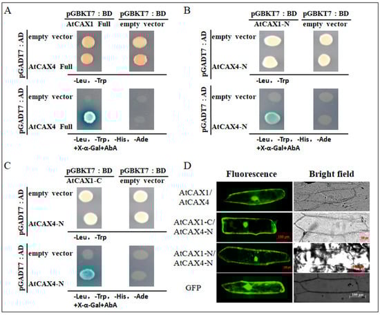
Figure 1.
AtCAX1 interacts with AtCAX4. (A–C) Yeast two-hybrid assay. The combinations of plasmids: full length of pGBKT7-AtCAX1 and pGADT7-AtCAX4 (A), C-terminus of pGBKT7-AtCAX1 and N-terminus sequence of pGADT7-AtCAX4 (B), N-terminus of pGBKT7-AtCAX1 and N-terminus sequences of pGADT7-AtCAX4 (C) were co-transformed into yeast Y2HGold cells. Transformed yeast cells were grown on SD-Leu-Trp and SD-Leu-Trp-His-Ade+X-a-gal+AbA medium for 2–3 days at 30 °C. (D) Bimolecular fluorescence complementation (BiFC) in onion epidermal cells. The combinations of plasmids (full length of AtCAX1-nGFP and AtCAX4-cGFP, C-terminus of AtCAX1 and N-terminus of AtCAX4, N-terminus of AtCAX1 and N-terminus of AtCAX4) are shown on the left. GFP was used a control. Scale bar = 100 μm.
The interaction between AtCAX1 and AtCAX4 was verified in vivo using a BiFC assay. Green fluorescence was observed when AtCAX1-nGFP and AtCAX4-cGFP, or when the N-terminal sequences of AtCAX1-nGFP (AtCAX1-N) or C-terminal sequences of AtCAX1-nGFP (AtCAX1-C) and the N-terminal sequence of AtCAX4-cGFP (AtCAX4-N) were expressed together in onion cells (Figure 1D). These results demonstrate that AtCAX1 can interact with the N-terminal sequence of AtCAX4 in an endomembrane system in vivo. The observed localization pattern was also consistent with findings reported in a previous study [28].
2.2. Expression of AtCAX1 and AtCAX4 in Response to Salt and Ion Stress
RNA was extracted from root, stem, leaf, flower, and silique tissues of Arabidopsis plants grown for 3 weeks under favorable growth conditions to investigate the pattern of AtCAX1 and AtCAX4 expression. Reverse transcription quantitative PCR (RT-qPCR) analysis conducted on various tissues indicated that AtCAX1 and AtCAX4 are expressed in all Arabidopsis plant organs, with the expression level of AtCAX1 being higher in silique and leaf tissues, while AtCAX4 was expressed more highly in root tissues (Figure 2A). The level of expression of AtCAX1 and AtCAX4 mRNA in response to salt and ion stress was also assessed (Figure 2B–E). AtCAX1 mRNA peaked at 12 h in response to NaCl and then declined at 24 h in both leaves and roots, while AtCAX4 mRNA increased gradually in leaves and roots in response to NaCl (Figure 2B,C). AtCAX1 gene expression was also induced in response to CaCl2 and peaked at 12 h or 6 h after treatment in leaf and root tissues, respectively, and then declined. In contrast, AtCAX4 mRNA expression gradually increased in both leaf and root tissues, peaking at 24 h (Figure 2D,E). These results indicate that AtCAX1 and AtCAX4 are involved in plant response to salt and ion stress.
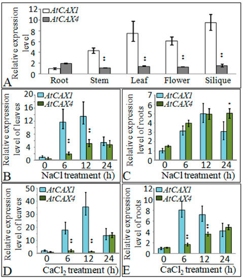
Figure 2.
Expression of AtCAX1 and AtCAX4 in plant tissues subjected to stress conditions. (A) RT-qPCR analysis of AtCAX1 and AtCAX4 in different organs of A. thaliana. (B–E) RT-qPCR analysis of the expression of AtCAX1 and AtCAX4 in response to various abiotic stresses (see Methods for details). AtCAX1 and AtCAX4 expression was normalized against Actin mRNA levels. The reported data are the means ± SE of three replicate experiments. Single and double asterisks indicate significant differences from wild type (WT) at p < 0.05 and p < 0.01, respectively.
2.3. Loss of Function Mutants of AtCAX1 and AtCAX4 Render Arabidopsis Plants Sensitive to Salt and Ion Stress
A T-DNA insertion located in the third exon of AtCAX1 and eight introns in AtCAX4 (Figure S2A) was confirmed by PCR-based genotype analysis (Figure S2B). PCR analysis was performed on the homozygous T-DNA insertion atcax1 and atcax4 mutants. The analysis demonstrated that they completely lack AtCAX1 or AtCAX4 transcripts (Figure S2B). F2 homozygous progeny double mutants atcax1/atcax4 were also identified by PCR analysis, and further analysis also indicated that they completely lack AtCAX1 and AtCAX4 transcripts (Figure S2B). Three Arabidopsis transgenic lines overexpressing AtCAX1 or AtCAX4 were generated under the control of the CaMV35S promoter (#1–#3) and identified by northern blotting (Figure S2C). Control samples demonstrated weak AtCAX1 or AtCAX4 signals, while the transgenic plants exhibited strong signals, indicating that they had been successfully transformed with AtCAX1 or AtCAX4.
After seed stratification on 1/2 Murashige and Skoog (MS) medium supplemented with different concentrations of NaCl (Figure 3A), assessment of germination indicated that a greater number of wild-type (WT) seeds germinated than seeds of atcax1, atcax4, and atcax1/atcax4 double mutants. WT plants also grew better than the transgenic lines on salt-amended media. In contrast, the growth rates of atcax1, atcax4, atcax1/atcax4, and WT did not obviously differ when seeds were germinated and grown on a medium that was not amended with NaCl (Figure 3A). Moreover, root lengths in atcax1, atcax4, and atcax1/atcax4 mutants were markedly shorter than those of WT plants when plants were grown in the presence of different concentrations of NaCl (125 and 135 mM) (Figure 3A). The sensitivity of atcax1, atcax4, and atcax1/atcax4 mutants to CaCl2 was also determined. A 65 mM CaCl2 treatment produced an abnormal phenotype in the mutant, while no obvious differences were observed between mutant and WT plants under control conditions (Figure 3B). Overall, the response of atcax1, atcax4, and atcax1/atcax4 double mutants to different types of stress indicated that the growth of WT plants was generally better than the growth of atcax1 and atcax4 single or atcax1/atcax4 double mutants. The sensitivity of plants to stress in vivo was associated with the deletion of either AtCAX1 or AtCAX4 alone or in combination.
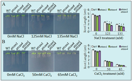
Figure 3.
Stress sensitivity of wild-type (WT) and atcax1, atcax4, and atcax1/atcax4 plants. Seeds were germinated on half-strength Murashige and Skoog (MS) medium containing either 125 or 135 mM NaCl (A), 50 or 65 mM CaCl2 (B). Photographs were taken 3 weeks after germination. Root length of WT and atcax1, atcax4, and atcax1/atcax4 plants was measured three weeks after germination. Data represent the mean ± SE of three replicates. Single and double asterisks indicate significant differences from WT at p < 0.05 and p < 0.01, respectively.
The response of AtCAX1 and AtCAX4 transgenic plants to salt and ion stress was further assessed in vivo to determine the effect of the AtCAX1 and AtCAX4 interaction on abiotic stress tolerance. Results indicated that the sensitivity of atcax1 and atcax4 mutants to NaCl stress increased with NaCl concentration, while the tolerance of plants overexpressing AtCAX1 or AtCAX4 to salt stress was significantly enhanced. This was mainly manifested in significantly longer root length in transgenic plants relative to WT plants, while atcax1 and atcax4 mutants had the shortest roots (Figure 4A). Similarly, atcax1 and atcax4 mutants treated with increasing concentrations of CaCl2 exhibited the weakest growth, while AtCAX1 and AtCAX4 transgenic plants exhibited better growth than WT plants. At 65 mM CaCl2, the growth of all the different lines of plants was inhibited; however, the AtCAX1 and AtCAX4 transgenic plants still exhibited better growth than atcax1 and atcax4 mutants, as well as WT plants (Figure 4B). Collectively, our results indicate that the overexpression of AtCAX1 or AtCAX4 genes can improve abiotic stress tolerance in Arabidopsis.
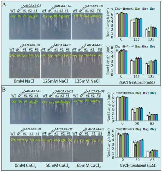
Figure 4.
Stress tolerance analysis of AtCAX1OE and AtCAX4OE transgenic plants treated with different concentrations of salt and ion stress. Phenotypes of WT, atcax1, atcax4, and AtCAX1OE and AtCAX4OE transgenic seedlings treated with 125 or 135 mM NaCl (A), 50 or 65 mM CaCl2 (B). Photographs were taken 3 weeks after germination. The root length of WT, atcax1, atcax4, and AtCAX1OE and AtCAX4OE transgenic plants were measured 3 weeks after treatment. Data represent the mean ± SE of three replicates. Single and double asterisks indicate significant differences from WT at p < 0.05 and p < 0.01, respectively.
3. Discussion
Calcium ions (Ca2+) are involved as a second messenger in many physiological processes, where they play a regulatory role in transducing cell signals involved in the growth and stress response of plants. The CAX family is thought to play an important role in regulating intracellular Ca2+ and the balance of other cations.
3.1. AtCAX1 and AtCAX4 form A Heterodimer
There are six CAX members in Arabidopsis thaliana. In the present study, we analyzed the CAX proteins in Arabidopsis thaliana for potential interactions using a yeast two-hybrid system. Results demonstrated that CAX1 and CAX4 proteins interact, which was verified by the co-transformation of yeast strains and BiFC assays (Figure 1). Previous studies revealed that an atcax1/atcax3 double mutant exhibited slower growth (shorter plants), relative to WT and single mutants, when planted in soil. When seeds were germinated and grown on a culture medium amended with the different levels of Ca2+, the atcax1/atcax3 double mutant exhibited a higher sensitivity to Ca2+, exhibiting poorer growth, producing shorter plants, and a greater level of leaf tip necrosis, relative to WT and single mutant plants. Notably, the growth rate of the atcax1/atcax3 double mutant on a medium containing 15 mM MgCl2 was better than the control and the atcax3 single mutant, while the poor growth rate of the atcax1/atcax3 double mutation was improved by MgCl2 [24]. In addition, the expression level of AtCAX3 and AtCAX4 in Arabidopsis thaliana was significantly increased in atcax1 inserted mutant plants [22]. These studies indicate that a potential interaction occurs between CAX members. The N-terminal regulatory region (NRR) of CAXs function as an autoinhibitory region in yeast and possess an autoinhibitory domain for Ca2+/H+ transport activity. The interaction of AtCAX1 (N terminal, AtCAX1-N) and AtCAX4 (N terminal, AtCAX4-N) was detected in the Y2H system used in the current study. The Ca2+ transport capacity of full-length AtCAX1 can be specifically activated by the CAX-interacting protein, CXIP4 [33]. The C-terminal region also has a regulatory function in some transporters, such as the mammalian Na+/H+ exchanger isoform 1 (NHE1) and the cyanobacterial Na+/H+ antiporter [34,35,36]. Our results indicate that AtCAX1 interacts with the N-terminal region of AtCAX4 and provides evidence that CAXs are capable of forming a heterodimeric protein through an interaction at the N-terminal region.
3.2. AtCAX1 and AtCAX4 Interact in Plants in Response to Environmental Stress
We investigated the expression pattern of AtCAX1 and AtCAX4 in Arabidopsis in response to different salt and ion treatments. In addition to the effect of NaCl, we also assessed the effect of Ca2+ treatments on the expression of CAX1 and CAX4. Results indicated that CAX1 and CAX4 expression was induced to different degrees in roots and leaves (Figure 2). This finding is consistent with a previous study reporting that CAX4 mRNA levels increased in response to a salt treatment [26]. CAX1 in Arabidopsis is highly expressed in leaf tissue and modestly expressed in roots, stems, and flowers [24]. CAX4 expression is relatively low in most tissues, relative to CAX1; however, when expressed at high levels in plants, its biochemical properties resemble other CAXs [17,26]. Previous studies, whose main focus was the response of CAX expression to Ca2+, have demonstrated that CAX1 and CAX4 are the main Ca2+ transporters in plants. However, in the present study, the response to NaCl was also analyzed, in addition to Ca2+. Our analysis of stress-induced expression indicated that CAX1 and CAX4 are involved in abiotic stress response in general, including the response to salt and ion stress (Figure 2). Bioinformatic analysis of Arabidopsis CAX family members revealed that AtCAX1 and AtCAX4 belong to the CAX IA developmental group, suggesting that the target of these two proteins may be similar or the same; CAX4 is most closely related to CAX1 at the amino acid level [11,25]. The loss-of-function mutant (cax4-1) and CAX4 RNA interference (CAX4 RNAi) lines were reported to exhibit altered root growth and development in response to Cd2+, Mn2+, and auxin [17]. These results suggest that CAX4 functions in root growth in plants under heavy metal stress conditions. Our current study also confirms that the CAX4 is most highly expressed in roots (Figure 2). Although CAX1 is most highly expressed in leaves, both CAX1 and CAX4 were induced to varying degrees in both roots and leaves in response to ion stress conditions (Figure 2). The growth of cax1, cax4, and cax1/cax4 mutants treated with stress levels of salt and ion was analyzed with a specific focus on root elongation. Results indicated no significant differences in root and shoot growth between cax1, cax4, and cax1/cax4 mutants under normal conditions, although the growth of wild-type plants was better than the mutant plants when plants were exposed to different cations (Figure 3). Previous studies demonstrated that CAXs can play compensatory roles in many cell functions. For example, plants that have lost a single CAX transport function are almost indistinguishable from the wild-type plants under normal growth conditions [11], but plants display severe growth defects in some double CAX deletion mutants, implying the compensatory function of CAX gene members [18,23]. These observations also imply that the interaction between CAX members is critical for plant growth, especially under abiotic stress conditions.
In the present study, we provide the first data demonstrating the response of cax1/cax4 to Ca2+. Notably, the overexpression of CAX1 or CAX4 genes can significantly improve plant tolerance to abiotic stress, relative to cax1 and cax4 mutants (Figure 4), indicating that overexpression may enhance the interaction between CAX1 and CAX4 in vivo, which leads to enhanced abiotic stress tolerance, and the interaction between CAX1 and CAX4 may be crucial to the enhancement of the tolerance of plants to abiotic stress. Changes in the relative expression of CAX transcripts in response to abiotic stresses have been previously reported. Studies in Arabidopsis and rice have indicated that CAX transcripts can either increase or decrease in response to dehydrative stresses, including drought, heat, cold, and salinity [37,38]. Enhanced CAX expression in plants in response to salt stress suggests that these transporters play a functional role in the response to salt stress [7,39]. CAX proteins are involved in a variety of abiotic stress response pathways, including as a modulator of cytosolic Ca2+ signaling [39]. The interaction between CAX1 and CAX4 may alleviate the self-inhibition resulting form the N-terminal region of CAX proteins. Thus, the role of the heterodimer in the regulation of intracellular Ca2+ signaling in plants is worthy of further study.
4. Materials and Methods
4.1. Yeast Two-Hybrid (Y2H) Screening
Y2H experiments were performed with the Matchmaker™Gold Yeast Two-Hybrid System as described by the manufacturer (Clontech Laboratories, CA, USA). Full-length AtCAX1 and AtCAX4, as well as the N-terminal truncated 108 aa of AtCAX1 (∆NAtCAX1 or AtCAX1-C) or AtCAX4 (∆NAtCAX4 or AtCAX4-C) and C-terminal truncated 36 aa sequences of AtCAX1 (∆CAtCAX1 or AtCAX1-N) or AtCAX4 (∆CAtCAX4 or AtCAX4-N) were amplified using the primers listed in Table S1 and cloned into the pGBKT7 and pGADT7 vectors. The constructs, pGBKT7-AtCAX1 and pGADT7-AtCAX4, were co-transformed into the Saccharomyces cerevisiae strain in the Y2HGold and cultured on SD media lacking leucine and tryptophan (SD/-Leu/-Trp) and SD media lacking leucine, tryptophan, histidine, and adenine and containing Aureobasidin A (AbA) (SD/–Leu/–Trp/–His/–Ade+AbA), as described in [40]. The interacting CAX1 and CAX4 proteins were detected on the SD/–Leu/–Trp/–His/–Ade+AbA+X-a-Gal medium to assess the expression of four reporter genes (HIS3, ADE2, AUR1-C, and MEL1), which were activated by a positive interaction between interacting CAX proteins.
4.2. Bimolecular Fluorescence Complementation (BiFC) Assay
The vector for BiFC was constructed by replacing the GFP in pBS-35S-GFP with the N-terminus (154 aa) or C-terminus (80 aa) of the mVenus plasmid, yielding pBS-35S: VN154 (nGFP) and pBS-35S:VC80 (cGFP), respectively [41]. Full-length AtCAX1 and AtCAX4, as well as the N-terminal truncated of AtCAX1 (∆NAtCAX1 or AtCAX1-C), and C-terminal truncated sequences of AtCAX1 (∆CAtCAX1 or AtCAX1-N) or AtCAX4 (∆CAtCAX4 or AtCAX4-N) were amplified using the primers listed in Table S1 and cloned into the pBS-35S: VN154 (nGFP) and pBS-35S:VC80 (cGFP) vectors, generating pBS-35S: AtCAX1-VC80 (AtCAX1-cGFP) and pBS-35S: AtCAX4-VN154 (AtCAX4-nGFP), pBS-35S: AtCAX1-VC80-N (AtCAX1-N-cGFP), pBS-35S: AtCAX1-VC80-C (AtCAX1-C-cGFP), and pBS-35S: AtCAX4-VN154 (AtCAX4-N-nGFP). A mixture of an nGFP construct and a cGFP construct (500 ng each) (AtCAX4-nGFP+AtCAX1-cGFP, AtCAX4-N-nGFP+ AtCAX1-N-cGFP, AtCAX4-N-nGFP+AtCAX1-C-cGFP) was used for particle bombardment to co-express proteins of interest in onion epidermal cells [42]. Images were viewed and recorded on a confocal scanning-laser imaging system (Olympus Fluoview, FV500).
4.3. Plant Material and Transformation
Arabidopsis thaliana ecotype Columbia-0 (Col-0) was used in this study as wild-type (WT), T-DNA insertion mutants, and to create transgenic plants. The AtCAX1 and AtCAX4 T-DNA insertion mutant SALK-108310 (atcax1) and SALK-201217C (atcax4) were obtained from the Arabidopsis Biological Resource Center (ABRC: http://www.arabidopsis.org/). The mutant lines atcax1 and atcax4 were crossed to generate double mutants (atcax1/atcax4), and homozygous double mutant plants were screened from the resulting F2 progeny by PCR. Homozygous mutant plants were identified using the gene-specific primers and T-DNA left border-specific primers listed in Table S1.
The following Arabidopsis (Arabidopsis thaliana) transgenic plants were generated: 35S::AtCAX1/Col-0 (AtCAX1OE), and 35S::AtCAX1/Col-0 (AtCAX4OE). The full-length coding sequence of AtCAX1 and AtCAX4 were separately amplified and individually ligated into the pBI121 vector under the control of the 35S promoter to generate transgenic AtCAX1OE and AtCAX4OE constructs. The resulting plasmids were introduced into wild-type Arabidopsis thaliana, ecotype Columbia plants using a floral dip infiltration method for Agrobacterium tumefaciens-mediated transformation [43]. Homozygous lines were identified and used in the subsequent analyses. The primers used for the generation of the constructs are listed in Table S1. Plants were grown under a 16-h light/8-h dark cycle at 22 °C.
For the stress tolerance assay, 30 seeds of Col-0, atcax1, atcax4, atcax1/atcax4 mutants, AtCAX1OE, and AtCAX4OE were surface sterilized and placed on 1/2 Murashige and Skoog (MS) medium supplemented with different concentrations of NaCl (125 and 135 mM), or CaCl2 (50 and 65 mM). After 14 days, seedling phenotypes were photographed, and the root length of the seedlings were measured. The experiment was repeated three times. Significant statistical differences between treatment means were determined at p < 0.05 using a Student’s t-tests.
4.4. RNA Isolation and Northern Blotting
Total RNA was isolated using a RNeasy plant Mini kit (Qiagen, Hilden, Germany) and treated with RNasefree DNaseI (Qiagen, Hilden, Germany). First-strand cDNA was synthesized using SuperScript III reverse transcriptase (Invitrogen, Carlsbad, CA, USA). Pairs of gene-specific primer pairs, AtCAX1-RT-FW and AtCAX1-RT-RV were used for AtCAX1, AtCAX4-RT-FW and AtCAX4-RT-RV were used for AtCAX4, while Actin-FW and Actin-RV were used to amplify the Actin gene (Table S1). Transcript abundance was determined by RT-qPCR using SYBR green I on a LightCycler®480 system II (Agilent Technologies, Palo Alto, CA, USA).
For northern blotting analysis, total RNA (10 µg) obtained from transgenic Arabidopsis lines was separated on a 1% (m/v) agarose–formaldehyde gels and transferred to Hybond N+ membranes. Hybridizations were carried out at 50 °C using AtCAX1 and AtCAX4 probes labeled with digoxigenin (DIG, Roche, Basel, Switzerland). Signals were detected with CDP-Star using a luminescent image analyzer (Fujifilm, LAS-4000mini, Tokyo, Japan). The single lines were named #1, #2, and #3, respectively.
4.5. Analysis of Gene Expression Using Reverse Transcription–Quantitative PCR (RT-qPCR)
Gene expression was quantified by RT-qPCR. Arabidopsis seeds were surface sterilized and placed on solid half MS medium. After 2 days of stratification at 4 °C, the plates were moved to a 22 °C incubator for propagation. The seedlings were transferred from the plates to a 1:1 mixture of soil and vermiculite and grown to maturity at 22 °C. The plants were grown under a 16-hlight/8-h-dark cycle in a growth chamber. Roots, stems, leaves, panicle, and siliques of two-month-old plants were sampled and used in the RT-qPCR analysis. A second batch of seedlings was pre-cultured for 2 weeks on 1/2 solid medium and then treated with different concentrations of NaCl (125 and 135 mM) and CaCl2 (50 and 65 mM). Shoots and roots were sampled after 0, 6, 12, and 24 h after treatment and used in the RT-qPCR analyses.
5. Conclusions
In the present study, the interaction between the N-terminal region of AtCAX4 with AtCAX1, CAX family members in Arabidopsis, was demonstrated using a yeast two-hybrid system and a bimolecular fluorescence complementation assay. This is the first study to report the interaction between CAX1 and CAX4 in plants. Further experiments demonstrated that the AtCAX1 and AtCAX4 expression is induced in roots and leaves in response to abiotic stress (NaCl and Ca2+). Functional analysis of AtCAX1 and AtCAX4 in Arabidopsis demonstrated that atcax1, atcax4 single mutants, and atcax1/atcax4 double mutants were more sensitive to abiotic stress than WT plants. In contrast, the overexpression of AtCAX1 or AtCAX4 in Arabidopsis improves abiotic stress tolerance. AtCAX1 and AtCAX4 may form a heterodimer in plants in response to abiotic stress.
Supplementary Materials
Supplementary Materials can be found at https://www.mdpi.com/1422-0067/22/2/856/s1. Table S1: Sequence of the primers used for PCR. Figure S1. C-terminus sequence of pGBKT7-AtCAX1 and C-terminus sequence of pGADT7-AtCAX4 were co-transformed into yeast Y2HGold cells. Transformed yeast cells were grown on SD-Leu-Trp and SD-Leu-Trp-His-Ade+X-a-gal+AbA medium for 2–3 days at 30 °C. Figure S2. Characterization of atcax1 and atcax4 T-DNA insertion mutants, and atcax1/atcax4 homozygous double mutants. (A) T-DNA insertion site in AtCAX1 and AtCAX4; gray boxes represent exons; black lines represent introns; (B) Reverse-transcription-quantitative PCR (RT-qPCR) analysis confirmed the knockout status of atcax1, atcax4, and homozygous double mutants atcax1/atcax4; Actin expression was used as an internal control in the RT-qPCR analysis; (C) RNA gel blot analysis of T3 transgenic plants expressing AtCAX1 or AtCAX4. WT: Arabidopsis thaliana ecotype Columbia-0; #1, #2, and #3: T3 seedlings expressing AtCAX1 or AtCAX4 in a Columbia-0 background.
Author Contributions
Conceptualization, Y.B. and S.L.; investigation, Y.B., J.C.; data curation, W.F.; writing—original draft preparation, Y.B.; writing—review and editing, S.L. and T.T.; funding acquisition, Y.B. and S.L. All authors have read and agreed to the published version of the manuscript.
Funding
This research was funded by Heilongjiang Province Government Postdoctoral Science Foundation (grant number: LBH-Q18008), and the Program for Changjiang Scholars and Innovative Research Team in University (grant number: No. IRT17R99).
Institutional Review Board Statement
Not applicable.
Informed Consent Statement
Not applicable.
Data Availability Statement
Not applicable.
Conflicts of Interest
The authors declare no conflict of interest. The funders had no role in the design of the study; in the collection, analyses, or interpretation of data; in the writing of the manuscript, or in the decision to publish the results.
Abbreviations
| CAXs | Ca2+/H+ antiporters |
| BiFC | Bimolecular fluorescence complementation |
| Ca2+ | Calcium ion |
| Ba2+ | Barium ion |
| AbA | Aureobasidin A |
| MS | Murashige and Skoog |
References
- Reddy, A.S.N.; Ali, G.S.; Celesnik, H.; Day, I.S. Coping with stresses: Roles of calcium-and calcium/calmodulin-regulated gene expression. Plant Cell 2011, 23, 2010–2032. [Google Scholar] [CrossRef]
- Steinhorst, L.; Kudla, J. Signaling in cells and organisms-calcium holds the line. Curr. Opin. Plant Biol. 2014, 22, 14–21. [Google Scholar] [CrossRef] [PubMed]
- Zhu, X.Y.; Dunand, C.; Snedden, W.; Galaud, J.P. CaM and CML emergence in the green lineage. Trends Plant Sci. 2015, 20, 483–489. [Google Scholar] [CrossRef] [PubMed]
- Clapham, D.E. Calcium signaling. Cell 2007, 13, 1047–1058. [Google Scholar] [CrossRef] [PubMed]
- Bose, J.; Pottosin, I.I.; Shabala, S.S.; Palmgren, M.G.; Shabala, S. Calcium efflux systems in stress signaling and adaptation in plants. Front. Plant Sci. 2011, 2, 85. [Google Scholar] [CrossRef]
- Shabala, S. Physiological and cellular aspects of phytotoxicity tolerance in plants: The role of membrane transporters and implications for crop breeding for waterlogging tolerance. New Phytol. 2011, 190, 289–298. [Google Scholar] [CrossRef]
- Shigaki, T.; Hirschi, K.D. Diverse functions and molecular properties emerging for CAX cation/H+ exchangers in plants. Plant Biol. 2006, 8, 419–429. [Google Scholar] [CrossRef]
- Pittman, J.K.; Bonza, M.C.; De Michelis, M.I. Ca2+ pumps and Ca2+ antiporters in plant development. Transp. Pumps Plant Signal. Signal. Commun. Plants 2011, 7, 133–161. [Google Scholar]
- Kamiya, T.; Maeshima, M. Residues in internal repeats of the rice cation/H+ exchanger are involved in the transport and selection of cations. J. Biol. Chem. 2004, 279, 812–819. [Google Scholar] [CrossRef]
- Shigaki, T.; Barkla, B.J.; Miranda-Vergara, M.C.; Zhao, J.; Pantoja, O.; Hirschi, K.D. Identification of a crucial histidine involved in metal transport activity in the Arabidopsis cation/H+ exchanger CAX1. J. Biol. Chem. 2005, 280, 30136–30142. [Google Scholar] [CrossRef]
- Shigaki, T.; Rees, I.; Nakhleh, L.; Hirschi, K.D. Identification of three distinct phylogenetic groups of CAX cation ⁄proton antiporters. J. Mol. Evol. 2006, 63, 815–825. [Google Scholar] [CrossRef] [PubMed]
- Hirschi, K.D.; Zhen, R.G.; Cunningham, K.W.; Rea, P.A.; Fink, G.R. CAX1, an H+/Ca2+ antiporter from Arabidopsis. Proc. Nat. Acad. Sci. USA 1996, 93, 8782–8786. [Google Scholar] [CrossRef] [PubMed]
- Liu, H.; Zhang, X.; Takano, T.; Liu, S. Characterization of a PutCAX1 gene from Puccinellia tenuiflora that confers Ca2+ and Ba2+ tolerance in yeast. Biochem. Biophys. Res. Commun. 2009, 383, 392–396. [Google Scholar] [CrossRef] [PubMed]
- Shigaki, T.; Mei, H.; Marshall, J.; Li, X.; Manohar, M.; Hirschi, K.D. The expression of the open reading frame of Arabidopsis CAX1, but not its cDNA, confers metal tolerance in yeast. Plant Biol 2010, 12, 935–939. [Google Scholar] [CrossRef] [PubMed]
- Kamiya, T.; Akahori, T.; Maeshima, M. Expression profile of the genes for rice cation⁄H+ exchanger family and functional analysis in yeast. Plant Cell Physiol. 2005, 46, 1735–1740. [Google Scholar] [CrossRef] [PubMed]
- Korenkov, V.; Hirschi, K.; Crutchfield, J.D.; Wagner, G.J. Enhancing tonoplast Cd/H antiport activity increases Cd, Zn, and Mn tolerance, and impacts root/shoot Cd partitioning in Nicotiana tabacum L. Planta 2007, 226, 1379–1387. [Google Scholar] [CrossRef]
- Mei, H.; Cheng, N.H.; Zhao, J.; Park, S.; Escareno, R.A.; Pittman, J.K.; Hirschi, K.D. Root development under metal stress in Arabidopsis thaliana requires the H+/cation antiporter CAX4. New Phytol. 2009, 183, 95–105. [Google Scholar] [CrossRef]
- Conn, S.J.; Gilliham, M.; Athman, A.; Schreiber, A.W.; Baumann, U.; Moller, I.; Cheng, N.H.; Stancombe, M.A.; Hirschi, K.D.; Webb, A.A.R.; et al. Cell-specific vacuolar calcium storage mediated by CAX1 regulates apoplastic calcium concentration, gas exchange, and plant productivity in Arabidopsis. Plant Cell 2011, 23, 240–257. [Google Scholar] [CrossRef]
- Pittman, J.K.; Sreevidya, C.S.; Shigaki, T.; Ueoka-Nakanishi, H.; Hirschi, K.D. Distinct N-terminal regulatory domains of Ca2+/H+ antiporters. Plant Physiol 2002, 130, 1054–1062. [Google Scholar] [CrossRef]
- Pittman, J.K.; Shigaki, T.; Marshall, J.L.; Morris, J.L.; Cheng, N.H.; Hirschi, K.D. Functional and regulatory analysis of the Arabidopsis thaliana CAX2 cation transporter. Plant. Mol. Biol. 2004, 56, 959–971. [Google Scholar] [CrossRef]
- Schaaf, G.; Catoni, E.; Fitz, M.; Schwacke, R.; Schneider, A.; von Wiren, N.; Frommer, W.B. A putative role for the vacuolar calcium⁄manganese proton antiporter AtCAX2 in heavy metal detoxification. Plant Biol. 2002, 4, 612–618. [Google Scholar] [CrossRef]
- Mei, H.; Zhao, J.; Pittman, J.K.; Lachmansingh, J.; Park, S.; Hirschi, K.D. In planta regulation of the Arabidopsis Ca2+/H+ antiporter CAX1. J. Exp. Bot. 2007, 58, 3419–3427. [Google Scholar] [CrossRef] [PubMed]
- Pittman, J.K.; Hirschi, K.D. Regulation of CAX1, an Arabidopsis Ca2+/H+ antiporter. Identification of an N-terminal autoinhibitory domain. Plant Physiol. 2001, 127, 1020–1029. [Google Scholar] [CrossRef] [PubMed]
- Pittman, J.K.; Shigaki, T.; Cheng, N.H.; Hirschi, K.D. Mechanism of N-terminal autoinhibition in the Arabidopsis Ca2+/H+ antiporter CAX1. J. Biol. Chem. 2002, 277, 26452–26459. [Google Scholar] [CrossRef]
- Cheng, N.H.; Pittman, J.K.; Barkla, B.J.; Shigaki, T.; Hirschi, K.D. The Arabidopsis cax1 mutant exhibits impaired ion homeostasis, development, and hormonal responses and reveals interplay among vacuolar transporters. Plant Cell 2003, 15, 347–364. [Google Scholar] [CrossRef]
- Cheng, N.H.; Pittman, J.K.; Shigaki, L.J.; LeClere, S.; Lahner, B.; Salt, D.E.; Hirschi, K.D. Functional association of Arabidopsis CAX1 and CAX3 is required for normal growth and ion homeostasis. Plant Physiol 2005, 138, 2048–2060. [Google Scholar] [CrossRef]
- Shigaki, T.; Hirschi, K. Characterization of CAX-like genes in plants: Implications for functional diversity. Gene 2000, 31, 291–298. [Google Scholar] [CrossRef]
- Cheng, N.H.; Pittman, J.K.; Shigaki, T.; Hirschi, K.D. Characterization of CAX4, an Arabidopsis H+/cation antiporter. Plant Physiol 2002, 128, 1245–1254. [Google Scholar] [CrossRef]
- Zhao, J.; Shigaki, T.; Mei, H.; Guo, Y.; Cheng, N.H.; Hirschi, K.D. Interaction between Arabidopsis Ca2+/H+ Exchangers CAX1 and CAX3. J. Biol. Chem. 2009, 284, 4605–4615. [Google Scholar] [CrossRef]
- Ma, Y.; Berkowitz, G.A. Multimeric CAX complexes and Ca2+ signaling-beyond humdrum housekeeping. J. Exp. Bot. 2017, 68, 3997–3999. [Google Scholar] [CrossRef]
- Loqué, D.; Lalonde, S.; Looger, L.L.; von Wire’n, N.; Frommer, W.B. A cytosolic trans-activation domain essential for ammonium uptake. Nature 2007, 446, 195–198. [Google Scholar] [CrossRef] [PubMed]
- Hocking, B.; Conn, S.J.; Manohar, M.; Xu, B.; Athman, A.; Stancombe, M.A.; Webb, A.R.; Hirschi, K.D.; Gilliham, M. Heterodimerization of Arabidopsis calcium/proton exchangers contributes to regulation of guard cell dynamics and plant defense responses. J. Exp. Bot. 2017, 68, 4171–4183. [Google Scholar] [CrossRef] [PubMed]
- Cheng, N.; Liub, J.; Nelsonb, R.S.; Hirschi, K.D. Characterization of CXIP4, a novel Arabidopsis protein that activates the H+/Ca2+ antiporter, CAX1. FEBS Lett. 2004, 559, 99–106. [Google Scholar] [CrossRef]
- Gebreselassie, D.; Rajarathnam, K.; Fliegel, L. Expression, purification, and characterization of the carboxyl-terminal region of the Na+/H+ exchanger. Biochem. Cell Biol. 1998, 76, 837–842. [Google Scholar] [CrossRef]
- Li, X.; Liu, Y.; Kay, C.M.; Muller-Esterl, W.; Fliegel, L. The Na+/H+ exchanger cytoplastic tail: Structure, function, and interactions with tescalcin. Biochemistry 2003, 42, 7448–7456. [Google Scholar] [CrossRef]
- Waditee, R.; Hibino, T.; Tanaka, Y.; Nakamura, T.; Incharoensakdi, A.; Takabe, T. Halotolerant cyanobacterium Aphanothece halophytica contains an Na+/H+ antiporter, homologous to eukaryotic ones, with novel ion specificity affected by C-terminal tail. J. Biol. Chem. 2001, 276, 36931–36938. [Google Scholar] [CrossRef]
- Bickerton, P.D.; Pittman, J.K. Role of cation/proton exchangers in abiotic stress signaling and stress tolerance in plants. In Elucidation of Abiotic Stress Signaling in Plants: Functional Genomics Perspectives, 1st ed.; Pandey, K.G., Ed.; Springer: New York, NY, USA, 2015; pp. 95–117. [Google Scholar]
- Pittman, J.K.; Hirschi, K.D. CAX-ing a wide net: Cation/H+ transporters in metal remediation and abiotic stress signaling. Plant Biol. 2016, 18, 741–749. [Google Scholar] [CrossRef]
- Manohar, M.; Shigaki, T.; Hirschi, K.D. Plant cation/H+ exchangers (CAXs): Biological functions and genetic manipulations. Plant Biol. 2011, 13, 561–569. [Google Scholar] [CrossRef]
- Guo, K.; Bu, Y.; Takano, T.; Liu, S.; Zhang, X. Arabidopsis cysteine proteinase inhibitor AtCYSb interacts with a Ca2+-dependent nuclease, AtCaN2. FEBS Lett. 2013, 587, 3417–3421. [Google Scholar] [CrossRef]
- Tsugama, D.; Liu, S.; Takano, T. A putative myristoylated 2C-type protein phosphatase, PP2C74, interacts with SnRK1 in Arabidopsis. FEBS Lett. 2012, 586, 693–698. [Google Scholar] [CrossRef]
- Zhang, C.Q.; Nishiuchi, S.; Liu, S.; Takano, T. Characterization of two plasma membrane protein 3 genes (PutPMP3) from the alkali grass, Puccinellia tenuiflora, and functional comparison of the rice homologues, OsLti6a/b from rice. BMB Rep. 2008, 41, 448–454. [Google Scholar]
- Clough, S.J.; Bent, A.F. Floral dip: A simplified method for Agrobacterium mediated transformation of Arabidopsis thaliana. Plant J. 1998, 16, 735–743. [Google Scholar] [CrossRef] [PubMed]


Publisher’s Note: MDPI stays neutral with regard to jurisdictional claims in published maps and institutional affiliations. |
© 2021 by the authors. Licensee MDPI, Basel, Switzerland. This article is an open access article distributed under the terms and conditions of the Creative Commons Attribution (CC BY) license (http://creativecommons.org/licenses/by/4.0/).