Communication

Road to Metastasis: The TWEAK Pathway as a Discriminant between Metastasizing and Non-Metastasizing Thick Melanomas

by
Canan Güvenç
1,*,
Asier Antoranz
2,
Anna Szumera-Ciećkiewicz
3,4,
Pawel P. Teterycz
5,
Piotr R. Rutkowski
5,
Robert V. Rawson
6,7,8,9,
Richard A. Scolyer
6,7,8,9,10,
John F. Thompson
6,7,11,
Julia Newton-Bishop
12,
Marguerite Stas
13,
Veerle Boecxstaens
13,
Oliver Bechter
14,
Jurgen Vercauteren
15,
Marjan Garmyn
1,
Joost van den Oord
2 and
Francesca Maria Bosisio
2,*
1
Department of Dermatology, University Hospitals Leuven, 3000 Leuven, Belgium
2
Department of Imaging and Pathology, Translational Cell and Tissue Research, University Hospitals Leuven, 3000 Leuven, Belgium
3
Department of Pathology and Laboratory Medicine, Maria Sklodowska-Curie National Research Institute of Oncology, 02-781 Warsaw, Poland
4
Diagnostic Hematology Department, Institute of Hematology and Transfusion Medicine, 02-776 Warsaw, Poland
5
Department of Soft Tissue/Bone Sarcoma and Melanoma, Maria Sklodowska-Curie National Research Institute of Oncology, 02-781 Warsaw, Poland
6
Melanoma Institute Australia, The University of Sydney, Sydney, NSW 2000, Australia
7
Faculty of Medicine and Health, Sydney Medical School, The University of Sydney, Sydney, NSW 2000, Australia
8
Tissue Pathology and Diagnostic Oncology, Royal Prince Alfred Hospital, Sydney, NSW 2000, Australia
9
NSW Health Pathology, Sydney, NSW 2000, Australia
10
Charles Perkins Centre, The University of Sydney, Sydney, NSW 2000, Australia
11
Department of Melanoma and Surgical Oncology, Royal Prince Alfred Hospital, Sydney, NSW 2000, Australia
12
Institute of Medical Research at St James’s, University of Leeds, Leeds LS2 9JT, UK
13
Department of Surgical Oncology, University Hospitals Leuven, 3000 Leuven, Belgium
14
Department of General Medical Oncology, University Hospitals Leuven, 3000 Leuven, Belgium
15
Department of Microbiology, Immunology and Transplantation, Rega Institute for Medical Research, Clinical and Epidemiological Virology, Katholieke Universiteit Leuven, 3000 Leuven, Belgium
*
Authors to whom correspondence should be addressed.
Int. J. Mol. Sci. 2021, 22(19), 10568; https://doi.org/10.3390/ijms221910568
Submission received: 29 August 2021 / Revised: 20 September 2021 / Accepted: 25 September 2021 / Published: 29 September 2021

Abstract

:
Cutaneous melanoma (CM) is the most aggressive form of skin cancer, and its worldwide incidence is rapidly increasing. Early stages can be successfully treated by surgery, but once metastasis has occurred, the prognosis is poor. However, some 5–10% of thick (≥2 mm) melanomas do not follow this scenario and run an unpredictable course. Little is known about the factors that contribute to metastasis in some patient with thick melanomas and the lack thereof in thick melanoma patients who never develop metastatic disease. We were therefore interested to study differential gene expression and pathway analysis and compare non-metastatic and metastatic thick melanomas. We found that the TNF-like weak inducer of apoptosis (TWEAK) pathway was upregulated in thick non-metastasizing melanomas. MAP3K14 (NIK1), BIRC2 (cIAP1), RIPK1, CASP7, CASP8, and TNF play an important role in inhibiting proliferation and invasion of tumor cells via the activation of the non-canonical NF-κB signaling pathway. In particular, this pathway sensitizes melanoma cells to TNF-alpha and activates the apoptosis module of the TWEAK pathway in thick non-metastasizing melanomas. Hence, our study suggests a potential role of the TWEAK pathway in inhibiting thick melanoma from metastasis. Exploitation of these genes and the pathway they control may open future therapeutic avenues.

1. Introduction

Cutaneous melanoma (CM) is a tumor that originates from melanocytes of the skin, and continues to carry the potential to be a deadly disease worldwide [1]. While early-stage CM is generally curable as a result of early detection and definitive surgery, diagnosis at a late stage and the consequential delay in treatment has an adverse effect on survival. This is due to the propensity of CM to metastasize, and to the resistance of metastatic cells to classical chemotherapy. Targeted therapies and immunological approaches to treat metastatic disease have dramatically improved survival in patient with CM in recent years [2,3]. Nevertheless, these therapies are not effective in all patients, and are burdened by adverse effects and the development of resistance. It has become crucial to identify biomarkers to assist in identifying patients at highest risk of disease progression to facilitate development of personalized therapy and follow-up care.
The prognosis of primary cutaneous melanoma is correlated with several clinical and histological variables including Breslow-thickness, ulceration, mitotic rate, pattern of tumor-infiltrating lymphocytes, age and gender of the patients and the tumor site. According to Breslow, the thickness of the primary melanoma, measured from the granular layer up to the deepest melanoma cell in the dermis, is by far the most significant prognostic variable and is correlated with outcome [4]. However, some 5–10% of thick classical (≥2 mm) CM do not show this association and have an excellent outcome [5,6,7]. Therefore, investigating the biology of thick melanomas with variable outcome can shed light on important processes driving tumor progression and metastasis. The latter process results from changes in the expression of proteins that favor tumor growth, invasion and migration, such as, for example, the expression of the vasoactive peptide Endothelin-1 or of matrix metalloproteinase 9 (MMP-9) by the CM cells [8]. Several signaling pathways have been identified as key regulators in CM progression, such as the activation of Ras/MAPK signaling pathway through the mutant BRAF or NRAS genes or alterations of CCND1 [9]. Additionally, frequently affected molecular pathways are the PI3K/AKT/mTOR pathways (e.g., PTEN loss of function), cell cycle regulators (e.g., CDKN2a, CDK4, CCND1), P53 (e.g., Tp53, MDM2), and pathways involved in epigenetic regulation (e.g., ARID2a) [10]. Although many studies have investigated the role of distinct features involved in CM spreading, no effort has been made yet to investigate the metastasis of thick CMs starting from a global, unbiased perspective through a bulk sequencing of the transcriptome instead of focusing on the analysis of specific pathways. Moreover, previous studies of gene expression profiles (GEP) in thick CM have not compared groups of patients that have been matched for all other prognostic features that may play a role in influencing the metastatic potential. As a result, the molecular differences between thick non-metastasizing (M−) and thick metastasizing (M+) CMs that may the basis of the different clinical behavior, are not yet fully elucidated. Here, we analyse the significance of classical prognostic parameters in thick CM, and compare the molecular composition of thick primary melanomas that did metastasize with a matched group of thick primary melanomas that did not metastasize, aiming to identify a gene signature associated with non-metastasizing melanomas.

2. Results

The analysis of clinicopathological parameters of thick CM are depicted in Supplementary Materials Table S1 (a + b). For the molecular analysis, demographic and clinicopathological characteristics of the study population (thick CM) are depicted in Table 1. The pathway analysis is summarized in Figure 1 and Supplementary Materials Table S2.

2.1. Analysis of Clinicopathological Predictors in Thick CM

The univariate analysis of thick CM (Breslow ≥ 2 mm) from the UZleuven database (n = 140), showed no significant association between the development of metastasis within five years and the classical prognostic variables (age, gender, Breslow thickness, ulceration, mitosis, site) plus the additional investigated histopathological variables (subtype, TIL, vascular invasion, solar damage and adjoining nevus). After adjustment for the classical prognostic variables, no significant associations were found between outcome and all investigated parameters in the multivariate analysis (Supplementary Materials Table S1a). We validated our findings in the Leeds database (n = 141), where we obtained the same results in the multivariate analysis (Supplementary Materials Table S1b) [14].

2.2. Demographic and Clinicopathological Characteristics of the Study Population for the Molecular Analysis

We included 24 patients with thick primary melanoma (≥2 mm) who consented to this study, of whom 6 (25%) were women and 18 (75%) men. We grouped 24 cases into 12 pairs of melanomas that were matched for all clinicopathological prognostic markers but that differed in outcome (i.e., distant metastasis within a follow-up period of 5 years) (Supplementary Table S3). Breslow thickness varied from 2.8 mm to 18 mm (median 5 mm, mean 5.7 mm). There were 8 (33.3%) patients younger than 60 years and 16 (66.7%) patients older than 60 years. The primary tumor was located in 11 (45.8%) patients on the extremities, in 6 (25%) on the trunk, and in 7 (29.2%) on the head and neck. Nodular melanoma was the predominant histological subtype of CM in 13 (54.2%) of patients, followed by superficial spreading melanoma (SSM) [4 (16.7%)] and acrolentiginous melanoma (AL) [7 (3.1%)]. Other clinicopathological characteristics are summarized in Table 1.

2.3. Molecular Analysis

Of the 24 samples, 23 samples passed the quality control and underwent RNA-sequencing and bioinformatics analysis.

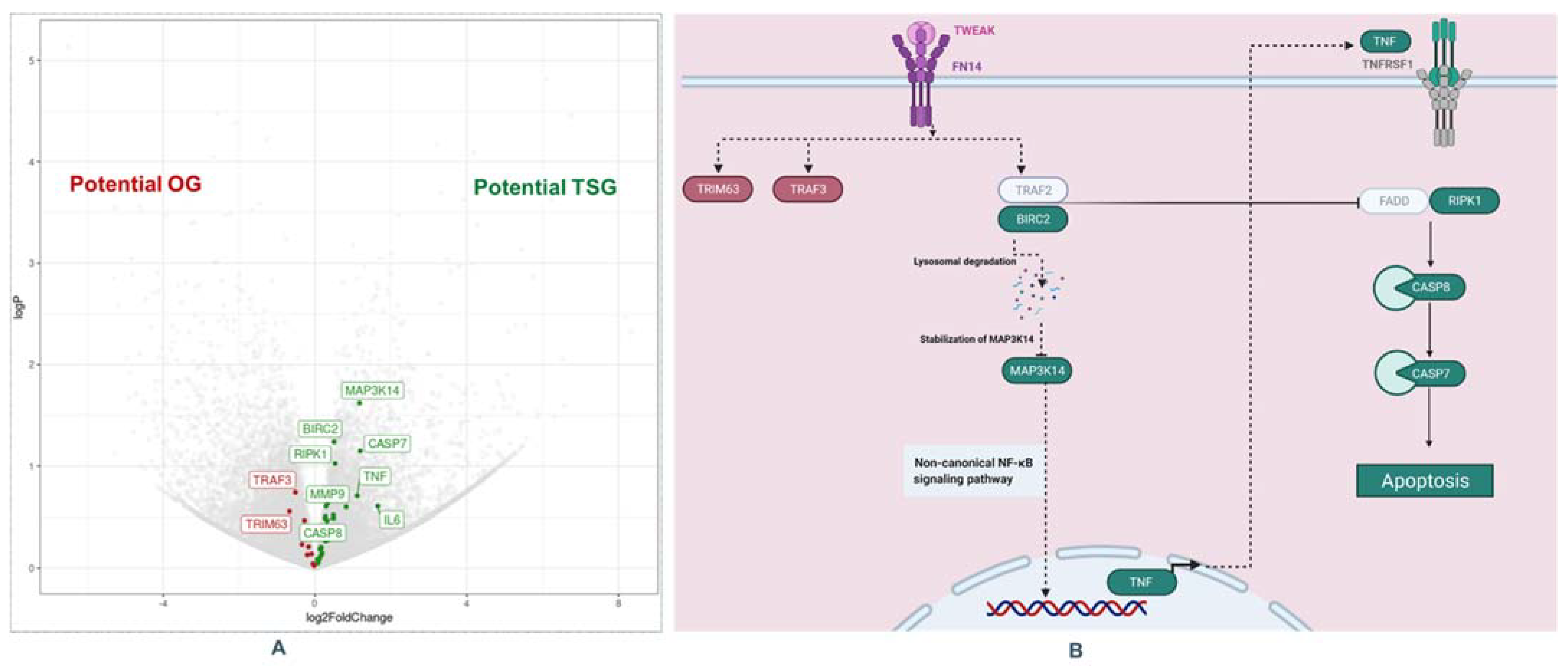
2.3.1. Differentially Expressed Genes in Thick CM

From the 29,457 genes included in the Leuven dataset, we identified 830 genes differentially expressed between M− and M+ (398 overexpressed in M− and 432 overexpressed in M+) However, after correction for multiple testing with the Benjamini–Hochberg method, no significantly differentially expressed genes (DEGs) were found.

2.3.2. Pathway Analysis: TNF-like Weak Inducer of Apoptosis (TWEAK) Pathway

Since complex and heterogeneous phenotypes are best explained by small but coordinated differences in functionally correlated genes, that may therefore not be statistically significant if analyzed separately, we performed pathway analysis. The top ten upregulated pathway in thick M− are depicted in Supplementary Materials Table S2. We found an upregulation of the TNF-like weak inducer of apoptosis (TWEAK) pathways in non-metastatic thick melanomas. The most affected genes in this pathway were two genes with higher expression in M+ patients (TRAF3 and TRIM63) and 8 genes with higher expression in M− patients (MAP3K14 (NIK1), BIRC2 (cIAP1), RIPK1, CASP7, CASP8, TNF, IL6, MMP9).

2.3.3. Validation in the Leeds Dataset

We validated our findings in the Leeds database. From the 703 primary cutaneous melanomas included in this dataset, 179 met our inclusion criteria (23 M− and 156 M+). From the 20,807 genes, we found 1864 differentially expressed (1016 upregulated in M− and 848 upregulated in M+). No genes remained significant after multiple testing adjustment. The TWEAK pathway in this dataset reported a p-value of 9.99 × 10⁻⁵ (Supplementary Materials Figure S1 Volcano plot Leeds database) [14,15].

3. Discussion

Despite recent advances in understanding its pathogenesis, melanoma is still the deadliest form of skin cancer. Understanding the molecular mechanisms involved in the development of thick CM, as well as the knowledge of their interplay, has great potential to assist in the clinical management of patients with thick CM. The metastatic process itself in CM is not yet fully understood and gene expression profiling studies for thick primary cutaneous melanomas that did not metastasize are scarce and understudied in the literature. The biology of thick CMs is still largely unexplored and, so far, the underlying mechanisms of metastasis in thick CM have not been investigated with a “multi-omics” approach. In this study we explored the clinical, pathological and molecular differences between non-metastasizing (M−) and metastasizing (M+) thick CMs in order to find elements that could help to explain their different clinical behavior and gain insight into mechanisms of metastasis in CM. Moreover, we tried to find metastatization hallmarks for thick melanomas in order to better stratify patients for prognosis and treatment.
A retrospective analysis of the clinicopathological data of 140 patients with thick CM (≥2.0 mm) from a UZ Leuven cohort with a follow up of at least 5 years revealed that the outcome of these patients was not correlated with any of the known clinical (gender, age, site, etc.) or histological (Breslow thickness, ulceration, mitotic count, TILS solar damage and vascular invasion, nevus) parameters. We also found these observations in the Leeds database. This confirmed that other parameters need to be found to better stratify these selected group of patients (Breslow ≥ 2 mm) than the ones already used in the clinic (according to AJCC8 based on the total group; stage I–IV patients) [4].
These preliminary data encouraged us to enroll patients with thick CM for advanced molecular analysis. In the UZ database and Leeds database, no significantly differentially expressed genes were found, which might be indicative for multifactorial drivers of metastasis, since complex and heterogeneous phenotypes are best explained by small differences in functionally correlated genes. To gain further insight into the molecular changes that occur in thick CM, we also carried out genes set analysis. Pathway enrichment analysis revealed the TWEAK pathway as a potential driving mechanism of metastasis, particularly the submodule that regulates cell death. Hence, our study confirmed for the first time that the apoptosis module of the TWEAK pathway is upregulated in thick M− and identified a gene signature associated with non-metastasizing melanomas. In addition to the TWEAK pathway, the oestrogen receptor pathway was also identified in our gene-set analysis, but we excluded this pathway because hormone-related studies are often multifactorial, and from the literature we could not draw a coherent conclusion on genes that were involved and upregulated in the estrogen receptor pathway in thick CM [16,17].
According to the literature, the TWEAK/Fn14 signaling pathway plays a key role in cancer. While TWEAK and Fn14 gene expression is low in normal healthy tissues, increased expression of these genes has been observed in many solid primary tumor types (kidney, liver, colon, ovarian, esophageal, and pancreatic cancer) [18,19,20,21,22,23,24]. In vivo, TWEAK is a pro-angiogenic [16,17,24] and pro-inflammatory [25,26,27,28] factor that can promote tumor vascularization and inflammation. In addition, studies in vitro have shown that TWEAK-triggered Fn14 activation in cancer cells themselves can stimulate both “protumorigenic/metastatic” and “anti-tumorigenic/metastatic” cellular responses, depending on the investigated cell lines. Armstrong et al. found that TWEAK:Fn14 engagement in cancer cells can positively or negatively regulate the invasive activity, depending on the cancer cell line [18]. They showed that TWEAK activation of the non-canonical NFKB pathway in prostate cancer stimulates proliferation and invasion, while in melanoma cells, it inhibits proliferation and invasion. In particular Fn14-TRAF-TNFR axis regulates intracellular signal transduction that triggers death signals in tumor cells via the non-canonical NF-κB signaling pathway. After the formation of the TRAF2-cIAP1 (BIRC2) complex, it undergoes Cathepsin B mediated degradation. The degradation of the TRAF2-cIAP1 complex leads to the stabilization of MAP3K14 (NIK) [11,12]. The latter induces the autocrine cellular apoptosis machinery by activating the RIPK1-FADDCaspase-8 complex and sensitizes tumor cells to death [11,12]. In our study, we identified six major up-regulated genes involved in the apoptosis module of the TWEAK pathway in thick M− melanoma, namely MAP3K14 (NIK1), BIRC2 (cIAP1), RIPK1, CASP7, CASP8, TNF, suggesting that RIPK1-FADDCaspase-8 complex-mediated apoptosis of melanoma cells may be a crucial mechanism in the prevention of metastasis in thick melanomas. The results presented are in line with a similar study, which also identified members of the NF-κB pathway [14]. Furthermore, we found the strongest log2FC regulation for IL6. IL6 is a classic inducer of STAT3 and induces inflammatory processes. STAT3 was shown to repress MITF and reduces the proliferation of melanoma cells [29].
Our results provide evidence regarding the differential biological behavior of thick melanoma; however, whether our data are also confirmed at the protein level awaits further studies using immunohistochemical techniques on larger numbers of thick melanomas. Our findings highlight the molecular biomarkers that can be used for prognostic purposes and provide information on the underlying mechanisms of outcome in thick CM. Detailed molecular knowledge of thick CM biology will further be instrumental in the specification of new biomarkers for risk stratification, which will improve the clinical management of this disease. In this regard, knowing the apoptotic biomarkers among thick CM patients may be helpful to find better treatment options.

4. Materials and Methods

4.1. Patient Populations

Patients with high Breslow thickness melanomas with available remaining FFPE tissue were identified from the files of the pathology departments at University Hospitals in Leuven, Belgium (study series; n = 4) and at the Melanoma Institute Australia in Sydney, Australia (n = 14) and Maria Sklodowska-Curie National Research Institute of Oncology in Warsaw, Poland (n = 6). These 24 cases were grouped into 12 pairs of melanomas that were matched for all clinic pathological prognostic markers but that differed in outcome (i.e., locoregional and distant metastasis within a follow-up period of five years). Hence, the study cohort consisted of 12 melanomas that did metastasize and 12 that did not. Each of these 12 pairs were matched for all other prognostic factors as depicted in Supplementary Materials Table S3. All patients were treated uniformly—i.e., complete excision of the primary melanoma that was followed by re-excision with margins appropriate for the thickness of the primary tumor.
Tissue collections and the specific study protocol were approved by the medical ethical committees and institutional review boards of the University Hospitals at the Katholieke Universiteit Leuven (S61610, approved on 16-08-2018), Melanoma Institute Australia and Maria Sklodowska-Curie National Research Institute of Oncology.
The pathology of each melanoma of our patient cohort was reviewed by four expert dermatopathologists (A. Szumera-Ciećkiewicz, J. J. van den Oord, F.M. Bosisio and R.V. Rawson). Clinical data available included age (<40, 40–59 years, ≥60 years), sex, site of involvement (extremity, head and neck, trunk), histological subtype, mitotic index, ulceration, Breslow thickness and AJCC stage at diagnosis [30]. The clinical and histopathologic characteristics of the study cohort are summarized in Table 1.

4.2. Tumor Tissue Samples: RNA Extraction and Purification

Seven consecutive 5 µm sections were prepared from FFPE tissue samples. The first and last sections were stained with hematoxylin & eosin (H & E; Agilent, Santa Clara, CA; USA) and used as a control for the presence of a vertical growth phase and a required tumor cell percentage of at least 10%. After deparaffinization with Histo-Clear and ethanol, tumor foci marked by a pathologist on the H & E section were manually macrodissected. Tissue was digested with proteinase K; RNA extraction was performed using RNeasy FFPE Kit (Qiagen, Hilden, Germany) according to the manufacturer’s instructions, and RNA was quantified using the Qubit 2.0 fluorometer (Thermo Fisher Scientific, Waltham, MA, USA). This study is based on analysis of bulk-RNA, derived from the vertical growth phase of thick CM, and therefore does not take heterogeneity in RNA expression into consideration.

4.3. Construction of RNA-Seq Library and Sequencing

Sequence libraries were prepared with the Lexogen QuantSeq 3′ mRNA-Seq Library prep kit according to the manufacturer’s protocol. Samples were indexed to allow for multiplexing. Library quality and size range was assessed using a Bioanalyzer (Agilent Technologies) with the DNA 1000 kit (Agilent Technologies, CA, USA) according to the manufacturer’s recommendations. Libraries were diluted to a final concentration of 2 nM and subsequently sequenced on an Illumina HiSeq4000 platform according to the manufacturer’s recommendations. Single-end reads of 50 bp length were produced with a minimum of 1 M reads per sample.

4.4. Bioinformatics Analysis

Quality control of raw reads was performed with FastQC v0.11.7 (Andrews S. (2010)). FastQC: a quality control tool for high throughput sequence data. Available online at: http://www.bioinformatics.babraham.ac.uk/projects/fastqc (accessed on 10 September 2021). Adapters were filtered with ea-utils fastq-mcf v1.05. (Erik Aronesty (2011). ea-utils: “Command-line tools for processing biological sequencing data”; Ea-utils: Fastq processing utilities. https://github.com/ExpressionAnalysis/ea-utils (accessed on 10 September 2021)) Splice-aware alignment was performed with STAR v2.6.1b against the human reference genome hg38 using the default parameters. Reads mapping to multiple loci in the reference genome were discarded [31]. Resulting BAM alignment files were handled with Samtools v1.5 [32].
Quantification of reads per gene was performed with HT-seq Count v2.7.14. Differential gene expression analysis was done comparing thick M- melanomas versus thick M+ melanomas with the DESeq2 [33] R-package (The R Foundation for Statistical Computing, Vienna, Austria). Reported p-values were adjusted for multiple comparisons with the Benjamini–Hochberg procedure, which controls false discovery rate (FDR). Pathway analysis was performed using Piano R package for enriched gene set analysis [34]. Within the piano framework, the following gene-set enrichment methods and gene-level statistics were used: Fisher (p-value), Stouffer (p-value), Reporter (p-value), PAGE (t-value), Tail Strength (p-value), GSEA (t-value), Mean (logFC), Median (logFC), Sum (logFC), MaxMean (t-value). For the gene-sets we used the Molecular Signatures Database, curated pathways (c2), canonical pathways (cp), version 7.2. Gene-sets with more than 100 genes capturing general biology were removed from downstream analysis (Raw mRNA expression data for UZ Leuven database: primary accession –PRJEB47586 and secondary accession-ERP131867).

4.5. Validation Using the Leeds Dataset

The results of our analysis were validated using the Leeds dataset [14]. The normalized expression data and the corresponding patient metadata was obtained directly from the authors. Raw mRNA expression data for Leeds Melanoma Cohort was downloaded from the European Genome Archive using the accession number EGAS00001002922. Thick melanomas were filtered by selecting those patients with a Breslow thickness ≥2 mm. Metastatic patients were defined as those with a time of relapse ≤5 years from the time of diagnosis. Patients with unknown time of relapse were excluded from downstream analysis. Differential gene expression and pathway analysis were performed as described for the Leuven dataset.

5. Conclusions

In the present study, we characterized the transcriptome of thick melanoma M− versus M+ in a precisely matched patient data set. We identified a gene signature associated with non-metastasizing melanomas. In particular, the TWEAK-induced activation of the non-canonical NF-κB signaling pathway, which induces production of TNF-alpha and sensitizes tumor cells to death, may play a protective role in thick non-metastasizing melanomas. The exploitation of these genes and pathway may open future therapeutic avenues.

Supplementary Materials

The following are available online at https://www.mdpi.com/article/10.3390/ijms221910568/s1.

Author Contributions

Conceptualization, J.v.d.O. and F.M.B.; methodology and software, J.v.d.O., F.M.B. and A.A.; validation and formal analysis, A.A. and C.G.; investigation, C.G.; data curation, C.G. and A.A.; writing—original draft preparation, C.G.; writing—review and editing, F.M.B., A.A., M.S., V.B., O.B., J.V., M.G., J.v.d.O., A.S.-C., P.P.T., P.R.R., R.V.R., R.A.S., J.F.T. and J.N.-B. All authors have read and agreed to the published version of the manuscript.

Funding

This work was supported by a National Health and Medical Research Council of Australia (NHMRC) Program Grant (APP1093017) (to R.A.S. and J.F.T.). R.A.S. is supported by an NHMRC Practitioner Fellowship (APP1141295). Support from Deborah McMurtrie and John McMurtire AM, the Ainsworth Foundation and The Cameron Family is also gratefully acknowledged.

Institutional Review Board Statement

The study was conducted according to the guidelines of the Declaration of Helsinki, and approved by the medical ethical committees and institutional review boards of the University Hospitals at the Katholieke Universiteit Leuven (S61610, 16-08-2018), Melanoma Institute Australia and Maria Sklodowska-Curie National Research Institute of Oncology.

Informed Consent Statement

Patient consent was waived due to the fact that it was a retrospective study on data (legal basis: public interest) and secondary use of samples (legal basis: opt-out).

Data Availability Statement

Raw RNAseq data is available at the European Nucleotide Archive (ENA): primary accession–PRJEB47586, secondary accession-ERP131867.

Acknowledgments

The authors sincerely thank EORTC Melanoma Group (Corporate Author), all their medical colleagues, residents, laboratory personnel (Cmol) and Genomics core Leuven (sequencing and raw data analysis), who helped to make this study possible. They also extend their heartfelt gratitude to all the patients who participated and helped advance melanoma research.

Conflicts of Interest

R.A.S. has received fees for professional services from Evaxion, Provectus Biopharmaceuticals Australia, Qbiotics, Novartis, Merck Sharp & Dohme, NeraCare, AMGEN Inc., Bristol-Myers Squibb, Myriad Genetics, GlaxoSmithKline. J.F.T. has received honoraria for advisory board participation from BMS Australia, MSD Australia, GSK and Provectus Inc., and travel and conference support from GSK, Provectus Inc. and Novartis. The other authors declare no conflict of interest.

Abbreviations

AJCCAmerican Joint Committee on Cancer
AKTAKT Serine/Threonine Kinase
ALacrolentiginous melanoma
ARID2AT-Rich Interaction Domain 2
BIRC2Baculoviral IAP Repeat Containing 2
BRAFB-Raf Proto-Oncogene, Serine/Threonine Kinase
CASP7Caspase 7
CASP8Caspase 8
CCND1Cyclin D1
CDK4Cyclin Dependent Kinase 4
CDKN2aCyclin Dependent Kinase Inhibitor 2A
cIAP1Cellular Inhibitor of Apoptosis 1
CMCutaneous melanoma
DEGdifferential expressed genes
FADDFas Associated Via Death Domain
Fn14Fibroblast growth factor-inducible 14
FFPEFormalin-Fixed Paraffin-Embedded
GSTP1Glutathione S-Transferase P
H & Ehematoxylin & eosin
HLA-DQA1Major Histocompatibility Complex, Class II, DQ Alpha 1
IGHMImmunoglobulin Heavy Constant Mu
IL6interleukin 6
JNKc-Jun N-terminal kinase
M−non-metastatic CMs
M+metastatic CMs
MAPMitogen-Activated Protein
MAP3K14Mitogen-Activated Protein Kinase Kinase Kinase 14
MDM2MDM2 Proto-Oncogene
METMET Proto-Oncogene, Receptor Tyrosine Kinase
MET, c-METMesenchymal-epithelial transition tyrosine kinase receptor
MMP9expression of matrix metalloproteinase 9
NIKNFkB Inducing Kinase
NMNodular melanoma
NRASNRAS Proto-Oncogene, GTPase
NSCLCNon-small-cell lung carcinoma
OGoncogene
P53Tumor Protein P53
PTENPhosphatase And Tensin Homolog
RAMP1Receptor Activity Modifying Protein 1
Ras/MAPKRas/Mitogen-Activated Protein Kinase
RIPK1Receptor Interacting Serine/Threonine Kinase 1
RNAribonucleic acid
SH3GL2SH3 Domain Containing GRB2 Like 2, Endophilin A1
SSMSuperficial spreading melanoma
STK19Serine/Threonine Kinase 19
TMOD1Tropomodulin 1
TNFtumor necrose factor
TNF-alphaTumor Necrosis Factor-alpha
TNFRtumor necrose factor receptor
TNFR1Tumor Necrosis Factor Receptor Type 1
TRAFTNF Receptor Associated Factor
TRIM63Tripartite Motif Containing 63
TSGtumor suppressor gene
TWEAKTNF-like weak inducer of apoptosis
TWIST2Twist Family BHLH Transcription Factor 2

References

  1. Arnold, M.; Holterhues, C.; Hollestein, L.M.; Coebergh, J.W.; Nijsten, T.; Pukkala, E.; Holleczek, B.; Tryggvadóttir, L.; Comber, H.; Bento, M.J.; et al. Trends in incidence and predictions of cutaneous melanoma across Europe up to 2015. J. Eur. Acad. Dermatol. Venereol. 2014, 28, 1170–1178. [Google Scholar] [CrossRef]
  2. Rajkumar, S.; Watson, I.R. Molecular characterisation of cutaneous melanoma: Creating a framework for targeted and immune therapies. Br. J. Cancer 2016, 115, 145–155. [Google Scholar] [CrossRef] [PubMed] [Green Version]
  3. Topalian, S.L.; Drake, C.G.; Pardoll, D.M. Immune checkpoint blockade: A common denominator approach to cancer therapy. Cancer Cell 2015, 27, 450–461. [Google Scholar] [CrossRef] [Green Version]
  4. Gershenwald, J.E.; Scolyer, R.A.; Hess, K.R.; Sondak, V.K.; Long, G.V.; Ross, M.I.; Lazar, A.J.; Faries, M.B.; Kirkwood, J.M.; McArthur, G.A.; et al. Melanoma staging: Evidence-based changes in the American Joint Committee on Cancer eighth edition cancer staging manual. CA Cancer J. Clin. 2017, 67, 472–492. [Google Scholar] [CrossRef] [Green Version]
  5. Spatz, A.; Shaw, H.M.; Crotty, K.A.; Thompson, J.F.; McCarthy, S.W. Analysis of histopathological factors associated with prolonged survival of 10 years or more for patients with thick melanomas (>5 mm). Histopathology 1998, 33, 406–413. [Google Scholar] [CrossRef]
  6. Blessing, K.; McLaren, K.M.; McLean, A.; Davidson, P. Thick malignant melanomas (greater than 3 mm Breslow) with good clinical outcome: A histological study and survival analysis. Histopathology 1991, 18, 143–148. [Google Scholar] [CrossRef]
  7. Whiteman, D.C.; Baade, P.D.; Olsen, C.M. More people die from thin melanomas (1 mm) than from thick melanomas (>4 mm) in Queensland, Australia. J. Invest. Dermatol. 2015, 135, 1190–1193. [Google Scholar] [CrossRef] [Green Version]
  8. Falzone, L.; Salemi, R.; Travali, S.; Scalisi, A.; McCubrey, J.A.; Candido, S.; Libra, M. MMP-9 overexpression is associated with intragenic hypermethylation of MMP9 gene in melanoma. Aging 2016, 8, 933–944. [Google Scholar] [CrossRef] [PubMed] [Green Version]
  9. Vizkeleti, L.; Ecsedi, S.; Rakosy, Z.; Orosz, A.; Lazar, V.; Emri, G.; Koroknai, V.; Kiss, T.; Ádány, R.; Balázs, M. The role of CCND1 alterations during the progression of cutaneous malignant melanoma. Tumour Biol. 2012, 33, 2189–2199. [Google Scholar] [CrossRef]
  10. The Cancer Genome Atlas Network. Genomic Classification of Cutaneous Melanoma. Cell 2015, 161, 1681–1696. [Google Scholar] [CrossRef] [PubMed] [Green Version]
  11. Ikner, A.; Ashkenazi, A. TWEAK induces apoptosis through a death-signaling complex comprising receptor-interacting protein 1 (RIP1), Fas-associated death domain (FADD), and caspase-8. J. Biol. Chem. 2011, 286, 21546–21554. [Google Scholar] [CrossRef] [Green Version]
  12. Vince, J.E.; Chau, D.; Callus, B.; Wong, W.W.; Hawkins, C.J.; Schneider, P.; McKinlay, M.; Benetatos, C.A.; Condon, S.M.; Chunduru, S.K.; et al. TWEAK-FN14 signaling induces lysosomal degradation of a cIAP1-TRAF2 complex to sensitize tumor cells to TNFalpha. J. Cell Biol. 2008, 182, 171–184. [Google Scholar] [CrossRef] [PubMed] [Green Version]
  13. Bhattacharjee, M.; Raju, R.; Radhakrishnan, A.; Nanjappa, V.; Muthusamy, B.; Singh, K.; Kuppusamy, D.; Lingala, B.T.; Pan, A.; Mathur, P.P.; et al. A Bioinformatics Resource for TWEAK-Fn14 Signaling Pathway. J. Signal Transduct. 2012, 2012, 376470. [Google Scholar] [CrossRef] [PubMed] [Green Version]
  14. Thakur, R.; Laye, J.P.; Lauss, M.; Diaz, J.M.S.; O’Shea, S.J.; Pozniak, J.; Filia, A.; Harland, M.; Gascoyne, J.; Randerson-Moor, J.A.; et al. Transcriptomic Analysis Reveals Prognostic Molecular Signatures of Stage I Melanoma. Clin. Cancer Res. 2019, 25, 7424–7435. [Google Scholar] [CrossRef] [Green Version]
  15. Raw mRNA Expressions. The Leeds Melanoma Cohort Gene Expression Data Access Committee. Available online: https://ega-archive.org/datasets/EGAD00010001562 (accessed on 10 May 2021).
  16. Schmidt, A.N.; Nanney, L.B.; Boyd, A.S.; King, L.E., Jr.; Ellis, D.L. Oestrogen receptorbeta expression in melanocytic lesions. Exp. Dermatol. 2006, 15, 971–980. [Google Scholar] [CrossRef] [PubMed]
  17. Glatthaar, H.; Katto, J.; Vogt, T.; Mahlknecht, U.; Glatthaar, H. Estrogen Receptor Alpha (ESR1) Single-Nucleotide Polymorphisms (SNPs) Affect Malignant Melanoma Susceptibility and Disease Course. Genet Epigenet. 2016, 8, 1–6. [Google Scholar] [CrossRef]
  18. Armstrong, C.L.; Galisteo, R.; Brown, S.A.; Winkles, J.A. TWEAK activation of the non-canonical NF-kappaB signaling pathway differentially regulates melanoma and prostate cancer cell invasion. Oncotarget 2016, 7, 81474–81492. [Google Scholar] [CrossRef] [Green Version]
  19. Gu, L.; Dai, L.; Cao, C.; Zhu, J.; Ding, C.; Xu, H.-B.; Qiu, L.; Di, W. Functional expression of TWEAK and the receptor Fn14 in human malignant ovarian tumors: Possible implication for ovarian tumor intervention. PLoS ONE 2013, 8, e57436. [Google Scholar]
  20. Ho, D.H.; Vu, H.; Brown, S.A.; Donohue, P.J.; Hanscom, H.N.; Winkles, J.A. Soluble tumor necrosis factor-like weak inducer of apoptosis overexpression in HEK293 cells promotes tumor growth and angiogenesis in athymic nude mice. Cancer Res. 2004, 64, 8968–8972. [Google Scholar] [CrossRef] [Green Version]
  21. Kawakita, T.; Shiraki, K.; Yamanaka, Y.; Yamaguchi, Y.; Saitou, Y.; Enokimura, N.; Yamamoto, N.; Okano, H.; Sugimoto, K.; Murata, K.; et al. Functional expression of TWEAK in human hepatocellular carcinoma: Possible implication in cell proliferation and tumor angiogenesis. Biochem. Biophys. Res. Commun. 2004, 318, 726–733. [Google Scholar] [CrossRef]
  22. Kawakita, T.; Shiraki, K.; Yamanaka, Y.; Yamaguchi, Y.; Saitou, Y.; Enokimura, N.; Yamamoto, N.; Okano, H.; Sugimoto, K.; Murata, K.; et al. Functional expression of TWEAK in human colonic adenocarcinoma cells. Int. J. Oncol. 2005, 26, 87–93. [Google Scholar] [CrossRef] [PubMed]
  23. Lin, B.-R.; Huang, M.-T.; Chen, S.-T.; Jeng, Y.-M.; Li, Y.-J.; Liang, J.-T.; Lee, P.-H.; Chang, K.-J.; Chang, C.-C. Prognostic significance of TWEAK expression in colorectal cancer and effect of its inhibition on invasion. Ann. Surg. Oncol. 2012, 19 (Suppl. 3), 385–394. [Google Scholar] [CrossRef]
  24. Yoriki, R.; Akashi, S.; Sho, M.; Nomi, T.; Yamato, I.; Hotta, K.; Takayama, T.; Matsumoto, S.; Wakatsuki, K.; Migita, K.; et al. Therapeutic potential of the TWEAK/Fn14 pathway in intractable gastrointestinal cancer. Exp. Ther. Med. 2011, 2, 103–108. [Google Scholar] [CrossRef]
  25. Burkly, L.C.; Michaelson, J.S.; Hahm, K.; Jakubowski, A.; Zheng, T.S. TWEAKing tissue remodeling by a multifunctional cytokine: Role of TWEAK/Fn14 pathway in health and disease. Cytokine 2007, 40, 1–16. [Google Scholar] [CrossRef]
  26. Fortin, S.P.; Ennis, M.J.; Savitch, B.A.; Carpentieri, D.; McDonough, W.S.; Winkles, J.A.; Loftus, J.C.; Kingsley, C.; Hostetter, G.; Tran, N.L. Tumor necrosis factor-like weak inducer of apoptosis stimulation of glioma cell survival is dependent on Akt2 function. Mol. Cancer Res. 2009, 7, 1871–1881. [Google Scholar] [CrossRef] [PubMed] [Green Version]
  27. Girgenrath, M.; Weng, S.; Kostek, C.A.; Browning, B.; Wang, M.; Brown, S.A.; Winkles, J.A.; Michaelson, J.S.; Allaire, N.; Schneider, P.; et al. TWEAK, via its receptor Fn14, is a novel regulator of mesenchymal progenitor cells and skeletal muscle regeneration. Embo J. 2006, 25, 5826–5839. [Google Scholar] [CrossRef] [PubMed] [Green Version]
  28. Lynch, C.N.; Wang, Y.C.; Lund, J.K.; Chen, Y.W.; Leal, J.A.; Wiley, S.R. TWEAK induces angiogenesis and proliferation of endothelial cells. J. Biol. Chem. 1999, 274, 8455–8459. [Google Scholar] [CrossRef] [Green Version]
  29. Swoboda, A.; Soukup, R.; Eckel, O.; Kinslechner, K.; Wingelhofer, B.; Schörghofer, D.; Sternberg, C.; Pham, H.T.T.; Vallianou, M.; Horvath, J.; et al. STAT3 promotes melanoma metastasis by CEBP-induced repression of the MITF pathway. Oncogene 2020, 40, 1091–1105. [Google Scholar] [CrossRef]
  30. Balch, C.M.; Gershenwald, J.E.; Soong, S.J.; Thompson, J.F.; Atkins, M.B.; Byrd, D.R.; Buzaid, A.C.; Cochran, A.J.; Coit, D.G.; Ding, S.; et al. Final version of 2009 AJCC melanoma staging and classification. J. Clin. Oncol. 2009, 27, 6199–6206. [Google Scholar] [CrossRef] [PubMed] [Green Version]
  31. Dobin, A.; Davis, C.A.; Schlesinger, F.; Drenkow, J.; Zaleski, C.; Jha, S.; Batut, P.; Chaisson, M.; Gingeras, T.R. STAR: Ultrafast universal RNA-seq aligner. Bioinformatics 2013, 29, 15–21. [Google Scholar] [CrossRef]
  32. Li, H.; Handsaker, B.; Wysoker, A.; Fennell, T.; Ruan, J.; Homer, N.; Marth, G.; Abecasis, G.; Durbin, R.; 1000 Genome Project Data Processing Subgroup. The Sequence Alignment/Map format and SAMtools. Bioinformatics 2009, 25, 2078–2079. [Google Scholar] [CrossRef] [PubMed] [Green Version]
  33. Love, M.I.; Huber, W.; Anders, S. Moderated estimation of fold change and dispersion for RNA-seq data with DESeq2. Genome Biol. 2014, 15, 550. [Google Scholar] [CrossRef] [PubMed] [Green Version]
  34. Varemo, L.; Nielsen, J.; Nookaew, I. Enriching the gene set analysis of genome-wide data by incorporating directionality of gene expression and combining statistical hypotheses and methods. Nucleic Acids Res. 2013, 41, 4378–4391. [Google Scholar] [CrossRef] [PubMed]
Figure 1. TNF-like weak inducer of apoptosis (TWEAK) Pathway in thick melanoma. (A): Volcano Plot for differentially gene expression: red boxes are genes with higher expression in thick M+ (left), and green boxes are genes with higher expression in thick M− (right). (B): Apoptosis module of the TWEAK pathway (Created with Biorender): Genes overexpressed in thick M− (green) and genes overexpressed in thick M+ (red), TWEAK/Fn14 interaction may trigger the degradation of cIAPs and then enhances apoptotic processes through TNFR1 [11,12,13]. This figure was adapted from Bhattacharjee et al. Abbreviations: OG: oncogene; TSG: tumor suppressor gene.
Ijms 22 10568 g001
Table 1. Clinical and histopathological parameters of all patients with thick CM.
Characteristics Frequency (n)Percent (%)
MetastasisNo1250
Yes1250
SubtypeSSM416.7
NM1354.2
AL729.2
GenderFemale625
Male1875
UlcerationNo833.3
Yes1666.7
MitosisNo416.7
Yes2083.3
Age40–59 y833.3
≥60 y1666.7
SiteExtremity1145.8
Trunk625
Head and neck729.2
Breslow thickness2–4 mm1250
>4 mm1250
Abbreviations: SSM: Superficial spreading melanoma; NM: nodular melanoma; AL: acrolentiginous melanoma. Age is expressed in years and Breslow thickness in mm.
Publisher’s Note: MDPI stays neutral with regard to jurisdictional claims in published maps and institutional affiliations.

