B Cell Lymphoma 2: A Potential Therapeutic Target for Cancer Therapy
Abstract
1. Introduction
2. Bcl-2 Family Proteins Mediated Apoptosis
3. Discovery of Bcl-2
4. Bcl-2 Structure and Function
5. Bcl-2 Mediated Cancer Development
6. Targeting of Bcl-2 as a Novel Anticancer Treatment
7. Bcl-2 Inhibitors
7.1. Oblimersen
7.2. Gossypol
7.3. Obatoclax
7.4. EGCG
7.5. HA14-1
7.6. ABT-737
7.7. ABT-263
7.8. ABT-199
7.9. TW-37
8. Limitations of Bcl-2 Inhibitors
9. Conclusions and Future Directions
Author Contributions
Funding
Institutional Review Board Statement
Informed Consent Statement
Data Availability Statement
Acknowledgments
Conflicts of Interest
Abbreviations
| Bcl-2 | B cell lymphoma 2 |
| BH | Bcl-2 homology domain |
| MMP | Mitochondrial membrane permeabilization |
| MBR | Major breakpoint region |
| CLL | Chronic lymphocytic leukemia |
| NSCLC | Non-small cell lung cancer |
| SCLC | Small-cell lung cancer |
| NHL | Non-Hodgkin lymphomas |
| SATB1 | Special AT-rich sequence binding protein 1 |
| IgH | Immunoglobulin heavy chain |
| TNF | Tumor necrosis factor |
| UTR | Untranslated region |
| PKC | Protein kinase C |
| EGCG | Epigallocatechin-3-gallate |
References
- Kiyoshima, T.; Yoshida, H.; Wada, H.; Nagata, K.; Fujiwara, H.; Kihara, M.; Hasegawa, K.; Someya, H.; Sakai, H. Chemoresistance to concanamycin a1 in human oral squamous cell carcinoma is attenuated by an hdac inhibitor partly via suppression of bcl-2 expression. PLoS ONE 2013, 8, e80998. [Google Scholar] [CrossRef] [PubMed]
- Gilormini, M.; Malesys, C.; Armandy, E.; Manas, P.; Guy, J.B.; Magné, N.; Rodriguez-Lafrasse, C.; Ardail, D. Preferential targeting of cancer stem cells in the radiosensitizing effect of abt-737 on hnscc. Oncotarget 2016, 7, 16731. [Google Scholar] [CrossRef]
- Hockenbery, D.; Nuñez, G.; Milliman, C.; Schreiber, R.D.; Korsmeyer, S.J. Bcl-2 is an inner mitochondrial membrane protein that blocks programmed cell death. Nature 1990, 348, 334–336. [Google Scholar] [CrossRef]
- Cory, S.; Adams, J.M. The bcl2 family: Regulators of the cellular life-or-death switch. Nat. Rev. Cancer 2002, 2, 647–656. [Google Scholar] [CrossRef]
- Yin, X.M.; Oltvai, Z.N.; Korsmeyer, S.J. Bh1 and bh2 domains of bcl-2 are required for inhibition of apoptosis and heterodimerization with bax. Nature 1994, 369, 321–323. [Google Scholar] [CrossRef]
- Nix, P.; Cawkwell, L.; Patmore, H.; Greenman, J.; Stafford, N. Bcl-2 expression predicts radiotherapy failure in laryngeal cancer. Br. J. Cancer 2005, 92, 2185–2189. [Google Scholar] [CrossRef]
- Jackel, M.C.; Dorudian, M.A.; Marx, D.; Brinck, U.; Schauer, A.; Steiner, W. Spontaneous apoptosis in laryngeal squamous cell carcinoma is independent of bcl-2 and bax protein expression. Cancer 1999, 85, 591–599. [Google Scholar] [CrossRef]
- Nor, J.E.; Christensen, J.; Liu, J.; Peters, M.; Mooney, D.J.; Strieter, R.M.; Polverini, P.J. Up-regulation of bcl-2 in microvascular endothelial cells enhances intratumoral angiogenesis and accelerates tumor growth. Cancer Res. 2001, 61, 2183–2188. [Google Scholar] [PubMed]
- Cho, H.Y.; Park, H.S.; Lin, Z.; Kim, I.; Joo, K.J.; Cheon, J. BCL6 gene mutations in transitional cell carcinomas. J. Int. Med. Res. 2007, 35, 224–230. [Google Scholar] [CrossRef]
- Hong, J.; Kim, A.J.; Park, J.S.; Lee, S.H.; Lee, K.C.; Park, J.; Sym, S.J.; Cho, E.K.; Shin, D.B.; Lee, J.H. Additional rituximab-CHOP (R-CHOP) versus involved-field radiotherapy after a brief course of R-CHOP in limited, non-bulky diffuse large B-cell lymphoma: A retrospective analysis. Korean. J. Hematol. 2010, 45, 253–259. [Google Scholar] [CrossRef] [PubMed]
- Yoshino, T.; Shiina, H.; Urakami, S.; Kikuno, N.; Yoneda, T.; Shigeno, K.; Igawa, M. BCL-2 expression as a predictive marker of hormone-refractory prostate cancer treated with taxane-based chemotherapy. Clin. Cancer Res. 2006, 12, 6116–6124. [Google Scholar] [CrossRef] [PubMed]
- Perini, G.F.; Ribeiro, G.N.; Neto, J.V.P.; Campos, L.T.; Hamerschlak, N. BCL-2 as therapeutic target for hematological malignancies. J. Hematol. Oncol. 2018, 11, 1–15. [Google Scholar] [CrossRef] [PubMed]
- Tsujimoto, Y. BCL-2 family of proteins: Life-or-death switch in mitochondria. Biosci. Rep. 2002, 22, 47–58. [Google Scholar] [CrossRef] [PubMed][Green Version]
- Hong, J.; Lee, Y.; Park, Y.; Kim, S.G.; Hwang, K.H.; Park, S.H.; Jeong, J.; Kim, K.H.; Ahn, J.Y.; Park, S.; et al. Role of FDG-PET/CT in detecting lymphomatous bone marrow involvement in patients with newly diagnosed diffuse large B-cell lymphoma. Ann. Hematol. 2012, 91, 687–695. [Google Scholar] [CrossRef] [PubMed]
- Deng, X.; Ruvolo, P.; Carr, B.; May, W.S., Jr. Survival function of erk1/2 as il-3-activated, staurosporine-resistant bcl2 kinases. Proc. Natl. Acad. Sci. USA 2000, 97, 1578–1583. [Google Scholar] [CrossRef]
- Mai, H.; May, W.S.; Gao, F.; Jin, Z.; Deng, X. A functional role for nicotine in BCL2 phosphorylation and suppression of apoptosis. J. Biol. Chem. 2003, 278, 1886–1891. [Google Scholar] [CrossRef]
- Hong, J.; Park, S.; Park, J.; Jang, S.J.; Ahn, H.K.; Sym, S.J.; Cho, E.K.; Shin, D.B.; Lee, J.H. CD99 expression and newly diagnosed diffuse large B-cell lymphoma treated with rituximab-CHOP immunochemotherapy. Ann. Hematol. 2012, 91, 1897–1906. [Google Scholar] [CrossRef]
- Miyashita, T.; Krajewski, S.; Krajewska, M.; Wang, H.G.; Lin, H.K.; Liebermann, D.A.; Hoffman, B.; Reed, J.C. Tumor suppressor p53 is a regulator of BCL-2 and bax gene expression in vitro and in vivo. Oncogene 1994, 9, 1799–1805. [Google Scholar]
- Miyashita, T.; Reed, J.C. Tumor suppressor p53 is a direct transcriptional activator of the human bax gene. Cell 1995, 80, 293–299. [Google Scholar]
- Sakuragi, N.; Salah-eldin, A.E.; Watari, H.; Itoh, T.; Inoue, S.; Moriuchi, T.; Fujimoto, S. Bax, BCL-2, and p53 expression in endometrial cancer. Gynecol. Oncol. 2002, 86, 288–296. [Google Scholar] [CrossRef]
- Mortenson, M.M.; Schlieman, M.G.; Virudalchalam, S.; Bold, R.J. Overexpression of BCL-2 results in activation of the akt/nf-kb cell survival pathway. J. Surg. Res. 2003, 114, 302. [Google Scholar] [CrossRef]
- Hong, J.; Park, S.; Park, J.; Kim, H.S.; Kim, K.H.; Ahn, J.Y.; Rim, M.Y.; Jung, M.; Sym, S.J.; Cho, E.K.; et al. Evaluation of prognostic values of clinical and histopathologic characteristics in diffuse large B-cell lymphoma treated with rituximab, cyclophosphamide, doxorubicin, vincristine, and prednisolone therapy. Leuk. Lymphoma 2011, 52, 1904–1912. [Google Scholar] [CrossRef]
- Cook, S.J.; Stuart, K.; Gilley, R.; Sale, M.J. Control of cell death and mitochondrial fission by erk1/2 map kinase signalling. FEBS J. 2017, 284, 4177–4195. [Google Scholar] [CrossRef]
- Lee, H.G.; Kim, S.Y.; Kim, I.; Kim, Y.K.; Kim, J.A.; Kim, Y.S.; Lee, H.S.; Park, J.; Kim, S.J.; Shim, H.; et al. Prediction of survival by applying current prognostic models in diffuse large B-cell lymphoma treated with R-CHOP followed by autologous transplantation. Blood. Res. 2015, 50, 160–166. [Google Scholar] [CrossRef]
- Lee, J.; Kang, Y.J.; Ahn, J.; Song, S.H. Indolent B-Cell Lymphoid Malignancy in the Spleen of a Man Who Handled Benzene: Splenic Marginal Zone Lymphoma. Saf. Health Work 2017, 8, 315–317. [Google Scholar] [CrossRef] [PubMed]
- Lee, S.H.; Kim, H.C.; Kim, Y.J. B-Cell Lymphoma in a Patient With a History of Foreign Body Injection. J. Craniofac. Surg. 2017, 28, 504–505. [Google Scholar] [CrossRef] [PubMed]
- Lee, K.C.; Lee, S.H.; Sung, K.; Ahn, S.H.; Choi, J.; Lee, S.H.; Lee, J.H.; Hong, J.; Park, S.H. A Case of Primary Breast Diffuse Large B-Cell Lymphoma Treated with Chemotherapy Followed by Elective Field Radiation Therapy: A Brief Treatment Pattern Review from a Radiation Oncologist’s Point of View. Case. Rep. Oncol. Med. 2015, 2015, 907978. [Google Scholar] [CrossRef] [PubMed]
- Lee, S.P.; Park, S.; Park, J.; Hong, J.; Ko, Y.H. Clinicopathologic characteristics of CD99-positive diffuse large B-cell lymphoma. Acta Haematol. 2011, 125, 167–174. [Google Scholar] [CrossRef] [PubMed]
- Subedi, L.; Gaire, B.P.; Do, M.H.; Lee, T.H.; Kim, S.Y. Anti-neuroinflammatory and neuroprotective effects of the Lindera neesiana fruit in vitro. Phytomedicine 2016, 23, 872–881. [Google Scholar] [CrossRef] [PubMed]
- Lai, D.; Visser-Grieve, S.; Yang, X. Tumour suppressor genes in chemotherapeutic drug response. Biosci. Rep. 2012, 32, 361–374. [Google Scholar] [CrossRef]
- Oda, E.; Ohki, R.; Murasawa, H.; Nemoto, J.; Shibue, T.; Yamashita, T.; Tokino, T.; Taniguchi, T.; Tanaka, N. Noxa, a BH3-only member of the BCL-2 family and candidate mediator of p53-induced apoptosis. Science 2000, 288, 1053–1058. [Google Scholar] [CrossRef]
- Lee, Y.; Hwang, K.H.; Hong, J.; Park, J.; Lee, J.H.; Ahn, J.Y.; Kim, J.H.; Lee, H.; Kim, S.G.; Shin, J.Y. Usefulness of (18)F-FDG PET/CT for the Evaluation of Bone Marrow Involvement in Patients with High-Grade Non-Hodgkin’s Lymphoma. Nucl. Med. Mol. Imaging 2012, 46, 269–277. [Google Scholar] [CrossRef][Green Version]
- Hong, J.; Yoon, H.H.; Ahn, H.K.; Sym, S.J.; Park, J.; Park, P.W.; Ahn, J.Y.; Park, S.; Cho, E.K.; Shin, D.B.; et al. Prognostic role of serum lactate dehydrogenase beyond initial diagnosis: A retrospective analysis of patients with diffuse large B cell lymphoma. Acta Haematol. 2013, 130, 305–311. [Google Scholar] [CrossRef]
- Jang, H.R.; Song, M.K.; Chung, J.S.; Yang, D.H.; Lee, J.O.; Hong, J.; Cho, S.H.; Kim, S.J.; Shin, D.H.; Park, Y.J.; et al. Maximum standardized uptake value on positron emission tomography/computed tomography predicts clinical outcome in patients with relapsed or refractory diffuse large B-cell lymphoma. Blood Res. 2015, 50, 97–102. [Google Scholar] [CrossRef]
- Lee, Y.S.; Jun, H.S. Anti-diabetic actions of glucagon-like peptide-1 on pancreatic beta-cells. Metabolism 2014, 63, 9–19. [Google Scholar] [CrossRef] [PubMed]
- Jeong, E.K.; Jang, H.J.; Kim, S.S.; Oh, M.Y.; Lee, D.H.; Eom, D.W.; Kang, K.S.; Kwan, H.C.; Ham, J.Y.; Park, C.S.; et al. Protective effect of eupatilin against renal ischemia-reperfusion injury in mice. Transplant. Proc. 2015, 47, 757–762. [Google Scholar] [CrossRef] [PubMed]
- Jung, M.Y.; Seo, C.S.; Baek, S.E.; Lee, J.; Shin, M.S.; Kang, K.S.; Lee, S.; Yoo, J.E. Analysis and Identification of Active Compounds from Gami-Soyosan Toxic to MCF-7 Human Breast Adenocarcinoma Cells. Biomolecules 2019, 9, 272. [Google Scholar] [CrossRef] [PubMed]
- Reed, J.C. Pro-apoptotic multi-domain BCL-2/bax-family proteins: Mechanisms, physiological roles, and therapeutic opportunities. Cell Death Differ. 2006, 13, 1378–1386. [Google Scholar] [CrossRef]
- Camisasca, D.R.; Honorato, J.; Bernardo, V.; da Silva, L.E.; da Fonseca, E.C.; de Faria, P.A.; Dias, F.L.; Lourenco Sde, Q. Expression of BCL-2 family proteins and associated clinicopathologic factors predict survival outcome in patients with oral squamous cell carcinoma. Oral Oncol. 2009, 45, 225–233. [Google Scholar] [CrossRef]
- Strasser, A.; Huang, D.C.; Vaux, D.L. The role of the BCL-2/ced-9 gene family in cancer and general implications of defects in cell death control for tumourigenesis and resistance to chemotherapy. Biochim. Biophys. Acta 1997, 1333, F151–F178. [Google Scholar] [CrossRef]
- Jordan, R.C.; Catzavelos, G.C.; Barrett, A.W.; Speight, P.M. Differential expression of BCL-2 and bax in squamous cell carcinomas of the oral cavity. Eur. J. Cancer Part B Oral Oncol. 1996, 32, 394–400. [Google Scholar] [CrossRef]
- Xie, X.; Clausen, O.P.; de Angelis, P.; Boysen, M. The prognostic value of spontaneous apoptosis, bax, BCL-2, and p53 in oral squamous cell carcinoma of the tongue. Cancer 1999, 86, 913–920. [Google Scholar] [CrossRef]
- Huang, D.C.; Strasser, A. BH3-only proteins-essential initiators of apoptotic cell death. Cell 2000, 103, 839–842. [Google Scholar] [CrossRef]
- Lavieille, J.P.; Gazzeri, S.; Riva, C.; Reyt, E.; Brambilla, C.; Brambilla, E. P53 mutations and p53, waf-1, bax and BCL-2 expression in field cancerization of the head and neck. Anticancer Res. 1998, 18, 4741–4749. [Google Scholar]
- Kim, J.; Hong, J.; Kim, S.G.; Hwang, K.H.; Kim, M.; Ahn, H.K.; Sym, S.J.; Park, J.; Cho, E.K.; Shin, D.B.; et al. Prognostic Value of Metabolic Tumor Volume Estimated by (18) F-FDG Positron Emission Tomography/Computed Tomography in Patients with Diffuse Large B-Cell Lymphoma of Stage II or III Disease. Nucl. Med. Mol. Imaging 2014, 48, 187–195. [Google Scholar] [CrossRef]
- Quinn, B.A.; Dash, R.; Azab, B.; Sarkar, S.; Das, S.K.; Kumar, S.; Oyesanya, R.A.; Dasgupta, S.; Dent, P.; Grant, S.; et al. Targeting mcl-1 for the therapy of cancer. Expert Opin. Investig. Drugs 2011, 20, 1397–1411. [Google Scholar] [CrossRef]
- Quinn, L.M.; Richardson, H. Bcl-2 in cell cycle regulation. Cell Cycle 2004, 3, 7–9. [Google Scholar] [CrossRef][Green Version]
- Ma, C.; Zhang, J.; Durrin, L.K.; Lv, J.; Zhu, D.; Han, X.; Sun, Y. The BCL2 major breakpoint region (mbr) regulates gene expression. Oncogene 2007, 26, 2649–2657. [Google Scholar] [CrossRef] [PubMed]
- Chipuk, J.E.; Moldoveanu, T.; Llambi, F.; Parsons, M.J.; Green, D.R. The BCL-2 family reunion. Mol. Cell 2010, 37, 299–310. [Google Scholar] [CrossRef] [PubMed]
- Youle, R.J.; Strasser, A. The BCL-2 protein family: Opposing activities that mediate cell death. Nat. Rev. Mol. Cell Biol. 2008, 9, 47–59. [Google Scholar] [CrossRef] [PubMed]
- Kim, S.S.; Jang, H.J.; Oh, M.Y.; Lee, J.H.; Kang, K.S. Tetrahydrocurcumin Enhances Islet Cell Function and Attenuates Apoptosis in Mouse Islets. Transplant. Proc. 2018, 50, 2847–2853. [Google Scholar] [CrossRef]
- Reed, J.C.; Cuddy, M.; Slabiak, T.; Croce, C.M.; Nowell, P.C. Oncogenic potential of BCL-2 demonstrated by gene transfer. Nature 1988, 336, 259–261. [Google Scholar] [CrossRef] [PubMed]
- Yunis, J.J.; Mayer, M.G.; Arnesen, M.A.; Aeppli, D.P.; Oken, M.M.; Frizzera, G. BCL-2 and other genomic alterations in the prognosis of large-cell lymphoma. N. Engl. J. Med. 1989, 320, 1047–1054. [Google Scholar] [CrossRef] [PubMed]
- Bakhshi, A.; Jensen, J.P.; Goldman, P.; Wright, J.J.; McBride, O.W.; Epstein, A.L.; Korsmeyer, S.J. Cloning the chromosomal breakpoint of t(14;18) human lymphomas: Clustering around jh on chromosome 14 and near a transcriptional unit on 18. Cell 1985, 41, 899–906. [Google Scholar] [CrossRef]
- Cleary, M.L.; Smith, S.D.; Sklar, J. Cloning and structural analysis of cdnas for BCL-2 and a hybrid BCL-2/immunoglobulin transcript resulting from the t(14;18) translocation. Cell 1986, 47, 19–28. [Google Scholar] [CrossRef]
- Tsujimoto, Y.; Finger, L.R.; Yunis, J.; Nowell, P.C.; Croce, C.M. Cloning of the chromosome breakpoint of neoplastic b cells with the t(14;18) chromosome translocation. Science 1984, 226, 1097–1099. [Google Scholar] [CrossRef]
- Limpens, J.; de Jong, D.; van Krieken, J.H.; Price, C.G.; Young, B.D.; van Ommen, G.J.; Kluin, P.M. BCL-2/jh rearrangements in benign lymphoid tissues with follicular hyperplasia. Oncogene 1991, 6, 2271–2276. [Google Scholar]
- Limpens, J.; Stad, R.; Vos, C.; de Vlaam, C.; de Jong, D.; van Ommen, G.J.; Schuuring, E.; Kluin, P.M. Lymphoma-associated translocation t(14;18) in blood b cells of normal individuals. Blood 1995, 85, 2528–2536. [Google Scholar] [CrossRef] [PubMed]
- Vaux, D.L.; Cory, S.; Adams, J.M. BCL-2 gene promotes haemopoietic cell survival and cooperates with c-myc to immortalize pre-b cells. Nature 1988, 335, 440–442. [Google Scholar] [CrossRef] [PubMed]
- McDonnell, T.J.; Deane, N.; Platt, F.M.; Nunez, G.; Jaeger, U.; McKearn, J.P.; Korsmeyer, S.J. BCL-2-immunoglobulin transgenic mice demonstrate extended b cell survival and follicular lymphoproliferation. Cell 1989, 57, 79–88. [Google Scholar] [CrossRef]
- McDonnell, T.J.; Korsmeyer, S.J. Progression from lymphoid hyperplasia to high-grade malignant lymphoma in mice transgenic for the t(14; 18). Nature 1991, 349, 254–256. [Google Scholar] [CrossRef]
- Cleary, M.; Rosenberg, S.A. The BCL-2 gene, follicular lymphoma, and hodgkin’s disease. J. Natl. Cancer Inst. 1990, 82, 808–809. [Google Scholar] [CrossRef]
- Horning, S.J.; Rosenberg, S.A. The natural history of initially untreated low-grade non-hodgkin’s lymphomas. N. Engl. J. Med. 1984, 311, 1471–1475. [Google Scholar] [CrossRef]
- Minn, A.J.; Velez, P.; Schendel, S.L.; Liang, H.; Muchmore, S.W.; Fesik, S.W.; Fill, M.; Thompson, C.B. BCL-xl forms an ion channel in synthetic lipid membranes. Nature 1997, 385, 353–357. [Google Scholar] [CrossRef]
- Ikegaki, N.; Katsumata, M.; Minna, J.; Tsujimoto, Y. Expression of BCL-2 in small cell lung carcinoma cells. Cancer Res. 1994, 54, 6–8. [Google Scholar] [PubMed]
- Tsujimoto, Y. Stress-resistance conferred by high level of BCL-2 alpha protein in human b lymphoblastoid cell. Oncogene 1989, 4, 1331–1336. [Google Scholar] [PubMed]
- Beham, A.; Marin, M.C.; Fernandez, A.; Herrmann, J.; Brisbay, S.; Tari, A.M.; Lopez-Berestein, G.; Lozano, G.; Sarkiss, M.; McDonnell, T.J. BCL-2 inhibits p53 nuclear import following DNA damage. Oncogene 1997, 15, 2767–2772. [Google Scholar] [CrossRef] [PubMed]
- Massaad, C.A.; Portier, B.P.; Taglialatela, G. Inhibition of transcription factor activity by nuclear compartment-associated BCL-2. J. Biol. Chem. 2004, 279, 54470–54478. [Google Scholar] [CrossRef]
- Seto, M.; Jaeger, U.; Hockett, R.D.; Graninger, W.; Bennett, S.; Goldman, P.; Korsmeyer, S.J. Alternative promoters and exons, somatic mutation and deregulation of the BCL-2-ig fusion gene in lymphoma. EMBO J. 1988, 7, 123–131. [Google Scholar] [CrossRef]
- Dwivedi, G.R.; Rai, R.; Pratap, R.; Singh, K.; Pati, S.; Sahu, S.N.; Kant, R.; Darokar, M.P.; Yadav, D.K. Drug resistance reversal potential of multifunctional thieno[3,2-c]pyran via potentiation of antibiotics in MDR P. aeruginosa. Biomed. Pharmacother. 2021, 142, 112084. [Google Scholar] [CrossRef]
- Lee, D.; Kim, K.H.; Lee, W.Y.; Kim, C.E.; Sung, S.H.; Kang, K.B.; Kang, K.S. Multiple Targets of 3-Dehydroxyceanothetric Acid 2-Methyl Ester to Protect Against Cisplatin-Induced Cytotoxicity in Kidney Epithelial LLC-PK1 Cells. Molecules 2019, 24, 878. [Google Scholar] [CrossRef]
- Zhang, J.; Ma, C.; Han, X.; Durrin, L.K.; Sun, Y. The BCL-2 major breakpoint region (mbr) possesses transcriptional regulatory function. Gene 2006, 379, 127–131. [Google Scholar] [CrossRef]
- Gong, F.; Sun, L.; Wang, Z.; Shi, J.; Li, W.; Wang, S.; Han, X.; Sun, Y. The BCL2 gene is regulated by a special at-rich sequence binding protein 1-mediated long range chromosomal interaction between the promoter and the distal element located within the 3′-utr. Nucleic Acids Res. 2011, 39, 4640–4652. [Google Scholar] [CrossRef]
- Dong, L.; Wang, W.; Wang, F.; Stoner, M.; Reed, J.C.; Harigai, M.; Samudio, I.; Kladde, M.P.; Vyhlidal, C.; Safe, S. Mechanisms of transcriptional activation of BCL-2 gene expression by 17beta-estradiol in breast cancer cells. J. Biol. Chem. 1999, 274, 32099–32107. [Google Scholar] [CrossRef] [PubMed]
- Chen-Levy, Z.; Nourse, J.; Cleary, M.L. The BCL-2 candidate proto-oncogene product is a 24-kilodalton integral-membrane protein highly expressed in lymphoid cell lines and lymphomas carrying the t(14;18) translocation. Mol. Cell. Biol. 1989, 9, 701–710. [Google Scholar] [CrossRef] [PubMed]
- Liu, Z.; Wild, C.; Ding, Y.; Ye, N.; Chen, H.; Wold, E.A.; Zhou, J. Bh4 domain of BCL-2 as a novel target for cancer therapy. Drug Discov. Today 2016, 21, 989–996. [Google Scholar] [CrossRef]
- Petros, A.M.; Medek, A.; Nettesheim, D.G.; Kim, D.H.; Yoon, H.S.; Swift, K.; Matayoshi, E.D.; Oltersdorf, T.; Fesik, S.W. Solution structure of the anti-apoptotic protein BCL-2. Proc. Natl. Acad. Sci. USA 2001, 98, 3012–3017. [Google Scholar] [CrossRef]
- Huang, D.C.; Adams, J.M.; Cory, S. The conserved n-terminal bh4 domain of BCL-2 homologues is essential for inhibition of apoptosis and interaction with ced-4. EMBO J. 1998, 17, 1029–1039. [Google Scholar] [CrossRef]
- Hanada, M.; Aime-Sempe, C.; Sato, T.; Reed, J.C. Structure-function analysis of BCL-2 protein. Identification of conserved domains important for homodimerization with BCL-2 and heterodimerization with bax. J. Biol. Chem. 1995, 270, 11962–11969. [Google Scholar] [CrossRef] [PubMed]
- Bruey, J.M.; Bruey-Sedano, N.; Luciano, F.; Zhai, D.; Balpai, R.; Xu, C.; Kress, C.L.; Bailly-Maitre, B.; Li, X.; Osterman, A.; et al. Bcl-2 and BCL-xl regulate proinflammatory caspase-1 activation by interaction with nalp1. Cell 2007, 129, 45–56. [Google Scholar] [CrossRef] [PubMed]
- Wang, H.G.; Rapp, U.R.; Reed, J.C. Bcl-2 targets the protein kinase raf-1 to mitochondria. Cell 1996, 87, 629–638. [Google Scholar] [CrossRef]
- Jin, Z.; May, W.S.; Gao, F.; Flagg, T.; Deng, X. Bcl2 suppresses DNA repair by enhancing c-myc transcriptional activity. J. Biol. Chem. 2006, 281, 14446–14456. [Google Scholar] [CrossRef] [PubMed]
- Hardwick, J.M.; Soane, L. Multiple functions of BCL-2 family proteins. Cold Spring Harb. Perspect. Biol. 2013, 5, a008722. [Google Scholar] [CrossRef]
- Raffo, A.J.; Perlman, H.; Chen, M.W.; Day, M.L.; Streitman, J.S.; Buttyan, R. Overexpression of BCL-2 protects prostate cancer cells from apoptosis in vitro and confers resistance to androgen depletion in vivo. Cancer Res. 1995, 55, 4438–4445. [Google Scholar] [PubMed]
- Campbell, K.J.; Tait, S.W.G. Targeting BCL-2 regulated apoptosis in cancer. Open Biol. 2018, 8, 180002. [Google Scholar] [CrossRef]
- Fulda, S.; Meyer, E.; Debatin, K.M. Inhibition of trail-induced apoptosis by BCL-2 overexpression. Oncogene 2002, 21, 2283–2294. [Google Scholar] [CrossRef]
- Mishra, R. Glycogen synthase kinase 3 beta: Can it be a target for oral cancer. Mol. Cancer 2010, 9, 144. [Google Scholar] [CrossRef]
- Hanahan, D.; Weinberg, R.A. The hallmarks of cancer. Cell 2000, 100, 57–70. [Google Scholar] [CrossRef]
- Deng, X.; Gao, F.; Flagg, T.; May, W.S., Jr. Mono- and multisite phosphorylation enhances BCL2’s anti-apoptotic function and inhibition of cell cycle entry functions. Proc. Natl. Acad. Sci. USA 2004, 101, 153–158. [Google Scholar] [CrossRef]
- Zhou, M.; Zhang, Q.; Zhao, J.; Liao, M.; Wen, S.; Yang, M. Phosphorylation of BCL-2 plays an important role in glycochenodeoxycholate-induced survival and chemoresistance in hcc. Oncol. Rep. 2017, 38, 1742–1750. [Google Scholar] [CrossRef]
- Eischen, C.M.; Packham, G.; Nip, J.; Fee, B.E.; Hiebert, S.W.; Zambetti, G.P.; Cleveland, J.L. Bcl-2 is an apoptotic target suppressed by both c-myc and e2f-1. Oncogene 2001, 20, 6983–6993. [Google Scholar] [CrossRef]
- Gautschi, O.; Tschopp, S.; Olie, R.A.; Leech, S.H.; Simoes-Wust, A.P.; Ziegler, A.; Baumann, B.; Odermatt, B.; Hall, J.; Stahel, R.A.; et al. activity of a novel BCL-2/BCL-xl-bispecific antisense oligonucleotide against tumors of diverse histologic origins. J. Natl. Cancer Inst. 2001, 93, 463–471. [Google Scholar] [CrossRef] [PubMed]
- Jiang, Z.; Zheng, X.; Rich, K.M. Down-regulation of BCL-2 and BCL-xl expression with bispecific antisense treatment in glioblastoma cell lines induce cell death. J. Neurochem. 2003, 84, 273–281. [Google Scholar] [CrossRef]
- Zangemeister-Wittke, U.; Leech, S.H.; Olie, R.A.; Simoes-Wust, A.P.; Gautschi, O.; Luedke, G.H.; Natt, F.; Haner, R.; Martin, P.; Hall, J.; et al. A novel bispecific antisense oligonucleotide inhibiting both BCL-2 and BCL-xl expression efficiently induces apoptosis in tumor cells. Clin. Cancer Res. 2000, 6, 2547–2555. [Google Scholar] [PubMed]
- Wang, J.L.; Zhang, Z.J.; Choksi, S.; Shan, S.; Lu, Z.; Croce, C.M.; Alnemri, E.S.; Korngold, R.; Huang, Z. Cell permeable Bcl-2 binding peptides: A chemical approach to apoptosis induction in tumor cells. Cancer Res. 2000, 60, 1498–1502. [Google Scholar] [PubMed]
- Degterev, A.; Lugovskoy, A.; Cardone, M.; Mulley, B.; Wagner, G.; Mitchison, T.; Yuan, J. Identification of small-molecule inhibitors of interaction between the BH3 domain and BCL-xl. Nat. Cell Biol. 2001, 3, 173–182. [Google Scholar] [CrossRef] [PubMed]
- Tzung, S.P.; Kim, K.M.; Basanez, G.; Giedt, C.D.; Simon, J.; Zimmerberg, J.; Zhang, K.Y.; Hockenbery, D.M. Antimycin a mimics a cell-death-inducing BCL-2 homology domain 3. Nat. Cell Biol. 2001, 3, 183–191. [Google Scholar] [CrossRef] [PubMed]
- Lowe, S.L.; Rubinchik, S.; Honda, T.; McDonnell, T.J.; Dong, J.Y.; Norris, J.S. Prostate-specific expression of bax delivered by an adenoviral vector induces apoptosis in lncap prostate cancer cells. Gene Ther. 2001, 8, 1363–1371. [Google Scholar] [CrossRef] [PubMed][Green Version]
- O’Brien, S.M.; Cunningham, C.C.; Golenkov, A.K.; Turkina, A.G.; Novick, S.C.; Rai, K.R. Phase I to II multicenter study of oblimersen sodium, a BCL-2 antisense oligonucleotide, in patients with advanced chronic lymphocytic leukemia. J. Clin. Oncol. 2005, 23, 7697–7702. [Google Scholar] [CrossRef] [PubMed]
- Rudin, C.M.; Salgia, R.; Wang, X.; Hodgson, L.D.; Masters, G.A.; Green, M.; Vokes, E.E. Randomized phase II study of carboplatin and etoposide with or without the BCL-2 antisense oligonucleotide oblimersen for extensive-stage small-cell lung cancer: Calgb 30103. J. Clin. Oncol. 2008, 26, 870–876. [Google Scholar] [CrossRef] [PubMed]
- Zhai, D.; Jin, C.; Satterthwait, A.C.; Reed, J.C. Comparison of chemical inhibitors of anti-apoptotic BCL-2-family proteins. Cell Death Differ. 2006, 13, 1419–1421. [Google Scholar] [CrossRef] [PubMed]
- Masuda, M.; Suzui, M.; Weinstein, I.B. Effects of epigallocatechin-3-gallate on growth, epidermal growth factor receptor signaling pathways, gene expression, and chemosensitivity in human head and neck squamous cell carcinoma cell lines. Clin. Cancer Res. 2001, 7, 4220–4229. [Google Scholar] [PubMed]
- Leone, M.; Zhai, D.; Sareth, S.; Kitada, S.; Reed, J.C.; Pellecchia, M. Cancer prevention by tea polyphenols is linked to their direct inhibition of anti-apoptotic BCL-2-family proteins. Cancer Res. 2003, 63, 8118–8121. [Google Scholar]
- Li, R.; Zang, Y.; Li, C.; Patel, N.S.; Grandis, J.R.; Johnson, D.E. Abt-737 synergizes with chemotherapy to kill head and neck squamous cell carcinoma cells via a noxa-mediated pathway. Mol. Pharmacol. 2009, 75, 1231–1239. [Google Scholar] [CrossRef] [PubMed]
- Vogler, M. Targeting BCL2-proteins for the treatment of solid tumours. Adv. Med. 2014, 2014, 943648. [Google Scholar] [CrossRef] [PubMed]
- Roberts, A.W.; Seymour, J.F.; Brown, J.R.; Wierda, W.G.; Kipps, T.J.; Khaw, S.L.; Carney, D.A.; He, S.Z.; Huang, D.C.; Xiong, H.; et al. Substantial susceptibility of chronic lymphocytic leukemia to BCL2 inhibition: Results of a phase i study of navitoclax in patients with relapsed or refractory disease. J. Clin. Oncol. 2012, 30, 488–496. [Google Scholar] [CrossRef]
- Tse, C.; Shoemaker, A.R.; Adickes, J.; Anderson, M.G.; Chen, J.; Jin, S.; Johnson, E.F.; Marsh, K.C.; Mitten, M.J.; Nimmer, P.; et al. Abt-263: A potent and orally bioavailable BCL-2 family inhibitor. Cancer Res. 2008, 68, 3421–3428. [Google Scholar] [CrossRef]
- Pan, R.; Hogdal, L.J.; Benito, J.M.; Bucci, D.; Han, L.; Borthakur, G.; Cortes, J.; DeAngelo, D.J.; Debose, L.; Mu, H. Selective Bcl-2 inhibition by abt-199 causes on-target cell death in acute myeloid leukemia. Cancer Discov. 2014, 4, 362–375. [Google Scholar] [CrossRef]
- Vaillant, F.; Merino, D.; Lee, L.; Breslin, K.; Pal, B.; Ritchie, M.E.; Smyth, G.K.; Christie, M.; Phillipson, L.J.; Burns, C.J.; et al. Targeting BCL-2 with the BH3 mimetic abt-199 in estrogen receptor-positive breast cancer. Cancer Cell 2013, 24, 120–129. [Google Scholar] [CrossRef]
- Ashimori, N.; Zeitlin, B.D.; Zhang, Z.; Warner, K.; Turkienicz, I.M.; Spalding, A.C.; Teknos, T.N.; Wang, S.; Nor, J.E. Tw-37, a small-molecule inhibitor of BCL-2, mediates s-phase cell cycle arrest and suppresses head and neck tumor angiogenesis. Mol. Cancer Ther. 2009, 8, 893–903. [Google Scholar] [CrossRef]
- Wang, Z.; Song, W.; Aboukameel, A.; Mohammad, M.; Wang, G.; Banerjee, S.; Kong, D.; Wang, S.; Sarkar, F.H.; Mohammad, R.M. Tw-37, a small-molecule inhibitor of BCL-2, inhibits cell growth and invasion in pancreatic cancer. Int. J. Cancer 2008, 123, 958–966. [Google Scholar] [CrossRef]
- Oliver, C.L.; Bauer, J.A.; Wolter, K.G.; Ubell, M.L.; Narayan, A.; O’Connell, K.M.; Fisher, S.G.; Wang, S.; Wu, X.; Ji, M.; et al. In vitro effects of the BH3 mimetic, (−)-gossypol, on head and neck squamous cell carcinoma cells. Clin. Cancer Res. 2004, 10, 7757–7763. [Google Scholar] [CrossRef]
- Chen, J.; Freeman, A.; Liu, J.; Dai, Q.; Lee, R.M. The apoptotic effect of ha14-1, a BCL-2-interacting small molecular compound, requires bax translocation and is enhanced by pk11195. Mol. Cancer Ther. 2002, 1, 961–967. [Google Scholar] [PubMed]
- Huang, S.; Okumura, K.; Sinicrope, F.A. BH3 mimetic obatoclax enhances trail-mediated apoptosis in human pancreatic cancer cells. Clin. Cancer Res. 2009, 15, 150–159. [Google Scholar] [CrossRef] [PubMed]
- Wang, J.L.; Liu, D.; Zhang, Z.-J.; Shan, S.; Han, X.; Srinivasula, S.M.; Croce, C.M.; Alnemri, E.S.; Huang, Z. Structure-based discovery of an organic compound that binds BCL-2 protein and induces apoptosis of tumor cells. Proc. Natl. Acad. Sci. USA 2000, 97, 7124–7129. [Google Scholar] [CrossRef]
- Sinicrope, F.A.; Penington, R.C.; Tang, X.M. Tumor necrosis factor-related apoptosis-inducing ligand-induced apoptosis is inhibited by BCL-2 but restored by the small molecule BCL-2 inhibitor, ha 14-1, in human colon cancer cells. Clin. Cancer Res. 2004, 10, 8284–8292. [Google Scholar] [CrossRef]
- Hoffmann, T.K.; Leenen, K.; Hafner, D.; Balz, V.; Gerharz, C.D.; Grund, A.; Ballo, H.; Hauser, U.; Bier, H. Anti-tumor activity of protein kinase c inhibitors and cisplatin in human head and neck squamous cell carcinoma lines. Anticancer Drugs 2002, 13, 93–100. [Google Scholar] [CrossRef] [PubMed]
- Casara, P.; Davidson, J.; Claperon, A.; Le Toumelin-Braizat, G.; Vogler, M.; Bruno, A.; Chanrion, M.; Lysiak-Auvity, G.; Le Diguarher, T.; Starck, J.B.; et al. S55746 is a novel orally active BCL-2 selective and potent inhibitor that impairs hematological tumor growth. Oncotarget 2018, 9, 20075–20088. [Google Scholar] [CrossRef]
- Emi, M.; Kim, R.; Tanabe, K.; Uchida, Y.; Toge, T. Targeted therapy against BCL-2-related proteins in breast cancer cells. Breast Cancer Res. 2005, 7, R940–R952. [Google Scholar] [CrossRef]
- Maiuri, M.C.; Le Toumelin, G.; Criollo, A.; Rain, J.C.; Gautier, F.; Juin, P.; Tasdemir, E.; Pierron, G.; Troulinaki, K.; Tavernarakis, N.; et al. Functional and physical interaction between BCL-x(l) and a BH3-like domain in beclin-1. EMBO J. 2007, 26, 2527–2539. [Google Scholar] [CrossRef]
- Kim, R.; Emi, M.; Matsuura, K.; Tanabe, K. Antisense and nonantisense effects of antisense BCL-2 on multiple roles of BCL-2 as a chemosensitizer in cancer therapy. Cancer Gene Ther. 2007, 14, 1–11. [Google Scholar] [CrossRef][Green Version]
- Moreira, J.N.; Santos, A.; Simoes, S. Bcl-2-targeted antisense therapy (oblimersen sodium): Towards clinical reality. Rev. Recent Clin. Trials 2006, 1, 217–235. [Google Scholar] [CrossRef]
- Marshall, J.; Chen, H.; Yang, D.; Figueira, M.; Bouker, K.B.; Ling, Y.; Lippman, M.; Frankel, S.R.; Hayes, D.F. A phase I trial of a BCL-2 antisense (g3139) and weekly docetaxel in patients with advanced breast cancer and other solid tumors. Ann. Oncol. 2004, 15, 1274–1283. [Google Scholar] [CrossRef]
- Tolcher, A.W. Targeting BCL-2 protein expression in solid tumors and hematologic malignancies with antisense oligonucleotides. Clin. Adv. Hematol. Oncol. 2005, 3, 635–642. [Google Scholar]
- Withers, W.A.; Carruth, F.E. Gossypol—A toxic substance in cottonseed. A preliminary note. Science 1915, 41, 324. [Google Scholar] [CrossRef]
- Ohuchi, K.; Watanabe, M.; Hirasawa, N.; Tsurufuji, S.; Ozeki, T.; Fujiki, H. Inhibition by gossypol of tumor promoter-induced arachidonic acid metabolism in rat peritoneal macrophages. Biochim. Biophys. Acta 1988, 971, 85–91. [Google Scholar] [PubMed]
- Hu, F.; Mah, K.; Teramura, D.J. Gossypol effects on cultured normal and malignant melanocytes. In Vitro Cell. Dev. Biol. 1986, 22, 583–588. [Google Scholar] [CrossRef] [PubMed]
- Huang, Y.W.; Wang, L.S.; Chang, H.L.; Ye, W.; Dowd, M.K.; Wan, P.J.; Lin, Y.C. Molecular mechanisms of (−)-gossypol-induced apoptosis in human prostate cancer cells. Anticancer Res. 2006, 26, 1925–1933. [Google Scholar] [PubMed]
- Volate, S.R.; Kawasaki, B.T.; Hurt, E.M.; Milner, J.A.; Kim, Y.S.; White, J.; Farrar, W.L. Gossypol induces apoptosis by activating p53 in prostate cancer cells and prostate tumor-initiating cells. Mol. Cancer Ther. 2010, 9, 461–470. [Google Scholar] [CrossRef]
- Bushunow, P.; Reidenberg, M.M.; Wasenko, J.; Winfield, J.; Lorenzo, B.; Lemke, S.; Himpler, B.; Corona, R.; Coyle, T. Gossypol treatment of recurrent adult malignant gliomas. J. Neurooncol. 1999, 43, 79–86. [Google Scholar] [CrossRef]
- Kang, M.H.; Reynolds, C.P. Bcl-2 inhibitors: Targeting mitochondrial apoptotic pathways in cancer therapy. Clin. Cancer Res. 2009, 15, 1126–1132. [Google Scholar] [CrossRef]
- Pan, J.; Cheng, C.; Verstovsek, S.; Chen, Q.; Jin, Y.; Cao, Q. The BH3-mimetic gx15-070 induces autophagy, potentiates the cytotoxicity of carboplatin and 5-fluorouracil in esophageal carcinoma cells. Cancer Lett. 2010, 293, 167–174. [Google Scholar] [CrossRef]
- Voss, V.; Senft, C.; Lang, V.; Ronellenfitsch, M.W.; Steinbach, J.P.; Seifert, V.; Kogel, D. The pan-BCL-2 inhibitor (−)-gossypol triggers autophagic cell death in malignant glioma. Mol. Cancer Res. 2010, 8, 1002–1016. [Google Scholar] [CrossRef]
- Simon, G.; Sharma, A.; Li, X.; Hazelton, T.; Walsh, F.; Williams, C.; Chiappori, A.; Haura, E.; Tanvetyanon, T.; Antonia, S.; et al. Feasibility and efficacy of molecular analysis-directed individualized therapy in advanced non-small-cell lung cancer. J. Clin. Oncol. 2007, 25, 2741–2746. [Google Scholar] [CrossRef]
- Olberding, K.E.; Wang, X.; Zhu, Y.; Pan, J.; Rai, S.N.; Li, C. Actinomycin d synergistically enhances the efficacy of the BH3 mimetic abt-737 by downregulating mcl-1 expression. Cancer Biol. Ther. 2010, 10, 918–929. [Google Scholar] [CrossRef]
- Trudel, S.; Li, Z.H.; Rauw, J.; Tiedemann, R.E.; Wen, X.Y.; Stewart, A.K. Pre-clinical studies of the pan-BCL inhibitor obatoclax (gx015-070) in multiple myeloma. Blood 2007, 109, 5430–5438. [Google Scholar] [CrossRef] [PubMed]
- Smoot, R.L.; Blechacz, B.R.; Werneburg, N.W.; Bronk, S.F.; Sinicrope, F.A.; Sirica, A.E.; Gores, G.J. A bax-mediated mechanism for obatoclax-induced apoptosis of cholangiocarcinoma cells. Cancer Res. 2010, 70, 1960–1969. [Google Scholar] [CrossRef]
- Perez-Galan, P.; Roue, G.; Villamor, N.; Montserrat, E.; Campo, E.; Colomer, D. The proteasome inhibitor bortezomib induces apoptosis in mantle-cell lymphoma through generation of ros and noxa activation independent of p53 status. Blood 2006, 107, 257–264. [Google Scholar] [CrossRef]
- Masood, A.; Azmi, A.S.; Mohammad, R.M. Small molecule inhibitors of BCL-2 family proteins for pancreatic cancer therapy. Cancers 2011, 3, 1527–1549. [Google Scholar] [CrossRef] [PubMed]
- Lambert, J.D.; Yang, C.S. Cancer chemopreventive activity and bioavailability of tea and tea polyphenols. Mutat. Res. 2003, 523, 201–208. [Google Scholar] [CrossRef]
- Yang, C.S.; Lambert, J.D.; Ju, J.; Lu, G.; Sang, S. Tea and cancer prevention: Molecular mechanisms and human relevance. Toxicol. Appl. Pharmacol. 2007, 224, 265–273. [Google Scholar] [CrossRef]
- Mukhtar, H.; Ahmad, N. Tea polyphenols: Prevention of cancer and optimizing health. Am. J. Clin. Nutr. 2000, 71, 1698S–1702S. [Google Scholar] [CrossRef]
- Yang, C.S.; Maliakal, P.; Meng, X. Inhibition of carcinogenesis by tea. Annu. Rev. Pharmacol. Toxicol. 2002, 42, 25–54. [Google Scholar] [CrossRef]
- Tang, S.N.; Singh, C.; Nall, D.; Meeker, D.; Shankar, S.; Srivastava, R.K. The dietary bioflavonoid quercetin synergizes with epigallocathechin gallate (egcg) to inhibit prostate cancer stem cell characteristics, invasion, migration and epithelial-mesenchymal transition. J. Mol. Signal. 2010, 5, 14. [Google Scholar] [CrossRef]
- Adhami, V.M.; Ahmad, N.; Mukhtar, H. Molecular targets for green tea in prostate cancer prevention. J. Nutr. 2003, 133, 2417S–2424S. [Google Scholar] [CrossRef] [PubMed]
- Chung, L.Y.; Cheung, T.C.; Kong, S.K.; Fung, K.P.; Choy, Y.M.; Chan, Z.Y.; Kwok, T.T. Induction of apoptosis by green tea catechins in human prostate cancer du145 cells. Life Sci. 2001, 68, 1207–1214. [Google Scholar] [CrossRef]
- Amin, A.R.; Khuri, F.R.; Chen, Z.G.; Shin, D.M. Synergistic growth inhibition of squamous cell carcinoma of the head and neck by erlotinib and epigallocatechin-3-gallate: The role of p53-dependent inhibition of nuclear factor-kappab. Cancer Prev. Res. 2009, 2, 538–545. [Google Scholar] [CrossRef] [PubMed]
- Shankar, S.; Ganapathy, S.; Hingorani, S.R.; Srivastava, R.K. Egcg inhibits growth, invasion, angiogenesis and metastasis of pancreatic cancer. Front. Biosci. 2008, 13, 440–452. [Google Scholar] [CrossRef]
- Shankar, S.; Ganapathy, S.; Srivastava, R.K. Green tea polyphenols: Biology and therapeutic implications in cancer. Front. Biosci. 2007, 12, 4881–4899. [Google Scholar] [CrossRef]
- Lin, H.Y.; Hou, S.C.; Chen, S.C.; Kao, M.C.; Yu, C.C.; Funayama, S.; Ho, C.T.; Way, T.D. (−)-Epigallocatechin gallate induces fas/cd95-mediated apoptosis through inhibiting constitutive and il-6-induced jak/stat3 signaling in head and neck squamous cell carcinoma cells. J. Agric. Food Chem. 2012, 60, 2480–2489. [Google Scholar] [CrossRef]
- Zhao, J.; Blayney, A.; Liu, X.; Gandy, L.; Jin, W.; Yan, L.; Ha, J.H.; Canning, A.J.; Connelly, M.; Yang, C.; et al. Egcg binds intrinsically disordered n-terminal domain of p53 and disrupts p53-mdm2 interaction. Nat. Commun. 2021, 12, 986. [Google Scholar] [CrossRef]
- Lee, H.G.; Choi, Y.; Kim, S.Y.; Kim, I.; Kim, Y.K.; Kim, Y.S.; Lee, H.S.; Kim, S.J.; Kim, J.A.; Park, B.B.; et al. R-CHOP chemoimmunotherapy followed by autologous transplantation for the treatment of diffuse large B-cell lymphoma. Blood. Res. 2014, 49, 107–114. [Google Scholar] [CrossRef]
- Manero, F.; Gautier, F.; Gallenne, T.; Cauquil, N.; Gree, D.; Cartron, P.F.; Geneste, O.; Gree, R.; Vallette, F.M.; Juin, P. The small organic compound ha14-1 prevents BCL-2 interaction with bax to sensitize malignant glioma cells to induction of cell death. Cancer Res. 2006, 66, 2757–2764. [Google Scholar] [CrossRef]
- Tian, D.; Das, S.G.; Doshi, J.M.; Peng, J.; Lin, J.; Xing, C. Sha 14-1, a stable and ros-free antagonist against anti-apoptotic BCL-2 proteins, bypasses drug resistances and synergizes cancer therapies in human leukemia cell. Cancer Lett. 2008, 259, 198–208. [Google Scholar] [CrossRef]
- Campas, C.; Cosialls, A.M.; Barragan, M.; Iglesias-Serret, D.; Santidrian, A.F.; Coll-Mulet, L.; de Frias, M.; Domingo, A.; Pons, G.; Gil, J. Bcl-2 inhibitors induce apoptosis in chronic lymphocytic leukemia cells. Exp. Hematol. 2006, 34, 1663–1669. [Google Scholar] [CrossRef]
- Skommer, J.; Wlodkowic, D.; Matto, M.; Eray, M.; Pelkonen, J. Ha14-1, a small molecule BCL-2 antagonist, induces apoptosis and modulates action of selected anticancer drugs in follicular lymphoma b cells. Leuk. Res. 2006, 30, 322–331. [Google Scholar] [CrossRef] [PubMed]
- Doshi, J.M.; Tian, D.; Xing, C. Ethyl-2-amino-6-bromo-4-(1-cyano-2-ethoxy-2-oxoethyl)-4h- chromene-3-carboxylate (ha 14-1), a prototype small-molecule antagonist against anti-apoptotic BCL-2 proteins, decomposes to generate reactive oxygen species that induce apoptosis. Mol. Pharm. 2007, 4, 919–928. [Google Scholar] [CrossRef] [PubMed]
- Dai, Y.; Grant, S. Targeting multiple arms of the apoptotic regulatory machinery. Cancer Res. 2007, 67, 2908–2911. [Google Scholar] [CrossRef] [PubMed]
- Oltersdorf, T.; Elmore, S.W.; Shoemaker, A.R.; Armstrong, R.C.; Augeri, D.J.; Belli, B.A.; Bruncko, M.; Deckwerth, T.L.; Dinges, J.; Hajduk, P.J.; et al. An inhibitor of BCL-2 family proteins induces regression of solid tumours. Nature 2005, 435, 677–681. [Google Scholar] [CrossRef] [PubMed]
- Zhang, H.; Zhong, X.; Zhang, X.; Shang, D.; Zhou, Y.I.; Zhang, C. Enhanced anticancer effect of abt-737 in combination with naringenin on gastric cancer cells. Exp. Ther. Med. 2016, 11, 669–673. [Google Scholar] [CrossRef]
- Li, Y.L.; Sun, J.; Hu, X.; Pan, Y.N.; Yan, W.; Li, Q.Y.; Wang, F.; Lin, N.M.; Zhang, C. Epothilone b induces apoptosis and enhances apoptotic effects of abt-737 on human cancer cells via pi3k/akt/mtor pathway. J. Cancer Res. Clin. Oncol. 2016, 142, 2281–2289. [Google Scholar] [CrossRef]
- Vogler, M.; Furdas, S.D.; Jung, M.; Kuwana, T.; Dyer, M.J.; Cohen, G.M. Diminished sensitivity of chronic lymphocytic leukemia cells to abt-737 and abt-263 due to albumin binding in blood. Clin. Cancer Res. 2010, 16, 4217–4225. [Google Scholar] [CrossRef]
- Gandhi, L.; Camidge, D.R.; Ribeiro de Oliveira, M.; Bonomi, P.; Gandara, D.; Khaira, D.; Hann, C.L.; McKeegan, E.M.; Litvinovich, E.; Hemken, P.M.; et al. Phase I study of navitoclax (abt-263), a novel BCL-2 family inhibitor, in patients with small-cell lung cancer and other solid tumors. J. Clin. Oncol. 2011, 29, 909–916. [Google Scholar] [CrossRef]
- Rudin, C.M.; Hann, C.L.; Garon, E.B.; Ribeiro de Oliveira, M.; Bonomi, P.D.; Camidge, D.R.; Chu, Q.; Giaccone, G.; Khaira, D.; Ramalingam, S.S.; et al. Phase II study of single-agent navitoclax (abt-263) and biomarker correlates in patients with relapsed small cell lung cancer. Clin. Cancer Res. 2012, 18, 3163–3169. [Google Scholar] [CrossRef] [PubMed]
- Roberts, A.W.; Advani, R.H.; Kahl, B.S.; Persky, D.; Sweetenham, J.W.; Carney, D.A.; Yang, J.; Busman, T.B.; Enschede, S.H.; Humerickhouse, R.A.; et al. Phase 1 study of the safety, pharmacokinetics, and antitumour activity of the BCL2 inhibitor navitoclax in combination with rituximab in patients with relapsed or refractory cd20+ lymphoid malignancies. Br. J. Haematol. 2015, 170, 669–678. [Google Scholar] [CrossRef] [PubMed]
- Mason, K.D.; Carpinelli, M.R.; Fletcher, J.I.; Collinge, J.E.; Hilton, A.A.; Ellis, S.; Kelly, P.N.; Ekert, P.G.; Metcalf, D.; Roberts, A.W. Programmed anuclear cell death delimits platelet life span. Cell 2007, 128, 1173–1186. [Google Scholar] [CrossRef] [PubMed]
- Vogler, M.; Hamali, H.A.; Sun, X.-M.; Bampton, E.T.W.; Dinsdale, D.; Snowden, R.T.; Dyer, M.J.S.; Goodall, A.H.; Cohen, G.M. Bcl2/BCL-xl inhibition induces apoptosis, disrupts cellular calcium homeostasis, and prevents platelet activation. Blood J. Am. Soc. Hematol. 2011, 117, 7145–7154. [Google Scholar] [CrossRef]
- Schoenwaelder, S.M.; Yuan, Y.; Josefsson, E.C.; White, M.J.; Yao, Y.; Mason, K.D.; O’Reilly, L.A.; Henley, K.J.; Ono, A.; Hsiao, S. Two distinct pathways regulate platelet phosphatidylserine exposure and procoagulant function. Blood J. Am. Soc. Hematol. 2009, 114, 663–666. [Google Scholar] [CrossRef]
- Zhang, H.; Nimmer, P.M.; Tahir, S.K.; Chen, J.; Fryer, R.M.; Hahn, K.R.; Iciek, L.A.; Morgan, S.J.; Nasarre, M.C.; Nelson, R.; et al. Bcl-2 family proteins are essential for platelet survival. Cell Death Differ. 2007, 14, 943–951. [Google Scholar] [CrossRef]
- Cang, S.; Iragavarapu, C.; Savooji, J.; Song, Y.; Liu, D. Abt-199 (venetoclax) and BCL-2 inhibitors in clinical development. J. Hematol. Oncol. 2015, 8, 129. [Google Scholar] [CrossRef] [PubMed]
- Souers, A.J.; Leverson, J.D.; Boghaert, E.R.; Ackler, S.L.; Catron, N.D.; Chen, J.; Dayton, B.D.; Ding, H.; Enschede, S.H.; Fairbrother, W.J.; et al. Abt-199, a potent and selective BCL-2 inhibitor, achieves anti-tumor activity while sparing platelets. Nat. Med. 2013, 19, 202–208. [Google Scholar] [CrossRef] [PubMed]
- Peirs, S.; Matthijssens, F.; Goossens, S.; Van de Walle, I.; Ruggero, K.; de Bock, C.E.; Degryse, S.; Cante-Barrett, K.; Briot, D.; Clappier, E.; et al. Abt-199 mediated inhibition of BCL-2 as a novel therapeutic strategy in t-cell acute lymphoblastic leukemia. Blood 2014, 124, 3738–3747. [Google Scholar] [CrossRef]
- DiNardo, C.D.; Pratz, K.; Pullarkat, V.; Jonas, B.A.; Arellano, M.; Becker, P.S.; Frankfurt, O.; Konopleva, M.; Wei, A.H.; Kantarjian, H.M.; et al. Venetoclax combined with decitabine or azacitidine in treatment-naive, elderly patients with acute myeloid leukemia. Blood 2019, 133, 7–17. [Google Scholar] [CrossRef] [PubMed]
- Klanova, M.; Andera, L.; Soukup, J.; Jan, B.; Svadlenka, J.; Benesova, S.; Prukova, D.; Vejmelkova, D.; Jaksa, R.; Helman, K. Mcl1 Targeting Agent Homoharringtonine Exerts Strong Cytotoxicity towards Diffuse Large B-Cell Lymphoma (dlBCL) Cells and Synergizes with BCL2 Targeting Agent abt199 in Eliminating BCL2-Positive dlBCL Cells; American Society of Hematology: Washington, DC, USA, 2014. [Google Scholar]
- Davids, M.S.; Roberts, A.W.; Seymour, J.F.; Pagel, J.M.; Kahl, B.S.; Wierda, W.G.; Puvvada, S.; Kipps, T.J.; Anderson, M.A.; Salem, A.H. Phase I first-in-human study of venetoclax in patients with relapsed or refractory non-hodgkin lymphoma. J. Clin. Oncol. 2017, 35, 826. [Google Scholar] [CrossRef]
- Wang, G.; Nikolovska-Coleska, Z.; Yang, C.Y.; Wang, R.; Tang, G.; Guo, J.; Shangary, S.; Qiu, S.; Gao, W.; Yang, D.; et al. Structure-based design of potent small-molecule inhibitors of anti-apoptotic BCL-2 proteins. J. Med. Chem. 2006, 49, 6139–6142. [Google Scholar] [CrossRef] [PubMed]
- Zeitlin, B.D.; Joo, E.; Dong, Z.; Warner, K.; Wang, G.; Nikolovska-Coleska, Z.; Wang, S.; Nor, J.E. Anti-angiogenic effect of tw37, a small-molecule inhibitor of BCL-2. Cancer Res. 2006, 66, 8698–8706. [Google Scholar] [CrossRef]
- Wang, Z.; Azmi, A.S.; Ahmad, A.; Banerjee, S.; Wang, S.; Sarkar, F.H.; Mohammad, R.M. Tw-37, a small-molecule inhibitor of BCL-2, inhibits cell growth and induces apoptosis in pancreatic cancer: Involvement of notch-1 signaling pathway. Cancer Res. 2009, 69, 2757–2765. [Google Scholar] [CrossRef] [PubMed]
- Azmi, A.S.; Wang, Z.; Burikhanov, R.; Rangnekar, V.M.; Wang, G.; Chen, J.; Wang, S.; Sarkar, F.H.; Mohammad, R.M. Critical role of prostate apoptosis response-4 in determining the sensitivity of pancreatic cancer cells to small-molecule inhibitor-induced apoptosis. Mol. Cancer Ther. 2008, 7, 2884–2893. [Google Scholar] [CrossRef] [PubMed]
- D’Aguanno, S.; del Bufalo, D. Inhibition of anti-apoptotic BCL-2 proteins in pre-clinical and clinical studies: Current overview in cancer. Cells 2020, 9, 1287. [Google Scholar] [CrossRef] [PubMed]
- Suryani, S.; Carol, H.; Chonghaile, T.N.; Frismantas, V.; Sarmah, C.; High, L.; Bornhauser, B.; Cowley, M.J.; Szymanska, B.; Evans, K. Cell and molecular determinants of in vivo efficacy of the BH3 mimetic abt-263 against pediatric acute lymphoblastic leukemia xenografts. Clin. Cancer Res. 2014, 20, 4520–4531. [Google Scholar] [CrossRef]
- Sengupta, P.; Chattopadhyay, S.; Chatterjee, S. G-quadruplex surveillance in BCL-2 gene: A promising therapeutic intervention in cancer treatment. Drug Discov. Today 2017, 22, 1165–1186. [Google Scholar] [CrossRef]
- Van Delft, M.F.; Wei, A.H.; Mason, K.D.; Vandenberg, C.J.; Chen, L.; Czabotar, P.E.; Willis, S.N.; Scott, C.L.; Day, C.L.; Cory, S.; et al. The BH3 mimetic abt-737 targets selective BCL-2 proteins and efficiently induces apoptosis via bak/bax if mcl-1 is neutralized. Cancer Cell 2006, 10, 389–399. [Google Scholar] [CrossRef]
- Vogler, M.; Butterworth, M.; Majid, A.; Walewska, R.J.; Sun, X.M.; Dyer, M.J.; Cohen, G.M. Concurrent up-regulation of BCL-xl and BCL2a1 induces approximately 1000-fold resistance to abt-737 in chronic lymphocytic leukemia. Blood 2009, 113, 4403–4413. [Google Scholar] [CrossRef]
- Soderquist, R.S.; Danilov, A.V.; Eastman, A. Gossypol increases expression of the pro-apoptotic BH3-only protein noxa through a novel mechanism involving phospholipase a2, cytoplasmic calcium, and endoplasmic reticulum stress. J. Biol. Chem. 2014, 289, 16190–16199. [Google Scholar] [CrossRef]
- Soderquist, R.; Bates, D.J.; Danilov, A.V.; Eastman, A. Gossypol overcomes stroma-mediated resistance to the BCL2 inhibitor abt-737 in chronic lymphocytic leukemia cells ex vivo. Leukemia 2013, 27, 2262–2264. [Google Scholar] [CrossRef] [PubMed][Green Version]
- Kapoor, I.; Bodo, J.; Hill, B.T.; Hsi, E.D.; Almasan, A. Targeting BCL-2 in b-cell malignancies and overcoming therapeutic resistance. Cell Death Dis. 2020, 11, 941. [Google Scholar] [CrossRef] [PubMed]
- Tahir, S.K.; Smith, M.L.; Hessler, P.; Rapp, L.R.; Idler, K.B.; Park, C.H.; Leverson, J.D.; Lam, L.T. Potential mechanisms of resistance to venetoclax and strategies to circumvent it. BMC Cancer 2017, 17, 399. [Google Scholar] [CrossRef] [PubMed]
- Fresquet, V.; Rieger, M.; Carolis, C.; Garcia-Barchino, M.J.; Martinez-Climent, J.A. Acquired mutations in BCL2 family proteins conferring resistance to the BH3 mimetic abt-199 in lymphoma. Blood 2014, 123, 4111–4119. [Google Scholar] [CrossRef] [PubMed]
- Soderquist, R.S.; Eastman, A. Bcl2 inhibitors as anticancer drugs: A plethora of misleading BH3 mimetics. Mol. Cancer Ther. 2016, 15, 2011–2017. [Google Scholar] [CrossRef]
- Oh, S.Y.; Kim, W.S.; Kim, J.S.; Chae, Y.S.; Lee, G.W.; Eom, H.S.; Ryoo, H.M.; Lee, S.; Kim, S.J.; Yoon, D.H.; et al. A phase II study of oxaliplatin and prednisone for patients with relapsed or refractory marginal zone lymphoma: Consortium for Improving Survival of Lymphoma trial. Leuk. Lymphoma 2016, 57, 1406–1412. [Google Scholar] [CrossRef] [PubMed]
- Oh, S.Y.; Kim, W.S.; Kim, J.S.; Kim, S.J.; Yoon, D.H.; Yang, D.H.; Lee, W.S.; Kim, H.J.; Yhim, H.Y.; Jeong, S.H.; et al. Phase II study of R-CVP followed by rituximab maintenance therapy for patients with advanced marginal zone lymphoma: Consortium for improving survival of lymphoma (CISL) study. Cancer Commun. 2019, 39, 58. [Google Scholar] [CrossRef] [PubMed]
- Oh, Y.S.; Lee, Y.J.; Park, K.; Choi, H.H.; Yoo, S.; Jun, H.S. Treatment with glucokinase activator, YH-GKA, increases cell proliferation and decreases glucotoxic apoptosis in INS-1 cells. Eur. J. Pharm. Sci. 2014, 51, 137–145. [Google Scholar] [CrossRef] [PubMed]
- Oh, Y.S.; Seo, E.; Park, K.; Jun, H.S. Compound 19e, a Novel Glucokinase Activator, Protects against Cytokine-Induced Beta-Cell Apoptosis in INS-1 Cells. Front. Pharmacol. 2017, 8, 169. [Google Scholar] [CrossRef][Green Version]
- Park, C.S.; Jang, H.J.; Lee, J.H.; Oh, M.Y.; Kim, H.J. Tetrahydrocurcumin Ameliorates Tacrolimus-Induced Nephrotoxicity Via Inhibiting Apoptosis. Transplant. Proc. 2018, 50, 2854–2859. [Google Scholar] [CrossRef] [PubMed]
- Park, H.; Chung, J.W.; Kim, A.J.; Park, S.Y.; Rim, M.Y.; Jang, Y.R.; Lee, J.H.; Park, S. A case of rectal mucosa-associated lymphoid tissue lymphoma diagnosed by endoscopic unroofing technique. Korean J. Gastroenterol. 2012, 59, 428–432. [Google Scholar] [CrossRef] [PubMed]
- Park, M.J.; Park, S.H.; Park, P.W.; Seo, Y.H.; Kim, K.H.; Seo, J.Y.; Jeong, J.H.; Kim, M.J.; Ahn, J.Y.; Hong, J. Prognostic impact of concordant and discordant bone marrow involvement and cell-of-origin in Korean patients with diffuse large B-cell lymphoma treated with R-CHOP. J. Clin. Pathol. 2015, 68, 733–738. [Google Scholar] [CrossRef]
- Park, S.; Cho, H.Y.; Ha, S.Y.; Chung, D.H.; Kim, N.R.; An, J.S. Marginal zone B-cell lymphoma of MALT in small intestine associated with amyloidosis: A rare association. J. Korean Med. Sci. 2011, 26, 686–689. [Google Scholar] [CrossRef]
- Park, S.; Kim, K.M.; Kim, J.J.; Lee, J.H.; Rhee, J.C.; Ko, Y.H. Methylation of p16INK4A and mitotic arrest defective protein 2 (MAD2) genes in gastric marginal-zone B-cell lymphomas. Acta Haematol. 2008, 120, 217–224. [Google Scholar] [CrossRef]
- Seo, J.Y.; Hong, J.; Chun, K.; Jeong, J.; Cho, H.; Kim, K.H.; Park, J.; Ahn, J.Y.; Park, P.W.; Lee, J.H. Prognostic significance of PCR-based molecular staging in patients with diffuse large B-cell lymphoma treated with R-CHOP immunochemotherapy. Leuk. Lymphoma 2017, 58, 357–365. [Google Scholar] [CrossRef]
- Son, E.S.; Kim, S.H.; Kim, Y.O.; Lee, Y.E.; Kyung, S.Y.; Jeong, S.H.; Kim, Y.J.; Park, J.W. Coix lacryma-jobi var. ma-yuen Stapf sprout extract induces cell cycle arrest and apoptosis in human cervical carcinoma cells. BMC Complement. Altern. Med. 2019, 19, 312. [Google Scholar] [CrossRef]
| Agents | Chemical Structure | IC50 for Bcl-2 (μM) | IC50 for Bcl-xL (μM) | Used for Treatment | Clinical Status | References |
|---|---|---|---|---|---|---|
| G3139 |  | NA | NA | Solid Tumor (ST), SCLC, Melanoma Leukemia, etc. | Phase 1 | [99,100] |
| EGCG | 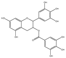 | 0.45 | 0.59 | HNSCC, OSCC, other cancers | Phase 1/2 | [101,102,103] |
| ABT-737 |  | 0.12 | 0.064 | HNSCC, ST, PC, Leukemia, etc. | Phase 1/2 | [101,104,105] |
| ABT-263 |  | NA | NA | ST, Haemato logical malig- nancies, SCLC | Phase 1/2 | [106,107] |
| ABT-199 |  | 0.1 | NA | ST, Breast cancer | Approved for use in CLL | [108,109] |
| TW-37 |  | NA | NA | HNSCC, Prostrate Cancer, PC, | Phase 1/2 | [110,111] |
| Gossypol |  | 0.28–10 | 0.4–3.03 | HNSCC, ST, PC | Phase 1/2 | [101,112] |
| GX15-070 (Obatoclax) |  | NA | NA | HNSCC, PC, ST, NSCLC | Phase 1 | [113,114] |
| HA14-1 |  | ~9 | NA | HNSCC, leukemia, lymphoma, colon cancer, etc. | Pre-clinical | [113,115,116] |
| Chelerythrine |  | ~10 | ~10 | HNSCC, ST, etc. | [101,117] | |
| S55746 |  | NA | NA | Hematological tumor | Phase 1 | [118] |
Publisher’s Note: MDPI stays neutral with regard to jurisdictional claims in published maps and institutional affiliations. |
© 2021 by the authors. Licensee MDPI, Basel, Switzerland. This article is an open access article distributed under the terms and conditions of the Creative Commons Attribution (CC BY) license (https://creativecommons.org/licenses/by/4.0/).
Share and Cite
Alam, M.; Ali, S.; Mohammad, T.; Hasan, G.M.; Yadav, D.K.; Hassan, M.I. B Cell Lymphoma 2: A Potential Therapeutic Target for Cancer Therapy. Int. J. Mol. Sci. 2021, 22, 10442. https://doi.org/10.3390/ijms221910442
Alam M, Ali S, Mohammad T, Hasan GM, Yadav DK, Hassan MI. B Cell Lymphoma 2: A Potential Therapeutic Target for Cancer Therapy. International Journal of Molecular Sciences. 2021; 22(19):10442. https://doi.org/10.3390/ijms221910442
Chicago/Turabian StyleAlam, Manzar, Sabeeha Ali, Taj Mohammad, Gulam Mustafa Hasan, Dharmendra Kumar Yadav, and Md. Imtaiyaz Hassan. 2021. "B Cell Lymphoma 2: A Potential Therapeutic Target for Cancer Therapy" International Journal of Molecular Sciences 22, no. 19: 10442. https://doi.org/10.3390/ijms221910442
APA StyleAlam, M., Ali, S., Mohammad, T., Hasan, G. M., Yadav, D. K., & Hassan, M. I. (2021). B Cell Lymphoma 2: A Potential Therapeutic Target for Cancer Therapy. International Journal of Molecular Sciences, 22(19), 10442. https://doi.org/10.3390/ijms221910442


