Review

The Multiple Faces of Integrin–ECM Interactions in Inflammatory Bowel Disease

1
IRCCS Humanitas Research Hospital, Rozzano, 20089 Milan, Italy
2
Department of Pharmaceutical Sciences, University of Piemonte Orientale ‘A. Avogadro’, 28100 Novara, Italy
3
Department of Biomedical Sciences, Humanitas University, Pieve Emanuele, 20072 Milan, Italy
*
Author to whom correspondence should be addressed.
Int. J. Mol. Sci. 2021, 22(19), 10439; https://doi.org/10.3390/ijms221910439
Submission received: 7 September 2021 / Revised: 20 September 2021 / Accepted: 21 September 2021 / Published: 28 September 2021

Abstract

:
Inflammatory Bowel Disease (IBD) comprises a series of chronic and relapsing intestinal diseases, with Crohn’s disease and ulcerative colitis being the most common. The abundant and uncontrolled deposition of extracellular matrix, namely fibrosis, is one of the major hallmarks of IBD and is responsible for the progressive narrowing and closure of the intestine, defined as stenosis. Although fibrosis is usually considered the product of chronic inflammation, the substantial failure of anti-inflammatory therapies to target and reduce fibrosis in IBD suggests that fibrosis might be sustained in an inflammation-independent manner. Pharmacological therapies targeting integrins have recently shown great promise in the treatment of IBD. The efficacy of these therapies mainly relies on their capacity to target the integrin-mediated recruitment and functionality of the immune cells at the damage site. However, by nature, integrins also act as mechanosensitive molecules involved in the intracellular transduction of signals and modifications originating from the extracellular matrix. Therefore, understanding integrin signaling in the context of IBD may offer important insights into mechanisms of matrix remodeling, which are uncoupled from inflammation and could underlie the onset and persistency of intestinal fibrosis. In this review, we present the currently available knowledge on the role of integrins in the etiopathogenesis of IBD, highlighting their role in the context of immune-dependent and independent mechanisms.

1. Introduction

Inflammatory Bowel Disease (IBD) defines a group of chronic, relapsing, and remitting enteropathies, including Crohn’s disease (CD) and Ulcerative colitis (UC) [1]. The leading causes of IBD onset and progression are poorly understood. However, it is well known that genetic susceptibility, alteration in the gut microbiota compositions, and environmental factors are strongly involved [2]. This multifactorial scenario sustains a chronic and aberrant immune response, triggering extensive intestinal tissue damage at the inflammatory site [3,4,5]. While in physiological conditions the tissue damage response leads to the correct restoration of the tissue homeostasis [6,7], the persistency of the inflammatory insult during IBDs triggers an unbalanced wound healing response resulting in chronic tissue damage and fibrosis, ultimately leading to organ dysfunction [8]. Intestinal fibrosis is a common complication in IBD and can occur in both UC and CD, although it is more prevalent in CD [9]. Tissue fibrosis is responsible for the onset of common IBD complications, such as strictures formation, also termed stenosis, which narrows the intestinal lumen and impairs bowel functionality [10,11,12].
Current anti-inflammatory therapies for IBD patients do not prevent or solve stenotic formations, especially in the latest stages of the pathology progression. These anti-inflammatory biological molecules can modulate inflammation but are ineffective in treating fibrosis onset and progression [13,14]. Therefore, despite the availability of these novel biological treatments, the incidence of strictures formation has not changed and requires the development of different therapeutic strategies [15]. This evidence suggests that IBD and its related complications, among which fibrosis is the most debilitating, are not only governed by inflammation but might be sustained and perpetuated by additional inflammation-independent mechanisms which need to be understood in depth and taken into consideration to develop new therapeutic strategies able to target these stenotic complications [16].
Molecular players that have recently become prominent in IBD research and clinic are represented by members of the integrin family [17,18]. Many studies have reported the central role of integrins in the recruitment of immune cells at the damaged tissue area and in supporting the progression of inflammatory mechanisms [17,19]. In addition, integrins exert fundamental roles in the cell interaction with the ECM, in both sensing and transducing the modifications of the extracellular milieu, and acting as mechanosensitive molecules. For this dual role in participating in both inflammation-dependent and -independent mechanisms, integrins have been investigated in the context of the intestinal disease and other organ districts [19].
In this review article, we aim to summarize the involvement of integrins in IBD-associated intestinal fibrosis development, taking into consideration their functional role in both immune cells and other cell types, primarily fibroblasts, highly implicated in the development of fibrotic complications.

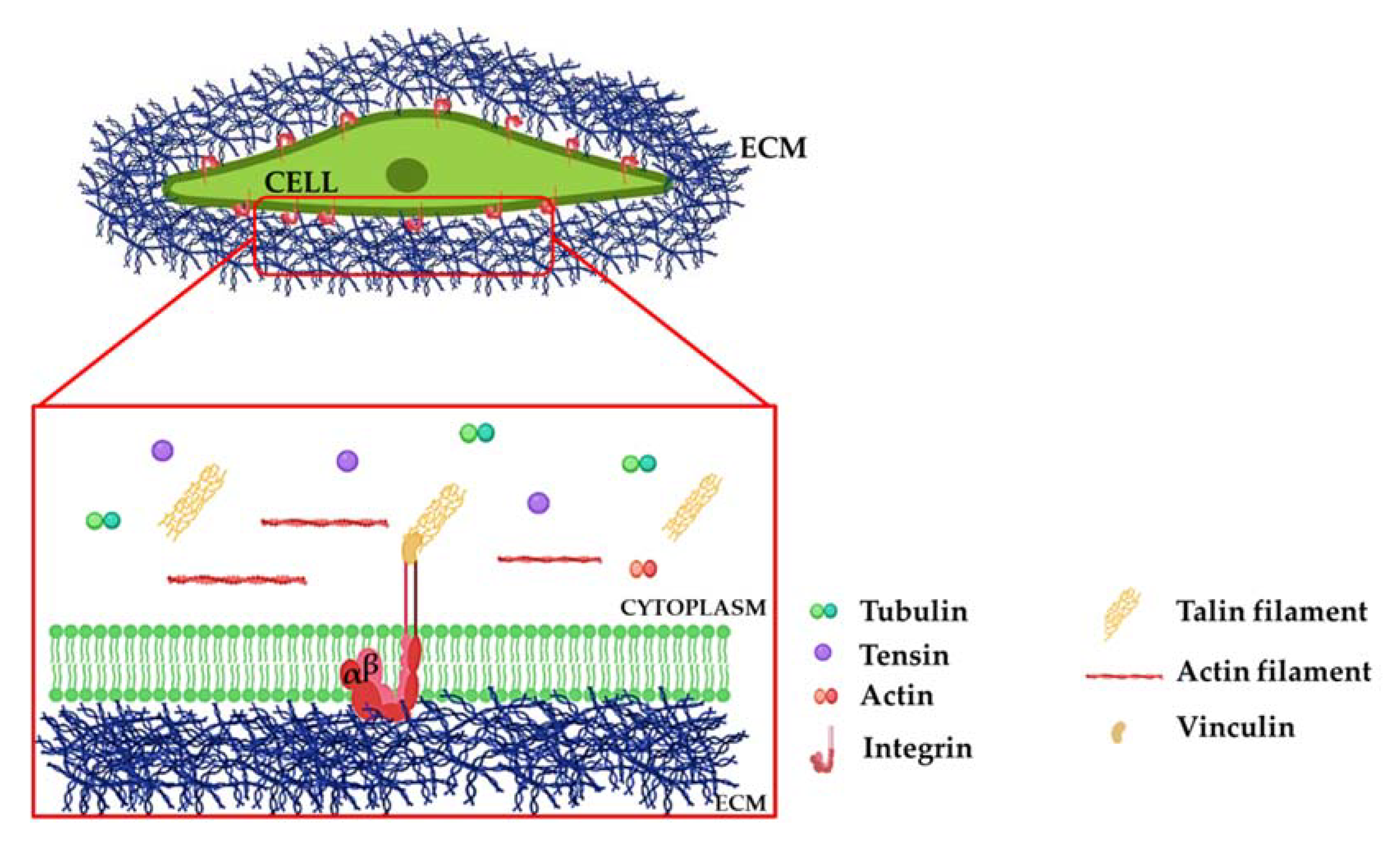
2. Integrins: A Brief Overview

Integrins are heterodimeric transmembrane receptors composed of α and β subunits acting as a bridge between the ECM and the cellular cytoskeleton [20,21]. Integrins are about 90–120 kDa molecules characterized by a multi-domain molecular structure, with a large extracellular domain linked through a transmembrane portion to a cytosolic tail directly interacting with the cytoskeleton [18,21,22]. The integrin family consists of 18 alpha and 8 beta subunits that can combine to form 24 different homo- or heterodimeric receptors, which bind specific ligands [23]. β-subunits are characterized by a cytoplasmic binding site involved in recognition of cytoskeletal cell components and transduction of downstream signalling. In contrast, the α-domains are engaged in establishing ligand type specificity with the outer ECM components [24]. In recent years, it has become clear that integrins have profound effects on the development of various diseases such as cancer and fibrotic diseases [19,25]. Moreover, many of the key cell–cell and cell-matrix interactions that regulate these pathologies are mediated by members of the integrin family. Collagens, laminins, and RGD-domains represent the preferential extracellular binding partners of integrins. In addition, through their cytosolic domain, integrins can cooperate with various intracellular proteins such as talin, vinculin, and actin (Figure 1) [26].

Molecular Mechanisms of Integrin Signaling

Integrin affinity for ECM molecules stabilizes the connection between the cell cytoskeleton and the ECM, activating intracellular signaling pathways involved in regulating biological functions such as proliferation, apoptosis, and differentiation [27]. This interaction starts with the translocation of integrins from the cytosol to the plasma membrane and the subsequent creation of integrin clusters sensing the extracellular microenvironment and transducing external signals [28,29]. This activation, in turn, triggers the activation of the downstream molecules focal adhesion kinase (FAK), proto-oncogene non-receptor tyrosine kinase (SRC), serine-threonine protein kinase (AKT), and small GTPases belonging to the Rho family [29] (Figure 2).
Signaling pathways triggered by integrin activation are often the same activated by soluble molecular factors. For example, epidermal growth factor (EGF), platelet-derived growth factor (PDGF), and lipoprotein A (LPA) require cell adhesion to ECM, a function that is exerted by integrins. This explains why integrins and soluble molecular factors share common signaling axes [30].
In addition, integrins play a crucial role in maintaining a balance between apoptosis and cell proliferation in normal cells. In the first case, the function is based on the activation of the phosphatidylinositol 3-kinase (PI3K) signaling pathway, and in the second is based on the extracellular signal-regulated kinase (ERK), cyclin D1 cascade, and cyclin E-cyclin dependent kinase (CDK) 2 complex in early G1 phase. Furthermore, the effect of the cyclin E-CDK2 complex seems to be mediated by the downregulation of the CDK inhibitors p21 and p27. These findings suggest the involvement of integrins in regulating cell cycle progression [31,32].
Another fundamental function regulated by integrins is cell motility. Integrins influence the phosphorylation status of the cell motility machinery, enhancing the activation of the myosin light chain kinases (MLCK) and the phosphorylation of the myosin light chain (MLC) molecule [33]. It has been studied that ERK can also phosphorylate MLCK, inducing MLC activation in a calmodulin-dependent manner [34,35]. In addition, survival signaling pathways are associated with both growth factor receptors and cell adhesion molecules, and are relevant in protecting cells from pathological and homeostatic cell death. For example, molecular growth factors such as EGF, PDGF, and insulin can induce survival of serum-starved epithelial cells [36,37,38]. At the same time, binding of integrins such as αvβ3 [39], α5β1, and α6β1 [40,41,42] to the appropriate extracellular matrix protein can inhibit cell death.

3. The Role of Integrins in Inflammation-Dependent Mechanisms in IBD

Homing of immune cells to the injured tissue is mediated by chemo-attractive mechanisms and facilitated by interactions between integrins expressed by leucocytes and adhesion molecules presented on the endothelial cell surface. Various preclinical studies have elucidated the critical role of leukocyte-expressed integrins in different inflammatory diseases, demonstrating that their deficiencies may significantly moderate both the pathogenesis and the development of IBDs [43].
Integrin heterodimers mainly involved in mediating the homing of lymphocytes to the gut mucosa are α4β1, α4β7, and αEβ7. α4β1 and α4β7 integrins are highly expressed on CD4+ memory T cells [44,45], and they interact, respectively, with the vascular cell adhesion molecule 1 (VCAM-1) expressed on the endothelium [46], and the mucosal addressin cell adhesion molecule 1 (MAdCAM-1) expressed in the gut-associated lymphoid tissues (GALT) [47]. The fundamental function of the α4 integrins in intestinal inflammation and immune cell recruitment was demonstrated early on by immunoblockade of α4 integrin in a cotton-top tamarin model of colitis [48,49]. Adoptive transfer of α4 null T cells in immunodeficient mice significantly attenuated chronic colitis and confirmed the defective homing of T cells to the inflamed intestinal mucosa [50].
MAdCAM-1 expression is largely restricted to the intestinal tissue, therefore its interaction with α4β7 integrin is responsible for most of the T cells homing in the gut. Both MAdCAM-1 and VCAM-1 were reported to be increased in inflamed intestinal biopsies from CD and UC patients [51,52], and this upregulation enhances the α4β1- and α4β7-mediated recruitment of inflammatory lymphocytes. MAdCAM-1 expression has also been reported on venules of lymphoid aggregates in CD patients [51]. Additionally, α4β7 expression facilitates the gut entry of regulatory T cells, while blocking α4β7 reduced the intestinal homing of both effector and regulatory T cells [53].
Parallel to the recruiting and homing functions of α4β1 and α4β7 heterodimers, the αEβ7 integrin instead exerts its major role in retaining T cells and dendritic cells in the intestinal mucosa. The expression of αEβ7 on the surface of T lymphocytes, induced by the local TGFβ [54,55], allows these immune cells to be retained in the intestinal epithelial layer thanks to the engagement with its ligand E-cadherin [56,57,58]. The pro-colitogenic role of the αEβ7-expressing lymphocytes is well documented. αEβ7 was expressed by a subset of pro-inflammatory colonic CD4+ T cells, which display Th1 and Th17 inflammatory profile and reduced expression of regulatory T cell-associated genes such as FOXP3, IL-10, CTL-4, and ICOS [52]. Furthermore, αE expression on a subset of resident memory CD4+CD69+ T cells, which accumulate in the mucosa of IBD patients, is predictive for the development of flares [59]. αEβ7 was also shown in CD to be expressed on an expanded subset of CD4+ T cells expressing the NKG2D receptor and displaying inflammatory and cytotoxic properties [60]. Elevated expression of αEβ7 was noted on Th9 CD4+ and CD8+ cells, in comparison to α4β7 being expressed by the Th2 and Th17 T cells [55]. In addition to CD4+ and CD8+ cells, αEβ7 was found upregulated on a subset of γδ-T lymphocytes found in the mesenteric lymph node and the intestinal tissue in a mouse model of T cell-mediated colitis and the SAMP spontaneous chronic ileitis [61]. In addition to being physically retained in the epithelium, a direct cytotoxic activity of the αEβ7-expressing T lymphocytes on the intestinal epithelial cells has been reported [52,62].
In line with the functional role of αEβ7 highlighted by all these studies, administration of a monoclonal antibody against αEβ7 (Table 1) was able to reduce the accumulation of CD4+ T cells in the lamina propria and their IFNγ production, thus ameliorating immunization-induced colitis in the IL2 knockout mouse model [63]. Moreover, etrolizumab-mediated blockade of αEβ7 was more effective than vedolizumab-mediated targeting of α4β7 in reducing the colonic numbers of CD8+ and Th9 cells [55].
Integrin αE is also expressed in the dendritic cells (DCs) in the subepithelial lamina propria and the mesenteric lymphnodes [64,65]. In physiological conditions, these αE+ DCs exert a tolerogenic function in the regulation of T cell homing [66,67]. The frequency and functionality of αE+ DCs were reported to be altered in the inflamed intestinal mucosa [68]. Specifically, the function of αE+ DCs was shown to be altered in the colon of UC patients. These DCs displayed an impaired capacity to generate regulatory T cells and induced a Th1/Th2/Th17 phenotype in CD4+ effector T cells [69].
Overall, these studies highlighted the integrin-mediated infiltration and retention of immune cells, as well as their switch to a pro-inflammatory phenotype, as fundamental mechanisms underlying IBD pathogenesis.

4. Anti-Integrin Therapies for IBD Based on Immune-Dependent Functions

The finding that integrin-mediated leucocytes trafficking plays a crucial role in IBD and could therefore represent a valuable therapeutic strategy has led to the development of monoclonal antibodies against leukocyte-expressed integrins. In contrast to the traditional immune-suppressive therapies, certainly able to modulate the inflammatory response but also generating harmful immunosuppression, anti-integrin therapies offered the opportunity to develop a more targeted approach, thus overcoming potential systemic side effects. In line with the knowledge acquired through in vitro and in vivo studies, the most diffused and effective anti-integrin treatments currently available are directed against integrins α4β1, α4β7, and αEβ7 (Table 1).
Natalizumab is a humanized monoclonal IgG4 antibody against the α4-integrin, acting on both α4β1 and α4β7 heterodimers. It inhibits the interaction between α4β7 and endothelial MAdCAM-1, blocking lymphocytes recruitment in the gastrointestinal lymphoid tissue [70]. Phase II and III studies show that natalizumab is effective as an induction and maintenance therapy in CD patients [71,72] and it has usually been used in patients not responding to other previously administered treatments [73]. However, its use has been limited due to the reported risk of Progressive Multifocal Leukoencephalopathy (PML), a rare but potentially fatal demyelinating brain disorder caused by polyomavirus JC which commonly occurs in HIV positive people, but is also reported in patients receiving long-term immunosuppression [74,75,76]. Natalizumab is thought to increase PML risk by preventing lymphocytes from adhering to the endothelium of the blood–brain barrier, therefore reducing their migration and suppressing T cell-mediated immune response in the brain [77].
Vedolizumab is a humanized monoclonal immunoglobulin G1 against α4β7 integrin. Its role in modulating lymphocytes trafficking is exerted in the gut but not in the brain, avoiding the predisposition to PML [78]. Benefits in patients with IBD have been elucidated in phase II clinical trials in patients with moderately to severely active Crohn’s disease [79].
Etrolizumab is another humanized monoclonal antibody selectively binding the β7 subunit of both α4β7 and αEβ7 heterodimers, antagonizing both α4β7-MAdCAM mediated lymphocyte recruitment and the αEβ7-E-cadherin interaction [80], which mediates T cells adhesion to epithelial cells [81]. Results of phase II clinical trials indicated that etrolizumab as induction therapy leads to a significant higher proportion of clinical remission compared to placebo, in patients with moderate-to-severe UC [82].
AJM300 is a small molecule orally administered and able to antagonize α4 integrins. Promising outcomes have been illustrated from several preclinical studies of experimental IBD animal models such as the DSS- and TNBS-induced. In 2009, Takazoe et al. reported the results obtained during a randomized study involving Japanese active CD-affected patients treated with AJM300 compound or placebo [83]. The clinical trial adopted AJM at three different doses (40 mg three times a day (TID), 120 mg TID, or 240 mg TID), or placebo, orally for eight weeks. AJM300 was safe and well-tolerated. However, the study was not pursued as no significant difference between the AJM300 treatment groups and the placebo group was reported. AJM300 evaluation efficacy, therefore, requires further investigation [83].
Abrilumab is an anti-α4β7 monoclonal antibody acting on immune cell trafficking, used in phase IIb clinical trials. Abrilumab can inhibit leukocytes recruitment and extravasation from the blood vessels and a specific sub-population of CD4+T cells (CD4+T-CD45RA). Nowadays, Abrilumab has been tested in UC patients, specifically in refractory patients with mild to severe UC. In fact, in 2019, Sandborn et al. tested the anti-α4β7 integrin monoclonal antibody at different doses (7, 21, 70, and 210 mg) in UC patients through subcutaneous administration [84]. Significant clinical responsiveness and mucosal healing were observed at week 8 of treatment, in the regimen with repeated 70 mg dosages and the single 210 mg subcutaneous administration, suggesting its promising potential as a new treatment for IBD patients [84].

5. Integrins in IBD-Associated Fibrosis: A Key Player in Its Onset, Independent of Inflammation

5.1. Fibrosis and Tissue Stiffness

Anti-inflammatory therapies for IBD are ineffective in treating pathological consequences of chronic inflammation such as fibrosis development. Despite stenotic complications, usually following the IBD pathology’s relapsing and remitting inflammatory fluctuations, it seems that intestinal fibrosis can partly occur independently from inflammation at the later stages of the disease, and that biologicals display some effects in inflammatory, but not fibrotic, strictures [13,14,85]. This can be explained by an auto-propagation of the fibrotic mechanisms supported by the persistent interaction between cellular compartments and the ECM matrix. The dysregulated release of ECM molecules supports the increase in matrix stiffness, a prominent feature of CD-associated intestinal fibrosis [86]. During homeostatic conditions, matrix stiffness degree is tightly regulated by the balance between deposition of ECM and tissue remodeling operated by enzymes such as metalloproteinases (MMPs) [87]. However, matrix deposition and turnover can be highly subverted during injury and CD-associated tissue damage, leading to a pathological scenario [88]. Matrix stiffness has been reported to be one of the mechanical forces sensed by cells able to modulate many biological functions, such as cell growth, survival, and motility. It is documented that an increment of matrix stiffness can modulate cell proliferation and differentiation [89]. Cells, especially related to the epithelial and mesenchymal compartment, can sense mechanical stiffness derived from the outer ECM and the adjacent cells. The transduction of extracellular mechanical stimuli and conversion of those signals into downstream signaling pathways is referred to as mechanotransduction and has been deeply studied in the recent decade in the field of fibrosis [90]. The mechano-signaling is a finely regulated process that relies on various proteins such as integrins and focal adhesion molecules, intracellular and cytoskeletal proteins, and nuclear responders [91]. Genetic ablation of α6 integrin in collagen-expressing mesenchymal cells or pharmacological blockade of matrix stiffness-regulated α6-expression was shown to protect mice against experimental lung fibrosis [88]. These findings suggest that integrins may be considered matrix stiffness-regulated mechanosensitive molecules that orchestrate the invasive fibroblast phenotype and mediate experimental lung fibrosis. Therefore, an alteration of matrix stiffness degree is sensed by integrins, leading to aberrant activation of cellular biological functions, eventually leading to tissue dysfunction, such as intestinal fibrosis. Intestinal fibrosis is characterized by increased tissue stiffness, distinguished from a healthy bowel by mechanical measurement and ultrasound elasticity imaging [92]. Ex vivo colonic tissue from normal, strictured, and the unaffected surgical margin was used to determine the stiffness of fibrotic compared to the normal colon for the development of an in vitro cell culture model of tissue stiffness [93]. This study showed that CD-associated strictured colon is nearly 6-fold stiffer than the normal bowel or the unaffected margin. Moreover, expression of genes involved in fibrogenic matrix remodeling, barrier function, and inflammatory was significantly increased in the CD strictures, therefore indicating concurrent fibrosis and inflammation. It has been demonstrated that increased matrix stiffness associated with the fibrostenotic disease alters colonic myofibroblast activation, producing a fibrogenic phenotype and auto-propagating fibrosis [94]. Indeed, activated colonic myofibroblasts function as key mediators of fibrosis and tissue stiffness by two mechanisms: (1) excessive extracellular collagen production which stiffens the ECM and (2) active contraction of the ECM, which exerts additional mechanical force on the matrix [95].

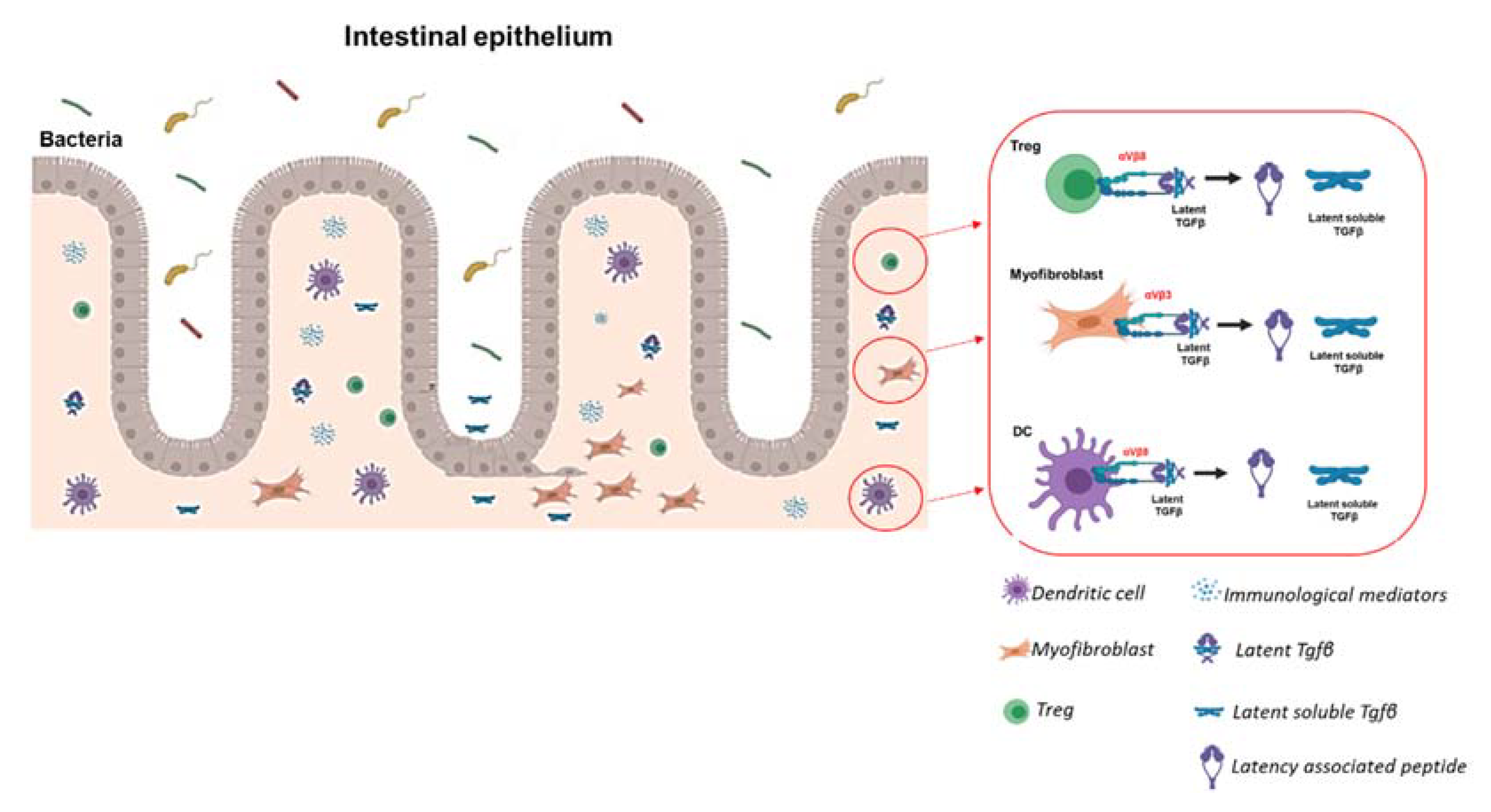
5.2. The Role of ECM in Intestinal Fibrosis: Not Only Support but Also a Reservoir of Molecular Mediators

The ECM is a three-dimensional molecular structure that provides an architecture to the organ tissue. Its molecular composition serves as a ligand for cell membrane receptors, such as integrins and various adhesion molecules [22]. The relevance of ECM has been demonstrated through many in vivo loss of function studies, in which genes encoding for fundamental ECM proteins have been silenced, leading to the onset of several pathologies characterized by mice mortality at early embryonic stages [96,97]. The ECM is composed of over 300 proteins, including laminins, fibronectin, and collagens. These molecules are the most representative, and their crucial role has been demonstrated in various diseases, supporting the idea that ECM balance is essential in all organs [98]. The interstitial ECM is mainly produced by cells belonging to the mesenchymal cell compartment. Differently, basement membrane-ECM plays a role in maintaining the epithelial cell layer and spatially separates it from the underlying intestinal lamina propria. The interstitial ECM is the most fundamental in fibrosis development. It contains molecular fibrotic mediators, such as epidermal growth factor (EGF), fibroblast growth factor (FGF), and TGFβ in a latent state. Among those, the most documented is the activation of TGFβ, the principal molecular factor involved in fibrosis development. Integrins and the downstream focal adhesion (FAK) complex proteins, known as mechano-sensors and mechanotransducers [23], sense and transduce external mechanical stimuli into biochemical signals [99]. The downstream signalling pathways induced by integrin-FAK complexes in response to matrix stiffness is bidirectional, such that there is a close relationship between the matrix, which exerts force on the cell via integrins, and the cellular cytoskeleton, which in turn resists this force [100].

5.3. Mechanisms of Integrin-Induced Release of Latent TGFβ

Integrins are involved in the direct activation of soluble molecular factors, which in homeostatic conditions are imprisoned in a latent form by the ECM network. In intestinal fibrosis, integrins have been involved in activating latent TGFβ [25,101,102], which is highly secreted by CD-associated stricture myofibroblasts [103]. TGFβ molecule is released in the extracellular microenvironment and conserved in a latent state at high concentration and directly cross-linked to the ECM. TGFβ activation is carried out by the interaction of integrins to a linear arginine-glycine-aspartic acid (RGD) motif present in an N-terminal fragment of TGFβ called a latency-associated peptide (LAP) [104].
Integrin-mediated TGFβ activation can be carried out in a protease-dependent or protease-independent manner. Integrin αvβ8 has been shown to bind to the RGD peptide in LAP and subsequently recruits MMP14, which then releases TGFβ by proteolytic cleavage. Similarly, integrin αvβ3 can act as a docking site for MMP2 and MMP9 [105].
Non-proteolytic activation instead occurs through cell traction forces due to increased matrix stiffness as it happens during fibrosis. The increased matrix stiffness provokes a tension of the cellular cytoskeleton, which modifies the interaction between integrin and the latent-TGFβ. This connection can cleave LAP and release the mature growth factor in the extracellular microenvironment [104]. Furthermore, these forces stimulate integrins to induce conformational changes of the TGFβ–LAP–LTBP complex, by which active TGFβ is released and able to interact with its receptors directly [106]. Interestingly, a recent study highlighted that activation of TGFβ by αvβ8 can occur independently from cytoskeletal forces and does not require the release from the latent peptide [107] (Figure 3).
Several integrins have been investigated in the pathogenesis of fibrosis in many organs, and their role in activating latent TGFβ has been well elucidated [25]. αv and α5 are the major group of integrins detected in intestinal fibrosis, especially for their involvement in the activation of TGFβ. αvβ3, αvβ5, and αvβ6, and their role in activating TGFβ, have been very well demonstrated in many in vitro studies [106]. In in vivo studies, the upregulation of αvβ6 in the epidermis has provoked elevated TGFβ1 activation leading to chronic ulcers formations and severe fibrosis [108].
In support of this evidence, in vivo studies demonstrated that αvβ6 and αvβ8 ablation recapitulates TGFβ knockout mouse phenotype and mutations in the RGD peptide share similar alterations observed in the TGFβ knockout mouse model [109].
Latent TGFβ1 was also reported to be activated by the αvβ3 RGD domain expressed in human and rat intestinal smooth muscles [110], and that an αvβ3 RGD inhibitor could be a novel treatment to diminish excess TGF-β1 activation, collagen I production, and development of fibrosis in CD [111]. In addition, an interaction between TGFβ, Smad3, αvβ6, and mTOR was observed in the development of intestinal fibrosis [112]. Integrins can also potentiate the action of the TGFβ pathway, supporting the over-expression of its downstream related genes. For example, the over-expression of α3β1 was shown to enhance MMP9 expression levels in keratinocytes [113], and the up-regulation of integrin β1 was demonstrated to induce p38/MAPK signaling during the EMT process [114]. Moreover, SMAD2 and SMAD3 cascade were found to be reduced as a consequence of αV blocking [115].

6. Functional Contribution of Integrins Expressed by Fibrosis-Associated Fibroblasts

Fibroblasts are active and fundamental players in the wound healing response and the progression to fibrosis [116,117]. By the fibroblast-to-myofibroblasts conversion process, these cells become activated and enabled to produce and deposit large amounts of ECM, which progressively accumulates during the fibrotic process. The fibrotic matrix itself, in turn, participates in the fibroblasts’ conversion through mechanotransduction processes [118].
A functional role for the fibroblast-expressed integrins has been demonstrated in multiple fibrotic disease models. To date, most of the work has been focused on the αV subunit [119]. Fibroblasts can express four types of αv heterodimers: αvβ1, αvβ3, αvβ5, and αvβ8. Conditional deletion of the Itgav gene (encoding integrin αv) in Pdgfrβ+ myofibroblasts was shown to protect mice from tetrachloride-induced hepatic fibrosis [120]. In vitro experiments using genetic or pharmacological inhibition of αv integrin confirmed the reduction in the profibrotic phenotype (collagen I and III, and αSMA expression) in these fibroblasts. Additionally, genetic deletion of αv integrin exerted a protective role also in bleomycin-induced pulmonary fibrosis and Unilateral Ureteral Obstruction (UUO)-induced renal fibrosis [115]. The high expression of αvβ1 mediates fibroblasts’ adhesion to LAP and activation of latent TGFβ [25,101,121]. The relevant heterodimer involved in this context was speculated to be αvβ1 as the global or conditional knockout of all the other possible binding partners (β3, β5, β6, and β8) failed to protect against hepatic fibrosis. The pre-clinical use of the αv small molecule inhibitor CWHM12 demonstrated to be effective in attenuating liver and lung fibrosis, even when fibrosis was already established [115], and the subsequent design of an αvβ1 small molecule inhibitor further proved the efficacy of targeting this integrin complex [101].
A functional role for fibroblast-expressed integrin αvβ8 was shown in the lung disease where conditional deletion or neutralization of integrin β8 in lung fibroblasts inhibits airway inflammation and fibrosis [44,122]. Furthermore, the αvβ8-mediated activation of TGFβ by fibroblasts was shown to be crucial for directing dendritic cell trafficking [44]. The fact that αvβ8 functionality in fibrosis-associated fibroblasts may be organ-dependent is indicated by the fact that conditional knockout of β8 in the hepatic fibroblast did not exert any protection from hepatic fibrosis.
αvβ3 expression in intestinal smooth muscle cells leads to activation of latent TGFβ and subsequent collagen production and fibrosis in the muscolaris mucosa in the TNBS-induced experimental model of intestinal fibrosis [123]. The RGD-containing αvβ3 integrin inhibitor Cilengitide administration decreased TGFβ1 activation, collagen production, and diminished TNBS-induced fibrosis.
Compared to the other αv-containing heterodimers, the αvβ6 integrin is upregulated exclusively in epithelial lineages during an injury in multiple models. αvβ6 expression has been reported in the renal epithelium [124], cholangiocyte and hepatic progenitors [125,126], pneumocytes, and alveolar epithelium [127,128]. In all these studies, genetic and pharmacological blocking of αvβ6 proved effective in reducing lung, kidney, and biliary fibrosis. All these models demonstrated that the activation of latent TGFβ mediates the mechanism of αvβ6-induced fibrogenesis. In addition, αvβ6 was shown to be up-regulated in the submucosa and serosa of TNBS treated mice [112]; however, no detailed investigation on its precise expression and functionality in the intestinal epithelium has been conducted.

7. Future Perspectives

Despite significant advancement in the treatment of IBD obtained with immunomodulators, there is still a substantial portion of IBD patients who are refractory or acquire resistance to these therapies. Targeted anti-integrin therapies represent a promising therapeutic option for managing the disease in this fraction of patients [17]. The therapeutic strategy underlying these anti-integrins is represented by blocking the functionality of integrins expressed by immune cells and mediating their homing and retention to the damaged intestine [17].
It has become increasingly appreciated that inflammation is not the only component of IBD disease. The majority of complications, such as strictures and stenosis, arise due to fibrotic processes, particularly in Crohn’s patients [129,130]. The fact that anti-inflammatory therapies are substantially ineffective towards these fibrotic complications suggests that, although it might indeed arise as a consequence of inflammation, once established, the fibrotic response could progress and self-perpetuate independently from inflammation, as also suggested by mounting experimental evidence [131,132]. Fibroblasts represent the major cell type involved in the establishment and evolution of fibrosis. As reviewed above, various types of integrin heterodimers are expressed by fibrosis-associated fibroblasts, and their functional involvement has been demonstrated in multiple fibrotic disease models, including liver, kidney, and lung [101,115]. On the contrary, the phenotypic and functional characterization of integrin expression in the fibrotic fibroblasts associated with intestinal fibrosis is very limited and would certainly deserve further investigation.
The vast majority of the studies utilizing genetic or pharmacological inhibition of fibroblast-expressed integrins have identified the activation of the latent TGFβ peptide as the major mechanism responsible for the induction of the integrin-mediated pro-fibrotic phenotype [101,133]. In this regard, numerous open questions remain to be answered concerning the intracellular signaling pathways engaged as a consequence of the activation of the integrin signaling in fibroblasts. Moreover, considering that integrins function as mechano-transducers translating stimuli from the external ECM into intracellular changes, understanding the functional role of fibroblast-expressed integrins from a mechanotransduction perspective may shed some light on the complex reciprocal interplay between intestinal matrix remodeling and fibroblasts functionality. This would potentially provide insights into the mechanisms of fibrosis propagated by tissue remodeling events and uncoupled from inflammatory processes and, in turn, explain why blocking intestinal inflammation alone is ineffective in limiting fibrotic processes.

8. Closing Remarks

Considering that multiple intracellular pathways are activated by integrins, and the way that ECM changes modulate much fibroblast functionality, it is possible to predict that the activation of the latent TGFβ is not the only molecular pathway activated by fibroblast-expressed integrins. Extending our understanding of the complex interplay between inflammation and tissue remodeling in IBD to the multiple cell types potentially involved, and considering the fibrotic matrix as an active player rather than just a scaffold may provide novel mechanistic insights to further our comprehension of the disease and design novel therapies for the IBD-associated fibrotic complications.

Author Contributions

V.G. conceptually designed the review, searched the literature, prepared the figures and the table, and wrote and edited the manuscript. S.L. supervised the conceptual design of the review, helped performing the literature search, provided intellectual input, helped writing, and edited the manuscript. S.V. and S.D. provided intellectual input, participated in the conceptual design of the review, and edited the manuscript. All authors approved the final version. All authors have read and agreed to the published version of the manuscript.

Funding

Research in the Vetrano and Danese laboratories related to the topic of this review is supported by the Horizon 2020 ERA-Net Cofund Action EuroNanoMed III grant (RUNNING) to S.D., and by the Innovative Medicines Initiative 2 Joint Undertaking (JU) under grant agreement No.853995 to S.D. and S.V. The JU receives support from the European Union’s Horizon 2020 research and innovation programme and EFPIA. The content provided in this review reflects only the author’s view and neither the IMI JU nor the European Commission are responsible for any use that may be made of the information it contains. S.L. is supported by the European Union’s Horizon 2020 research and innovation program under the Marie Sklodowska-Curie grant agreement #101029427 and was also supported by Fondazione Umberto Veronesi (2020 Post-Doctoral Fellowship).

Institutional Review Board Statement

Not applicable.

Informed Consent Statement

Not applicable.

Data Availability Statement

Not applicable.

Acknowledgments

The authors wish to thank Angelica Iato for assistance with the literature search, and Ghada Shalaby for English language proofreading. We apologize to any author whose work might have been unintentionally omitted.

Conflicts of Interest

S.D. has served as a speaker, consultant, and advisory board member for Schering-Plough, Abbott (AbbVie) Laboratories, Merck and Co, UCB Pharma, Ferring, Cellerix, Millenium Takeda, Nycomed, Pharmacosmos, Actelion, Alfa Wasserman, Genentech, Grunenthal, Pfizer, Astra Zeneca, Novo Nordisk, Vifor, and Johnson and Johnson. The other authors declare no conflict of interest.

References

  1. Rieder, F.; Fiocchi, C. Intestinal fibrosis in IBD—A dynamic, multifactorial process. Nat. Rev. Gastroenterol. Hepatol. 2009, 6, 228–235. [Google Scholar] [CrossRef] [PubMed]
  2. Danese, S.; Fiocchi, C. Etiopathogenesis of inflammatory bowel diseases. World J. Gastroenterol. 2006, 12, 4807–4812. [Google Scholar] [CrossRef]
  3. M’Koma, A. The Multifactorial Etiopathogeneses Interplay of Inflammatory Bowel Disease: An Overview. Gastrointest. Disord. 2018, 1, 75–105. [Google Scholar] [CrossRef] [Green Version]
  4. Jostins, L.; Ripke, S.; Weersma, R.K.; Duerr, R.H.; McGovern, D.P.; Hui, K.Y.; Lee, J.C.; Schumm, L.P.; Sharma, Y.; Anderson, C.A.; et al. Host-microbe interactions have shaped the genetic architecture of inflammatory bowel disease. Nature 2012, 491, 119–124. [Google Scholar] [CrossRef] [PubMed] [Green Version]
  5. Lakatos, P.L.; Szamosi, T.; Lakatos, L. Smoking in inflammatory bowel diseases: Good, bad or ugly? World J. Gastroenterol. 2007, 13, 6134–6139. [Google Scholar] [CrossRef] [PubMed]
  6. Rieder, F.; Brenmoehl, J.; Leeb, S.; Schölmerich, J.; Rogler, G. Wound healing and fibrosis in intestinal disease. Gut 2007, 56, 130–139. [Google Scholar] [CrossRef]
  7. Gabbiani, G. The myofibroblast in wound healing and fibrocontractive diseases. J. Pathol. 2003, 200, 500–503. [Google Scholar] [CrossRef]
  8. Otte, J.M.; Rosenberg, I.M.; Podolsky, D.K. Intestinal myofibroblasts in innate immune responses of the intestine. Gastroenterology 2003, 124, 1866–1878. [Google Scholar] [CrossRef]
  9. Lewis, A.; Correale, C.; Szabo, H.; Montorsi, M. Intestinal Fibrosis in Crohn’s Disease. Inflamm. Bowel Dis. 2015, 21, 1141–1150. [Google Scholar] [CrossRef]
  10. Cosnes, J.; Cattan, S.; Blain, A.; Beaugerie, L.; Carbonnel, F.; Parc, R.; Gendre, J.P. Long-term evolution of disease behavior of Crohn’s disease. Inflamm. Bowel Dis. 2002, 8, 244–250. [Google Scholar] [CrossRef]
  11. de Bruyn, J.R.; Meijer, S.L.; Wildenberg, M.E.; Bemelman, W.A.; van den Brink, G.R.; D’Haens, G.R. Development of fibrosis in acute and longstanding ulcerative colitis. J. Crohn’s Colitis 2015, 9, 966–972. [Google Scholar] [CrossRef] [PubMed] [Green Version]
  12. Alfredsson, J.; Wick, M.J. Mechanism of fibrosis and stricture formation in Crohn’s disease. Scand. J. Immunol. 2020, 92, 1–23. [Google Scholar] [CrossRef]
  13. de Bruyn, J.R.; Becker, M.A.; Steenkamer, J.; Wildenberg, M.E.; Meijer, S.L.; Buskens, C.J.; Bemelman, W.A.; Löwenberg, M.; Ponsioen, C.Y.; van den Brink, G.R.; et al. Intestinal fibrosis is associated with lack of response to Infliximab therapy in Crohn’s disease. PLoS ONE 2018, 13, e0190999. [Google Scholar] [CrossRef]
  14. Bharadwaj, S.; Fleshner, P.; Shen, B. Therapeutic armamentarium for stricturing Crohn’s disease: Medical versus endoscopic versus surgical approaches. Inflamm. Bowel Dis. 2015, 21, 2194–2213. [Google Scholar] [CrossRef] [PubMed]
  15. Crespi, M.; Dulbecco, P.; De Ceglie, A.; Conio, M. Strictures in Crohn’s Disease: From Pathophysiology to Treatment. Dig. Dis. Sci. 2020, 65, 1904–1916. [Google Scholar] [CrossRef] [PubMed]
  16. Yoo, J.H.; Holubar, S.; Rieder, F. Fibrostenotic strictures in Crohn’s disease. Intest. Res. 2020, 18, 379–401. [Google Scholar] [CrossRef] [PubMed]
  17. Dotan, I.; Allez, M.; Danese, S.; Keir, M.; Tole, S.; McBride, J. The role of integrins in the pathogenesis of inflammatory bowel disease: Approved and investigational anti-integrin therapies. Med. Res. Rev. 2020, 40, 245–262. [Google Scholar] [CrossRef] [PubMed] [Green Version]
  18. Sun, Z.; Guo, S.S.; Fässler, R. Integrin-mediated mechanotransduction. J. Cell Biol. 2016, 215, 445–456. [Google Scholar] [CrossRef]
  19. Goodman, S.L.; Picard, M. Integrins as therapeutic targets. Trends Pharmacol. Sci. 2012, 33, 405–412. [Google Scholar] [CrossRef]
  20. Hynes, R.O. Integrins: Bidirectional, allosteric signaling machines. Cell 2002, 110, 673–687. [Google Scholar] [CrossRef] [Green Version]
  21. Wiesner, S.; Legate, K.R.; Fässler, R. Integrin-actin interactions. Cell. Mol. Life Sci. 2005, 60, 1081–1099. [Google Scholar] [CrossRef]
  22. Hynes, R.O. The extracellular matrix: Not just pretty fibrils. Science 2009, 326, 1216–1219. [Google Scholar] [CrossRef] [Green Version]
  23. Kechagia, J.Z.; Ivaska, J.; Roca-Cusachs, P. Integrins as biomechanical sensors of the microenvironment. Nat. Rev. Mol. Cell Biol. 2019, 20, 457–473. [Google Scholar] [CrossRef]
  24. Mezu-Ndubuisi, O.J.; Maheshwari, A. The role of integrins in inflammation and angiogenesis. Pediatr. Res. 2021, 89, 1619–1626. [Google Scholar] [CrossRef]
  25. Henderson, N.C.; Sheppard, D. Integrin-mediated regulation of TGFβ in fibrosis. Biochim. Biophys. Acta—Mol. Basis Dis. 2013, 1832, 891–896. [Google Scholar] [CrossRef] [PubMed] [Green Version]
  26. Latella, G.; Di Gregorio, J.; Flati, V.; Rieder, F.; Lawrance, I.C. Mechanisms of initiation and progression of intestinal fibrosis in IBD. Scand. J. Gastroenterol. 2014, 50, 53–65. [Google Scholar] [CrossRef] [PubMed]
  27. Zamir, E.; Geiger, B. Molecular complexity and dynamics of cell-matrix adhesions. J. Cell Sci. 2001, 114, 3583–3590. [Google Scholar] [CrossRef] [PubMed]
  28. Hamidi, H.; Ivaska, J. Every step of the way: Integrins in cancer progression and metastasis. Nat. Rev. Cancer 2018, 18, 1–16. [Google Scholar] [CrossRef] [Green Version]
  29. De Franceschi, N.; Miihkinen, M.; Hamidi, H.; Alanko, J.; Mai, A.; Picas, L.; Guzmán, C.; Lévy, D.; Mattjus, P.; Goult, B.T.; et al. ProLIF—Quantitative integrin protein–protein interactions and synergistic membrane effects on proteoliposomes. J. Cell Sci. 2019, 132, jcs214270. [Google Scholar] [CrossRef] [Green Version]
  30. Assoian, R.K. Anchorage-dependent cell cycle progression. J. Cell Biol. 1997, 136, 1–4. [Google Scholar] [CrossRef] [Green Version]
  31. Danen, E.H.J.; Yamada, K.M. Fibronectin, integrins, and growth control. J. Cell. Physiol. 2001, 189, 1–13. [Google Scholar] [CrossRef] [PubMed]
  32. Walker, J.L.; Assoian, R.K. Integrin-dependent signal transduction regulating cyclin D1 expression and G1 phase cell cycle progression. Cancer Metastasis Rev. 2005, 24, 383–393. [Google Scholar] [CrossRef]
  33. Klemke, R.L.; Cai, S.; Giannini, A.L.; Gallagher, P.J.; Lanerolle, P.D.; Cheresh, D.A. Regulation of cell motility by mitogen-activated protein kinase. J. Cell Biol. 1997, 137, 481–492. [Google Scholar] [CrossRef]
  34. Schlaepfer, D.D.; Hunter, T. Integrin signalling and tyrosine phosphorylation: Just the FAKs? Trends Cell Biol. 1998, 8, 151–157. [Google Scholar] [CrossRef]
  35. Tanimura, S.; Takeda, K. ERK signalling as a regulator of cell motility. J. Biochem. 2017, 162, 145–154. [Google Scholar] [CrossRef] [Green Version]
  36. Merlo, A.; Herman, J.G.; Mao, L.; Lee, D.J.; Gabrielson, E.; Burger, P.C.; Baylin, S.B.; Sidransky, D. 5′ CpG Island methylation is associated with transcriptional silencing of the tumour suppressor p16/CDKN2/MTS1 in human cancers. Nat. Med. 1995, 1, 686–692. [Google Scholar] [CrossRef]
  37. Rampalli, A.M.; Zelenka, P.S. Insulin regulates expression of c-fos and c-jun and suppresses apoptosis of lens epithelial cells. Cell Growth Differ. 1995, 6, 945–953. [Google Scholar] [PubMed]
  38. Rodeck, U.; Jost, M.; DuHadaway, J.; Kari, C.; Jensen, P.J.; Risse, B.; Ewert, D.L. Regulation of Bcl-xL expression in human keratinocytes by cell-substratum adhesion and the epidermal growth factor receptor. Proc. Natl. Acad. Sci. USA 1997, 94, 5067–5072. [Google Scholar] [CrossRef] [Green Version]
  39. Strömblad, S.; Becker, J.C.; Yebra, M.; Brooks, P.C.; Cheresh, D.A. Suppression of p53 activity and p21(WAF1/CIP1) expression by vascular cell integrin αvβ3 during angiogenesis. J. Clin. Investig. 1996, 98, 426–433. [Google Scholar] [CrossRef]
  40. Howlett, A.R.; Bailey, N.; Damsky, C.; Petersen, O.W.; Bissell, M.J. Cellular growth and survival are mediated by β1 integrins in normal human breast eqithelium but not in breast carcinoma. J. Cell Sci. 1995, 108, 1945–1957. [Google Scholar] [CrossRef]
  41. Wewer, U.M.; Thornell, L.E.; Loechel, F.; Zhang, X.; Durkin, M.E.; Amano, S.; Burgeson, R.E.; Engvall, E.; Albrechtsen, R.; Virtanen, I.; et al. Extrasynaptic localization of laminin β2 chain in developing and adult human skeletal muscle. Am. J. Pathol. 1997, 151, 621–631. [Google Scholar]
  42. Farrelly, N.; Lee, Y.J.; Oliver, J.; Dive, C.; Streuli, C.H. Extracellular matrix regulates apoptosis in mammary epithelium through a control on insulin signaling. J. Cell Biol. 1999, 144, 1337–1347. [Google Scholar] [CrossRef]
  43. Yonekawa, K.; Harlan, J.M. Targeting leukocyte integrins in human diseases. J. Leukoc. Biol. 2005, 77, 129–140. [Google Scholar] [CrossRef]
  44. Minagawa, S.; Lou, J.; Seed, R.I.; Cormier, A.; Wu, S.; Cheng, Y.; Murray, L.; Tsui, P.; Connor, J.; Herbst, R.; et al. Selective targeting of TGF-β activation to treat Fibroinflammatory airway disease. Sci. Transl. Med. 2014, 6, 241ra79. [Google Scholar] [CrossRef] [Green Version]
  45. Erle, D.J.; Briskin, M.J.; Butcher, E.C.; Garcia-Pardo, A.; Lazarovits, A.I.; Tidswell, M. Expression and function of the MAdCAM-1 receptor, integrin alpha 4 beta 7, on human leukocytes. J. Immunol. 1994, 153, 517–528. [Google Scholar] [PubMed]
  46. Elices, M.J.; Osborn, L.; Takada, Y.; Crouse, C.; Luhowskyj, S.; Hemler, M.E.; Lobb, R.R. VCAM-1 on activated endothelium interacts with the leukocyte integrin VLA-4 at a site distinct from the VLA-4/Fibronectin binding site. Cell 1990, 60, 577–584. [Google Scholar] [CrossRef]
  47. Berlin, C.; Berg, E.L.; Briskin, M.J.; Andrew, D.P.; Kilshaw, P.J.; Holzmann, B.; Weissman, I.L.; Hamann, A.; Butcher, E.C. α4β7 integrin mediates lymphocyte binding to the mucosal vascular addressin MAdCAM-1. Cell 1993, 74, 185–195. [Google Scholar] [CrossRef]
  48. Podolsky, D.K.; Lobb, R.; King, N.; Benjamin, C.D.; Pepinsky, B.; Sehgal, P.; DeBeaumont, M. Attenuation of colitis in the cotton-top tamarin by anti-α4 integrin monoclonal antibody. J. Clin. Investig. 1993, 92, 372–380. [Google Scholar] [CrossRef] [PubMed] [Green Version]
  49. Hesterberg, P.E.; Winsor-Hines, D.A.; Briskin, M.J.; Soler-Ferran, D.U.; Merrill, C.H.; Mackay, C.R.; Newman, W.A.; Ringler, D.J. Rapid resolution of chronic colitis in the cotton-top tamarin with an antibody to a gut-homing integrin α4β7. Gastroenterology 1996, 111, 1373–1380. [Google Scholar] [CrossRef]
  50. Kurmaeva, E.; Lord, J.D.; Zhang, S.; Bao, J.R.; Kevil, C.G.; Grisham, M.B.; Ostanin, D.V. T cell-associated α4 β7 but not α4 β1 integrin is required for the induction and perpetuation of chronic colitis. Mucosal Immunol. 2014, 7, 1354–1365. [Google Scholar] [CrossRef] [PubMed] [Green Version]
  51. Arihiro, S.; Ohtani, H.; Suzuki, M.; Murata, M.; Ejima, C.; Oki, M.; Kinouchi, Y.; Fukushima, K.; Sasaki, I.; Nakamura, S.; et al. Differential expression of mucosal addressin cell adhesion molecule-1 (MAdCAM-1) in ulcerative colitis and Crohn’s disease. Pathol. Int. 2002, 52, 367–374. [Google Scholar] [CrossRef]
  52. Lamb, C.A.; Mansfield, J.C.; Tew, G.W.; Gibbons, D.; Long, A.K.; Irving, P.; Diehl, L.; Eastham-Anderson, J.; Price, M.B.; O’Boyle, G.; et al. αEβ7 Integrin Identifies Subsets of Pro-Inflammatory Colonic CD4+ T Lymphocytes in Ulcerative Colitis. J. Crohn’s Colitis 2017, 11, 610–620. [Google Scholar] [CrossRef] [Green Version]
  53. Fischer, A.; Zundler, S.; Atreya, R.; Rath, T.; Voskens, C.; Hirschmann, S.; López-Posadas, R.; Watson, A.; Becker, C.; Schuler, G.; et al. Differential effects of α4β7 and GPR15 on homing of effector and regulatory T cells from patients with UC to the inflamed gut in vivo. Gut 2016, 65, 1642–1664. [Google Scholar] [CrossRef] [PubMed] [Green Version]
  54. El-Asady, R.; Yuan, R.; Liu, K.; Wang, D.; Gress, R.E.; Lucas, P.J.; Drachenberg, C.B.; Hadley, G.A. TGF-β-dependent CD103 expression by CD8+ T cells promotes selective destruction of the host intestinal epithelium during graft-versus-host disease. J. Exp. Med. 2005, 201, 1647–1657. [Google Scholar] [CrossRef] [Green Version]
  55. Zundler, S.; Schillinger, D.; Fischer, A.; Atreya, R.; López-Posadas, R.; Watson, A.; Neufert, C.; Atreya, I.; Neurath, M.F. Blockade of αeβ7 integrin suppresses accumulation of CD8 + and Th9 lymphocytes from patients with IBD in the inflamed gut in vivo. Gut 2017, 66, 1936–1948. [Google Scholar] [CrossRef] [PubMed] [Green Version]
  56. Briskin, M.; Winsor-Hines, D.; Shyjan, A.; Cochran, N.; Bloom, S.; Wilson, J.; McEvoy, L.M.; Butcher, E.C.; Kassam, N.; Mackay, C.R.; et al. Human mucosal addressin cell adhesion molecule-1 is preferentially expressed in intestinal tract and associated lymphoid tissue. Am. J. Pathol. 1997, 151, 97–110. [Google Scholar] [PubMed]
  57. Schön, M.P.; Arya, A.; Murphy, E.A.; Adams, C.M.; Strauch, U.G.; Agace, W.W.; Marsal, J.; Donohue, J.P.; Her, H.; Beier, D.R. Mucosal T Lymphocyte Numbers Are Selectively Reduced in Integrin Alpha E (CD103)-Deficient Mice. J. Immunol. 1999, 162, 6641–6649. [Google Scholar] [PubMed]
  58. Wagner, N.; Löhler, J.; Kunkel, E.J.; Ley, K.; Leung, E.; Krissansen, G.; Rajewsky, K.; Müller, W. Critical role for β7 integrins in formation of the gut-associated lymphoid tissue. Nature 1996, 382, 366–370. [Google Scholar] [CrossRef]
  59. Zundler, S.; Becker, E.; Spocinska, M.; Slawik, M.; Parga-Vidal, L.; Stark, R.; Wiendl, M.; Atreya, R.; Rath, T.; Leppkes, M.; et al. Hobit- and Blimp-1-driven CD4+ tissue-resident memory T cells control chronic intestinal inflammation. Nat. Immunol. 2019, 20, 288–300. [Google Scholar] [CrossRef]
  60. Allez, M.; Tieng, V.; Nakazawa, A.; Treton, X.; Pacault, V.; Dulphy, N.; Caillat–Zucman, S.; Paul, P.; Gornet, J.M.; Douay, C.; et al. CD4+NKG2D+ T Cells in Crohn’s Disease Mediate Inflammatory and Cytotoxic Responses through MICA Interactions. Gastroenterology 2007, 132, 2346–2358. [Google Scholar] [CrossRef]
  61. Do, J.S.; Kim, S.; Keslar, K.; Jang, E.; Huang, E.; Fairchild, R.L.; Pizarro, T.T.; Min, B. γδ T Cells Coexpressing Gut Homing α4β7 and αE Integrins Define a Novel Subset Promoting Intestinal Inflammation. J. Immunol. 2017, 198, 908–915. [Google Scholar] [CrossRef] [Green Version]
  62. Roberts, A.I.; O’Connell, S.M.; Biancone, L.; Brolin, R.E.; Ebert, E.C. Spontaneous cytotoxicity of intestinal intraepithelial lymphocytes: Clues to the mechanism. Clin. Exp. Immunol. 1993, 94, 527–532. [Google Scholar] [CrossRef]
  63. Lúdvíksson, B.R.; Strober, W.; Nishikomori, R.; Hasan, S.K.; Ehrhardt, R.O. Administration of mAb against alpha E beta 7 prevents and ameliorates immunization-induced colitis in IL-2-/- mice. J. Immunol. 1999, 162, 4975–4982. [Google Scholar]
  64. Iliev, I.D.; Spadoni, I.; Mileti, E.; Matteoli, G.; Sonzogni, A.; Sampietro, G.M.; Foschi, D.; Caprioli, F.; Viale, G.; Rescigno, M.; et al. Human intestinal epithelial cells promote the differentiation of tolerogenic dendritic cells. Gut 2009, 58, 1481–1489. [Google Scholar] [CrossRef] [PubMed]
  65. Jaensson, E.; Uronen-Hansson, H.; Pabst, O.; Eksteen, B.; Tian, J.; Coombes, J.L.; Berg, P.L.; Davidsson, T.; Powrie, F.; Johansson-Lindbom, B.; et al. Small intestinal CD103+ dendritic cells display unique functional properties that are conserved between mice and humans. J. Exp. Med. 2008, 205, 2139–2149. [Google Scholar] [CrossRef] [Green Version]
  66. Quan, T.E.; Cowper, S.; Wu, S.P.; Bockenstedt, L.K.; Bucala, R. Circulating fibrocytes: Collagen-secreting cells of the peripheral blood. Int. J. Biochem. Cell Biol. 2004, 36, 598–606. [Google Scholar] [CrossRef] [PubMed]
  67. Scott, C.L.; Aumeunier, A.M.; Mowat, A.M.I. Intestinal CD103 + dendritic cells: Master regulators of tolerance? Trends Immunol. 2011, 32, 412–419. [Google Scholar] [CrossRef] [PubMed]
  68. Nguyen, D.T.; Nagarajan, N.; Zorlutuna, P. Effect of Substrate Stiffness on Mechanical Coupling and Force Propagation at the Infarct Boundary. Biophys. J. 2018, 115, 1966–1980. [Google Scholar] [CrossRef] [PubMed] [Green Version]
  69. Mann, E.R.; Bernardo, D.; Ng, S.C.; Rigby, R.J.; Al-Hassi, H.O.; Landy, J.; Peake, S.T.; Spranger, H.; English, N.R.; Thomas, L.V.; et al. Human gut dendritic cells drive aberrant gut-specific T-cell responses in ulcerative colitis, characterized by increased IL-4 production and loss of IL-22 and IFNγ. Inflamm. Bowel Dis. 2014, 20, 2299–2307. [Google Scholar] [CrossRef] [PubMed] [Green Version]
  70. Hamann, A.; Andrew, D.P.; Jablonski-Westrich, D.; Holzmann, B.; Butcher, E.C. Role of alpha 4-integrins in lymphocyte homing to mucosal tissues in vivo. J. Immunol. 1994, 152, 3282–3293. [Google Scholar]
  71. Ghosh, S.; Goldin, E.; Gordon, F.H.; Malchow, H.A.; Rask-Madsen, J.; Rutgeerts, P.; Vyhnálek, P.; Zádorová, Z.; Palmer, T.; Donoghue, S. Natalizumab for Active Crohn’s Disease. N. Engl. J. Med. 2003, 348, 24–32. [Google Scholar] [CrossRef] [PubMed]
  72. Targan, S.R.; Feagan, B.G.; Fedorak, R.N.; Lashner, B.A.; Panaccione, R.; Present, D.H.; Spehlmann, M.E.; Rutgeerts, P.J.; Tulassay, Z.; Volfova, M.; et al. Natalizumab for the Treatment of Active Crohn’s Disease: Results of the ENCORE Trial. Gastroenterology 2007, 132, 1672–1683. [Google Scholar] [CrossRef] [Green Version]
  73. D’Haens, G.; Rieder, F.; Feagan, B.G.; Higgins, P.D.; Panes, J.; Maaser, C.; Rogler, G.; Löwenberg, M.; van der Voort, R.; Pinzani, M.; et al. Challenges in the Pathophysiology, Diagnosis and Management of Intestinal Fibrosis in Inflammatory Bowel Disease. Gastroenterology 2019. [Google Scholar] [CrossRef] [PubMed]
  74. Langer-Gould, A.; Atlas, S.W.; Green, A.J.; Bollen, A.W.; Pelletier, D. Progressive multifocal leukoencephalopathy in a patient treated with natalizumab. N. Engl. J. Med. 2005, 28, 375–381. [Google Scholar] [CrossRef] [Green Version]
  75. Yousry, T.A.; Major, E.O.; Ryschkewitsch, C.; Fahle, G.; Fischer, S.; Hou, J.; Curfman, B.; Miszkiel, K.; Mueller-Lenke, N.; Sanchez, E.; et al. Evaluation of Patients Treated with Natalizumab for Progressive Multifocal Leukoencephalopathy. N. Engl. J. Med. 2006, 354, 924–933. [Google Scholar] [CrossRef] [Green Version]
  76. Tan, C.S.; Koralnik, I.J. Progressive multifocal leukoencephalopathy and other disorders caused by JC virus: Clinical features and pathogenesis. Lancet Neurol. 2010, 9, 425–437. [Google Scholar] [CrossRef] [Green Version]
  77. Khoy, K.; Mariotte, D.; Defer, G.; Petit, G.; Toutirais, O.; Le Mauff, B. Natalizumab in Multiple Sclerosis Treatment: From Biological Effects to Immune Monitoring. Front. Immunol. 2020, 11, 1–7. [Google Scholar] [CrossRef]
  78. Card, T.; Xu, J.; Liang, H.; Bhayat, F. What Is the Risk of Progressive Multifocal Leukoencephalopathy in Patients with Ulcerative Colitis or Crohn’s Disease Treated with Vedolizumab? Inflamm. Bowel Dis. 2018, 24, 953–959. [Google Scholar] [CrossRef] [Green Version]
  79. Sandborn, W.J.; Feagan, B.G.; Rutgeerts, P.; Hanauer, S.; Colombel, J.F.; Sands, B.E.; Lukas, M.; Fedorak, R.N.; Lee, S.; Bressler, B.; et al. Vedolizumab as Induction and Maintenance Therapy for Crohn’s Disease. N. Engl. J. Med. 2013, 369, 711–721. [Google Scholar] [CrossRef] [Green Version]
  80. Lin, K.K.; Mahadevan, U. Etrolizumab: Anti-β7—A Novel Therapy for Ulcerative Colitis. Gastroenterology 2014, 146, 307–309. [Google Scholar] [CrossRef] [PubMed]
  81. Cepek, K.L.; Parker, C.M.; Madara, J.L.; Brenner, M.B. Integrin α(E)β7 mediates adhesion of T lymphocytes to epithelial cells. J. Immunol. 1993, 150, 3459–3470. [Google Scholar]
  82. Vermeire, S.; O’Byrne, S.; Keir, M.; Williams, M.; Lu, T.T.; Mansfield, J.C.; Lamb, C.A.; Feagan, B.G.; Panes, J.; Salas, A.; et al. Etrolizumab as induction therapy for ulcerative colitis: A randomised, controlled, phase 2 trial. Lancet 2014, 384, 309–318. [Google Scholar] [CrossRef] [Green Version]
  83. Takazoe, M.; Watanabe, M.; Kawaguchi, T.; Matsumoto, T.; Oshitani, N.; Hiwatashi, N.; Hibi, T. S1066 Oral Alpha-4 Integrin Inhibitor (AJM300) in Patients with Active Crohn’s Disease—A Randomized, Double-Blind, Placebo-Controlled Trial. Gastroenterology 2009, 136, A-181. [Google Scholar] [CrossRef]
  84. Sandborn, W.J.; Cyrille, M.; Hansen, M.B.; Feagan, B.G.; Loftus, E.V., Jr.; Rogler, G.; Vermeire, S.; Cruz, M.L.; Yang, J.; Boedigheimer, M.J.; et al. Efficacy and Safety of Abrilumab in a Randomized, Placebo-Controlled Trial for Moderate-to-Severe Ulcerative Colitis. Gastroenterology 2019, 156, 946–957. [Google Scholar] [CrossRef] [Green Version]
  85. Laukens, D. Inflammation-Independent Mechanisms of Intestinal Fibrosis: The Role of the Extracellular Matrix. In Fibrostenotic Inflammatory Bowel Disease; Springer International Publishing: Basel, Switzerland, 2018; pp. 77–95. [Google Scholar] [CrossRef]
  86. Wight, T.N.; Potter-Perigo, S. The extracellular matrix: An active or passive player in fibrosis? Am. J. Physiol. Gastrointest. Liver Physiol. 2011, 301, 950–955. [Google Scholar] [CrossRef] [PubMed] [Green Version]
  87. Bonnans, C.; Chou, J.; Werb, Z. Remodelling the extracellular matrix in development and disease. Nat. Rev. Mol. Cell Biol. 2014, 15, 786–801. [Google Scholar] [CrossRef]
  88. Bosman, F.T.; Stamenkovic, I. Functional structure and composition of the extracellular matrix. J. Pathol. 2003, 200, 423–428. [Google Scholar] [CrossRef]
  89. Wells, R.G. The role of matrix stiffness in regulating cell behavior. Hepatology 2008, 47, 1394–1400. [Google Scholar] [CrossRef] [PubMed]
  90. Wang, L.; Xia, J.; Li, J.; Hagemann, T.L.; Jones, J.R.; Fraenkel, E.; Weitz, D.A.; Zhang, S.C.; Messing, A.; Feany, M.B.; et al. Tissue and cellular rigidity and mechanosensitive signaling activation in Alexander disease. Nat. Commun. 2018, 9, 1899. [Google Scholar] [CrossRef] [PubMed]
  91. Wang, N.; Tytell, J.D.; Ingber, D.E. Mechanotransduction at a distance: Mechanically coupling the extracellular matrix with the nucleus. Nat. Rev. Mol. Cell Biol. 2009, 10, 75–82. [Google Scholar] [CrossRef]
  92. Kim, K.; Johnson, L.A.; Jia, C.; Joyce, J.C.; Rangwalla, S.; Higgins, P.D.; Rubin, J.M. Noninvasive Ultrasound Elasticity Imaging (UEI) of Crohn’s Disease: Animal Model. Ultrasound Med. Biol. 2008, 34, 902–912. [Google Scholar] [CrossRef] [PubMed] [Green Version]
  93. Hinz, B. Tissue stiffness, latent TGF-β1 Activation, and mechanical signal transduction: Implications for the pathogenesis and treatment of fibrosis. Curr. Rheumatol. Rep. 2009, 11, 120–126. [Google Scholar] [CrossRef]
  94. Johnson, L.A.; Rodansky, E.S.; Sauder, K.L.; Horowitz, J.C.; Mih, J.D.; Tschumperlin, D.J.; Higgins, P.D. Matrix Stiffness Corresponding to Strictured Bowel Induces a Fibrogenic Response in Human Colonic Fibroblasts. Inflamm. Bowel Dis. 2013, 19, 891–903. [Google Scholar] [CrossRef] [PubMed] [Green Version]
  95. Tomasek, J.J.; Gabbiani, G.; Hinz, B.; Chaponnier, C.; Brown, R.A. Myofibroblasts and mechano-regulation of connective tissue remodelling. Nat. Rev. Mol. Cell Biol. 2002, 3, 349–363. [Google Scholar] [CrossRef] [PubMed]
  96. Bateman, J.F.; Boot-Handford, R.P.; Lamandé, S. Genetic diseases of connective tissues: Cellular and extracellular effects of ECM mutations. Nat. Rev. Genet. 2009, 10, 173–183. [Google Scholar] [CrossRef]
  97. Rozario, T.; DeSimone, D.W. The extracellular matrix in development and morphogenesis: A dynamic view. Dev. Biol. 2010, 341, 126–140. [Google Scholar] [CrossRef] [Green Version]
  98. Hynes, R.O.; Naba, A. Overview of the matrisome-An inventory of extracellular matrix constituents and functions. Cold Spring Harb. Perspect. Biol. 2012, 4, a004903. [Google Scholar] [CrossRef] [Green Version]
  99. Discher, D.E.; Janmey, P.; Wang, Y.L. Tissue cells feel and respond to the stiffness of their substrate. Science 2005, 310, 1139–1143. [Google Scholar] [CrossRef] [Green Version]
  100. Bershadsky, A.; Kozlov, M.; Geiger, B. Adhesion-mediated mechanosensitivity: A time to experiment, and a time to theorize. Curr. Opin. Cell Biol. 2006, 18, 472–481. [Google Scholar] [CrossRef]
  101. Reed, N.I.; Jo, H.; Chen, C.; Tsujino, K.; Arnold, T.D.; DeGrado, W.F.; Sheppard, D. The αvβ1 integrin plays a critical in vivo role in tissue fibrosis. Sci. Transl. Med. 2015, 7, 288ra79. [Google Scholar] [CrossRef] [Green Version]
  102. Dong, X.; Hudson, N.E.; Lu, C.; Springer, T.A. Structural determinants of integrin β-subunit specificity for latent TGF-β. Nat. Struct. Mol. Biol. 2014, 21, 1091–1096. [Google Scholar] [CrossRef] [Green Version]
  103. Di Sabatino, A.; Rovedatti, L.; Rosado, M.M.; Carsetti, R.; Corazza, G.R.; MacDonald, T.T. Increased expression of mucosal addressin cell adhesion molecule 1 in the duodenum of patients with active celiac disease is associated with depletion of integrin α4β7-positive T cells in blood. Hum. Pathol. 2009, 40, 699–704. [Google Scholar] [CrossRef]
  104. Miyazono, K.; Heldin, C.H. Latent forms of TGF-beta: Molecular structure and mechanisms of activation. Ciba Found. Symp. 1991, 157, 81–89. [Google Scholar] [CrossRef]
  105. Rolli, M.; Fransvea, E.; Pilch, J.; Saven, A.; Felding-Habermann, B. Activated integrin αvβ3 cooperates with metalloproteinase MMP-9 in regulating migration of metastatic breast cancer cells. Proc. Natl. Acad. Sci. USA 2003, 100, 9482–9487. [Google Scholar] [CrossRef] [Green Version]
  106. Wipff, P.J.; Rifkin, D.B.; Meister, J.J.; Hinz, B. Myofibroblast contraction activates latent TGF-β1 from the extracellular matrix. J. Cell Biol. 2007, 179, 1311–1323. [Google Scholar] [CrossRef] [PubMed] [Green Version]
  107. Campbell, M.G.; Cormier, A.; Ito, S.; Seed, R.I.; Bondesson, A.J.; Lou, J.; Marks, J.D.; Baron, J.L.; Cheng, Y.; Nishimura, S.L.; et al. Cryo-EM Reveals Integrin-Mediated TGF-β Activation without Release from Latent TGF-β. Cell 2020, 180, 490–501. [Google Scholar] [CrossRef] [PubMed]
  108. Eslami, A.; Gallant-Behm, C.L.; Hart, D.A.; Wiebe, C.; Honardoust, D.; Gardner, H.; Häkkinen, L.; Larjava, H.S. Expression of integrin αvβ6 and TGF-β in Scarless vs Scar-forming wound healing. J. Histochem. Cytochem. 2009, 57, 543–557. [Google Scholar] [CrossRef] [Green Version]
  109. Aluwihare, P.; Mu, Z.; Zhao, Z.; Yu, D.; Weinreb, P.H.; Horan, G.S.; Violette, S.M.; Munger, J.S. Mice that lack activity of αvβ6- and αvβ8-integrins reproduce the abnormalities of Tgfb1- and Tgfb3-null mice. J. Cell Sci. 2009, 122, 227–232. [Google Scholar] [CrossRef] [PubMed] [Green Version]
  110. Li, Z.B.; Cai, W.; Cao, Q.; Chen, K.; Wu, Z.; He, L.; Chen, X. 64Cu-labeled tetrameric and octameric RGD peptides for small-animal PET of tumor αvβ3 integrin expression. J. Nucl. Med. 2007, 48, 1162–1171. [Google Scholar] [CrossRef]
  111. Li, G.; Jin, R.; Norris, R.A.; Zhang, L.; Yu, S.; Wu, F.; Markwald, R.R.; Nanda, A.; Conway, S.J.; Smyth, S.S.; et al. Periostin mediates vascular smooth muscle cell migration through the integrins ανβ3 and ανβ5 and focal adhesion kinase (FAK) pathway. Atherosclerosis 2010, 208, 358–365. [Google Scholar] [CrossRef] [Green Version]
  112. Latella, G.; Vetuschi, A.; Sferra, R.; Speca, S.; Gaudio, E. Localization of αvβ6 integrin-TGF-β1/Smad3, mTOR and PPARγ in experimental colorectal fibrosis. Eur. J. Histochem. 2013, 57, e40. [Google Scholar] [CrossRef] [Green Version]
  113. Missan, D.S.; Mitchell, K.; Subbaram, S.; Di Persio, C.M. Integrin α3β1 signaling through MEK/ERK determines alternative polyadenylation of the MMP-9 mRNA transcript in immortalized mouse keratinocytes. PLoS ONE 2015, 10, e0119539. [Google Scholar] [CrossRef] [PubMed] [Green Version]
  114. Grande, M.; Franzen, A.; Karlsson, J.O.; Ericson, L.E.; Heldin, N.E.; Nilsson, M. Transforming growth factor-β and epidermal growth factor synergistically stimulate epithelial to mesenchymal transition (EMT) through a MEK-dependent mechanism in primary cultured pig thyrocytes. J. Cell Sci. 2002, 115, 4227–4236. [Google Scholar] [CrossRef] [PubMed] [Green Version]
  115. Henderson, N.C.; Arnold, T.D.; Katamura, Y.; Giacomini, M.M.; Rodriguez, J.D.; McCarty, J.H.; Pellicoro, A.; Raschperger, E.; Betsholtz, C.; Ruminski, P.G.; et al. Targeting of αv integrin identifies a core molecular pathway that regulates fibrosis in several organs. Nat. Med. 2013, 19, 1617–1624. [Google Scholar] [CrossRef] [PubMed] [Green Version]
  116. Kalluri, R. The biology and function of fibroblasts in cancer. Nat. Rev. Cancer 2016, 16, 582–598. [Google Scholar] [CrossRef]
  117. Plikus, M.V.; Wang, X.; Sinha, S.; Forte, E.; Thompson, S.M.; Herzog, E.L.; Driskell, R.R.; Rosenthal, N.; Biernaskie, J.; Horsley, V. Fibroblasts: Origins, definitions, and functions in health and disease. Cell 2021, 184, 3852–3872. [Google Scholar] [CrossRef]
  118. Humphrey, J.D.; Dufresne, E.R.; Schwartz, M.A. Mechanotransduction and extracellular matrix homeostasis. Nat. Rev. Mol. Cell Biol. 2014, 15, 802–812. [Google Scholar] [CrossRef] [Green Version]
  119. Conroy, K.P.; Kitto, L.J.; Henderson, N.C. αv integrins: Key regulators of tissue fibrosis. Cell Tissue Res. 2016, 365, 511–519. [Google Scholar] [CrossRef] [Green Version]
  120. Shang, L.; Hosseini, M.; Liu, X.; Kisseleva, T.; Brenner, D.A. Human hepatic stellate cell isolation and characterization. J. Gastroenterol. 2018, 53, 6–17. [Google Scholar] [CrossRef] [Green Version]
  121. Chang, Y.; Lau, W.L.; Jo, H.; Tsujino, K.; Gewin, L.; Reed, N.I.; Atakilit, A.; Nunes, A.C.; DeGrado, W.F.; Sheppard, D.; et al. Pharmacologic blockade of αvβ1 integrin ameliorates renal failure and fibrosis in Vivo. J. Am. Soc. Nephrol. 2017, 28, 1998–2005. [Google Scholar] [CrossRef] [Green Version]
  122. Kitamura, H.; Cambier, S.; Somanath, S.; Barker, T.; Minagawa, S.; Markovics, J.; Goodsell, A.; Publicover, J.; Reichardt, L.; Jablons, D.; et al. Mouse and human lung fibroblasts regulate dendritic cell trafficking, airway inflammation, and fibrosis through integrin αvβ8—Mediated activation of TGF-β. J. Clin. Investig. 2011, 121, 2863–2875. [Google Scholar] [CrossRef] [Green Version]
  123. Li, C.; Flynn, R.S.; Grider, J.R.; Murthy, K.S.; Kellum, J.M.; Akbari, H.; Kuemmerle, J.F. Increased activation of latent TGF-β1 by αVβ3 in human Crohn’s disease and fibrosis in TNBS colitis can be prevented by cilengitide. Inflamm. Bowel Dis. 2013, 19, 2829–2839. [Google Scholar] [CrossRef] [PubMed] [Green Version]
  124. Hahm, K.; Lukashev, M.E.; Luo, Y.; Yang, W.J.; Dolinski, B.M.; Weinreb, P.H.; Simon, K.J.; Wang, L.C.; Leone, D.R.; Lobb, R.R.; et al. αvβ6 Integrin Regulates Renal Fibrosis and Inflammation in Alport Mouse. Am. J. Pathol. 2007, 170, 110–125. [Google Scholar] [CrossRef] [PubMed] [Green Version]
  125. Peng, Z.W.; Ikenaga, N.; Liu, S.B.; Sverdlov, D.Y.; Vaid, K.A.; Dixit, R.; Weinreb, P.H.; Violette, S.; Sheppard, D.; Schuppan, D.; et al. Integrin αvβ6 critically regulates hepatic progenitor cell function and promotes ductular reaction, fibrosis, and tumorigenesis. Hepatology 2016, 63, 217–232. [Google Scholar] [CrossRef] [PubMed] [Green Version]
  126. Wang, B.; Dolinski, B.M.; Kikuchi, N.; Leone, D.R.; Peters, M.G.; Weinreb, P.H.; Violette, S.M.; Bissell, D.M. Role of αvβ6 integrin in acute biliary fibrosis. Hepatology 2007, 46, 1404–1412. [Google Scholar] [CrossRef] [PubMed] [Green Version]
  127. Horan, G.S.; Wood, S.; Ona, V.; Li, D.J.; Lukashev, M.E.; Weinreb, P.H.; Simon, K.J.; Hahm, K.; Allaire, N.E.; Rinaldi, N.J.; et al. Partial inhibition of integrin αvβ6 prevents pulmonary fibrosis without exacerbating inflammation. Am. J. Respir. Crit. Care Med. 2008, 177, 56–65. [Google Scholar] [CrossRef]
  128. Thia, K.T.; Loftus, E.V.; Sandborn, W.J.; Yang, S.K. An update on the epidemiology of inflammatory bowel disease in Asia. Am. J. Gastroenterol. 2008, 103, 3167–3182. [Google Scholar] [CrossRef]
  129. Li, J.; Mao, R.; Kurada, S.; Wang, J.; Lin, S.; Chandra, J.; Rieder, F. Pathogenesis of fibrostenosing Crohn’s disease. Trans. Res. 2019, 209, 39–54. [Google Scholar] [CrossRef] [PubMed]
  130. Zidar, N.; Langner, C.; Jerala, M.; Boštjančič, E.; Drobne, D.; Tomažič, A. Pathology of Fibrosis in Crohn’s Disease—Contribution to Understanding Its Pathogenesis. Front. Med. 2020, 7, 167. [Google Scholar] [CrossRef]
  131. Johnson, L.A.; Luke, A.; Sauder, K.; Moons, D.S.; Horowitz, J.C.; Higgins, P.D. Intestinal fibrosis is reduced by early elimination of inflammation in a mouse model of IBD: Impact of a ‘top-Down’ approach to intestinal fibrosis in mice. Inflamm. Bowel Dis. 2012, 18, 460–471. [Google Scholar] [CrossRef] [Green Version]
  132. Hünerwadel, A.; Fagagnini, S.; Rogler, G.; Lutz, C.; Jaeger, S.U.; Mamie, C.; Weder, B.; Ruiz, P.A.; Hausmann, M. Severity of local inflammation does not impact development of fibrosis in mouse models of intestinal fibrosis. Sci. Rep. 2018, 8, 15182. [Google Scholar] [CrossRef] [PubMed] [Green Version]
  133. Györfi, A.H.; Matei, A.-E.; Distler, J.H.W. Targeting TGF-β signaling for the treatment of fibrosis. Matrix Biol. 2018, 68–69, 8–27. [Google Scholar] [CrossRef] [PubMed]
Figure 1. Force transmission through integrins. Schematic representation of a mesenchymal cell expressing integrins and ECM molecules. Integrins transduce messages from the extracellular matrix (ECM) to the actin cytoskeleton by providing a link across the plasma membrane. Any force applied to the ECM leads to ECM molecule rearrangement and is transmitted as a mechanical signal to ligand-bound integrins. The zoom represents the positioning of integrins with respect to ECM components. Integrins do not directly communicate with the cytoskeleton. However, they are composed of a cytoplasmic domain called the tail, connected to actin-binding adaptor proteins such as talin, tensin, or actin, which transmit forces from the integrins to the cytoskeleton.
Ijms 22 10439 g001
Figure 2. Integrin-mediated signaling pathways leading to cell proliferation and survival. Schematic representation of the multiple signaling pathways activated by integrins and affecting cell proliferation and survival. Integrins regulate cell survival via Ras/MEK/ERK, ILK/Akt (both requiring the Shc subunit and an adaptor protein), and PI3K (through Akt or Rac1) signaling pathways. In addition, proliferation is mediated by Ras/MEK/ERK, RhoA, PI3K, and FAK, the latter also being involved in the regulation of focal adhesions.
Ijms 22 10439 g002
Figure 3. Latent and active TGFβ in IBD. TGF-β production as a soluble ligand is sustained by immune cells (mainly dendritic cells (DCs) and regulatory T cells (Tregs)) and other cell compartments such as epithelial cells and fibroblasts. TGF-β is usually imprisoned in a latent form in the ECM by association with the latency-associated peptide (LAP). Following ECM remodeling, latent TGF-β is converted into the functionally active form by the action of αvβ3, αvβ6, and αvβ8 integrins. As a result, released TGF-β can bind to its receptors, usually residing on immune cells (T-cells, B-cells, DCs, and macrophages), epithelial cells, and fibroblasts.
Ijms 22 10439 g003
Table 1. Anti-integrin therapies in IBD.
DrugTargetClinical TrialsIndication in IBD
NATALIZUMABα4 integrinENCOREInduction and maintenance in CD
AJM300α4 integrinPhase IIaInduction in UC
VEDOLIZUMABα4β7 integrinGemini1, Gemini2, Gemini3Induction and maintenance in UC (Gemini1)
Induction and maintenance in CD (Gemini2,3)
ETROLIZUMABβ7 integrinEucalyptus, Bergamot, HickoryInduction in UC (Eucalyptus)
Induction in CD (Bergamot)
Induction in CD (Hickory)
ABRILUMABα4β7 integrinPhase IIbInduction in UC and CD
UC: Ulcerative Colitis; CD: Crohn’s Disease.
Publisher’s Note: MDPI stays neutral with regard to jurisdictional claims in published maps and institutional affiliations.

