
Folate-Targeted Monodisperse PEG-Based Conjugates Made by Chemo-Enzymatic Methods for Cancer Diagnosis and Treatment

 , ,
, ,  , , , , , ,
Abstract
:1. Introduction
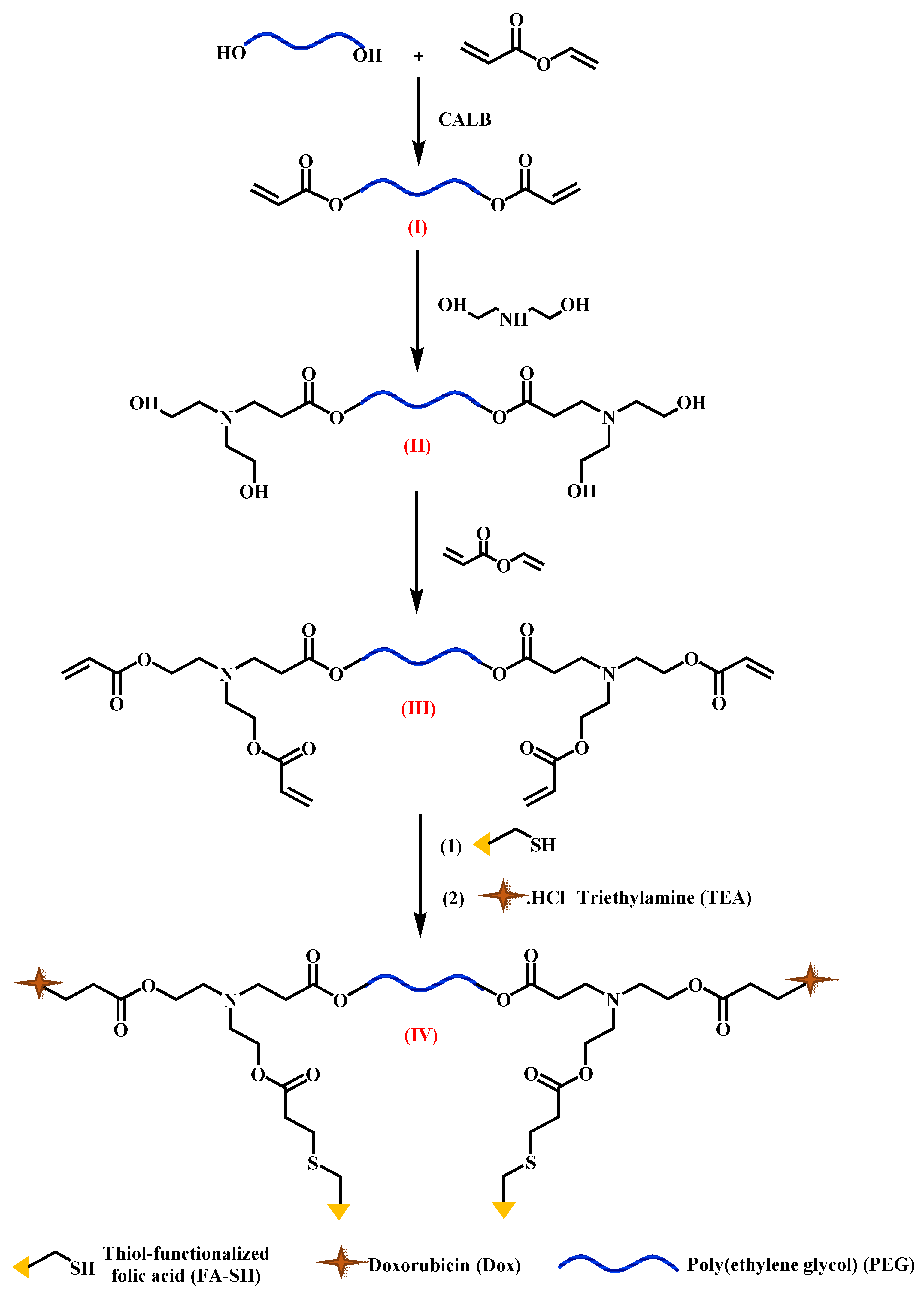
2. Results
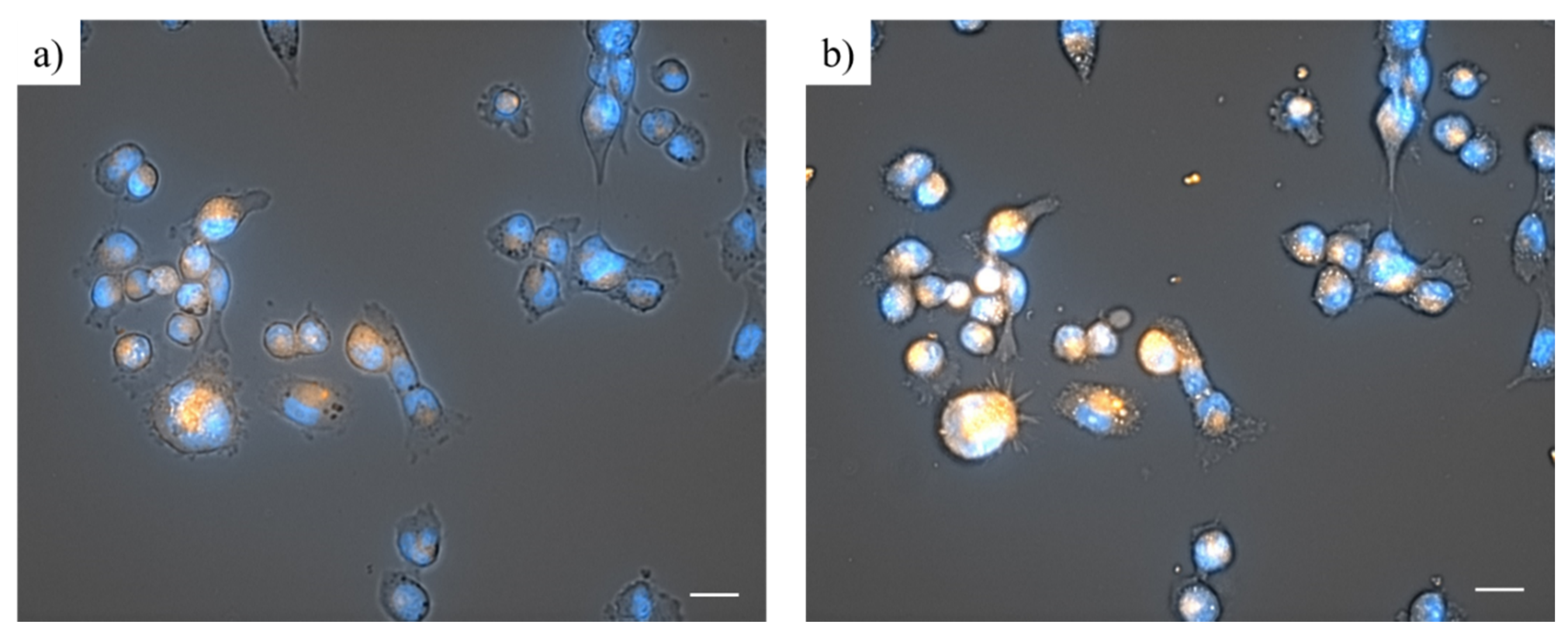
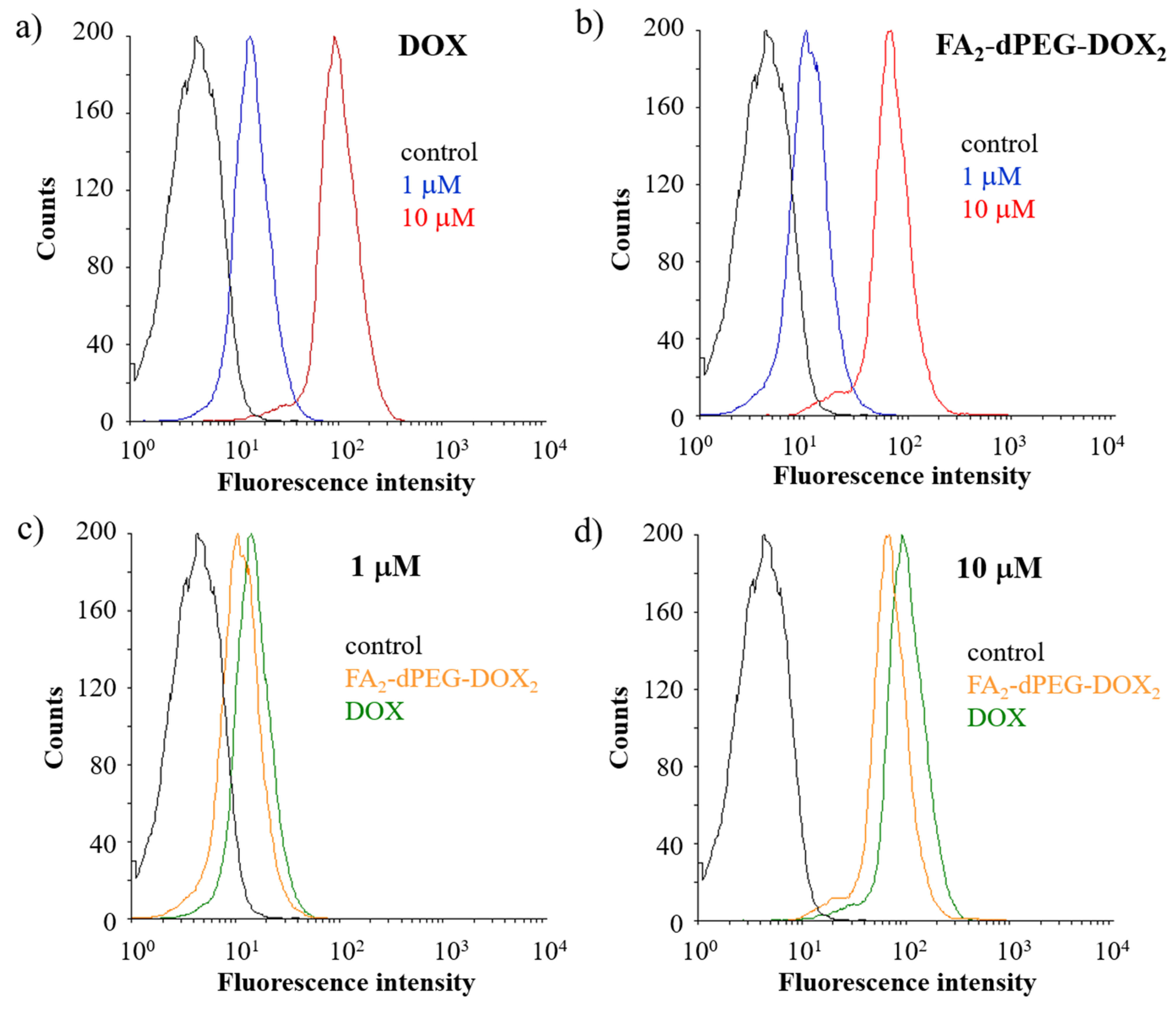
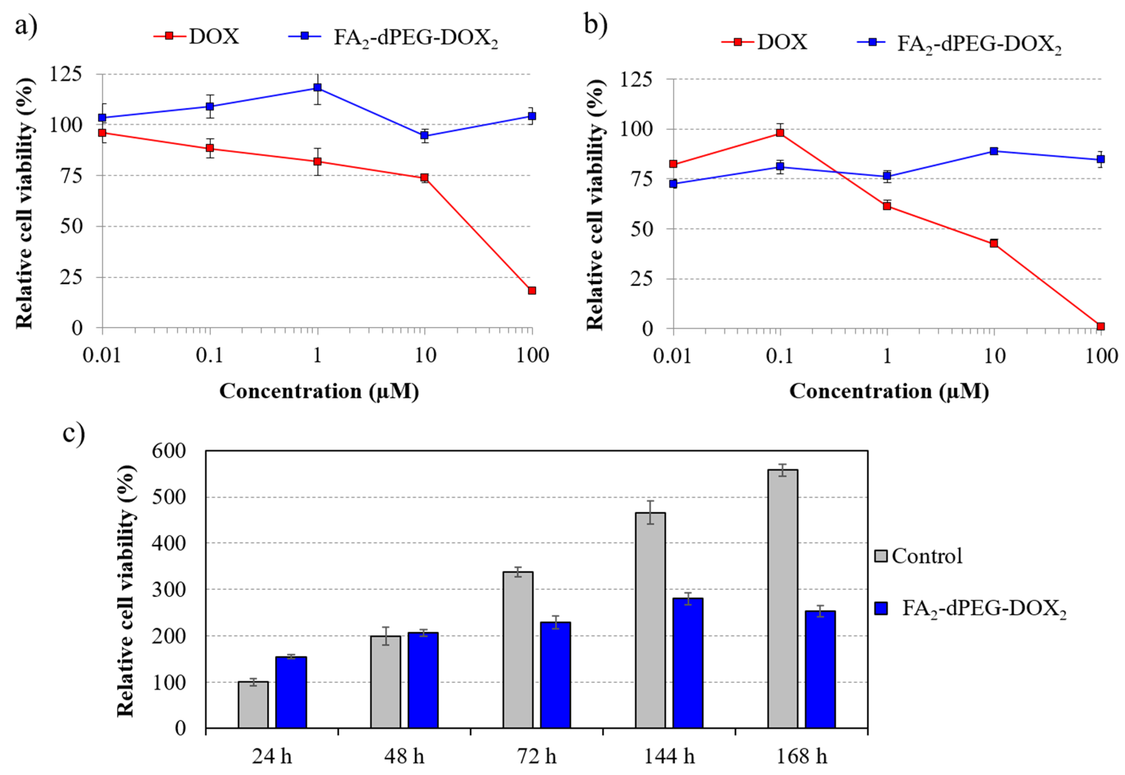
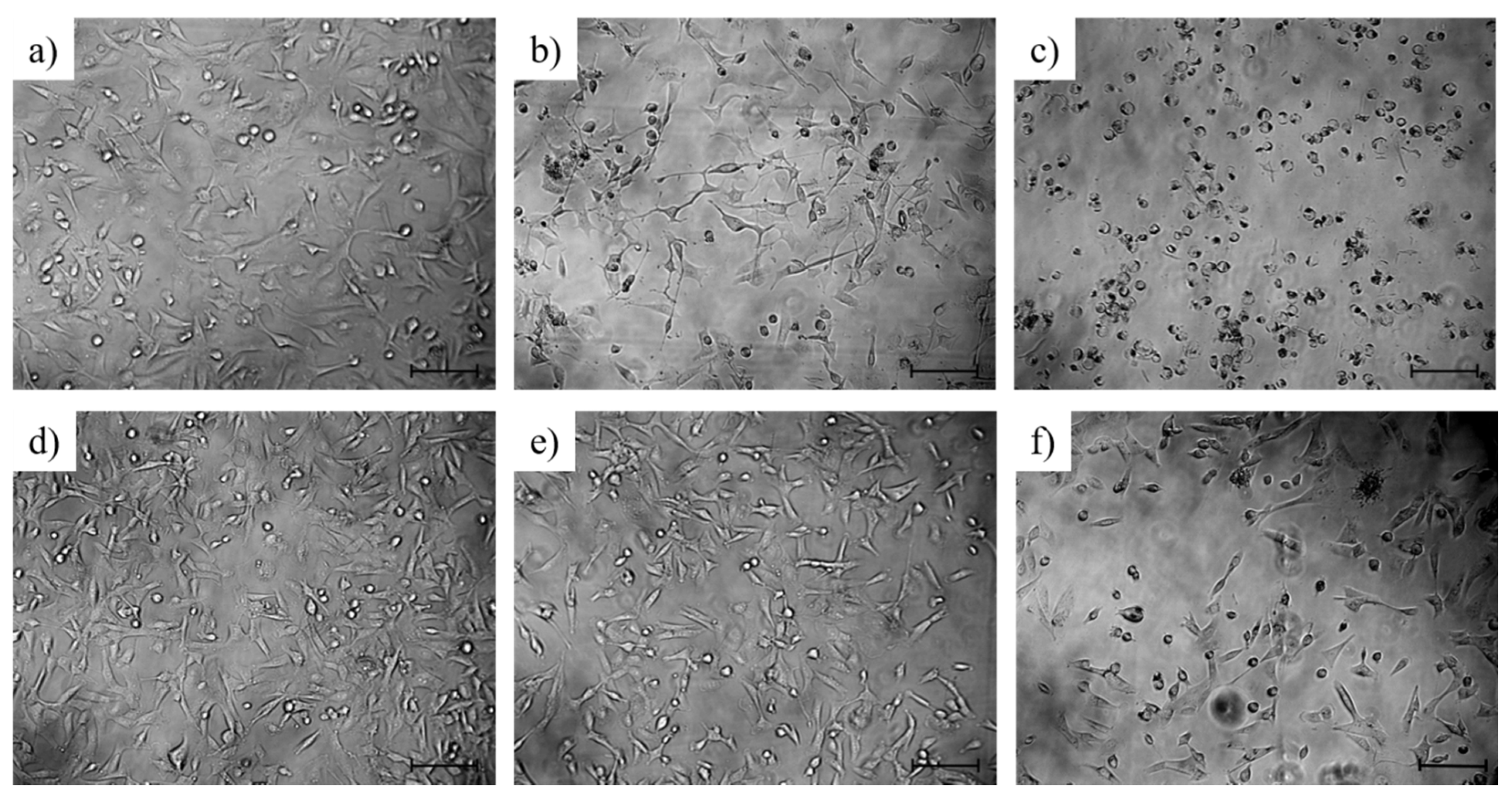
2.1. In Vitro Study
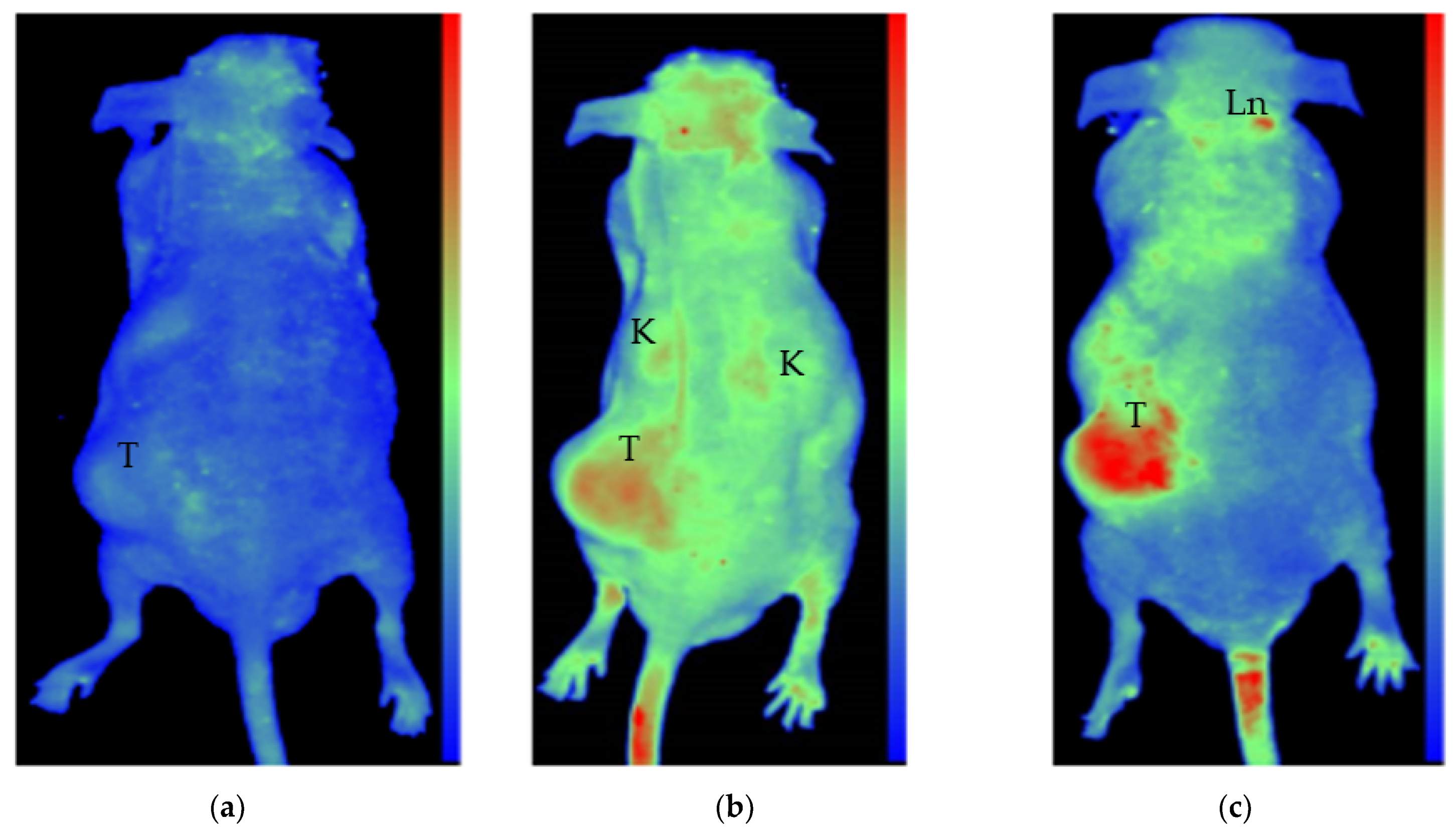
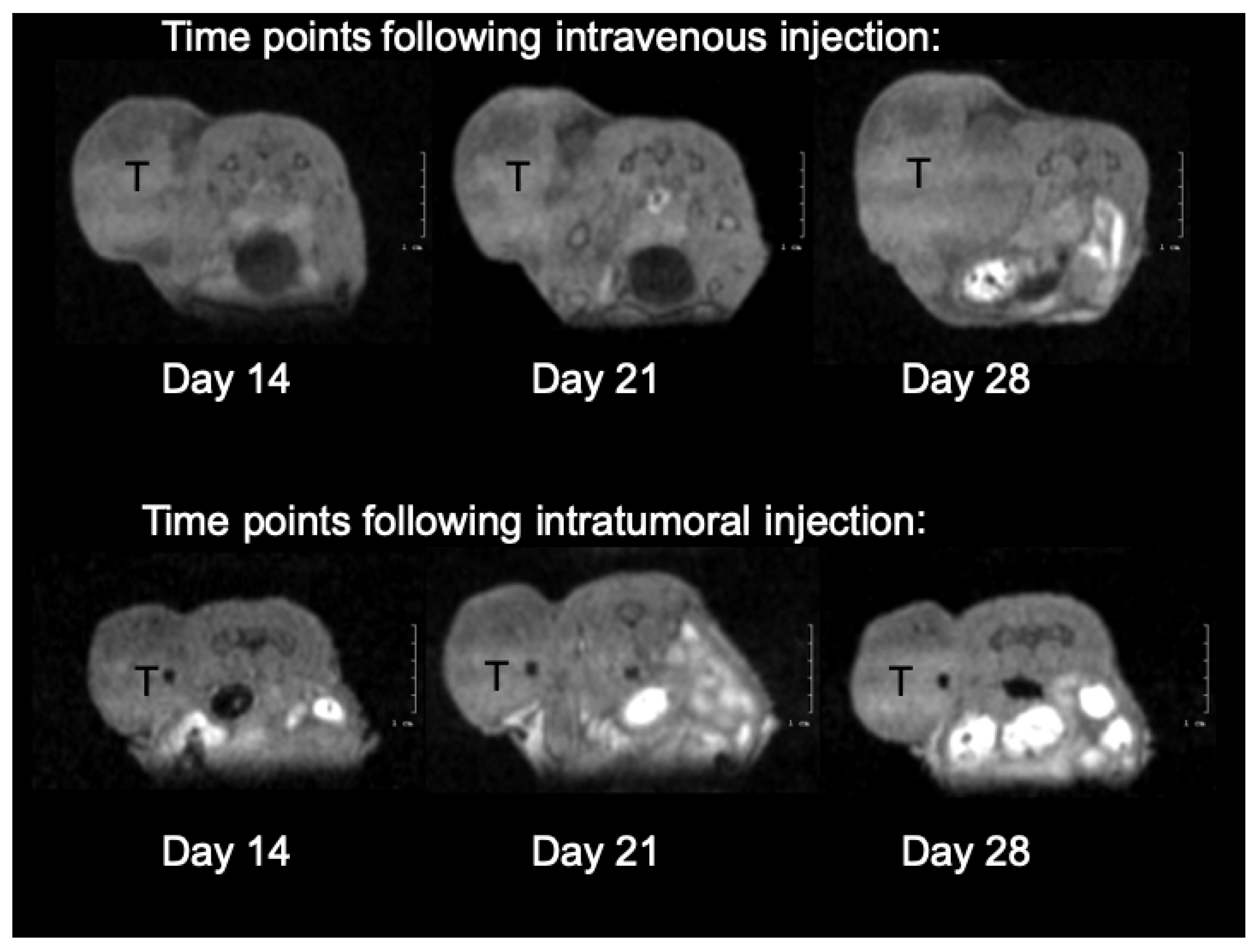
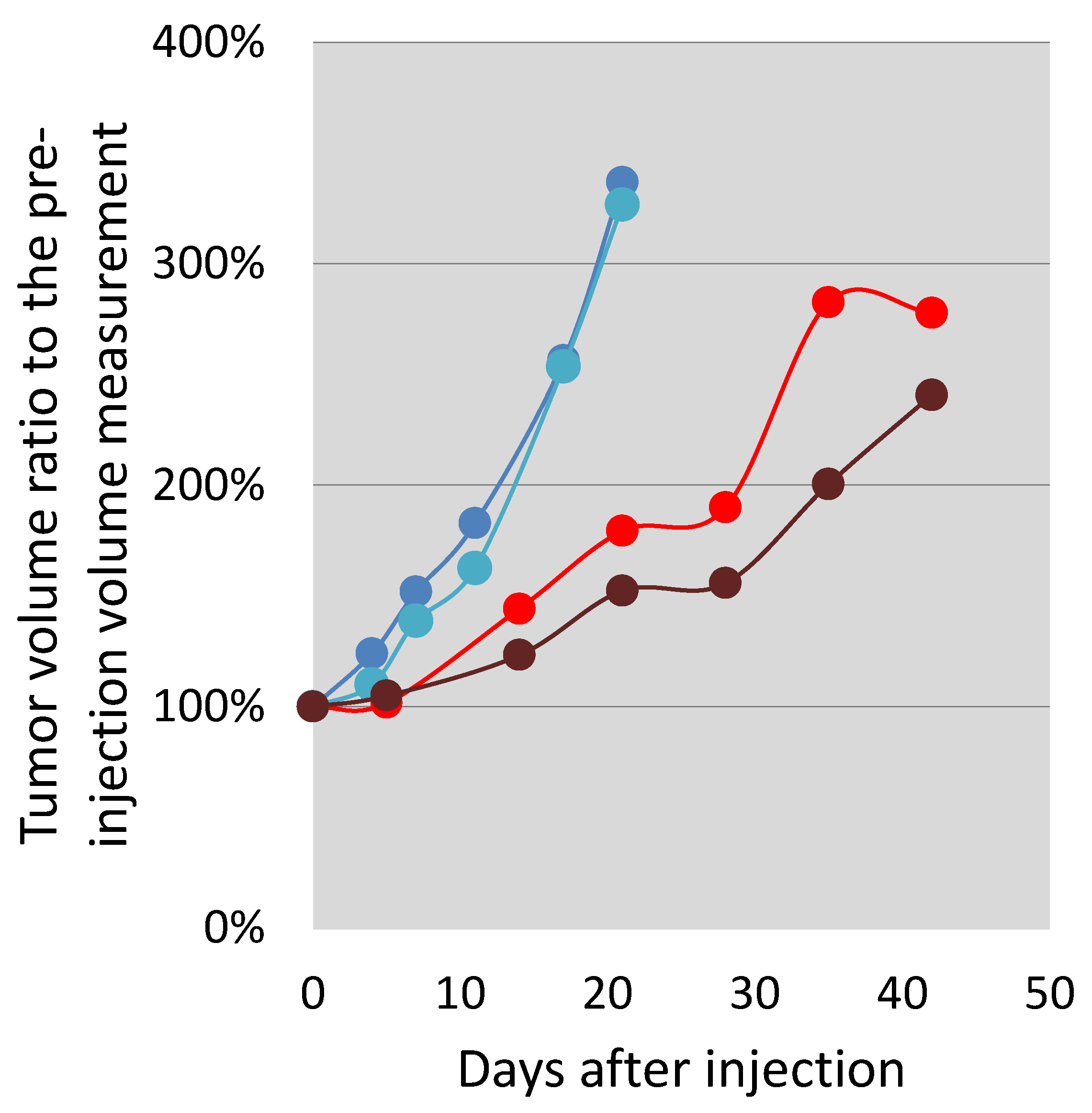
2.2. In Vivo Testing
3. Discussion
4. Materials and Methods
4.1. Cytotoxicity Study
4.2. Phase-Contrast and Fluorescence Microscopy
4.3. Flow Cytometry
4.4. In Vivo Imaging—Preliminary Studies
4.5. Magnetic Resonance Imaging (MRI)
4.6. In Vivo Fluorescent Imaging
4.7. In Vitro Absorbance and Fluorescence Tests
Supplementary Materials
Author Contributions
Funding
Institutional Review Board Statement
Informed Consent Statement
Data Availability Statement
Acknowledgments
Conflicts of Interest
References
- Zhong, L.; Li, Y.; Xiong, L.; Wang, W.; Wu, M.; Yuan, T.; Yang, W.; Tian, C.; Miao, Z.; Wang, T.; et al. Small molecules in targeted cancer therapy: Advances, challenges, and future perspectives. Signal Transduct. Target. Ther. 2021, 6, 201. [Google Scholar] [CrossRef]
- Puskas, J.E.; Molnar, K.; Krisch, E. Toward the effective synthesis of bivalent Folate-targeted PEGylated cancer diagnostic and therapeutic agents using chemo-enzymatic processes. J. Mol. Liq. 2020, 310, 113218–113227. [Google Scholar] [CrossRef]
- Low, P.S.; Henne, W.A.; Doorneweerd, D.D. Discovery and Development of Folic-Acid-Based Receptor Targeting for Imaging and Therapy of Cancer and Inflammatory Diseases. Acc. Chem. Res. 2008, 41, 120–129. [Google Scholar] [CrossRef] [PubMed]
- Vlahov, I.R.; Santhapuram, H.K.R.; Wang, Y.; Kleindl, P.J.; You, F.; Howard, S.J.; Westrick, E.; Reddy, J.A.; Leamon, C.P. An Assembly Concept for the Consecutive Introduction of Unsymmetrical Disulfide Bonds: Synthesis of a Releasable Multidrug Conjugate of Folic Acid. J. Org. Chem. 2007, 72, 5968–5972. [Google Scholar] [CrossRef] [PubMed]
- Zhong, Y.-J.; Shao, L.-H.; Li, Y. Cathepsin B-cleavable doxorubicin prodrugs for targeted cancer therapy. Int. J. Oncol. 2013, 42, 373–383. [Google Scholar] [CrossRef] [PubMed] [Green Version]
- Mindt, T.L.; Müller, C.; Melis, M.; de Jong, M.; Schibli, R. “Click-to-Chelate”: In Vitro and In Vivo Comparison of a 99m Tc(CO)3-Labeled N(τ)-Histidine Folate Derivative with Its Isostructural, Clicked 1,2,3-Triazole Analogue. Bioconjug. Chem. 2008, 19, 1689–1695. [Google Scholar] [CrossRef] [PubMed]
- Available online: http://www.endocyte.com (accessed on 2 February 2021).
- Naumann, R.W.; Coleman, R.L. Management Strategies for Recurrent Platinum-Resistant Ovarian Cancer. Drugs 2011, 71, 1397–1412. [Google Scholar] [CrossRef] [PubMed]
- Pribble, P.; Edelman, M.J. EC145: A novel targeted agent for adenocarcinoma of the lung. Expert Opin. Investig. Drugs 2012, 21, 755–761. [Google Scholar] [CrossRef]
- Coward, J.I.G.; Middleton, K.; Murphy, F. New perspectives on targeted therapy in ovarian cancer. Int. J. Womens Health 2015, 7, 189. [Google Scholar] [CrossRef] [Green Version]
- Naumann, R.W.; Coleman, R.L.; Burger, R.A.; Sausville, E.A.; Kutarska, E.; Ghamande, S.A.; Gabrail, N.Y.; DePasquale, S.E.; Nowara, E.; Gilbert, L.; et al. PRECEDENT: A Randomized Phase II Trial Comparing Vintafolide (EC145) and Pegylated Liposomal Doxorubicin (PLD) in Combination Versus PLD Alone in Patients with Platinum-Resistant Ovarian Cancer. J. Clin. Oncol. 2013, 31, 4400–4406. [Google Scholar] [CrossRef]
- MERCK. Available online: https://www.mrknewsroom.com/newsroom/news-releases/news-details/2014/Merck-and-Endocyte-Announce-Independent-DSMB-Recommends-Vintafolide-PROCEED-Phase-3-Trial-Be-Stopped-for-Futility-Following-Interim-Analysis/default.aspx (accessed on 2 March 2021).
- A Study of Vintafolide (MK-8109) in Participants With Advanced Solid Tumor (MK-8109-011). Available online: https://clinicaltrials.gov/ct2/show/NCT02049281 (accessed on 7 July 2021).
- Hanna, N.; Juhász, E.; Cainap, C.; Gladkov, O.; Ramlau, R.; Juan-Vidal, O.; Lal, R.; Symanowski, J.; Perez, W.; Nguyen, B.; et al. Target: A Randomized, Phase Ii Trial Comparing Vintafolide Versus Vintafolide Plus Docetaxel, Versus Docetaxel Alone in Second-Line Treatment of Folate-Receptor-Positive Non-Small Cell Lung Cancer (Nsclc) Patients. Ann. Oncol. 2014, 25, v1. [Google Scholar] [CrossRef]
- Jedrzejczyk, M.; Wisniewska, K.; Kania, K.D.; Marczak, A.; Szwed, M. Transferrin-Bound Doxorubicin Enhances Apoptosis and DNA Damage through the Generation of Pro-Inflammatory Responses in Human Leukemia Cells. Int. J. Mol. Sci. 2020, 21, 9390. [Google Scholar] [CrossRef] [PubMed]
- Illa, O.; Olivares, J.-A.; Gaztelumendi, N.; Martínez-Castro, L.; Ospina, J.; Abengozar, M.-Á.; Sciortino, G.; Maréchal, J.-D.; Nogués, C.; Royo, M.; et al. Chiral Cyclobutane-Containing Cell-Penetrating Peptides as Selective Vectors for Anti-Leishmania Drug Delivery Systems. Int. J. Mol. Sci. 2020, 21, 7502. [Google Scholar] [CrossRef] [PubMed]
- Woźniak, M.; Pastuch-Gawołek, G.; Makuch, S.; Wiśniewski, J.; Krenács, T.; Hamar, P.; Gamian, A.; Szeja, W.; Szkudlarek, D.; Krawczyk, M.; et al. In Vitro and In Vivo Efficacy of a Novel Glucose–Methotrexate Conjugate in Targeted Cancer Treatment. Int. J. Mol. Sci. 2021, 22, 1748. [Google Scholar] [CrossRef] [PubMed]
- Li, Q.; Li, W.; Xu, K.; Xing, Y.; Shi, H.; Jing, Z.; Li, S.; Hong, Z. PEG Linker Improves Antitumor Efficacy and Safety of Affibody-Based Drug Conjugates. Int. J. Mol. Sci. 2021, 22, 1540. [Google Scholar] [CrossRef] [PubMed]
- Mondal, U.K.; Doroba, K.; Shabana, A.M.; Adelberg, R.; Alam, M.R.; Supuran, C.T.; Ilies, M.A. PEG Linker Length Strongly Affects Tumor Cell Killing by PEGylated Carbonic Anhydrase Inhibitors in Hypoxic Carcinomas Expressing Carbonic Anhydrase IX. Int. J. Mol. Sci. 2021, 22, 1120. [Google Scholar] [CrossRef]
- Zaręba, M.; Sareło, P.; Kopaczyńska, M.; Białońska, A.; Uram, Ł.; Walczak, M.; Aebisher, D.; Wołowiec, S. Mixed-Generation PAMAM G3-G0 Megamer as a Drug Delivery System for Nimesulide: Antitumor Activity of the Conjugate Against Human Squamous Carcinoma and Glioblastoma Cells. Int. J. Mol. Sci. 2019, 20, 4998. [Google Scholar] [CrossRef] [Green Version]
- Ekladious, I.; Colson, Y.L.; Grinstaff, M.W. Polymer–drug conjugate therapeutics: Advances, insights and prospects. Nat. Rev. Drug Discov. 2019, 18, 273–294. [Google Scholar] [CrossRef]
- Baker, J.R. Dendrimer-based nanoparticles for cancer therapy. Hematology 2009, 2009, 708–719. [Google Scholar] [CrossRef] [Green Version]
- Mullen, D.G.; Desai, A.M.; Waddell, J.N.; Cheng, X.; Kelly, C.V.; McNerny, D.Q.; Majoros, I.J.; Baker, J.R.; Sander, L.M.; Orr, B.G.; et al. The Implications of Stochastic Synthesis for the Conjugation of Functional Groups to Nanoparticles. Bioconjug. Chem. 2008, 19, 1748–1752. [Google Scholar] [CrossRef] [Green Version]
- Das, D.; Koirala, N.; Li, X.; Khan, N.; Dong, F.; Zhang, W.; Mulay, P.; Shrikhande, G.; Puskas, J.; Drazba, J.; et al. Screening of Polymer-Based Drug Delivery Vehicles Targeting Folate Receptors in Triple-Negative Breast Cancer. J. Vasc. Interv. Radiol. 2020, 31, 1866–1873.e2. [Google Scholar] [CrossRef] [PubMed]
- Puskas, J.E.; Castano, M.; Mulay, P.; Dudipala, V.; Wesdemiotis, C. Method for the Synthesis of γ-PEGylated Folic Acid and Its Fluorescein-Labeled Derivative. Macromolecules 2018, 51, 9069–9077. [Google Scholar] [CrossRef]
- Puskas, J.E.; Sen, M.Y. Process of Preparing Functionalized Polymers via Enzymatic Catalysis. U.S. Patent 8,710,156, 29 April 2014. [Google Scholar]
- Puskas, J.E.; Sen, M.Y. Process of Preparing Functionalized Polymers via Enzymatic Catalysis. U.S. Patent 9,885,070, 6 February 2018. [Google Scholar]
- Hong, S.; Leroueil, P.R.; Majoros, I.J.; Orr, B.G.; Baker, J.R.; Banaszak Holl, M.M. The Binding Avidity of a Nanoparticle-Based Multivalent Targeted Drug Delivery Platform. Chem. Biol. 2007, 14, 107–115. [Google Scholar] [CrossRef] [PubMed] [Green Version]
- McGowan, J.V.; Chung, R.; Maulik, A.; Piotrowska, I.; Walker, J.M.; Yellon, D.M. Anthracycline Chemotherapy and Cardiotoxicity. Cardiovasc. Drugs Ther. 2017, 31, 63–75. [Google Scholar] [CrossRef] [PubMed] [Green Version]
- Koirala, N.; Das, D.; Fayazzadeh, E.; Sen, S.; McClain, A.; Puskas, J.E.; Drazba, J.A.; McLennan, G. Folic acid conjugated polymeric drug delivery vehicle for targeted cancer detection in hepatocellular carcinoma. J. Biomed. Mater. Res. Part A 2019, 107, 2522–2535. [Google Scholar] [CrossRef] [PubMed]
- Luo, Y.; Humayun, A.; Murray, T.A.; Kemp, B.S.; McFarland, A.; Liu, X.; Mills, D.K. Cellular Analysis and Chemotherapeutic Potential of a Bi-Functionalized Halloysite Nanotube. Pharmaceutics 2020, 12, 962. [Google Scholar] [CrossRef] [PubMed]
- Alberts, B.; Johnson, A.; Lewis, J.; Raff, M.; Roberts, K.; Walter, P. Transport into the Cell from the Plasma Membrane: Endocytosis. In Molecular Biology of the Cell, 4th ed.; Garland Science: New York, NY, USA, 2002; ISBN 0-8153-3218-1. [Google Scholar]


| Amount | Unit | Compound | APITitle 3 |
|---|---|---|---|
| 128.2 | mg | FA2-PEG-DOX | API |
| 50 | uL | DMSO | 2.50% |
| 2000 | uL | Saline | |
| 62.5 | mg/mL | Final API concentration |
Publisher’s Note: MDPI stays neutral with regard to jurisdictional claims in published maps and institutional affiliations. |
© 2021 by the authors. Licensee MDPI, Basel, Switzerland. This article is an open access article distributed under the terms and conditions of the Creative Commons Attribution (CC BY) license (https://creativecommons.org/licenses/by/4.0/).
Share and Cite
Nagy, K.S.; Toth, K.; Pallinger, E.; Takacs, A.; Kohidai, L.; Jedlovszky-Hajdu, A.; Mathe, D.; Kovacs, N.; Veres, D.S.; Szigeti, K.; et al. Folate-Targeted Monodisperse PEG-Based Conjugates Made by Chemo-Enzymatic Methods for Cancer Diagnosis and Treatment. Int. J. Mol. Sci. 2021, 22, 10347. https://doi.org/10.3390/ijms221910347
Nagy KS, Toth K, Pallinger E, Takacs A, Kohidai L, Jedlovszky-Hajdu A, Mathe D, Kovacs N, Veres DS, Szigeti K, et al. Folate-Targeted Monodisperse PEG-Based Conjugates Made by Chemo-Enzymatic Methods for Cancer Diagnosis and Treatment. International Journal of Molecular Sciences. 2021; 22(19):10347. https://doi.org/10.3390/ijms221910347
Chicago/Turabian StyleNagy, Krisztina S., Krisztina Toth, Eva Pallinger, Angela Takacs, Laszlo Kohidai, Angela Jedlovszky-Hajdu, Domokos Mathe, Noemi Kovacs, Daniel S. Veres, Krisztian Szigeti, and et al. 2021. "Folate-Targeted Monodisperse PEG-Based Conjugates Made by Chemo-Enzymatic Methods for Cancer Diagnosis and Treatment" International Journal of Molecular Sciences 22, no. 19: 10347. https://doi.org/10.3390/ijms221910347
APA StyleNagy, K. S., Toth, K., Pallinger, E., Takacs, A., Kohidai, L., Jedlovszky-Hajdu, A., Mathe, D., Kovacs, N., Veres, D. S., Szigeti, K., Molnar, K., Krisch, E., & Puskas, J. E. (2021). Folate-Targeted Monodisperse PEG-Based Conjugates Made by Chemo-Enzymatic Methods for Cancer Diagnosis and Treatment. International Journal of Molecular Sciences, 22(19), 10347. https://doi.org/10.3390/ijms221910347