Article

Isolation and Functional Characterization of Two SHORT VEGETATIVE PHASE Homologous Genes from Mango

State Key Laboratory for Conservation and Utilization of Subtropical Agro-Bioresources, College of Agriculture, Guangxi University, 100 East Daxue Road, Nanning 530004, China
*
Author to whom correspondence should be addressed.
These authors contributed equally to this work.
Int. J. Mol. Sci. 2021, 22(18), 9802; https://doi.org/10.3390/ijms22189802
Submission received: 23 July 2021 / Revised: 2 September 2021 / Accepted: 7 September 2021 / Published: 10 September 2021
(This article belongs to the Section Molecular Plant Sciences)

Abstract

:
The SHORT VEGETATIVE PHASE (SVP) gene is a transcription factor that integrates flowering signals and plays an important role in the regulation of flowering time in many plants. In this study, two full-length cDNA sequences of SVP homologous genes—MiSVP1 and MiSVP2—were obtained from ‘SiJiMi’ mango. Sequence analysis showed that the MiSVPs had typical MADS-box domains and were highly conserved between each other. The analysis of expression patterns showed that the MiSVPs were expressed during flower development and highly expressed in vegetative tissues, with low expression in flowers/buds. The MiSVPs could responded to low temperature, NaCl, and PEG treatment. Subcellular localization revealed that MiSVP1 and MiSVP2 were localized in the nucleus. Transformation of Arabidopsis revealed that overexpression of MiSVP1 delayed flowering time, overexpression of MiSVP2 accelerated flowering time, and neither MiSVP1 nor MiSVP2 had an effect on the number of rosette leaves. Overexpression of MiSVP1 increased the expression of AtFLC and decreased the expression of AtFT and AtSOC1, and overexpression of MiSVP2 increased the expression levels of AtSOC1 and AtFT and decreased the expression levels of AtFLC. Point-to-point and bimolecular fluorescence complementation (BiFC) assays showed that MiSVP1 and MiSVP2 could interact with SEP1-1, SOC1D, and AP1-2. These results suggest that MiSVP1 and MiSVP2 may play a significant roles in the flowering process of mango.

1. Introduction

Flower transformation is the transition from vegetative growth to reproductive growth and is an important stage of the plant life cycle. Plant flowering is a hotspot in botanical research [1]. This process is jointly affected by environmental conditions and the internal growth and developmental state. In Arabidopsis thaliana, flowering regulation consists of six signaling pathways: the photoperiod, age, ambient temperature, gibberellin, vernalization pathways and the autonomous pathway comprising a combination of epigenetic factors and post-transcriptional gene regulation [2,3,4,5,6,7]. These regulatory pathways are independent and interact with each other to form a complex gene regulatory network to jointly regulate plant flowering [8].
SHORT VEGETATIVE PHASE (SVP) homologous genes belonging to the StMADS11 group of the MADS-box gene family are critical flowering repressors in Arabidopsis and have important functions in regulating floral transition and inflorescence structure in many plants [9]. SVP is regulated by autonomous and thermosensitive gibberellin pathways [10]. Recent research has suggested that SVPs may be another central regulators of the flowering regulatory network because SVPs can repress SUPPRESSOR OF OVEREXPRESSION OF CONSTANS1 (SOC1), FLOWERING LOCUS T (FT), and TWIN SISTER OF FT (TSF) transcription in the meristem and leaf [10,11]. SVP can also bind to the promoter regions of FT and SOC1, and SVP can negatively regulate the expression of FT by binding to the CArG motif in the FT promoter sequence, which is one of the molecular mechanisms regulating flowering time under the condition of temperature fluctuations [12]. SVP can also interact with FLM-β in response to the regulation of ambient temperature, and SVP is an important mediator within the thermosensitive pathway [13]. In the early stages of flower development, AGAMOUS-LIKE 24 (AGL24), SVP, and APETALA1 (AP1) form the dimers AP1-AGL24 and AP1-SVP, which interact with the LEUNIG (LUG)-SEUSS (SEU) compound to regulate the expression of AGAMOUS (AG), thereby affecting the construction of flower organs [14].
Mango (Mangifera indica L.) is an economically important tropical fruit and is cultivated in many countries. The flowering and fruit setting of mango are important factors affecting its production. Studies have shown that mango flowering is affected by many factors, including drought, low temperature, age of branches, and gibberellin and paclobutrazole treatment [15]. Some flowering-related genes of mango, such as MiAP1, MiSOC1, MiFT, and MiCO, have been isolated and functionally identified [16,17,18,19]. However, the functions of the SVP genes in mango have not been reported. In this study, two SVP homologue genes were isolated, and their functions were studied in transgenic A. thaliana.

2. Results

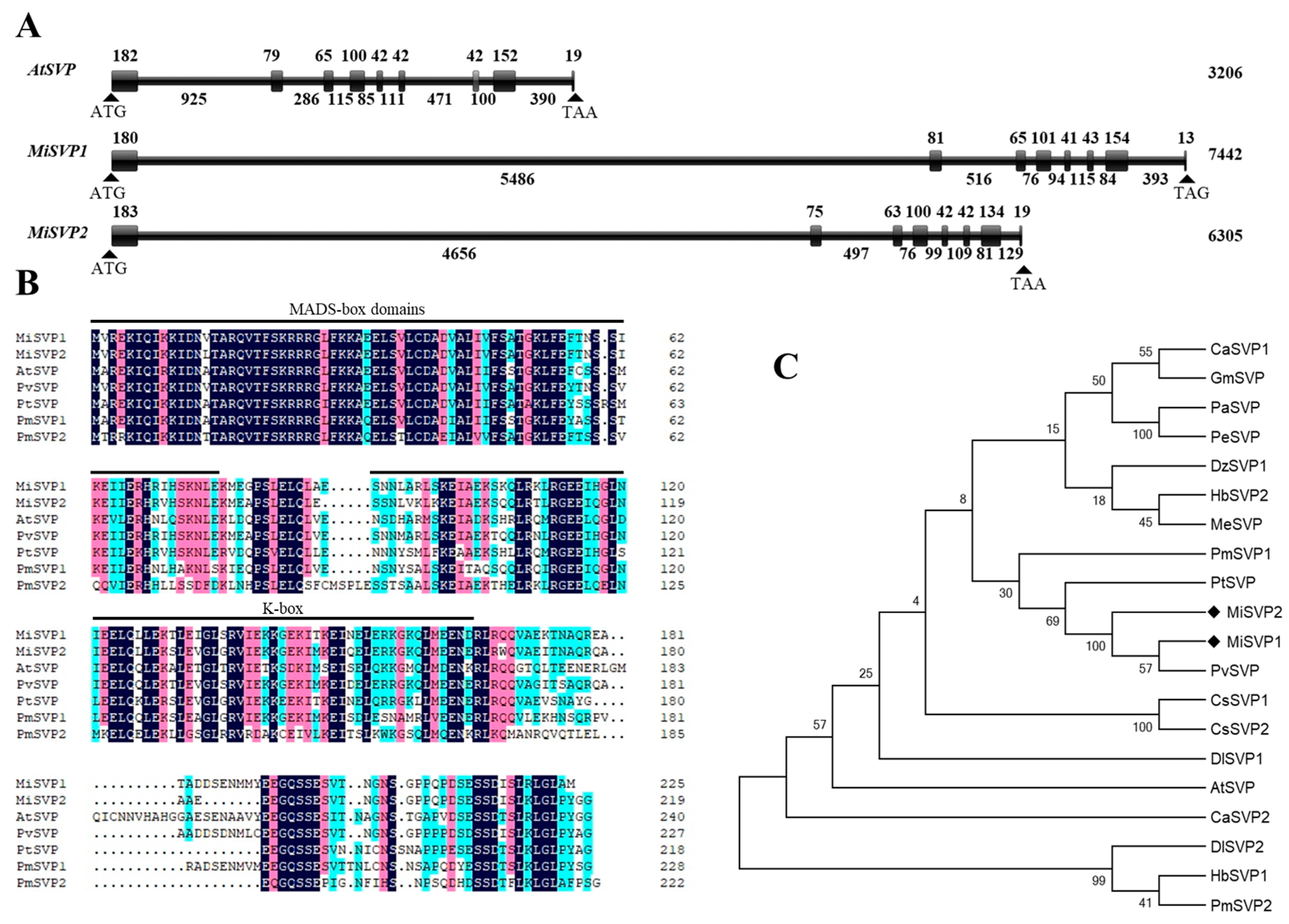
2.1. Cloning and Bioinformatics Analysis of MiSVPs

Two MiSVP genes, named MiSVP1 (MZ542518) and MiSVP2 (MZ542519), were identified from transcriptomic and genomic data (unpublished data) of ‘SiJiMi’ mango. The DNA sequences of MiSVP1 and MiSVP2 were 7442 bp and 6305 bp, respectively, in length, and included eight exons and seven introns (Figure 1A). The open reading frames (ORFs) were 675 bp and 657 bp and encoded 225 and 219 amino acids (aa), respectively. Their sequences were highly similar to each other. The molecular weight (MW) of MiSVP1 and MiSVP2 was estimated to be 25.55 and 24.77 kDa, respectively, and the theoretical isoelectric point (pI) of MiSVP1 and MiSVP2 was estimated to be 6.33 and 6.58, respectively. Sequence analysis indicated that the two MiSVP proteins contained typical MADS-box and K-box domains (Figure 1B). Phylogenetic tree analysis showed that MiSVP1 and MiSVP2 were clustered within the same branch and closely related to Pistacia vera PvSVP (XP_031287661) (Figure 1C).

2.2. Expression Analysis of MiSVPs

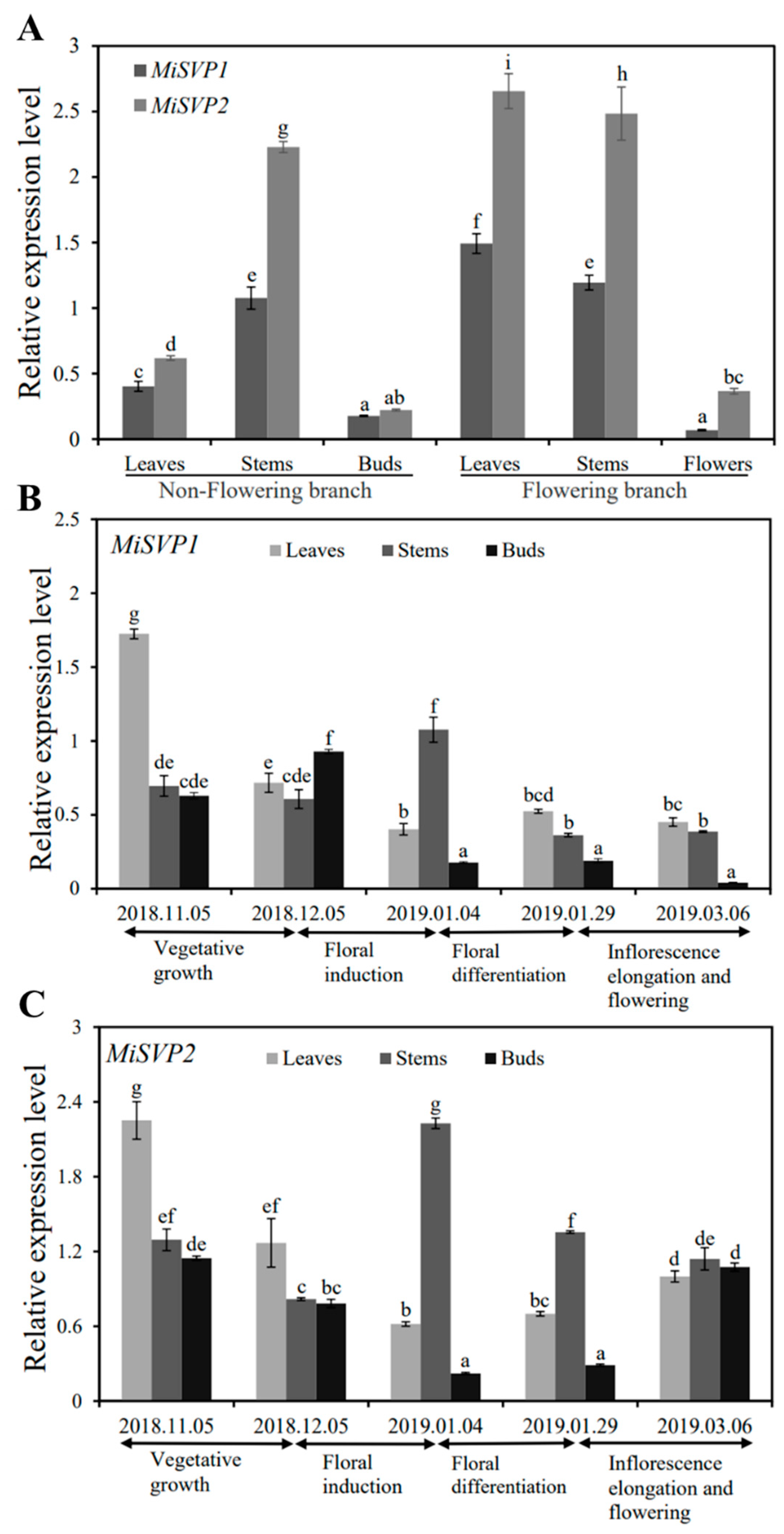
2.2.1. Tissue Expression Analysis of MiSVPs

To research the function of MiSVP1 and MiSVP2 in the growth and development of mango, the expression levels of MiSVP1 and MiSVP2 in all tissues of ‘SiJiMi’—including the nonflowering branches of leaves, stems, buds, and flowering branches of leaves, stems, and flowers—were investigated. The real-time quantitative reverse transcription polymerase chain reaction (qRT-PCR) results indicated that the MiSVP1 and MiSVP2 genes were expressed in all tested tissues (Figure 2A), and both MiSVP genes were more highly expressed in the leaves of flowering branches than in nonflowering branch tissues. The lowest expression level of MiSVP1 was found in flowers, and the lowest expression of MiSVP2 was found in buds. MiSVP2 was more highly expressed than MiSVP1 in all tested tissues.

2.2.2. Temporal Expression Analysis of MiSVPs

To further study the role of MiSVP1 and MiSVP2 in mango, the expression levels of MiSVP1 and MiSVP2 in stems, leaves and buds from November 2018 to March 2019 were analyzed (Figure 2B,C). The qRT-PCR results showed that MiSVP1 and MiSVP2 were present in samples at all stages of the test phase but at different transcriptional levels. In leaves, the expression of MiSVP1 was highest in the vegetative growth phase, followed by the early stage of floral induction and the period of floral differentiation. The expression level of MiSVP1 was lowest in the early stage of floral differentiation, at which point the inflorescences had begun to differentiate. The expression level of MiSVP1 in stems was highest in the early stage of floral differentiation, followed by the vegetative growth phase and the early stage of floral induction, and the lowest expression levels were observed during the late stage of floral differentiation. The expression level of MiSVP1 in buds was highest in the early stage of floral induction, followed by the vegetative growth phase and the late period of floral differentiation, and the lowest expression levels were observed during inflorescence elongation and flowering. MiSVP2 had a different expression pattern than MiSVP1. The expression level of MiSVP2 in leaves and buds showed a trend of first decreasing and then increasing, and the lowest expression levels occurred during floral differentiation. The expression level of MiSVP2 in stems was highest in the early stage of floral differentiation, followed by the late period of floral differentiation and the vegetative growth phase, and the lowest expression levels were observed during the early stage of floral induction. The expression level of MiSVP2 was higher than that of MiSVP1 from 5 November to 6 March.

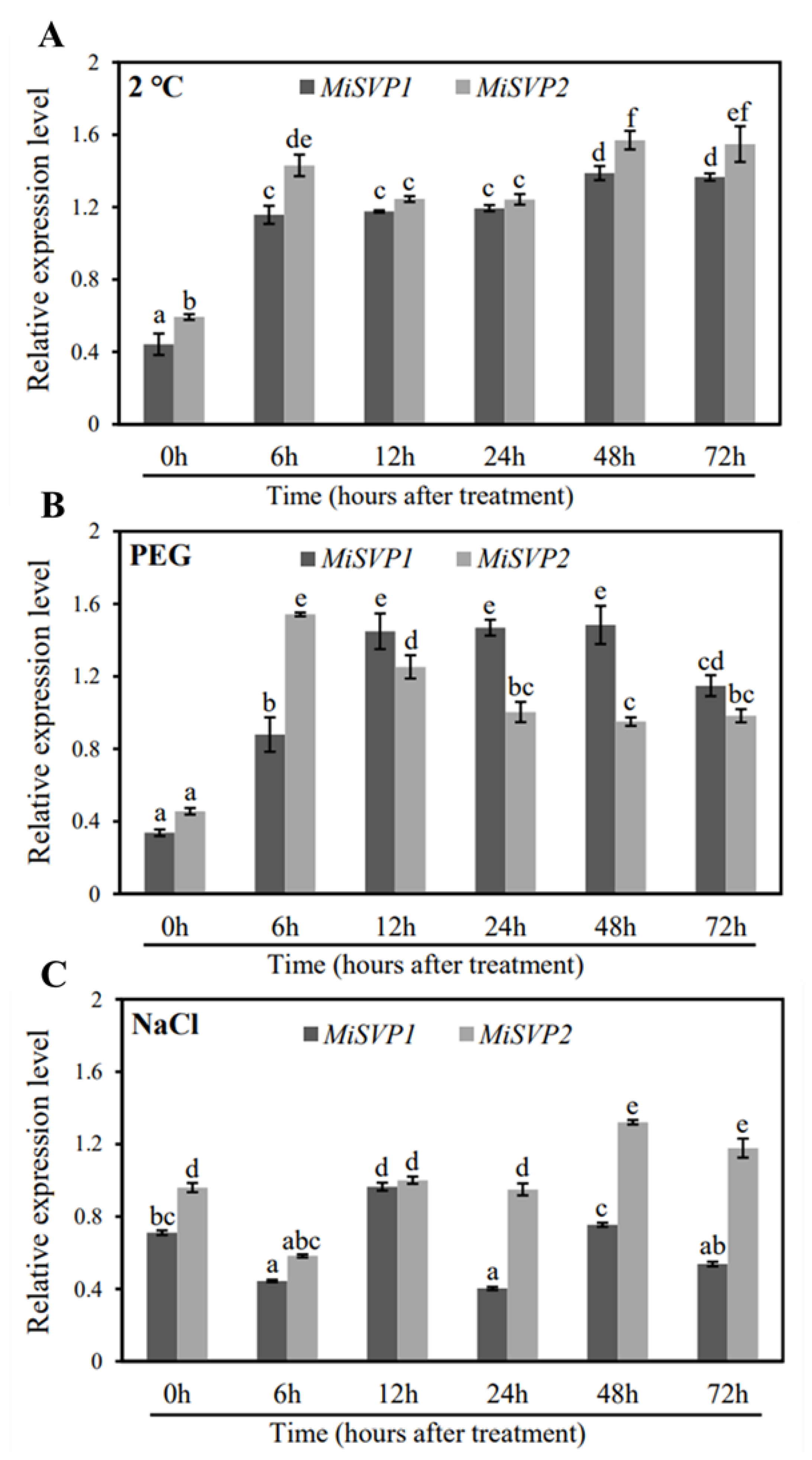
2.2.3. Effects of Adversity Treatments on the Alteration of MiSVPs Expression Levels

To explore the impact of adversity on the alteration of MiSVPs expression level, the expression levels of MiSVPs were detected after one of several stress treatments. Low temperature treatment significantly upregulated the expression levels of MiSVP1 and MiSVP2 compared with those at 0 h, with expression peaking at 48 h after treatment (Figure 3A). After 30% PEG treatment, MiSVP1 and MiSVP2 were significantly upregulated compared with 0 h levels and reached peak expression at 6 h (MiSVP2) and 48 h (MiSVP1) after treatment (Figure 3B). After salt stress treatment, the expression of MiSVP1 was significantly downregulated at 6 h; then upregulated, reaching a peak at 12 h; and finally downregulated again. The expression of MiSVP2 was significantly downregulated at 6 h, returned to the pretreatment level between 12–24 h, and then was significantly upregulated at 48–72 h (Figure 3C).

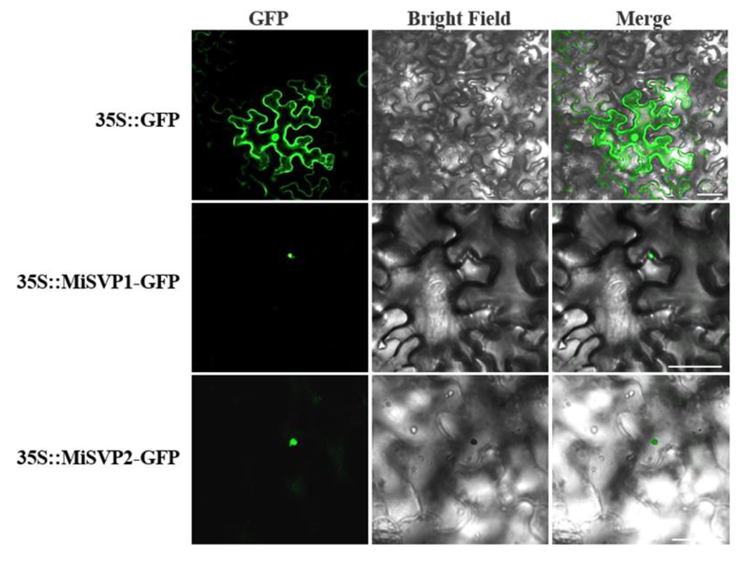
2.3. Subcellular Localization of MiSVPs

To ascertain the specific locations of MiSVP proteins in the cell, 35S::MiSVP1-GFP and 35S::MiSVP2-GFP fusion protein carriers were transferred into Agrobacterium tumefaciens (EHA105) and then injected into tobacco leaves to observe the fluorescence signal. The green fluorescence of 35S::GFP was used as a control and could be found in the cell membrane and nucleus, while the green fluorescence of the 35S::MiSVP1-GFP and 35S::MiSVP2-GFP fusion proteins were only observed in the nucleus (Figure 4), which showed that the MiSVP1 and MiSVP2 proteins were located in the nucleus.

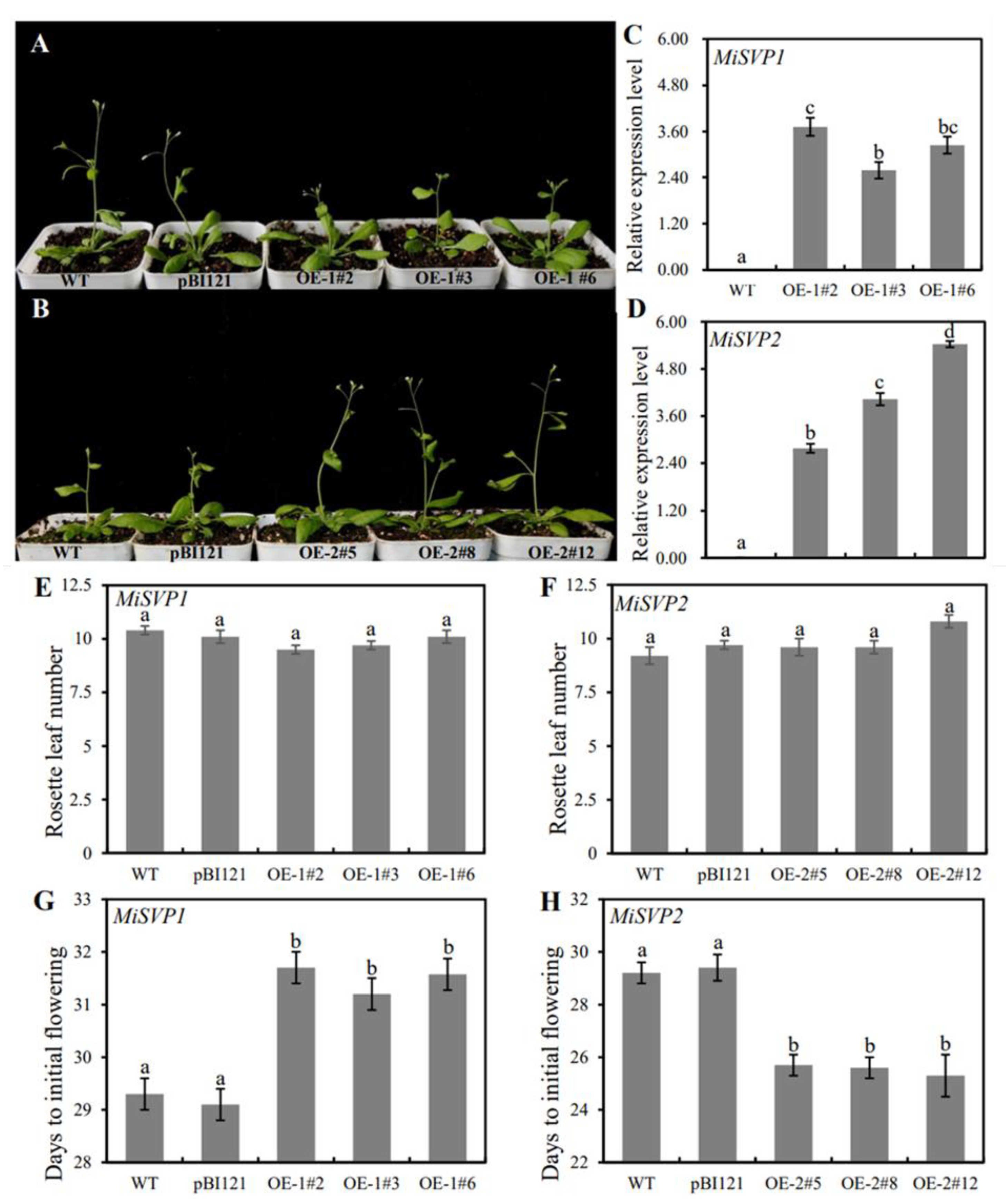
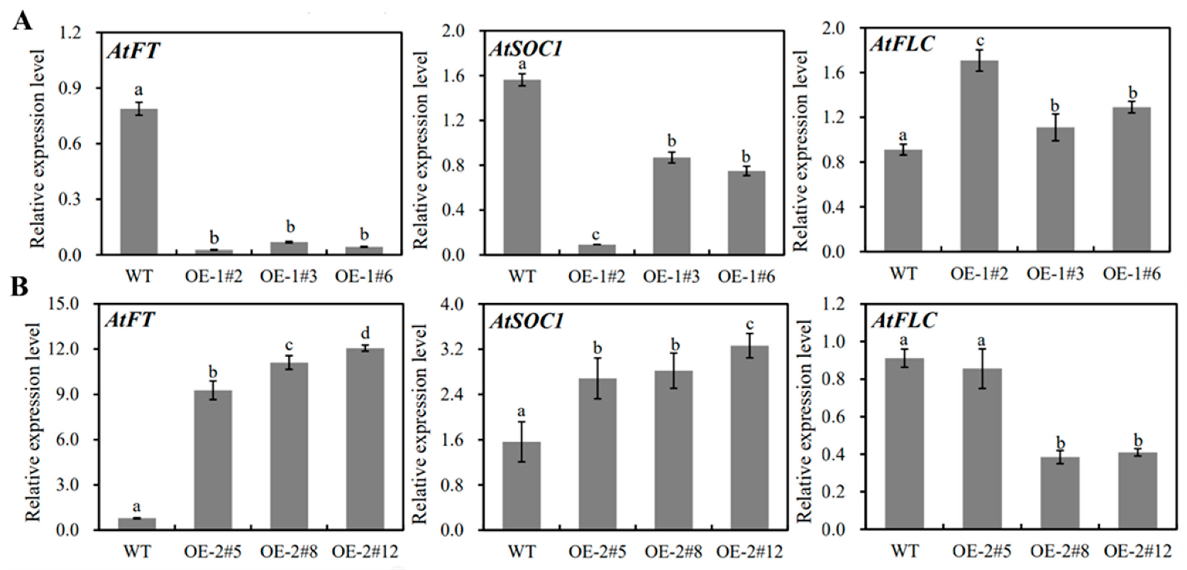
2.4. Functional Analysis of MiSVPs in Arabidopsis thaliana

To study the role of the two MiSVP genes in plant flowering, the MiSVPs genes were transfected into wild-type (WT) A. thaliana. Three homozygous transgenic lines of MiSVP1 (OE-1#2, OE-1#3, and OE-1#6) and of MiSVP2 (OE-2#5, OE-2#8, and OE-2#12) were selected for functional analysis. As shown in Figure 5A,B, overexpression of MiSVP1 delayed flowering time. However, overexpression of MiSVP2 advanced flowering time. Compared with those in the WT, the expression levels of the MiSVP1 and MiSVP2 genes in transgenic plants were high (Figure 5C,D). Overexpression of the MiSVPs resulted in no significant changes in the number of rosette leaves (Figure 5E,F) but caused significant changes in days to initial flowering (Figure 5G,H).
To analyze the influence of overexpression of MiSVPs on the expression of endogenous genes in A. thaliana, leaves of transgenic plants were collected before blooming, RNA was extracted, and the expression levels of endogenous genes associated with flowering were analyzed by qRT-PCR. The expression of the endogenous genes AtFT and AtSOC1 decreased in MiSVP1 transgenic plants compared with WT, while that of the endogenous gene AtFLC increased (Figure 6A). The expression trends of endogenous genes in MiSVP2 transgenic plants were opposite those in MiSVP1 transgenic plants. In MiSVP2 transgenic plants compared with WT, the expression of the endogenous genes AtFT and AtSOC1 increased, while that of the endogenous gene AtFLC decreased (Figure 6B).

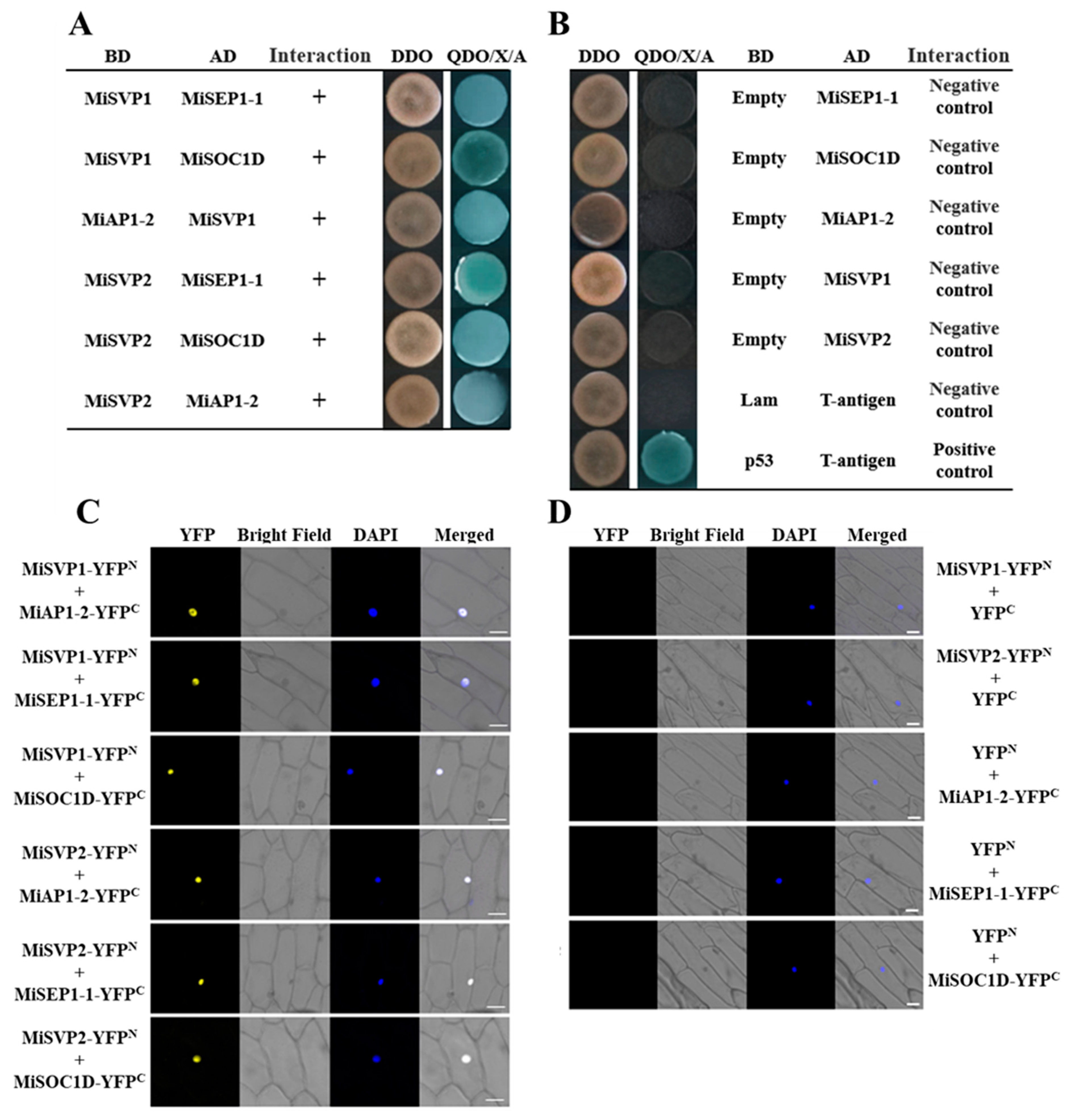
2.5. Screening and Validation of MiSVPs Interacting Proteins

To investigate the regulation of MiSVPs in the flowering network, the non-self-activated yeast pGADT7-MiSVP vector was combined with the mango cDNA library previously constructed in our laboratory, and a total of 17 blue monoclonal colonies were obtained by screening on selective media (SDO/–Trp/–Leu/–His/–Ade). Due to the high false positive rate of the yeast two-hybrid system, three flowering-related proteins were selected for point-to-point and bimolecular fluorescence complementation (BiFC) assays. These three proteins were SUPPRESSOR OF OVEREXPRESSION OF CONSTANS 1 D (SOC1D), APETALA 1-2 (AP1-2), and SEPALLATA 1-1 (SEP1-1). As shown in Figure 7, the point-to-point and BiFC assays of the three candidate proteins with MiSVPs showed that MiSVP1 and MiSVP2 interacted with SEP1-1, AP1-2 and SOC1D (Figure 7A,C).

3. Discussion

Different members of the MADS-box gene family play a wide range of roles in the regulation of flowering time, the determination of floral meristem and floral organ characteristics, and the regulation of root, leaf, ovule and fruit development [20]. SVP belongs to the STMADS11 group of the MADS-box gene family. In A. thaliana, SVP regulates flowering time and determines floral meristem and floral organ characteristics [9,21]. Recent advances in research on SVPs suggest that SVP may be another central regulator of the flowering regulatory network [10]. In this study, we isolated and identified two SVP homologous genes, MiSVP1 and MiSVP2, from mango. The exon-intron structural analysis of MiSVP1 and MiSVP2 suggests that the intron-exon length and the intron number differ between MiSVP1 and MiSVP2. There were seven introns from the start codon to the stop codon of MiSVP1 and MiSVP2 (Figure 2A), which is the same number as in Prunus mume and different from that in A. thaliana, with eight introns in Poncirus trifoliata L. Raf. [22,23]. These results indicated that SVP gene sequences differ among different species. Sequence analysis indicated that both MiSVP1 and MiSVP2 belonged to the MADS-box gene family, with typical MADS-box motifs and high conservatism between them (Figure 2B). Phylogenetic tree analysis showed that MiSVP1 and MiSVP2 belong to the same branch, but that MiSVP2 is more closely related than MiSVP1 to the SVP-like gene PvSVP of Pistacia vera (Figure 2C), which indicated that MiSVP2 and PvSVP are very closely related; their protein structures may also be very similar. Therefore, it is speculated that the MiSVP2 protein may have a similar function to the PvSVP protein. The genome sequence of ‘Alfonso’ mango has been published; although there are two base differences between the SVP1 gene of ‘SiJiMi’ and the SVP1 gene of ‘Alfonso’, there is no difference in amino acid sequence. The sequence of the SVP2 gene in ‘SiJiMi’ is the same as that in ‘Alfonso’.
In A. thaliana, AtSVP is mainly expressed in vegetative tissues but not in flowers and pods [9]. Strawberry FaSVPs and kiwifruit AcSVPs are expressed only in vegetative organs and not in reproductive organs, which is consistent with the patterns in A. thaliana [24,25,26]. However, some different expression patterns have been found; for example, for Medicago MtSVP, Chrysanthemum morifolium CmSVP, grape lily LoSVP, Eriobotrya japonica EjSVPs, Prunus mume PmSVPSs, Malus × domestica MdSVPs, and another apple SVP-like gene, MdMADS50, are all of highly expressed in vegetative tissues but weakly expressed in reproductive organs [23,27,28,29,30,31,32]. In this study, the qRT-PCR results suggested that the expression patterns of MiSVP1 and MiSVP2 were the same; however, the transcription levels of MiSVP1 and MiSVP2 were different. MiSVP1 and MiSVP2 were highly expressed in vegetative tissues, but their expression levels were low in flowers/buds. These findings suggest that the expression and function of SVP homologous proteins may be different in different species.
The temporal expression patterns indicated that the expression of MiSVP1 in buds increased during the vegetative growth period but decreased during flower development. This result is consistent with the expression patterns of SVP-like genes in other species, such as Poncirus trifoliata L. Raf., Cymbidium goeringii, and Crocus sativus L. [33,34,35]. However, the expression of MiSVP2 decreased during the vegetative stage and flower induction stage and increased during flower development. These results suggest that the SVP gene may have evolved different temporal expression patterns, a possibility that needs further study.
The MiSVPs showed sensitivity to low temperature, PEG and NaCl treatments, especially low temperature; PEG treatment significantly promoted their expression. The SVP gene is a key factor in the response to low temperature [13]. In A. thaliana, lily, and Cymbidium goeringii, low temperature can promote the expression of SVP genes, which agrees with the results of the present study [28,34,36]. In previous work, 20% PEG and 150 mM NaCl treatment did not significantly change the expression levels of SVPs in A. thaliana, which differs from the results of the present study [36]. The expression level of MiSVPs in mango seedlings changed significantly under treatment with 30% PEG and 300 mM NaCl. PEG treatment can significantly promote the expression of MiSVPs. After NaCl treatment, the expression of MiSVP1 was significantly downregulated at 6 h, upregulated to a peak at 12 h, and then downregulated again. The expression of MiSVP2 was significantly downregulated at 6 h, restored to the pretreatment level at 12–24 h, and significantly increased at 48–72 h, indicating that the expression of MiSVPs is regulated by PEG and NaCl. The reason for the different results between studies may be that the concentrations of PEG and NaCl used in Arabidopsis were different from those in this study.
SVP homologous genes in most species mainly inhibit flowering, participate in the development of floral organs and maintain flower meristem specificity. In Arabidopsis, the SVP mutant shows early flowering and a reduced number of rosette leaves and stem leaves [9]. Loquat EjSVP1 and EjSVP2 and Brassica BcSVP have been found to delay flowering and induce floral organ variation [30,37]. Overexpression of OsMADS22, OsMADS47 and OsMADS55 in rice can delay flowering, but only the transgenic plants of OsMADS22 and OsMADS47 have been found to show floral organ variation [38]. Overexpression of Prunus mume PmSVP1 and PmSVP2 showed floral organ variation, but only PmSVP1 delays flowering [23]. Overexpression of four kiwifruit AcSVP genes results in floral organ variation, but only AcSVP1 delays flowering [26,39,40]. In addition, SVP genes play a role in promoting flowering in some species. For example, overexpression of Epimedium sagittatum EsSVP can produce early flowering lines and late flowering lines [41]. Overexpression of the Polypogon fugax SVP homologous gene PfMADS16 leads to early flowering in transgenic plants [42]. Overexpression of Phyllostachys violascens PvSVP1 and PvSVP2 can induce early flowering and floral organ variation [43]. In this study, overexpression of MiSVP1 and MiSVP2 had no effect on the inflorescence structure or the number of rosette leaves in transgenic plants. MiSVP1 delayed flowering, and MiSVP2 resulted in early flowering. These results suggest that SVP genes play different roles in different species and that different SVP homologous genes in the same species play different roles in plant growth.
In A. thaliana, SVP and FLC can interact with each other to form a heterodimer, SVP-FLC, which can be combined with the promoter regions of SOC1 and FT to regulate the expression of SOC1 and FT, thereby regulating the flowering in A. thaliana [12,25]. Research has found that SVP can interact with AP1 and participate in the regulation of the flowering stage [42]. In this study, overexpression of the MiSVP gene in A. thaliana caused no significant changes in the number of rosette leaves; however, it had a significant impact on flowering time. MiSVP1 can significantly delay the flowering time of transgenic plants compared to that of WTplant, under MiSVP1 overexpression, the expression of endogenous genes AtFT and AtSOC1 in Arabidopsis decreased, while the expression of the endogenous gene AtFLC increased, which is consistent with the effects of PmSVP2 [23]. MiSVP2 significantly accelerated the flowering time of transgenic plants; additionally, the expression levels of endogenous genes exhibited opposite changes to those under MiSVP1: the expression levels of the endogenous genes AtFT and AtSOC1 increased, while the expression of the endogenous gene AtFLC decreased, consistent with the effects of SVP-like gene PfMADS16 [42]. In conclusion, MiSVPs regulate flowering time in transgenic plants by regulating the expression levels of the FT and SOC1 genes.
Among the four SVP-like genes of kiwifruit, SVP1 and SVP4 can interact with SOC1 but not with AP1 and FLC, and SVP2 and SVP3 can interact with AP1, SOC1, and FLC, playing a redundant regulatory role [25,26]. In pecan, CcSVP can interact with CcAP1 and CcSOC1 but not with CcFLC [44]. In rice, OsMADS22 and OsMADS55, which are encoded by SVP homologous genes, interact with AGL24 and AP1, while only OsMADS55 interacts with FLC [38]. In Cymbidium goeringii, CgSVP can interact with CgAP1 and CgSOC1 [34]. These observations indicate that the numbers and regulatory mechanisms of SVP genes differ among different plants, suggesting that the SVP homologous genes from different species have evolved different functions. In this study, MiSVP1 and MiSVP2 were found able to interact with SEP1-1, AP1-2, and SOC1D. Our results are similar to those of previous studies.

4. Materials and Methods

4.1. Plant Materials

The test materials used in this study were from 10-month-old and 5-year-old ‘SiJiMi’ mango (Mangifera indica L.) trees growing in the Fruit Tree Specimen Garden of the Agricultural College of Guangxi University (Nanning, China). The experimental materials for cloning from the leaves of ‘SiJiMi’ mainly included healthy young stems, flowers, leaves of 5-year-old seedlings and leaves of 10-month-old seedlings of ‘SiJiMi’. Leaves of ‘SiJiMi’ were collected once a month starting in November 2018 from flower bud differentiation to the flowering stage, for a total of five times. Mature leaves of 10-month-old seedlings of ‘SiJiMi’ were treated with 300 mM NaCl, 30% PEG or 2 °C, and leaves were collected at 0, 6, 12, 24, 48, and 72 h after treatment to analyze the effects of NaCl, PEG, or 2 °C treatment on the expression levels of MiSVPs. The samples used in the experiment were all collected from the healthy leaves of the fourth round from the top bud on the new branches; the flowers were all unopened or half-opened. The stems were collected from the new branches of the mango in the same year, and the buds were the top buds on the mango branches. All samples were stored in a refrigerator at −80 °C to prevent RNA degradation.
Wild-type A. thaliana plant were preserved in our laboratory only as transgenic receptors. Nicotiana tobacco was preserved in our laboratory only as a receptor for subcellular localization experiments. Arabidopsis and tobacco were cultured at 22 °C under for a long photoperiod (16 h light/8 h dark cycle).

4.2. Cloning and Bioinformatics Analysis of MiSVPs

The total RNA extraction kit of TIGANDAN DP441 polysaccharide polyphenol plants was used to extract the total RNA of mango, and Moloney mouse leukemia virus (M-MLV) reverse transcriptase (TaKaRa, Dalian, China) and primer AUP1 were used to perform reverse transcription following the manufacturer’s instructions to obtain cDNA. The specific primers MiSVP1/2u and MiSVP1d, MiSVP2d were designed, and PCR with cDNA as a template was performed to obtain MiSVP1 and MiSVP2 fragments (Table S1).
Identification of MiSVP nucleotide sequences was completed through the National Center for Biotechnology Information (NCBI) BLAST program (http://www.ncbi.nlm.nih.gov/BLAST, accessed on 1 June 2018). Exon–intron structures of MiSVP1 and MiSVP2 were generated using IBS 1.0 [45]. The aa sequence, MW and pI of the MiSVP proteins were determined via BioXM 2.6 software (Ji Huang, Nanjing, China). Multiple alignments of the two MiSVP genes and SVP-like aa sequences in different species were performed using DNAMAN software (Lynnon Biosoft, Vaudreuil-Dorion, Quebec, Canada). The phylogenetic tree of the two MiSVP and SVP-like proteins was constructed via MEGA 6.0 software (Koichiro Tamura, Hachioji, Tokyo, Japan), with 1000 bootstrap replicates. SVP-like aa sequences in different species were downloaded from NCBI.

4.3. Expression Analysis of MiSVPs

The qRT-PCR-specific primers for MiSVP1 and MiSVP2 were designed in Primer3 Input (https://bioinfo.ut.ee/primer3-0.4.0/, accessed on 13 January 2019) (Table S1). The expression levels of MiSVP1 and MiSVP2 were analyzed on an ABI 7500 instrument (Applied Biosystems, Foster, America) by qRT-PCR. The qRT-PCR conditions followed the manufacturer’s instructions. Each qRT-PCR mixture contained 10 µL SYBR Premix Ex Taq II (TakaRa, Dalian, China), 0.5 µL (10 µM) upstream and downstream specific primers of MiSVPs (Table S1), 0.4 µL ROX reference dye II and 2 µL cDNA (100 ng·µL−1) as template, and sterile water was added to bring the total system volume to 20 µL. The mango MiACT1 gene was used as the internal reference gene [46], and the relative expression levels of MiSVPs were calculated by the 2−ΔΔCt method [47].

4.4. Subcellular Localization

The MiSVP gene and the vector p1300 plasmid were connected through SacI and BamHI cleavage sites, and the vector p1300-MiSVP1-GFP and p1300-MiSVP2-GFP fusion protein carriers were constructed and transferred into EHA105 cells. EHA105 cells, which contained plasmids carrying the target gene, were incubated overnight in 1 mL YEP liquid culture medium (50 µL/100 mL KANA, 25 µL/100 mL Rif) at 28 °C and oscillated at 200 rpm. The bacteria were collected by centrifugation (8000 rpm, 5 min), resuspended to OD600 = 0.6–1.0, transferred into tobacco by the EHA105-mediated method, cultured at 25 °C for 2 days, and then observed through confocal laser scanning microscopy (TCS-SP8 MP, Leica, Germany).

4.5. MiSVP Vector Construction and Transformation of Arabidopsis

The pBI121-GUS-MiSVP1 and pBI121-GUS-MiSVP2 overexpression vectors were constructed and transformed into EHA105 cells. WT A. thaliana was transformed by the floral dipping method with Agrobacterium tumefaciens containing the target gene [48]. Positive seedlings (T1) were obtained by resistance screening on 1/2 MS solid medium supplemented with kanamycin (100 mg/L). The leaves of T1 generation transgenic plants were collected to extract DNA by the CTAB method, and PCR was used to detect whether the T1 generation plants contained target genes. Seeds of T1 generation plants were further screened in the resistant medium. The positive seedlings with the ratio of positive seedlings to negative seedlings of 3:1 in the resistant medium were transplanted, then T2 generation plants were obtained. The leaves of T2 generation transgenic plants were collected for RNA extraction. The expression levels of MiSVPs and original genes in WT and transgenic plants were analyzed by qRT-PCR, and the primers used in this study are shown in Table S1. Seeds of T2 generation plants were collected and further screened in the resistant medium. If all the seedlings survive in the resistant medium, it is the T3 homozygous transgenic line. Phenotypic analysis was performed with the WT, pBI121-GUS and T3 homozygous transgenic lines. The number of rosette leaves was recorded when the bolting height of the plant was 0.5–1 cm. The days to initial flowering was calculated as the period from sowing to the first bloom. The leaves of WT plants and transgenic plants employed for detection were collected on the same day before flowering to extract total RNA by the Easy Pure®® Plant RNA Kit (Transgen Biotech, Being, China) following the manufacturer’s instructions. With Arabidopsis AtACTIN as an internal reference gene (Table S1), the relative expression levels of the MiSVP genes and endogenous genes were calculated by the 2−ΔΔCt method [46,47].

4.6. Yeast Two-Hybrid Assay

The pGBKT7-MiSVP1 and pGBKT7-MiSVP2 fusion protein vectors were constructed and transferred into Y2H Gold competent cells. Specific primers were used for PCR detection to confirm successful transformation, and then self-activation and toxicity tests were performed (Table S1). Protein–protein interaction screening was performed using pGBKT7- MiSVPs and the ‘SiJiMi’ mango cDNA library following the manufacturer’s instructions (Clontech). The incubated mixed products were initially screened in SDO/–Trp/–Leu/–His/–Ade (QDO). The products of preliminary screening were further screened on SDO/–Trp/–Leu/–His/–Ade/X-alpha-gal/AbA (QDO/X/A) solid medium. The common primers T7 and 3′AD were used to detect the obtained blue monocolonies (Table S1). The plasmid of the candidate protein was extracted and retransformed into the competent Y187 yeast cells. The pGBKT7-T was used to detect the self-activation of candidate proteins. A point-to-point assay was performed to verify the interaction between nonself-activated candidate proteins and MiSVPs.

4.7. Bimolecular Fluorescence Complementation (BiFC) Assay

The full-length sequence of the MiSVP protein was constructed into puc-sPYNE, the full-length sequence of the candidate protein was constructed into puc-sPYCE, and the fusion vector was transferred into EHA105. Agrobacterium-mediated technology was used to transfer Agrobacterium containing the fusion vector into onion epidermis, which was then observed through confocal laser scanning microscopy (TCS-SP8MP, Leica, Germany).

4.8. Statistical Analysis

IBM SPSS 19.0 (SPSS Inc., Chicago, IL, United States) was used to analyze the experimental data, Duncan’s multiple range test was used to determine the statistical significance of the results, and p < 0.05 indicated a significant difference.

5. Conclusions

In this study, two MiSVP genes were isolated and identified from mango. The temporal and tissue expression analysis and the results of low temperature, PEG and NaCl treatment showed that the MiSVPs were expressed in various stages of flower development, mainly in vegetative tissues, and that MiSVPs can respond to plant regulation of low temperature, drought, and salt stress. Furthermore, the overexpression of MiSVPs had no significant effects on the number of rosette leaves; however, it had a significant impact on flowering time. Point-to-point and BiFC assays showed that the two MiSVP genes could interact with the flowering-related proteins AP1-2, SEP1-1, and SOC1D. These findings provide important insights into the regulation of the mango flowering stage; however, the main reason why the functions of the two MiSVP genes differ in regulating flowering time needs further investigation.

Supplementary Materials

The following are available online at https://www.mdpi.com/article/10.3390/ijms22189802/s1.

Author Contributions

C.L. and X.H. designed and financed this experiment. X.M. and J.C. performed the experiments. H.Y., Y.L., X.X. and Z.F. helped with the experiments and data analysis. X.M. and C.L. wrote the manuscript. All authors have read and agreed to the published version of the manuscript.

Funding

This research was supported by the National Natural Science Foundation of China (31860541), State Key Laboratory for Conservation and Utilization of Subtropical Agro-bioresources (SKLCUSA-a201906, SKLCUSA-c201901), Innovation Team of Guangxi Mango Industry Project (nycytxgxcxtd-2021-06-1), The six one’ special action of “strengthening agriculture and enriching people” by science and technology Vanguard (Guangxi Agricultural Science and technology League 202104).

Institutional Review Board Statement

Not applicable.

Informed Consent Statement

Not applicable.

Data Availability Statement

Not applicable.

Conflicts of Interest

The authors declare no conflict of interest.

References

  1. Chen, J.; Yang, X.F.; Qin, Q.P. Research progress on flowering pathway of woody fruit trees. Mod. Agric. Sci. Technol. 2010, 24, 111–112. [Google Scholar]
  2. Song, Y.H.; Ito, S.; Imaizumi, T. Flowering time regulation: Photoperiod- and temperature-sensing in leaves. Trends Plant Sci. 2013, 18, 575–583. [Google Scholar] [CrossRef] [PubMed] [Green Version]
  3. Chew, Y.H.; Smith, R.W.; Jones, H.J.; Seaton, D.D.; Grima, R.; Halliday, K.J. Mathematical models light up plant signaling. Plant Cell 2014, 26, 5–20. [Google Scholar] [CrossRef] [Green Version]
  4. Kazan, K.; Lyons, R. The link between flowering time and stress tolerance. J. Exp. Bot. 2016, 67, 295–301. [Google Scholar] [CrossRef] [Green Version]
  5. Capovilla, G.; Schmid, M.; Posé, D. Control of flowering by ambient temperature. J. Exp. Bot. 2015, 66, 59–69. [Google Scholar] [CrossRef] [PubMed] [Green Version]
  6. Bergonzi, S.; AIbani, M.C.; Themaat, E.V.L.V.; Nordstrom, K.J.V.; Wang, R.H.; Schneeberger, K.; Perry, D.; Moerland, P.D.; George, C. Mechanisms of Age-Dependent response to winter temperature in perennial flowering of Arabisalpina. Science 2013, 340, 1094–1097. [Google Scholar] [CrossRef] [PubMed]
  7. Simpson, G.G. The autonomous pathway: Epigenetic and post-transcriptional gene regulation in the control of Arabidopsis flowering time. Curr. Opin. Plant. Biol. 2004, 7, 570–574. [Google Scholar] [CrossRef] [PubMed]
  8. Horvath, D.P.; Sung, S.; Kim, D.; Chao, W.; Anderson, J. Characterization, expression and function of DORMANCY ASSOCIATED MADS-BOX genes from leafy spurge. Plant Mol. Biol. 2010, 73, 169–179. [Google Scholar] [CrossRef] [PubMed]
  9. Hartmann, U.; Höhmann, S.; Nettesheim, K.; Wisman, H.; Saedler, P.; Huijser, P. Molecular cloning of SVP: A negative regulator of the floral transition in Arabidopsis. Plant. J. 2000, 21, 351–360. [Google Scholar] [CrossRef] [PubMed]
  10. Li, D.; Liu, C.; Shen, L.; Wu, Y.; Chen, H.; Robertson, M.; Helliwell, C.A.; Ito, T.; Meyerowitz, E.; Yu, H. A repressor complex governs the integration of flowering signals in Arabidopsis. Dev. Cell. 2008, 15, 110–120. [Google Scholar] [CrossRef] [PubMed] [Green Version]
  11. Jang, S.; Torti, S.; Coupland, G. Genetic and spatial interactions between FT, TSF and SVP during the early stages of floral induction in Arabidopsis. Plant. J. 2009, 60, 614–625. [Google Scholar] [CrossRef] [PubMed]
  12. Lee, J.H.; Yoo, S.J.; Park, S.H.; Hwang, I.; Lee, J.S.; Ahn, J.H. Role of SVP in the control of flowering time by ambient temperature in Arabidopsis. Genes Dev. 2007, 21, 397–402. [Google Scholar] [CrossRef] [PubMed] [Green Version]
  13. Lee, J.H.; Ryu, H.S.; Chung, K.S.; Posé, D.; Kim, S.; Schmid, M.; Ahn, J.H. Regulation of temperature-responsive flowering by MADS-box transcription factor repressors. Science 2013, 342, 628–632. [Google Scholar] [CrossRef] [PubMed]
  14. Gregis, V.; Sessa, A.; Colombo, L.; Kater, M.M. AGL24, SHORT VEGETATIVE PHASE, and APETALA1 redundantly control AGAMOUS during early stages of flower development in Arabidopsis. Plant Cell 2006, 18, 1373–1382. [Google Scholar] [CrossRef] [PubMed] [Green Version]
  15. Luo, C.; Yu, H.X.; Fan, Y.; Zhang, X.J.; He, X.H. Research advance on the flowering mechanism of mango. Acta Hortic. 2019, 1, 17–22. [Google Scholar] [CrossRef]
  16. Yu, H.X.; Luo, C.; Fan, Y.; Zhang, X.J.; Huang, F.; Li, M.; He, X.H. Isolation and characterization of two APETALA1-Like genes from mango (Mangifera indica L.). Sci. Hortic. 2020, 259, 108814. [Google Scholar] [CrossRef]
  17. Wei, J.; Liu, D.; Liu, G.; Tang, J.; Chen, Y.Y. Molecular Cloning, Characterization, and Expression of MiSOC1: A Homolog of the Flowering Gene SUPPRESSOR OF OVEREXPRESSION OF CONSTANS1 from Mango (Mangifera indica L). Front Plant Sci. 2016, 7, 1758. [Google Scholar] [CrossRef] [Green Version]
  18. Fan, Z.Y.; He, X.H.; Fan, Y.; Yu, H.X.; Wang, Y.H.; Xie, X.J.; Liu, Y.; Mo, X.; Wang, J.Y.; Luo, C. Isolation and functional characterization of three MiFTs genes from mango. Plant Physiol. Biochem. 2020, 155, 169–176. [Google Scholar] [CrossRef]
  19. Liu, Y.; Luo, C.; Zhang, X.J.; Lu, X.X.; Yu, H.X.; Xie, X.J.; Fan, Z.Y.; Mo, X.; He, X.H. Overexpression of the mango MiCO gene delayed flowering time in transgenic Arabidopsis. Plant Cell Tissue Organ Cult. 2020, 143, 219–228. [Google Scholar] [CrossRef]
  20. Lu, S.H.; Meng, Z. Gene duplication and functional diversity of MADS-box gene family. Chin. Bull. Bot. 2007, 24, 60–70. [Google Scholar]
  21. Michaels, S.D.; Ditta, G.; Gustafson-Brown, C.; Pelaz, S.; Yanofsky, M.; Amasino, R.M. AGL24 acts as a promoter of flowering in Arabidopsis and is positively regulated by vernalization. Plant. J. 2003, 33, 867–874. [Google Scholar] [CrossRef] [PubMed]
  22. Li, Z.M.; Zhang, J.Z.; Mei, L.; Deng, X.X.; Hu, C.G.; Yao, J.L. PtSVP, an SVP homolog from trifoliate orange (Poncirus trifoliata L. Raf.), shows seasonal periodicity of meristem determination and affects flower development in transgenic Arabidopsis and tobacco plants. Plant Mol. Biol. 2010, 74, 129–142. [Google Scholar] [CrossRef]
  23. Li, Y.S.; Zhou, Y.Z.; Yang, W.R.; Cheng, T.G.; Wang, J.; Zhang, Q.X. Isolation and functional characterization of SVP-like genes in Prunus mume. Sci. Hortic. 2017, 215, 91–101. [Google Scholar] [CrossRef]
  24. Zhao, Q. Isolation, Expression and Function Analysis of SVP-Like Genes in Strawberry; Shenyang Agricultural University: Shenyang, China, 2016. [Google Scholar]
  25. Wu, R.M.; Walton, E.F.; Richardson, A.C.; Wood, M.; Hellens, R.P.; Varkonyi-Gasic, E. Conservation and divergence of four kiwifruit SVP-like MADS-box genes suggest distinct roles in kiwifruit bud dormancy and flowering. J. Exp. Bot. 2012, 63, 797–807. [Google Scholar] [CrossRef] [Green Version]
  26. Wu, R.; Wang, T.C.; Warren, B.A.W.; Allan, A.C.; Macknight, R.C.; Varkonyi-Gasic, E. Kiwifruit SVP2 gene prevents premature budbreak during dormancy. J. Exp. Bot. 2017, 68, 1071–1082. [Google Scholar] [CrossRef]
  27. Jaudal, M.; Monash, J.; Zhang, L.; Wen, J.; Mysore, K.S.; Macknight, R.; Putterill, J. Overexpression of Medicago SVP genes causes floral defects and delayed flowering in Arabidopsis but only affects floral development in Medicago. J. Exp. Bot. 2014, 65, 429–442. [Google Scholar] [CrossRef] [PubMed] [Green Version]
  28. Gao, Y.; Gao, Y.; Fan, M.; Yuan, L.; Wu, Z.; Zhang, Q. Overexpression of Chrysanthemum morifolium SVP gene delays blossoming and regulates inflorescence architecture in transgenic Arabidopsis. Can J. Plant Sci. 2017, 97, 1130–1139. [Google Scholar]
  29. Tang, X.L.; Liang, M.X.; Han, J.J.; Cheng, J.S.; Zhang, H.X.; Liu, X.H. Ectopic expression of LoSVP, a MADS-domain transcription factor from lily, leads to delayed flowering in transgenic Arabidopsis. Plant Cell Rep. 2020, 39, 289–298. [Google Scholar] [CrossRef]
  30. Jiang, Y.; Peng, J.; Zhang, Z.; Lin, S.; Lin, S.; Yang, X. The role of EjSVPs in flower initiation in Eriobotrya japonica. Int. J. Mol. Sci. 2019, 20, 5933. [Google Scholar] [CrossRef] [Green Version]
  31. Wu, R.; Tomes, S.; Karunairetnam, S.; Tustin, S.D.; Hellens, R.P.; Allan, A.C.; Macknight, R.C.; Varkonyi-Gasic, E. SVP-like MADS box genes control dormancy and budbreak in apple. Front Plant Sci. 2017, 8, 477. [Google Scholar] [CrossRef]
  32. Wang, S.X.; Zuo, X.Y.; Xing, L.B.; Fan, S.; Zhuang, D.; Han, M.Y.; Zhuang, L.S. Cloning, expression and promoter activity analysis of apple flower suppressor protein SVP gene. Acta Hortic. Sin. 2019, 46, 1445–1457. [Google Scholar]
  33. Sun, L.M.; Zhang, J.Z.; Hu, C.G. Characterization and expression analysis of PtAGL24, a SHORT VEGETATIVE PHASE/AGAMOUS-LIKE 24 (SVP/AGL24)-type MADS-Box gene from Trifoliate Orange (Poncirus trifoliata L. Raf.). Front Plant Sci. 2016, 7, 823. [Google Scholar] [CrossRef] [Green Version]
  34. Yang, F.; Zhu, G.; Wei, Y.; Gao, J.; Liang, G.; Peng, L.; Lu, C.; Jin, J. Low-temperature-induced changes in the transcriptome reveal a major role of CgSVP genes in regulating flowering of Cymbidium goeringii. BMC Genom. 2019, 20, 53. [Google Scholar] [CrossRef] [Green Version]
  35. Haghighi, R.; Tabatabaei, B.E.S.T.; Maibody, S.A.M.M.; Talebi, M.; Molina, R.V.; Nebauer, S.G.; Renau-Morata, B. A flowering inhibitor of the temperature-dependent pathway in Crocus sativus L. Mol. Biol. Rep. 2020, 47, 2171–2179. [Google Scholar] [CrossRef] [PubMed]
  36. Wang, Z.; Wang, F.X.; Hong, Y.C.; Yao, J.J.; Ren, Z.Z.; Shi, H.Z.; Zhu, J.K. The flowering repressor SVP confers drought resistance in Arabidopsis by regulating abscisic acid catabolism. Mol. Plant. 2018, 11, 1184–1197. [Google Scholar] [CrossRef] [Green Version]
  37. Lee, J.H.; Park, S.H.; Lee, J.S.; Ahn, J.H. A conserved role of SHORT VEGETATIVE PHASE (SVP) in controlling flowering time of Brassica plants. BBA-Bioenerg. 2007, 1769, 455–461. [Google Scholar] [CrossRef] [PubMed]
  38. Lee, S.; Choi, S.C.; An, G. Rice SVP-group MADS-box proteins, OsMADS22 and OsMADS55, are negative regulators of brassinosteroid responses. Plant J. 2010, 54, 93–105. [Google Scholar] [CrossRef] [PubMed]
  39. Wu, R.M.; Wang, T.C.; Tony, M.G.; Charlotte, V.; Andrew, C.A.; Roger, P.H.; Erika, V.G. Overexpression of the kiwifruit SVP3 gene affects reproductive development and suppresses anthocyanin biosynthesis in petals, but has no effect on vegetative growth, dormancy, or flowering time. J. Exp. Bot. 2014, 65, 4985–4995. [Google Scholar] [CrossRef] [PubMed] [Green Version]
  40. Wu, R.M.; Wang, T.C.; Andrew, C.A.; Richard, C.M.; Erika, V.G. Overexpression of both AcSVP1 and AcSVP4 delays budbreak in kiwifruit A. chinensis var. deliciosa, but only AcSVP1 delays flowering in model plants. Environ. Exp. Bot. 2018, 153, 262–270. [Google Scholar] [CrossRef]
  41. Li, Z.; Zeng, S.; Li, Y.; Li, M.; Souer, E. Leaf-like sepals induced by ectopic expression of a SHORT VEGETATIVE PHASE (SVP)-Like MADS-box gene from the basal eudicot Epimedium sagittatum. Front Plant Sci. 2016, 28, 1461. [Google Scholar] [CrossRef] [Green Version]
  42. Zhou, F.Y.; Yu, Q.; Zhang, Y.; Yao, C.C.; Han, Y.J. StMADS11 subfamily gene PfMADS16 from Polypogon fugax regulates early flowering and seed development. Front Plant Sci. 2020, 11, 525. [Google Scholar] [CrossRef] [PubMed]
  43. Liu, S.N. Cloning and Functional Analysis of SVP-Like and SOC1-Like Genes in Phyllostachys Violascens; Beijing Forestry University: Beijing, China, 2016. [Google Scholar]
  44. Wu, D.S. Study on the Function of Carva Cathayensis SVP Gene; Zhejiang Agriculture & Forestry University: Hangzhou, China,, 2019. [Google Scholar]
  45. Liu, W.Z.; Xie, Y.B.; Ma, J.Y.; Luo, X.T.; Nie, P.; Zuo, Z.X.; Lahrmann, U.; Zhao, Q.; Zheng, Y.Y.; Zhao, Y.; et al. IBS: An illustrator for the presentation and visualization of biological sequences. Bioinformatics 2015, 31, 3359–3361. [Google Scholar] [CrossRef] [PubMed] [Green Version]
  46. Luo, C.; He, X.H.; Chen, H.; Hu, Y.; Ou, S.J. Molecular cloning and expression analysis of four actin genes (MiACT) from mango. Biol. Plantarum. 2013, 57, 238–244. [Google Scholar] [CrossRef]
  47. Livak, K.J.; Schmittgen, T.D. Analysis of relative gene expression data using real-time quantitative PCR and the 2−ΔΔCt method. Methods 2001, 25, 402–408. [Google Scholar] [CrossRef]
  48. Clough, S.J.; Bent, A.F. Floral dip: A simplifed method for Agrobacterium-mediated transformation of Arabidopsis thaliana. Plant. J. 1998, 16, 735–743. [Google Scholar] [CrossRef] [Green Version]
Figure 1. Sequence, structural, and phylogenetic analyses of MiSVP1 and MiSVP2. (A) Schematic diagram of intron/exon structures of MiSVP1, MiSVP2, and AtSVP. The lines represent introns, and the boxes represent exons. The number represents the length of the exons/introns in base pairs. (B) Multiple amino acid sequence alignment of MiSVPs and SVP-like proteins in different species. The underscores denote the MADS-box and K-box domains. Black indicates that the sequences are identical, red indicates ≥ 75% similarity, and blue indicates ≥ 50% similarity. (C) Phylogenetic analysis of SVP proteins from mango and other species. MiSVP1 and MiSVP2 are marked by black diamonds. The protein sequences used in this study were downloaded from NCBI, and were as follows (gene accession numbers in parentheses): CaSVP1 (Coffea arabica, AHW58026), CaSVP2 (Coffea arabica, AHW58042), GmSVP (Glycine max, NP_001240951), PaSVP (Populus alba, XP_034933033), PeSVP (Populus euphratica, XP_011021845), DzSVP1 (Durio zibethinus, XP_022751126), HbSVP1 (Hevea brasiliensis, ARQ16479), HbSVP2 (Hevea brasiliensis, ARQ16481), MeSVP (Manihot esculenta, XP_021631111), PmSVP1 (Prunus mume, AML81015), PmSVP2 (Prunus mume, AML81016), PtSVP (Citrus trifoliata, ACJ09169), PvSVP (Pistacia vera, XP_031287661), CsSVP1 (Camellia sinensis, XP_028064640), CsSVP2 (Camellia sinensis, XP_028064641), DlSVP1 (Dimocarpus longan, AIY25020), DlSVP2 (Dimocarpus longan, AIY25021), AtSVP (Arabidopsis thaliana, AF211171).
Ijms 22 09802 g001
Figure 2. Tissue and temporal expression analyses of MiSVP1 and MiSVP2 in mango. (A) Tissue expression analysis of MiSVP1 and MiSVP2 in the different tissues of mango. (B,C) Temporal expression analysis of MiSVP1 and MiSVP2 at different floral development stages. The vegetative growth phase was 5 November 2018, to 5 December 2018. Floral induction was 5 December 2018 to 4 January 2019. The floral differentiation phase was 4 January 2019 to 29 January 2019. The inflorescence elongation and flowering phase was 29 January 2019 to 6 March 2019. The MiActin1 gene served as an internal control. Duncan’s multiple range test was used to analyze the significance of differences among multiple groups of samples, and p < 0.05 indicated a significant difference. The error bars represents the standard error of the mean (±SE) among three replicates of the same sample.
Ijms 22 09802 g002
Figure 3. Expression patterns of MiSVP1 and MiSVP2 in leaves under stress treatments. (A) 2 °C (low temperature) treatment. (B) PEG treatment. (C) NaCl treatment. Duncan’s multiple range test was used to analyze the significance of differences among multiple groups of samples, and p < 0.05 indicated a significant difference. The error bars represent the standard error of the mean (±SE) among three replicates of the same sample.
Ijms 22 09802 g003
Figure 4. Subcellular localization of MiSVPs in tobacco leaves. GFP: Signaling of a GFP-fused protein by fluorescence microscopy; Merged: superimposed GFP and bright-field images. Bars = 50 μm.
Ijms 22 09802 g004
Figure 5. Effects of overexpression of MiSVP1 and MiSVP2 on the phenotype of Arabidopsis. (A) Phenotypes of MiSVP1 transgenic lines and controls. (B) Phenotypes of MiSVP2 transgenic lines and controls. The controls were WT and pBI121-GUS plants. (C,D) The expression levels of MiSVP1 and MiSVP2 in transgenic plants. (E,F) The number of rosette leaves of MiSVP1 and MiSVP2 transgenic lines and the WT. (G,H) The days to initial flowering of MiSVP1 and MiSVP2 transgenic lines and the WT. Duncan’s multiple range test was used to analyze the significance of differences among multiple groups of samples, and p < 0.05 indicated a significant difference. The error bars represent the standard error of the mean (±SE) among 12 replicates of the same sample.
Ijms 22 09802 g005
Figure 6. Expression level analysis of endogenous genes in Arabidopsis. (A) Expression of AtFT, AtSOC1, and AtFLC in 35S::MiSVP1 transgenic plants. (B) Expression of AtFT, AtSOC1, and AtFLC in 35S::MiSVP2 transgenic plants. Relative expression level was determined by comparing the expression levels of AtFT, AtSOC1, and AtFLC with the expression level of AtACTIN. Duncan’s multiple range test was used to analyze the significance of differences among multiple groups of samples, and p < 0.05 indicated a significant difference. The error bars represent the standard error of the mean (±SE) among three replicates of the same sample.
Ijms 22 09802 g006
Figure 7. Identification of MiSVP1 and MiSVP2 interacting proteins. (A) Point-to-point verification of MiSVP1and MiSVP2 with candidate proteins. (B) The self-activation verification of candidate proteins. (C) BiFC assays of MiSVP1and MiSVP2 with candidate proteins. (D) BiFC assays of MiSVPs and candidate proteins with empty vector. YFP: Signaling of a YFP-fused protein by fluorescence microscopy; DAPI: staining shows cell nucleus; Merged: superimposed YFP, DAPI, and bright-field images. Bars = 20 μm.
Ijms 22 09802 g007
Publisher’s Note: MDPI stays neutral with regard to jurisdictional claims in published maps and institutional affiliations.

