Review

The Role of the BH4 Cofactor in Nitric Oxide Synthase Activity and Cancer Progression: Two Sides of the Same Coin

by
Diego Assis Gonçalves
1,2,
Miriam Galvonas Jasiulionis
3 and
Fabiana Henriques Machado de Melo
4,5,*
1
Micro-Imuno-Parasitology Department, Universidade Federal de São Paulo, São Paulo 04023-062, Brazil
2
Department of Parasitology, Microbiology and Immunology, Federal University of Juiz de Fora, Juiz de Fora 36036-900, Brazil
3
Pharmacology Department, Universidade Federal de São Paulo, São Paulo 04023-062, Brazil
4
Department of Pharmacology, Institute of Biomedical Science, University of São Paulo, São Paulo 05508-000, Brazil
5
Institute of Medical Assistance to Public Servants of the State (IAMSPE), São Paulo 04039-000, Brazil
*
Author to whom correspondence should be addressed.
Int. J. Mol. Sci. 2021, 22(17), 9546; https://doi.org/10.3390/ijms22179546
Submission received: 1 August 2021 / Revised: 22 August 2021 / Accepted: 23 August 2021 / Published: 2 September 2021
(This article belongs to the Special Issue Nitric Oxide Synthases: Regulation and Function 2021)

Abstract

:
Cancer development is associated with abnormal proliferation, genetic instability, cell death resistance, metabolic reprogramming, immunity evasion, and metastasis. These alterations are triggered by genetic and epigenetic alterations in genes that control cell homeostasis. Increased reactive oxygen and nitrogen species (ROS, RNS) induced by different enzymes and reactions with distinct molecules contribute to malignant transformation and tumor progression by modifying DNA, proteins, and lipids, altering their activities. Nitric oxide synthase plays a central role in oncogenic signaling modulation and redox landscape. Overexpression of the three NOS isoforms has been found in innumerous types of cancer contributing to tumor growth and development. Although the main function of NOS is the production of nitric oxide (NO), it can be a source of ROS in some pathological conditions. Decreased tetrahydrobiopterin (BH4) cofactor availability is involved in NOS dysfunction, leading to ROS production and reduced levels of NO. The regulation of NOSs by BH4 in cancer is controversial since BH4 has been reported as a pro-tumoral or an antitumoral molecule. Therefore, in this review, the role of BH4 in the control of NOS activity and its involvement in the capabilities acquired along tumor progression of different cancers was described.

1. Background

Cancers are a set of diseases characterized by genetic instability, abnormal cell proliferation, cell death resistance, metabolic reprogramming, angiogenesis, metastasis capability, and immune response evasion caused by genetic and epigenetic alterations in oncogenes and tumor suppressor genes [1]. These alterations contribute to malignant transformation and tumor development with the consequent acquisition of an increasingly aggressive phenotype through dysregulation of signaling pathways that maintain cell homeostasis. Despite the advancement of medical technology and the development of new target therapies [2,3], the incidence of different cancers is rising worldwide and will continue to increase over the course of this century according to the International Agency for the Research on Cancer (IARC) GLOBOCAN database within the Global Cancer Observatory (COSMIC v94, released 28-MAY-21). This scenario is mainly attributed to high life expectancy, the obesity epidemic, increased ultraviolet (UV) radiation exposure, and infectious pathogens classified as human carcinogens [4,5,6].
Among other cause effects, tobacco, obesity, and related metabolic syndromes, infections, and UV are associated with inflammation, loss of redox homeostasis, and oxidative stress, contributing to cancer development [7,8,9,10]. A pro-oxidant milieu alters redox signaling pathways improving the acquisition of cancer-related hallmarks. Moreover, nitric oxide (NO) and reactive nitrogen species (RNS) metabolism are also altered in inflammation [11]. High reactive oxygen species (ROS) levels found in tumors are caused by different mechanisms, including increased NADPH oxidase expression and activity, mitochondrial damage, endoplasmic reticulum stress, and nitric oxide synthase dysfunction (NOS) [12,13,14,15,16].

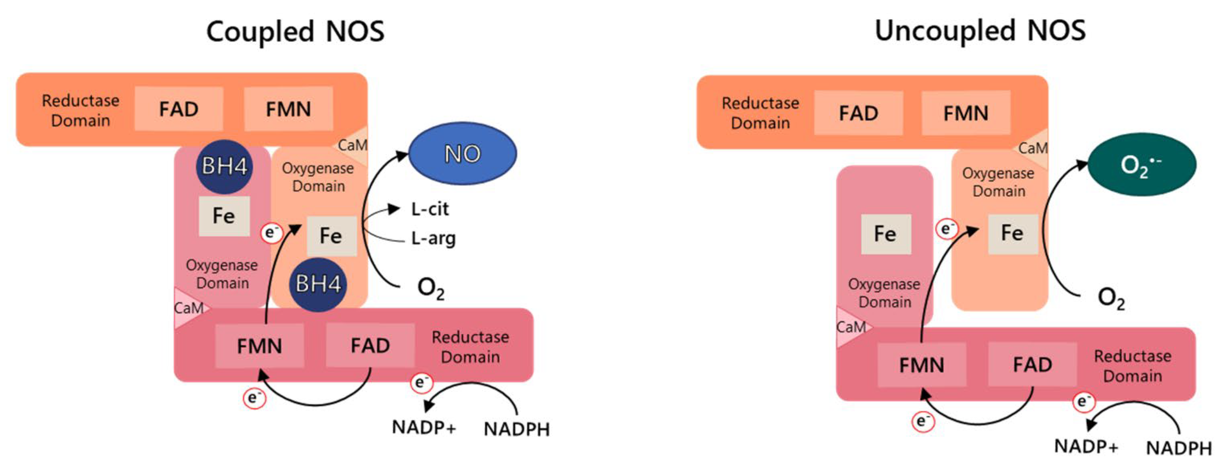
2. Nitric Oxide Synthase

Three NOS (EC 1.14.13.39) isoforms have been described in mammalian cells. NOS isoforms, named neuronal NOS (NOS1), endothelial NOS (NOS3), and inducible NOS (NOS2), are encoded by different genes localized in distinct chromosomes. All three isoforms are homodimeric and each monomer contains a C-terminal reductase domain and an N-terminal oxygenase domain connected by a calmodulin-binding peptide linker. The binding sites for the cofactors flavin mononucleotide (FMN), flavin adenine dinucleotide (FAD), and nicotinamide adenine dinucleotide phosphate (NADPH) are found in the reductase domain, which shares homology with cytochrome P450 (CYT P450) reductases while the oxygenase domain contains binding sites for heme, the cofactor (6R)-5,6,7,8-tetrahydrobiopterin (BH4) and the substrate L-arginine. The electron transfer occurs from the NADPH cofactor into the oxygenase domain, where the L-arginine hydroxylation and subsequently NO formation takes place. Stable dimerization between the two NOS monomers is required for efficient NOS activity, as electrons flow from the reductase domain of one monomer to the oxidase domain localized in the other monomer (Figure 1) [17,18]. NOSs activity is regulated by integrating mechanisms, including post-translational modifications, protein–protein interactions, and substrate and cofactor availability, being BH4 concentration determinant [19,20,21].
NO is a pleiotropic gaseous molecule with a reactive free radical activity. NO regulates innumerous physiological processes, including vascular (blood flow, angiogenesis, platelet aggregation) and neurological functions (nervous system development and neurotransmission) [21]. Decreased NO concentration is associated with distinct diseases, including cardiovascular diseases, diabetes, autism, and cancer [22,23,24].
Different studies have shown that NO can promote or attenuate cancer progression through the regulation of diverse signaling pathways. Effects of NO on neoplasia depend on its concentration and exposure duration, NOS localization, NO-induced post-translational modifications of proteins, and cancer type and stage. The role of NO on cancer development is also associated with its source, tumor, or stromal cells (cancer-associated fibroblasts, endothelial or immune cells) [25,26]. At relatively high concentrations, NO induces cytotoxic and genotoxic events such as protein activity inhibition, mitochondria dysfunction, DNA and protein damage, and cell death while in low doses can trigger cell proliferation and angiogenesis. Many studies have reported the dual effect of NO in cancer cells [21,26]. In melanoma cells, increased NO concentration causes growth arrest and apoptosis [13,27]. Evodiamine treatment induced p38 and NF-κB activation, which in turn triggers NO-derived iNOS, increasing apoptosis in a p53-dependent manner [28]. Moreover, NO stabilizes p53 by phosphorylation, stimulating apoptosis in different cancers [29,30]. On the other hand, NO elicits HIF-1α accumulation in the nucleus of different tumor cells, enhancing gene expression of hypoxia-target genes, which in turn contributes to cancer proliferation, angiogenesis, and metastasis [31,32,33]. Furthermore, Newton et al. found that iNOS inhibition improved immunotherapy in combination with radiotherapy in solid tumors by modulating the tumor immune microenvironment. Decreased NO levels induced CD8+ effector cells activation and intratumoral infiltration, attenuating tumor growth [34].
Dysfunctional NOS is a source of superoxide anion (O2•−), which is associated with the development of cardiovascular diseases, diabetes, neurodegenerative disorders, and more recently, its role in cancer has been reported [13,35,36,37]. The role of ROS in malignant transformation and tumor progression depends on its concentration and cancer stage, contributing to carcinogenesis or tumor cell death [13,16,33].
Although increased expression of the three NOS isoforms in different cancers has been related in innumerous studies, not all the studies describe NOS activity [13,38,39,40,41]. High NOS expression is associated with increased cell proliferation, metastasis, and chemotherapy resistance, in some cases being associated with poor prognosis [26,40,42]. Therefore, the knowledge of NOS activity is very important to understand its real role in oncogenesis and to develop an efficient therapeutic strategy.

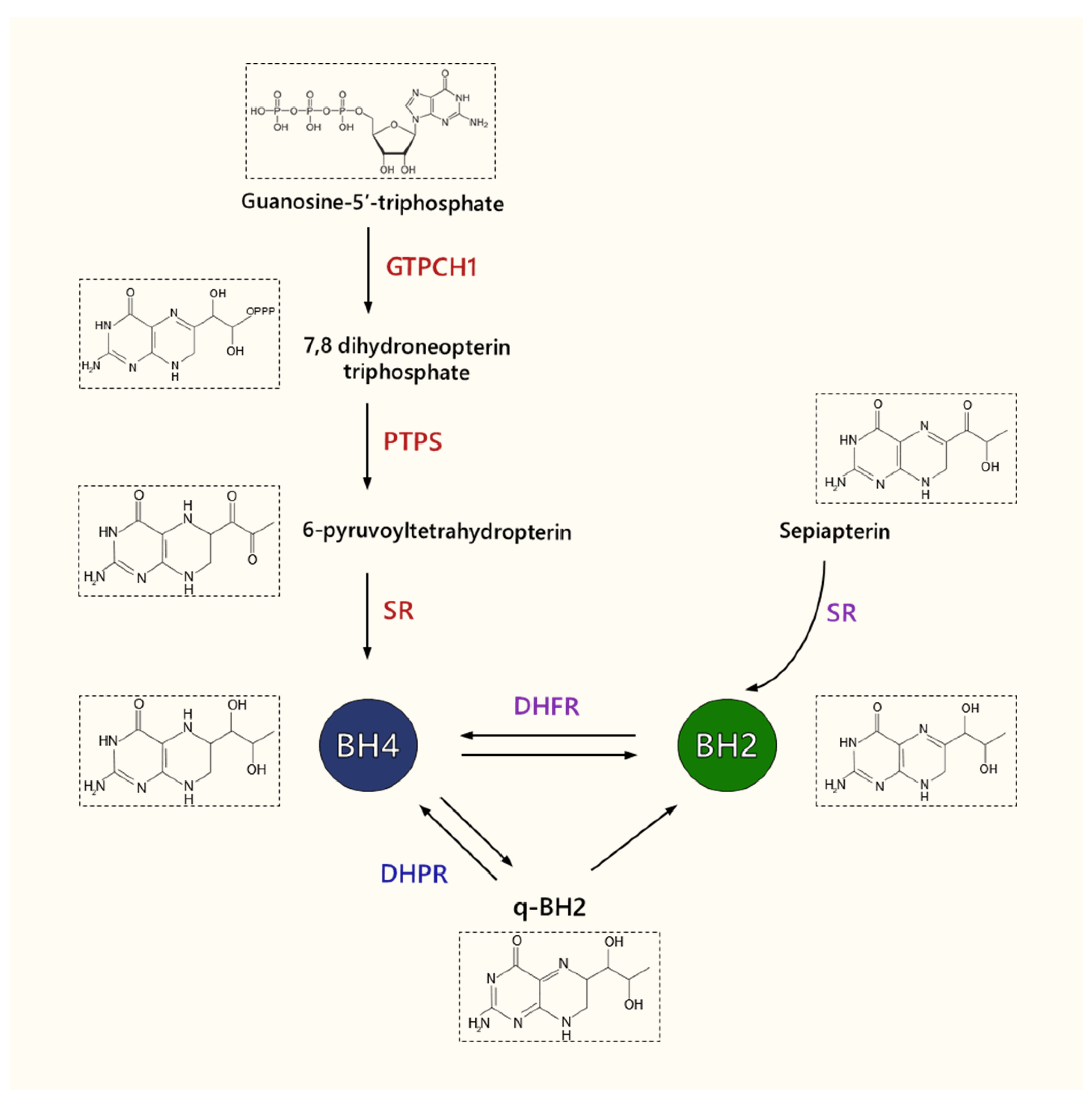
3. Tetrahydrobiopterin

BH4 plays an essential function as a cofactor of a set of metabolic enzymes, including NOS, four aromatic amino acid hydroxylases, alkylglycerol monooxygenase (AGMO), and the brain-specific tryptophan hydroxylase isomer TPH2. BH4 is a pteridine bicyclic molecule comprising a pyrimidine ring fused to a pyrazine ring. Biopterin’s redox activity is conferred by their heteroatomic rings that can be oxidized (biopterin), partially reduced (dihydrobiopterin, BH2), and fully reduced (BH4) [19]. BH4 also has the ability of scavenging ROS by preventing polyunsaturated fatty acyl (PUFA) chains depletion and lipid peroxidation [43] acting as an anti-oxidant molecule [44]. As BH4 is easily oxidized, an increase in ROS and RNS like peroxynitrite leads to BH4 depletion.
Intracellular BH4 amount is tightly regulated by de novo, salvage, and recycle synthesis pathways (Figure 2) [45]. In de novo BH4 biosynthesis, BH4 is synthesized from GTP in a three-step reaction being GTPCH1 (GTP cyclohydrolase 1) (EC 3.5.4.16), the first and rate-limiting enzyme, followed by PTPS (6-pyruvoyl-tetrahydrobiopterin synthase) (EC 4.6.1.10) and SR (sepiapterin reductase) (EC 1.1.1.153) [46]. In the salvage pathway, SR catalyzes the reduction of sepiapterin into BH2 which is then converted into BH4 by DHFR (dihydrofolate reductase) (EC 1.5.1.3). In the recycling, quinoid-BH2 is reduced by DHPR (dihydropteridine reductase) (EC 1.5.1.34) into BH4. Other enzymes share with SR the same substrates, namely, AR (aldose reductase) and CR (carbonyl reductase). Additionally, BH4 can also be synthesized by AKR1C3 (3α-hydroxysteroid dehydrogenase type 3) (EC 1.1.1.357) and AR [47]. GTPCH1 activity is modulated in transcriptional and post-transcriptional levels and by interaction with the proteins GFRP (GTP cyclohydrolase I feedback regulator protein) and caveolin-1 [46,48,49]. GTPCH1 expression is triggered by pro-inflammatory cytokines, ROS, and NRF2 transcription factor in different cell lines [50,51].
BH4 binding to the heme active site at the interface between the two monomers is indispensable for NO synthesis through increased L-arginine substrate interaction and dimer stabilization. Decreased intracellular BH4 concentration promotes NOS destabilization and the reduction of NO production. This dysfunctional state of NOS is referred to as uncoupling because the oxidation of NADPH and the reduction of oxygen are uncoupled from arginine hydroxylation and NO formation. However, the electron transfer from NADPH through the flavin domains to molecular oxygen is not inhibited, resulting in the generation of O2•− and hydrogen peroxide (H2O2) (Figure 1) [47]. Although BH2 can bind to the active site of NOS with the same affinity of BH4, it has no cofactor activity, competing with and displacing BH4 from the oxygenase domain. Therefore, the BH4/BH2 ratio also determines NOS function.

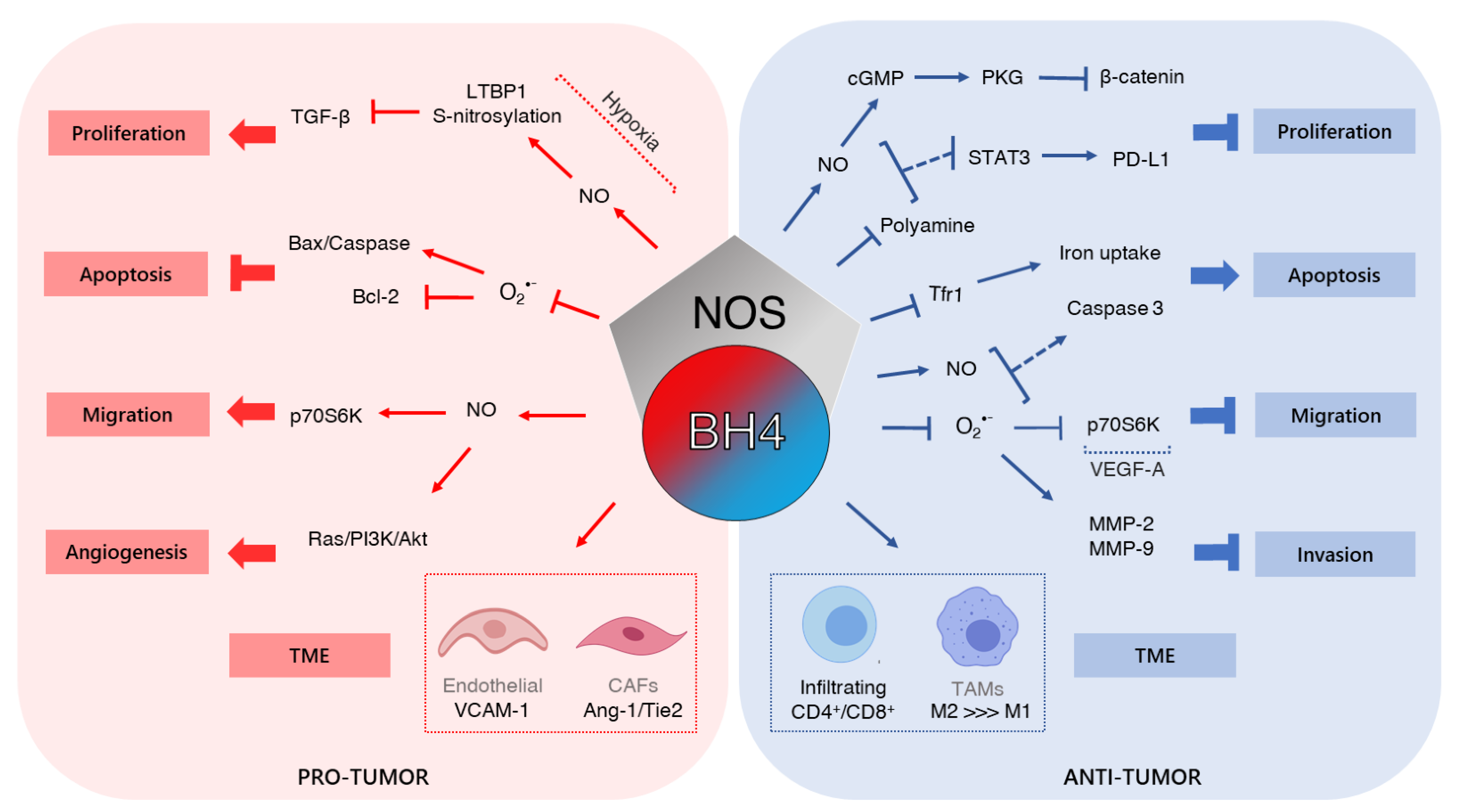
4. Tetrahydrobiopterin and Cancer

Different molecules have been described to contribute positively or negatively to tumor progression, which is associated with several factors, including the type of neoplasia and the stage of tumor development, reflecting the cross-talking between altered oncogenic signaling pathways and the interaction with other molecules. The compartmentalization within the cell and the location in the cancer microenvironment in addition to the expression and/or concentration of such factors also cooperate with its role.
As discussed below, the involvement of tetrahydrobiopterin as a pro- or anti-tumoral molecule has been demonstrated in different biological processes, including tumor microenvironmental reprogramming, cell growth, metabolism, and metastasis, contributing or impairing cancer development (Table 1).

4.1. Cell Growth

Proliferation is a biological process controlled by innumerous signaling pathways triggered by ligand-receptor interaction, which in turn, among other processes, regulates cell cycle progression, nutrition status, and genome integrity. In cancer, uncontrolled cell growth is associated with genetic and epigenetic alterations of key genes that maintain cell homeostasis and also by cell metabolism dysregulation [1,69].
As discussed, NOS coupling does not only depend on the total amount of BH4. The BH4/BH2 ratio and the stoichiometric balance between BH4 and NOS have been shown to determine the functional state of the enzyme [47,70]. In this context, some studies have shown that the increase in BH4/BH2 ratio, due to BH4 or precursor sepiapterin supplementation, impaired or improved cancer development, by regulating tumor growth by different mechanisms.
Rabender et al. observed decreased BH4/BH2 ratio in human breast cancer cells grown in culture, as xenografts, or in a spontaneous mouse breast cancer model (MMTVneu). Besides that, a low BH4/BH2 ratio was found in human colorectal carcinoma biopsies compared with the adjacent normal colon tissue. Treatment with L-sepiapterin partially reduced O2•− and increased cGMP levels in MCF-7 and MDA-231 cancer breast cells, which suggested NOS recoupling. Moreover, L-sepiapterin supplementation of MCF-7 cells induced cGMP-dependent protein kinase (PKG) activation, which in turn promoted β-catenin expression and decreased TCF4 promoter activity [35]. The pro-inflammatory transcription factor NF-κB is associated with carcinogenesis since it induces the expression of different oncogenes [71]. The activity of NF-κB is both positively and negatively modulated by ROS/RNS since Tyr181 nitration of IκBa, an NF-κB inhibitor, triggers NF-κB activation. On the other hand, CYs38 S-nitrosylation of the NF-κB p65 subunit induces its inhibition. Treatment of MCF-7 breast cancer cells with L-sepiapterin abrogated Tyr nitration of IκBa and increased p65 S-nitrosylation, reducing NF-κB activity. Since L-sepiapterin decreases O2•− and increases NO amount, we can conclude that BH4 metabolism alteration modulates the role of NF-κB in cancer and probably the activity of other signaling pathways. Finally, L-sepiapterin inhibited clonogenic capability of MCF-7 and MDA-231 cells in vitro which was shown to be dependent on NO and PKG. MDA-231 xenografts tumor growth and progression were also impaired in the presence of L-sepiapterin. Although MCF-7 and MDA-231 breast carcinoma cells express both iNOS and eNOS isoforms, these results suggested that iNOS is uncoupled since the iNOS selective inhibitor 1400 W impaired the L-sepiapterin-induced cGMP levels [35].
Previously, the same group described that L-sepiapterin impaired chemically induced murine colitis and azoxymethane-induced colorectal cancer [52]. Inflammation is one of the hallmarks of colorectal cancer contributing to malignant transformation and tumor progression [71,72]. Activation of pro-inflammatory NF-κB signaling pathway inducing increased iNOS expression is one of the mechanisms of colorectal cancer development [38,73]. Besides that, the participation of eNOS isoform in colorectal cancer carcinogenesis has been shown by other authors [38,39,73,74,75]. Interestingly, Gochman et al. demonstrated increased iNOS expression and nitrotyrosine content in colitis and colorectal carcinoma, which is caused by peroxynitrite, an RNS and biomarker of oxidative damage, formed by a reaction between nitric oxide and O2•− [76]. Since peroxynitrite formation is a very fast reaction, O2•− and NO must be in close proximity [77], which normally occurs when both species are produced by NOS. Moreover, Youn et al. demonstrated that resveratrol, an antioxidant, abrogated superexpression of iNOS and DSS-induced colitis, indicating that iNOS is uncoupled and producing O2•− [78]. Burhanuddin et al. also found increased iNOS expression in DSS/AMO-induced colorectal cancer, but not measured NOS activity [79]. ROS is also induced by inflammation [80], implicating NOS uncoupling in colorectal cancer development. Cardnell et al. showed that sepiapterin decreases inflammation through reduction of infiltrating neutrophils and macrophages and proinflammatory cytokine expression, which in turn impaired tumor growth. Reduction in BH4/BH2 ratio was inconclusive since there was a trend for lower BH4/BH2 after DSS treatment that was rescued by sepiapterin. On the other hand, sepiapterin reversed the increase in cGMC levels and Tyr nitration, a hallmark of peroxynitrite formation, after DSS treatment. In conclusion, these results suggested for the first time the involvement of NOS uncoupling in colitis-induced colorectal carcinoma [52].
Campos et al. demonstrated for the first time evidence of the association between NOS uncoupling and murine melanocyte malignant transformation. Melanocytes submitted to a chronic stress condition showed increased Nos3 expression. Moreover, O2•− was suppressed by L-NAME (N-Nitro-L-arginine methyl ester), a NOS inhibitor [81]. The same group proposed that NOS uncoupling in murine melanoma cells was a result of decreased BH4 bioavailability since L-sepiapterin treatment restored NO amount and reduced O2•− concentration [53]. Recently, our group showed that although there is no difference between the absolute concentration of BH4 in human melanoma cells when compared to normal melanocytes, BH4/BH2 ratio was lower in tumor cells. In addition, these melanoma cells showed an increase in iNOS and eNOS protein expression. In endothelial cells, NOS uncoupling can be a result of altered NOS and BH4 stoichiometry which means that there is not enough BH4 to catalyze NO formation. Supporting this finding, melanoma cells produced higher levels of O2•− and lower levels of NO. Treatment with BH4 increased the BH4/BH2 ratio, NO concentration, and decreased O2•−. More importantly, BH4 attenuated cell proliferation and induced apoptosis of metastatic melanoma cells, but not melanocytes, pointing to its use as a promising therapy [13]. The hypothesis of NOS/BH4 stoichiometry loss was demonstrated by eNOS suppression in metastatic melanoma cells. Decreased Nos3 expression abrogated the increase in O2•− and the reduction of NO. Moreover, cell growth in vitro and in vivo was attenuated (Melo et al., unpublished). Increased expression of all three isoforms has been shown in melanoma cells and tissues [13,40,41]. However, as mentioned, not all studies investigated NOS activity. This information is fundamental since NO and O2•− modulate different signaling pathways in melanoma, provoking different tumor phenotypes. Furthermore, innumerous authors suggested that the imbalance between NO and O2•− results in cancer growth and progression or tumor development impairment.
Recent data showed that GTPCH1/BH4/NO axis impaired hepatocellular carcinoma (HCC) development. GCH1 is downregulated in HCC tissues and different cell lines by promoter methylation. Decreased GCH1 expression is significantly associated with a higher Barcelona Clinic Liver Cancer stage and larger tumor size. Moreover, GCH1 is correlated with poor clinical outcomes in HCC patients. GCH1 silencing promotes proliferation in vitro and in vivo through inhibition of oxidative stress-induced AKT/p38 activation, indicating the involvement of uncoupled NOS in HCC carcinogenesis [54]. Although the status of NOS expression was not showed, the results implicate altered NOS activity. On the other hand, different studies showed the pro-tumoral activity of NO-derived eNOS/iNOS in HCC growth [82,83]. González et al. reported that sorafenib, a receptor tyrosine kinase, and MAPK pathway inhibitor, decreased eNOS activity and NO synthesis, attenuating proliferation and inducing apoptosis in HCC cell lines [83]. Moreover, the NOS3 T-786C polymorphism (rs2070744) genotype, resulting in reduced activity of NOS3 gene promoter and NO production, is associated with better clinical outcomes in patients treated with sorafenib [84,85]. Rahman et al. showed that higher NOS2 and PTGS-2 expression was correlated with poor prognosis in hepatitis C virus–positive HCC patients. However, NOS activity was not analyzed [86]. Sorafenib has been eligible for the standard treatment for patients with advanced unresectable HCC and the participation of uncoupled NOS was evaluated in primary operable HCC. Accordingly, NOS can have a dual role in HCC progression as was shown in other cancers [87].
On the other hand, Zhang et al. reported an oncogenic role of BH4 in breast cancer development. Increased sepiapterin reductase protein (SR) was found in breast cancer tissues when compared to the adjacent non-tumorigenic tissue and correlated with tumor aggressiveness. SR downregulation inhibited metastatic breast cancer cell proliferation and induced ROS-mediated apoptosis. Although NO amount was not evaluated, decreased BH4 concentration was reported in MDA-MB-231 and MDA-MB-468 breast cancer cells, suggesting the involvement of uncoupled NOS in metastatic breast cancer survival impairment [55]. The reduction of sepiapterin reductase can affect another pathway, since its inhibition impairs ornithine decarboxylase activity and decrease polyamines concentration in neuroblastomas, attenuating tumor growth [88,89]. The shift in arginine metabolism leading to high levels of polyamines will reduce NO synthesis affecting NOS function [90]. Therefore, although SR and GTPCH1 are on the same metabolic pathway, their contributions to breast cancer progression may not be the same.
In glioblastoma (GBM), in silico analyses showed that increased GCH1 expression was correlated with higher glioma grade, recurrence, and worse survival. Furthermore, GCH1 overexpression and BH4 levels elevation in GBM cells increase proliferation and decrease survival in an intracranial GBM-mouse model, which is correlated with brain tumor-initiating cells maintenance and ROS suppression [56]. GBM development is associated with high ROS levels; however, exceeded production can induce cell death [91,92]. On the other hand, increased NOS expression and NO is correlated with glioma cell proliferation [41,56,93]. Therefore, GTPCH1/BH4/NO signaling activation in GBM cells may represent a chemoresistance mechanism, preventing cell death as in melanocyte and keratinocyte cells submitted to radiation-induced ROS [51].
Increased expression of iNOS and eNOS have been related to esophageal squamous cell carcinoma (ESCC). However, neither the regulation of NOS by BH4 nor the mechanism of contribution to the pathogenesis of ESCC have not been investigated. AU-rich element RNA-binding factor 1 (AUF1) is a family of proteins involved in the post-transcriptional regulation process of mRNA and has been related as a promoter or inhibitor of cancer progression [57,94,95,96]. Gao and colleagues found that AUF1 expression is higher in ESCC when compared to normal or tumor-adjacent tissues. Interestingly, the AUF1 silencing reduced GCH1 expression and NO amount, which in turn decreased cell growth and increased apoptosis in squamous carcinoma Eca-109 cells [57]. Although not explored by the authors, the data indicate that AUF1 downregulation causes NOS uncoupling via GTPCH1 regulation in ESCC cells. Since ESCC growth suppression was triggered by endoplasmic reticulum stress activation and increased ROS levels [97,98], NOS uncoupling can be associated with oxidative stress-induced cell death. However, to confirm this hypothesis BH4, BH4/BH2, and O2•− analyses are required.
In a very elegant approach, Soula et al. showed that BH4 abrogates lipid peroxidation–induced ferroptosis caused by glutathione peroxidase 4 (GPX4) inhibition [58]. Using metabolism-focused CRISPR-Cas9 genetic screens it was shown that decreased expression of enzymes from the de novo BH4 synthesis pathway is associated with cell death triggered by RSL3, a GPX4 inhibitor, in different cancer cell lines. Exogenous BH4 supplementation increased proliferation and restored the resistance of T- and B-cell acute lymphoblastic leukemia and lymphomas to GPX4 inhibitors, indicating BH4 synthesis as a new target therapy in those tumors. The authors showed that ferroptosis impairment was caused by the ability of BH4 to act as a radical-trapping antioxidant in lipid membranes, avoiding lipid peroxidation in accordance with another study using fibroblasts [43]. It was also suggested that the protective role of BH4 was by an eNOS-independent mechanism. However, it was assumed that NO formation by NOS promotes the oxidation of BH4 to BH2, reducing BH4 bioavailability, which in turn triggers NOS uncoupling and O2•− generation. BH4 is not consumed in the reaction catalyzed by NOS, instead, it is regenerated by the enzyme itself, not resulting in a BH4 decrease [99]. Moreover, BH4, NO, and O2•− levels were not evaluated to be able to infer about NOS activity. NOS inhibition or eNOS silencing renders leukemic cells more sensitive to RSL3, suggesting the protective function of NO. This data is in agreement with other studies showing that NOS activity is important in leukemia cell survival [41,55].
In the early stages of colorectal cancers, PTPS was found to be elevated. Under hypoxia, a common characteristic of aggressive cancers, AMPK-induced PTPS phosphorylation, which in turn promotes LTBP (Latent TGF-β binding protein)-PTPS-iNOS interaction leading to LTBP nitrosylation and ubiquitin-dependent degradation. In this condition, TGF-β secretion is increased contributing to colorectal tumor growth. PTS silencing impaired LTBP nitrosylation and cell proliferation, showing the involvement of the BH4/NO pathway in colorectal cancer progression [59]. The apparent conflict with the study from Cardnell et al. can be explained by the fact that the authors used an AMS/DSS-induced murine colorectal cancer model. The treatment with AMS/DSS induces oxidative stress, causing BH4 decrease and consequently, NOS uncoupling [52], contributing to malignant transformation. In this case, L-sepiapterin supplementation decreased tumor incidence, but not tumor growth, suggesting the role of uncoupled NOS in colorectal malignant transformation instead of cancer progression.

4.2. Tumor Microenvironment and Angiogenesis

Heterogeneous cell types, including fibroblasts, endothelial, and immune cells, along with extracellular matrix components comprise the tumor microenvironment (TME). Through the secretion of various factors, cancer cells can reprogram and control the function of the surrounding environment, contributing to biological processes, such as angiogenesis [100].
Angiogenesis is a physiological process consisting of the growth of new blood vessels from pre-existing ones, providing oxygen and nutrient supplies. In cancer, pathological angiogenesis contributes to tumor development, increasing proliferation, growth, and metastasis [1,101]. Angiogenesis is maintained by the overproduction of proangiogenic factors, including growth factors, matrix proteinases, and extracellular matrix molecules [100,102]. Different approaches have already shown that eNOS-derived NO signaling in endothelial cells is a critical mechanism of vascular homeostasis, mediating angiogenesis and contributing to increased cell proliferation and migration of endothelial cells and consequently to tumor progression [103,104,105]. Since eNOS activity requires BH4 as a cofactor for NO production, it was shown that the maintenance of its bioavailability is essential for angiogenesis by many groups using different technical procedures [60,61,106,107].
Experiments in vitro demonstrated that increased GCHI expression and L-sepiapterin or BH4 supplementation augmented NO production, cell proliferation, migration, and the capacity of endothelial cells of different species to form capillary-like structures through PI3K/AKT signaling pathway activation [61,106,108]. Chen et al. showed that L-sepiapterin triggers GTP-bound wild-type Ras, increasing AKTSer473 and eNOSSei1177 phosphorylation and NO production. Increased cell proliferation, migration, and tube formation required PI3K signaling since LY treatment, a PI3K signaling inhibitor, abrogated it. Furthermore, Ras activation and PI3K/Akt/eNOS up-regulation were impaired by L-NAME, suggesting a positive feedback mechanism. It was shown that NO caused S-nitrosylation (SNO) of p21Ras in Cys118, triggering the activation of ERK/MAPK signaling pathway and the proliferation of innumerous cells as neural stem cells, breast cancer, and endothelial cells [109,110,111]. Accordingly, AKT activates eNOS, increasing NO synthesis that promotes S-nitrosylation of Ras in Cys118, which drives the PI3K/Akt pathway to maintain angiogenesis and consequently growth of tumors.
On the other hand, the treatment with 2,4-diamino-6-hydroxypyrimidine (DAHP), a GTPCHI inhibitor, and decreased GCHI expression in vitro impaired angiogenesis through abrogation of AKT activation, eNOS phosphorylation, NO production, and consequently reduction of endothelial cell proliferation, migration, and tubulogenesis [60,62,112]. The treatment of endothelial cells with ribavirin, an IMP dehydrogenase inhibitor, decreased GMP synthesis, the GTP cyclohydrolase substrate, causing BH4 and consequently NO reduction, which in turn impaired cell proliferation and tube formation [106].
Many authors evaluate the participation of BH4 in angiogenesis in vivo. To analyze the proangiogenic potential of BH4 in tumor stromal fibroblasts, BALB/c SCID mice were implanted with a construction where the gene GCH1 was cloned into a plasmid that contains tetracycline responsive element under cytomegalovirus promoter control. In the presence of doxycycline (DOX), GCH1 expression is abrogated. Mice with tumors around 100 mm3 in size (25th day after implantation) were then fed with DOX in drinking water to inhibit GCH1 expression or injected with DAHP (300 mg/kg/day) for 7 days. Although angiogenesis was reduced, as showed by decreased CD34-positive microvessels, tumor growth was not inhibited [61]. Interestingly, eNOS expression was also reduced in DOX- or DAHP-treated mice. However, the BH4 concentration was not reduced in DAHP-treated animals, only after the treatment with DOX, suggesting that angiogenesis reduction was BH4-independent. DAHP may have additional inhibitory effects on tumor angiogenesis through downregulating of eNOS expression. Furthermore, it was shown that DAHP reduced the expression of cytokine-induced expression of vascular cell adhesion molecule 1 (VCAM-1) and the transcription factor NF-κB in endothelial cells, both proteins involved in the angiogenic process [113,114,115,116,117]. Therefore, the decreased neovascularization observed after DAHP treatment can be associated with the inhibition of other angiogenic factors.
On the other hand, Dai et al. observed that DAHP treatment (80 mg/kg/day) for two weeks once tumors reached 100 mm3 in size inhibited AKT/eNOS pathway activation and decreased BH4 and NO concentration in hepatocellular carcinoma tissue. Hence, CD31 staining was significantly lower in DAHP-treated mice showing impairment of angiogenesis and tumor growth [60]. Some factors can be associated with these different results between the two studies. Although they observed reduced angiogenesis in DAHP-treated animals, only the treatment with 80 mg/kg/day for two weeks abrogated tumor growth, suggesting that this phenomenon is dose and time-dependent. Moreover, the animals used by Dai et al. were male mice aged 4–6 weeks, while Chen et al. worked with 6- to 8-week-old females [61]. Finally, and more importantly, Chen et al. did not observe a decrease in BH4 content in tumor stromal fibroblasts after DAHP treatment. However, it was reported that the growth of cancer-associated fibroblast (CAFs) is BH4-dependent [63].
CAFs are found in the tumor microenvironmental surrounding the malignant lesion and, through dysregulated cell signaling and communication with tumor cells, contribute to malignant transformation, proliferation, metabolic program, and metastasis [118]. Furthermore, the presence of CAFs has been correlated with poor outcomes in different cancers, including breast cancer [119,120,121]. GTPCH1 was overexpressed in stromal fibroblasts of breast cancer when compared with normal tissue. Moreover, high GTPCH1 was significantly associated with aggressiveness and low recurrence-free survival [63]. Interestingly, GTPCH1-expressing fibroblasts induced BH4-dependent breast cancer proliferation and migration by secreting angiopoietin-1 (Ang-1), which in turn triggers Tie2 phosphorylation and the activation of Ras/PI3K/AKT and ERK pathways. Furthermore, breast tumor growth in vivo and angiogenesis was also enhanced by GTPCH-expressing fibroblast complaints [63]. The authors suggested that Ang1/Tie2 interaction increases breast cancer growth through activation of Ras/PI3K/Akt signaling pathway as was shown in endothelial cells [61]. This study did not investigate NO/O2•− amount or NOS expression; therefore, the relationship between BH4 and enzyme activity was not discussed. However, studies have indicated that NO can induce Ang1/Tie2 interaction [122,123]. Zacharek et al. showed that treatment with a NO donor, (Z)-1-[N-(2-aminoethyl)-N-(2-ammonioethyl) aminio] diazen-1-ium-1,2-diolate (DETA-NONOate), increased Ang1 and Tie2 expression in stroke rats. Furthermore, DETA-NONOate induced capillary tube formation in mouse brain endothelial cells, attenuated by Ang1 blockage [123]. Since NO-derived NOS also stimulates endothelial cell proliferation by Ras/PI3K/AKT activation and NO is associated with breast cancer growth [124,125], BH4-induced NO can contribute to breast cancer progression. Moreover, eNOS activity is also increased by Ang1/Tie2-induced Ras/PI3K/AKT activation, suggesting a feedback mechanism [126]. Therefore, further studies are needed to investigate whether Ang1 induction is related to NO production by NOS coupling or directly by increased BH4 in CAFs.
Showing that the anti-angiogenic and consequently anti-tumor role of DAHP is time-dependent, Pickert et al. showed that the administration of DAHP (100 mg/kg) twice daily orally immediately after tumor cells inoculation reduced the formation of blood vessels as indicated by decreased CD31 and αvβ3 integrin staining [62]. Importantly, DAHP treatment diminished BH4 plasma concentration. Reduction of angiogenesis attenuated tumor growth of B16F10 murine melanoma cells and HT29 human colon cancer cells in vivo. Blood vessels formation in shGCH1-transduced HT29 xenotransplants was analyzed to investigate if the production and release of BH4 from cancer cells also contribute to angiogenesis. In addition to decreasing CD31 and αvβ3 integrin amount, the development of shGCH1-transduced HT29 xenotransplants was reduced when compared with control cells. Moreover, DAHP treatment has been associated with impaired immune-induced tumorigenesis, since tumor-associated macrophages (TAMs) with an antitumoral M1-like phenotype were infiltrating tumor microenvironmental. These data are in accordance with the studies showing that macrophages polarized towards an anti-inflammatory M2 phenotype instead of M1-like phenotype are involved in high angiogenic activity [127,128]. However, the mechanism underlying increased angiogenesis by pro-tumoral M2 polarized macrophages is not associated with increased NOS expression but instead with elevated arginase activity and consequently with reduced NO concentration [129]. Therefore, the axis GTPCH1/BH4/NOS/NO could be a compensatory pathway to maintain NO-induced angiogenesis.
Important components of the TME in various types of cancer, TAMs exhibit a wide spectrum of phenotypic and functional profiles regulated by different signals of the surrounding environment [130,131]. Therefore, functional reprogramming of TAMs to a more anti-tumorigenic profile has been explored as presenting a therapeutic potential [132,133]. CA1d human breast cancer cells and M2 macrophages treated with sepiapterin produced higher levels of NO than polyamines. This rise in NO/polyamine ratio was accompanied by p-STAT3 inhibition and programmed death-ligand 1 (PD-L1) downregulation in mammary tumor cells. Furthermore, sepiapterin treatment of M2-macrophages increased iNOS, STAT1, and IL-12 (M1 macrophage markers) expression, while decreasing M2 markers (CD163, IL-10, and STAT3) [64]. NO and polyamines are products of arginine metabolism by NOS and arginase, respectively. Studies have reported that beyond substrate competition, these two pathways can inhibit each other [134,135]. Furthermore, both have an important role in macrophage polarization, so that NOS/NO is related to M1; on the other hand, arginase/polyamine to the M2 pro-tumor phenotype [129]. In fact, elevated levels of polyamines have been related to different types of cancer and associated with a poor prognosis of breast cancers [136,137]. Interestingly, ex vivo analysis showed that 100 μM sepiapterin reduced PD-L1 expression and tumor epithelial density (cytokeratin 14 staining), while reprogramming TAMs from M2 to M1 phenotype in culture tumors derived from MMTV-PyMT mice [64]. PD-L1 expression has not only been associated with poor prognosis in breast cancer patients but was also considered a target for immunotherapy [138,139,140], with an important role of TAMs in this scenario [141]. Therefore, redirecting arginine metabolism by BH4 synthesis in cancer cells and TAMs can improve breast cancer immunogenicity.
Recently, the effects of BH4 on T cells and its role in breast cancer were also investigated. Although the normal development of T cells in mice knocked to Gch1 specifically for these cells, there was a reduction in BH4 levels accompanied by a decrease in the proliferation of mature T cells induced via TCR (T cell receptors). A similar result was shown by inhibiting sepiapterin reductase through the SPRi3 inhibitor. On the other hand, the increased BH4 levels by GCH1 overexpression or L-sep/BH4 treatment enhanced the proliferation of stimulated CD4+ and CD8+ T cells. Interestingly, on the orthotopic breast cancer model (E0771 cells), the tumor growth was rejected in GCH1-overexpressing mice. Consistently, BH4 supplementation (100 mg/kg−1/day, intraperitoneally) decreased tumor growth and increased tumor-infiltrating CD4+ and CD8+ T cells. Furthermore, stimulated BH4-deficient T cells showed decreased iron levels and reduced mitochondrial respiration and oxygen consumption [65]. Thus, the authors indicate mitochondrial dysfunction via defective iron-redox cycling of cytochrome c as a mechanism for the effects of BH4 deficiency in TCR-activated T cells. However, the antioxidant activity of BH4 is controversial [19] and its role as a cofactor of NOS cannot be discarded. Although there is no difference in iNOS expression in stimulated T cells, other NOS isoforms could be involved, since its activity has been described in these cells [142,143]. In addition, the fact that Gch1-ablated T cells do not show a difference in nitrite levels compared to control cells does not completely exclude the participation of NOS, since the enzyme in stimulated T cells may be in a state of partial uncoupling, that is, producing NO and O2•− [144]. Consistently, increased O2•− levels were found in stimulated T cells, which were potentiated in BH4-deficient cells [65].
The fact that hypoxia induces GTPCH1 expression and BH4 synthesis corroborate their critical role in angiogenesis [62]. Reduced oxygen apport triggers the expression of the transcription factor hypoxia-inducible transcription factor (HIF-α), which in turn induces the expression of more than 60 genes, including VEGF that induces NO signaling-mediated angiogenesis [145].
Angiogenic vasculature structure found in solid tumors is poorly organized, leading to heterogeneous blood flow, increased interstitial pressure, and intermittent hypoxia contributing to radioresistance and the low ratio of drug delivery [146]. One of the mechanisms associated with dysfunctional tumor vasculature is NOS uncoupling caused by the decreased bioavailability of BH4 [147]. Normalization of abnormal cancer vasculature can result in a transient period to ameliorate drug delivery [125,148]. In a spontaneous breast tumor mice model, L-sepiapterin induced NOS recoupling, which in turn reduced hypoxia, restored perfusion leading to increased doxycycline uptake and cell death. Moreover, sepiapterin also increased radiation-induced apoptosis [66]. Although NO-derived NOS improves tubulogenesis through stimulation of endothelial cell proliferation and migration, uncoupling NOS also contributes to disruption of angiogenic vasculature impairing treatment. It is important to note that there is a therapeutic window where sepiapterin supplementation can be used to abrogate cancer cell resistance to radio- and chemotherapy.

4.3. Migration and Invasion

Metastasis is a complex process comprising a sequence of steps that begins with local invasion (migration and extracellular matrix degradation), then intravasation into lymphatic and blood vessels, survival in hematogenous and lymphatic systems, extravasation into the parenchyma of distant tissues, and finally the formation and growth of small niches of cells, the metastatic lesions [149,150].
Kanugula et al. showed that statin treatment reduced invasion and induced apoptosis of triple-negative breast cancer cells, by iNOS-mediated NO production [67]. Statins are inhibitors of 3-hydroxy-3-methylglutaryl coenzyme A (HMG-CoA) reductase (a rate-limiting enzyme of the mevalonate pathway) and have been used in the treatment of patients with cardiovascular diseases [151,152]. Fluvastatin enhanced iNOS expression, NO production, and nitrite levels in breast cancer cells, while reduced transferrin receptor (Tfr1) expression and iron uptake leading to cell death. These effects were abrogated in the presence of mevalonate, ADMA, or 1400 W. In addition, this statin decreased H2O2 levels and downregulated MMP-2 and MMP-9 transcripts, inhibiting the invasive potential of the cells, which was reversed in the presence of a catalase inhibitor [67]. Since studies reported that fluvastatin and others statins increased eNOS-mediated NO production via upregulation of GTPCH and consequent increase BH4 levels [153,154,155], the antitumor effects of fluvastatin found by Kanagula and colleagues can be associated with iNOS coupling. Consistently, the authors showed that sepiapterin treatment (50 μM for 24 h) was able to reduce Tfr1 expression and iron uptake in MDA-MB-231 cells. Furthermore, inhibition of iNOS by 1400 W reversed H2O2 levels reduction and the anti-invasive effect of fluvastatin in these cells [67]. Although not exploring the role of BH4/NOS, other studies have also demonstrated the anti-tumor propriety of fluvastatin on breast, hepatocellular, ovarian, and prostate cancer cells [156,157,158].
In ovarian cancer cells, sepiapterin abrogated the increase in migration and proliferation triggered by vascular endothelial growth factor-A (VEGF-A) and p70S6K-dependent VEGRF2 expression in a NO-independent mechanism. Surprisingly, sepiapterin increased migration and proliferation of ovarian cancer cells in the absence of growth factors, which is mediated by a NO-dependent activation of ERK, AKT, and p70S6K signaling [68]. Since VGFR2 activation promotes ROS increase which is associated with ovarian cancer development [159,160,161], impairment of cell growth in the presence of sepiapterin can be attributed to NOS recoupling. These results suggest that the role of sepiapterin in cancer is related to the modulation of signaling pathways by NO or ROS. Elevated expression of the three NOS isoforms has been associated with ovarian cancer carcinogenesis [40,162]. Some authors documented that increased iNOS expression is correlated with poor prognosis. In iNOS-positive patients, the authors observed an increase in the risk of disease relapse, lower disease-free survival period, and high death incidence [40,163,164]. However, NOS activity was not evaluated. So, expression of the NOS isoforms can be useful as a prognostic factor in ovarian cancer; however, as a therapeutic target, its real activity must be taken into account. It is also important to note, that NOS uncoupling can be partial, implicating the NO and O2•− synthesis at the same time.
Some important acquisitions of cancer cells during tumor progression and metastatic colonization are the ability of anchorage-independent growth and resistance to a programmed cell death known as anoikis. This type of apoptosis is induced by loss of attachment to extracellular matrix or inappropriate cell-matrix interactions, preventing cell adhesion to inadequate locations [165,166]. Campos et al. also showed that uncoupled eNOS contributes to anoikis-resistance of melanocytes submitted to anchorage impediment [81]. Superoxide production was higher in melanocytes maintained in suspension, which was reversed in the presence of L-sep or inhibiting NOS by L-NAME. Curiously, DAHP treatment enhanced O2•− and reduced NO levels in these cells. Furthermore, sepiapterin and Mn(III)TBAP (a superoxide scavenger) were able to decrease melanocyte and metastatic melanoma cell survival in suspension, but not in adhesion [53]. In fact, malignant melanocyte transformation has been associated with higher production of ROS, characterizing disrupted redox homeostasis [167]. The control in ROS and NO concentrations can mediate the acquisition of the appropriate phenotype in different steps of tumor progression and prevents cancer cell death under adverse conditions [168]. Cell lines corresponding to distinct stages of melanoma progression increased O2•− production and decreased NO levels compared to parental melanocytes. In addition, in vivo analysis showed that the treatment with L-NAME before and during sequential cycles of anchorage blockade impairs melanocyte malignant transformation [53]. Taken together, these results indicate that BH4 synthesis and eNOS coupling has anti-tumor effects on melanoma development.

5. Conclusions

Altered signaling pathways are associated with malignant transformation and the acquisition of an aggressiveness phenotype. The expression, activity, and cell localization of these transduction signals are regulated by innumerous factors, including ROS and NO-induced post-translational modifications. Different studies have shown that ROS and NO amount and the balance between these reactive species contribute to tumor growth, chemoresistance, and metastasis. Moreover, the contribution of ROS and RNS from tumor microenvironmental in cancer development has to be considered. Since NOS can be a source of both ROS and NO, understanding its activity is essential to improve cancer management. Therefore, the dual role of the BH4/NOS pathway in carcinogenesis can be explained by the status of transduction pathways found in distinct cancers (Figure 3).

Author Contributions

F.H.M.d.M., D.A.G. and M.G.J. wrote the paper. All authors have read and agreed to the published version of the manuscript.

Funding

This work had financial support from Fundação de Amparo à Pesquisa do Estado de São Paulo—FAPESP. FHMM had been supported by FAPESP (2019/27517-0), DAG by CAPES and MGJ by FAPESP (2011/12306-1).

Conflicts of Interest

The authors declare no conflict of interest.

References

  1. Hanahan, D.; Weinberg, R.A. Hallmarks of cancer: The next generation. Cell 2011, 144, 646–674. [Google Scholar] [CrossRef] [Green Version]
  2. Gotwals, P.; Cameron, S.; Cipolletta, D.; Cremasco, V.; Crystal, A.; Hewes, B.; Mueller, B.; Quaratino, S.; Sabatos-Peyton, C.; Petruzzelli, L.; et al. Prospects for combining targeted and conventional cancer therapy with immunotherapy. Nat. Rev. Cancer 2017, 17, 286–301. [Google Scholar] [CrossRef]
  3. Montor, W.R.; Salas, A.R.O.S.E.; Melo, F.H.M. Receptor tyrosine kinases and downstream pathways as druggable targets for cancer treatment: The current arsenal of inhibitors. Mol. Cancer 2018, 17, 1–18. [Google Scholar] [CrossRef]
  4. Arnold, M.; Pandeya, N.; Byrnes, G.; Renehan, P.A.G.; Stevens, G.A.; Ezzati, P.M.; Ferlay, J.; Miranda, J.J.; Romieu, I.; Dikshit, R.; et al. Global burden of cancer attributable to high body-mass index in 2012: A population-based study. Lancet Oncol. 2015, 16, 36. [Google Scholar] [CrossRef]
  5. Arnold, M.; de Vries, E.; Whiteman, D.C.; Jemal, A.; Bray, F.; Parkin, D.M.; Soerjomataram, I. Global burden of cutaneous melanoma attributable to ultraviolet radiation in 2012. Int. J. Cancer 2018, 143, 1305–1314. [Google Scholar] [CrossRef] [PubMed]
  6. Soerjomataram, I.; Bray, F. Planning for tomorrow: Global cancer incidence and the role of prevention 2020–2070. Nat. Rev. Clin. Oncol. 2021, in press. [Google Scholar] [CrossRef]
  7. Kolb, R.; Sutterwala, F.S.; Zhang, W. Obesity and cancer: Inflammation bridges the two. Curr. Opin. Pharmacol. 2016, 29, 77. [Google Scholar] [CrossRef] [Green Version]
  8. Avgerinos, K.I.; Spyrou, N.; Mantzoros, C.S.; Dalamaga, M. Obesity and cancer risk: Emerging biological mechanisms and perspectives. Metab. Clin. Exp. 2019, 92, 121–135. [Google Scholar] [CrossRef] [PubMed]
  9. Mrityunjaya, M.; Pavithra, V.; Neelam, R.; Janhavi, P.; Halami, P.M.; Ravindra, P.V. Immune-boosting, antioxidant and anti-inflammatory food supplements targeting pathogenesis of COVID-19. Front. Immunol. 2020, 11, 2337. [Google Scholar] [CrossRef] [PubMed]
  10. Bang, E.; Kim, D.H.; Chung, H.Y. Protease-activated receptor 2 induces ROS-mediated inflammation through Akt-mediated NF-κB and FoxO6 modulation during skin photoaging. Redox Biol. 2021, 44, 102022. [Google Scholar] [CrossRef] [PubMed]
  11. Anavi, S.; Tirosh, O. iNOS as a metabolic enzyme under stress conditions. Free Radic. Biol. Med. 2020, 146, 16–35. [Google Scholar] [CrossRef]
  12. Alves-Fernandes, D.K.; de Oliveira, É.A.; Faião-Flores, F.; Alicea-Rebecca, G.; Weeraratna, A.T.; Smalley, K.S.M.; Barros, S.B.M.; Maria-Engler, S.S. ER stress promotes antitumor effects in BRAFi/MEKi resistant human melanoma induced by natural compound 4-nerolidylcathecol (4-NC). Pharmacol. Res. 2019, 141, 63–72. [Google Scholar] [CrossRef] [PubMed]
  13. Gonçalves, D.A.; Xisto, R.; Gonçalves, J.D.; da Silva, D.B.; Moura Soares, J.P.; Icimoto, M.Y.; Sant’Anna, C.; Gimenez, M.; de Angelis, K.; Llesuy, S.; et al. Imbalance between nitric oxide and superoxide anion induced by uncoupled nitric oxide synthase contributes to human melanoma development. Int. J. Biochem. Cell Biol. 2019, 115, 105592. [Google Scholar] [CrossRef] [PubMed]
  14. Lin, Y.; Jiang, M.; Chen, W.; Zhao, T.; Wei, Y. Cancer and ER stress: Mutual crosstalk between autophagy, oxidative stress and inflammatory response. Biomed. Pharmacother. 2019, 118, 109249. [Google Scholar] [CrossRef] [PubMed]
  15. Guan, Y.; Wang, Y.; Li, B.; Shen, K.; Li, Q.; Ni, Y.; Huang, L. Mitophagy in carcinogenesis, drug resistance and anticancer therapeutics. Cancer Cell Int. 2021, 21, 1–9. [Google Scholar] [CrossRef] [PubMed]
  16. Vermot, A.; Petit-Härtlein, I.; Smith, S.M.E.; Fieschi, F. NADPH oxidases (NOX): An overview from discovery, molecular mechanisms to physiology and pathology. Antioxidants 2021, 10, 890. [Google Scholar] [CrossRef] [PubMed]
  17. Poulos, T.L.; Li, H. Nitric oxide synthase and structure-based inhibitor design. Nitric Oxide 2017, 63, 68–77. [Google Scholar] [CrossRef] [Green Version]
  18. Stuehr, D.J.; Haque, M.M. Nitric oxide synthase enzymology in the 20 years after the Nobel Prize. Br. J. Pharmacol. 2019, 176, 177–188. [Google Scholar] [CrossRef] [Green Version]
  19. Vásquez-Vivar, J. Tetrahydrobiopterin, superoxide and vascular dysfunction. Free Radic. Biol. Med. 2009, 47, 1108. [Google Scholar] [CrossRef] [PubMed] [Green Version]
  20. Looft-Wilson, R.C.; Billaud, M.; Johnstone, S.R.; Straub, A.C.; Isakson, B.E. Interaction between nitric oxide signaling and gap junctions: Effects on vascular function. Biochim. Biophys. Acta Biomembr. 2012, 1818, 1895–1902. [Google Scholar] [CrossRef] [Green Version]
  21. Gantner, B.N.; LaFond, K.M.; Bonini, M.G. Nitric oxide in cellular adaptation and disease. Redox Biol. 2020, 34, 101550. [Google Scholar] [CrossRef]
  22. Pannirselvam, M.; Simon, V.; Verma, S.; Anderson, T.; Triggle, C.R. Chronic oral supplementation with sepiapterin prevents endothelial dysfunction and oxidative stress in small mesenteric arteries from diabetic (db/db) mice. Br. J. Pharmacol. 2003, 140, 701. [Google Scholar] [CrossRef] [Green Version]
  23. İnci, A.; Özaslan, A.; Okur, İ.; Biberoğlu, G.; Güney, E.; Ezgü, F.S.; Tümer, L.; İşeri, E. Autism: Screening of inborn errors of metabolism and unexpected results. Autism Res. 2021, 14, 887–896. [Google Scholar] [CrossRef]
  24. Infante, T.; Costa, D.; Napoli, C. Novel insights regarding nitric oxide and cardiovascular diseases. Angiology 2021, 72, 411–425. [Google Scholar] [CrossRef] [PubMed]
  25. Chang, C.-F.; Diers, A.R.; Hogg, N. Cancer cell metabolism and the modulating effects of nitric oxide. Free Radic. Biol. Med. 2015, 79, 324. [Google Scholar] [CrossRef] [Green Version]
  26. Somasundaram, V.; Basudharm, D.; Bharadwaj, G. Molecular mechanisms of nitric oxide in cancer progression, signal transduction, and metabolism. Antioxid. Redox Signal. 2019, 30, 1124–1143. [Google Scholar] [CrossRef] [PubMed]
  27. Paskas, S.; Mazzon, E.; Basile, M.S.; Cavalli, E.; Al-Abed, Y.; He, M.; Rakocevic, S.; Nicoletti, F.; Mijatovic, S.; Maksimovic-Ivanic, D. Lopinavir-NO, a nitric oxide-releasing HIV protease inhibitor, suppresses the growth of melanoma cells in vitro and in vivo. Investig. New Drugs 2019, 37, 1014–1028. [Google Scholar] [CrossRef] [PubMed]
  28. Yang, J.; Wu, L.-J.; Tashiro, S.-I.; Onodera, S.; Ikejima, T. Nitric oxide activated by p38 and NF-κB facilitates apoptosis and cell cycle arrest under oxidative stress in evodiamine-treated human melanoma A375-S2 cells. Free Radic. Res. 2009, 42, 1–11. [Google Scholar] [CrossRef] [PubMed]
  29. Li, C.-Q.; Pang, B.; Kiziltepe, T.; Trudel, L.J.; Engelward, B.P.; Dedon, P.C.; Wogan, G.N. Threshold effects of nitric oxide-Induced toxicity and cellular responses in wild-type and p53-null human lymphoblastoid cells. Chem. Res. Toxicol. 2006, 19, 399. [Google Scholar] [CrossRef] [PubMed] [Green Version]
  30. Mijatovic, S.; Maksimovic-Ivanic, D.; Mojic, M.; Malaponte, G.; Libra, M.; Cardile, V.; Miljkovic, D.; Harhaji, L.; Dabideen, D.; Cheng, K.F.; et al. Novel nitric oxide-donating compound (S,R)-3-phenyl-4,5-dihydro-5-isoxazole acetic acid–nitric oxide (GIT-27NO) induces p53 mediated apoptosis in human A375 melanoma cells. Nitric Oxide 2008, 19, 177–183. [Google Scholar] [CrossRef]
  31. Branco-Price, C.; Evans, C.E.; Johnson, R.S.; Branco-Price, C.; Evans, C.E.; Johnson, R.S. Endothelial hypoxic metabolism in carcinogenesis and dissemination: HIF-A isoforms are a NO metastatic phenomenon. Oncotarget 2013, 4, 2567–2576. [Google Scholar] [CrossRef] [Green Version]
  32. Capparelli, C.; Whitaker-Menezes, D.; Guido, C.; Balliet, R.; Pestell, T.G.; Howell, A.; Sneddon, S.; Pestell, R.G.; Martinez-Outschoorn, U.; Lisanti, M.P.; et al. CTGF drives autophagy, glycolysis and senescence in cancer-associated fibroblasts via HIF1 activation, metabolically promoting tumor growth. Cell Cycle 2012, 11, 2272–2284. [Google Scholar] [CrossRef] [Green Version]
  33. Mijatović, S.; Savić-Radojević, A.; Plješa-Ercegovac, M.; Simić, T.; Nicoletti, F.; Maksimović-Ivanić, D. The double-faced role of nitric oxide and reactive oxygen species in solid tumors. Antioxidants 2020, 9, 374. [Google Scholar] [CrossRef] [PubMed]
  34. Newton, J.M.; Hanoteau, A.; Liu, H.-C.; Gaspero, A.; Parikh, F.; Gartrell-Corrado, R.D.; Hart, T.D.; Laoui, D.; Ginderachter, J.A.; Dharmaraj, N.; et al. Immune microenvironment modulation unmasks therapeutic benefit of radiotherapy and checkpoint inhibition. J. Immunother. Cancer 2019, 7, 216. [Google Scholar] [CrossRef] [PubMed] [Green Version]
  35. Rabender, C.S.; Alam, A.; Sundaresan, G.; Cardnell, R.J.; Yakovlev, V.A.; Mukhopadhyay, N.D.; Graves, P.; Zweit, J.; Mikkelsen, R.B. The role of nitric oxide synthase uncoupling in tumor progression. Mol. Cancer Res. 2015, 13, 1034–1043. [Google Scholar] [CrossRef] [PubMed] [Green Version]
  36. Daiber, A.; Kröller-Schön, S.; Oelze, M.; Hahad, O.; Li, H.; Schulz, R.; Steven, S.; Münzel, T. Oxidative stress and inflammation contribute to traffic noise-induced vascular and cerebral dysfunction via uncoupling of nitric oxide synthases. Redox Biol. 2020, 34, 101506. [Google Scholar] [CrossRef] [PubMed]
  37. Pérez-Torres, I.; Manzano-Pech, L.; Rubio-Ruíz, M.E.; Soto, M.E.; Guarner-Lans, V. Nitrosative stress and its association with cardiometabolic disorders. Molecules 2020, 25, 2555. [Google Scholar] [CrossRef]
  38. Campanholo, V.M.L.P.; Silva, R.M.; Silva, T.D.; Neto, R.A.; Ribeiro Paiotti, A.P.; Ribeiro, D.A.; Forones, N.M. Oral concentrated grape juice suppresses expression of NF-kappa B, TNF-α and iNOS in experimentally induced colorectal carcinogenesis in wistar rats. Asian Pac. J. Cancer Prev. 2015, 16, 947–952. [Google Scholar] [CrossRef] [Green Version]
  39. Peñarando, J.; López-Sánchez, L.M.; Mena, R.; Guil-Luna, S.; Conde, F.; Hernández, V.; Toledano, M.; Gudiño, V.; Raponi, M.; Billard, C.; et al. A role for endothelial nitric oxide synthase in intestinal stem cell proliferation and mesenchymal colorectal cancer. BMC Biol. 2018, 16, 3. [Google Scholar] [CrossRef] [Green Version]
  40. Kielbik, M.; Szulc-Kielbik, I.; Klink, M. The potential role of iNOS in ovarian cancer progression and chemoresistance. Int. J. Mol. Sci. 2019, 20, 1751. [Google Scholar] [CrossRef] [Green Version]
  41. Lawal, B.; Lee, C.-Y.; Mokgautsi, N.; Sumitra, M.R.; Khedkar, H.; Wu, A.T.H.; Huang, H.-S. mTOR/EGFR/iNOS/MAP2K1/FGFR/TGFB1 are druggable candidates for N-(2,4-Difluorophenyl)-2′,4′-Difluoro-4-Hydroxybiphenyl-3-Carboxamide (NSC765598), with consequent anticancer implications. Front. Oncol. 2021, 11, 932. [Google Scholar] [CrossRef]
  42. Thomas, D.D.; Wink, D.A. NOS2 as an emergent player in progression of cancer. Antioxid. Redox Signal. 2017, 26, 963–965. [Google Scholar] [CrossRef] [PubMed]
  43. Kraft, V.A.N.; Bezjian, C.T.; Pfeiffer, S.; Ringelstetter, L.; Müller, C.; Zandkarimi, F.; Merl-Pham, J.; Bao, X.; Anastasov, N.; Kössl, J.; et al. GTP Cyclohydrolase 1/Tetrahydrobiopterin counteract ferroptosis through lipid remodeling. ACS Cent. Sci. 2019, 6, 41–53. [Google Scholar] [CrossRef] [PubMed] [Green Version]
  44. Kirsch, M.; Korth, H.-G.; Stenert, V.; Sustmann, R.; Groot, H. The autoxidation of tetrahydrobiopterin revisited: Proof of superoxide formation from reaction of tetrahydrobiopterin with molecular oxygen. J. Biol. Chem. 2003, 278, 24481–24490. [Google Scholar] [CrossRef] [Green Version]
  45. Thöny, B.; Auerbach, G.; Blau, N. Tetrahydrobiopterin biosynthesis, regeneration and functions. Biochem. J. 2000, 347, 1–16. [Google Scholar] [CrossRef] [PubMed]
  46. Werner, E.R.; Blau, N.; Thöny, B. Tetrahydrobiopterin: Biochemistry and pathophysiology. Biochem. J. 2011, 438, 397–414. [Google Scholar] [CrossRef] [PubMed] [Green Version]
  47. Crabtree, M.J.; Channon, K.M. Synthesis and recycling of tetrahydrobiopterin in endothelial function and vascular disease. Nitric Oxide 2011, 25, 81–88. [Google Scholar] [CrossRef] [Green Version]
  48. Peterson, T.E.; d’Uscio, L.V.; Cao, S.; Wang, X.-L.; Katusic, Z.S. Guanosine triphosphate cyclohydrolase I expression and enzymatic activity are present in caveolae of endothelial cells. Hypertension 2009, 53, 189–195. [Google Scholar] [CrossRef]
  49. Tatham, A.L.; Crabtree, M.J.; Warrick, N.; Cai, S.; Alp, N.J.; Channon, K.M. GTP cyclohydrolase I expression, protein, and activity determine intracellular tetrahydrobiopterin levels, independent of GTP cyclohydrolase feedback regulatory protein expression. J. Biol. Chem. 2009, 284, 13660–13668. [Google Scholar] [CrossRef] [Green Version]
  50. Shimizu, S.; Hiroi, T.; Ishii, M.; Hagiwara, T.; Wajima, T.; Miyazaki, A.; Kiuchi, Y. Hydrogen peroxide stimulates tetrahydrobiopterin synthesis through activation of the Jak2 tyrosine kinase pathway in vascular endothelial cells. Int. J. Biochem. Cell Biol. 2008, 40, 755–765. [Google Scholar] [CrossRef]
  51. Xue, J.; Yu, C.; Sheng, W.; Zhu, W.; Luo, J.; Zhang, Q.; Yang, H.; Cao, H.; Wang, W.; Zhou, J.; et al. The Nrf2/GCH1/BH4 axis ameliorates radiation-induced skin injury by modulating the ROS cascade. J. Investig. Dermatol. 2017, 137, 2059–2068. [Google Scholar] [CrossRef] [Green Version]
  52. Cardnell, R.J.G.; Rabender, C.S.; Ross, G.R.; Guo, C.; Howlett, E.L.; Alam, A.; Wang, X.Y.; Akbarali, H.I.; Mikkelsen, R.B. Sepiapterin ameliorates chemically induced murine colitis and azoxymethane-induced colon cancer. J. Pharmacol. Exp. Ther. 2013, 347, 117–125. [Google Scholar] [CrossRef] [Green Version]
  53. Melo, F.H.M.; Molognoni, F.; Morais, A.S.; Toricelli, M.; Mouro, M.G.; Higa, E.M.S.; Lopes, J.D.; Jasiulionis, M.G. Endothelial nitric oxide synthase uncoupling as a key mediator of melanocyte malignant transformation associated with sustained stress conditions. Free Radic. Biol. Med. 2011, 50, 1263–1273. [Google Scholar] [CrossRef] [Green Version]
  54. Zhong, G.C.; Zhao, Z.B.; Cheng, Y.; Wang, Y.B.; Qiu, C.; Mao, L.H.; Hu, J.J.; Cai, D.; Liu, Y.; Gong, J.P.; et al. Epigenetic silencing of GCH1promotes hepatocellular carcinoma growth by activating superoxide anion-mediated ASK1/p38 signaling via inhibiting tetrahydrobiopterin de novo biosynthesis. Free Radic. Biol. Med. 2021, 168, 81–94. [Google Scholar] [CrossRef] [PubMed]
  55. Zhang, X.; Chen, Y.; Wang, K.; Tang, J.; Chen, Y.; Jin, G.; Liu, X. The knockdown of the sepiapterin reductase gene suppresses the proliferation of breast cancer by inducing ROS-mediated apoptosis. Int. J. Clin. Exp. Pathol. 2020, 13, 2228–2239. [Google Scholar] [PubMed]
  56. Tran, A.N.; Walker, K.; Harrison, D.G.; Chen, W.; Mobley, J.; Hocevar, L.; Hackney, J.R.; Sedaka, R.S.; Pollock, J.S.; Goldberg, M.S.; et al. Reactive species balance via GTP cyclohydrolase i regulates glioblastoma growth and tumor initiating cell maintenance. Neuro-Oncology 2018, 20, 1055–1067. [Google Scholar] [CrossRef] [PubMed]
  57. Gao, Y.; Wang, W.; Cao, J.; Wang, F.; Geng, Y.; Cao, J.; Xu, X.; Zhou, J.; Liu, P.; Zhang, S. Upregulation of AUF1 is involved in the proliferation of esophageal squamous cell carcinoma through GCH1. Int. J. Oncol. 2016, 49, 2001–2010. [Google Scholar] [CrossRef]
  58. Soula, M.; Weber, R.A.; Zilka, O.; Alwaseem, H.; La, K.; Yen, F.; Molina, H.; Garcia-Bermudez, J.; Pratt, D.A.; Birsoy, K. Metabolic determinants of cancer cell sensitivity to canonical ferroptosis inducers. Nat. Chem. Biol. 2020, 16, 1351–1360. [Google Scholar] [CrossRef] [PubMed]
  59. Zhao, Q.; Zheng, K.; Ma, C.; Li, J.; Zhuo, L.; Huang, W.; Chen, T.; Jiang, Y. PTPS Facilitates compartmentalized LTBP1 S-Nitrosylation and promotes tumor growth under hypoxia. Mol. Cell 2020, 77, 95–107.e5. [Google Scholar] [CrossRef]
  60. Dai, Y.; Cui, J.; Gan, P.; Li, W. Downregulation of tetrahydrobiopterin inhibits tumor angiogenesis in BALB/c-nu mice with hepatocellular carcinoma. Oncol. Rep. 2016, 36, 669–675. [Google Scholar] [CrossRef] [Green Version]
  61. Chen, L.; Zeng, X.; Wang, J.; Briggs, S.S.; O’Neill, E.; Li, J.; Leek, R.; Kerr, D.J.; Harris, A.L.; Cai, S. Roles of tetrahydrobiopterin in promoting tumor angiogenesis. Am. J. Pathol. 2010, 177, 2671–2680. [Google Scholar] [CrossRef] [Green Version]
  62. Pickert, G.; Lim, H.Y.; Weigert, A.; Häussler, A.; Myrczek, T.; Waldner, M.; Labocha, S.; Ferreirós, N.; Geisslinger, G.; Lötsch, J.; et al. Inhibition of GTP cyclohydrolase attenuates tumor growth by reducing angiogenesis and M2-like polarization of tumor associated macrophages. Int. J. Cancer 2013, 132, 591–604. [Google Scholar] [CrossRef]
  63. Chen, L.; Zeng, X.; Kleibeuker, E.; Buffa, F.; Barberis, A.; Leek, R.D.; Roxanis, I.; Zhang, W.; Worth, A.; Beech, J.S.; et al. Paracrine effect of GTP cyclohydrolase and angiopoietin-1 interaction in stromal fibroblasts on tumor Tie2 activation and breast cancer growth. Oncotarget 2016, 7, 9353–9367. [Google Scholar] [CrossRef] [PubMed] [Green Version]
  64. Zheng, X.; Fernando, V.; Sharma, V.; Walia, Y.; Letson, J.; Furuta, S. Correction of arginine metabolism with sepiapterin—The precursor of nitric oxide synthase cofactor BH4—induces immunostimulatory-shift of breast cancer. Biochem. Pharmacol. 2020, 176, 113887. [Google Scholar] [CrossRef]
  65. Cronin, S.J.F.; Seehus, C.; Weidinger, A.; Talbot, S.; Reissig, S.; Seifert, M.; Pierson, Y.; McNeill, E.; Longhi, M.S.; Turnes, B.L.; et al. The metabolite BH4 controls T–cell proliferation in autoimmunity and cancer. Nature 2018, 563, 564. [Google Scholar] [CrossRef]
  66. Rabender, C.S.; Bruno, N.; Alam, A.; Sundaresan, G.; Zweit, J.; Mikkelsen, R.B. Sepiapterin enhances tumor radio- and chemosensitivities by promoting vascular normalization. J. Pharmacol. Exp. Ther. 2018, 365, 536–543. [Google Scholar] [CrossRef]
  67. Kanugula, A.K.; Gollavilli, P.N.; Vasamsetti, S.B.; Karnewar, S.; Gopoju, R.; Ummanni, R.; Kotamraju, S. Statin-induced inhibition of breast cancer proliferation and invasion involves attenuation of iron transport: Intermediacy of nitric oxide and antioxidant defence mechanisms. FEBS J. 2014, 281, 3719–3738. [Google Scholar] [CrossRef]
  68. Cho, Y.R.; Kim, S.H.; Ko, H.Y.; Kim, M.D.; Choi, S.W.; Seo, D.W. Sepiapterin inhibits cell proliferation and migration of ovarian cancer cells via down-regulation of p70 S6K-dependent VEGFR-2 expression. Oncol. Rep. 2011, 26, 861–867. [Google Scholar] [CrossRef] [PubMed]
  69. Kroemer, G.; Pouyssegur, J. Tumor cell metabolism: Cancer’s Achilles’ heel. Cancer Cell 2008, 13, 472–482. [Google Scholar] [CrossRef] [PubMed]
  70. Bendall, J.K.; Alp, N.J.; Warrick, N.; Cai, S.; Adlam, D.; Rockett, K.; Yokoyama, M.; Kawashima, S.; Channon, K.M. Stoichiometric relationships between endothelial tetrahydrobiopterin, endothelial NO synthese (eNOS) activity, and eNOS coupling in vivo: Insights from transgenic mice with endothelial-targeted GTP cyclohydrolase 1 and eNOS overexpression. Circ. Res. 2005, 97, 864–871. [Google Scholar] [CrossRef] [PubMed]
  71. Taniguchi, K.; Karin, M. NF-B, inflammation, immunity and cancer: Coming of age. Nat. Rev. Immunol. 2018, 18, 309–324. [Google Scholar] [CrossRef] [PubMed]
  72. Cooks, T.; Pateras, I.S.; Tarcic, O.; Solomon, H.; Schetter, A.J.; Wilder, S.; Lozano, G.; Pikarsky, E.; Forshew, T.; Rozenfeld, N.; et al. Mutant p53 prolongs NF-κB activation and promotes chronic inflammation and inflammation-associated colorectal cancer. Cancer Cell 2013, 23, 634–646. [Google Scholar] [CrossRef] [PubMed] [Green Version]
  73. Gao, Y.; Xiao, X.; Zhang, C.; Yu, W.; Guo, W.; Zhang, Z.; Li, Z.; Feng, X.; Hao, J.; Zhang, K.; et al. Melatonin synergizes the chemotherapeutic effect of 5-fluorouracil in colon cancer by suppressing PI3K/AKT and NF-κB/iNOS signaling pathways. J. Pineal Res. 2017, 62, 12380. [Google Scholar] [CrossRef] [PubMed]
  74. Gao, Y.; Zhou, S.; Pang, L.; Yang, J.; Li, H.J.; Huo, X.; Qian, S.Y. Celastrol suppresses nitric oxide synthases and the angiogenesis pathway in colorectal cancer. Free Radic. Res. 2019, 53, 324–334. [Google Scholar] [CrossRef]
  75. Krzystek-Korpacka, M.; Szczęśniak-Sięga, B.; Szczuka, I.; Fortuna, P.; Zawadzki, M.; Kubiak, A.; Mierzchała-Pasierb, M.; Fleszar, M.G.; Lewandowski, Ł.; Serek, P.; et al. L-arginine/nitric oxide pathway is altered in colorectal cancer and can be modulated by novel derivatives from oxicam class of non-steroidal anti-inflammatory drugs. Cancers 2020, 12, 2594. [Google Scholar] [CrossRef]
  76. Gochman, E.; Mahajna, J.; Shenzer, P.; Dahan, A.; Blatt, A.; Elyakim, R.; Reznick, A.Z. The expression of iNOS and nitrotyrosine in colitis and colon cancer in humans. Acta Histochem. 2012, 114, 827–835. [Google Scholar] [CrossRef]
  77. Bartesaghi, S.; Radi, R. Fundamentals on the biochemistry of peroxynitrite and protein tyrosine nitration. Redox Biol. 2018, 14, 618–625. [Google Scholar] [CrossRef]
  78. Youn, J.; Lee, J.S.; Na, H.K.; Kundu, J.K.; Surh, Y.J. Resveratrol and piceatannol inhibit iNOS expression and NF-kappaB activation in dextran sulfate sodium-induced mouse colitis. Nutr. Cancer 2009, 61, 847–854. [Google Scholar] [CrossRef]
  79. Burhannudin, M.N.; Widyarini, S.; Purnomosari, D. Chemopreventive effects of Edible Canna (Canna edulis Kerr.) against colorectal carcinogenesis: Effects on expression of adenomatous polyposis coli and inducible nitric oxide synthase in rat inflammatory model. Asian Pac. J. Cancer Prev. 2018, 19, 839–844. [Google Scholar] [CrossRef]
  80. Lin, S.; Li, Y.; Zamyatnin, A.A.; Werner, J.; Bazhin, A.V. Reactive oxygen species and colorectal cancer. J. Cell. Physiol. 2018, 233, 5119–5132. [Google Scholar] [CrossRef]
  81. Campos, A.C.E.; Molognoni, F.; Melo, F.H.M.; Galdieri, L.C.; Carneiro, C.R.W.; D’Almeida, V.; Correa, M.; Jasiulionis, M.G. Oxidative stress modulates DNA methylation during melanocyte anchorage blockade associated with malignant transformation. Neoplasia 2007, 9, 1111–1121. [Google Scholar] [CrossRef] [Green Version]
  82. Fu, H.; Tang, B.; Lang, J.; Du, Y.; Cao, B.; Jin, L.; Fang, M.; Hu, Z.; Cheng, C.; Liu, X.; et al. High-fat diet promotes macrophage-mediated hepatic inflammation and aggravates diethylnitrosamine-induced hepatocarcinogenesis in mice. Front. Nutr. 2020, 7, 247. [Google Scholar] [CrossRef]
  83. González, R.; Rodríguez-Hernández, M.A.; Negrete, M.; Ranguelova, K.; Rossin, A.; Choya-Foces, C.; Cruz-Ojeda, P.; Miranda-Vizuete, A.; Martínez-Ruiz, A.; Rius-Pérez, S.; et al. Downregulation of thioredoxin-1-dependent CD95 S-nitrosation by Sorafenib reduces liver cancer. Redox Biol. 2020, 34, 101528. [Google Scholar] [CrossRef]
  84. Marisi, G.; Petracci, E.; Raimondi, F.; Faloppi, L.; Foschi, F.G.; Lauletta, G.; Iavarone, M.; Canale, M.; Valgiusti, M.; Neri, L.M.; et al. ANGPT2 and NOS3 polymorphisms and clinical outcome in advanced hepatocellular carcinoma patients receiving sorafenib. Cancers 2019, 11, 1023. [Google Scholar] [CrossRef] [Green Version]
  85. Casadei-Gardini, A.; Marisi, G.; Dadduzio, V.; Gramantieri, L.; Faloppi, L.; Ulivi, P.; Foschi, F.G.; Tamburini, E.; Vivaldi, C.; Rizzato, M.D.; et al. Association of NOS3 and ANGPT2 gene polymorphisms with survival in patients with hepatocellular carcinoma receiving sorafenib: Results of the multicenter prospective innovate study. Clin. Cancer Res. 2020, 26, 4485–4493. [Google Scholar] [CrossRef] [PubMed]
  86. Rahman, M.A.; Dhar, D.K.; Yamaguchi, E.; Maruyama, S.; Sato, T.; Hayashi, H.; Ono, T.; Yamanoi, A.; Kohno, H.; Nagasue, N. Coexpression of inducible nitric oxide synthase and COX-2 in hepatocellular carcinoma and surrounding liver. Clin. Cancer Res. 2001, 7, 1325–1332. [Google Scholar]
  87. Vannini, F.; Kashfi, K.; Nath, N. The dual role of iNOS in cancer. Redox Biol. 2015, 6, 334. [Google Scholar] [CrossRef] [PubMed] [Green Version]
  88. Lange, I.; Geerts, D.; Feith, D.J.; Mocz, G.; Koster, J.; Bachmann, A.S. Novel interaction of ornithine decarboxylase with sepiapterin reductase regulates neuroblastoma cell proliferation. J. Mol. Biol. 2014, 426, 332–346. [Google Scholar] [CrossRef] [Green Version]
  89. Gamble, L.D.; Purgato, S.; Murray, J.; Xiao, L.; Yu, D.M.T.; Hanssen, K.M.; Giorgi, F.M.; Carter, D.R.; Gifford, A.J.; Valli, E.; et al. Inhibition of polyamine synthesis and uptake reduces tumor progression and prolongs survival in mouse models of neuroblastoma. Sci. Transl. Med. 2019, 11, 1099. [Google Scholar] [CrossRef] [Green Version]
  90. Kus, K.; Kij, A.; Zakrzewska, A.; Jasztal, A.; Stojak, M.; Walczak, M.; Chlopicki, S. Alterations in arginine and energy metabolism, structural and signalling lipids in metastatic breast cancer in mice detected in plasma by targeted metabolomics and lipidomics. Breast Cancer Res. 2018, 20, 1–13. [Google Scholar] [CrossRef] [PubMed]
  91. Singer, E.; Judkins, J.; Salomonis, N.; Matlaf, L.; Soteropoulos, P.; McAllister, S.; Soroceanu, L. Reactive oxygen species-mediated therapeutic response and resistance in glioblastoma. Cell Death Dis. 2015, 6, e1601. [Google Scholar] [CrossRef] [PubMed] [Green Version]
  92. Rocha, C.R.R.; Kajitani, G.S.; Quinet, A.; Fortunato, R.S.; Menck, C.F.M. NRF2 and glutathione are key resistance mediators to temozolomide in glioma and melanoma cells. Oncotarget 2016, 7, 48081. [Google Scholar] [CrossRef] [Green Version]
  93. Palumbo, P.; Miconi, G.; Cinque, B.; Lombardi, F.; La Torre, C.; Dehcordi, S.R.; Galzio, R.; Cimini, A.; Giordano, A.; Cifone, M.G. NOS2 expression in glioma cell lines and glioma primary cell cultures: Correlation with neurosphere generation and SOX-2 expression. Oncotarget 2017, 8, 25582. [Google Scholar] [CrossRef] [PubMed] [Green Version]
  94. Zucconi, B.E.; Wilson, G.M. Modulation of neoplastic gene regulatory pathways by the RNA-binding factor AUF1. Front. Biosci. 2011, 16, 2307. [Google Scholar] [CrossRef] [Green Version]
  95. Al-Khalaf, H.H.; Aboussekhra, A. AUF1 positively controls angiogenesis through mRNA stabilizationdependent up-regulation of HIF-1α and VEGF-A in human osteosarcoma. Oncotarget 2019, 10, 4868–4879. [Google Scholar] [CrossRef] [PubMed] [Green Version]
  96. Tian, X.Y.; Li, J.; Liu, T.H.; Li, D.N.; Wang, J.J.; Zhang, H.; Deng, Z.L.; Chen, F.J.; Cai, J.P. The overexpression of AUF1 in colorectal cancer predicts a poor prognosis and promotes cancer progression by activating ERK and AKT pathways. Cancer Med. 2020, 9, 8612–8623. [Google Scholar] [CrossRef] [PubMed]
  97. An, K.; Zhang, Y.; Liu, Y.; Yan, S.; Hou, Z.; Cao, M.; Liu, G.; Dong, C.; Gao, J.; Liu, G. Neferine induces apoptosis by modulating the ROS-mediated JNK pathway in esophageal squamous cell carcinoma. Oncol. Rep. 2020, 44, 1116–1126. [Google Scholar] [CrossRef]
  98. Liu, Q.; He, J.; Zhou, X.; Han, M.; Li, J.; Liu, C.; Yuan, H. ACP-5862 suppresses esophageal squamous cell carcinoma growth through inducing apoptosis via activation of endoplasmic reticulum stress and ROS production. Biochem. Biophys. Res. Commun. 2021, 534, 995–1002. [Google Scholar] [CrossRef]
  99. Giovanelli, J.; Campos, K.L.; Kaufman, S. Tetrahydrobiopterin, a cofactor for rat cerebellar nitric oxide synthase, does not function as a reactant in the oxygenation of arginine. Proc. Natl. Acad. Sci. USA 1991, 88, 7091–7095. [Google Scholar] [CrossRef] [Green Version]
  100. Chung, J.Y.-F.; Chan, M.K.-K.; Li, J.S.-F.; Chan, A.S.-W.; Tang, P.C.-T.; Leung, K.-T.; To, K.-F.; Lan, H.-Y.; Tang, P.M.-K. TGF-β Signaling: From tissue fibrosis to tumor microenvironment. Int. J. Mol. Sci. 2021, 22, 7575. [Google Scholar] [CrossRef]
  101. Chambers, A.; Kundranda, M.; Rao, S.; Mahmoud, F.; Niu, J. Anti-angiogenesis revisited: Combination with immunotherapy in solid tumors. Curr. Oncol. Reports 2021, 23, 1–9. [Google Scholar] [CrossRef]
  102. Ribatti, D.; Solimando, A.G.; Pezzella, F. The anti-VEGF(R) drug discovery legacy: Improving attrition rates by breaking the vicious cycle of angiogenesis in cancer. Cancers 2021, 13, 3433. [Google Scholar] [CrossRef]
  103. Brouet, A.; Dewever, J.; Martinive, P.; Havaux, X.; Bouzin, C.; Sonveaux, P.; Feron, O. Antitumor effects of in vivo caveolin gene delivery are associated with the inhibition of the proangiogenic and vasodilatory effects of nitric oxide. FASEB J. 2005, 19, 1–15. [Google Scholar] [CrossRef] [Green Version]
  104. Lin, M.I.; Yu, J.; Murata, T.; Sessa, W.C. Caveolin-1—Deficient mice have increased tumor microvascular permeability, angiogenesis, and growth. Cancer Res. 2007, 67, 2849–2856. [Google Scholar] [CrossRef] [Green Version]
  105. Wei, X.; Guan, L.; Fan, P.; Liu, X.; Liu, R.; Liu, Y.; Bai, H. Direct current electric field stimulates nitric oxide production and promotes NO-dependent angiogenesis: Involvement of the PI3K/Akt signaling pathway. J. Vasc. Res. 2020, 57, 195–205. [Google Scholar] [CrossRef] [PubMed]
  106. Michaelis, M.; Michaelis, R.; Suhan, T.; Schmidt, H.; Mohamed, A.; Doerr, H.W.; Cinatl, J. Ribavirin inhibits angiogenesis by tetrahydrobiopterin depletion. FASEB J. 2007, 21, 81–87. [Google Scholar] [CrossRef] [PubMed]
  107. Wu, S.H.; Zhang, F.; Yao, S.; Tang, L.; Zeng, H.T.; Zhu, L.P.; Yang, Z. Shear stress triggers angiogenesis of late endothelial progenitor cells via the PTEN/Akt/GTPCH/BH4 pathway. Stem Cells Int. 2020, 2020, 5939530. [Google Scholar] [CrossRef] [PubMed]
  108. Marinos, R.S.; Zhang, W.; Wu, G.; Kelly, K.A.; Meininger, C.J. Tetrahydrobiopterin levels regulate endothelial cell proliferation. Am. J. Physiol.-Heart Circ. Physiol. 2001, 281, H482–H489. [Google Scholar] [CrossRef]
  109. Marshall, H.E.; Foster, M.W. S-nitrosylation of Ras in breast cancer. Breast Cancer Res. 2012, 14, 113. [Google Scholar] [CrossRef] [Green Version]
  110. Santos, A.I.; Carreira, B.P.; Izquierdo-Álvarez, A.; Ramos, E.; Lourenço, A.S.; Filipa Santos, D.; Morte, M.I.; Ribeiro, L.F.; Marreiros, A.; Sánchez-López, N.; et al. S-Nitrosylation of ras mediates nitric oxide-dependent post-injury neurogenesis in a seizure model. Antioxid. Redox Signal. 2018, 28, 15–30. [Google Scholar] [CrossRef] [Green Version]
  111. Batista, W.L.; Ogata, F.T.; Curcio, M.F.; Miguel, R.B.; Arai, R.J.; Matsuo, A.L.; Moraes, M.S.; Stern, A.; Monteiro, H.P. S-nitrosoglutathione and endothelial nitric oxide synthase-derived nitric oxide regulate compartmentalized ras S-nitrosylation and stimulate cell proliferation. Antioxid. Redox Signal. 2013, 18, 221–238. [Google Scholar] [CrossRef] [PubMed]
  112. Sugiyama, T.; Levy, B.D.; Michel, T. Tetrahydrobiopterin recycling, a key determinant of endothelial nitric-oxide synthase-dependent signaling pathways in cultured vascular endothelial cells. J. Biol. Chem. 2009, 284, 12691–12700. [Google Scholar] [CrossRef] [Green Version]
  113. Fukushi, J.; Ono, M.; Morikawa, W.; Iwamoto, Y.; Kuwano, M. The activity of soluble VCAM-1 in angiogenesis stimulated by IL-4 and IL-13. J. Immunol. 2000, 165, 2818–2823. [Google Scholar] [CrossRef]
  114. Nakao, S.; Kuwano, T.; Ishibashi, T.; Kuwano, M.; Ono, M. Synergistic effect of TNF-α in soluble VCAM-1-induced angiogenesis through α 4 integrins. J. Immunol. 2003, 170, 5704–5711. [Google Scholar] [CrossRef] [PubMed] [Green Version]
  115. Xie, T.-X.; Xia, Z.; Zhang, N.; Gong, W.; Huang, S. Constitutive NF-kappaB activity regulates the expression of VEGF and IL-8 and tumor angiogenesis of human glioblastoma. Oncol. Rep. 2010, 23, 725–732. [Google Scholar] [CrossRef] [PubMed]
  116. Zhu, C.; Chen, C.; Xu, Z.; Zhao, J.; Ou, B.; Sun, J.; Zheng, M.; Zong, Y.; Lu, A. CCR6 promotes tumor angiogenesis via the AKT/NF-κB/VEGF pathway in colorectal cancer. Biochim. Biophys. Acta Mol. Basis Dis. 2018, 1864, 387–397. [Google Scholar] [CrossRef]
  117. Ikemoto, K.; Matsumoto, T.; Ohtsuki, M.; Itoh, M.; Tada, S.; Udagawa, Y.; Sumi-Ichinose, C.; Kondo, K.; Nomura, T. 2,4-Diamino-6-hydroxypyrimidine (DAHP) suppresses cytokine-induced VCAM-1 expression on the cell surface of human umbilical vein endothelial cells in a BH4-independent manner. Biochim. Biophys. Acta Gen. Subj. 2008, 1780, 960–965. [Google Scholar] [CrossRef] [PubMed]
  118. Bu, L.; Baba, H.; Yoshida, N.; Miyake, K.; Yasuda, T.; Uchihara, T.; Tan, P.; Ishimoto, T. Biological heterogeneity and versatility of cancer-associated fibroblasts in the tumor microenvironment. Oncogene 2019, 38, 4887–4901. [Google Scholar] [CrossRef]
  119. Abubakar, M.; Zhang, J.; Ahearn, T.U.; Koka, H.; Guo, C.; Lawrence, S.M.; Mutreja, K.; Figueroa, J.D.; Ying, J.; Lissowska, J.; et al. Tumor-associated stromal cellular density as a predictor of recurrence and mortality in breast cancer: Results from ethnically diverse study populations. Cancer Epidemiol. Prev. Biomark. 2021, 30, 1397–1407. [Google Scholar] [CrossRef]
  120. Ji, Z.; Tian, W.; Gao, W.; Zang, R.; Wang, H.; Yang, G. Cancer-associated fibroblast-derived interleukin-8 promotes ovarian cancer cell stemness and malignancy through the Notch3-mediated signaling. Front. Cell Dev. Biol. 2021, 9, 1655. [Google Scholar] [CrossRef]
  121. Zhang, Q.; Wang, Y.; Xia, C.; Ding, L.; Pu, Y.; Hu, X.; Cai, H.; Hu, Q. Integrated analysis of single-cell RNA-seq and bulk RNA-seq reveals distinct cancer-associated fibroblasts in head and neck squamous cell carcinoma. Ann. Transl. Med. 2021, 9, 1017. [Google Scholar] [CrossRef]
  122. Cui, X.; Chen, J.; Zacharek, A.; Roberts, C.; Yang, Y.; Chopp, M. Nitric oxide donor up-regulation of SDF1/CXCR4 and Ang1/Tie2 promotes neuroblast cell migration after stroke. J. Neurosci. Res. 2009, 87, 86–95. [Google Scholar] [CrossRef] [Green Version]
  123. Zacharek, A.; Chen, J.; Zhang, C.; Cui, X.; Roberts, C.; Jiang, H.; Teng, H.; Chopp, M. Nitric oxide regulates Angiopoietin1/Tie2 expression after stroke. Neurosci. Lett. 2006, 404, 28–32. [Google Scholar] [CrossRef] [PubMed] [Green Version]
  124. Palczewski, M.B.; Kuschman, H.P.; Bovee, R.; Hickok, J.R.; Thomas, D.D. Vorinostat exhibits anticancer effects in triple-negative breast cancer cells by preventing nitric oxide-driven histone deacetylation. Biol. Chem. 2021, 402, 501–512. [Google Scholar] [CrossRef] [PubMed]
  125. Liu, Z.; Zhao, Q.; Zheng, Z.; Liu, S.; Meng, L.; Dong, L.; Jiang, X. Vascular normalization in immunotherapy: A promising mechanisms combined with radiotherapy. Biomed. Pharmacother. 2021, 139, 111607. [Google Scholar] [CrossRef]
  126. Hwang, B.; Lee, S.H.; Kim, J.S.; Moon, J.H.; Jeung, I.C.; Lee, N.G.; Park, J.; Hong, H.J.; Cho, Y.L.; Jung, H.; et al. Stimulation of angiogenesis and survival of endothelial cells by human monoclonal Tie2 receptor antibody. Biomaterials 2015, 51, 119–128. [Google Scholar] [CrossRef]
  127. Jetten, N.; Verbruggen, S.; Gijbels, M.J.; Post, M.J.; De Winther, M.P.J.; Donners, M.M.P.C. Anti-inflammatory M2, but not pro-inflammatory M1 macrophages promote angiogenesis in vivo. Angiogenesis 2014, 17, 109–118. [Google Scholar] [CrossRef] [PubMed]
  128. Zhu, C.; Kros, J.M.; Cheng, C.; Mustafa, D. The contribution of tumor-associated macrophages in glioma neo-angiogenesis and implications for anti-angiogenic strategies. Neuro-Oncol. 2017, 19, 1435–1446. [Google Scholar] [CrossRef] [Green Version]
  129. Rath, M.; Müller, I.; Kropf, P.; Closs, E.I.; Munder, M. Metabolism via arginase or nitric oxide synthase: Two competing arginine pathways in macrophages. Front. Immunol. 2014, 5, 532. [Google Scholar] [CrossRef] [PubMed] [Green Version]
  130. Pan, Y.; Yu, Y.; Wang, X.; Zhang, T. Tumor-associated macrophages in tumor immunity. Front. Immunol. 2020, 11, 3151. [Google Scholar] [CrossRef] [PubMed]
  131. Vitale, I.; Manic, G.; Coussens, L.M.; Kroemer, G.; Galluzzi, L. Macrophages and metabolism in the tumor microenvironment. Cell Metab. 2019, 30, 36–50. [Google Scholar] [CrossRef] [PubMed]
  132. Zheng, X.; Mansouri, S.; Krager, A.; Grimminger, F.; Seeger, W.; Pullamsetti, S.S.; Wheelock, C.E.; Savai, R. Metabolism in tumour-associated macrophages: A quid pro quo with the tumour microenvironment. Eur. Respir. Rev. 2020, 29, 1–14. [Google Scholar] [CrossRef] [PubMed]
  133. Crezee, T.; Rabold, K.; de Jong, L.; Jaeger, M.; Netea-Maier, R.T. Metabolic programming of tumor associated macrophages in the context of cancer treatment. Ann. Transl. Med. 2020, 8, 1028. [Google Scholar] [CrossRef]
  134. Zou, S.; Wang, X.; Liu, P.; Ke, C.; Xu, S. Arginine metabolism and deprivation in cancer therapy. Biomed. Pharmacother. 2019, 118, 109210. [Google Scholar] [CrossRef] [PubMed]
  135. Clemente, G.S.; van Waarde, A.; Antunes, I.F.; Dömling, A.; Elsinga, P.H. Arginase as a potential biomarker of disease progression: A molecular imaging perspective. Int. J. Mol. Sci. 2020, 21, 5291. [Google Scholar] [CrossRef]
  136. Akinyele, O.; Wallace, H.M. Characterising the response of human breast cancer cells to polyamine modulation. Biomolecules 2021, 11, 743. [Google Scholar] [CrossRef]
  137. Cervelli, M.; Pietropaoli, S.; Signore, F.; Amendola, R.; Mariottini, P. Polyamines metabolism and breast cancer: State of the art and perspectives. Breast Cancer Res. Treat. 2014, 148, 233–248. [Google Scholar] [CrossRef]
  138. Muenst, S.; Schaerli, A.R.; Gao, F.; Däster, S.; Trella, E.; Droeser, R.A.; Muraro, M.G.; Zajac, P.; Zanetti, R.; Gillanders, W.E.; et al. Expression of programmed death ligand 1 (PD-L1) is associated with poor prognosis in human breast cancer. Breast Cancer Res. Treat. 2014, 146, 15–24. [Google Scholar] [CrossRef] [Green Version]
  139. Planes-Laine, G.; Rochigneux, P.; Bertucci, F.; Chrétien, A.-S.; Viens, P.; Sabatier, R.; Gonçalves, A. PD-1/PD-L1 Targeting in Breast Cancer: The First Clinical Evidences are Emerging—A Literature Review. Cancers 2019, 11, 1033. [Google Scholar] [CrossRef] [PubMed] [Green Version]
  140. Huang, W.; Ran, R.; Shao, B.; Li, H. Prognostic and clinicopathological value of PD-L1 expression in primary breast cancer: A meta-analysis. Breast Cancer Res. Treat. 2019, 178, 17–33. [Google Scholar] [CrossRef]
  141. Santoni, M.; Romagnoli, E.; Saladino, T.; Foghini, L.; Guarino, S.; Capponi, M.; Giannini, M.; Cognigni, P.D.; Ferrara, G.; Battelli, N. Triple negative breast cancer: Key role of tumor-associated macrophages in regulating the activity of anti-PD-1/PD-L1 agents. Biochim. Biophys. Acta Rev. Cancer 2018, 1869, 78–84. [Google Scholar] [CrossRef] [PubMed]
  142. García-Ortiz, A.; Serrador, J.M. Nitric oxide signaling in T cell-mediated immunity. Trends Mol. Med. 2018, 24, 412–427. [Google Scholar] [CrossRef]
  143. Williams, M.S.; Noguchi, S.; Henkart, P.A.; Osawa, Y. Nitric oxide synthase plays a signaling role in TCR-triggered apoptotic death. J. Immunol. 1998, 161, 6526. [Google Scholar] [PubMed]
  144. Pall, M.L. Nitric oxide synthase partial uncoupling as a key switching mechanism for the NO/ONOO—Cycle. Med. Hypotheses 2007, 69, 821–825. [Google Scholar] [CrossRef]
  145. Jin, X.; Dai, L.; Ma, Y.; Wang, J.; Liu, Z. Implications of HIF-1α in the tumorigenesis and progression of pancreatic cancer. Cancer Cell Int. 2020, 20, 1–11. [Google Scholar] [CrossRef] [PubMed]
  146. Viallard, C.; Larrivée, B. Tumor angiogenesis and vascular normalization: Alternative therapeutic targets. Angiogenes 2017, 20, 409–426. [Google Scholar] [CrossRef] [PubMed]
  147. Paulo, M.; Costa, D.E.F.R.; Bonaventura, D.; Lunardi, C.N.; Bendhack, L.M. Nitric oxide donors as potential drugs for the treatment of vascular diseases due to endothelium dysfunction. Curr. Pharm. Des. 2020, 26, 3748–3759. [Google Scholar] [CrossRef]
  148. Channon, K.M. Tetrahydrobiopterin: A vascular redox target to improve endothelial function. Curr. Vasc. Pharmacol. 2012, 10, 705. [Google Scholar] [CrossRef]
  149. Suhail, Y.; Cain, M.P.; Vanaja, K.; Kurywchak, P.A.; Levchenko, A.; Kalluri, R. Kshitiz systems biology of cancer metastasis. Cell Syst. 2019, 9, 109–127. [Google Scholar] [CrossRef] [Green Version]
  150. Zeeshan, R.; Mutahir, Z. Cancer metastasis—Tricks of the trade. Bosn. J. Basic Med. Sci. 2017, 17, 172. [Google Scholar] [CrossRef] [Green Version]
  151. Hope, H.F.; Binkley, G.M.; Fenton, S.; Kitas, G.D.; Verstappen, S.M.M.; Symmons, D.P.M. Systematic review of the predictors of statin adherence for the primary prevention of cardiovascular disease. PLoS ONE 2019, 14, e0201196. [Google Scholar] [CrossRef]
  152. Adhyaru, B.B.; Jacobson, T.A. Safety and efficacy of statin therapy. Nat. Rev. Cardiol. 2018, 15, 757–769. [Google Scholar] [CrossRef] [PubMed]
  153. Ma, S.; Ma, C.C.H. Recent developments in the effects of nitric oxide-donating statins on cardiovascular disease through regulation of tetrahydrobiopterin and nitric oxide. Vascul. Pharmacol. 2014, 63, 63–70. [Google Scholar] [CrossRef]
  154. Aoki, C.; Nakano, A.; Tanaka, S.; Yanagi, K.; Ohta, S.; Jojima, T.; Kasai, K.; Takekawa, H.; Hirata, K.; Hattori, Y. Fluvastatin upregulates endothelial nitric oxide synthase activity via enhancement of its phosphorylation and expression and via an increase in tetrahydrobiopterin in vascular endothelial cells. Int. J. Cardiol. 2012, 156, 55–61. [Google Scholar] [CrossRef]
  155. Antoniades, C.; Bakogiannis, C.; Leeson, P.; Guzik, T.J.; Zhang, M.-H.; Tousoulis, D.; Antonopoulos, A.S.; Demosthenous, M.; Marinou, K.; Hale, A.; et al. Rapid, direct effects of statin treatment on arterial redox state and nitric oxide bioavailability in human atherosclerosis via tetrahydrobiopterin-mediated endothelial nitric oxide synthase coupling. Circulation 2011, 124, 335. [Google Scholar] [CrossRef] [PubMed]
  156. Fahmy, U.A. Augmentation of Fluvastatin cytotoxicity against prostate carcinoma PC3 Cell line utilizing alpha lipoic—Ellagic acid nanostructured lipid carrier formula. AAPS Pharm. Sci. Tech. 2018, 19, 3454–3461. [Google Scholar] [CrossRef]
  157. Badr-Eldin, S.M.; Alhakamy, N.A.; Fahmy, U.A.; Ahmed, O.A.A.; Asfour, H.Z.; Althagafi, A.A.; Aldawsari, H.M.; Rizg, W.Y.; Mahdi, W.A.; Alghaith, A.F.; et al. Cytotoxic and pro-apoptotic effects of a sub-toxic concentration of fluvastatin on OVCAR3 ovarian cancer cells after its optimized formulation to melittin nano-conjugates. Front. Pharmacol. 2021, 11, 2500. [Google Scholar] [CrossRef]
  158. Salis, O.; Okuyucu, A.; Bedir, A.; Gör, U.; Kulcu, C.; Yenen, E.; Kılıç, N. Antimetastatic effect of fluvastatin on breast and hepatocellular carcinoma cells in relation to SGK1 and NDRG1 genes. Tumor Biol. 2015, 37, 3017–3024. [Google Scholar] [CrossRef] [PubMed]
  159. Zhuang, J.; Jiang, T.; Lu, D.; Luo, Y.; Zheng, C.; Feng, J.; Yang, D.; Chen, C.; Yan, X. NADPH oxidase 4 mediates reactive oxygen species induction of CD146 dimerization in VEGF signal transduction. Free Radic. Biol. Med. 2010, 49, 227–236. [Google Scholar] [CrossRef]
  160. Liu, W.-J.; Huang, Y.-X.; Wang, W.; Zhang, Y.; Liu, B.-J.; Qiu, J.-G.; Jiang, B.-H.; Liu, L.-Z. NOX4 signaling mediates cancer development and therapeutic resistance through HER3 in ovarian cancer cells. Cells 2021, 10, 1647. [Google Scholar] [CrossRef]
  161. You, Y.; Fan, Q.; Huang, J.; Wu, Y.; Lin, H.; Zhang, Q. Ferroptosis-related gene signature promotes ovarian cancer by influencing immune infiltration and invasion. J. Oncol. 2021, 2021, 1–16. [Google Scholar] [CrossRef]
  162. Hefler, L.A.; Grimm, C.; Lantzsch, T.; Lampe, D.; Koelbl, H.; Lebrecht, A.; Heinze, G.; Tempfer, C.; Reinthaller, A.; Zeillinger, R. Polymorphisms of the endothelial nitric oxide synthase gene in breast cancer. Breast Cancer Res. Treat. 2006, 98, 151–155. [Google Scholar] [CrossRef]
  163. Ekmekcioglu, S.; Ellerhorst, J.; Smid, C.M.; Prieto, V.G.; Munsell, M.; Buzaid, A.C.; Grimm, E.A. Inducible nitric oxide synthase and nitrotyrosine in human metastatic melanoma tumors correlate with poor survival. Clin. Cancer Res. 2000, 6, 4768–4775. [Google Scholar]
  164. Garrido, P.; Shalaby, A.; Walsh, E.M.; Keane, N.; Webber, M.; Keane, M.M.; Sullivan, F.J.; Kerin, M.J.; Callagy, G.; Ryan, A.E.; et al. Impact of inducible nitric oxide synthase (iNOS) expression on triple negative breast cancer outcome and activation of EGFR and ERK signaling pathways. Oncotarget 2017, 8, 80568. [Google Scholar] [CrossRef] [Green Version]
  165. Cao, Z.; Livas, T.; Kyprianou, N. Anoikis and EMT: Lethal “Liaisons” during cancer progression. Crit. Rev. Oncog. 2016, 21, 155. [Google Scholar] [CrossRef] [Green Version]
  166. Tajbakhsh, A.; Rivandi, M.; Abedini, S.; Pasdar, A.; Sahebkar, A. Regulators and mechanisms of anoikis in triple-negative breast cancer (TNBC): A review. Crit. Rev. Oncol. Hematol. 2019, 140, 17–27. [Google Scholar] [CrossRef] [PubMed]
  167. Denat, L.; Kadekaro, A.L.; Marrot, L.; Leachman, S.; Abdel-Malek, Z.A. Melanocytes as instigators and victims of oxidative stress. J. Investig. Dermatol. 2014, 134, 1512. [Google Scholar] [CrossRef] [PubMed] [Green Version]
  168. Monteiro, H.P.; Rodrigues, E.G.; Amorim Reis, A.K.C.; Longo, L.S.; Ogata, F.T.; Moretti, A.I.S.; da Costa, P.E.; Teodoro, A.C.S.; Toledo, M.S.; Stern, A. Nitric oxide and interactions with reactive oxygen species in the development of melanoma, breast, and colon cancer: A redox signaling perspective. Nitric Oxide 2019, 89, 1–13. [Google Scholar] [CrossRef] [PubMed]
Figure 1. Nitric oxide synthase (NOS) activity. NOSs are homodimers and each monomer contains a reductase domain and an oxygenase domain connected by a calmodulin-binding peptide linker (CaM) (for eNOS and nNOS). In the normal NOS coupled state, electron (e) flow through the nicotinamide adenine dinucleotide phosphate (NADPH) and flavin domains, adenine dinucleotide (FAD), and flavin mononucleotide (FMN), from the reductase domain of one monomer to the iron of the heme group (Fe) localized in oxidase domain of the other monomer. (6R)-5,6,7,8-tetrahydrobiopterin (BH4) binding to the heme active site at the interface between the two monomers increased L-arg (L-arginine) substrate interaction and dimer stabilization, producing nitric oxide (NO) and L-cit (L-citrulline). In the uncoupled state caused by the absence of BH4, the electron transfer and the reduction of oxygen (O2) are uncoupled from L-arginine oxidation resulting in the generation of superoxide anion (O2).
Ijms 22 09546 g001
Figure 2. Tetrahydrobiopterin (BH4) biosynthesis pathway. De novo (red): BH4 is produced from guanosine-5′-triphosphate (GTP) by enzymes GTP cyclohydrolase 1 (GTPCH1), 6-pyruvoyl-tetrahydrobiopterin synthase (PTPS), and sepiapterin reductase (SR). Salvage (purple): Sepiapterin reduction into dihydrobiopterin (BH2) by SR, and BH2 conversion into BH4 by dihydrofolate reductase (DHFR). Recycling (blue): quinoid-BH2 reduction into BH4 by dihydropteridine reductase (DHPR).
Ijms 22 09546 g002
Figure 3. Schematic presentation of the dual mechanisms of the BH4/NOS axis in acquired capabilities along with cancer progression. BH4 pro-tumor activity comprises proliferation induction through TGF-β pathway activation, apoptosis abrogation by Bax inhibition and Bcl-2 increase, p70S6K-dependent promotion of migration, and angiogenesis stimulation by Ras/PI3K/Akt signaling. As anti-tumor, BH4/NOS axis induces apoptosis due to caspase-3 activation and iron uptake decrease, reduces proliferation by decreasing polyamine concentration, and impairs migration and invasion by reducing VEGF-A/p70S6K pathway and MMP-2 and MMP-9 activity, respectively.
Ijms 22 09546 g003
Table 1. Studies showing the role of BH4 and NOS activity in the progression of different types of cancers.
Cancer TypeAltered Tumor
Capability
NOS
Isoforms
Management Methods
of BH4 Levels
BH4/NOS RoleReference
MelanomaGrowth/Apoptosis
in vitro
eNOS, iNOSBH4/L-sep
supplementation
Anti-tumor[13]
BreastGrowth
in vitro/in vivo
eNOS, iNOSL-sep supplementationAnti-tumor[35]
ColorectalGrowth in vivoNOSL-sep supplementationAnti-tumor[52]
MelanomaAnoikis in vitro,
Growth in vivo
eNOSL-sep/DAHP
supplementation
Anti-tumor[53]
HCCGrowth
in vitro/in vivo
NOSGCH1 silencing
BH4 supplementation
Anti-tumor[54]
BreastGrowth/Apoptosis
in vitro
-SPR silencingPro-tumor[55]
GlioblastomaGrowth
in vitro/in vivo
-GCH1 overexpression
GCH1 silencing
Pro-tumor[56]
ESCCGrowth/Apoptosis
in vitro
-GCH1 silencing
GCH1 regulation
Pro-tumor[57]
Leukemia,
lymphoma
Growth in vitroeNOSGCH1/SPR/PTS knockout
BH2 supplementation
Pro-tumor[58]
ColorectalGrowth in vitro/in vivoiNOSBH4 supplementation
PTPS silencing
Pro-tumor[59]
HCCAngiogenesis/Growth
in vivo
eNOSDAHP supplementationPro-tumor[60]
CAFsGrowth/Angiogenesis
in vivo
eNOSDAHP supplementation
GCH1 silencing
Pro-tumor[61]
Colon, Breast,
melanoma, TAMs
Angiogenesis/Growth
in vivo/in vivo
eNOSDAHP supplementation,
GCH1 silencing
Pro-tumor[62]
Breast, CAFsGrowth in vitro/in vivo
Angiogenesis in vivo
-DAHP supplementation
GCH1 silencing
Pro-tumor[63]
Breast, TAMsGrowth ex vivoeNOS, iNOSL-sep supplementationAnti-tumor[64]
Breast, T cellsGrowth in vivoiNOSBH4/L-sep supplementation
GCH1 overexpression/silencing
Anti-tumor[65]
BreastAngiogenesis/Apoptosis
in vivo
NOSL-sep supplementationAnti-tumor[66]
BreastInvasion/Apoptosis
in vitro
iNOSL-sep supplementationAnti-tumor[67]
OvarianGrowth/migration
in vitro
NOSL-sep supplementationAnti/pro-
tumor
[68]
BH4: Tetrahydrobiopterin; NOS: nitric oxide synthase; iNOS: inducible nitric oxide synthase protein; eNOS: endothelial nitric oxide synthase protein; DAHP: 2,4-diamino-6-hydroxypyrimidine; L-sep: L-sepiapterin; BH2: dihydrobiopterin; GCH1: cyclohydrolase gene; PTPS: 6-pyruvoyltetrahydrobiopterin synthase protein; SPR: sepiapterin reductase gene; TAMs: tumor-associated macrophages; CAFs: cancer-associated fibroblast; ESCC: esophageal squamous cell carcinoma; HCC: hepatocellular carcinoma.
Publisher’s Note: MDPI stays neutral with regard to jurisdictional claims in published maps and institutional affiliations.

