Article

Cell-Permeable Oct4 Gene Delivery Enhances Stem Cell-like Properties of Mouse Embryonic Fibroblasts

1
Department of Biological Sciences and Biotechnology, School of Biological Sciences, College of Natural Sciences, Chungbuk National University, Cheongju 28644, Korea
2
Department of Dental Regenerative Bioengineering and Dental Research Institute, School of Dentistry, Seoul National University, Seoul 03080, Korea
3
Central Research Institute, Nano Intelligent Biomedical Engineering Corporation (NIBEC), School of Dentistry, Seoul National University, Seoul 03080, Korea
*
Author to whom correspondence should be addressed.
Int. J. Mol. Sci. 2021, 22(17), 9357; https://doi.org/10.3390/ijms22179357
Submission received: 10 August 2021 / Accepted: 26 August 2021 / Published: 28 August 2021
(This article belongs to the Special Issue Peptide Based Smart Molecules in Regenerative Medicine)

Abstract

:
Direct conversion of one cell type into another is a trans-differentiation process. Recent advances in fibroblast research revealed that epithelial cells can give rise to fibroblasts by epithelial-mesenchymal transition. Conversely, fibroblasts can also give rise to epithelia by undergoing a mesenchymal to epithelial transition. To elicit stem cell-like properties in fibroblasts, the Oct4 transcription factor acts as a master transcriptional regulator for reprogramming somatic cells. Notably, the production of gene complexes with cell-permeable peptides, such as low-molecular-weight protamine (LMWP), was proposed to induce reprogramming without cytotoxicity and genomic mutation. We designed a complex with non-cytotoxic LMWP to prevent the degradation of Oct4 and revealed that the positively charged cell-permeable LMWP helped condense the size of the Oct4-LMWP complexes (1:5 N:P ratio). When the Oct4-LMWP complex was delivered into mouse embryonic fibroblasts (MEFs), stemness-related gene expression increased while fibroblast intrinsic properties decreased. We believe that the Oct4-LMWP complex developed in this study can be used to reprogram terminally differentiated somatic cells or convert them into stem cell-like cells without risk of cell death, improving the stemness level and stability of existing direct conversion techniques.

1. Introduction

Trans-differentiation processes can directly convert one cell type into another [1]. During these processes, transcription factors can induce the direct conversion or reprogramming of somatic cells into other differentiated lineages without an intermediate stage, which is important for the development of navigator cells [2,3,4,5]. Fibroblasts are abundant connective tissue cells that are biologically dynamic and morphologically heterogeneous, with diverse structures depending on their location and activity [6,7]. The main function of fibroblasts is to maintain the structural integrity of connective tissues by continuously secreting extracellular matrix precursors. Recent advances in our knowledge of the pathophysiologic features of fibroblasts revealed that while epithelial cells can give rise to fibroblasts by epithelial–mesenchymal transition, fibroblasts may also give rise to epithelia by undergoing a mesenchymal to epithelial transition [8,9,10].
Given their ability to differentiate into other cells, fibroblasts may provide a novel clue for in situ tissue repair and contribute to the cellular mechanisms of mesenchymal stem cell-like features in normal or pathological conditions [11,12]. Takahashi and Yamanaka [13] reported that inducible pluripotency in fibroblast cells represents a major breakthrough in cellular reprogramming. The advantage of this regenerative therapeutic technology is that the repair of damaged tissue can be generated from fibroblasts collected from a relatively easier isolation method than mesenchymal stem cell acquisition [10,14,15]. To elicit stem cell-like properties in fibroblasts, many studies have suggested that cellular functionality can be improved by regulating the tissue microenvironment [16,17,18]. Furthermore, factor-driven reprogramming of fibroblasts has been reported to directly yield other somatic cell types, such as cardiomyocytes [19], chondrocytes [20], hepatocytes [21], blood [9,22], and neural progenitors [23,24]. These reports demonstrated the ability to generate progenitors and deliver stemness factors directly without establishing pluripotency.
Among the stemness factors, Oct4 (also called POU5F1) is a transcription factor that functions as a master regulator during the reprogramming of somatic cells, as a transcriptional activator, and plays an essential role in the reprogramming or direct conversion of somatic cells [25]. Additionally, the expression of key hematopoietic regulators and reprogramming with remodeling factors such as Brg1, Baf155, and INO80 act as partners of Oct4 [25]. Many strategies have been reported to transduce transcription factors, including Oct4, in cells by gene delivery [11,26,27]. However, the cell membrane consists of a phospholipid bilayer with embedded proteins and carries a net negative charge; therefore, biomaterials that have a positive charge are required to transport the genes through the cell membrane.
The commonly used method for reprogramming somatic cells is the introduction of transcription factors by delivering their DNA using viruses [28]. However, viral delivery has been reported to cause tumorigenicity due to unwanted genomic mutations [29]. Other methods for delivering genes includes the use of excisable transposons or non-integrating episomal vectors that can promote the non-specific insertion of DNA into the chromosome, but they have demonstrable cytotoxicity and low transfection efficiency [30]. The application of cell-permeable peptides (CPP), such as low-molecular-weight protamine (LMWP), was proposed to induce reprogramming without cytotoxicity and genomic mutation in the direct treatment into the somatic cells [31,32,33].
Herein, we propose a complex of the LMWP peptide and Oct4 gene to induce stem cell-like properties in fibroblasts. We demonstrate that the Oct4-LMWP complex was activated in fibroblasts, and this activation increased stem cell-like properties in this cell system. It is anticipated that the Oct4-LMWP complex can be used to reprogram terminally differentiated somatic cells or give them stem cell-like properties without risk of cell death by improving stemness level and stability of existing direct conversion technologies.

2. Results

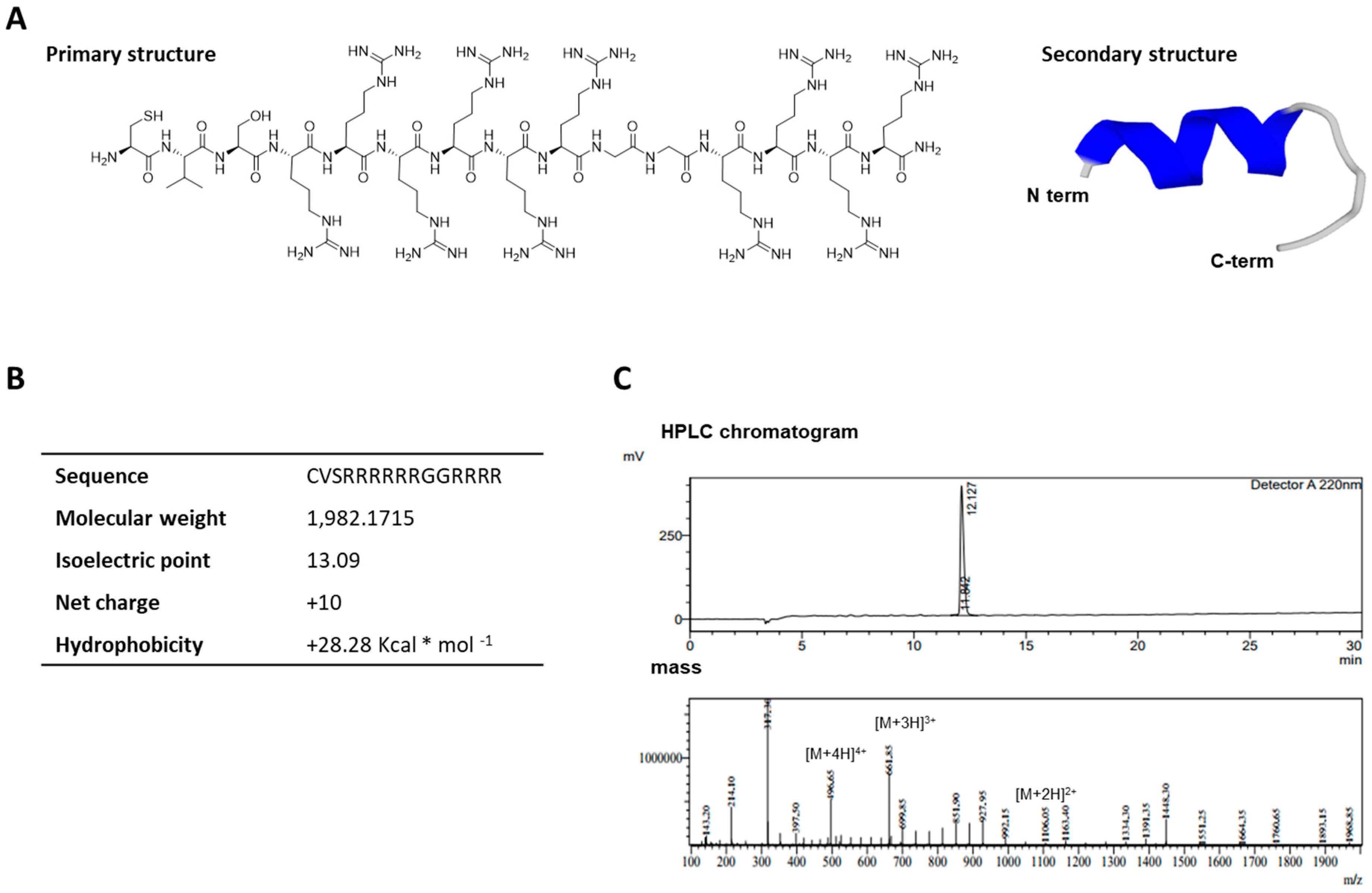
2.1. Characterization of the Cell-Permeable Peptide, LMWP

The primary and secondary structures of the synthetic LMWP derived from human protamine are shown in Figure 1. The LMWP sequence is composed of 15 amino acids (CVSRRRRRRGGRRRR) and the N-terminus of LMWP has an α-helix structure (Figure 1A). The guanidine group makes a hole in the lipid bilayer of the cell’s membrane, which closes after LMWP internalization. Therefore, ten guanidine groups of arginine residues in LMWP can effectively induce the penetration of cells or tissues. The molecular mass of the synthesized LMWP was 1982.1715 (Figure 1B) and the purity of LMWP was 99% (Figure 1C).

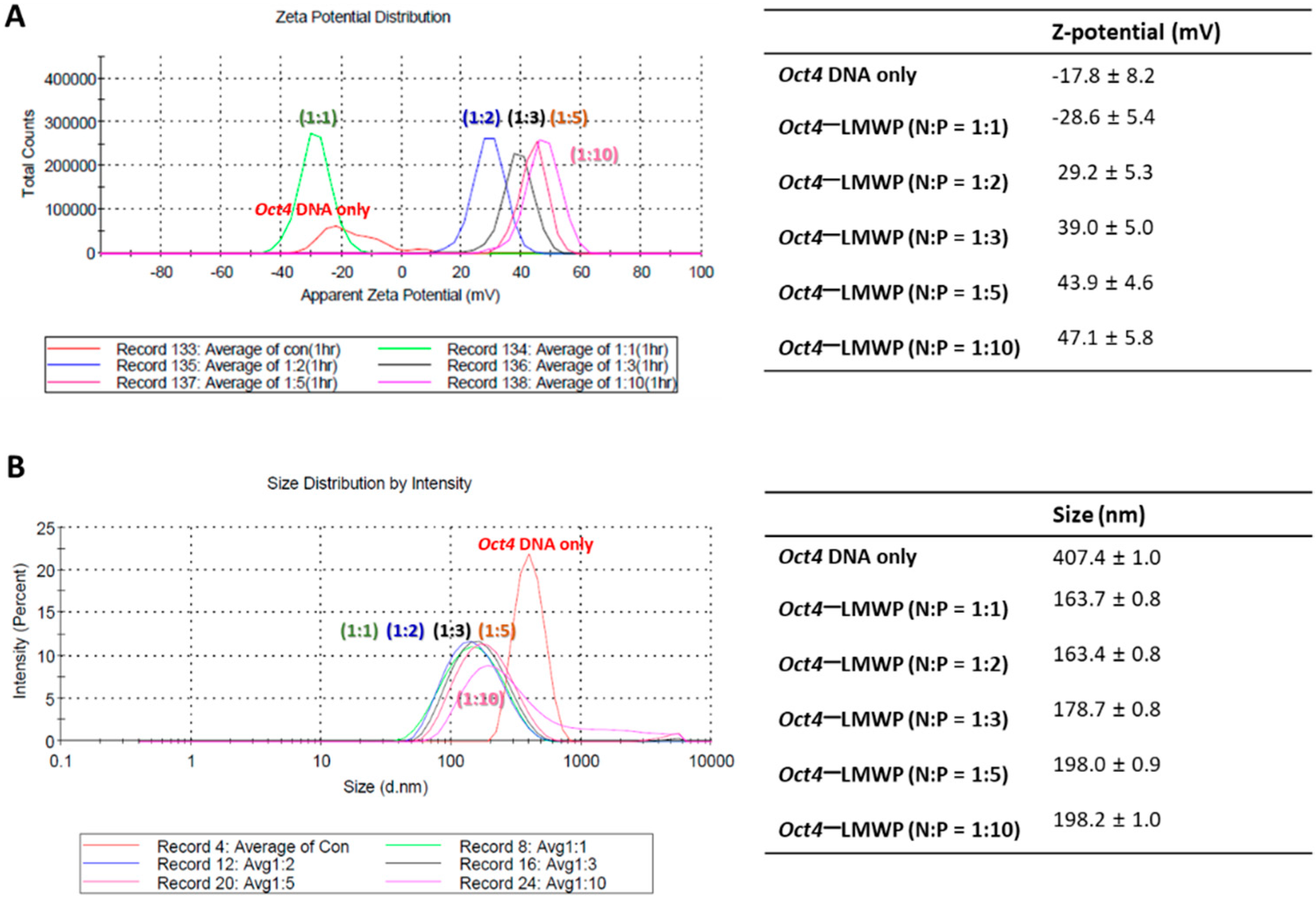
2.2. Characterization of the Oct4-LMWP Complex

The Oct4-LMWP complexes with different molar ratios with LMWP (N:P = 1:1, 1:2, 1:3, 1:5, and 1:10) were formulated and applied for measuring their surface charge and size. The distribution of the mean zeta potential and particle size diameter of Oct4-LMWP complexes were determined for each of the five tested ratios (Figure 2). The apparent zeta potential distribution of Oct4 DNA was −17.8 ± 8.2 mV (Figure 2A). The Oct4-LMWP complexes had zeta potential distributions of −28.6 ± 5.4 mV (N:P = 1:1), 29.2 ± 5.3 mV (N:P = 1:2), 39.0 ± 5.0 mV (N:P = 1:3), 43.9 ± 4.6 mV (N:P = 1:5), and 47.1 ± 5.8 mV (N:P = 1:10), respectively. The zeta potential of the Oct4 DNA increased to 47.1 ± 5.8 mV when in a complex formation with LMWP. The mean particle size of the Oct4-LMWP complex was decreased to less than 200 nm, which was significantly smaller than that of native Oct4 DNA (407.4 ± 1.0 nm; Figure 2B).

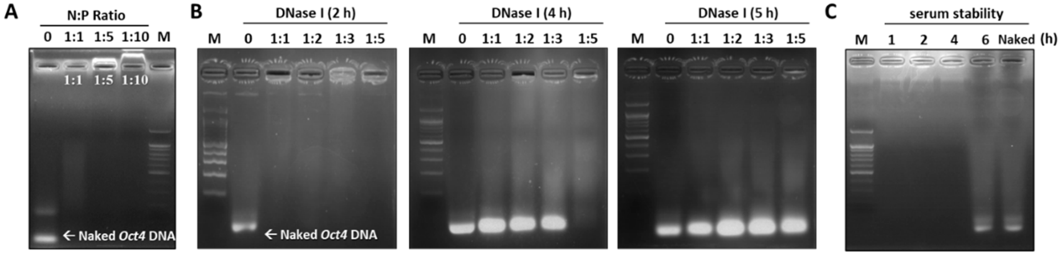
2.3. Assessing the Stability of the Oct4-LMWP Complex

The stability of the Oct4-LMWP complex against enzyme and serum treatment was assessed using an agarose gel retardation assay (Figure 3). To form Oct4-LMWP, different ratios of CPP (1:1, 1:5, and 1:10) were added to Oct4 DNA, while naked Oct4 DNA was used as a control (Figure 3A). While the Oct4-LMWP complex (N:P = 1:10) had an upward migration pattern, the naked Oct4 DNA rapidly migrated downward within 20 min. The Oct4-LMWP complexes (N:P = 1:1 and 1:5) were maintained inside the loading comb, indicating the optimum N:P molar ratio of the complex was 1:1 and 1:5 (Figure 3A). We then examined the stability of the Oct4-LMWP complex against nuclease degradation. DNase I (50 units) was added to different molar ratios of the Oct4-LMWP complex, followed by incubation of the reaction mixtures for 2, 4, or 5 h at 37 °C. Oct4-LMWP (N:P = 1:5) was stable for 4 h, indicating the complex was protected against nuclease digestion (Figure 3B). We next investigated the serum stability of the Oct4-LMWP complex (N:P = 1:5) with incubation in 50% FBS solution for 1, 2, 4, and 6 h. Naked Oct4 DNA was used as a control and rapidly migrated downward; however, no migration of the Oct4-LMWP complex was observed until 4 h (Figure 3C). According to the gel retardation, DNase I, and serum stability assays, the N:P ratio of 1:5 was the optimal ratio for Oct4-LMWP complex formation and was stable and active for at least 4 h under the DNase I and serum-containing conditions.

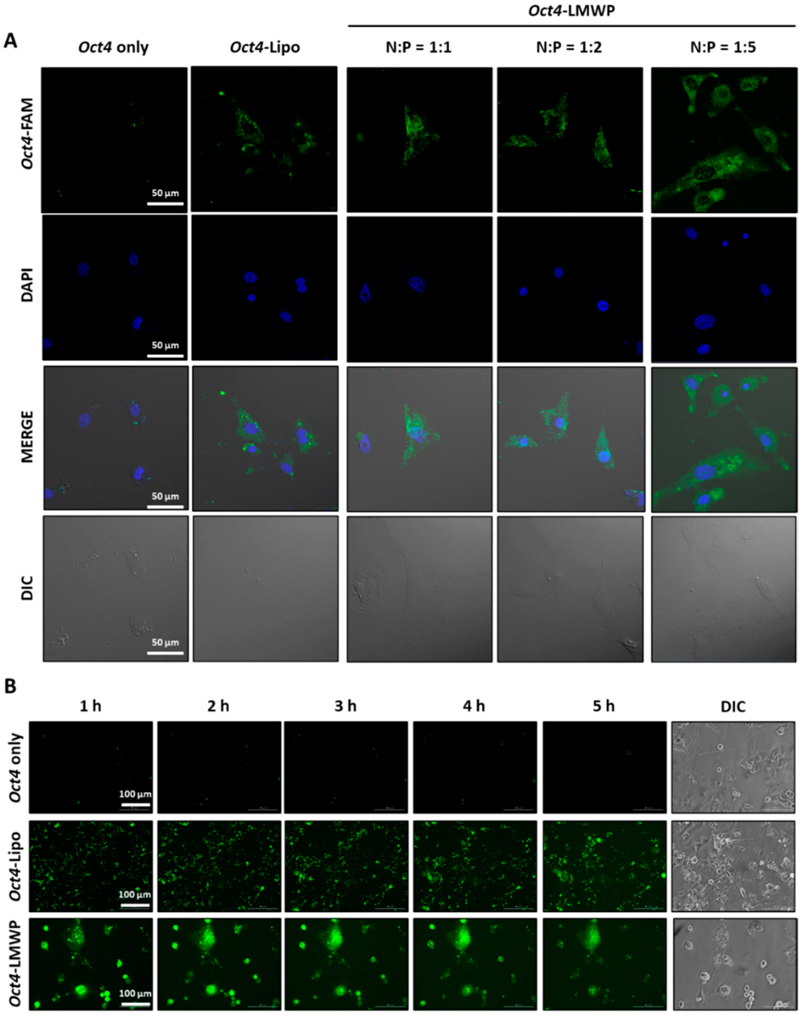
2.4. Intracellular Uptake of the Oct4-LMWP Complex

The cellular uptake and intracellular localization of the Oct4-LMWP complex were analyzed using MEF cells by confocal microscopy and real-time live imaging (Figure 4). To determine complex delivery efficacy, cellular uptake of naked Oct4 DNA, lipofectamine-transfected Oct4 DNA (Oct4-Lipo), and the Oct4-LMWP complex at varying N:P ratios were investigated by pre-labeling Oct4 with the FAM fluorophore and examining their intracellular distributions. As shown in Figure 4A,B, no intercellular fluorescence was observed in the naked Oct4 DNA samples. In cells transfected with Oct4 DNA (Oct4-Lipo), fluorescence was observed in the intracellular region, while the apparent cell-permeable capacity increased in Oct4-LMWP-treated cells in an N:P molar-ratio-dependent manner. The complex with an N:P ratio of 1:5 showed the most apparent intracellular uptake capacity. Thus, the 1:5 Oct4-LMWP complex was chosen as the optimum condition for Oct4 delivery to induce the direct conversion of MEF into MSC-like cells and was used for further experiments. The delivery efficacy of this Oct4-LMWP complex was monitored for 5 h. In comparison with naked Oct4 and Oct4-Lipo, the Oct4-LMWP complex maintained its intracellular distribution for 5 h.

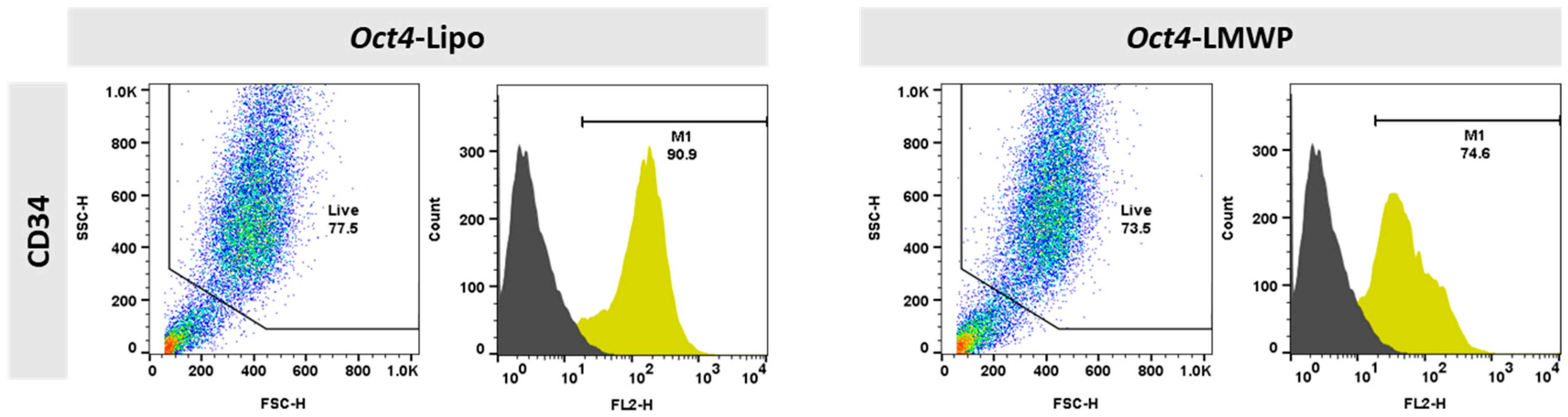
2.5. Changes in MEF Cell Surface Markers by the Oct4-LMWP Complex Delivery

The phenotypic characteristics of the Oct4-Lipo and Oct4-LMWP complexes were assessed using CD34 as a representative fibroblast surface marker (Figure 5). Using FACS, we found that 90.9% of Oct4-Lipo cells expressed CD34, while, in comparison, 74.6% of cells treated with Oct4-LMWP expressed CD34. These results indicated a decrease in fibroblast-specific characteristics when Oct4 was delivered with LMWP.
The CD34 expression (yellow peak) results indicated that Oct4-delivered MEF cells presented increased stemness, which was determined by increased CD34 surface marker expression and decreased fibroblastic characteristics.

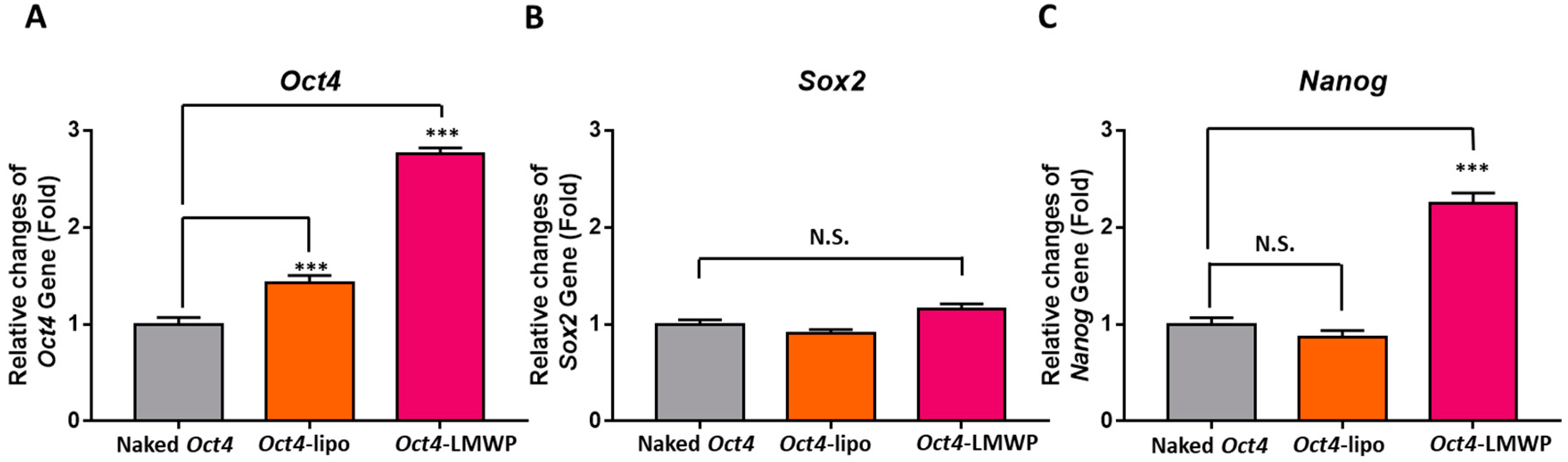
2.6. Fibroblast-Specific and Stemness Marker Gene Expression in Response to Oct4-LMWP Treatment

The changes in fibroblast-specific marker gene expression in Oct4-delivered MEF cells were examined by quantitative RT-PCR analysis of fibroblast-specific protein (FSP1), fibronectin, α-smooth muscle actin (α-SMA), and vimentin (Figure 6A–D). With Oct4-LMWP treatment, the FSP1 and fibronectin gene expression was significantly decreased 0.43 ± 0.07-fold and 0.37 ± 0.09-fold compared to naked Oct4 treated MEF cells, respectively; there was no significant change in α-SMA and vimentin expression in these cells.
The change expression of stem cell-specific marker genes, including Oct4, Sox2, and Nanog, with Oct4 delivery was further evaluated (Figure 7). Oct4-Lipo cells showed significantly increased expression that was 1.4 ± 0.1-fold compared to control MEF cells. However, there were no significant change in Sox2 and Nanog gene expression. The Oct4-LMWP-treated group presented significantly increased expression of Oct4 and Nanog genes that were 2.8 ± 0.1- and 2.3 ± 0.1-fold, respectively, compared to the control. Although there were few differences in Sox2 gene expression among the experimental groups, Oct4-LMWP cells showed an increased pattern in the Sox2 gene in naked Oct4 treated MEFs and Oct4-Lipo groups. These results suggest that Oct4-delivered MEFs possess improved MSC-specific characteristics, indicating increased expression of stem cell marker genes.

3. Discussion

The current study demonstrates the direct conversion of MEFs into MSC-like stem cells by delivering Oct4 using the cell-permeable peptide, LMWP, which can enhance the in situ regenerative potential of somatic cells. MEF cells have stem cell-like properties via Oct4-dependent cellular reprogramming factors, especially when complexed with LMWP. The development of effective gene carriers requires many essential factors, including the efficient delivery capacity of target genes into the target cells, their stability in living systems, and their biocompatibility, which reduces toxicity [34,35,36]. Therefore, this study was designed to create a complex between Oct4 and the non-cytotoxic LMWP to prevent the degradation of Oct4 DNA (Figure 1, Figure 3 and Figure 4). Here, we revealed that the positively charged cell-permeable LMWP peptide helped condense the size of Oct4 DNA complexes when in a 1:5 N:P ratio (Figure 2 and Figure 3). When the Oct4-LMWP complex was delivered into MEFs, stemness gene expression increased while fibroblast intrinsic properties decreased. These results suggest that endogenous cells can be converted through the derivation of stem cell transcription factors.
In the field of regenerative medicine, direct conversion has advantages in situ and in the generation of clinically relevant cells, including hard-to-achieve cells, from easily obtained cells such as fibroblasts [37,38]. The viral carrier transport efficiency of gene delivery can be high, depending on the type of vector used [15,27]; however, the risk of integration of viral DNA into the host genome raises potential safety issues for potential therapeutics and poses concerns like inefficient/unpredictable reprogramming outcomes, genomic integration, and unwarranted immune responses and toxicity. Therefore, several non-viral delivery methods have been reported as alternative ways to incorporate the transcription factors necessary for direct cell fate conversion or to promote stem cell-like properties [39,40]. Emerging studies have reported LMWP fragments as possible nontoxic substitutes for protamine in clinical heparin neutralization [41]. These LMWP peptides, which displayed arginine-rich content in their composition, retained a nearly complete heparin-neutralizing ability but with significantly lower toxicity in vivo [35,42].
In this study, MEF cells treated with Oct4-LMWP successfully expressed mesenchymal stem cell-like properties, including cell surface markers and gene expression. As shown in Figure 7, the increased Oct4 and Nanog expression levels in the Oct4-LMWP group indicated an increase in stem cell-like properties. The correlation between Oct4 and Nanog is known to be proportionally related to the probability of cell differentiation and pluripotency [43], which suggests that Oct4 DNA might affect Nanog expression levels, playing a role in maintaining pluripotency and in the reprogramming relationship between Oct4 and Nanog [13].
Our analysis of fibroblast-specific gene expression in cells treated with the Oct4-LMWP complex showed differential responses to Oct4 induction. According to the relationship between α-SMA and vimentin with stemness, α-SMA expression in somatic fibroblasts is correlated with stemness and proliferation markers [44]. In addition, previous studies have reported that vimentin, which is a biomarker of epithelial to mesenchymal transition, is required for cancer stem cell metastasis [45]. Mechanistic studies are still needed, but our results indicate that only the Oct4-LMWP complex changed fibroblast-specific characteristics after intracellular uptake of Oct4 in MEFs. In addition, the Oct4-LMWP complex significantly increased the expression of stemness-specific markers without cell death.
Except for the viral carrier method, general protein-based direct conversion studies still have limitations. For example, the expression of mammalian-cell-derived proteins in E. coli often resulting in inclusion bodies [46]. In the process of denaturation and refolding in inclusion bodies, the recovery yield is reduced [47]. In addition, the inclusion bodies may re-aggregate due to misfolding during the refolding process or protein activity may be lost. Our results suggest that normal somatic cells can be converted into multipotent cells simply by using LMWP peptide as a target gene carrier. Moreover, we showed the effective intracellular uptake delivery of Oct4 DNA by increasing the surface charge of the target gene through the complex formation with LMWP (Figure 4).
The stable delivery of recombinant reprogramming factors is still considered a major challenge for reprogramming strategies [48]. Oct4 is a master regulator that needs to be maintained at high levels for effective reprogramming to induce stem cell-like properties, as well as for the direct conversion of cells into other lineages. In conclusion, the Oct4-LMWP complex is a relatively safe and efficient method of cellular reprogramming because it is intracellularly delivered and maintains transcriptional regulatory activity. Nevertheless, the human fibroblasts were not directly assessed in this study, and therefore, a further study on human fibroblasts with Oct4-LMWP would be necessary to validate the direct conversion for clinical therapy. The Oct4-LMWP complex could contribute to the development of feasible somatic cell reprogramming technology and thereby, contribute to enhanced regenerative efficacy.

4. Materials and Methods

4.1. Synthesis of Cell Permeable Peptides

Cell-permeable peptides, LMWP, were manufactured using a peptide synthesizer (Prelude, Protein Technologies Inc., Tucson, AZ, USA) based on standard 9-fluorenylmethoxycarbonyl (Fmoc) chemistry. Rink amide methylbenzhydrylamine (MBHA) resin (GL Biochem, Shanghai, China) was pre-swollen in dimethylformamide (DMF, 50 mg/mL) and the Fmoc-protecting groups of resin were removed with 20% piperidine in DMF. Amino acid coupling occurred in the presence of 10 equiv. DIPEA, 5 eq. HBTU, and 5 eq. Fmoc-protected amino acids according to the resin loading of amino acids. Cleavage and sidechain deprotection of the peptide resin was conducted for 4 h using a cleavage cocktail (trifluoroacetic acid: phenol: water: thioanisole: 1,2-ethanedithiol = 82.5:5:5:5:2.5). Solutions containing the cleaved peptides were precipitated by adding cold ether. Peptides were purified using preparative reverse-phase high-performance liquid chromatography (RP-HPLC; Shimadzu, Kyoto, Japan) with a Kromasil 100-10-C18 column (Nouryon, Bhous, Sweden) at 220 nm and a 40 min gradient from 95% to 5% water/acetonitrile containing 0.1% trifluoroacetic acid (TFA). Peptide purity was determined to be >98% by HPLC (Shimadzu, Kyoto, Japan) and liquid chromatography–mass spectrometry (LC-MS, Shimadzu, Kyoto, Japan).

4.2. Isolation and Culture of MEFs

Female pregnant C57BL/6J mice were supplied by Daehan Bio Link Inc. (Eumseong, Korea). All animal experiments were approved by the Chungbuk National University Institutional Animal Care and Use Committee (IACUC ID: CBNUA-1418-20-01). MEF cells were isolated from mouse embryos according to the guidelines. Briefly, embryos were acquired from pregnant mice at 13.5 d and washed three times with Dulbecco’s phosphate-buffered saline (DPBS; Welgene Inc., Gyeongsan, Korea). The embryos were separated from the uterine horn and the placenta. The isolated embryos were mechanically digested and then the tissues were chemically digested with DNase I (Sigma-Aldrich, St. Louis, MO, USA). The digested tissues were incubated with 0.25% trypsin-EDTA (Gibco, Grand Island, NY, USA) at 37 °C for 10 min.
The mononuclear cells were seeded in high-glucose Dulbecco’s modified Eagle’s medium (DMEM-HG; Welgene Inc.) supplemented with 10% fetal bovine serum (FBS: certified, US origin, Gibco), 1% antibiotics/antimycotics (A/A), and penicillin-streptomycin (P/S; Gibco) and cultured at 37 °C in a humidified 5% CO2 incubator. Cells were allowed to adhere to culture plates for 12 h and adherent mononuclear cells were used as MEFs. When the MEF cultures reached 80–90% confluence, they were treated with 0.25% trypsin-EDTA (Gibco) for 3 min. The detached cells were washed twice with PBS and collected by centrifugation at 3000 rpm for 5 min (Eppendorf, Hamburg, Germany).

4.3. Preparation of Oct4-LMWP Complexes

The Oct4-LMWP complexes were formulated by adding varying ratios of LMWP (1:1, 1:2, 1:3, 1:5, 1:10 N:P ratio) to Oct4 DNA (10 μg/mL). The complex was agitated at 37 °C for 1, 2, 3, 4, or 5 h. The optimal N:P ratio of LMWP for stable and effective complex formation with Oct4 DNA was determined by gel retardation, serum stability, and DNase I stability assays. The Oct4 DMA sequences of thiol oligonucleotide and 6-fluorescein amidite (6-FAM)-labeling were synthesized by Bioneer Technologies, Inc. (Daejeon, Korea) as follows: 5′-(Thiol)ATG GCA TAC TGT GGA CCT CAG GTT G(6-FAM)-3′. For the control Oct4-Lipo complex, transfection of Oct4 DNA was carried out using LipofectamineTM 2000 (Invitrogen, Schwerte, Germany) according to the protocol of the manufacturer’s instructions. The day before transfection, 2.0 × 10⁵ MEFs were cultured in Opti-MEM® (Invitrogen) and transfected with Oct4 DNA.

4.4. Measurement of Oct4-LMWP Complex Particle Size and Zeta Potential

The particle size and surface charge of each Oct4-LMWP complex sample formed at different N:P ratios and Oct4-Lipo were determined at 25 °C with a scattering angle of 90° using a Zetasizer (Malvern, UK). The Oct4-LMWP complexes were prepared in deionized water to ensure that their evaluation was conducted under low-ionic-strength conditions so that the surface charge of the particles could be accurately determined.

4.5. DNase I and Serum Stability of the Oct4-LMWP Complex

DNase I (50 units) was added to a solution of Oct4 DNA alone and in the LMWP complex and mixed solutions were incubated for 1, 2, 4, or 6 h at 37 °C. During incubation, 50 μL of each suspension was collected at the different time points, mixed with 75 μL quenching solution (4 M ammonium acetate, 20 mM EDTA, and 2 mg/mL glycogen), and placed on ice. After incubation, each complex was subjected to 1% agarose gel electrophoresis. The stability of the Oct4-LMWP complex was investigated by incubating 100 μL of the complex in 100 μL 50% FBS solution for 1, 2, 4, or 6 h at room temperature. The incubated complex solutions were subjected to 1% agarose gel electrophoresis. Naked Oct4 DNA was used as a control.

4.6. Cellular Uptake Assay

To confirm the cell membrane permeability of the Oct4-LMWP complex, MEF cells were seeded (1.5 × 10⁵) onto a 6-well plate and incubated at 37 °C under 5% CO2. The FAM-Oct4-LMWP complex was added to the medium at each N:P ratio. Each group was fixed with 4% paraformaldehyde (Biosesang, Seongnam, Korea) and washed three times with PBS. Nuclei were counterstained with DAPI for 3 min. The cells were observed and imaged with a confocal microscope (LSM-880 with Airyscan, Oberkochen, ZEISS). For real-time live imaging of cellular uptake, cells were treated with DNA, lipofectamine, or Oct4-LMWP complex before live-cell imaging using laser scanning microscopy (Lionheart FX, Biotek, Korea). The cells were observed at 1 h intervals, maintained at 37 °C under 5% CO2 for 6 h.

4.7. Fluorescence-Activated Cell Sorting (FACS) Analysis

The MEF cells (1.0 × 10⁴ cells) from the Oct4-Lipo and Oct4-LMWP (N:P = 1:5) groups were incubated with phycoerythrin (PE)-conjugated monoclonal antibodies against Isotype-PE and PE-conjugated CD34 (BD Biosciences, San Jose, CA, USA) for 30 min at 4 °C. Cell populations were analyzed using a FACScan instrument (FACSCalibur-S). Untreated MEF cells and isotype-PE Ig controls for each wavelength were used as controls. Data were analyzed using the FlowJo software (BD Biosciences).

4.8. Quantitative RT-PCR

To obtain RNA, MEF cells were seeded onto a 6-well plate and incubated at 37 °C under 5% CO2 until 70–80% confluence. Cells were incubated for 2 h in DMEM containing 0.5% serum for starvation. Total RNA from MEF cells was extracted using TRIzol reagent according to the manufacturer’s instructions (Life Technologies, Darmstadt, Germany). Isolated RNA was treated with DNase I (Thermo Scientific, Schwerte, Germany) to remove possible genomic DNA contamination and used for cDNA synthesis with Superscript III Transcriptase (Life Technologies) and oligo dT (Thermo Scientific) at 42 °C for 1 h. Each sample was run in triplicate. Quantitative RT-PCR was conducted using Luna Universal qPCR Master Mix (NEB, Massachusetts, USA) and 0.1 μM primers on the Magnetic Induction Cycler Real-Time PCR System (BMS, Coomera, Australia) under the following cycle conditions: primary denaturation at 95 °C for 5 min, 42 cycles of 30 s at 95 °C, 40 s at 60 °C, and 30 s at 72 °C, followed by fluorescence measurement. The primer sequences used were as follows: (1) FSP1: forward, 5′-GAT GAG CAA CTT GGA CAG CA-3′; reverse, 5′- ATG TGC GAA GAA GCC AGA GT-3′. (2) Fibronectin: forward, 5′-CCG TGG GAT GTT TGA GAC-3′; reverse, 5′- GGC AAA AGA AAG CAG AGG-3′. (3) α-SMA: forward, 5′-CTG ACA GAG GCA CCA CTG AA-3′; reverse, 5′-CAT CTC CAG AGT CCA GCA CA-3′. (4) Vimentin: forward, 5′-TCC AGA TCG ATG TGG ACG TT-3′; reverse, 5′-ATA CTG CTG GCG CAC ATC AC-3′. (5) Oct4: forward, 5′-CAG ACC ACC ATC TGT CGC TTC-3′; reverse, 5′-AGA CTC CAC CTC ACA CGG TTC TC-3′. (6) Sox2: forward, 5′-TGG CGA ACC ATC TCT GTG GT-3′; reverse, 5′-GGA AAG TTG GGA TCG AAC AAA AGC-3′. (7) Nanog: forward, 5′-GTC CCA AAG GCA AAC AAC CC-3′; reverse, 5′-GCT GGG TGG AAG AGA ACA CA-3′. (8) β-actin: forward, 5′-GAC AAC GGC TCC GGC ATG TG-3′; reverse, 5′-TGG CTG GGG TGT TGA AGG TC-3′.

4.9. Statistical Analysis

All data are presented as means ± standard deviation (S.D.) and were obtained from at least three independent sets of experiments to ensure the reproducibility of the results. The statistical significances of differences among each group (control, Oct4-Lipo, and Oct4-LMWP) were determined using one-way analysis of variance (ANOVA) followed by Tukey’s honestly significant difference test using GraphPad Prism 7 software (GraphPad Software Inc., La Jolla, CA, USA). Statistical significance between groups was analyzed using the Student’s t-test. The level of significance is represented as * p < 0.05, ** p < 0.01, or *** p < 0.001.

Author Contributions

Y.S.P. and D.H.C. designed the experiments. J.-Y.L. and Y.J.P. synthesized the peptides. D.H.C., K.E.L. and J.P. performed the experiments. D.H.C. and Y.S.P. were the major contributors to the writing of the manuscript. All authors have read and agreed to the published version of the manuscript.

Funding

This research was funded by the Basic Science Research Program through the National Research Foundation of Korea (NRF) funded by the Ministry of Science, ICT, and Future Planning (Nos. 2019M3A9H1032376, 2020R1A2C1101340).

Institutional Review Board Statement

The study was conducted according to the guidelines of the Chungbuk National University Institutional Animal Care and Use Committee and approved by the Institution (IACUC ID: CBNUA-1418-20-01).

Informed Consent Statement

Not applicable.

Data Availability Statement

The data presented in this study are available on request from the corresponding author.

Conflicts of Interest

The authors declare no conflict of interest.

References

  1. Mollinari, C.; Zhao, J.; Lupacchini, L.; Garaci, E.; Merlo, D.; Pei, G. Transdifferentiation: A new promise for neurodegenerative diseases. Cell Death Dis. 2018, 9, 830. [Google Scholar] [CrossRef] [PubMed]
  2. Im, G.I.; Kim, T.K. Overcoming Current Dilemma in Cartilage Regeneration: Will Direct Conversion Provide a Breakthrough? Tissue Eng. Regen. Med. 2020, 17, 829–834. [Google Scholar] [CrossRef]
  3. Vierbuchen, T.; Wernig, M. Direct lineage conversions: Unnatural but useful? Nat. Biotechnol. 2011, 29, 892–907. [Google Scholar] [CrossRef] [PubMed] [Green Version]
  4. Horisawa, K.; Suzuki, A. Direct cell-fate conversion of somatic cells: Toward regenerative medicine and industries. Proc. Jpn. Acad. Ser. B Phys. Biol. Sci. 2020, 96, 131–158. [Google Scholar] [CrossRef] [Green Version]
  5. Grath, A.; Dai, G. Direct cell reprogramming for tissue engineering and regenerative medicine. J. Biol. Eng. 2019, 13, 14. [Google Scholar] [CrossRef] [PubMed]
  6. Chang, Y.; Li, H.; Guo, Z. Mesenchymal stem cell-like properties in fibroblasts. Cell. Physiol. Biochem. 2014, 34, 703–714. [Google Scholar] [CrossRef] [PubMed]
  7. Sriram, G.; Bigliardi, P.L.; Bigliardi-Qi, M. Fibroblast heterogeneity and its implications for engineering organotypic skin models in vitro. Eur. J. Cell Biol. 2015, 94, 483–512. [Google Scholar] [CrossRef] [Green Version]
  8. Stone, R.C.; Pastar, I.; Ojeh, N.; Chen, V.; Liu, S.; Garzon, K.I.; Tomic-Canic, M. Epithelial-mesenchymal transition in tissue repair and fibrosis. Cell Tissue Res. 2016, 365, 495–506. [Google Scholar] [CrossRef]
  9. Szabo, E.; Rampalli, S.; Risueno, R.M.; Schnerch, A.; Mitchell, R.; Fiebig-Comyn, A.; Levadoux-Martin, M.; Bhatia, M. Direct conversion of human fibroblasts to multilineage blood progenitors. Nature 2010, 468, 521–526. [Google Scholar] [CrossRef]
  10. Yamamoto, K.; Kishida, T.; Nakai, K.; Sato, Y.; Kotani, S.I.; Nishizawa, Y.; Yamamoto, T.; Kanamura, N.; Mazda, O. Direct phenotypic conversion of human fibroblasts into functional osteoblasts triggered by a blockade of the transforming growth factor-beta signal. Sci. Rep. 2018, 8, 8463. [Google Scholar] [CrossRef]
  11. Mitchell, R.R.; Szabo, E.; Benoit, Y.D.; Case, D.T.; Mechael, R.; Alamilla, J.; Lee, J.H.; Fiebig-Comyn, A.; Gillespie, D.C.; Bhatia, M. Activation of neural cell fate programs toward direct conversion of adult human fibroblasts into tri-potent neural progenitors using OCT-4. Stem Cells Dev. 2014, 23, 1937–1946. [Google Scholar] [CrossRef] [Green Version]
  12. Cha, H.H.; Park, H.H.; Park, J.H. Direct conversion of human fibroblasts into osteoblasts triggered by histone deacetylase inhibitor valproic acid. Appl. Sci. 2020, 10, 7372. [Google Scholar] [CrossRef]
  13. Takahashi, K.; Yamanaka, S. Induction of pluripotent stem cells from mouse embryonic and adult fibroblast cultures by defined factors. Cell 2006, 126, 663–676. [Google Scholar] [CrossRef] [PubMed] [Green Version]
  14. Ichim, T.E.; O’Heeron, P.; Kesari, S. Fibroblasts as a practical alternative to mesenchymal stem cells. J. Transl. Med. 2018, 16, 212. [Google Scholar] [CrossRef] [Green Version]
  15. Steens, J.; Unger, K.; Klar, L.; Neureiter, A.; Wieber, K.; Hess, J.; Jakob, H.G.; Klump, H.; Klein, D. Direct conversion of human fibroblasts into therapeutically active vascular wall-typical mesenchymal stem cells. Cell. Mol. Life Sci. 2020, 77, 3401–3422. [Google Scholar] [CrossRef] [Green Version]
  16. Kalluri, R. The biology and function of fibroblasts in cancer. Nat. Rev. Cancer 2016, 16, 582–598. [Google Scholar] [CrossRef] [PubMed]
  17. Quail, D.F.; Joyce, J.A. Microenvironmental regulation of tumor progression and metastasis. Nat. Med. 2013, 19, 1423–1437. [Google Scholar] [CrossRef] [PubMed]
  18. Alkasalias, T.; Moyano-Galceran, L.; Arsenian-Henriksson, M.; Lehti, K. Fibroblasts in the Tumor Microenvironment: Shield or Spear? Int. J. Mol. Sci. 2018, 19, 1532. [Google Scholar] [CrossRef] [Green Version]
  19. Yang, L. From fibroblast cells to cardiomyocytes: Direct lineage reprogramming. Stem Cell Res. Ther. 2011, 2, 1–2. [Google Scholar] [CrossRef] [Green Version]
  20. Shi, J.W.; Zhang, T.T.; Liu, W.; Yang, J.; Lin, X.L.; Jia, J.S.; Shen, H.F.; Wang, S.C.; Li, J.; Zhao, W.T. Direct conversion of pig fibroblasts to chondrocyte-like cells by c-Myc. Cell Death. Discov. 2019, 5, 55. [Google Scholar] [CrossRef]
  21. Nakamori, D.; Akamine, H.; Takayama, K.; Sakurai, F.; Mizuguchi, H. Direct conversion of human fibroblasts into hepatocyte-like cells by ATF5, PROX1, FOXA2, FOXA3, and HNF4A transduction. Sci. Rep. 2017, 7, 16675. [Google Scholar] [CrossRef]
  22. Pulecio, J.; Alejo-Valle, O.; Capellera-Garcia, S.; Vitaloni, M.; Rio, P.; Mejia-Ramirez, E.; Caserta, I.; Bueren, J.A.; Flygare, J.; Raya, A. Direct Conversion of Fibroblasts to Megakaryocyte Progenitors. Cell Rep. 2016, 17, 671–683. [Google Scholar] [CrossRef] [Green Version]
  23. Xiao, D.; Liu, X.; Zhang, M.; Zou, M.; Deng, Q.; Sun, D.; Bian, X.; Cai, Y.; Guo, Y.; Liu, S. Direct reprogramming of fibroblasts into neural stem cells by single non-neural progenitor transcription factor Ptf1a. Nat. Commun. 2018, 9, 2865. [Google Scholar] [CrossRef] [Green Version]
  24. Their, M.; Worsdorfer, P.; Lakes, Y.B.; Gorris, R.; Herms, S.; Opitz, T.; Seiferling, D.; Quandel, T.; Hoffmann, P.; Nothen, M.M. Direct conversion of fibroblasts into stably expandable neural stem cells. Cell Stem Cell 2012, 10, 473–479. [Google Scholar] [CrossRef] [Green Version]
  25. Chen, K.; Long, Q.; Xing, G.; Wang, T.; Wu, Y.; Li, L.; Qi, J.; Zhou, Y.; Ma, B.; Scholer, H.R. Heterochromatin loosening by the Oct4 linker region facilitates Klf4 binding and iPSC reprogramming. EMBO J. 2020, 39, e99165. [Google Scholar] [CrossRef]
  26. Lee, J.Y.; Park, J.H. Efficient production of cell-permeable oct4 protein using 30Kc19 protein originating from silkworm. Biotechnol. Bioprocess Eng. 2019, 24, 964–971. [Google Scholar] [CrossRef]
  27. Tammam, S.; Malak, P.; Correa, D.; Rothfuss, O.; Azzazy, H.M.; Lamprecht, A.; Schulze-Osthoff, K. Nuclear delivery of recombinant OCT4 by chitosan nanoparticles for transgene-free generation of protein-induced pluripotent stem cells. Oncotarget 2016, 7, 37728–37739. [Google Scholar] [CrossRef] [PubMed] [Green Version]
  28. Um, S.H. Delivering factors for reprogramming a somatic cell to pluripotency. Int. J. Stem Cells 2012, 5, 6–11. [Google Scholar] [CrossRef] [Green Version]
  29. Das, S.K.; Menezes, M.E.; Bhatia, S.; Wang, X.Y.; Emdad, L.; Sarkar, D.; Fisher, P.B. Gene Therapies for Cancer: Strategies, Challenges and Successes. J. Cell. Physiol. 2015, 230, 259–271. [Google Scholar] [CrossRef] [PubMed] [Green Version]
  30. Tipanee, J.; Chai, Y.C.; VandenDriessche, T.; Chuah, M.K. Preclinical and clinical advances in transposon-based gene therapy. Biosci. Rep. 2017, 37, BSR20160614. [Google Scholar] [CrossRef] [PubMed] [Green Version]
  31. Park, Y.J.; Liang, J.F.; Ko, K.S.; Kim, S.W.; Yang, V.C. Low molecular weight protamine as an efficient and nontoxic gene carrier: In vitro study. J. Gene Med. 2003, 5, 700–711. [Google Scholar] [CrossRef] [PubMed] [Green Version]
  32. He, H.; Ye, J.; Liu, E.; Liang, Q.; Liu, Q.; Yang, V.C. Low molecular weight protamine (LMWP): A nontoxic protamine substitute and an effective cell-penetrating peptide. J. Control. Release 2014, 193, 63–73. [Google Scholar] [CrossRef] [PubMed]
  33. Choi, Y.S.; Lee, J.Y.; Suh, J.S.; Kwon, Y.M.; Lee, S.J.; Chung, J.K.; Lee, D.S.; Yang, V.C.; Chung, C.P.; Park, Y.J. The systemic delivery of siRNAs by a cell penetrating peptide, low molecular weight protamine. Biomaterials 2010, 31, 1429–1443. [Google Scholar] [CrossRef] [PubMed]
  34. Suh, J.S.; Lee, H.J.; Nam, H.; Jo, B.S.; Lee, D.W.; Kim, J.H.; Lee, J.Y.; Chung, C.P.; Lee, G.; Park, Y.J. Control of cancer stem cell like population by intracellular target identification followed by the treatment with peptide-siRNA complex. Biochem. Biophys. Res. Commun. 2017, 491, 827–833. [Google Scholar] [CrossRef]
  35. Suh, J.S.; Lee, J.Y.; Choi, Y.J.; You, H.K.; Hong, S.D.; Chung, C.P.; Park, Y.J. Intracellular delivery of cell-penetrating peptide-transcriptional factor fusion protein and its role in selective osteogenesis. Int. J. Nanomed. 2014, 9, 1153–1166. [Google Scholar]
  36. Suh, J.S.; Lee, J.Y.; Choi, Y.S.; Chung, C.P.; Park, Y.J. Peptide-mediated intracellular delivery of miRNA-29b for osteogenic stem cell differentiation. Biomaterials 2013, 34, 4347–4359. [Google Scholar] [CrossRef]
  37. Abdal, D.A.; Lee, S.B.; Kim, K.; Lim, K.M.; Jeon, T.I.; Seok, J.; Cho, A.S. Production of Mesenchymal Stem Cells Through Stem Cell Reprogramming. Int. J. Mol. Sci. 2019, 20, 1922. [Google Scholar] [CrossRef] [Green Version]
  38. Flores-Huerta, N.; Silva-Cazares, M.B.; Arriaga-Pizano, L.A.; Prieto-Chavez, J.L.; Lopez-Camarillo, C. LncRNAs and microRNAs as Essential Regulators of Stemness in Breast Cancer Stem Cells. Biomolecules 2021, 11, 380. [Google Scholar] [CrossRef]
  39. Adler, A.F.; Grigsby, C.L.; Kulangara, K.; Wang, H.; Yasuda, R.; Leong, K.W. Nonviral direct conversion of primary mouse embryonic fibroblasts to neuronal cells. Mol. Ther. Nucleic Acids 2012, 1, e32. [Google Scholar] [CrossRef]
  40. Gantenbein, B.; Tang, S.; Guerrero, J.; Higuita-Castro, N.; Salazar-Puerta, A.I.; Croft, A.S.; Gazdhar, A.; Purmessur, D. Non-viral Gene Delivery Methods for Bone and Joints. Front. Bioeng. Biotechnol. 2020, 8, 598466. [Google Scholar] [CrossRef]
  41. Choi, Y.S.; Lee, J.Y.; Suh, J.S.; Lee, S.J.; Yang, V.C.; Chung, C.P.; Park, Y.J. Cell penetrating peptides for tumor targeting. Curr. Pharm. Biotechnol. 2011, 12, 1166–1182. [Google Scholar] [CrossRef] [PubMed]
  42. Kikuchi, J.; Hayashi, N.; Osada, N.; Sugitani, M.; Furukawa, Y. Conversion of human fibroblasts into multipotent cells by cell-penetrating peptides. Biochem. Biophys. Res. Commun. 2019, 518, 134–140. [Google Scholar] [CrossRef] [PubMed]
  43. Lim, J.; Kim, J.; Kang, J.; Jo, D. Partial somatic to stem cell transformations induced by cell-permeable reprogramming factors. Sci. Rep. 2014, 4, 4361. [Google Scholar] [CrossRef] [PubMed] [Green Version]
  44. Patel, A.K.; Vipparthi, K.; Thatikonda, V.; Arun, I.; Bhattacharjee, S.; Sharan, R.; Arun, P.; Singh, S. A subtype of cancer-associated fibroblasts with lower expression of alpha-smooth muscle actin suppresses stemness through BMP4 in oral carcinoma. Oncogenesis 2018, 7, 78. [Google Scholar] [CrossRef] [PubMed]
  45. Nomura, S. Identification, Friend or Foe: Vimentin and alpha-Smooth Muscle Actin in Cancer-Associated Fibroblasts. Ann. Surg. Oncol. 2019, 26, 4191–4192. [Google Scholar] [CrossRef] [PubMed]
  46. Thwaite, R.; Ji, J.; Torrealba, D.; Coll, J.; Sabes, M.; Villaverde, A.; Roher, N. Protein Nanoparticles Made of Recombinant Viral Antigens: A Promising Biomaterial for Oral Delivery of Fish Prophylactics. Front. Immunol. 2018, 9, 1652. [Google Scholar] [CrossRef] [PubMed]
  47. Singh, A.; Upadhyay, V.; Upadhyay, A.K.; Singh, S.M.; Panda, A.K. Protein recovery from inclusion bodies of Escherichia coli using mild solubilization process. Microb. Cell Factories 2015, 14, 41. [Google Scholar] [CrossRef] [Green Version]
  48. Gonzalez, F.; Boue, S.; Izpisua, J.C. Methods for making induced pluripotent stem cells: Reprogramming a la carte. Nat. Rev. Genet. 2011, 12, 231–242. [Google Scholar] [CrossRef]
Figure 1. Synthesis of the LMWP cell-permeable peptide. (A) The primary and secondary structure of LMWP. (B) Sequence information of the synthesized LMWP. (C) HPLC chromatogram peak and mass value of LMWP.
Ijms 22 09357 g001
Figure 2. Size and surface charge distribution of the Oct4-LMWP complex. (A) Distribution of zeta potential and (B) size distribution of the naked Oct4 DNA and Oct4-LMWP complex at different N:P molar ratios.
Ijms 22 09357 g002
Figure 3. Gel retardation and serum stability assays of the Oct4-LMWP complex. To determine the optimum N:P molar ratio of complexes, (A) Oct4 DNA and LMWP were mixed at different molar ratios and analyzed by gel retardation with 1% agarose gel electrophoresis. (B) The DNase I protection and (C) serum (50%) stability of the Oct4-LMWP complex (N:P = 1:5) were evaluated using an agarose gel retardation method at the indicated time points. M: size marker.
Ijms 22 09357 g003
Figure 4. Intracellular uptake of Oct4-LMWP. (A) The cellular uptake and intracellular localization of the Oct4-LMWP complex were monitored at different molar ratios and analyzed using a confocal microscopy. Scale bar = 50 μm. (B) Real-time detection of cellular uptake of Oct4-LMWP complexes by MEF cells. Scale bar = 100 μm.
Ijms 22 09357 g004
Figure 5. Surface marker expression in Oct4-treated MEF cells was characterized and compared using FACS analysis. Oct4 was delivered by either lipofectamine (Oct4-lipo) or in complex with LMWP (Oct4-LMWP). CD34 fibroblast-specific cell surface marker was examined in each delivery system. Negative control peak is indicated as dark gray color, CD34 are indicated as yellow peaks, respectively.
Ijms 22 09357 g005
Figure 6. Gene expression of fibroblast-specific markers in MEF cells. Changes in the expression of fibroblast-specific marker genes (A) FSP1, (B) fibronectin, (C) a-SMA, and (D) vimentin in MEF cells with Oct4 delivered by lipofectamine or Oct4-LMWP complex were confirmed by quantitative RT-PCR. The bar graph represents the relative fold changes in gene expression, which were determined by the cT value of each sample normalized to that of β-actin. Statistical significance between groups was analyzed using the Student’s t-test. The level of significance is represented as ** p < 0.001, *** p < 0.001.
Ijms 22 09357 g006
Figure 7. Gene expression of stemness markers in MEF cells. Changes in the expression of stemness marker genes (A) Oct4, (B) Sox2, and (C) Nanog in MEF cells treated with Oct4-Lipo or Oct4-LMWP complexes were confirmed by quantitative RT-PCR. The bar graph represents the relative fold changes in gene expression, which were determined by the cT value of each sample normalized to that of β-actin. Statistical significance between groups was analyzed using the Student’s t-test. The level of significance is represented as *** p < 0.001.
Ijms 22 09357 g007
Publisher’s Note: MDPI stays neutral with regard to jurisdictional claims in published maps and institutional affiliations.

