
Direct Synthesis of Polyaromatic Cyclophanes Containing Bis-Methylene-Interrupted Z-Double Bonds and Study of Their Antitumor Activity In Vitro

 ,
,  ,
,
Abstract
:
1. Introduction
2. Results
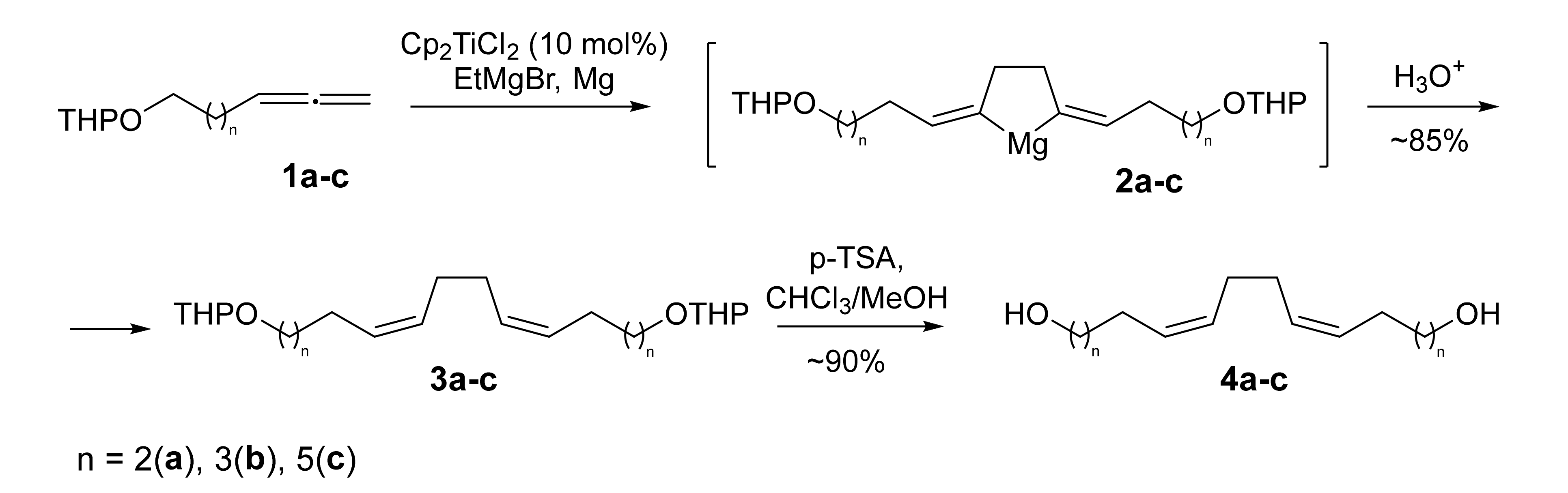
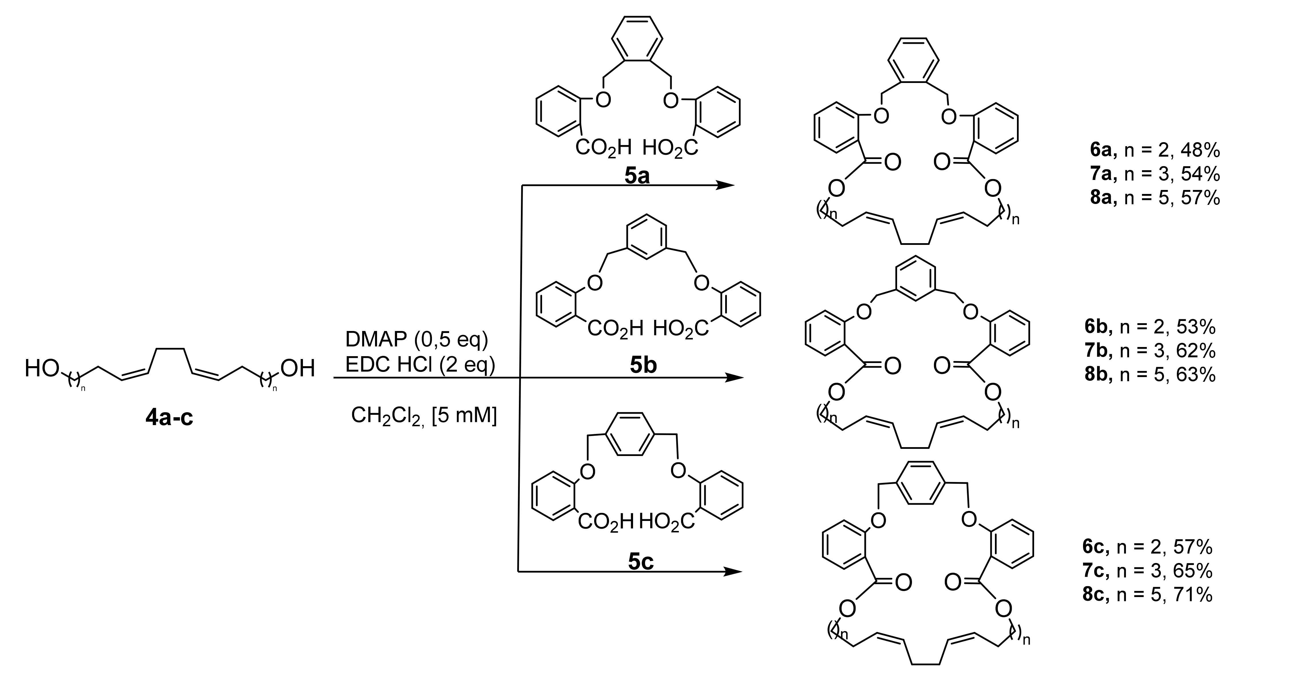
2.1. Chemistry
2.2. Biological Evaluation
2.2.1. The In Vitro Antitumor Activity of Cyclophanes (6–8)
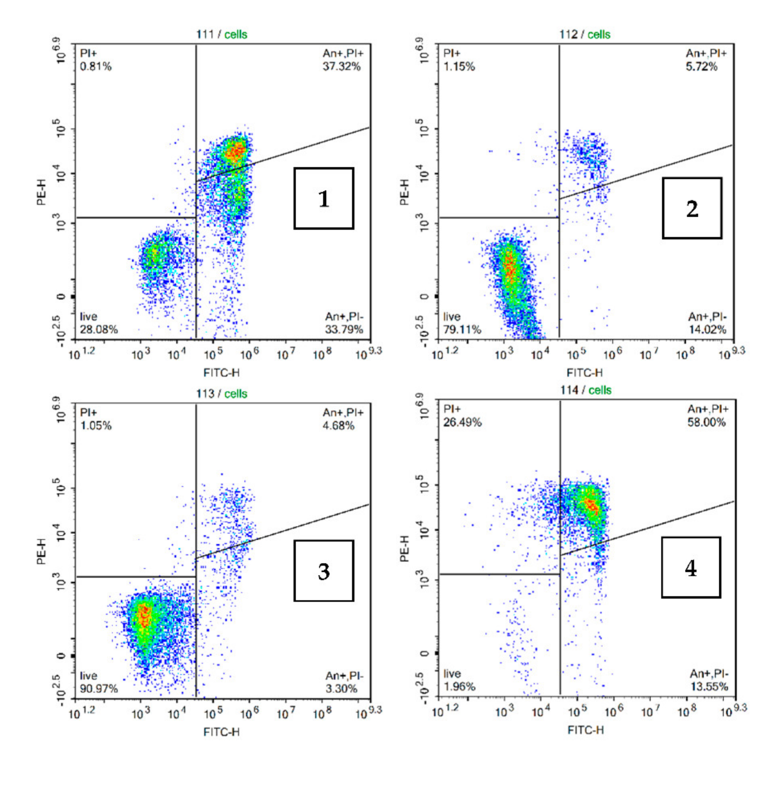
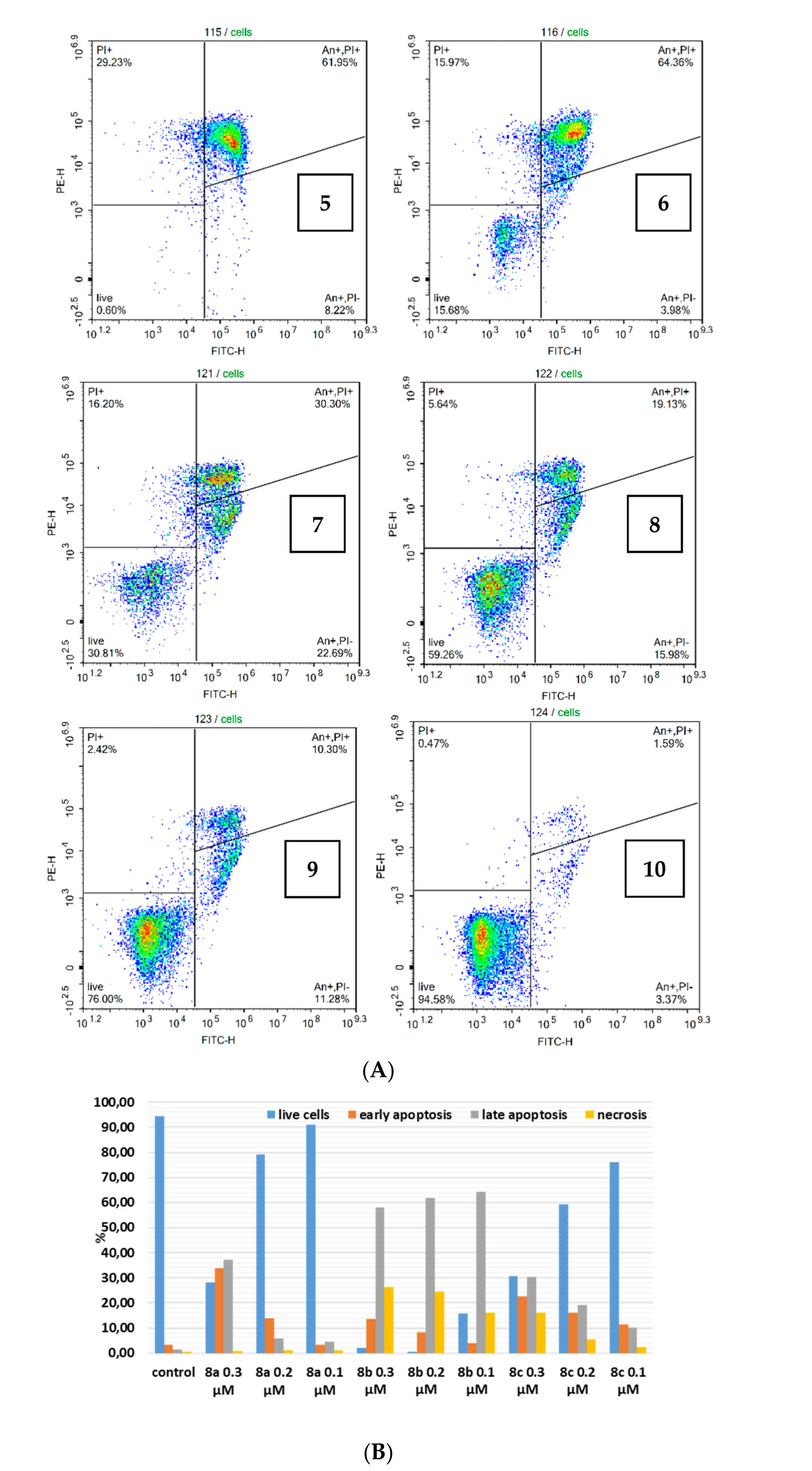
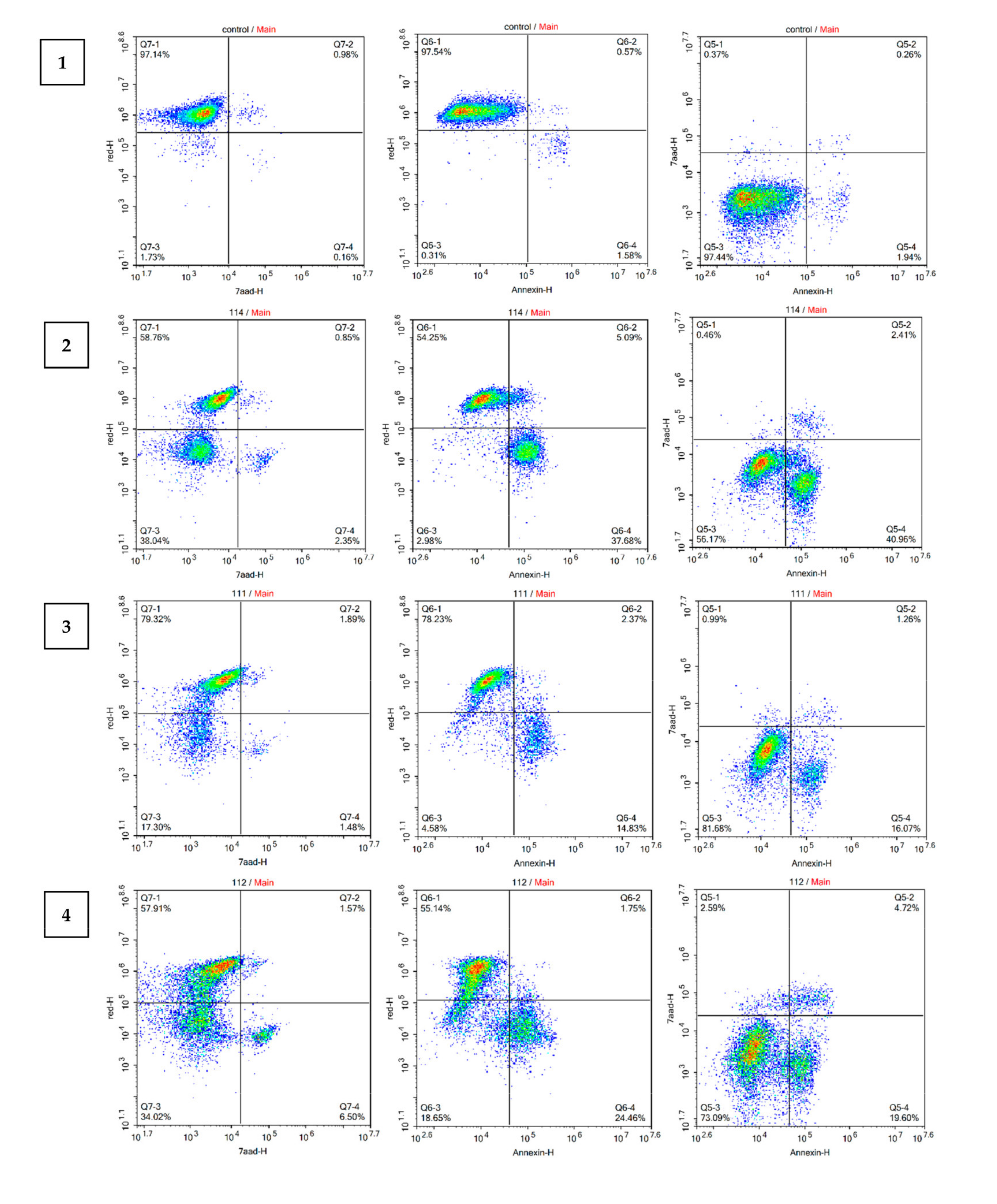
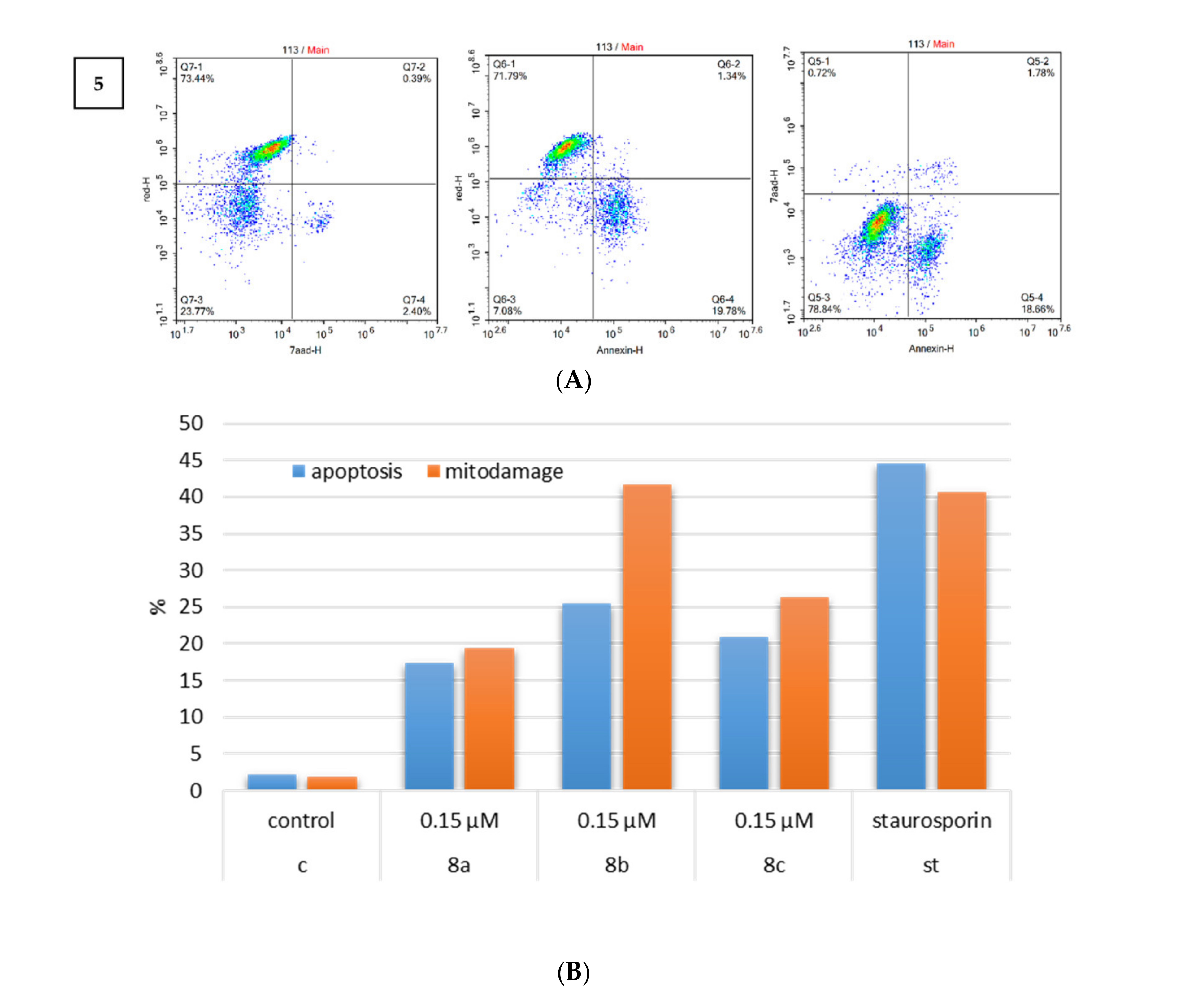
2.2.2. Induction of Apoptosis
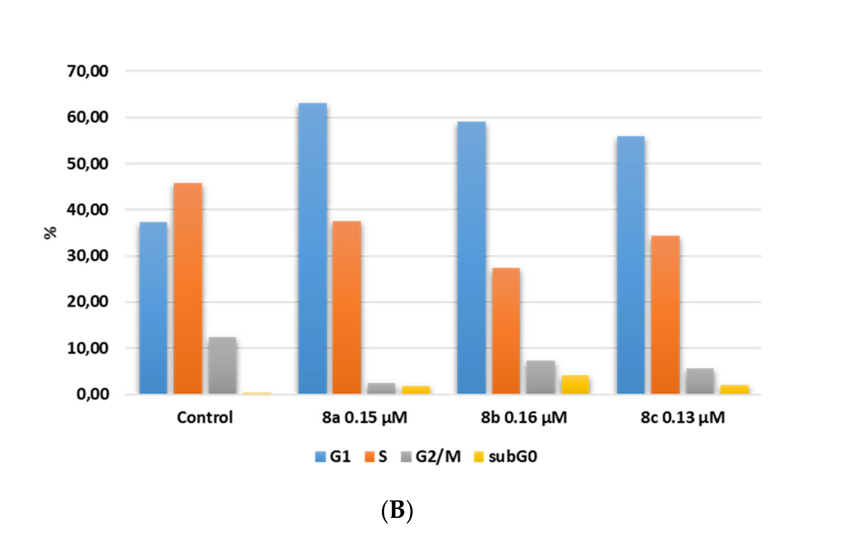
2.2.3. Cell Cycle Analysis
3. Materials and Methods
3.1. Chemistry
3.2. Chemical Experimental Data
General Procedure Synthesis of Cyclophanes
3.3. Biology Studies
3.3.1. Cell Culturing
3.3.2. Cytotoxicity Assay
3.3.3. Viability and Apoptosis
3.3.4. Cell Cycle Analysis
3.3.5. Mitochondrial Damage
4. Conclusions
Supplementary Materials
Author Contributions
Funding
Institutional Review Board Statement
Informed Consent Statement
Data Availability Statement
Acknowledgments
Conflicts of Interest
References
- Diederich, F. Cyclophanes; The Royal Society of Chemistry: Cambridge, UK, 1991; 313p. [Google Scholar] [CrossRef]
- Vögtle, F. Cyclophane Chemistry: Synthesis, Structures and Reactions; John Wiley & Sons Ltd.: West Sussex, UK, 1993; 501p. [Google Scholar]
- Ploutno, A.; Carmeli, S. Nostocyclyne A, a Novel Antimicrobial Cyclophane from the Cyanobacterium Nostoc sp. J. Nat. Prod. 2000, 63, 1524–1526. [Google Scholar] [CrossRef]
- Fürstner, A.; Stelzer, F.; Rumbo, A.; Krause, H. Total Synthesis of the Turrianes and Evaluation of Their DNA-Cleaving Properties. Chem. A Eur. J. 2002, 8, 1856–1871. [Google Scholar] [CrossRef]
- Jolly, C.; Thoison, O.; Martin, M.-T.; Dumontet, V.; Gilbert, A.; Pfeiffer, B.; Léonce, S.; Sévenet, T.; Guéritte, F.; Litaudon, M. Cytotoxic turrianes of Kermadecia elliptica from the New Caledonian rainforest. Phytochemistry 2008, 69, 533–540. [Google Scholar] [CrossRef]
- Beniddir, M.A.; Simonin, A.-L.; Martin, M.-T.; Dumontet, V.; Poullain, C.; Gueritte, F.; Litaudon, M. Turrianes from Kermadecia rotundifolia as new acetylcholinesterase inhibitors. Phytochem. Lett. 2010, 3, 75–78. [Google Scholar] [CrossRef]
- Garrido, L.; Zubia, E.; Ortega, M.J.; Salva, J. Haouamines A and B: A New Class of Alkaloids from the Ascidian Aplidium haouarianum. J. Org. Chem. 2003, 68, 293–299. [Google Scholar] [CrossRef] [PubMed]
- Baran, P.S.; Burns, N.Z. Total Synthesis of (±)-Haouamine A. J. Am. Chem. Soc. 2006, 128, 3908–3909. [Google Scholar] [CrossRef] [PubMed]
- Shen, L.; Maddox, M.M.; Adhikari, S.; Bruhn, D.F.; Kumar, M.; Lee, R.E.; Hurdle, J.G.; Lee, R.E.; Sun, D. Syntheses and evaluation of macrocyclic engelhardione analogs as antitubercular and antibacterial agents. J. Antibiot. 2013, 66, 319–325. [Google Scholar] [CrossRef] [Green Version]
- Ettmayer, P.; Billich, A.; Hecht, P.; Rosenwirth, A.B.; Gstach, H. Paracyclophanes: A Novel Class of Water-Soluble Inhibitors of HIV Proteinase. J. Med. Chem. 1996, 39, 3291–3299. [Google Scholar] [CrossRef] [PubMed]
- Asakawa, Y. Progress in the Chemistry of Organic Natural Products: 65; Springer: New York, NY, USA, 1995; 473p. [Google Scholar]
- Niu, C.; Qu, J.-B.; Lou, H.-X. Antifungal Bis[bibenzyls] from the Chinese Liverwort Marchantia polymorpha L. Chem. Biodivers. 2006, 3, 34–40. [Google Scholar] [CrossRef]
- Kostiuk, S.L.; Woodcock, T.; Dudin, L.F.; Howes, P.D.; Harrowven, D. Unified Syntheses of Cavicularin and Riccardin C: Addressing the Synthesis of an Arene Adopting a Boat Configuration. Chem. A Eur. J. 2011, 17, 10906–10915. [Google Scholar] [CrossRef]
- Moore, B.S.; Chen, J.-L.; Patterson, G.M.; Moore, R.E. Structures of cylindrocyphanes a-f. Tetrahedron 1992, 48, 3001–3006. [Google Scholar] [CrossRef]
- Smith, A.B.; Adams, C.M.; Kozmin, S.A.; Paone, D.V. Total synthesis of (-)-cylindrocyclophanes A and F exploiting the reversible nature of the olefin cross metathesis reaction. J. Am. Chem. Soc. 2001, 123, 5925–5937. [Google Scholar] [CrossRef] [PubMed]
- D’Yakonov, V.A.; Islamov, I.I.; Khusainova, E.M.; Dzhemilev, U. Original catalytic synthesis of macrodiolides containing a 1Z,5Z-diene moiety. Mendeleev Commun. 2018, 28, 503–504. [Google Scholar] [CrossRef]
- D’Yakonov, V.A.; Islamov, I.I.; Dzhemileva, L.U.; Khusainova, E.M.; Yunusbaeva, M.; Dzhemilev, U.M. Targeted synthesis of macrodiolides containing bis-methylene-separated Z-double bonds and their antitumor activity in vitro. Tetrahedron 2018, 74, 4606–4612. [Google Scholar] [CrossRef]
- Dzhemileva, L.; D’Yakonov, V.A.; Islamov, I.I.; Yunusbaeva, M.M.; Dzhemilev, U.M. New 1Z,5Z-diene macrodiolides: Catalytic synthesis, anticancer activity, induction of mitochondrial apoptosis, and effect on the cell cycle. Bioorganic Chem. 2020, 99, 103832. [Google Scholar] [CrossRef]
- Carballeira, N. New advances in fatty acids as antimalarial, antimycobacterial and antifungal agents. Prog. Lipid Res. 2008, 47, 50–61. [Google Scholar] [CrossRef] [PubMed] [Green Version]
- Carballeira, N.M.; Betancourt, J.E.; Orellano, E.A.; González, F.A. Total Synthesis and Biological Evaluation of (5Z,9Z)-5,9-Hexadecadienoic Acid, an Inhibitor of Human Topoisomerase I. J. Nat. Prod. 2002, 65, 1715–1718. [Google Scholar] [CrossRef]
- Carballeira, N.M.; Montano, N.; Amador, L.A.; Rodríguez, A.D.; Golovko, M.; Golovko, S.A.; Reguera, R.M.; Álvarez-Velilla, R.; Balana-Fouce, R. Novel Very Long-Chain α-Methoxylated Δ5,9 Fatty Acids from the SpongeAsteropus nigerAre Effective Inhibitors of Topoisomerases IB. Lipids 2016, 51, 245–256. [Google Scholar] [CrossRef] [Green Version]
- Nemoto, T.; Yoshino, G.; Ojika, M.; Sakagami, Y. Amphimic acids and related long-chain fatty acids as DNA topoisomerase I inhibitors from an Australian sponge, Amphimedon sp.: Isolation, structure, synthesis, and biological evaluation. Tetrahedron 1997, 53, 16699–16710. [Google Scholar] [CrossRef]
- Chen, S.-J.; Hsu, C.-P.; Li, C.-W.; Lu, J.-H.; Chuang, L.-T. Pinolenic acid inhibits human breast cancer MDA-MB-231 cell metastasis in vitro. Food Chem. 2011, 126, 1708–1715. [Google Scholar] [CrossRef] [PubMed]
- Xie, K.; Miles, E.; Calder, P.C. A review of the potential health benefits of pine nut oil and its characteristic fatty acid pinolenic acid. J. Funct. Foods 2016, 23, 464–473. [Google Scholar] [CrossRef] [Green Version]
- D’Yakonov, V.A.; Makarov, A.A.; Dzhemileva, L.U.; Makarova, E.K.; Khusnutdinova, E.K.; Dzhemilev, U. The facile synthesis of the 5Z,9Z-dienoic acids and their topoisomerase I inhibitory activity. Chem. Commun. 2013, 49, 8401–8403. [Google Scholar] [CrossRef] [Green Version]
- D’Yakonov, V.A.; Dzhemileva, L.; Makarov, A.A.; Mulukova, A.R.; Baev, D.; Khusnutdinova, E.; Tolstikova, T.G.; Dzhemilev, U.M. Stereoselective synthesis of 11-phenylundeca-5Z,9Z-dienoic acid and investigation of its human topoisomerase I and IIα inhibitory activity. Bioorganic Med. Chem. Lett. 2015, 25, 2405–2408. [Google Scholar] [CrossRef]
- D’Yakonov, V.A.; Dzhemileva, L.; Makarov, A.A.; Mulyukova, A.R.; Baev, D.; Khusnutdinova, E.; Tolstikova, T.G.; Dzhemilev, U. nZ,(n + 4)Z-Dienoic fatty acids: A new method for the synthesis and inhibitory action on topoisomerase I and IIα. Med. Chem. Res. 2016, 25, 30–39. [Google Scholar] [CrossRef]
- D’Yakonov, V.A.; Dzhemileva, L.; Tuktarova, R.A.; Makarov, A.A.; Islamov, I.I.; Mulyukova, A.R.; Dzhemilev, U.M. Catalytic cyclometallation in steroid chemistry III: Synthesis of steroidal derivatives of 5Z,9Z-dienoic acid and investigation of its human topoisomerase I inhibitory activity. Steroids 2015, 102, 110–117. [Google Scholar] [CrossRef]
- Naveen, M.; Babu, S.A. Ring-closing metathesis reaction-based synthesis of new classes of polyether macrocyclic systems. Tetrahedron 2015, 71, 7758–7781. [Google Scholar] [CrossRef]
- Ishihara, K.; Ohara, S.; Yamamoto, H. Direct Condensation of Carboxylic Acids with Alcohols Catalyzed by Hafnium(IV) Salts. Science 2000, 290, 1140–1142. [Google Scholar] [CrossRef] [PubMed]
- De Léséleuc, M.; Collins, S.K. Direct synthesis of macrodiolides via hafnium(iv) catalysis. Chem. Commun. 2015, 51, 10471–10474. [Google Scholar] [CrossRef] [PubMed]
- Naveen, M.; Babu, S.A. EDC/DMAP-mediated direct condensation of dicarboxylic acids and diols: A concise synthesis of extra large polyether macrocyclic lactones and their X-ray structures. Tetrahedron Lett. 2016, 57, 5801–5807. [Google Scholar] [CrossRef]


| Comp. | Jurkat (IC50, µM) * | K562 (IC50, µM) * | U937 (IC50, µM) * | HL-60 (IC50, µM) * | Hek293 (IC50, µM) * | Fibrobl. (IC50, µM) * |
|---|---|---|---|---|---|---|
| 6a | 0.39 ± 0.03 | 0.61 ± 0.05 | 0.49 ± 0.04 | 0.43 ± 0.03 | 1.94 ± 0.17 | 4.71 ± 0.42 |
| 6b | 0.48 ± 0.05 | 0.64 ± 0.06 | 0.51 ± 0.05 | 0.44 ± 0.04 | 2.18 ± 0.21 | 5.09 ± 0.47 |
| 6c | 0.34 ± 0.03 | 0.58 ± 0.05 | 0.46 ± 0.04 | 0.40 ± 0.03 | 1.76 ± 0.16 | 4.22 ± 0.39 |
| 7a | 0.49 ± 0.04 | 0.76 ± 0.08 | 0.56 ± 0.06 | 0.94 ± 0.08 | 3.86 ± 0.34 | 7.12 ± 0.59 |
| 7b | 0.94 ± 0.08 | 1.49 ± 0.11 | 1.17 ± 0.09 | 0.97 ± 0.09 | 4.21 ± 0.39 | 7.69 ± 0.72 |
| 7c | 0.35 ± 0.03 | 0.59 ± 0.04 | 0.39 ± 0.03 | 0.89 ± 0.09 | 3.47 ± 0.31 | 6.55 ± 0.61 |
| 8a | 0.15 ± 0.02 | 0.37 ± 0.02 | 0.30 ± 0.02 | 0.22 ± 0.02 | 1.44 ± 0.11 | 3.37 ± 0.29 |
| 8b | 0.16 ± 0.01 | 0.39 ± 0.04 | 0.31 ± 0.03 | 0.24 ± 0.02 | 1.68 ± 0.14 | 3.48 ± 0.32 |
| 8c | 0.13 ± 0.01 | 0.34 ± 0.03 | 0.27 ± 0.02 | 0.19 ± 0.01 | 1.21 ± 0.09 | 3.19 ± 0.28 |
Publisher’s Note: MDPI stays neutral with regard to jurisdictional claims in published maps and institutional affiliations. |
© 2021 by the authors. Licensee MDPI, Basel, Switzerland. This article is an open access article distributed under the terms and conditions of the Creative Commons Attribution (CC BY) license (https://creativecommons.org/licenses/by/4.0/).
Share and Cite
D’yakonov, V.A.; Islamov, I.I.; Dzhemileva, L.U.; Makarova, E.K.; Dzhemilev, U.M. Direct Synthesis of Polyaromatic Cyclophanes Containing Bis-Methylene-Interrupted Z-Double Bonds and Study of Their Antitumor Activity In Vitro. Int. J. Mol. Sci. 2021, 22, 8787. https://doi.org/10.3390/ijms22168787
D’yakonov VA, Islamov II, Dzhemileva LU, Makarova EK, Dzhemilev UM. Direct Synthesis of Polyaromatic Cyclophanes Containing Bis-Methylene-Interrupted Z-Double Bonds and Study of Their Antitumor Activity In Vitro. International Journal of Molecular Sciences. 2021; 22(16):8787. https://doi.org/10.3390/ijms22168787
Chicago/Turabian StyleD’yakonov, Vladimir A., Ilgiz I. Islamov, Lilya U. Dzhemileva, Elina Kh. Makarova, and Usein M. Dzhemilev. 2021. "Direct Synthesis of Polyaromatic Cyclophanes Containing Bis-Methylene-Interrupted Z-Double Bonds and Study of Their Antitumor Activity In Vitro" International Journal of Molecular Sciences 22, no. 16: 8787. https://doi.org/10.3390/ijms22168787
APA StyleD’yakonov, V. A., Islamov, I. I., Dzhemileva, L. U., Makarova, E. K., & Dzhemilev, U. M. (2021). Direct Synthesis of Polyaromatic Cyclophanes Containing Bis-Methylene-Interrupted Z-Double Bonds and Study of Their Antitumor Activity In Vitro. International Journal of Molecular Sciences, 22(16), 8787. https://doi.org/10.3390/ijms22168787