Review

Autophagy and Metabolism in Normal and Malignant Hematopoiesis

by
Ioanna E. Stergiou
and
Efstathia K. Kapsogeorgou
*
Department of Pathophysiology, School of Medicine, National and Kapodistrian University of Athens, 11527 Athens, Greece
*
Author to whom correspondence should be addressed.
Int. J. Mol. Sci. 2021, 22(16), 8540; https://doi.org/10.3390/ijms22168540
Submission received: 8 July 2021 / Revised: 3 August 2021 / Accepted: 5 August 2021 / Published: 9 August 2021
(This article belongs to the Special Issue Metabolic Disturbances in Hematologic Malignancies)

Abstract

:
The hematopoietic system relies on regulation of both metabolism and autophagy to maintain its homeostasis, ensuring the self-renewal and multipotent differentiation potential of hematopoietic stem cells (HSCs). HSCs display a distinct metabolic profile from that of their differentiated progeny, while metabolic rewiring from glycolysis to oxidative phosphorylation (OXPHOS) has been shown to be crucial for effective hematopoietic differentiation. Autophagy-mediated regulation of metabolism modulates the distinct characteristics of quiescent and differentiating hematopoietic cells. In particular, mitophagy determines the cellular mitochondrial content, thus modifying the level of OXPHOS at the different differentiation stages of hematopoietic cells, while, at the same time, it ensures the building blocks and energy for differentiation. Aberrations in both the metabolic status and regulation of the autophagic machinery are implicated in the development of hematologic malignancies, especially in leukemogenesis. In this review, we aim to investigate the role of metabolism and autophagy, as well as their interconnections, in normal and malignant hematopoiesis.

1. Introduction

Hematopoiesis is a stepwise process through which hematopoietic stem cells (HSCs) differentiate to progenitor cells that demonstrate a restricted potential and eventually further differentiate to form all mature blood and immune cells. Effective hematopoiesis depends on the strict regulation of quiescence, self-renewal, and differentiation of HSCs. Healthy HSCs rely heavily on bone marrow oxygen tension to retain their stemness, and they do so by altering their gene expression to fit a more glycolytic rather than mitochondrial metabolic profile [1,2]. Differentiation commitment is reflected by a metabolic switch from Warburg glycolysis to the mitochondrial tricarboxylic acid cycle (TCA) and oxidative phosphorylation (OXPHOS) [1,2]. Malignant cells tend to rewire their metabolism to meet up with their high energy requirements for growth and proliferation. Metabolic reprogramming serves two main goals: first, rapid adenosine triphosphate (ATP) production and adequate supply of intermediates for the synthesis of nucleotides, amino acids, lipids, and redox molecules; second, the rewiring of nutrient sensing pathways [3]. Metabolic pathway aberrations, including glycolysis, the TCA cycle, and fatty acid metabolism, have been implicated in hematologic malignancies, especially leukemia [4].
Autophagy is the major intracellular degradation system by which cytoplasmic components are delivered to and degraded in the lysosome. Double-membrane vesicles, termed autophagosomes, engulf long-lived proteins or damaged organelles and transport these cargos to the lysosomes. There, the outer membrane of the autophagosome fuses with the lysosomal membrane, and the inner vesicle, together with its cargo, is degraded. The resulting macromolecules can be recycled back to the cytosol. The autophagic process serves a dual role: first, removal of damaged intracellular organelles; second, recycling of cellular contents, to support metabolites and basic building blocks required in anabolic processes, such as cell growth, proliferation, remodeling, and differentiation [5]. Autophagy has been shown to be essential for HSCs maintenance [6], while transcription factors, considered to be master regulators of hematopoiesis, exert their transcriptional control on autophagy-related genes [7,8]. Autophagy works to satisfy distinct metabolic demands during HSCs differentiation. The cellular mitochondrial content, as well as mitochondrial quality, defines the adequacy of OXPHOS, and thus ATP production, but, at the same time, the reactive oxygen species (ROS) levels. Mitophagy regulation during normal hematopoiesis serves the different metabolic requirements of hematopoietic cells from the HSC stage to mature blood cells and also controls the degree of DNA damage, through ROS modulation. Distinct levels of mitochondrial content and activity are a prerequisite for the maintenance and differentiation of hematopoietic stem/progenitor cells (HSPCs); hence, an optimal amount of mitophagy is critical. Hyperactivated mitophagy results in diminished mitochondrial activity, resulting in various hematopoietic disorders, such as aplastic anemia, hematological malignancies, and aging-associated diseases. Though research in leukemogenesis concentrates on recurrent genetic and epigenetic aberrations [9], recent data unravel that epigenome modulations contribute to the metabolic reprogramming of leukemic cells, while metabolites may conversely regulate epigenetic events [10]. This review focuses on the role of autophagy and metabolism during normal hematopoiesis, as well as on their perturbations during malignant hematopoiesis, with a focus on leukemogenesis.

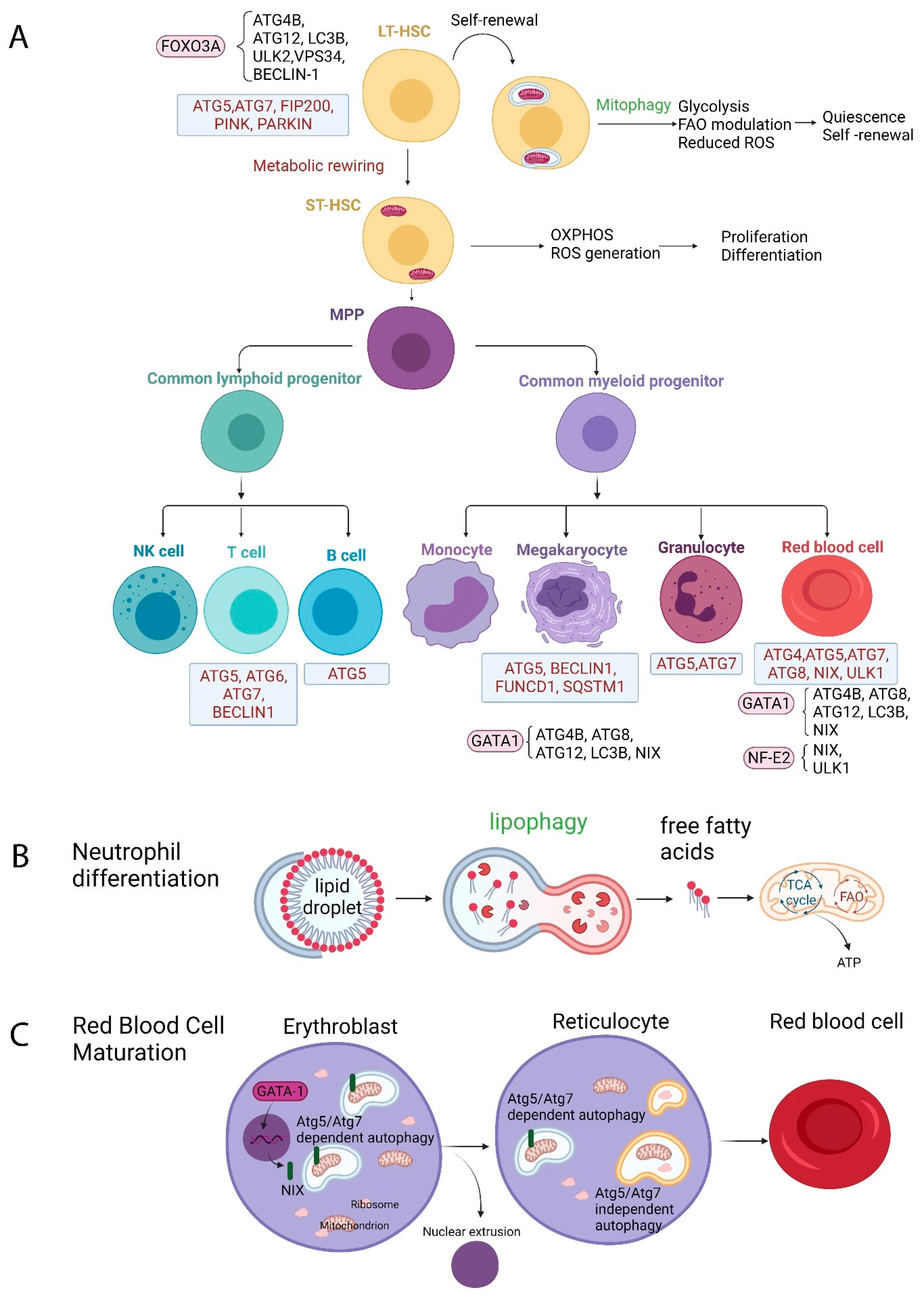
2. The Role of Autophagy in Normal Hematopoiesis

2.1. Autophagy and HSCs

HSCs are unique cells capable of self-renewal through symmetric division and multi-lineage differentiation. They consist the pool that gives rise to both myeloid and lymphoid cells throughout the lifetime and are characterized by the ability to reconstitute the whole blood system of the recipient upon transplantation [11]. HSCs are generally characterized by being in a quiescent state, but upon specific stimuli, they self-renew and/or differentiate into hematopoietic progenitor cells (HPCs) that, in turn, give rise to mature hematopoietic cells [12]. Evidence supports that autophagy may play a role in the regulation of quiescence, self-renewal, and differentiation of HSCs [13,14].
Data supporting the notion that autophagy is an essential regulator of HSCs maintenance have initially been obtained from mouse models. Mortensen et al. demonstrated that conditional deletion of the essential autophagy gene Atg7 in the hematopoietic system resulted in impaired self-renewal capacity, both in vitro (failure to form secondary colonies in colony-forming cell assays) and in vivo (repopulation assays). HSCs within the Lin(-) Sca-1(+) c-Kit(+) (LSK) compartment were significantly reduced, while Atg7-deficient LSK cells failed to reconstitute hematopoiesis in lethally irradiated mice. Autophagy impairment had a negative impact on the differentiation of HSCs into mature white and red blood cells, with Atg7-deficient mice displaying reduced myeloid and lymphoid progenitors, as well as severe anemia. Furthermore, decreased absolute counts of T, B, and NK cells in the peripheral blood were observed in these mice.
FIP200 (200-kDa FAK-family interacting protein) plays important roles in mammalian autophagy. Conditional deletion of FIP200 in hematopoietic cells leads to perinatal lethality and severe anemia in mouse models. FIP200 is required for the maintenance and function of fetal HSCs, since FIP200-deficient HSCs were unable to reconstitute lethally irradiated recipients. FIP200 ablation increased the rate of HSC proliferation and myeloid expansion, which may account for the depletion of fetal HSCs [15].
It is well established that the transcription factor FOXO3A regulates the expression of genes involved in the autophagic machinery, namely, LC3B, Atg12, Atg4b, Ulk2, Vps34, and Beclin-1 [16]. It has been shown that mouse HSCs robustly induce autophagy after ex vivo cytokine withdrawal and in vivo calorie restriction, with FOXO3A being a key player in maintaining a gene expression program that ensures rapid autophagy induction upon starvation [7]. Salemi et al. demonstrated that in vitro cultured human HSCs show a high rate of autophagy, through which they probably ensure their capacity for self-renewal and differentiation. Inhibition of autophagy, either pharmacologically by 3-methyladenine or by ATG5 shRNA, resulted in HSCs’ failure to form colonies in in vitro colony-forming assays and to differentiate to neutrophils [17].
Mitophagy is a form of autophagy specific to the elimination of defective mitochondria. Normal modulation of the level of mitophagy regulates the maintenance of HSCs. One of the key regulators of mitophagy is the phosphatase and tensin homolog (PTEN)-induced putative kinase Pink1. Ito et al. showed that silencing of Pink1 or Parkin, which encodes an E3 ubiquitin ligase necessary for Pink1 binding on the mitochondrial outer membrane and subsequent mitophagy induction, abrogates the ability of highly purified HSCs to expand in vitro but also affects their maintenance potential [18]. Mitophagy is generally considered a mechanism contributing to the maintenance of normal HSCs function. Excessive mitophagy, though, seems to be detrimental to HSCs maintenance and appears to block commitment to the hematopoietic lineage at the progenitor stage. Jin et al. identified the AAA+ -ATPase Atad3a as a regulator of Pink1-dependent mitophagy. Mice with hematopoietic system-specific deficiency in Atad3a exhibited hyperactivated mitophagy that resulted in aberrant hematopoietic homeostasis [19].

2.2. Autophagy and Erythropoiesis

Depletion of membrane organelles and extrusion of the nucleus are hallmarks of mammalian erythropoiesis. Betin et al. showed that autophagy is induced at the polychromatic erythroid stage, and that autophagosomes remain abundant until enucleation [20]. This stimulation of autophagy coincides with the transcriptional upregulation of many autophagy genes, namely, all Atg8 mammalian paralog family members and a subset of Atg4 family members [20]. Atg7 deletion in mice leads to the development of severe anemia, with erythroblasts accumulating damaged mitochondria with an altered membrane potential, a phenomenon that eventually leads to cell death [21].
GATA-1, the master regulator transcription factor of erythropoiesis, has been shown to activate the essential autophagy component microtubule-associated protein 1A/1B-light chain 3 (LC3), as well as other autophagy-related genes [8].
Programmed mitochondrial clearance via autophagy is a prerequisite for reticulocyte maturation. The proapoptotic factor NIX (BCL2/adenovirus E1B 19 kDa protein-interacting protein 3 ligand (BNIP3L)), a BH3-only protein of the outer mitochondrial membrane, is upregulated during terminal erythroid differentiation [22,23]. NIX induces mitochondrial membrane depolarization and subsequent recruitment of LC3B/Gamma-aminobutyric acid receptor-associated proteins (GABARAP) to mitochondria [23]. Schweers et al. demonstrated that NIX is necessary for mitochondrial clearance in reticulocytes, since its deletion in vivo impairs mitophagy and erythroid maturation, resulting in anemia with reticulocytosis [23]. However, other autophagic processes such as ribosome clearance are not affected by NIX deletion. Kundu et al. demonstrated that Unc-51-like kinase 1 (ULK1) plays a critical role in the autophagic removal both of mitochondria and ribosomes during terminal erythroid maturation, though ULK1 deficiency leads to impaired, but not completely abolished, mitochondrial clearance [24]. Although Atg5 and Atg7 are indispensable for macroautophagy, their role in mitochondrial clearance remains controversial. It has been shown that mammalian cells use conventional Atg5/Atg7-dependent macroautophagy as well as an alternative ULK1-dependent Atg5/Atg7-independent macroautophagy process [25]. Honda et al. showed that mitochondrial clearance in fetal reticulocytes is ULK-1-dependent and Atg-5-independent, while ULK1-dependent macroautophagy was less involved in steady-state adult reticulocytes [26].
GATA-1, a transcription factor considered to be a master regulator of normal erythropoiesis, is implicated in the transcriptional regulation of mitophagy during erythrocyte maturation. GATA-1 transcriptionally controls and upregulates the expression of autophagy genes, namely, Atg8 homologs, Atg4b, and Atg12, as well as lysosomal genes, namely, Atp6v0e, Clcn7, Neu1, and Lamp1, in primary human erythroblasts [8]. NIX expression is also upregulated by GATA-1 in primary human erythroblasts, thus rendering GATA-1 a regulator of mitophagy during erythroid maturation [23]. The KRAB/KAP1-miRNA regulatory cascade negatively regulates the miRNA-351 which targets NIX, thus increasing mitophagy during erythrocyte maturation. In mouse models, Kap1 deletion leads to reticulocytosis, mitochondrial retention, and fatal anemia [27]. Nuclear factor-erythroid 2 (NF-E2), another transcription factor crucial for normal erythroid maturation, upregulates the expression of BNIPL1 and ULK1, thus regulating mitophagy [28].

2.3. Autophagy and Granulopoiesis

Atg7 deletion at the granulocyte-monocyte progenitor (GMP) stage inhibits neutrophil differentiation, resulting in accumulation of immature precursors. Impaired lipophagy was identified as responsible for the neutrophil differentiation defect upon Atg7 deletion [29]. Moreover, autophagy-deficient mice with Atg5 or Atg7 deletion demonstrate impaired neutrophil function with defective nicotinamide adenine dinucleotide phosphate (NADPH) oxidase-mediated ROS generation and impaired neutrophil degranulation [30]. It has also been shown that the secretion of the proinflammatory cytokine IL-1β by neutrophils is mediated by autophagy [31].

2.4. Autophagy and Megakaryopoiesis

Megakaryocytic maturation, similar to erythroid maturation, is also regulated by the transcription factor GATA-1, which induces the expression of several autophagic genes [8]. Colosetti et al. used the chronic myeloid leukemia (CML) cell line K562 to prove that autophagy induction is an important event for megakaryocytic differentiation and is also accompanied by increased expression of Beclin 1 and p62/sequestosome 1 (SQSTM1) [32]. Using an Atg7 hematopoietic conditional knockout mouse model, Cao et al. demonstrated that loss of autophagy resulted in the accumulation of mitochondria and ROS, impeding megakaryopoiesis and megakaryocyte differentiation, as well as thrombopoiesis, leading to abnormal platelets, larger in size and fewer in number, with deficient functions of activation and aggregation [33]. Disruption of the autophagic flux, by means of pharmacological inhibition, leads to impairment of platelet aggregation and adhesion in vitro, while Becn1 heterozygous knockout mice display a prolonged bleeding time and reduced platelet aggregation [34]. Zhang et al. demonstrated that hypoxia induces mitophagy in platelets in a FUNDC1-dependent manner, regulating mitochondrial quality and quantity and controlling platelet activation during ischemia/reperfusion injury [35].
An overview of autophagy in normal hematopoiesis is described in Figure 1.

2.5. Autophagy and Lymphopoiesis

2.5.1. T Lymphocytes

Atg5 or Atg7 deletion in murine T cells leads to a decrease in thymocyte and peripheral T cell numbers, with Atg5-deficient T cells demonstrating decreased cell survival [36,37,38]. Stephenson et al. showed that Atg5 deletion resulted in increased mitochondrial mass in peripheral T cells, while they also observed a correlation between mitochondrial mass and Annexin-V staining, proposing that autophagy is critical for mitochondrial maintenance and T cell survival [37]. Accordingly, Pua et al. showed that autophagy-deficient mature T cells fail to reduce their mitochondrial content in vivo and have increased ROS production, as well as an imbalance in pro- and antiapoptotic protein expression [38]. Recent findings highlight how the activation of transient receptor potential vanilloid 1 (TRPV1) induces autophagy in mice thymocytes through the ROS-regulated 5’ adenosine monophosphate-activated protein kinase (AMPK) pathway. TRPV1-triggered autophagy is Atg6/Beclin-1-dependent and involves ROS- and AMPK-mediated upregulation of Beclin-1 expression. Autophagy seems to be activated as a pro-survival process, as its inhibition triggers the apoptosis of thymocytes [39]. Moreover, Beclin-1-deficient mice display a dramatic reduction in thymic cellularity, since Beclin-1 expression is necessary for the maintenance of undifferentiated/early lymphocyte progenitor populations [40].

2.5.2. B Lymphocytes

Autophagy has been shown to be differentially required at different stages of B cell development. Atg5 deletion in B lymphocyte progenitors results in a significant defect in B cell development at the pro- to pre-B cell transition, which is associated with increased cell death, indicating that Atg5 is important for B cell survival. Moreover, while B-1 B cells require Atg5 for their maintenance in the periphery, B-2 B cells are not affected by Atg5 deletion [41]. Autophagy has also been shown to be implicated in the processes of plasma cell differentiation and in the maintenance of memory B cells [42,43].

3. Autophagy in Malignant Hematopoiesis

3.1. Autophagy in Acute Myeloid Leukemia (AML)

AML is a clonal hematopoietic disorder originating from leukemic stem cells (LSCs), which are responsible for disease initiation, chemotherapy resistance or failure, and disease relapse. LSCs originate from transformed HSCs or HPCs [44].
As it has already been analyzed, autophagy gene expression regulates HSCs maintenance. Deletion of the autophagy gene Atg7 in HSCs resulted in pre-leukemic myeloproliferation in a mouse model and impaired production of both myeloid and lymphoid progenitors, while HSPCs displayed accumulation of mitochondria and ROS, as well as increased proliferation and DNA damage [45]. The loss of Atg5 results in a similar myeloproliferative pre-leukemic phenotype to that described in mouse HSPCs models with Atg7 loss, confirming the role of autophagy in HSPCs regulation [46].
Heterozygous deletions, missense mutations, or copy number changes in key autophagy genes have been identified with high frequency in AML patients, particularly those with complex karyotypes [46,47,48]. Jin at al. demonstrated that human AML blasts show lower transcript levels of ULK1, FIP200, Atg14, Atg5, Atg7, Atg3, Atg4b, and Atg4d compared to healthy donor granulocytes [49]. Autophagy-related genes Atg10 and Atg12, GABARAPL2 and MAP1LC3B, and GABARAP are, respectively, located at chromosome regions 5q, 16q, and 17p. Heterozygous chromosomal losses in these regions are frequently detected in AML patients, thus leading to decreased autophagic protein expression [46].
The reduced expression of autophagy genes found in human AML blasts and the subsequent decreased autophagic flux lead to accumulation of unhealthy mitochondria, implying that deficient autophagy may be beneficial for human AML. More importantly, heterozygous loss of autophagy in a mixed lineage leukemia-eleven nineteen (MLL-ENL) model of AML favored proliferation in vitro, and enhanced glycolysis along with leukemia aggressiveness in vivo [46]. Another study supporting the beneficial role of autophagy in AML comes from Sumitomo et al., who showed that inactivation of autophagy by Atg5 or Atg7 deletion prolonged survival in leukemic mice and reduced functional leukemia-initiating cells (LICs) [50]. Atg7-deficient LICs displayed enhanced mitochondrial activity and increased ROS production along with increased cell death. In addition, Atg7 deletion markedly decreased leukemic cells in the peripheral blood, a phenomenon attributed to increased apoptosis, suggesting a higher dependency on autophagy compared to bone marrow leukemia cells. Finally, the authors suggested that autophagy may contribute to chemotherapy resistance, since cytarabine (AraC) treatment activated autophagy in LICs, and Atg7 deletion potentiated the therapeutic effects of AraC, which included decreased LICs and prolonged survival [50]. Therefore, a reduced autophagic flux may provide an advantage for leukemogenesis.
However, Folkerts et al. demonstrated that autophagy levels were high in poor-risk AML, compared to favorable- and intermediate-risk AML [51]. Upon subdividing the CD34+ AML cells into ROS-low and ROS-high fractions, the authors showed that the AML CD34+ ROS-low cells exhibited higher levels of basal autophagy and decreased survival when treated with the inhibitor of autophagic lysosomal fusion, hydroxychloroquine [51]. Moreover, Atg5 knockdown inhibited the in vivo maintenance of AML CD34+ cells in NOD scid gamma (NSG) mice [51]. This observation is in contrast to the previous published literature and supports a favorable role of autophagy in tumor proliferation.
Reports from MLL-AF9 AML, the most common alteration in infant AML, support that ATG5 or ATG7 is necessary for the initiation of AML, but once AML is established, autophagy is no longer required for the disease maintenance [52,53,54,55]. In an MLL-AF9-induced murine AML model, Saito et al. demonstrated that deletion of AMPK, a positive regulator of autophagy, significantly delayed leukemogenesis by increasing oxidative stress and DNA damage in LSCs [56]. LSCs overexpress FIS1, a regulator of mitochondrial dynamics that mediates mitophagy activity. Depletion of FIS1 attenuates mitophagy and leads to a profound loss of the LSCs self-renewal potential [57].
Nguyen et al. showed that loss of the selective autophagy receptor p62 impaired the expansion and colony-forming ability of leukemia cells, through mitophagy impairment and accumulation of defective mitochondria with subsequent increased ROS production, while it prolonged the latency of leukemia development in mouse models in vivo [58].
Rudat et al. showed that mechanistic target of rapamycin complex 1 (mTORC1)-mediated autophagy suppression stabilizes mutant Fms Related Receptor Tyrosine Kinase 3 (FLT3) in AML, while receptor tyrosine kinase inhibition increases autophagy and leads to FLT3 depletion [59]. On the contrary, Heydt at al. demonstrated that FLT3-internal tandem duplication (ITD) expression increases basal autophagy in AML cells, with the transcription factor ATF4 being identified as a key player in FLT3-ITD-induced autophagy [60]. ATF4 downregulation inhibited autophagy-dependent AML cell proliferation [60].

3.2. Autophagy in Myelodysplastic Syndromes (MDS)

Aberrations in the autophagic machinery have also been implicated in the pathophysiology of bone marrow failure syndromes. MDS comprise a heterogenous group of clonal hematopoietic disorders characterized by ineffective hematopoiesis and an increased risk of transformation to AML [61]. Mutations in autophagy genes are enriched in MDS and prevalent in higher-risk MDS patients [62].
In a mouse model, Atg7 knockout in HSCs led to accumulation of mitochondria with an altered mitochondrial membrane potential in erythrocytes, causing cell death and anemia, similar to the MDS phenotype [21]. Erythroid precursors from patients with low-risk MDS demonstrate ultrastructural features of enhanced mitophagy [63], while erythroblasts from patients with high-risk MDS are characterized by the accumulation of enlarged and abnormal mitochondria [64]. Our team demonstrated that the mitophagy of abnormal mitochondria and autophagic death are prominently featured in the MDS myeloid lineage [65].
By assessing the transcript levels of the mitophagy receptor BNIP3, Lazarini et al. demonstrated decreased BNIP3 expression in MDS compared to healthy controls [66]. In another study, Jiang et al. reported decreased autophagosome formation, as well as decreased expression of the mitophagy receptor NIX and the autophagy promotor genes AMPK and ULK1, in bone marrow nucleated red blood cells from patients with high-risk MDS compared to controls and low-risk MDS patients. The high-risk group demonstrated a higher mitochondrial content and ROS production, whereas the mitochondrial membrane potential was lower, indicating mitochondrial dysfunction [67].

3.3. Autophagy in Chronic Myeloid Leukemia (CML)

CML is a myeloproliferative neoplasm characterized by the chromosomal translocation t(9;22)(q34.1;q11.2), which results in the formation of the BCR-ABL1 fusion gene, encoding the BCR-ABL1 kinase [61]. BCR-ABL, through phosphoinositide-3 kinase (PI3K)/serine-threonine protein kinase B (AKT)/FOXO4 signaling, transcriptionally upregulates activating transcription factor 5 (ATF5) expression, which, in turn, stimulates the transcription of mechanistic target of rapamycin (mTOR), resulting in autophagy suppression in vitro [68]. Altman et al. demonstrated that BCR-ABL-expressing cells have low basal levels of autophagy but are highly dependent on this process and rapidly undergo apoptosis upon disruption of autophagy in vitro. This dependence on autophagy was extended in vivo, as Atg3 deletion also prevented BCR-ABL-mediated leukemogenesis in a cell transfer model [69]. In vivo studies of a murine model of CML demonstrated that long-term (LT)-HSCs isolated from leukemic mice have higher basal autophagy levels compared with non-leukemic LT-HSCs and more mature leukemic cells [70].
Targeted (namely, tyrosine kinase inhibitor (TKI) imatinib mesylate) or non-targeted treatment in CML cell lines or patient samples has been shown to induce an increased autophagic response [71,72], attributed to ROS generation, endoplasmic reticulum (ER) stress, and increased autophagic gene and protein expression [71,73,74]. This increase in autophagic flux results in the senescence rather than the apoptosis of CML cells, rendering them resistant to treatment [75]. ATG7 and ATG4B, both key enzymes in the LC3 conjugation cascade, have been shown to play a critical role in CML CD34+ cell survival, while their genetic inhibition resulted in increased apoptosis of CML stem and progenitor cells and sensitized them to TKI therapy [76,77]. Karvela et al. demonstrated that loss of ATG7 resulted in metabolic reprogramming of CML cells, with impaired glycolysis and a shift towards OXPHOS, leading to ROS accumulation that caused CML CD34+ cells to differentiate towards the erythroid lineage [77]. Liu et al. showed that the AHI-1-BCR-ABL-DNM2 complex regulates endocytosis and ROS generation, both of which drive autophagy induction in CML [73]. Ianniciello et al. demonstrated that autophagy is required for CML CD34+ cell commitment as they transition from hypoxia to normoxia, simulating the leukemic commitment as cells migrate away from the hypoxic bone marrow niche [78].

3.4. Autophagy in Acute Lymphoblastic Leukemia (ALL)

Translocation t(12;21)(p13;q22), resulting in the ETV6-RUNX1 fusion protein (also known as TEL-AML1), is present in 25% of pediatric patients with B cell precursor ALL [79]. The autophagy-initiating lipidase VPS34 demonstrates increased expression in ALL patients with the ETV6-RUNX1 translocation compared to normal HSPCs. The causal relationship between ETV6-RUNX1 and autophagy was proved by the fact that VPS34 induction is transcriptionally regulated by ETV6-RUNX1 and correlates with increased levels of autophagy [80].
The BCR-ABL fusion protein is detected in 25–30% of adult and in 2–10% of pediatric B-ALL cases, and its detection is associated with poor prognosis [81]. In a murine model, hematopoietic progenitor cells expressing the p185 form of BCR-ABL had low basal levels of autophagy but were highly dependent on this process and rapidly underwent apoptosis upon disruption of autophagy through Atg3 deletion or treatment with chemical autophagy inhibitors. This dependence on autophagy was extended in vivo, as Atg3 deletion also prevented BCR-ABL-mediated leukemogenesis in a cell transfer model [69]. We can therefore conclude that hematopoietic cells expressing the BCR-ABL kinase depend on autophagy for survival, as well as for leukemic transformation.
A transcript variant of the Beclin 1 gene carrying a deletion of exon 11, which encoded a C-terminal truncation of the Beclin 1 isoform, has been identified in the ALL 697 cell line, while this truncated Beclin 1 isoform displayed a reduced activity in induction of autophagy by starvation, indicating that it might function as a dominant negative modulator of autophagy [82].
High mobility group box 1 (HMGB1) is a nuclear DNA-binding protein which functions as a damage-associated molecular pattern molecule (DAMP), and its expression has been found increased in childhood ALL. As a positive regulator of autophagy, intracellular HMGB1 interacts with Beclin 1 in leukemia cells, leading to autophagosome formation. Additionally, exogenous HMGB1 directly induces autophagy and cell survival in leukemia cells [83].
Glucocorticoids, used widely for the treatment of B-ALL, have been shown to activate autophagy, subsequently inducing cell death [84]. Obatoclax, a bcl-2 inhibitor, overcomes glucocorticoid resistance in B-ALL cells, inducing ATG5-dependent autophagy [85]. Everolimus, an mTORC1 inhibitor, induces autophagy in B-ALL cells, a phenomenon that is alleviated by Beclin-1 downregulation by the siRNA strategy [86]. Patients with B-ALL resistant to glucocorticoid therapy showed different patterns of autophagy gene expression (26 upregulated and 10 downregulated) compared to patients with glucocorticoid-sensitive B-ALL [87]. L-asparaginase, used for the treatment of ALL, hydrolyzes glutamine to glutamic acid and asparagine to aspartic acid, reducing aerobic metabolism [88]. Treatment with L-asparaginase results in autophagy induction by ALL cells to compensate for the metabolic stress, while the use of the autophagy inhibitor chloroquine increases the sensitivity of ALL to L-asparaginase [89].
Glucocorticoids decrease the cellular intake of glucose and glutamine, while they increase glutamine synthesis [90]. The decrease in nutrient intake leads to an increase in autophagic flux to catabolically compensate the nutrient deficiency. This catabolic process produces ammonia, which is used by glutamate-ammonia ligase for intracellular glutamine synthesis, allowing the cells to ensure their own glucose supply [90].

3.5. Autophagy in Chronic Lymphocytic Leukemia (CLL) and Lymphoma

Autophagy was shown to have a pro-survival role in CLL cells, since autophagy inhibition by RNA interference targeting key autophagy genes or by using chloroquine or 3-methyladenine in peripheral blood mononuclear cells (PBMCs) from CLL patients decreased CLL cell viability [91]. On the other hand, Gade et al. demonstrated that inhibition of death associated protein kinase 1 (DAPK1), an autophagy-associated gene, reduces autophagy and promotes CLL growth [92]. A positive prognostic marker in CLL is the deletion of 13q14. This chromosomal region contains micro-RNA-15 and -16, which target the antiapoptotic protein BCL-2, with 13q14 deletion leading to BCL-2 upregulation [93]. BCL-2 binds to and negatively regulates BECN1, with BCL2 upregulation leading to suppression of BECN1 and autophagy inhibition [94]. SLAMF1 expression constitutes another positive prognostic factor in CLL and is involved in autophagosome recruitment and autophagic flux activation [95]. The opposing effects on autophagy regulation by the above-mentioned positive prognostic markers reveal the heterogenous impact of molecular CLL aberrations on autophagy.
Germinal center B cell-like-derived (GCB) diffuse large B cell lymphoma (DLBCL) is characterized by BCL-2 overexpression [61]. BCL-2 binds directly to BECN1, thus inhibiting autophagy [96]. Patients with decreased BCL-2 levels show increased BECN1 expression, which correlates with a favorable clinical outcome [97]. Monoallelic deletion of BECN1 in mice has been shown to promote tumorigenesis [98]. Bertolo et al. demonstrated that constitutive repression of autophagy responses in BCL6-driven lymphomas may contribute to lymphomagenesis [99]. On the other hand, Li et al. demonstrated that Cullin4B (CULB4) (a scaffold protein of the CUL4B-RING E3 ubiquitin ligase complex, highly expressed in DLBCL) is implicated in the positive regulation of autophagy, thus contributing to DLBCL progression. CUL4B deletion leads to inhibition of proliferation, an effect that could be attributed to the pro-survival ability mediated by autophagy [100]. Pharmacological inhibition of autophagy or knockdown of LC3B, ATG5, or p62/SQSTM1 in DLBCL OCI-Ly1 cells inhibited cell proliferation and induced apoptosis in vitro [101]. These data demonstrate the complex mechanisms implicated in autophagy regulation in DLBCL.
Autophagy-related genes were found to be upregulated in follicular lymphoma (FL) lymph node tissues compared to reactive B cells [102]. In the same study, FL samples showed significantly decreased levels of both p62 and LC3 compared with reactive lymphoproliferation and DLBCL, indicative of increased autophagy activity in FL [102]. Wang at al. demonstrated that mutations in the ATP6V1B2 subunit of the vacuolar-type H+-translocating ATPase (v-ATPase) found in FL activate autophagic flux and maintain mTOR in an active state [103].
In mantle cell lymphoma (MCL), the t(11;14) translocation leads to cyclin D1 overexpression and high cyclin dependent kinase 4 (CDK4) activity. Inhibition of cyclin D1/CDK4 activity significantly reduces proteasome inhibitor-mediated stabilization of the proapoptotic BH3-only protein NOXA, mainly driven by an autophagy-mediated proteolysis. Combined treatment with the proteasome inhibitor bortezomib and autophagy inhibitors enhances NOXA stability, leading to super-induction of the NOXA protein, contributing to the apoptotic effect of proteasome inhibitor treatment [104]. Zhang et al. demonstrated that transglutaminase TG2 and nuclear factor-κB (NF-κB) signaling coordinates the survival of MCL cells via IL6-mediated autophagy [105], while Chen et al. demonstrated that ROS-induced C-X-C motif chemokine receptor 4 (CXCR4)/stromal cell derived factor 1 (SDF-1) signaling stimulates autophagy in MCL cells, promoting their survival in the bone marrow microenvironment [106].

4. Metabolism in Normal Hematopoiesis

The maintenance of the mammalian HSC system depends on the self-renewal of quiescent LT-HSCs and subsequent generation of short-term (ST)-HSCs, multipotent progenitors (MPPs) that will, in turn, differentiate to mature myeloid or lymphoid lineage cells [107,108]. Cells generate ATP, the major currency for energy-consuming reactions, through central carbon metabolism, including glycolysis and mitochondrial OXPHOS. It has been demonstrated that LT-HSCs contain fewer mitochondria compared to MPPs, thus relying on glycolysis to meet their energy demands [109,110]. This metabolic profile protects LT-HSCS from oxidative stress, maintaining low endogenous ROS levels, since aberrant ROS generation could abrogate various stem cell properties including cell cycle quiescence, self-renewal, survival, and multi-lineage differentiation capacity [111,112].

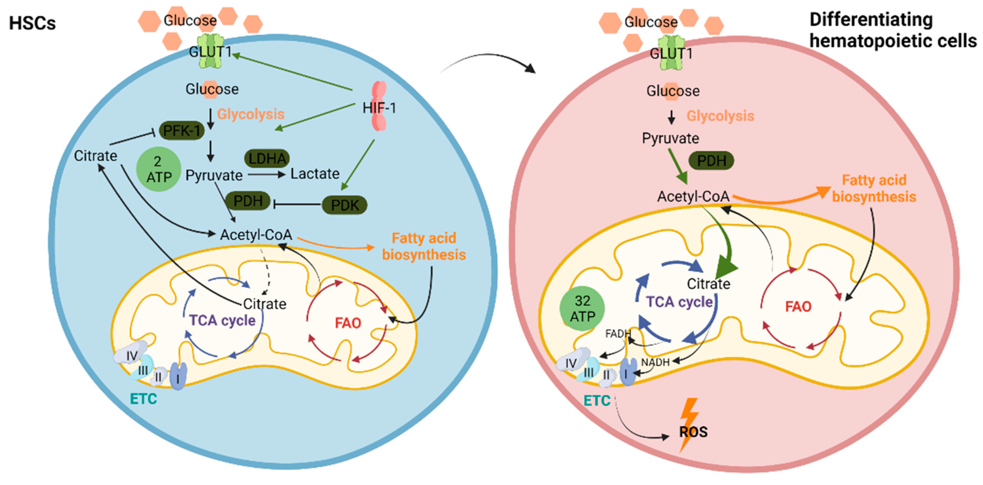
4.1. Regulation of HSC Metabolism–Glycolysis Versus OXPHOS

Precise regulation of the levels of hypoxia inducible factor-1α (HIF-1α) ensures cell cycle quiescence of LT-HSCs that are maintained in a hypoxic state in the bone marrow niche [113,114]. The ability of pyruvate to enter the TCA cycle in the mitochondria is regulated by pyruvate dehydrogenase kinase-1 (PDK-1). PDK-1 expression has been shown to be under the transcriptional control of HIF-1α [115,116]. PDK-1 suppresses the activity of pyruvate dehydrogenase (PDH) and limits the influx of pyruvate into the TCA cycle. Suppression of pyruvate entry into the TCA cycle results in decreased citrate production. In growing cells, the citrate exported from mitochondria into the cytosol suppresses glycolysis at the level of phosphofructose kinase-1 (PFK-1), allowing a greater amount of glucose-6-phosphate to be diverted to nucleotide biosynthesis and glycosylation reactions. Furthermore, the cytosolic degradation of citrate provides the major source of acetyl-CoA for lipid and isoprenoid synthesis during cell growth. Therefore, HIF-1α-enhanced PDK-1 activity results in a decline in mitochondrial citrate production, leading to a derepression of PFK-1 activity and a concomitant suppression of lipid synthesis [117]. HIF-1α also controls the expression of lactate dehydrogenase A (LDH-A), an enzyme responsible for the production of lactate from pyruvate [118]. As LDH-A levels increase, cellular pyruvate is preferentially converted to lactate and secreted into the extracellular space [119]. Thus, under the transcriptional control of HIF-1α, the intracellular glucose utilization is diverted from lipid and nucleotide synthesis to ATP production via anaerobic glycolysis (Figure 2).
HIF-1α-dependent upregulation of PDK-1 in LT-HSCs represses the influx of glycolytic intermediates into mitochondria. This leads to an LT-HSC metabolic profile characterized by increased glycolysis levels compared to MPPs, necessary for the maintenance of cell cycle quiescence [120]. While LT-HSCs are characterized by a low mitochondrial potential and utilize glycolysis for ATP production, the differentiating hematopoietic cells rely on OXPHOS to meet their increased energy demands [110]. Pyruvate production during glycolysis generates only two molecules of ATP per molecule of glucose during anaerobic respiration, while upon entering the mitochondria to be used for OXPHOS, pyruvate generates 32 molecules of ATP per molecule of glucose (Figure 2). Although the ratio of ATP generation to glucose consumption under anaerobic glycolysis is inefficient compared to that generated by OXPHOS, the rate of ATP production under hypoxia potentially increases 100-fold compared to that supported by mitochondrial energy production under normoxia [121]. However, when dye-independent methods are used, namely, mitochondrial DNA quantification and enumeration of mitochondrial nucleotides, it has been shown that HSCs and MPPs have a higher mitochondrial mass than lineage-committed progenitors and mature cells. They present, though, a compromised respiratory and mitochondrial turnover capacity. Despite their similar mitochondrial mass, the respiratory capacity of MPPs exceeds that of HSCs. [122]. Luchinger at al. demonstrated that the mitochondrial protein mitofusin 2 (MFN2), which is implicated in mitochondrial fusion, is essential for the maintenance of HSCs with an extensive lymphoid potential [123]. Another study by Mohrin et al. showed that the mitochondrial unfolded protein response (UPRmt) is activated as HSCs transition from quiescence to the proliferation state [124]. Sirtuin 7 (SIRT7) is a component of the UPRmt. Its inactivation causes reduced quiescence, increased mitochondrial protein folding stress, and a compromised regenerative capacity of HSCs. SIRT7 expression is reduced in aged HSCs, and SIRT7 upregulation improves the regenerative capacity of aged HSCs [125].
A study by Ansó et al. demonstrated that loss of the mitochondrial complex III subunit Rieske iron–sulfur protein (RISP) in fetal mouse HSCs allows them to proliferate but impairs their differentiation, resulting in anemia and prenatal death, while RISP inactivation in adult HSCs also impaired respiration, resulting in loss of quiescence concomitant with severe pancytopenia and lethality. They, thus, concluded that mitochondrial respiration is dispensable for adult or fetal HSC proliferation, but essential for fetal HSC differentiation and maintenance of adult HSC quiescence [126].
The glycolytic phenotype of HSCs can be considered a protective mechanism that reduces ROS generation, which would cause oxidative stress and induce differentiation [127]. Mitochondria are the major source of ROS. However, HSCs rely on mitochondria when a metabolic switch is required. The transcription factor FOXO3 is implicated in the regulation of HSC metabolism. Studies show that FOXO3 exerts a protective effect on HSPCs regarding DNA damage, as FOXO3 deletion in HSCs alters mitochondrial function, leading to a deleterious ROS accumulation [128,129]. Tuberous sclerosis complex 1 (TSC1) is a negative regulator of mTOR, thus affecting the regulation of cellular metabolism [130]. Tsc1 deletion in HSCs results in a transition from quiescence to active cell cycling, with increased mitochondrial biogenesis and elevated levels of ROS, dramatically reducing both hematopoiesis and the self-renewal of HSCs [131]. Luo et al. demonstrated that mTOR overactivation contributes to HSCs’ premature exhaustion, in part, via induction of HSC senescence, while its inhibition promotes the ex vivo expansion and long-term hematopoietic reconstitution capacity of HSCs [132]. mTORC1 signaling is enhanced to promote the translation of mitochondria-associated transcripts during erythroid differentiation, controlling the mitochondrial content or erythroid lineage cells [133].

4.2. Fatty Acid Oxidation (FAO) in HSCs

FAO, another mitochondrial metabolic process, also plays a role in HSCs maintenance. Ito et al. identified a novel promyelocytic leukemia gene (PML)-peroxisome proliferator-activated receptor delta (PPARδ)-FAO pathway implicated in HSCs maintenance. Loss of PPARδ profoundly affects the maintenance of HSCs. Moreover, treatment with PPARδ agonists improves these HSCs functions, whereas, conversely, inhibition of mitochondrial FAO induces loss of the HSC compartment. Importantly, the authors demonstrated that PML exerts its essential role in HSCs maintenance through regulation of PPAR signaling and FAO. Mechanistically, the PML-PPARδ-FAO pathway controls HSC asymmetric division. Depletion of Ppard or Pml, as well as FAO inhibition, results in symmetric commitment of HSC daughter cells, while, conversely, PPARδ activation increases asymmetric division [134]. In line with these findings, Yu et al. showed that downregulation of the mitochondrial phosphatase protein tyrosine phosphatase 1 (PTPMT1) shifts the metabolic profile from glycolysis and FAO to mitochondrial OXPHOS, leading to ineffective hematopoiesis. This phenomenon is attributed to accumulating HSCs unable to differentiate due to increased entry of stem cells in the cell cycle followed by a pause in the G1 phase [135].

5. Metabolism in Malignant Hematopoiesis

5.1. Glucose Metabolism

It has been demonstrated that AML cell lines, as well as human primary AML blasts, show enhanced levels of glycolysis [136]. This glycolytic metabolic profile is regulated by the PI3K/AKT and mTOR pathways [137,138]. Published data indicate that increased glycolysis and a low efficiency of OXPHOS may contribute to drug resistance in AML. The expression of HIF-1α and GLUT1, as well as that of two of the key enzymes controlling the glycolytic flux, namely, hexokinase 2 (HK2) and LDH, was increased in bone marrow cells from AML patients with no remission (NR), compared to healthy control individuals and patients with complete remission (CR) and partial remission (PR). Studies indicate that highly glycolytic AML blasts are resistant to chemotherapeutic agents [136,139]. Herst et al. investigated the level of glycolytic metabolism in leukemic blasts as a prognostic marker in AML, demonstrating that highly glycolytic AML blasts were more resistant to apoptosis induced by all-trans-retinoic acid (ATRA) and/or arsenic trioxide in vitro, while high levels of glycolytic metabolism at diagnosis were predictive of a significantly improved duration of CR1 and overall survival following AML remission induction chemotherapy [136]. Chen et al., using a set of metabolite markers to create a prognostic model for AML, demonstrated that the low-prognostic risk score group showed enhanced glycolysis and an enhanced TCA cycle [139]. Evidence supporting the above results also comes from studies in mouse HSCs/HSPCs, where the deletion of LDHA and the M2 pyruvate kinase isoform (PKM2), both enzymes that regulate the last steps of glycolysis, inhibits leukemia initiation [140].

5.2. Glutamine Metabolism

Glutaminolysis ensures nitrogen supply for protein and nucleotide synthesis, while it also contributes to redox maintenance via NADPH production. Inhibition of glutamine uptake has been shown to induce apoptosis in AML cells, while treatment with L-asparaginase, an antitumor agent that also bears glutaminase activity, inhibits mTORC1 and protein synthesis in AML cells, thus producing a strong apoptotic response [141]. Stevens et al. showed that LSCs isolated from de novo AML patients rely on amino acid metabolism for OXPHOS and survival, with pharmacological inhibition of amino acid metabolism reducing OXPHOS and inducing cell death. On the contrary, LSCs obtained from relapsed AML patients are not reliant on amino acid metabolism due to their ability to compensate through increased fatty acid metabolism [142]. Gallipoli et al. identified glutaminase, the first enzyme in glutamine metabolism, as synthetically lethal with FLT3 inhibitor treatment in FLT3-ITD-mutated AML. They also demonstrated that glutamine metabolism, through its ability to support both mitochondrial function and cellular redox metabolism, becomes a metabolic dependency of FLT3-ITD AML, specifically unmasked by FLT3-TKI treatment [143]. Furthermore, it has been shown that PI3K/AKT signaling causes a metabolic switch from glutaminolysis to aerobic glycolysis in Notch-dependent T-ALL [144].

5.3. Fatty Acid Metabolism

Since LSCs use glutamine to support oxidative metabolism, their glutamine demands can be covered by fatty acid metabolism. Recent in vitro studies have shown that distinct genetic changes in AML are associated with enhanced dynamics and metabolism of lipid species in AML cells [145]. Isocitrate dehydrogenases (IDH1/2) are key metabolic enzymes, mutated in more than 15% of AML patients, with IDH1-mutant AML cells displaying a strongly reprogrammed lipid anabolism with dysregulation of fatty acid metabolism and fluxes [145].
Adipose tissue is one of the main sources of glutamine for cells. Bone marrow adipocytes from the tumor microenvironment support the survival and proliferation of malignant cells from patients with AML. AML blasts alter metabolic processes in adipocytes activating lipolysis, thus enabling the transfer of fatty acids from adipocytes to AML blasts [146]. Ye et al., using a mouse model of blast crisis CML, showed that adipose tissue provides leukemic cells with the necessary free fatty acids (FFA) to support their energy demands. High expression of the fatty acid transporter CD36 characterized the LSCs associated with adipose tissue [147]. Perea et al. also identified a CD36-expressing subpopulation within the CD34+ LSC compartment of human AML that was characterized by increased FFA uptake and FAO [148]. Studies from Tucci et al. demonstrated that ALL cells stimulate adipocyte lipolysis and utilize adipocyte-derived free fatty acids for proliferation, and this interaction may contribute to ALL resistance to chemotherapy [149,150]. A STAT-driven aberrant lipoprotein lipase (LPL) expression in CLL cells provides them with FFAs, supporting their oxidative metabolic capacity [151].
FAO is used by cancer cells to support their survival [152]. FAO generates acetyl-CoA that enters the TCA cycle, as well as flavin adenine dinucleotide (FADH2) and NADH that enter the ETC to generate ATP. It has been shown that carnitine palmitoyl transferase 1A (CPT1A) and carnitine transporter CT2, functioning in favor of FAO, are overexpressed in AML, with their inhibition compromising the growth and viability of AML cells [153,154]. Samudio et al. suggested that pharmacologic inhibition of FAO inhibited proliferation and sensitized human leukemia cells to apoptosis induction by ABT-737, a molecule that releases proapoptotic Bcl-2 proteins such as Bak from antiapoptotic family members [155].
Phospholipid metabolism alterations are frequently observed in cancer. Overexpression of choline kinases, responsible for the conversion of choline to phosphocholine (PC), results in increased PC levels that contribute to neoplastic transformation and cancer cell proliferation [156]. It has been demonstrated that PC levels in AML patients’ sera are higher in those with intermediate-risk AML compared to those with a favorable risk [4]. Lo Presti et al. demonstrated that deregulations in phospholipid metabolism can negatively affect prognosis, with PC and phosphoethanolamine (PE) being potential aggressiveness biomarkers [157]. A link between phospholipid metabolism and AML stemness has also been demonstrated, highlighting the important role of phosphatidylserine (PS) [158].
De novo fatty acid synthesis provides the membrane components required for the rapid proliferation of neoplastic cells, but it can also alter the physicochemical properties of the lipid bilayers, conferring with signaling and drug resistance. A lipidomic approach has been used for the stratification of AML patients, with t(8;21) and inv(16) patients (favorable-risk groups) presenting a substantial modulation of ceramide/sphingolipid synthesis compared to patients with normal karyotypes [159].
FAO was also shown to be implicated in lymphomagenesis. A subset of DLBCLs that do not require the activation of the B cell receptor for survival and demonstrate an OXPHOS gene expression signature catabolizes fatty acids at a higher rate than other subtypes of DLBCL and is highly dependent on FAO for survival and growth [160,161].

5.4. Mitochondrial Metabolism

Compared with normal hematopoietic cells, AML cells have a higher mitochondrial mass, without a concomitant increase in respiratory chain complex activity, thus displaying a lower spare reserve capacity in the respiratory chain that renders them more susceptible to oxidative stress [162]. Cytarabine-resistant AML cells display high levels of ROS, with an increased mitochondrial mass and active polarized mitochondria, findings consistent with a high OXPHOS status. Cytarabine residual cells exhibit increased FAO, upregulated CD36 expression, and a high OXPHOS gene signature, predictive for treatment response in patient-derived xenografts and patients with AML [163].
Both in normal and malignant hematopoiesis, the redox status is defined by the production of ROS and the function of antioxidant systems. ROS production depends on two major sources: the mitochondria, and the nicotinamide adenine dinucleotide phosphate oxidases (NOX) activity. NOX functional polymorphisms in de novo AML patients have been associated with response to induction therapy, with higher ROS generation leading to improved complete remission rates [164]. Constitutive activation of NOX contributes to extracellular ROS production in patients with AML, while mitochondrial ROS production is rarely observed [165]. These increased ROS levels were shown to promote AML blast proliferation [165]. Activating mutations of the FLT3 receptor are detected in approximately one third of newly diagnosed AML cases, with ITD mutations being associated with an adverse prognosis [166]. The increased levels of endogenous ROS demonstrated in FLT3-ITD-mutated AML could be associated with the aggressiveness and poor prognosis of these AML cases [167]. Moreover, it seems that chemo-resistant AML displays high levels of ROS, along with an active OXPHOS status [163]. Redox balance deregulation in de novo AML patients has been correlated with the molecular status and overall survival [168]. However, compared to normal hematopoietic cells, AML cells display a higher mitochondrial mass, though without increased respiratory chain complex activity, resulting in lower spare reserve capacity of the respiratory chain and increased susceptibility to oxidative stress [162]. It has been shown that NOX2-derived superoxide stimulates mitochondrial transfer from bone marrow stromal cells (BMSC) to AML blasts through AML-derived tunneling nanotubules [169].
LSCs are characterized by a distinct metabolic profile different from that of healthy HSCs. Lagadinou et al. demonstrated that quiescent LSCs are deficient in their ability to employ glycolysis and are highly reliant on OXPHOS while still maintaining low ROS. These ROS-low LSCs are unable to utilize glycolysis when mitochondrial respiration is inhibited, thus rendering the maintenance of mitochondrial function essential for LSC survival [170].
It has been shown that in MLL-AF9-induced murine AML, deletion of AMPK, a positive regulator of autophagy, significantly delays leukemogenesis and depletes LSCs by reducing the expression of glucose transporter 1 (Glut1), compromising glucose flux, and increasing oxidative stress and DNA damage [56]. FIS1 is essential for mitophagy activity in LSCs. Inhibition of AMPK, which lies upstream of FIS1, recapitulates the biological effect of FIS1 loss, attenuating mitophagy and leading to a decreased self-renewal potential in LSCs [57].
Increased ROS production leads to DNA damage and genomic instability, and it has, therefore, been implicated in bone marrow failure [171]. In mouse models, ablation of the proofreading activity of the mitochondrial DNA polymerase gamma (Polg) leads to accumulation of mitochondrial DNA mutations and dysfunctional mitochondria. These animals develop fatal megaloblastic anemia that is associated with both erythrodysplasia and impaired lymphopoiesis, findings frequently observed in human patients with MDS [172]. Mitochondrial DNA mutations have been found in patients with MDS [173,174,175]. Metabolic analyses of the MDS stem cell compartment show a profound activation of the protein synthesis machinery and increased oxidative phosphorylation [176]. Poulaki et al. demonstrated that myeloid bone marrow cells from MDS patients with low blast counts exhibit accumulation of glycolytic intermediates due to failing mitochondria, while their counterparts from MDS patients with high blast counts display a functional ETC and improved redox, compensating for the Warburg disruption [177].
Metabolic analyses on both stem cell-enriched (CD34+ and CD34+CD38-) and differentiated (CD34-) cells derived from individuals with CML indicate that primitive CML cells rely on upregulated oxidative metabolism for their survival [178]. Combination treatment with imatinib and tigecycline, an antibiotic that inhibits mitochondrial protein translation, selectively eradicates CML LSCs both in vitro and in a xenotransplantation model of human CML [178].
ALL cells may also have high levels of mitochondrial oxidative metabolism, since inhibition of mitochondrial respiration by tigecycline in primary lymphocytes and CD34 progenitors from ALL patients inhibits growth and induces apoptosis [179]. Zhong et al. reported that T cell acute lymphoblastic leukemia (T-ALL) cells are characterized by increased oxidative phosphorylation and robust ATP production. They demonstrate that oxysterol binding protein-related protein 4 (ORP4L) is expressed in T-ALL but not normal T cells, and its abundance is proportional to cellular ATP [180].
When studying therapy-naïve CLL patients, Jitschin et al. identified a metabolic profile characterized by increased oxidative stress, linked to alterations in the lymphoid compartment. Increased oxidative phosphorylation without increased aerobic glycolysis characterized the CLL cells. CLL cells adapted to intrinsic oxidative stress by upregulating the stress-responsive heme-oxygenase-1 (HO-1), thus implying that HO-1, besides its function as an antioxidant, may promote mitochondrial biogenesis [181].
A subset of DLBCLs that do not require the activation of the B cell receptor for survival has an OXPHOS gene expression signature, indicating that they are highly dependent on mitochondrial metabolism for survival [160,161].
Figure 3 summarizes the main pathways implicated in LSCs metabolism.

6. Links between Autophagy and Metabolism

Nutrient deprivation dramatically increases autophagosome formation, which, in turn, leads to the generation of amino acids, glucose, free fatty acids, and ATP, through autophagic degradation of metabolic macromolecules [182]. The highest levels of autophagy are induced by nitrogen or amino acid deprivation in yeast and cultured mammalian cells, respectively. This comes as no surprise given that the main products of autophagy are protein-derived amino acids. Once the cellular levels of amino acids are restored, the reactivation of the mTORC1 complex leads to autophagy termination [183]. During starvation, protein anabolism is generally downregulated, but it has been suggested that autophagy-derived amino acids are used for the synthesis of proteins essential for adaptation to starvation. These include lysosomal enzymes, respiratory chain proteins, antioxidant enzymes, and proteins implicated in the amino acid synthesis pathway [184,185]. Autophagy-defective yeast mutants, being unable to synthesize these proteins, lose their respiratory function and accumulate high levels of ROS, further decreasing their mitochondrial DNA content [185]. Autophagy can also contribute to ATP production, since autophagy-derived amino acids can be converted to TCA cycle intermediates. Guo et al. showed that autophagy maintains TCA cycle metabolites in Ras-expressing neoplastic cells [186]. Moreover, lipophagy, which is the degradation of lipid droplets by autophagy, also contributes to energy production [187].
Glucose metabolism, through glycolysis, leads to pyruvate formation, which is subsequently oxidized to generate ATP via the TCA cycle and mitochondrial OXPHOS. Insulin is known to stimulate glycolysis and lipogenesis and to suppress gluconeogenesis [188]. During insulin resistance, Sirtuin1, an NAD-dependent deacetylase, is downregulated [189]. Nutrient depletion leads to NAD+ accumulation at the expense of NADH and induces autophagy via Sirtuin1 activation [190]. Cancer cells use glucose as their major energy source, through metabolic pathways different from those used by non-cancer cells. During aerobic glycolysis, normal cells use glycolysis metabolites for ATP production through mitochondrial oxidative phosphorylation. The oxygen-deprived cancer microenvironment leads cancer cells to perform anaerobic glycolysis, during which glucose is transformed to lactate for ATP production. Cancer cells preferentially use aerobic glycolysis for ATP production even under sufficient oxygen supply, a phenomenon known as the Warburg effect [191]. HIF-1 is one of the major drivers of the Warburg metabolism adopted by cancer cells [192]. HIF-1 stabilization leads to the expression of glucose transporters and glycolytic enzymes, favoring the conversion of pyruvate to lactate over its incorporation into the TCA cycle [193]. Furthermore, HIF-1 also induces the pro-mitophagic receptors BNIP3 and ΝΙΧ, promoting mitophagy and therefore mitochondrial mass reduction, which results in reduced oxygen consumption [194]. During normal hematopoiesis, HIF-1 stabilization determines the glycolytic metabolic profile of LT-LSCs, necessary for the maintenance of cell cycle quiescence [120].
Autophagy has been identified as an essential protective mechanism of HSCs against metabolic stress [45]. Watson et al. demonstrated that healthy human and mouse HSPCs show an enhanced basal autophagic flux compared to more committed/mature hematopoietic cells, thus ensuring autophagic mitochondrial clearance and lower ROS levels [46]. Since mitochondria are a major source of ROS, mitophagy in HSCs acts a “quality control” mechanism. Elevated ROS levels may be deleterious for HSCs, since, after an initial hyperproliferative HSC state, they eventually lead to HSC exhaustion through apoptosis [195,196]. Healthy HSCs have a lower number of mitochondria compared to more differentiated hematopoietic cells, reflecting their dependency on glycolytic metabolism to meet their energy requirements, while, as differentiation proceeds, mitochondrial OXPHOS ensures an adequate energy supply to meet the increasing energy demands [110,197].
Impairment of autophagy in HSCs leads to an increased mitochondrial content. Both ATG7 and FIP200 deletions in mice HSCs resulted in an increased mitochondrial mass and upregulated ROS production compared to healthy HSCs [15,45]. Pereira et al. investigated how signaling pathways and autophagy are implicated in the metabolic regulation of AML cell lines. They demonstrated that constitutive coactivation of AMPK and mTORC1 and increased autophagy are associated with OXPHOS metabolism in AML cells, while activation of AKT/mTORC1 and low autophagy flux are associated with a glycolytic profile of AML cells [138]. Karvela et al. demonstrated that autophagy inhibition, via ATG7 knockdown, in Philadelphia chromosome-positive cells results in decreased glycolysis and increased carbon flux in the mitochondrial TCA cycle, leading to increased oxidative phosphorylation and mitochondrial ROS accumulation [77].
Mitochondrial biogenesis is regulated by peroxisome proliferator-activated receptor gamma coactivator-1α (PGC-1α) and c-Myc. Under hypoxia, HIF-1 downregulates PGC-1α to ensure engagement in glycolytic metabolism [198]. The MAPK pathway stabilizes PINK1 and promotes mitophagy in response to ROS-induced stress, leading to the clearance of damaged mitochondria [199]. Hyperactivated PINK1-dependent mitophagy in hematopoietic cells results in aberrant hematopoietic homeostasis [19]. Our team demonstrated that pseudohypoxic HIF-1 stabilization drives mitophagy in differentiating myeloid bone marrow cells from patients with MDS, thus contributing to ineffective hemopoiesis [65].
Mitochondrial biogenesis and mitophagy can generate a pool of mitochondria better suited to catabolize fatty acids via FAO [200]. FAO provides metabolic fuel for cancer cells when extra ATP is needed [152]. Fatty acid oxidation leads to increased lipid-derived acetyl-CoA, which works as a source of carbon for histone acetylation [201]. PGC1α could orchestrate the metabolic pathway of FAO, since it has been shown that its upregulation promotes FAO [202]. PGC1α may connect lipid metabolism, mitophagy, and mitochondrial homeostasis, since it is known to regulate mitophagy during myogenesis, buffering ROS-mediated removal of mitochondria [203]. Activation of the PPAR (peroxisome proliferator-activated receptor)–fatty acid oxidation pathway promotes the expansion of highly purified HSCs through enhanced Parkin recruitment in mitochondria, which drives mitophagy initiation [18]. During normal granulocyte differentiation, lysosomal breakdown of lipids supports the required substrates for mitochondrial FAO, thus contributing to the metabolic switch from glycolysis to mitochondrial OXPHOS. In maturing granulocytes, this switch is accompanied by a decrease in lipid droplets in an autophagy-dependent manner. Riffelmacher at al. demonstrated that Atg7 deletion in granulocyte precursors leads to lipid droplet accumulation, proving that this lipophagy defect hinders the required neutrophil maturation metabolic reprogramming to OXPHOS. This phenotype could be rescued in vitro by free fatty acid supplementation or pyruvate, an alternative to fatty acid-derived acetyl-CoA for oxidative phosphorylation [29].
Table 1 summarizes autophagy aberrations implicated in myeloid leukemogenesis and their effects on the metabolism.

7. Conclusions

Autophagy and metabolism are highly conserved cellular functions that ensure the quiescence, self-renewal capacity, and differentiation potential of HSCs, as well as the normal maturation of differentiated hematopoietic cells. It is well established that autophagy is a key regulator of the metabolic rewiring during normal hematopoiesis. Aberrations in metabolic pathways and the autophagic machinery have been described in malignant hematopoiesis. Currently published studies indicate a controversial role of autophagy in leukemogenesis, with some identifying deficient autophagic machinery in the leukemic cell, and others supporting the notion of increased autophagic flux. There is no doubt, though, that dysregulated autophagy affects the metabolic profile of hematopoietic cells, contributing to ineffective hematopoiesis and hematopoietic malignancy, while it has also been shown to participate in responses to treatment, especially in the leukemia setting. Further research is definitely needed to unravel these interconnections between autophagy and metabolism in normal and malignant hematopoiesis, since their targeting may affect future therapeutic approaches.

Author Contributions

Conceptualization, I.E.S. and E.K.K.; writing—original draft preparation, I.E.S.; writing—review and editing, E.K.K.; visualization, I.E.S.; supervision, E.K.K. All authors have read and agreed to the published version of the manuscript.

Funding

This research received no external funding.

Institutional Review Board Statement

Not applicable.

Informed Consent Statement

Not applicable.

Data Availability Statement

Data sharing not applicable.

Acknowledgments

The authors would like to thank Michael Voulgarelis for constructive criticism and scientific support.

Conflicts of Interest

The authors declare no conflict of interest.

References

  1. Ito, K.; Bonora, M.; Ito, K. Metabolism as master of hematopoietic stem cell fate. Int. J. Hematol. 2019, 109, 18–27. [Google Scholar] [CrossRef] [Green Version]
  2. Kohli, L.; Passegué, E. Surviving change: The metabolic journey of hematopoietic stem cells. Trends Cell Biol. 2014, 24, 479–487. [Google Scholar] [CrossRef] [Green Version]
  3. Schiliro, C.; Firestein, B.L. Mechanisms of Metabolic Reprogramming in Cancer Cells Supporting Enhanced Growth and Proliferation. Cells 2021, 10, 1056. [Google Scholar] [CrossRef]
  4. Wang, Y.; Zhang, L.; Chen, W.L.; Wang, J.H.; Li, N.; Li, J.M.; Mi, J.Q.; Zhang, W.N.; Li, Y.; Wu, S.F.; et al. Rapid diagnosis and prognosis of de novo acute myeloid leukemia by serum metabonomic analysis. J. Proteome Res. 2013, 12, 4393–4401. [Google Scholar] [CrossRef] [PubMed]
  5. Mizushima, N.; Komatsu, M. Autophagy: Renovation of cells and tissues. Cell 2011, 147, 728–741. [Google Scholar] [CrossRef] [PubMed] [Green Version]
  6. Warr, M.R.; Kohli, L.; Passegué, E. Born to survive: Autophagy in hematopoietic stem cell maintenance. Cell Cycle 2013, 12, 1979–1980. [Google Scholar] [CrossRef] [PubMed] [Green Version]
  7. Warr, M.R.; Binnewies, M.; Flach, J.; Reynaud, D.; Garg, T.; Malhotra, R.; Debnath, J.; Passegué, E. FOXO3A directs a protective autophagy program in haematopoietic stem cells. Nature 2013, 494, 323–327. [Google Scholar] [CrossRef] [Green Version]
  8. Kang, Y.-A.; Sanalkumar, R.; O’Geen, H.; Linnemann, A.K.; Chang, C.-J.; Bouhassira, E.E.; Farnham, P.J.; Keles, S.; Bresnick, E.H. Autophagy driven by a master regulator of hematopoiesis. Mol. Cell. Biol. 2012, 32, 226–239. [Google Scholar] [CrossRef] [PubMed] [Green Version]
  9. Papaemmanuil, E.; Gerstung, M.; Bullinger, L.; Gaidzik, V.I.; Paschka, P.; Roberts, N.D.; Potter, N.E.; Heuser, M.; Thol, F.; Bolli, N.; et al. Genomic Classification and Prognosis in Acute Myeloid Leukemia. N. Engl. J. Med. 2016, 374, 2209–2221. [Google Scholar] [CrossRef]
  10. Karagianni, P.; Giannouli, S.; Voulgarelis, M. From the (Epi)Genome to Metabolism and Vice Versa; Examples from Hematologic Malignancy. Int. J. Mol. Sci. 2021, 22, 6321. [Google Scholar] [CrossRef]
  11. Orkin, S.H.; Zon, L.I. Hematopoiesis: An evolving paradigm for stem cell biology. Cell 2008, 132, 631–644. [Google Scholar] [CrossRef] [Green Version]
  12. Wilson, A.; Laurenti, E.; Oser, G.; van der Wath, R.C.; Blanco-Bose, W.; Jaworski, M.; Offner, S.; Dunant, C.F.; Eshkind, L.; Bockamp, E.; et al. Hematopoietic stem cells reversibly switch from dormancy to self-renewal during homeostasis and repair. Cell 2008, 135, 1118–1129. [Google Scholar] [CrossRef] [Green Version]
  13. Guan, J.L.; Simon, A.K.; Prescott, M.; Menendez, J.A.; Liu, F.; Wang, F.; Wang, C.; Wolvetang, E.; Vazquez-Martin, A.; Zhang, J. Autophagy in stem cells. Autophagy 2013, 9, 830–849. [Google Scholar] [CrossRef]
  14. Phadwal, K.; Watson, A.S.; Simon, A.K. Tightrope act: Autophagy in stem cell renewal, differentiation, proliferation, and aging. Cell Mol. Life Sci. 2013, 70, 89–103. [Google Scholar] [CrossRef] [Green Version]
  15. Liu, F.; Lee, J.Y.; Wei, H.; Tanabe, O.; Engel, J.D.; Morrison, S.J.; Guan, J.L. FIP200 is required for the cell-autonomous maintenance of fetal hematopoietic stem cells. Blood 2010, 116, 4806–4814. [Google Scholar] [CrossRef] [PubMed] [Green Version]
  16. Van der Vos, K.E.; Gomez-Puerto, C.; Coffer, P.J. Regulation of autophagy by Forkhead box (FOX) O transcription factors. Adv. Biol. Regul. 2012, 52, 122–136. [Google Scholar] [CrossRef] [PubMed] [Green Version]
  17. Salemi, S.; Yousefi, S.; Constantinescu, M.A.; Fey, M.F.; Simon, H.-U. Autophagy is required for self-renewal and differentiation of adult human stem cells. Cell Res. 2012, 22, 432–435. [Google Scholar] [CrossRef] [PubMed]
  18. Ito, K.; Turcotte, R.; Cui, J.; Zimmerman, S.E.; Pinho, S.; Mizoguchi, T.; Arai, F.; Runnels, J.M.; Alt, C.; Teruya-Feldstein, J.; et al. Self-renewal of a purified Tie2+ hematopoietic stem cell population relies on mitochondrial clearance. Science 2016, 354, 1156–1160. [Google Scholar] [CrossRef] [PubMed] [Green Version]
  19. Jin, G.; Xu, C.; Zhang, X.; Long, J.; Rezaeian, A.H.; Liu, C.; Furth, M.E.; Kridel, S.; Pasche, B.; Bian, X.W.; et al. Atad3a suppresses Pink1-dependent mitophagy to maintain homeostasis of hematopoietic progenitor cells. Nat. Immunol. 2018, 19, 29–40. [Google Scholar] [CrossRef] [PubMed]
  20. Betin, V.M.; Singleton, B.K.; Parsons, S.F.; Anstee, D.J.; Lane, J.D. Autophagy facilitates organelle clearance during differentiation of human erythroblasts: Evidence for a role for ATG4 paralogs during autophagosome maturation. Autophagy 2013, 9, 881–893. [Google Scholar] [CrossRef] [Green Version]
  21. Mortensen, M.; Ferguson, D.J.; Edelmann, M.; Kessler, B.; Morten, K.J.; Komatsu, M.; Simon, A.K. Loss of autophagy in erythroid cells leads to defective removal of mitochondria and severe anemia in vivo. Proc. Natl. Acad. Sci. USA 2010, 107, 832–837. [Google Scholar] [CrossRef] [Green Version]
  22. Aerbajinai, W.; Giattina, M.; Lee, Y.T.; Raffeld, M.; Miller, J.L. The proapoptotic factor Nix is coexpressed with Bcl-xL during terminal erythroid differentiation. Blood 2003, 102, 712–717. [Google Scholar] [CrossRef]
  23. Schweers, R.L.; Zhang, J.; Randall, M.S.; Loyd, M.R.; Li, W.; Dorsey, F.C.; Kundu, M.; Opferman, J.T.; Cleveland, J.L.; Miller, J.L.; et al. NIX is required for programmed mitochondrial clearance during reticulocyte maturation. Proc. Natl. Acad. Sci. USA 2007, 104, 19500–19505. [Google Scholar] [CrossRef] [PubMed] [Green Version]
  24. Kundu, M.; Lindsten, T.; Yang, C.Y.; Wu, J.; Zhao, F.; Zhang, J.; Selak, M.A.; Ney, P.A.; Thompson, C.B. Ulk1 plays a critical role in the autophagic clearance of mitochondria and ribosomes during reticulocyte maturation. Blood 2008, 112, 1493–1502. [Google Scholar] [CrossRef] [PubMed] [Green Version]
  25. Nishida, Y.; Arakawa, S.; Fujitani, K.; Yamaguchi, H.; Mizuta, T.; Kanaseki, T.; Komatsu, M.; Otsu, K.; Tsujimoto, Y.; Shimizu, S. Discovery of Atg5/Atg7-independent alternative macroautophagy. Nature 2009, 461, 654–658. [Google Scholar] [CrossRef] [PubMed]
  26. Honda, S.; Arakawa, S.; Nishida, Y.; Yamaguchi, H.; Ishii, E.; Shimizu, S. Ulk1-mediated Atg5-independent macroautophagy mediates elimination of mitochondria from embryonic reticulocytes. Nat. Commun. 2014, 5, 4004. [Google Scholar] [CrossRef] [PubMed]
  27. Barde, I.; Rauwel, B.; Marin-Florez, R.M.; Corsinotti, A.; Laurenti, E.; Verp, S.; Offner, S.; Marquis, J.; Kapopoulou, A.; Vanicek, J.; et al. A KRAB/KAP1-miRNA cascade regulates erythropoiesis through stage-specific control of mitophagy. Science 2013, 340, 350–353. [Google Scholar] [CrossRef] [Green Version]
  28. Gothwal, M.; Wehrle, J.; Aumann, K.; Zimmermann, V.; Gründer, A.; Pahl, H.L. A novel role for nuclear factor-erythroid 2 in erythroid maturation by modulation of mitochondrial autophagy. Haematologica 2016, 101, 1054–1064. [Google Scholar] [CrossRef] [Green Version]
  29. Riffelmacher, T.; Clarke, A.; Richter, F.C.; Stranks, A.; Pandey, S.; Danielli, S.; Hublitz, P.; Yu, Z.; Johnson, E.; Schwerd, T.; et al. Autophagy-Dependent Generation of Free Fatty Acids Is Critical for Normal Neutrophil Differentiation. Immunity 2017, 47, 466–480.e465. [Google Scholar] [CrossRef] [Green Version]
  30. Bhattacharya, A.; Wei, Q.; Shin, J.N.; Fattah, E.A.; Bonilla, D.L.; Xiang, Q.; Eissa, N.T. Autophagy Is Required for Neutrophil-Mediated Inflammation. Cell Rep. 2015, 12, 1731–1739. [Google Scholar] [CrossRef] [Green Version]
  31. Iula, L.; Keitelman, I.A.; Sabbione, F.; Fuentes, F.; Guzman, M.; Galletti, J.G.; Gerber, P.P.; Ostrowski, M.; Geffner, J.R.; Jancic, C.C.; et al. Autophagy Mediates Interleukin-1β Secretion in Human Neutrophils. Front. Immunol. 2018, 9, 269. [Google Scholar] [CrossRef] [Green Version]
  32. Colosetti, P.; Puissant, A.; Robert, G.; Luciano, F.; Jacquel, A.; Gounon, P.; Cassuto, J.P.; Auberger, P. Autophagy is an important event for megakaryocytic differentiation of the chronic myelogenous leukemia K562 cell line. Autophagy 2009, 5, 1092–1098. [Google Scholar] [CrossRef] [PubMed] [Green Version]
  33. Cao, Y.; Cai, J.; Zhang, S.; Yuan, N.; Li, X.; Fang, Y.; Song, L.; Shang, M.; Liu, S.; Zhao, W.; et al. Loss of autophagy leads to failure in megakaryopoiesis, megakaryocyte differentiation, and thrombopoiesis in mice. Exp. Hematol. 2015, 43, 488–494. [Google Scholar] [CrossRef] [PubMed]
  34. Feng, W.; Chang, C.; Luo, D.; Su, H.; Yu, S.; Hua, W.; Chen, Z.; Hu, H.; Liu, W. Dissection of autophagy in human platelets. Autophagy 2014, 10, 642–651. [Google Scholar] [CrossRef] [PubMed] [Green Version]
  35. Zhang, W.; Ren, H.; Xu, C.; Zhu, C.; Wu, H.; Liu, D.; Wang, J.; Liu, L.; Li, W.; Ma, Q.; et al. Hypoxic mitophagy regulates mitochondrial quality and platelet activation and determines severity of I/R heart injury. eLife 2016, 5, e21407. [Google Scholar] [CrossRef]
  36. Pua, H.H.; Dzhagalov, I.; Chuck, M.; Mizushima, N.; He, Y.W. A critical role for the autophagy gene Atg5 in T cell survival and proliferation. J. Exp. Med. 2007, 204, 25–31. [Google Scholar] [CrossRef]
  37. Stephenson, L.M.; Miller, B.C.; Ng, A.; Eisenberg, J.; Zhao, Z.; Cadwell, K.; Graham, D.B.; Mizushima, N.N.; Xavier, R.; Virgin, H.W.; et al. Identification of Atg5-dependent transcriptional changes and increases in mitochondrial mass in Atg5-deficient T lymphocytes. Autophagy 2009, 5, 625–635. [Google Scholar] [CrossRef] [Green Version]
  38. Pua, H.H.; Guo, J.; Komatsu, M.; He, Y.W. Autophagy is essential for mitochondrial clearance in mature T lymphocytes. J. Immunol. 2009, 182, 4046–4055. [Google Scholar] [CrossRef]
  39. Farfariello, V.; Amantini, C.; Santoni, G. Transient receptor potential vanilloid 1 activation induces autophagy in thymocytes through ROS-regulated AMPK and Atg4C pathways. J. Leukoc. Biol. 2012, 92, 421–431. [Google Scholar] [CrossRef]
  40. Arsov, I.; Adebayo, A.; Kucerova-Levisohn, M.; Haye, J.; MacNeil, M.; Papavasiliou, F.N.; Yue, Z.; Ortiz, B.D. A role for autophagic protein beclin 1 early in lymphocyte development. J. Immunol. 2011, 186, 2201–2209. [Google Scholar] [CrossRef] [Green Version]
  41. Miller, B.C.; Zhao, Z.; Stephenson, L.M.; Cadwell, K.; Pua, H.H.; Lee, H.K.; Mizushima, N.N.; Iwasaki, A.; He, Y.W.; Swat, W.; et al. The autophagy gene ATG5 plays an essential role in B lymphocyte development. Autophagy 2008, 4, 309–314. [Google Scholar] [CrossRef] [Green Version]
  42. Conway, K.L.; Kuballa, P.; Khor, B.; Zhang, M.; Shi, H.N.; Virgin, H.W.; Xavier, R.J. ATG5 regulates plasma cell differentiation. Autophagy 2013, 9, 528–537. [Google Scholar] [CrossRef] [Green Version]
  43. Chen, M.; Hong, M.J.; Sun, H.; Wang, L.; Shi, X.; Gilbert, B.E.; Corry, D.B.; Kheradmand, F.; Wang, J. Essential role for autophagy in the maintenance of immunological memory against influenza infection. Nat. Med. 2014, 20, 503–510. [Google Scholar] [CrossRef] [Green Version]
  44. Jordan, C.T. The leukemic stem cell. Best Pract. Res. Clin. Haematol. 2007, 20, 13–18. [Google Scholar] [CrossRef]
  45. Mortensen, M.; Soilleux, E.J.; Djordjevic, G.; Tripp, R.; Lutteropp, M.; Sadighi-Akha, E.; Stranks, A.J.; Glanville, J.; Knight, S.; Jacobsen, S.E.; et al. The autophagy protein Atg7 is essential for hematopoietic stem cell maintenance. J. Exp. Med. 2011, 208, 455–467. [Google Scholar] [CrossRef]
  46. Watson, A.S.; Riffelmacher, T.; Stranks, A.; Williams, O.; de Boer, J.; Cain, K.; MacFarlane, M.; McGouran, J.; Kessler, B.; Khandwala, S.; et al. Autophagy limits proliferation and glycolytic metabolism in acute myeloid leukemia. Cell Death Discov. 2015, 1, 15008. [Google Scholar] [CrossRef] [PubMed]
  47. Visconte, V.; Przychodzen, B.; Han, Y.; Nawrocki, S.T.; Thota, S.; Kelly, K.R.; Patel, B.J.; Hirsch, C.; Advani, A.S.; Carraway, H.E.; et al. Complete mutational spectrum of the autophagy interactome: A novel class of tumor suppressor genes in myeloid neoplasms. Leukemia 2017, 31, 505–510. [Google Scholar] [CrossRef] [PubMed]
  48. Walter, M.J.; Payton, J.E.; Ries, R.E.; Shannon, W.D.; Deshmukh, H.; Zhao, Y.; Baty, J.; Heath, S.; Westervelt, P.; Watson, M.A.; et al. Acquired copy number alterations in adult acute myeloid leukemia genomes. Proc. Natl. Acad. Sci. USA 2009, 106, 12950–12955. [Google Scholar] [CrossRef] [PubMed] [Green Version]
  49. Jin, J.; Britschgi, A.; Schläfli, A.M.; Humbert, M.; Shan-Krauer, D.; Batliner, J.; Federzoni, E.A.; Ernst, M.; Torbett, B.E.; Yousefi, S.; et al. Low Autophagy (ATG) Gene Expression Is Associated with an Immature AML Blast Cell Phenotype and Can Be Restored during AML Differentiation Therapy. Oxid. Med. Cell Longev. 2018, 2018, 1482795. [Google Scholar] [CrossRef]
  50. Sumitomo, Y.; Koya, J.; Nakazaki, K.; Kataoka, K.; Tsuruta-Kishino, T.; Morita, K.; Sato, T.; Kurokawa, M. Cytoprotective autophagy maintains leukemia-initiating cells in murine myeloid leukemia. Blood 2016, 128, 1614–1624. [Google Scholar] [CrossRef] [Green Version]
  51. Folkerts, H.; Hilgendorf, S.; Wierenga, A.T.J.; Jaques, J.; Mulder, A.B.; Coffer, P.J.; Schuringa, J.J.; Vellenga, E. Inhibition of autophagy as a treatment strategy for p53 wild-type acute myeloid leukemia. Cell Death Dis. 2017, 8, e2927. [Google Scholar] [CrossRef] [Green Version]
  52. Piya, S.; Kornblau, S.M.; Ruvolo, V.R.; Mu, H.; Ruvolo, P.P.; McQueen, T.; Davis, R.E.; Hail, N., Jr.; Kantarjian, H.; Andreeff, M.; et al. Atg7 suppression enhances chemotherapeutic agent sensitivity and overcomes stroma-mediated chemoresistance in acute myeloid leukemia. Blood 2016, 128, 1260–1269. [Google Scholar] [CrossRef] [PubMed] [Green Version]
  53. Liu, Q.; Chen, L.; Atkinson, J.M.; Claxton, D.F.; Wang, H.-G. Atg5-dependent autophagy contributes to the development of acute myeloid leukemia in an MLL-AF9-driven mouse model. Cell Death Dis. 2016, 7, e2361. [Google Scholar] [CrossRef]
  54. Amy, H.P.; Lucie Leveque-El, M.; Therese, V.; Claudia, B.; Joanne, S.; Sebastien, J.; Geoffrey, R.H.; Kelli, P.A.M.; Steven, W.L. Acute myeloid leukemia stem cell function is preserved in the absence of autophagy. Haematologica 2017, 102, e344–e347. [Google Scholar] [CrossRef] [Green Version]
  55. Chen, X.; Clark, J.; Wunderlich, M.; Fan, C.; Davis, A.; Chen, S.; Guan, J.-L.; Mulloy, J.C.; Kumar, A.; Zheng, Y. Autophagy is dispensable for Kmt2a/Mll-Mllt3/Af9 AML maintenance and anti-leukemic effect of chloroquine. Autophagy 2017, 13, 955–966. [Google Scholar] [CrossRef] [Green Version]
  56. Saito, Y.; Chapple, R.H.; Lin, A.; Kitano, A.; Nakada, D. AMPK Protects Leukemia-Initiating Cells in Myeloid Leukemias from Metabolic Stress in the Bone Marrow. Cell Stem Cell 2015, 17, 585–596. [Google Scholar] [CrossRef] [PubMed] [Green Version]
  57. Pei, S.; Minhajuddin, M.; Adane, B.; Khan, N.; Stevens, B.M.; Mack, S.C.; Lai, S.; Rich, J.N.; Inguva, A.; Shannon, K.M.; et al. AMPK/FIS1-Mediated Mitophagy Is Required for Self-Renewal of Human AML Stem Cells. Cell Stem Cell 2018, 23, 86–100.e106. [Google Scholar] [CrossRef] [PubMed] [Green Version]
  58. Nguyen, T.D.; Shaid, S.; Vakhrusheva, O.; Koschade, S.E.; Klann, K.; Thölken, M.; Baker, F.; Zhang, J.; Oellerich, T.; Sürün, D.; et al. Loss of the selective autophagy receptor p62 impairs murine myeloid leukemia progression and mitophagy. Blood 2019, 133, 168–179. [Google Scholar] [CrossRef] [Green Version]
  59. Rudat, S.; Pfaus, A.; Cheng, Y.Y.; Holtmann, J.; Ellegast, J.M.; Bühler, C.; Marcantonio, D.D.; Martinez, E.; Göllner, S.; Wickenhauser, C.; et al. RET-mediated autophagy suppression as targetable co-dependence in acute myeloid leukemia. Leukemia 2018, 32, 2189–2202. [Google Scholar] [CrossRef]
  60. Heydt, Q.; Larrue, C.; Saland, E.; Bertoli, S.; Sarry, J.E.; Besson, A.; Manenti, S.; Joffre, C.; Mansat-De Mas, V. Oncogenic FLT3-ITD supports autophagy via ATF4 in acute myeloid leukemia. Oncogene 2018, 37, 787–797. [Google Scholar] [CrossRef] [Green Version]
  61. World Health Organization (WHO). Classification of Tumours of Haematopoietic and Lymphoid Tissues, WHO Classification of Tumours, 4th ed.; International Agency for Research on Cancer (IARC): Lyon, France, 2017; Volume 2. [Google Scholar]
  62. Visconte, V.; Przychodzen, B.P.; Nawrocki, S.T.; Thota, S.; Kelly, K.R.; Patel, B.; Hirsch, C.M.; Carraway, H.E.; Parker, Y.; Lindner, D.; et al. Genetic and Epigenetic Defects in the Autophagy Machinery in Myelodysplastic Syndromes. Blood 2016, 128, 4301. [Google Scholar] [CrossRef]
  63. Houwerzijl, E.J.; Pol, H.W.; Blom, N.R.; van der Want, J.J.L.; Wolf, J.d.; Vellenga, E. Erythroid precursors from patients with low-risk myelodysplasia demonstrate ultrastructural features of enhanced autophagy of mitochondria. Leukemia 2009, 23, 886–891. [Google Scholar] [CrossRef]
  64. Van de Loosdrecht, A.A.; Brada, S.J.; Blom, N.R.; Hendriks, D.W.; Smit, J.W.; van den Berg, E.; de Wolf, J.T.; Vellenga, E. Mitochondrial disruption and limited apoptosis of erythroblasts are associated with high risk myelodysplasia. An ultrastructural analysis. Leuk. Res. 2001, 25, 385–393. [Google Scholar] [CrossRef]
  65. Stergiou, I.E.; Kambas, K.; Poulaki, A.; Giannouli, S.; Katsila, T.; Dimitrakopoulou, A.; Vidali, V.; Mouchtouris, V.; Kloukina, I.; Xingi, E.; et al. Exploiting the Role of Hypoxia-Inducible Factor 1 and Pseudohypoxia in the Myelodysplastic Syndrome Pathophysiology. Int. J. Mol. Sci. 2021, 22, 4099. [Google Scholar] [CrossRef]
  66. Lazarini, M.; Machado-Neto, J.A.; Duarte, A.d.S.S.; Pericole, F.V.; Vieira, K.P.; Niemann, F.S.; Alvarez, M.; Traina, F.; Saad, S.T.O. BNIP3L in myelodysplastic syndromes and acute myeloid leukemia: Impact on disease outcome and cellular response to decitabine. Haematologica 2016, 101, e445–e448. [Google Scholar] [CrossRef] [PubMed] [Green Version]
  67. Jiang, H.; Yang, L.; Guo, L.; Cui, N.; Zhang, G.; Liu, C.; Xing, L.; Shao, Z.; Wang, H. Impaired Mitophagy of Nucleated Erythroid Cells Leads to Anemia in Patients with Myelodysplastic Syndromes. Oxidative Med. Cell. Longev. 2018, 2018, 6328051. [Google Scholar] [CrossRef] [PubMed]
  68. Sheng, Z.; Ma, L.; Sun, J.E.; Zhu, L.J.; Green, M.R. BCR-ABL suppresses autophagy through ATF5-mediated regulation of mTOR transcription. Blood 2011, 118, 2840–2848. [Google Scholar] [CrossRef] [Green Version]
  69. Altman, B.J.; Jacobs, S.R.; Mason, E.F.; Michalek, R.D.; MacIntyre, A.N.; Coloff, J.L.; Ilkayeva, O.; Jia, W.; He, Y.W.; Rathmell, J.C. Autophagy is essential to suppress cell stress and to allow BCR-Abl-mediated leukemogenesis. Oncogene 2011, 30, 1855–1867. [Google Scholar] [CrossRef] [Green Version]
  70. Baquero, P.; Dawson, A.; Mukhopadhyay, A.; Kuntz, E.M.; Mitchell, R.; Olivares, O.; Ianniciello, A.; Scott, M.T.; Dunn, K.; Nicastri, M.C.; et al. Targeting quiescent leukemic stem cells using second generation autophagy inhibitors. Leukemia 2019, 33, 981–994. [Google Scholar] [CrossRef] [PubMed] [Green Version]
  71. Bellodi, C.; Lidonnici, M.R.; Hamilton, A.; Helgason, G.V.; Soliera, A.R.; Ronchetti, M.; Galavotti, S.; Young, K.W.; Selmi, T.; Yacobi, R.; et al. Targeting autophagy potentiates tyrosine kinase inhibitor-induced cell death in Philadelphia chromosome-positive cells, including primary CML stem cells. J. Clin. Investig. 2009, 119, 1109–1123. [Google Scholar] [CrossRef]
  72. Crowley, L.C.; Elzinga, B.M.; O’Sullivan, G.C.; McKenna, S.L. Autophagy induction by Bcr-Abl-expressing cells facilitates their recovery from a targeted or nontargeted treatment. Am. J. Hematol. 2011, 86, 38–47. [Google Scholar] [CrossRef] [PubMed]
  73. Liu, X.; Rothe, K.; Yen, R.; Fruhstorfer, C.; Maetzig, T.; Chen, M.; Forrest, D.L.; Humphries, R.K.; Jiang, X. A novel AHI-1-BCR-ABL-DNM2 complex regulates leukemic properties of primitive CML cells through enhanced cellular endocytosis and ROS-mediated autophagy. Leukemia 2017, 31, 2376–2387. [Google Scholar] [CrossRef] [PubMed]
  74. Yu, Y.; Yang, L.; Zhao, M.; Zhu, S.; Kang, R.; Vernon, P.; Tang, D.; Cao, L. Targeting microRNA-30a-mediated autophagy enhances imatinib activity against human chronic myeloid leukemia cells. Leukemia 2012, 26, 1752–1760. [Google Scholar] [CrossRef]
  75. Drullion, C.; Trégoat, C.; Lagarde, V.; Tan, S.; Gioia, R.; Priault, M.; Djavaheri-Mergny, M.; Brisson, A.; Auberger, P.; Mahon, F.X.; et al. Apoptosis and autophagy have opposite roles on imatinib-induced K562 leukemia cell senescence. Cell Death Dis. 2012, 3, e373. [Google Scholar] [CrossRef] [PubMed]
  76. Rothe, K.; Lin, H.; Lin, K.B.; Leung, A.; Wang, H.M.; Malekesmaeili, M.; Brinkman, R.R.; Forrest, D.L.; Gorski, S.M.; Jiang, X. The core autophagy protein ATG4B is a potential biomarker and therapeutic target in CML stem/progenitor cells. Blood 2014, 123, 3622–3634. [Google Scholar] [CrossRef]
  77. Karvela, M.; Baquero, P.; Kuntz, E.M.; Mukhopadhyay, A.; Mitchell, R.; Allan, E.K.; Chan, E.; Kranc, K.R.; Calabretta, B.; Salomoni, P.; et al. ATG7 regulates energy metabolism, differentiation and survival of Philadelphia-chromosome-positive cells. Autophagy 2016, 12, 936–948. [Google Scholar] [CrossRef] [Green Version]
  78. Ianniciello, A.; Dumas, P.-Y.; Drullion, C.; Guitart, A.; Villacreces, A.; Peytour, Y.; Chevaleyre, J.; de la Grange, P.B.; Vigon, I.; Desplat, V.; et al. Chronic myeloid leukemia progenitor cells require autophagy when leaving hypoxia-induced quiescence. Oncotarget 2017, 8, 96984–96992. [Google Scholar] [CrossRef] [Green Version]
  79. Golub, T.R.; Barker, G.F.; Bohlander, S.K.; Hiebert, S.W.; Ward, D.C.; Bray-Ward, P.; Morgan, E.; Raimondi, S.C.; Rowley, J.D.; Gilliland, D.G. Fusion of the TEL gene on 12p13 to the AML1 gene on 21q22 in acute lymphoblastic leukemia. Proc. Natl. Acad. Sci. USA 1995, 92, 4917–4921. [Google Scholar] [CrossRef] [Green Version]
  80. Polak, R.; Bierings, M.B.; van der Leije, C.S.; Sanders, M.A.; Roovers, O.; Marchante, J.R.M.; Boer, J.M.; Cornelissen, J.J.; Pieters, R.; den Boer, M.L.; et al. Autophagy inhibition as a potential future targeted therapy for ETV6-RUNX1-driven B-cell precursor acute lymphoblastic leukemia. Haematologica 2019, 104, 738–748. [Google Scholar] [CrossRef]
  81. Mullighan, C.G. The molecular genetic makeup of acute lymphoblastic leukemia. Hematol. Am. Soc. Hematol. Educ. Program 2012, 2012, 389–396. [Google Scholar] [CrossRef] [Green Version]
  82. Niu, Y.N.; Liu, Q.Q.; Zhang, S.P.; Yuan, N.; Cao, Y.; Cai, J.Y.; Lin, W.W.; Xu, F.; Wang, Z.J.; Chen, B.; et al. Alternative messenger RNA splicing of autophagic gene Beclin 1 in human B-cell acute lymphoblastic leukemia cells. Asian Pac. J. Cancer Prev. 2014, 15, 2153–2158. [Google Scholar] [CrossRef] [Green Version]
  83. Yu, Y.; Xie, M.; Kang, R.; Livesey, K.M.; Cao, L.; Tang, D. HMGB1 is a therapeutic target for leukemia. Am. J. Blood Res. 2012, 2, 36–43. [Google Scholar]
  84. Laane, E.; Tamm, K.P.; Buentke, E.; Ito, K.; Kharaziha, P.; Oscarsson, J.; Corcoran, M.; Björklund, A.C.; Hultenby, K.; Lundin, J.; et al. Cell death induced by dexamethasone in lymphoid leukemia is mediated through initiation of autophagy. Cell Death Differ. 2009, 16, 1018–1029. [Google Scholar] [CrossRef]
  85. Heidari, N.; Hicks, M.A.; Harada, H. GX15-070 (obatoclax) overcomes glucocorticoid resistance in acute lymphoblastic leukemia through induction of apoptosis and autophagy. Cell Death Dis. 2010, 1, e76. [Google Scholar] [CrossRef] [Green Version]
  86. Neri, L.M.; Cani, A.; Martelli, A.M.; Simioni, C.; Junghanss, C.; Tabellini, G.; Ricci, F.; Tazzari, P.L.; Pagliaro, P.; McCubrey, J.A.; et al. Targeting the PI3K/Akt/mTOR signaling pathway in B-precursor acute lymphoblastic leukemia and its therapeutic potential. Leukemia 2014, 28, 739–748. [Google Scholar] [CrossRef] [PubMed]
  87. Sarang, Z.; Gyurina, K.; Scholtz, B.; Kiss, C.; Szegedi, I. Altered expression of autophagy-related genes might contribute to glucocorticoid resistance in precursor B-cell-type acute lymphoblastic leukemia. Eur. J. Haematol. 2016, 97, 453–460. [Google Scholar] [CrossRef]
  88. Kumar, K.; Kaur, J.; Walia, S.; Pathak, T.; Aggarwal, D. L-asparaginase: An effective agent in the treatment of acute lymphoblastic leukemia. Leuk. Lymphoma 2014, 55, 256–262. [Google Scholar] [CrossRef] [PubMed]
  89. Takahashi, H.; Inoue, J.; Sakaguchi, K.; Takagi, M.; Mizutani, S.; Inazawa, J. Autophagy is required for cell survival under L-asparaginase-induced metabolic stress in acute lymphoblastic leukemia cells. Oncogene 2017, 36, 4267–4276. [Google Scholar] [CrossRef] [PubMed] [Green Version]
  90. Dyczynski, M.; Vesterlund, M.; Björklund, A.C.; Zachariadis, V.; Janssen, J.; Gallart-Ayala, H.; Daskalaki, E.; Wheelock, C.E.; Lehtiö, J.; Grandér, D.; et al. Metabolic reprogramming of acute lymphoblastic leukemia cells in response to glucocorticoid treatment. Cell Death Dis. 2018, 9, 846. [Google Scholar] [CrossRef] [Green Version]
  91. El-Khoury, V.; Pierson, S.; Szwarcbart, E.; Brons, N.H.C.; Roland, O.; Cherrier-De Wilde, S.; Plawny, L.; van Dyck, E.; Berchem, G. Disruption of autophagy by the histone deacetylase inhibitor MGCD0103 and its therapeutic implication in B-cell chronic lymphocytic leukemia. Leukemia 2014, 28, 1636–1646. [Google Scholar] [CrossRef] [PubMed] [Green Version]
  92. Gade, P.; Kimball, A.S.; DiNardo, A.C.; Gangwal, P.; Ross, D.D.; Boswell, H.S.; Keay, S.K.; Kalvakolanu, D.V. Death-associated Protein Kinase-1 Expression and Autophagy in Chronic Lymphocytic Leukemia Are Dependent on Activating Transcription Factor-6 and CCAAT/Enhancer-binding Protein-β. J. Biol. Chem. 2016, 291, 22030–22042. [Google Scholar] [CrossRef] [Green Version]
  93. Cimmino, A.; Calin, G.A.; Fabbri, M.; Iorio, M.V.; Ferracin, M.; Shimizu, M.; Wojcik, S.E.; Aqeilan, R.I.; Zupo, S.; Dono, M.; et al. miR-15 and miR-16 induce apoptosis by targeting BCL2. Proc. Natl. Acad. Sci. USA 2005, 102, 13944–13949. [Google Scholar] [CrossRef] [Green Version]
  94. Levine, B.; Sinha, S.; Kroemer, G. Bcl-2 family members: Dual regulators of apoptosis and autophagy. Autophagy 2008, 4, 600–606. [Google Scholar] [CrossRef] [PubMed] [Green Version]
  95. Bologna, C.; Buonincontri, R.; Serra, S.; Vaisitti, T.; Audrito, V.; Brusa, D.; Pagnani, A.; Coscia, M.; D’Arena, G.; Mereu, E.; et al. SLAMF1 regulation of chemotaxis and autophagy determines CLL patient response. J. Clin. Investig. 2016, 126, 181–194. [Google Scholar] [CrossRef] [PubMed] [Green Version]
  96. Marquez, R.T.; Xu, L. Bcl-2:Beclin 1 complex: Multiple, mechanisms regulating autophagy/apoptosis toggle switch. Am. J. Cancer Res. 2012, 2, 214–221. [Google Scholar] [PubMed]
  97. Huang, J.J.; Zhu, Y.J.; Lin, T.Y.; Jiang, W.Q.; Huang, H.Q.; Li, Z.M. Beclin 1 expression predicts favorable clinical outcome in patients with diffuse large B-cell lymphoma treated with R-CHOP. Hum. Pathol. 2011, 42, 1459–1466. [Google Scholar] [CrossRef]
  98. Qu, X.; Yu, J.; Bhagat, G.; Furuya, N.; Hibshoosh, H.; Troxel, A.; Rosen, J.; Eskelinen, E.L.; Mizushima, N.; Ohsumi, Y.; et al. Promotion of tumorigenesis by heterozygous disruption of the beclin 1 autophagy gene. J. Clin. Investig. 2003, 112, 1809–1820. [Google Scholar] [CrossRef] [Green Version]
  99. Bertolo, C.; Roa, S.; Sagardoy, A.; Mena-Varas, M.; Robles, E.F.; Martinez-Ferrandis, J.I.; Sagaert, X.; Tousseyn, T.; Orta, A.; Lossos, I.S.; et al. LITAF, a BCL6 target gene, regulates autophagy in mature B-cell lymphomas. Br. J. Haematol. 2013, 162, 621–630. [Google Scholar] [CrossRef] [PubMed] [Green Version]
  100. Li, Y.; Zhou, X.; Zhang, Y.; Yang, J.; Xu, Y.; Zhao, Y.; Wang, X. CUL4B regulates autophagy via JNK signaling in diffuse large B-cell lymphoma. Cell Cycle 2019, 18, 379–394. [Google Scholar] [CrossRef] [Green Version]
  101. Yuan, H.; He, M.; Cheng, F.; Bai, R.; da Silva, S.R.; Aguiar, R.C.T.; Gao, S.-J. Tenovin-6 inhibits proliferation and survival of diffuse large B-cell lymphoma cells by blocking autophagy. Oncotarget 2017, 8, 14912–14924. [Google Scholar] [CrossRef] [Green Version]
  102. McCarthy, A.; Marzec, J.; Clear, A.; Petty, R.D.; Coutinho, R.; Matthews, J.; Wilson, A.; Iqbal, S.; Calaminici, M.; Gribben, J.G.; et al. Dysregulation of autophagy in human follicular lymphoma is independent of overexpression of BCL-2. Oncotarget 2014, 5, 11653–11668. [Google Scholar] [CrossRef] [Green Version]
  103. Wang, F.; Gatica, D.; Ying, Z.X.; Peterson, L.F.; Kim, P.; Bernard, D.; Saiya-Cork, K.; Wang, S.; Kaminski, M.S.; Chang, A.E.; et al. Follicular lymphoma-associated mutations in vacuolar ATPase ATP6V1B2 activate autophagic flux and mTOR. J. Clin. Investig. 2019, 129, 1626–1640. [Google Scholar] [CrossRef]
  104. Heine, S.; Kleih, M.; Giménez, N.; Böpple, K.; Ott, G.; Colomer, D.; Aulitzky, W.E.; van der Kuip, H.; Silkenstedt, E. Cyclin D1-CDK4 activity drives sensitivity to bortezomib in mantle cell lymphoma by blocking autophagy-mediated proteolysis of NOXA. J. Hematol. Oncol. 2018, 11, 112. [Google Scholar] [CrossRef] [PubMed]
  105. Zhang, H.; Chen, Z.; Miranda, R.N.; Medeiros, L.J.; McCarty, N. TG2 and NF-κB Signaling Coordinates the Survival of Mantle Cell Lymphoma Cells via IL6-Mediated Autophagy. Cancer Res. 2016, 76, 6410–6423. [Google Scholar] [CrossRef] [PubMed] [Green Version]
  106. Chen, Z.; Teo, A.E.; McCarty, N. ROS-Induced CXCR4 Signaling Regulates Mantle Cell Lymphoma (MCL) Cell Survival and Drug Resistance in the Bone Marrow Microenvironment via Autophagy. Clin. Cancer Res. 2016, 22, 187–199. [Google Scholar] [CrossRef] [Green Version]
  107. Zon, L.I. Intrinsic and extrinsic control of haematopoietic stem-cell self-renewal. Nature 2008, 453, 306–313. [Google Scholar] [CrossRef] [PubMed]
  108. Seita, J.; Weissman, I.L. Hematopoietic stem cell: Self-renewal versus differentiation. Wiley Interdiscip. Rev. Syst. Biol. Med. 2010, 2, 640–653. [Google Scholar] [CrossRef] [Green Version]
  109. Kim, M.; Cooper, D.D.; Hayes, S.F.; Spangrude, G.J. Rhodamine-123 staining in hematopoietic stem cells of young mice indicates mitochondrial activation rather than dye efflux. Blood 1998, 91, 4106–4117. [Google Scholar] [CrossRef]
  110. Simsek, T.; Kocabas, F.; Zheng, J.; Deberardinis, R.J.; Mahmoud, A.I.; Olson, E.N.; Schneider, J.W.; Zhang, C.C.; Sadek, H.A. The distinct metabolic profile of hematopoietic stem cells reflects their location in a hypoxic niche. Cell Stem Cell 2010, 7, 380–390. [Google Scholar] [CrossRef] [Green Version]
  111. Miyamoto, K.; Araki, K.Y.; Naka, K.; Arai, F.; Takubo, K.; Yamazaki, S.; Matsuoka, S.; Miyamoto, T.; Ito, K.; Ohmura, M.; et al. Foxo3a is essential for maintenance of the hematopoietic stem cell pool. Cell Stem Cell 2007, 1, 101–112. [Google Scholar] [CrossRef] [Green Version]
  112. Kobayashi, C.I.; Suda, T. Regulation of reactive oxygen species in stem cells and cancer stem cells. J. Cell Physiol. 2012, 227, 421–430. [Google Scholar] [CrossRef]
  113. Takubo, K.; Goda, N.; Yamada, W.; Iriuchishima, H.; Ikeda, E.; Kubota, Y.; Shima, H.; Johnson, R.S.; Hirao, A.; Suematsu, M.; et al. Regulation of the HIF-1alpha level is essential for hematopoietic stem cells. Cell Stem Cell 2010, 7, 391–402. [Google Scholar] [CrossRef] [Green Version]
  114. Parmar, K.; Mauch, P.; Vergilio, J.A.; Sackstein, R.; Down, J.D. Distribution of hematopoietic stem cells in the bone marrow according to regional hypoxia. Proc. Natl. Acad. Sci. USA 2007, 104, 5431–5436. [Google Scholar] [CrossRef] [PubMed] [Green Version]
  115. Kim, J.W.; Tchernyshyov, I.; Semenza, G.L.; Dang, C.V. HIF-1-mediated expression of pyruvate dehydrogenase kinase: A metabolic switch required for cellular adaptation to hypoxia. Cell Metab. 2006, 3, 177–185. [Google Scholar] [CrossRef] [Green Version]
  116. Papandreou, I.; Cairns, R.A.; Fontana, L.; Lim, A.L.; Denko, N.C. HIF-1 mediates adaptation to hypoxia by actively downregulating mitochondrial oxygen consumption. Cell Metab. 2006, 3, 187–197. [Google Scholar] [CrossRef] [PubMed] [Green Version]
  117. Lum, J.J.; Bui, T.; Gruber, M.; Gordan, J.D.; DeBerardinis, R.J.; Covello, K.L.; Simon, M.C.; Thompson, C.B. The transcription factor HIF-1alpha plays a critical role in the growth factor-dependent regulation of both aerobic and anaerobic glycolysis. Genes Dev. 2007, 21, 1037–1049. [Google Scholar] [CrossRef] [Green Version]
  118. Semenza, G.L.; Jiang, B.H.; Leung, S.W.; Passantino, R.; Concordet, J.P.; Maire, P.; Giallongo, A. Hypoxia response elements in the aldolase A, enolase 1, and lactate dehydrogenase A gene promoters contain essential binding sites for hypoxia-inducible factor 1. J. Biol. Chem. 1996, 271, 32529–32537. [Google Scholar] [CrossRef] [Green Version]
  119. Fantin, V.R.; St-Pierre, J.; Leder, P. Attenuation of LDH-A expression uncovers a link between glycolysis, mitochondrial physiology, and tumor maintenance. Cancer Cell 2006, 9, 425–434. [Google Scholar] [CrossRef] [PubMed] [Green Version]
  120. Takubo, K.; Nagamatsu, G.; Kobayashi, C.I.; Nakamura-Ishizu, A.; Kobayashi, H.; Ikeda, E.; Goda, N.; Rahimi, Y.; Johnson, R.S.; Soga, T.; et al. Regulation of glycolysis by Pdk functions as a metabolic checkpoint for cell cycle quiescence in hematopoietic stem cells. Cell Stem Cell 2013, 12, 49–61. [Google Scholar] [CrossRef] [PubMed] [Green Version]
  121. Voet, D.; Voet, J.G. Biochemistry, 4th ed.; Wiley: Hoboken, NJ, USA, 2010. [Google Scholar]
  122. De Almeida, M.J.; Luchsinger, L.L.; Corrigan, D.J.; Williams, L.J.; Snoeck, H.W. Dye-independent methods reveal elevated mitochondrial mass in hematopoietic stem cells. Cell Stem Cell 2017, 21, 725–729.e724. [Google Scholar] [CrossRef] [Green Version]
  123. Luchsinger, L.L.; de Almeida, M.J.; Corrigan, D.J.; Mumau, M.; Snoeck, H.W. Mitofusin 2 maintains haematopoietic stem cells with extensive lymphoid potential. Nature 2016, 529, 528–531. [Google Scholar] [CrossRef] [PubMed] [Green Version]
  124. Mohrin, M.; Widjaja, A.; Liu, Y.; Luo, H.; Chen, D. The mitochondrial unfolded protein response is activated upon hematopoietic stem cell exit from quiescence. Aging Cell 2018, 17, e12756. [Google Scholar] [CrossRef]
  125. Mohrin, M.; Shin, J.; Liu, Y.; Brown, K.; Luo, H.; Xi, Y.; Haynes, C.M.; Chen, D. Stem cell aging. A mitochondrial UPR-mediated metabolic checkpoint regulates hematopoietic stem cell aging. Science 2015, 347, 1374–1377. [Google Scholar] [CrossRef] [Green Version]
  126. Ansó, E.; Weinberg, S.E.; Diebold, L.P.; Thompson, B.J.; Malinge, S.; Schumacker, P.T.; Liu, X.; Zhang, Y.; Shao, Z.; Steadman, M.; et al. The mitochondrial respiratory chain is essential for haematopoietic stem cell function. Nat. Cell Biol. 2017, 19, 614–625. [Google Scholar] [CrossRef] [PubMed]
  127. Folmes, C.D.; Dzeja, P.P.; Nelson, T.J.; Terzic, A. Metabolic plasticity in stem cell homeostasis and differentiation. Cell Stem Cell 2012, 11, 596–606. [Google Scholar] [CrossRef] [PubMed] [Green Version]
  128. Rimmelé, P.; Liang, R.; Bigarella, C.L.; Kocabas, F.; Xie, J.; Serasinghe, M.N.; Chipuk, J.; Sadek, H.; Zhang, C.C.; Ghaffari, S. Mitochondrial metabolism in hematopoietic stem cells requires functional FOXO3. EMBO Rep. 2015, 16, 1164–1176. [Google Scholar] [CrossRef] [PubMed] [Green Version]
  129. Bigarella, C.L.; Li, J.; Rimmelé, P.; Liang, R.; Sobol, R.W.; Ghaffari, S. FOXO3 Transcription Factor Is Essential for Protecting Hematopoietic Stem and Progenitor Cells from Oxidative DNA Damage. J. Biol. Chem. 2017, 292, 3005–3015. [Google Scholar] [CrossRef] [PubMed] [Green Version]
  130. Huang, J.; Manning, B.D. The TSC1-TSC2 complex: A molecular switchboard controlling cell growth. Biochem. J. 2008, 412, 179–190. [Google Scholar] [CrossRef] [PubMed] [Green Version]
  131. Chen, C.; Liu, Y.; Liu, R.; Ikenoue, T.; Guan, K.L.; Liu, Y.; Zheng, P. TSC-mTOR maintains quiescence and function of hematopoietic stem cells by repressing mitochondrial biogenesis and reactive oxygen species. J. Exp. Med. 2008, 205, 2397–2408. [Google Scholar] [CrossRef] [Green Version]
  132. Luo, Y.; Li, L.; Zou, P.; Wang, J.; Shao, L.; Zhou, D.; Liu, L. Rapamycin enhances long-term hematopoietic reconstitution of ex vivo expanded mouse hematopoietic stem cells by inhibiting senescence. Transplantation 2014, 97, 20–29. [Google Scholar] [CrossRef] [Green Version]
  133. Liu, X.; Zhang, Y.; Ni, M.; Cao, H.; Signer, R.A.J.; Li, D.; Li, M.; Gu, Z.; Hu, Z.; Dickerson, K.E.; et al. Regulation of mitochondrial biogenesis in erythropoiesis by mTORC1-mediated protein translation. Nat. Cell Biol. 2017, 19, 626–638. [Google Scholar] [CrossRef] [Green Version]
  134. Ito, K.; Carracedo, A.; Weiss, D.; Arai, F.; Ala, U.; Avigan, D.E.; Schafer, Z.T.; Evans, R.M.; Suda, T.; Lee, C.-H.; et al. A PML–PPAR-δ pathway for fatty acid oxidation regulates hematopoietic stem cell maintenance. Nat. Med. 2012, 18, 1350–1358. [Google Scholar] [CrossRef] [PubMed] [Green Version]
  135. Yu, W.M.; Liu, X.; Shen, J.; Jovanovic, O.; Pohl, E.E.; Gerson, S.L.; Finkel, T.; Broxmeyer, H.E.; Qu, C.K. Metabolic regulation by the mitochondrial phosphatase PTPMT1 is required for hematopoietic stem cell differentiation. Cell Stem Cell 2013, 12, 62–74. [Google Scholar] [CrossRef] [Green Version]
  136. Herst, P.M.; Howman, R.A.; Neeson, P.J.; Berridge, M.V.; Ritchie, D.S. The level of glycolytic metabolism in acute myeloid leukemia blasts at diagnosis is prognostic for clinical outcome. J. Leukoc. Biol. 2011, 89, 51–55. [Google Scholar] [CrossRef]
  137. Poulain, L.; Sujobert, P.; Zylbersztejn, F.; Barreau, S.; Stuani, L.; Lambert, M.; Palama, T.L.; Chesnais, V.; Birsen, R.; Vergez, F.; et al. High mTORC1 activity drives glycolysis addiction and sensitivity to G6PD inhibition in acute myeloid leukemia cells. Leukemia 2017, 31, 2326–2335. [Google Scholar] [CrossRef] [PubMed]
  138. Pereira, O.; Teixeira, A.; Sampaio-Marques, B.; Castro, I.; Girão, H.; Ludovico, P. Signalling mechanisms that regulate metabolic profile and autophagy of acute myeloid leukaemia cells. J. Cell Mol. Med. 2018, 22, 4807–4817. [Google Scholar] [CrossRef]
  139. Chen, W.L.; Wang, J.H.; Zhao, A.H.; Xu, X.; Wang, Y.H.; Chen, T.L.; Li, J.M.; Mi, J.Q.; Zhu, Y.M.; Liu, Y.F.; et al. A distinct glucose metabolism signature of acute myeloid leukemia with prognostic value. Blood 2014, 124, 1645–1654. [Google Scholar] [CrossRef] [PubMed]
  140. Wang, Y.H.; Israelsen, W.J.; Lee, D.; Yu, V.W.C.; Jeanson, N.T.; Clish, C.B.; Cantley, L.C.; van der Heiden, M.G.; Scadden, D.T. Cell-state-specific metabolic dependency in hematopoiesis and leukemogenesis. Cell 2014, 158, 1309–1323. [Google Scholar] [CrossRef] [Green Version]
  141. Willems, L.; Jacque, N.; Jacquel, A.; Neveux, N.; Maciel, T.T.; Lambert, M.; Schmitt, A.; Poulain, L.; Green, A.S.; Uzunov, M.; et al. Inhibiting glutamine uptake represents an attractive new strategy for treating acute myeloid leukemia. Blood 2013, 122, 3521–3532. [Google Scholar] [CrossRef] [Green Version]
  142. Jones, C.L.; Stevens, B.M.; D’Alessandro, A.; Reisz, J.A.; Culp-Hill, R.; Nemkov, T.; Pei, S.; Khan, N.; Adane, B.; Ye, H.; et al. Inhibition of Amino Acid Metabolism Selectively Targets Human Leukemia Stem Cells. Cancer Cell 2018, 34, 724–740.e724. [Google Scholar] [CrossRef] [Green Version]
  143. Gallipoli, P.; Giotopoulos, G.; Tzelepis, K.; Costa, A.S.H.; Vohra, S.; Medina-Perez, P.; Basheer, F.; Marando, L.; Di Lisio, L.; Dias, J.M.L.; et al. Glutaminolysis is a metabolic dependency in FLT3(ITD) acute myeloid leukemia unmasked by FLT3 tyrosine kinase inhibition. Blood 2018, 131, 1639–1653. [Google Scholar] [CrossRef] [PubMed] [Green Version]
  144. Knoechel, B.; Aster, J.C. Metabolic Mechanisms of Drug Resistance in Leukemia. Cell Metab. 2015, 22, 759–760. [Google Scholar] [CrossRef] [Green Version]
  145. Stuani, L.; Riols, F.; Millard, P.; Sabatier, M.; Batut, A.; Saland, E.; Viars, F.; Tonini, L.; Zaghdoudi, S.; Linares, L.K.; et al. Stable Isotope Labeling Highlights Enhanced Fatty Acid and Lipid Metabolism in Human Acute Myeloid Leukemia. Int. J. Mol. Sci. 2018, 19, 3325. [Google Scholar] [CrossRef] [Green Version]
  146. Shafat, M.S.; Oellerich, T.; Mohr, S.; Robinson, S.D.; Edwards, D.R.; Marlein, C.R.; Piddock, R.E.; Fenech, M.; Zaitseva, L.; Abdul-Aziz, A.; et al. Leukemic blasts program bone marrow adipocytes to generate a protumoral microenvironment. Blood 2017, 129, 1320–1332. [Google Scholar] [CrossRef]
  147. Ye, H.; Adane, B.; Khan, N.; Sullivan, T.; Minhajuddin, M.; Gasparetto, M.; Stevens, B.; Pei, S.; Balys, M.; Ashton, J.M.; et al. Leukemic Stem Cells Evade Chemotherapy by Metabolic Adaptation to an Adipose Tissue Niche. Cell Stem Cell 2016, 19, 23–37. [Google Scholar] [CrossRef] [Green Version]
  148. Perea, G.; Domingo, A.; Villamor, N.; Palacios, C.; Juncà, J.; Torres, P.; Llorente, A.; Fernández, C.; Tormo, M.; de Llano, M.P.O.; et al. Adverse prognostic impact of CD36 and CD2 expression in adult de novo acute myeloid leukemia patients. Leuk. Res. 2005, 29, 1109–1116. [Google Scholar] [CrossRef] [PubMed]
  149. Tucci, J.; Sheng, X.; Mittelman, S.D. Abstract 4339: Acute lymphoblastic leukemia cells stimulate adipocyte lipolysis and utilize adipocyte-derived free-fatty acids for proliferation. Cancer Res. 2014, 74, 4339. [Google Scholar] [CrossRef]
  150. Tucci, J.; Chen, T.; Margulis, K.; Orgel, E.; Paszkiewicz, R.L.; Cohen, M.D.; Oberley, M.J.; Wahhab, R.; Jones, A.E.; Divakaruni, A.S.; et al. Adipocytes Provide Fatty Acids to Acute Lymphoblastic Leukemia Cells. Front. Oncol. 2021, 11, 665763. [Google Scholar] [CrossRef]
  151. Rozovski, U.; Grgurevic, S.; Bueso-Ramos, C.; Harris, D.M.; Li, P.; Liu, Z.; Wu, J.Y.; Jain, P.; Wierda, W.; Burger, J.; et al. Aberrant LPL Expression, Driven by STAT3, Mediates Free Fatty Acid Metabolism in CLL Cells. Mol. Cancer Res. 2015, 13, 944–953. [Google Scholar] [CrossRef] [Green Version]
  152. Carracedo, A.; Cantley, L.C.; Pandolfi, P.P. Cancer metabolism: Fatty acid oxidation in the limelight. Nat. Rev. Cancer 2013, 13, 227–232. [Google Scholar] [CrossRef] [PubMed]
  153. Wu, Y.; Hurren, R.; MacLean, N.; Gronda, M.; Jitkova, Y.; Sukhai, M.A.; Minden, M.D.; Schimmer, A.D. Carnitine transporter CT2 (SLC22A16) is over-expressed in acute myeloid leukemia (AML) and target knockdown reduces growth and viability of AML cells. Apoptosis 2015, 20, 1099–1108. [Google Scholar] [CrossRef] [PubMed]
  154. Shi, J.; Fu, H.; Jia, Z.; He, K.; Fu, L.; Wang, W. High Expression of CPT1A Predicts Adverse Outcomes: A Potential Therapeutic Target for Acute Myeloid Leukemia. EBioMedicine 2016, 14, 55–64. [Google Scholar] [CrossRef] [PubMed] [Green Version]
  155. Samudio, I.; Harmancey, R.; Fiegl, M.; Kantarjian, H.; Konopleva, M.; Korchin, B.; Kaluarachchi, K.; Bornmann, W.; Duvvuri, S.; Taegtmeyer, H.; et al. Pharmacologic inhibition of fatty acid oxidation sensitizes human leukemia cells to apoptosis induction. J. Clin. Investig. 2010, 120, 142–156. [Google Scholar] [CrossRef] [Green Version]
  156. de Molina, A.R.; Gallego-Ortega, D.; Sarmentero-Estrada, J.; Lagares, D.; Del Pulgar, T.G.; Bandrés, E.; García-Foncillas, J.; Lacal, J.C. Choline kinase as a link connecting phospholipid metabolism and cell cycle regulation: Implications in cancer therapy. Int. J. Biochem. Cell Biol. 2008, 40, 1753–1763. [Google Scholar] [CrossRef]
  157. Lo Presti, C.; Fauvelle, F.; Jacob, M.-C.; Mondet, J.; Mossuz, P. The metabolic reprogramming in acute myeloid leukemia patients depends on their genotype and is a prognostic marker. Blood Adv. 2021, 5, 156–166. [Google Scholar] [CrossRef]
  158. Xu, M.; Seneviratne, A.K.; Schimmer, A.D. Phospholipid metabolism regulates AML growth and stemness. Aging 2019, 11, 3895–3897. [Google Scholar] [CrossRef] [PubMed]
  159. Stefanko, A.; Thiede, C.; Ehninger, G.; Simons, K.; Grzybek, M. Lipidomic approach for stratification of acute myeloid leukemia patients. PLoS ONE 2017, 12, e0168781. [Google Scholar] [CrossRef] [PubMed] [Green Version]
  160. Monti, S.; Savage, K.J.; Kutok, J.L.; Feuerhake, F.; Kurtin, P.; Mihm, M.; Wu, B.; Pasqualucci, L.; Neuberg, D.; Aguiar, R.C.; et al. Molecular profiling of diffuse large B-cell lymphoma identifies robust subtypes including one characterized by host inflammatory response. Blood 2005, 105, 1851–1861. [Google Scholar] [CrossRef] [Green Version]
  161. Caro, P.; Kishan, A.U.; Norberg, E.; Stanley, I.A.; Chapuy, B.; Ficarro, S.B.; Polak, K.; Tondera, D.; Gounarides, J.; Yin, H.; et al. Metabolic signatures uncover distinct targets in molecular subsets of diffuse large B cell lymphoma. Cancer Cell 2012, 22, 547–560. [Google Scholar] [CrossRef] [Green Version]
  162. Sriskanthadevan, S.; Jeyaraju, D.V.; Chung, T.E.; Prabha, S.; Xu, W.; Skrtic, M.; Jhas, B.; Hurren, R.; Gronda, M.; Wang, X.; et al. AML cells have low spare reserve capacity in their respiratory chain that renders them susceptible to oxidative metabolic stress. Blood 2015, 125, 2120–2130. [Google Scholar] [CrossRef] [Green Version]
  163. Farge, T.; Saland, E.; de Toni, F.; Aroua, N.; Hosseini, M.; Perry, R.; Bosc, C.; Sugita, M.; Stuani, L.; Fraisse, M.; et al. Chemotherapy-resistant human acute myeloid leukemia cells are not enriched for leukemic stem cells but require oxidative metabolism. Cancer Discov. 2017, 7, 716. [Google Scholar] [CrossRef] [PubMed] [Green Version]
  164. Megías-Vericat, J.E.; Montesinos, P.; Herrero, M.J.; Moscardó, F.; Bosó, V.; Rojas, L.; Martínez-Cuadrón, D.; Rodríguez-Veiga, R.; Sendra, L.; Cervera, J.; et al. Impact of NADPH oxidase functional polymorphisms in acute myeloid leukemia induction chemotherapy. Pharmacogenom. J. 2018, 18, 301–307. [Google Scholar] [CrossRef] [PubMed]
  165. Hole, P.S.; Zabkiewicz, J.; Munje, C.; Newton, Z.; Pearn, L.; White, P.; Marquez, N.; Hills, R.K.; Burnett, A.K.; Tonks, A.; et al. Overproduction of NOX-derived ROS in AML promotes proliferation and is associated with defective oxidative stress signaling. Blood 2013, 122, 3322–3330. [Google Scholar] [CrossRef] [PubMed]
  166. Kennedy, V.E.; Smith, C.C. FLT3 Mutations in Acute Myeloid Leukemia: Key Concepts and Emerging Controversies. Front. Oncol. 2020, 10, 612880. [Google Scholar] [CrossRef] [PubMed]
  167. Sallmyr, A.; Fan, J.; Datta, K.; Kim, K.T.; Grosu, D.; Shapiro, P.; Small, D.; Rassool, F. Internal tandem duplication of FLT3 (FLT3/ITD) induces increased ROS production, DNA damage, and misrepair: Implications for poor prognosis in AML. Blood 2008, 111, 3173–3182. [Google Scholar] [CrossRef] [Green Version]
  168. Julie, M.; Caroline Lo, P.; Catherine, G.; Kristina, S.; Clara, M.; Sylvain, C.; Sophie, P.; Martin, C.; Claude-Eric, B.; Lysiane, M.; et al. Adult patients with de novo acute myeloid leukemia show a functional deregulation of redox balance at diagnosis which is correlated with molecular subtypes and overall survival. Haematologica 2019, 104, e393–e397. [Google Scholar] [CrossRef] [Green Version]
  169. Marlein, C.R.; Zaitseva, L.; Piddock, R.E.; Robinson, S.D.; Edwards, D.R.; Shafat, M.S.; Zhou, Z.; Lawes, M.; Bowles, K.M.; Rushworth, S.A. NADPH oxidase-2 derived superoxide drives mitochondrial transfer from bone marrow stromal cells to leukemic blasts. Blood 2017, 130, 1649–1660. [Google Scholar] [CrossRef]
  170. Lagadinou, E.D.; Sach, A.; Callahan, K.; Rossi, R.M.; Neering, S.J.; Minhajuddin, M.; Ashton, J.M.; Pei, S.; Grose, V.; O’Dwyer, K.M.; et al. BCL-2 inhibition targets oxidative phosphorylation and selectively eradicates quiescent human leukemia stem cells. Cell Stem Cell 2013, 12, 329–341. [Google Scholar] [CrossRef] [Green Version]
  171. Richardson, C.; Yan, S.; Vestal, C.G. Oxidative stress, bone marrow failure, and genome instability in hematopoietic stem cells. Int. J. Mol. Sci. 2015, 16, 2366–2385. [Google Scholar] [CrossRef] [Green Version]
  172. Chen, M.L.; Logan, T.D.; Hochberg, M.L.; Shelat, S.G.; Yu, X.; Wilding, G.E.; Tan, W.; Kujoth, G.C.; Prolla, T.A.; Selak, M.A.; et al. Erythroid dysplasia, megaloblastic anemia, and impaired lymphopoiesis arising from mitochondrial dysfunction. Blood 2009, 114, 4045–4053. [Google Scholar] [CrossRef] [Green Version]
  173. Gattermann, N. From sideroblastic anemia to the role of mitochondrial DNA mutations in myelodysplastic syndromes. Leuk. Res. 2000, 24, 141–151. [Google Scholar] [CrossRef]
  174. Linnartz, B.; Anglmayer, R.; Zanssen, S. Comprehensive scanning of somatic mitochondrial DNA alterations in acute leukemia developing from myelodysplastic syndromes. Cancer Res. 2004, 64, 1966–1971. [Google Scholar] [CrossRef] [Green Version]
  175. Wulfert, M.; Küpper, A.C.; Tapprich, C.; Bottomley, S.S.; Bowen, D.; Germing, U.; Haas, R.; Gattermann, N. Analysis of mitochondrial DNA in 104 patients with myelodysplastic syndromes. Exp. Hematol. 2008, 36, 577–586. [Google Scholar] [CrossRef]
  176. Stevens, B.M.; Khan, N.; D’Alessandro, A.; Nemkov, T.; Winters, A.; Jones, C.L.; Zhang, W.; Pollyea, D.A.; Jordan, C.T. Characterization and targeting of malignant stem cells in patients with advanced myelodysplastic syndromes. Nat. Commun. 2018, 9, 3694. [Google Scholar] [CrossRef] [PubMed]
  177. Poulaki, A.; Katsila, T.; Stergiou, I.E.; Giannouli, S.; Gόmez-Tamayo, J.C.; Piperaki, E.T.; Kambas, K.; Dimitrakopoulou, A.; Patrinos, G.P.; Tzioufas, A.G.; et al. Bioenergetic Profiling of the Differentiating Human MDS Myeloid Lineage with Low and High Bone Marrow Blast Counts. Cancers 2020, 12, 3520. [Google Scholar] [CrossRef] [PubMed]
  178. Kuntz, E.M.; Baquero, P.; Michie, A.M.; Dunn, K.; Tardito, S.; Holyoake, T.L.; Helgason, G.V.; Gottlieb, E. Targeting mitochondrial oxidative phosphorylation eradicates therapy-resistant chronic myeloid leukemia stem cells. Nat. Med. 2017, 23, 1234–1240. [Google Scholar] [CrossRef] [PubMed] [Green Version]
  179. Fu, X.; Liu, W.; Huang, Q.; Wang, Y.; Li, H.; Xiong, Y. Targeting mitochondrial respiration selectively sensitizes pediatric acute lymphoblastic leukemia cell lines and patient samples to standard chemotherapy. Am. J. Cancer Res. 2017, 7, 2395–2405. [Google Scholar]
  180. Zhong, W.; Yi, Q.; Xu, B.; Li, S.; Wang, T.; Liu, F.; Zhu, B.; Hoffmann, P.R.; Ji, G.; Lei, P.; et al. ORP4L is essential for T-cell acute lymphoblastic leukemia cell survival. Nat. Commun. 2016, 7, 12702. [Google Scholar] [CrossRef] [PubMed] [Green Version]
  181. Jitschin, R.; Hofmann, A.D.; Bruns, H.; Giessl, A.; Bricks, J.; Berger, J.; Saul, D.; Eckart, M.J.; Mackensen, A.; Mougiakakos, D. Mitochondrial metabolism contributes to oxidative stress and reveals therapeutic targets in chronic lymphocytic leukemia. Blood 2014, 123, 2663–2672. [Google Scholar] [CrossRef] [Green Version]
  182. He, C.; Klionsky, D.J. Regulation mechanisms and signaling pathways of autophagy. Annu. Rev. Genet. 2009, 43, 67–93. [Google Scholar] [CrossRef] [Green Version]
  183. Yu, L.; McPhee, C.K.; Zheng, L.; Mardones, G.A.; Rong, Y.; Peng, J.; Mi, N.; Zhao, Y.; Liu, Z.; Wan, F.; et al. Termination of autophagy and reformation of lysosomes regulated by mTOR. Nature 2010, 465, 942–946. [Google Scholar] [CrossRef]
  184. Onodera, J.; Ohsumi, Y. Autophagy is required for maintenance of amino acid levels and protein synthesis under nitrogen starvation. J. Biol. Chem. 2005, 280, 31582–31586. [Google Scholar] [CrossRef] [Green Version]
  185. Suzuki, S.W.; Onodera, J.; Ohsumi, Y. Starvation induced cell death in autophagy-defective yeast mutants is caused by mitochondria dysfunction. PLoS ONE 2011, 6, e17412. [Google Scholar] [CrossRef] [PubMed] [Green Version]
  186. Guo, J.Y.; Chen, H.Y.; Mathew, R.; Fan, J.; Strohecker, A.M.; Karsli-Uzunbas, G.; Kamphorst, J.J.; Chen, G.; Lemons, J.M.; Karantza, V.; et al. Activated Ras requires autophagy to maintain oxidative metabolism and tumorigenesis. Genes Dev. 2011, 25, 460–470. [Google Scholar] [CrossRef] [PubMed] [Green Version]
  187. Singh, R.; Kaushik, S.; Wang, Y.; Xiang, Y.; Novak, I.; Komatsu, M.; Tanaka, K.; Cuervo, A.M.; Czaja, M.J. Autophagy regulates lipid metabolism. Nature 2009, 458, 1131–1135. [Google Scholar] [CrossRef] [Green Version]
  188. Rui, L. Energy metabolism in the liver. Compr. Physiol. 2014, 4, 177–197. [Google Scholar] [CrossRef] [Green Version]
  189. Galluzzi, L.; Pietrocola, F.; Levine, B.; Kroemer, G. Metabolic control of autophagy. Cell 2014, 159, 1263–1276. [Google Scholar] [CrossRef] [PubMed] [Green Version]
  190. Houtkooper, R.H.; Pirinen, E.; Auwerx, J. Sirtuins as regulators of metabolism and healthspan. Nat. Rev. Mol. Cell Biol. 2012, 13, 225–238. [Google Scholar] [CrossRef] [Green Version]
  191. Van der Heiden, M.G.; Cantley, L.C.; Thompson, C.B. Understanding the Warburg effect: The metabolic requirements of cell proliferation. Science 2009, 324, 1029–1033. [Google Scholar] [CrossRef] [Green Version]
  192. Courtnay, R.; Ngo, D.C.; Malik, N.; Ververis, K.; Tortorella, S.M.; Karagiannis, T.C. Cancer metabolism and the Warburg effect: The role of HIF-1 and PI3K. Mol. Biol. Rep. 2015, 42, 841–851. [Google Scholar] [CrossRef]
  193. Semenza, G.L. Hypoxia-inducible factor 1: Regulator of mitochondrial metabolism and mediator of ischemic preconditioning. Biochim. Biophys. Acta 2011, 1813, 1263–1268. [Google Scholar] [CrossRef] [Green Version]
  194. Sowter, H.M.; Ratcliffe, P.J.; Watson, P.; Greenberg, A.H.; Harris, A.L. HIF-1-dependent regulation of hypoxic induction of the cell death factors BNIP3 and NIX in human tumors. Cancer Res. 2001, 61, 6669–6673. [Google Scholar]
  195. Tothova, Z.; Kollipara, R.; Huntly, B.J.; Lee, B.H.; Castrillon, D.H.; Cullen, D.E.; McDowell, E.P.; Lazo-Kallanian, S.; Williams, I.R.; Sears, C.; et al. FoxOs are critical mediators of hematopoietic stem cell resistance to physiologic oxidative stress. Cell 2007, 128, 325–339. [Google Scholar] [CrossRef] [Green Version]
  196. Gan, B.; Sahin, E.; Jiang, S.; Sanchez-Aguilera, A.; Scott, K.L.; Chin, L.; Williams, D.A.; Kwiatkowski, D.J.; DePinho, R.A. mTORC1-dependent and -independent regulation of stem cell renewal, differentiation, and mobilization. Proc. Natl. Acad. Sci. USA 2008, 105, 19384. [Google Scholar] [CrossRef] [PubMed] [Green Version]
  197. Joshi, A.; Kundu, M. Mitophagy in hematopoietic stem cells. Autophagy 2013, 9, 1737–1749. [Google Scholar] [CrossRef] [PubMed] [Green Version]
  198. Tan, Z.; Luo, X.; Xiao, L.; Tang, M.; Bode, A.M.; Dong, Z.; Cao, Y. The Role of PGC1α in Cancer Metabolism and its Therapeutic Implications. Mol. Cancer Ther. 2016, 15, 774–782. [Google Scholar] [CrossRef] [PubMed] [Green Version]
  199. Park, J.H.; Ko, J.; Park, Y.S.; Park, J.; Hwang, J.; Koh, H.C. Clearance of Damaged Mitochondria Through PINK1 Stabilization by JNK and ERK MAPK Signaling in Chlorpyrifos-Treated Neuroblastoma Cells. Mol. Neurobiol. 2017, 54, 1844–1857. [Google Scholar] [CrossRef] [PubMed]
  200. Drake, L.E.; Springer, M.Z.; Poole, L.P.; Kim, C.J.; Macleod, K.F. Expanding perspectives on the significance of mitophagy in cancer. Semin. Cancer Biol. 2017, 47, 110–124. [Google Scholar] [CrossRef]
  201. McDonnell, E.; Crown, S.B.; Fox, D.B.; Kitir, B.; Ilkayeva, O.R.; Olsen, C.A.; Grimsrud, P.A.; Hirschey, M.D. Lipids Reprogram Metabolism to Become a Major Carbon Source for Histone Acetylation. Cell Rep. 2016, 17, 1463–1472. [Google Scholar] [CrossRef] [Green Version]
  202. Du, Q.; Tan, Z.; Shi, F.; Tang, M.; Xie, L.; Zhao, L.; Li, Y.; Hu, J.; Zhou, M.; Bode, A.; et al. PGC1α/CEBPB/CPT1A axis promotes radiation resistance of nasopharyngeal carcinoma through activating fatty acid oxidation. Cancer Sci. 2019, 110, 2050–2062. [Google Scholar] [CrossRef] [PubMed]
  203. Baldelli, S.; Aquilano, K.; Ciriolo, M.R. PGC-1α buffers ROS-mediated removal of mitochondria during myogenesis. Cell Death Dis. 2014, 5, e1515. [Google Scholar] [CrossRef] [PubMed] [Green Version]
Figure 1. Overview of autophagy during normal hematopoiesis and its implications in metabolic reprogramming. (A) Components of the autophagic machinery required for normal hematopoiesis at different differentiation stages are shown in blue rectangular boxes, while transcription factors (FOXO3A, GATA1, NF-E2) regulators of hematopoiesis that control autophagic gene expression are shown in pink oval boxes. Long-term hematopoietic stem cells (LT-HSCs) maintain their self-renewal capacity and quiescence via mitophagy induction, which ensures a glycolytic metabolic profile and low reactive oxygen species (ROS) production, thus limiting DNA damage. Upon differentiation commitment, short-term hematopoietic stem cells (ST-HSCs) acquire a different metabolic profile, relying on oxidative phosphorylation (OXPHOS) to meet up with their increased energy requirements. (B) Lipophagy during neutrophil differentiation. Lipophagy delivers free fatty acids to the mitochondria, ensuring adequate substrates for the tricarboxylic acid cycle (TCA) and fatty acid oxidation (FAO) for the adenosine triphosphate (ATP) production needed during neutrophil differentiation. (C) Autophagy during terminal erythroid maturation. The proapoptotic factor NIX is necessary for mitochondrial clearance in reticulocytes, while it does not affect ribosome clearance. NIX expression is upregulated by the master regulator of erythropoiesis transcription factor GATA-1. ULK1 plays a critical role in the autophagic removal both of mitochondria and ribosomes during terminal erythroid maturation. FAO: fatty acid oxidation, LT-HSCs: long-term hematopoietic stem cells, MPP: multipotent progenitor, OXPHOS: oxidative phosphorylation, ROS: reactive oxygen species, ST-HSCs: short-term hematopoietic stem cells, TCA: tricarboxylic acid. Created with BioRender.com.
Ijms 22 08540 g001
Figure 2. Metabolism in hematopoietic stem cells (HSCs) and differentiating hematopoietic cells. Under the transcriptional control of HIF-1, HSCs adopt a glycolytic metabolic profile that ensures low mitochondrial respiration and thus low ROS production. Differentiating hematopoietic cells depend on mitochondrial metabolism to ensure adequate energy supply for the process of differentiation. Acetyl-CoA: acetyl-coenzyme A, ETC: electron transport chain, FADH: flavin adenine dinucleotide, FAO: fatty acid oxidation, GLUT1: glucose transporter 1, HIF-1: hypoxia inducible factor 1, LDHA: lactate dehydrogenase A, NADH: nicotinamide adenine dinucleotide, PDH: pyruvate dehydrogenase, PDK: pyruvate dehydrogenase kinase, PFK-1: phosphofructokinase, ROS: reactive oxygen species, TCA: tricarboxylic acid. Created with BioRender.com.
Ijms 22 08540 g002
Figure 3. Leukemic stem cell metabolism. Leukemic stem cells demonstrate a glycolytic metabolic profile, with the branching from the upper glycolysis pentose phosphate pathway (PPP) contributing to nucleotide synthesis. Fatty acid oxidation (FAO) provides acetyl-CoA, which supports the TCA cycle and is also used for lipid synthesis. Glutaminolysis supports oxidative metabolism and supplies nitrogen for protein and nucleotide synthesis. Acetyl-CoA: acetyl-coenzyme A, FAO: fatty acid oxidation, PPP: pentose phosphate pathway, TCA: tricarboxylic acid. Created with BioRender.com.
Ijms 22 08540 g003
Table 1. Summary of autophagy aberrations implicated in myeloid leukemogenesis and effects on metabolism.
Autophagy AberrationsLeukemia Model Metabolic Effect
ATG5 deletionMyeloproliferation
(mouse models)
[46,50]
ATG7 deletionMyeloproliferation
(mouse models)
Accumulation of mitochondria
Increased ROS
[45,50]
Low transcript levels of ULK1, FIP200, ATG14, ATG7, ATG3, ATG4B, ATG4DHuman AML blasts [49]
Loss of ATG10, ATG12, GABARAPL2, MAP1LC3B, GABARAPAML with deletions of 5q, 16q, 17p [46]
AMPK deletion (downregulation of autophagy)MLL-AF9-induced murine AML modelIncreased oxidative stress[56]
FIS deletion (mitophagy attenuation) Human LSCsReduced mitochondrial activity[57]
Loss of p62 (mitophagy impairment)Murine myeloid leukemiaIncreased ROS[58]
AKT/mTORC1 activation (downregulation of autophagy)AML cell linesGlycolysis enhancement[138]
Downregulation of ATF4 (autophagy inhibition)FLT3-ITD-mutated AML cell lines [60]
Publisher’s Note: MDPI stays neutral with regard to jurisdictional claims in published maps and institutional affiliations.

