Review

Dual Nature of Relationship between Mycobacteria and Cancer

1
Department of Immunology and Infectious Biology, Institute of Microbiology, Biotechnology and Immunology, Faculty of Biology and Environmental Protection, University of Lodz, Banacha 12/16, 90-237 Lodz, Poland
2
Tuberculosis and Lung Diseases Outpatient Clinic, Health Care Facility in Łęczyca, Zachodnia 6, 99-100 Łęczyca, Poland
3
Department of Internal Diseases and Clinical Pharmacology, Medical University of Lodz, Kniaziewicza 1/5, 91-347 Lodz, Poland
4
Department of General Biophysics, Institute of Biophysics, Faculty of Biology and Environmental Protection, University of Lodz, Pomorska 141/143, 90-236 Lodz, Poland
*
Author to whom correspondence should be addressed.
Int. J. Mol. Sci. 2021, 22(15), 8332; https://doi.org/10.3390/ijms22158332
Submission received: 31 May 2021 / Revised: 21 July 2021 / Accepted: 29 July 2021 / Published: 3 August 2021
(This article belongs to the Special Issue The Consequences of Infections on the Host Immune Microenvironment)

Abstract

:
Although the therapeutic effect of mycobacteria as antitumor agents has been known for decades, recent epidemiological and experimental studies have revealed that mycobacterium-related chronic inflammation may be a possible mechanism of cancer pathogenesis. Mycobacterium tuberculosis and non-tuberculous Mycobacterium avium complex infections have been implicated as potentially contributing to the etiology of lung cancer, whereas Mycobacterium ulcerans has been correlated with skin carcinogenesis. The risk of tumor development with chronic mycobacterial infections is thought to be a result of many host effector mechanisms acting at different stages of oncogenesis. In this paper, we focus on the nature of the relationship between mycobacteria and cancer, describing the clinical significance of mycobacteria-based cancer therapy as well as epidemiological evidence on the contribution of chronic mycobacterial infections to the increased lung cancer risk.

1. Introduction

Lung cancer and tuberculosis (TB) cause millions of deaths worldwide each year. Although 150 years have passed since the identification of the etiological agent of TB—Mycobacterium tuberculosis (M.tb)—it is not fully understood that chronic inflammation can lead to the development of neoplastic processes. It is believed that this scenario may also apply to pathogenic tubercle bacilli, the presence of which may stimulate the development of lung cancer. On the other hand, the vaccine strain of mycobacteria—Mycobacterium bovis BCG (Bacillus Calmette–Guérin)—has been used for decades in the treatment of bladder cancer. These facts indicate the dual nature of the mycobacteria. In this review, the mycobacterial participation in neoplastic (lung cancer) and anticancer mechanisms is described.

2. TB and Lung Cancer Epidemiology

Virulent M.tb, the causative agent of TB, is responsible for more than 2 million deaths annually, and an estimated one-quarter of the human population worldwide is believed to be asymptomatically infected by the pathogen, a status referred to as latent TB [1]. Active TB (a symptomatic form of infection) develops in about 5%–10% of infected people, mainly in the lungs. Worldwide, TB is one of the top 10 causes of death and the leading cause generated by a single infectious agent (above HIV/AIDS). Globally, an estimated 10.0 million (range 8.9–11.0 million) people fell ill with TB, and a total of 1.4 million people died from TB in 2019 [2]. TB is present in all countries and age groups. Men accounted for 56% of the individuals who developed TB in 2019, women for 32%, and children (aged <15 years) for 12%. The novel virulent coronavirus (CoV) named severe acute respiratory syndrome coronavirus-2 (SARS-CoV-2) causing the coronavirus disease 2019 (COVID-19) pandemic, which is now spreading around the world, may have an impact on the transmission and consequently on TB morbidity and mortality due to greater diagnostic delays, reduced hospitalization, and increased clinical severity. Although the effects of the COVID-19 pandemic are not yet apparent, rigorous monitoring across all national TB centers is needed to prevent the devastating consequences of the infection [3]
Lung cancer is one of the leading malignant neoplasms in the world’s mortality rankings [4]. Global trends show that in the near future, mainly due to the advances in the prevention and treatment of cardiovascular diseases, life expectancy will increase. As a result, cancer is expected to become the main cause of death, as can be seen from the example of epidemiological analyses conducted in many countries, including Poland [5]. Increased exposure to potentially carcinogenic environmental factors may also contribute to the rapidly growing incidence of cancer [6]. Epidemiological data indicate that in 2020, more than 2 million new cases of lung cancer were diagnosed in the world, resulting in 1.8 million deaths, representing 11.4% of the total estimated number of new cancer cases worldwide and 18% of cancer deaths [7]. Lung cancer is the leading cause of cancer morbidity and mortality in men, whereas, in women, it ranks third for incidence after breast and colorectal cancer and second for mortality after breast cancer (Figure 1). It is estimated that in 2020 in the United States (US), 140,730 people died from cancers of the respiratory system—135,720 of them from lung and bronchial cancers (96.4%) [8]. Nearly a quarter out of 606,520 death cases in the US caused by neoplastic diseases per year are therefore a consequence of lung and bronchial cancer [8]. This poses an enormous challenge for the health system and generates a financial burden on society. For comparison, in 2018, the Polish National Cancer Registry received information on almost 167,500 new cases and 101,400 cancer deaths. Malignant neoplasms were the cause of 25.9% of all male deaths and 23.1% of all female deaths [9]. Generally, in Poland, lung cancer is responsible for 30% of all deaths from malignant neoplasms, and the incidence of lung cancer is approximately 20,000 patients annually [10]. Of particular concern is the fact that the percentage of women smoking continues to rise, which is reflected in the percentage of newly diagnosed lung cancer in women [11]. Interestingly, although neoplastic diseases are identified mainly in smoking men, about 15% of lung cancer patients are women who have never smoked [12,13,14].

3. Association of M.tb Infection with Malignancy Development

Although tobacco smoke is the most influential etiological risk factor in the development of lung cancer [15] (it is recognized that the carcinogenic effects of tobacco smoke can be associated with over 90% of lung cancer cases) [16], the cancerogenic properties are also attributed to radon (colorless and odorless radioactive gas generated by the decay of radium) and asbestos (naturally occurring fibrous silicate mineral commonly used in commercial and industrial settings) [17,18,19]. Another factor that increases the incidence of lung cancer is infection with pathogens causing inflammation, which has been shown to promote carcinogenesis [20]. Human papillomavirus (HPV) infection is detected in 20%−31.3% of lung cancer cases, depending on the tested population. HPV infections are most frequently observed in the Asian population [21,22]. The oncogenic properties of the human immunodeficiency virus (HIV) are also associated with lung cancer [23]. Lung cancer may also be associated with infection by cytomegalovirus (CMV), simian virus 40 (SV40), and polyomaviruses (John Cunningham Virus (JCV), and BK virus (BKV)) [22,24]. The chronic inflammatory process asthma is also considered a factor significantly associated with an increased risk of lung cancer (odds ratio 1.44, 95% CI 1.31–1.59; p <0.00001) [25]. It has been documented that pulmonary TB caused by the intracellular pathogen M.tb increases the risk and mortality of lung cancer [26,27,28,29,30]. In a cohort study adjusted for comorbidities by Yu et al., the adjusted hazard ratio (aHR) for lung cancer in TB patients was 3.32 (95% CI: 2.704.09) [27]. It was estimated that lung cancer was 11 times more common in TB patients than in non-TB patients, and the association between lung cancer and prior TB (RR 3.43; 95% CI: 1.936.11) was confirmed in a systematic review by Liang et al. [31]. The most recent cohort study involving 20,252 participants in South Korea showed that when compared to the control group, the hazard ratio of lung cancer among individuals with old pulmonary TB was 3.24 (95% CI, 1.875.62). Furthermore, in patients with old pulmonary TB, the hazard ratios of lung cancer for never-smokers, ex-smokers, and current smokers were 3.52 (95% CI, 1.1710.63), 2.16 (95% CI, 0.895.24), and 3.71 (95% CI, 1.499.22), respectively, compared to the control group. This suggests that individuals with old pulmonary TB are at increased risk of developing lung cancer when compared to the general population without pulmonary TB [32]. On the other hand, cancer patients are known to have a higher incidence of TB. The aHR for TB was 1.67 (95% CI:1.421.96) in a retrospective cohort analysis of cancer patients [33]. Esophageal, oral, and lung cancers (6.4, 3.5, and 2.8, respectively) and hematological malignancies had the greatest incidence rate ratio (IRR) of TB (IRR 3.0). In a prospective study, the risk of TB in patients with solid-organ malignancies was found to be almost 4.5 times higher than in the non-malignant group [34].
The association between TB and lung cancer is not fully understood. Lung cancer can develop independently and decrease local immunity, leading to latent TB reactivation or new exogenous infection. Prolonged inflammatory response in TB with substantial remodeling of lung tissue may function as a cause of cancer. Chronic M.tb infection has also been linked to cell dysplasia and squamous cell lung cancer (SCC). By generating DNA damage, Mycobacterium-infected macrophages may play a role in TB-induced carcinogenesis. These findings support a causal relationship between TB and malignant transformation [35]. The main rationale for considering a strong association between TB and lung cancer is that carcinoma can develop from TB scars (scar carcinoma), occur by epithelium metaplasia of tuberculous cavities, develop in old TB lesions, and reactivate the old focus of TB [36]. TB-related chronic inflammation and fibrosis can cause genetic mutations and changes. Lung parenchyma tissue is involved in both TB and lung cancer. Furthermore, continuous cough in lung cancer, morphological vascular variations, lymphocytosis processes, and the production of certain immune mediators such as interleukins are factors that have led to the hypothesis that TB plays a role in lung cancer [26,31,37,38,39]. According to some studies, inducing necrosis and apoptosis or reactivating TB, especially in immunocompromised patients, can lead to an increase in interleukin (IL)-17 and tumor necrosis factor (TNF)-α, which can either decrease tumor protein 53 (TP53) activity or increase B-cell lymphoma 2 (Bcl-2) expression, decrease (Bcl-2-associated X protein) Bax expression, and inhibit caspase-3 expression by reducing mitochondrial cytochrome oxidase expression [40,41]. It has also been suggested that after receiving the BCG vaccine, the host immune system is strengthened, with increased levels of interferon (IFN)–gamma (γ), nitric oxide, and IL-2. As a result, CD4+ lymphocyte function is boosted, and the anti-cancer immunity is enhanced [42].
In the context of risk factors associated with lung cancer, individual genetic predispositions, including those located at the loci (10q25.2, 6q22.2, and 6p21.32), are not without significance [20]. Molecular changes in lung cancer can be divided into those concerning growth factor receptors (epidermal growth factor receptor (EGFR), fibroblast growth factor receptor (FGFR), mesenchymal epithelial transition factor (MET)), translocations (anaplastic lymphoma kinase (ALK), ROS proto-oncogene 1 (ROS-1), RET proto-oncogene (RET)), activation of oncogenes (Kirsten rat sarcoma viral oncogene homolog (KRAS), v-raf murine sarcoma viral oncogene homolog B1 (BRAF), discoidin domain receptor-2 (DDR2), phosphoinositide-3-kinase catalytic alpha polypeptide (PIK3CA)), inactivation of suppressor genes (tumor protein 53 (TP53), liver kinase B1 (LKB-1), phosphatase and tensin homolog (PTEN)), and epigenetic changes [43].
Another factor that might be considered to play a critical role in the malignancy development in the context of M.tb infection can be immunodeficiency defined as an inherited or acquired disorder with defects in the function of the immune system [44]. Genetic disorders underlying immunodeficiency may predispose to oncogenesis due to impaired immune surveillance of the tumor and abnormalities related to the course of infection and/or inflammation [45]. A number of disorders that impair the function of macrophages, dendritic cells, NK cells, and T lymphocytes have been found [46]. Specific mutations in the genes including signal transducer and activator of transcription 1 (STAT1), NF-κB essential modulator (NEMO), interferon regulatory factor 8 (IRF8), GATA binding protein 2 (GATA2), and cytochrome B-245 beta chain (CYBB) have been shown to predispose individuals to mycobacterial infections, including TB [46]. Studies of Mendelian susceptibility to mycobacterial disease (MSMD), a predisposition to clinical disease caused by weakly virulent mycobacteria, such as BCG and non-tuberculous mycobacteria (NTM), confirm the genetic basis of the increased susceptibility to TB [47]. Acquired immunodeficiencies along with immunosuppressive drugs used during the treatment may also predispose to the development of mycobacterial diseases. Childhood leukemia has been documented to be associated with a higher risk of developing TB and BCG or NTM infections following bone marrow transplantation or during chemotherapy [48,49,50]. Moreover, patients treated with immunosuppressive drugs for transplantation or other conditions such as severe aplastic anemia or hemophagocytic lymphohistiocytosis have also been found to have NTM, M.tb, and BCG infections [51,52,53,54,55]. However, infection with human immunodeficiency virus (HIV) is the most common risk factor for the development of active TB, leading to high mortality among patients with acquired immunodeficiency syndrome (AIDS) [56]. HIV infection strongly influences the pathogenesis of TB by causing a progressive decline in CD4 T cell immunity, resulting in a higher risk of clinical TB, with more frequent extrapulmonary involvement, and atypical radiographic signs [57]. HIV-positive patients, due to defective T-cell-mediated immunity, may be prone to the consequences of non-tuberculous mycobacterial infections, which can manifest as cavitary or noncavitary lung disease as well as disseminated disease [58].

4. Mycobacteria as Causative Agents of Cancer

A growing number of studies indicate an increased risk of cancer associated with chronic mycobacterial infections [27,59,60,61,62]. The connection between pathogen-induced persistent inflammation and tumorigenesis has been documented mainly for M.tb, but also some non-tuberculous species including Mycobacterium avium complex or Mycobacterium ulcerans [63,64,65].
The success of M.tb as a persistent pathogen is largely attributed to its ability to survive in the host tissues. It has acquired various strategies to reside and multiply within macrophages, frontline host immune defense cells, avoiding an acquired immune response or subverting its consequences [66,67]. By suppressing macrophage maturation and lysosomal acidification as well as inhibiting oxidative stress, apoptosis, and autophagy, M.tb is capable of remaining in the host for a long time. The hallmark of chronic tuberculous inflammation is the formation of granulomas comprising aggregates of immune cells, including macrophages, giant cells, and foamy macrophages in the center, surrounded by the lymphocyte-rich marginal zone [68]. The efficient maintenance of granulomas during M.tb infection, requiring the activity of a wide variety of immunocompetent cells, may generate a microenvironment predisposing to malignant transformation [69,70,71]. Many hypotheses based on in vitro and in vivo experiments try to explain the contribution of M.tb-induced inflammatory events to lung cancer development [59,60]. It is generally accepted that the process of tumorigenesis includes several steps: an initiation stage, involving DNA damage; the promotion stage, involving cell proliferation and the fixation of mutations from premutational lesions; and the tumor progression step, involving expansion of the mutant cells and subsequent tumor growth [72,73]. M.tb-associated cancer may arise as a consequence of chronic inflammatory changes that lead to metaplasia of epithelium in the lung caverns, in calcified lymph nodes, and old scars in the bronchi [36]. The most common forms of cancers are cavern carcinoma, carcinoma of the drainage bronchus, and peripheral lung scar cancer [74,75]. The initiation of the tumor development process is a result of attracting macrophages and other cells to the sites of M.tb infection [60,76,77]. A wide range of toxic agents, such as reactive oxygen intermediates, tissue-destructive proteases, as well as prostaglandins, leukotrienes, and cytokines, produced by activated macrophages and other leukocytes, elicit a profound inflammatory reaction leading to tissue damage and genomic alterations [60,76,77,78,79,80,81]. Activation of the proinflammatory pathway mediated by nuclear factor NF-κβ in macrophages and epithelial cells is accompanied by an increase in the proliferation rate of cells with damaged DNA, which in combination with increased angiogenesis stimulated by cyclooxygenase-2 products leads to the initiation of lung tumorigenesis [72,82]. Moreover, repairing the tissue damaged by M.tb-induced inflammatory reactions can lead to the fibrosis and scarring of the lung tissue, which is also linked to an increased risk of lung cancer. Lung tissues infected for years with M.tb undergo multiple processes of inflammation and tissue repair, which generates a favorable environment for tumorigenesis and increases the risk of lung cancer development [59,60,82]. Moreover, M.tb can induce the release of inflammatory mediators, e.g., tumor necrosis factor (TNF)-α and interleukin (IL)-1, IL-2, and IL-12, which can be viewed as cancer promotors [81].
The simultaneous or sequential occurrence of TB and a variety of histological types of lung cancer have been documented in numerous studies; however, the causal link between the two is still not clear [27,31,61,83,84,85,86,87,88]. Nationwide population-based cohort studies performed in Taiwan, the United States, and Lithuania have revealed that pulmonary TB is an independent risk factor for lung cancer [27,61,86,89]. A study conducted in India showed that M.tb lesions were found in the lungs in 30%-33% of patients with tumors compared with 7% in the general population [90]. The development of lung cancer at the site of scars and old TB lesions was also confirmed by other research groups [38,61]. In a mouse model, Nalbandian et al. showed that chronic M.tb infection in the lungs is sufficient to cause a multi-step transformation of cells associated with TB lung lesions through squamous cell dysplasia to malignant squamous cell carcinoma [91]. The authors observed that in addition to DNA-damaging reactive oxygen and nitrogen intermediates, M.tb-infected macrophages within TB lung lesions produced epiregulin, a protein belonging to the epidermal growth factor family, which may participate in the initiation and promotion of lung tumorigenesis. A study by Cao et al. suggested that M.tb might repress Th1 immune response and promote lung cancer metastasis through the programmed cell death protein 1 (PD-1)/programmed death ligand 1 (PD-L1) signaling pathway, while in the opinion of Holla et al. the inhibition of TNF-α-mediated apoptosis via downregulation of the expression of tumor suppressor p53 could be a possible mechanism of mycobacteria-assisted tumorigenicity in type II epithelial cells [92,93].
Mycobacterium leprae (M. leprae), an etiological factor of leprosy and non-tuberculous mycobacteria (NTM) causing other mycobacterioses, has also been implicated in increased cancer risk [64,94,95,96]. Ratoosh et al. reported an association between cutaneous malignancy and leprosy, suggesting that the coexistence of M. leprae and cancer in the same lesion is likely secondary to the present high bacterial load [94]. A study by Lande et al. showed high rates of lung squamous cell carcinoma among people with previous or coexisting lung infection with the Mycobacterium avium (M. avium) complex (MAC) [64]. A case of Mycobacterium xenopi infection in a patient with squamous cell carcinoma was also reported [97]. The chronic persistence of M. avium subsp. paratuberculosis, the cause of chronic idiopathic inflammatory bowel disease (IIBD), in intestine was found to be correlated with the development of IIBD-associated and sporadic colorectal cancer [98]. Mycobacterium ulcerans infections were found to be correlated with scar carcinogenesis in the skin and other organs, while chronic Mycobacterium marinum infections acted as tumor promoters in liver tissues of Japanese medaka fish [73,95]. Although much scientific evidence suggests that chronic mycobacterial infections are closely related to tumorigenesis, their causal relationship remains controversial. There is still an unanswered question as to whether the chronic inflammatory environment created by the infection might lead to the development of cancer or whether lung cancer could allow the development of mycobacterial infection. Both scenarios are likely true, but determining which one happens more often may be a challenge.
M.tb can affect other organs as well as the lungs. Five cases of primary liver malignancy co-existing with isolated hepatic TB have been reported so far [99]. It is suggested that mycobacteria-induced reactive forms of oxygen species can damage cell DNA and lead to the development of cancer. Moreover, purified protein derivative (PPD) of M.tb can upregulate the expression of vascular endothelial growth factor in lymphocytes, which has significant angiogenic and mitogenic properties [99]. On the other hand, cancer treatment can also contribute to the development of TB [100]. Transarterial chemoembolization in hepatocellular carcinoma and cirrhosis may result in the development of symptomatic active TB. In patients who have reactivated TB, coexisting diseases may be the greatest factor, but cirrhosis is the main factor in the development of active disease [101]. The use of a PD-1 inhibitor in the treatment of neoplasms, including melanoma, also influences the development of TB. Unfortunately, this problem has not been resolved. In murine models, a decrease in the number of TB antigen-specific T cells was observed, which may be related to the effect of inhibitors on infection [102]. In addition, the life of mice was shortened and severe lung necrosis was observed [103]. However, anti-TB therapy also poses a risk of developing liver cancer if cirrhosis occurs [104]. This explains the necessity of diagnosing patients with TB before starting anticancer treatment [100].

5. Mycobacteria as Therapeutic Agents

Although bacteria are mostly considered pathogens, it was already suspected over 150 years ago that they might be useful in the treatment of cancer. This hypothesis was put forward independently by two German doctors, W. Bush and F. Fehleisen, when they noticed regression of neoplastic tumors in hospitalized patients accidentally infected with Streptococcus pyogenes [105]. The use of microbes in cancer therapy dates back to the 19th century when Dr. William Coley (1862–1936) developed a mixture of bacterial microbes and, for the first time in the history of modern medicine, successfully treated certain types of cancer, which led him to become a pioneer of immunotherapy [106]. Knowing about the spontaneous regression of sarcomas in patients with severe bacterial infection, he undertook the development of an experimental therapy to administer Streptococcus pyogenes to a patient with inoperable bone sarcoma. The results were extremely promising as the patient was diagnosed with complete tumor regression [107].
The first microorganism that was widely used in the fight against cancer was Mycobacterium bovis (M. bovis) BCG. Studies carried out in the late 1970s showed that intravesical administration of these bacteria reduces the risk of recurrence of non-muscle invasive bladder cancer. Infusions of the suspension of these mycobacteria are currently considered as a standard element of therapy in the oncological treatment of high–intermediate risk cancer, where the exact immune mechanism underlying this therapy is not entirely clear, and the pattern and timing of bacterial administration is the subject of ongoing testing [108]. Bacille Calmette–Guérin (BCG) is a commonly used vaccine for the prevention of TB. The name of the vaccine is a combination of the former name of the mycobacteria and the names of the French scientists who developed the vaccine—Albert Calmette and Camille Guérin. BCG is an attenuated strain of M. bovis (bovine mycobacterium) obtained at the Pasteur Institute in Lille, France. Calmette and Guérin systematically studied the passage of bacteria on a culture medium containing potato, glycerol, and bile. After 231 passages and 13 years of work, they obtained a strain that lost its pathogenic properties and could be used for vaccination. The vaccine was applied for the first time in 1920, and originally it was administrated orally. Analyses conducted in Belgium and France in the first years after the introduction of BCG showed that the vaccine was extremely effective in children [109,110,111].
Upon entry into the host organism, the pathogen provokes a response from the cells of the immune system. The presence of mycobacteria induces a response from both innate and acquired immune cells. Innate immunity is based on cells constantly circulating in the bloodstream, such as monocytes, natural killer (NK) cells, or neutrophils, as well as macrophages and dendritic cells. They recognize the antigens of the pathogen through specific receptors, which provoke a cascade of reactions, such as the release of cytokines or phagocytosis. On the other hand, acquired immunity is based on the process of pathogen recognition by lymphocytes, which among others consequently leads to the production of antibodies against this pathogen and the formation of memory cells storing a “picture” of its antigens. Vaccination is based on this process [112]. The BCG vaccine is also known for its non-specific properties [113,114,115,116]. Monocytes previously stimulated with BCG can respond more effectively when reinfected with unrelated microorganisms [117,118]. This effect is most associated with two immune mechanisms. The first depends on the epigenetic reprogramming of innate immune cells during a process termed trained immunity. The second is related to heterological responses from Th1 and Th17 lymphocytes and cross-protection [119,120,121]. Recent research calls into question the paradigm that innate immunity is completely devoid of adaptability. The non-specific beneficial effects of vaccines are increasingly being presented and cannot be explained solely by the acquired immunity mechanisms. This is also indicated by studies on plants and invertebrates, which are completely devoid of acquired immunity and show protective immune mechanisms after vaccine application or exposure to infections. These observations lead to the hypothesis that certain infections or vaccines can induce the reprogramming of congenital response mechanisms, leading to the development of non-specific protection against reinfection [112]. The BCG vaccine may play an important role in other cases as well. The results of many clinical trials show the beneficial effects of the use of the BCG vaccine as a first-line drug in the case of non-muscle invasive bladder cancer (NMIBC) to inhibit progression to muscle-invasive disease and to prevent recurrence of the disease. Unfortunately, data from many randomized clinical trials do not provide clear dose guidelines or frequency of vaccine administration [122]. In 1929, Pearl reported in the article “Cancer and tuberculosis” the potential anti-cancer effects of infection caused by tubercle bacilli. The data came from autopsies he conducted at Johns Hopkins Hospital, during which he noted that TB patients had significantly fewer malignancies compared to people who had no TB [123]. The first well-documented study confirming the anti-bladder cancer effect of BCG was conducted by Lamm in 1980 [124,125]. Lamm observed the development of the disease in 92 randomized patients and an additional 30 high-risk patients in their 5-year study [125]. Only 20% of patients given BCG as bladder infusion reported a relapse compared to 52% in the control group. However, during studies, cases of complications after BCG therapy, manifested by the formation of unwanted tuberculous granulomas in the liver, have been reported and described [126], and recent observations indicate that the use of immunotherapy with immune checkpoint inhibitors (ICIs) may drastically increase the risk of developing infectious diseases, including those caused by Mycobacterium avium complex bacilli [127]. Not only mycobacteria can participate in anti-cancer therapy. The use of Salmonella species in cancer immunotherapy is also a subject of interest. Like mycobacteria, these bacteria are intracellular pathogens. They penetrate the cells, multiplying and causing the death of infected host cells. Using mice as the animal model, it was shown that due to the retention of bacteria in the tumor and infection of the cancer cells, the growth of the tumor was stopped, and even its elimination was noticed. The first clinical trials were undertaken, which unfortunately led to disappointment for scientists. In the patient organism, such a distinct advantage of the neoplastic localization of microorganisms over the rest of the tissues was not observed. Therefore, it was necessary to modify the bacteria so that they prefer cancerous tissues in the human body. Scientists from the Faculty of Biochemistry, Biophysics, and Biotechnology at Jagiellonian University, Poland, modified the disposition of Salmonella, due to which it could be used for anti-cancer therapy. It expressed carcinoembryonic antigen (CEA)-specific single-chain antibody fragments to increase tumor-specific targeting and produced more SipB (Salmonella invasion protein), which increased the ability of bacteria to destroy infected cells [128].
How BCG immunotherapy affects the tumor is still not fully understood. Studies in animal models have shown that BCG interacts with the epithelium lining the bladder. Certain surface structures of the bacterial cell wall interact with fibronectin in the epithelium. Two possible scenarios of this interaction have been described. The first one indicates a physicochemical interaction that damages the glycosaminoglycan layer and provides BCG with easier access to the bladder wall and facilitates binding to fibronectin. The second scenario concerns specific receptor/ligand binding via fibronectin. It seems the presence of antigen 85 and fibronectin attachment protein (FAP) is required for BCG retention and targeting cells [129]. After the introduction of BCG into the bladder, the activation of epithelial cells and antigen-presenting cells that produce cytokines and chemokines is observed. As a result, granulocytes and mononuclear cells begin to flow into the bladder. After the intravesical administration of BCG, the appearance of macrophages, dendritic cells, lymphocytes, and neutrophils is observed in the bladder wall [129]. In vitro studies with the use of human NIMBC cancer cell lines showed that BCG increases the production of IL-6 and IL-8, GM-CSF (granulocyte-macrophage colony-stimulating factor), and TNF. Furthermore, data obtained from studies on human subjects showed that after the introduction of BCG, neutrophils, macrophages, monocytes, T and B lymphocytes, and NK cells are present in the bladder, accompanied by increased levels of IL-1β, IL-8, IL-15, IL-18, CXCL10 (CXC motif chemokine ligand 10), CC motif chemokine ligand (CCL)2, CCL3, and GM-CSF. Neutrophils can directly affect tumor cells through their phagocytic activity and the ability to produce reactive oxygen species, secretion of lytic enzymes, and factors inducing apoptosis, e.g., TRAIL (TNF-related apoptosis-inducing ligand). In the case of successful immunotherapy, intensive secretion of IL-2, IL-12, IFN-γ, TNF-α, and TNF-β was observed, whereas in the case of failure the production of IL-4, IL-5, IL-6, and IL-10 was noticed [129].
Although the immune response initiated by BCG reduces the risk of recurrence and progression of bladder cancer, this therapy is not always effective. The reason for this situation has not yet been found. It cannot be ruled out that the diversity of BCG strains may play a role here. After BCG was developed and shipped around the world, it was cultured for decades under slightly different conditions, which resulted in the generation of several different substrains. A few daughter strains derived directly from the parental BCG have been identified, and they are characterized mainly by a deletion of the region of differentiation (RD) 1. The daughter strain group includes BCG Russia, Moreau, Japan, Sweden, and Birkhaug. Moreover, some “late” BCG substrains have been established (Prague, Glaxo, Danish, Tice, Frappier, Connaught, Phipps, Pasteur), which are generally distinguished by the elimination of RD2 [130]. The substrains differ especially in the composition and the number of certain components of their cell wall, which applies especially to mycolic acids, phthiocerol dimycocerosates (PDIM), and phenolic glycolipids (PGLs)—lipids that play a particular role in microbe–host interactions [130]. Although the majority of studies indicate the lack of advantage of any of the BCG substrains over the others in terms of effectiveness in the therapy of nonmuscle-invasive bladder cancer (NMIBC) [131,132,133], some reports indicate such differences. Comparing the effectiveness of the two commonly used BCG strains in the treatment of NMIBC Connaught and Tice, a significant difference in the length of the relapse-free period was observed in favor of the former, indicating a difference in the immunogenicity of the two strains [134]. Experiments on mice C57BL/6 showed that the Connaught strain induced greater priming of BCG-specific CD8+ T cells and caused stronger recruitment of CD45+ leukocytes, NK cells, NKT cells, CD3+ T cells, CD4+ T cells, and CD8+ T cells to the bladder than the Tice strain. It cannot be excluded that single nucleotide polymorphisms (SNPs) of BCG strains may be responsible, at least partially, for the differential host response. Sequencing of the strains revealed genetic loss of region RD15 in the Connaught strain and 12 SNPs common to Connaught and Tice strains. Two SNPs and one small insertion and deletion (INDEL) were present exclusively in the BCG Tice strain (not in the other BCG strains or M.tb). One of the SNPs and the INDEL influence the open reading frames of BCG_3474c, BCG_2151c proteins of unknown function, while the second SNP is in the gene encoding the copper–zinc superoxide dismutase C (sodC) and is involved in a nonsynonymous change from the highly conserved CAC (His) codon to a CAG (Gln) codon [134].

6. Conclusions

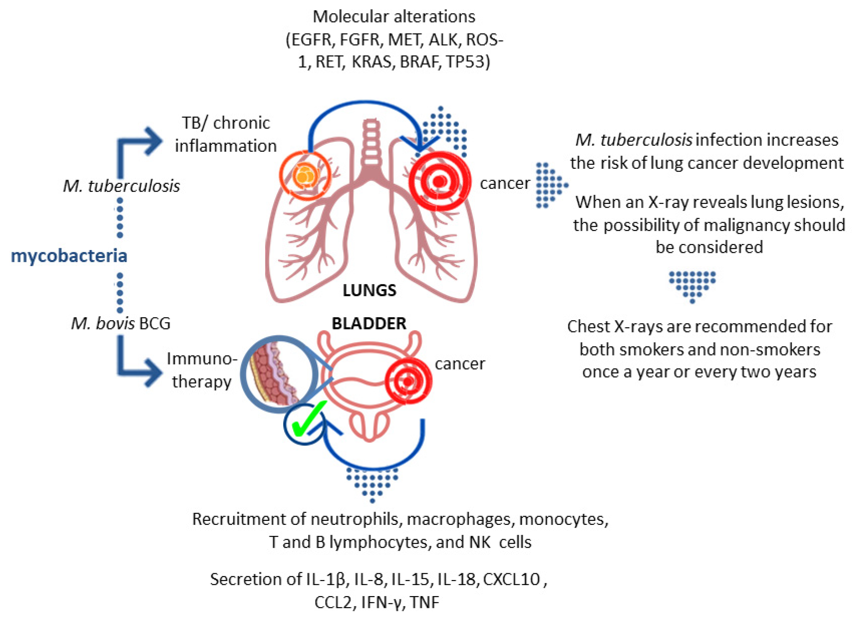
Some indications point out the link between lung cancer development and M.tb infection (Figure 2). The role of M.tb-induced inflammatory events in the development of lung cancer is still being intensively studied [59,60,76,77,78,79,80]. Interestingly, another member of the genus MycobacteriumM. bovis BCG—shows anti-tumor properties [123,124,125]. Mycobacterial suspension infusions are a standard therapy element in the oncological treatment of intermediate-risk bladder cancer [108]. The influence of mycobacteria on the immune system still hides many unclear issues, and recognizing and explaining them may prevent the development of neoplastic processes and/or make anticancer therapy more successful. It is widely known that infected individuals do not experience lung pain due to the lack of sensory innervation, and hence many lung diseases remain asymptomatic for a long time. This applies particularly to such diseases as TB and lung cancer. For this reason, and because of the worse epidemiological situation related to TB, until the 1990s, annual chest examinations were obligatory in Poland, either in the form of radiographic photography (photofluorography) or a standard X-ray image of the chest; this obligation was later abolished. From the perspective of over two decades, the consequence of this decision, in the opinion of many medical practitioners, is a delay in TB and lung cancer diagnosis. Therefore, it is increasingly postulated that non-smokers should have a chest X-ray at least once every two years, and smokers should have a prophylactic chest checkup at least once a year, which would be in the best interest of the patient.

Author Contributions

Conception, M.F. and M.D.; writing—original draft preparation, M.F., M.D., P.K., J.K. and P.B.; writing—review and editing, M.F., M.D. and P.B.; supervision, M.F. and M.D. All authors have read and agreed to the published version of the manuscript.

Funding

This research received no external funding.

Institutional Review Board Statement

Not applicable.

Informed Consent Statement

Not applicable.

Data Availability Statement

The data presented in this study are openly available under reference numbers.

Conflicts of Interest

The authors declare no conflict of interest.

References

  1. Houben, R.M.; Dodd, P.J. The global burden of latent tuberculosis infection: A re-estimation using mathematical modelling. PLoS Med. 2016, 13, e1002152. [Google Scholar] [CrossRef] [Green Version]
  2. World Health Organization (WHO). Global Tuberculosis Report. 2020. Available online: https://apps.who.int/iris/bitstream/handle/10665/336069/9789240013131-eng.pdf (accessed on 30 June 2021).
  3. Di Gennaro, F.; Gualano, G.; Timelli, L.; Vittozzi, P.; Di Bari, V.; Libertone, R.; Cerva, C.; Pinnarelli, L.; Nisii, C.; Ianniello, S.; et al. Increase in Tuberculosis Diagnostic Delay during First Wave of the COVID-19 Pandemic: Data from an Italian Infectious Disease Referral Hospital. Antibiotics 2021, 10, 272. [Google Scholar] [CrossRef] [PubMed]
  4. Religioni, U. Cancer incidence and mortality in Poland. Clin. Epidemiol Glob. Health 2020, 8, 329–334. [Google Scholar] [CrossRef] [Green Version]
  5. Obecny Stan Zwalczania Nowotworów w Polsce. 2014. Available online: http://www.walkazrakiem.pl/sites/default/files/library/files/obecny_stan_zwalczania_nowotworow_w_polsce_10-07-2014_2.pdf (accessed on 30 June 2021).
  6. Ren, H.; Xu, D.; Shi, X.; Xu, J.; Zhuang, D.; Yang, G. Characterization of gastric cancer and its relation to environmental factors: A case study in Shenqiu County, China. Int. J. Env. Health Res. 2016, 26, 1369–1619. [Google Scholar] [CrossRef]
  7. Sung, H.; Ferlay, J.; Siegel, R.L.; Laversanne, M.; Soerjomataram, I.; Jemal, A.; Bray, F. Global Cancer Statistics 2020: GLOBOCAN Estimates of incidence and mortality worldwide for 36 cancers in 185 countries. CA Cancer J. Clin. 2021, 71, 209–249. [Google Scholar] [CrossRef] [PubMed]
  8. Siegel, R.L.; Miller, K.D.; Jemal, A. Cancer Statistics, 2020. CA Cancer J. Clin. 2020, 70, 7–30. [Google Scholar] [CrossRef]
  9. Wojciechowska, U.; Didkowska, J.; Michałek, I.; Olasek, P.; Ciuba, A. Cancer in Poland in 2018. In Raport Narodowego Instytutu Onkologii; Narodowego Instytutu Onkologii: Warsaw, Poland, 2020. [Google Scholar]
  10. Modlińska, A.; Kowalczyk, A. Rak płuca–epidemiologia, obraz kliniczny oraz społeczne następstwa choroby. Psychoonkologia 2016, 20, 57–65. [Google Scholar] [CrossRef]
  11. Giovino, G.A.; Mirza, S.A.; Samet, J.M.; Gupta, P.C.; Jarvis, M.; Bhala, N.; Peto, R.; Zatonski, W.; Hsia, J.; Morton, J.; et al. Tobacco use in 3 billion individuals from 16 countries: An analysis of nationally representative cross-sectional household surveys. Lancet 2012, 380, 668–679. [Google Scholar] [CrossRef]
  12. Stiles, B.M.; Rahouma, M.; Hussein, M.K.; Nasar, A.; Nguyen, A.B.; Harrison, S.; Lee, B.; Port, J.L.; Altorki, N.K. Never smokers with resected lung cancer: Different demographics, similar survival. Eur. J. Cardio-Thorac Surg. 2018, 53, 842–848. [Google Scholar] [CrossRef]
  13. Saito, S.; Espinoza-Mercado, F.; Liu, H.; Sata, N.; Cui, X.; Soukiasian, H.J. Current status of research and treatment for non-small cell lung cancer in never-smoking females. Cancer Biol. Ther. 2017, 18, 359–368. [Google Scholar] [CrossRef] [Green Version]
  14. Barrera-Rodriguez, R.; Morales-Fuentes, J. Lung cancer in women. Lung Cancer 2012, 3, 79–89. [Google Scholar]
  15. Samet, J.M.; Wiggins, C.L.; Humble, C.G.; Pathak, D.R. Cigarette smoking and lung cancer in New Mexico. Am. Rev. Respir Dis. 1988, 137, 1110–1113. [Google Scholar] [CrossRef] [PubMed]
  16. Dela Cruz, C.S.; Tanoue, L.T.; Matthay, R.A. Lung cancer: Epidemiology, etiology, and prevention. Clin. Chest Med. 2011, 32, 605–644. [Google Scholar] [CrossRef] [Green Version]
  17. Seo, S.; Ha, W.H.; Kang, J.K.; Lee, D.; Park, S.; Kwon, T.E.; Jin, Y.W. Health effects of exposure to radon: Implications of the radon bed mattress incident in Korea. Epidemiol. Health 2019, 41, e2019004. [Google Scholar] [CrossRef] [Green Version]
  18. Hammond, E.C.; Selikoff, I.J.; Seidman, H. Asbestos exposure, cigarette smoking, and death rates. Ann. N. Y. Acad. Sci. 1979, 330, 473–490. [Google Scholar] [CrossRef]
  19. Alberg, A.J.; Brock, M.V.; Ford, J.G.; Samet, J.M.; Spivack, S.D. Epidemiology of lung cancer: Diagnosis and management of lung cancer, 3rd ed.: American college of chest physicians evidence-based clinical practice guidelines. Chest 2013, 143, e1S–e29S. [Google Scholar] [CrossRef] [Green Version]
  20. Corrales, L.; Rosell, R.; Cardona, A.F.; Martín, C.; Zatarain-Barrón, Z.L.; Arrieta, O. Lung cancer in never smokers: The role of different risk factors other than tobacco smoking. Crit. Rev. Oncol. Hematol. 2020, 148, 102895. [Google Scholar] [CrossRef] [PubMed]
  21. Bae, J.M.; Kim, E.H. Human papillomavirus infection and risk of lung cancer in never-smokers and women: An ‘adaptive’ meta-analysis. Epidemiol. Health 2015, 37, e2015052. [Google Scholar] [CrossRef] [PubMed]
  22. Giuliani, L.; Jaxmar, T.; Casadio, C.; Gariglio, M.; Manna, A.; D’Antonio, D.; Syrjanen, K.; Favalli, C.; Ciotti, M. Detection of oncogenic viruses (SV40, BKV, JCV, HCMV, HPV) and p53 codon 72 polymorphism in lung carcinoma. Lung Cancer 2007, 57, 273–281. [Google Scholar] [CrossRef]
  23. de Groot, P.M.; Wu, C.C.; Carter, B.W.; Munden, R.F. The epidemiology of lung cancer. Transl. Lung Cancer Res. 2018, 7, 220–233. [Google Scholar] [CrossRef]
  24. Zheng, H.; Aziz, H.A.; Nakanishi, Y.; Masuda, S.; Saito, H.; Tsuneyama, K.; Takano, Y. Oncogenic role of JC virus in lung cancer. J. Pathol. 2007, 212, 306–315. [Google Scholar] [CrossRef]
  25. Qu, Y.-L.; Liu, J.; Zhang, L.-X.; Wu, C.-M.; Chu, A.-J.; Wen, B.-L.; Ma, C.; Yan, X.-Y.; Zhang, X.; Wang, D.-M.; et al. Asthma and the risk of lung cancer: A meta-analysis. Oncotarget 2017, 8, 11614–11620. [Google Scholar] [CrossRef] [PubMed] [Green Version]
  26. Brenner, D.R.; McLaughlin, J.R.; Hung, R.J. Previous lung diseases and lung cancer risk: A systematic review and meta-analysis. PLoS ONE 2011, 6, e17479. [Google Scholar] [CrossRef]
  27. Yu, Y.H.; Liao, C.C.; Hsu, W.H.; Chen, H.J.; Liao, W.C.; Muo, C.H.; Sung, F.C.; Chen, C.Y. Increased lung cancer risk among patients with pulmonary tuberculosis: A population cohort study. J. Thorac. Oncol. 2011, 6, 32–37. [Google Scholar] [CrossRef] [Green Version]
  28. Seo, G.H.; Kim, M.J.; Seo, S.; Hwang, B.; Lee, E.; Yun, Y.; Choi, M.; Kim, M.; Kim, J.W.; Kim, E.S.; et al. Can-cer-specific incidence rates of tuberculosis: A 5-year nationwide population-based study in a country with an intermediate tu-berculosis burden. Medicine 2016, 95, e4919. [Google Scholar] [CrossRef] [PubMed]
  29. Vento, S.; Lanzafame, M. Tuberculosis and cancer: A complex and dangerous liaison. Lancet Oncol. 2011, 12, 520–522. [Google Scholar] [CrossRef]
  30. Simonsen, D.F.; Farkas, D.K.; Horsburgh, C.R.; Thomsen, R.W.; Sørensen, H.T. Increased risk of active tuberculosis after cancer diagnosis. J. Infect. 2017, 74, 590–598. [Google Scholar] [CrossRef] [PubMed]
  31. Liang, H.-Y.; Li, X.-L.; Yu, X.-S.; Guan, P.; Yin, Z.-H.; He, Q.-C.; Zhou, B.-S. Facts and fiction of the relationship between preexisting tuberculosis and lung cancer risk: A systematic review. Int. J. Cancer 2009, 125, 2936–2944. [Google Scholar] [CrossRef] [PubMed]
  32. Oh, C.M.; Roh, Y.H.; Lim, D.; Kong, H.J.; Cho, H.; Hwangbo, B.; Won, Y.J.; Jung, K.W.; Oh, K. Pulmonary tuberculosis is associated with elevated risk of lung cancer in Korea: The nationwide cohort study. J. Cancer 2020, 11, 1899–1906. [Google Scholar] [CrossRef] [Green Version]
  33. Wu, C.-Y.; Hu, H.-Y.; Pu, C.-Y.; Huang, N.; Shen, H.-C.; Li, C.-P.; Chou, Y.-J. Aerodigestive tract, lung and haematological cancers are risk factors for tuberculosis: An 8-year population-based study. Int. J. Tuberc. Lung Dis. 2011, 15, 125–130. [Google Scholar] [PubMed]
  34. Kim, H.-R.; Hwang, S.S.; Ro, Y.K.; Jeon, C.H.; Ha, D.Y.; Park, S.J.; Lee, C.-H.; Lee, S.-M.; Yoo, C.-G.; Kim, Y.W.; et al. Solid-organ malignancy as a risk factor for tuberculosis. Respirology 2008, 13, 413–419. [Google Scholar] [CrossRef]
  35. Skowroński, M.; Iwanik, K.; Halicka, A.; Barinow-Wojewódzki, A. Squamous cell lung cancer in a male with pulmonary tuberculosis. Pneumonol. Alergol. Pol. 2015, 83, 298–302. [Google Scholar] [CrossRef] [Green Version]
  36. Cukic, V. The association between lung carcinoma and tuberculosis. Med. Arch. 2017, 71, 212–214. [Google Scholar] [CrossRef] [PubMed] [Green Version]
  37. Engels, E.A.; Shen, M.; Chapman, R.S.; Pfeiffer, R.M.; Yu, Y.Y.; He, X.; Lan, Q. Tuberculosis and subsequent risk of lung cancer in Xuanwei, China. Int. J. Cancer 2009, 124, 1183–1187. [Google Scholar] [CrossRef] [PubMed]
  38. Cicènas, S.; Vencevičius, V. Lung cancer in patients with tuberculosis. World J. Surg. Oncol. 2007, 5, 22. [Google Scholar] [CrossRef] [PubMed] [Green Version]
  39. Bhatt, M.; Kant, S.; Bhaskar, R. Pulmonary tuberculosis as differential diagnosis of lung cancer. South Asian J. Cancer 2012, 1, 36–42. [Google Scholar] [CrossRef]
  40. Fried, L.E.; Arbiser, J.L. Honokiol, a multifunctional antiangiogenic and antitumor agent. Antioxid Redox Signal. 2009, 11, 1139–1148. [Google Scholar] [CrossRef] [PubMed]
  41. Liuzzo, G.; Trotta, F.; Pedicino, D. Interleukin-17 in atherosclerosis and cardiovascular disease: The good, the bad, and the unknown. Eur. Heart J. 2013, 34, 556–559. [Google Scholar] [CrossRef] [Green Version]
  42. Mariani, F.; Bocchino, M.; Cappelli, G.; Persechini, T.; Colizzi, V.; Bonanno, E.; Ponticiello, A.; Sanduzzi, A. Tuberculosis and lung cancer. An interesting case study. Monaldi Arch. Chest Dis 2001, 56, 30–32. [Google Scholar] [PubMed]
  43. Çiçek, Y.; Kosar, P.A.; Öztürk, Ö. Molecular genetics of lung cancer. Eurasian J. Pulmonol. 2018, 20, 111–117. [Google Scholar] [CrossRef]
  44. McCusker, C.; Warrington, R. Primary immunodeficiency. Allergy Asthma Clin. Immunol. 2011, 7 Suppl 1, S11. [Google Scholar] [CrossRef] [Green Version]
  45. Hauck, F.; Gennery, A.R.; Seidel, M.G. Editorial: The relationship between cancer predisposition and primary immunodeficiency. Front. Immunol. 2019, 30, 1781. [Google Scholar] [CrossRef] [PubMed]
  46. Boisson-Dupuis, S.; Bustamante, J.; El-Baghdadi, J.; Camcioglu, Y.; Parvaneh, N.; El Azbaoui, S.; Agader, A.; Hassani, A.; El Hafidi, N.; Mrani, N.A.; et al. Inherited and acquired immunodeficiencies underlying tuberculosis in childhood. Immunol. Rev. 2015, 264, 103–120. [Google Scholar] [CrossRef] [PubMed] [Green Version]
  47. Bustamante, J.; Boisson-Dupuis, S.; Abel, L.; Casanova, J.L. Mendelian susceptibility to mycobacterial disease: Genetic, immunological, and clinical features of inborn errors of IFN-γ immunity. Semin. Immunol. 2014, 26, 454–470. [Google Scholar] [CrossRef] [Green Version]
  48. Swinson, S.; Hall, G.; Pollard, A.J. Reactivation of the bacille Calmette-Guerin scar following immune reconstitution during treatment of infant acute lymphoblastic leukemia. J. Pediatr. Hematol. Oncol. 2004, 26, 112–115. [Google Scholar] [CrossRef]
  49. Arlotta, A.; Cefalo, M.G.; Maurizi, P.; Ruggiero, A.; Dodi, I.; Riccardi, R. Critical pulmonary infection due to nontuberculous mycobacterium in pediatric leukemia: Report of a difficult diagnosis and review of pediatric series. J. Pediatr. Hematol. Oncol. 2014, 36, 66–70. [Google Scholar] [CrossRef] [PubMed] [Green Version]
  50. Claass, A.; Claviez, A.; Westphal, E.; Rusch-Gerdes, S.; Schneppenheim, R. First case of disseminated Mycobacterium avium infection following chemotherapy for childhood acute myeloid leukemia. Infection 1995, 23, 301–302. [Google Scholar] [CrossRef]
  51. Gupta, S.; Basu, A.K.; Guliani, A.; Dhamija, A. Hemophagocytic lymphohistiocytosis complicating nontuberculous mycobacterial infection. Lung India 2019, 36, 266–267. [Google Scholar] [PubMed]
  52. Valdez, J.M.; Scheinberg, P.; Young, N.S.; Walsh, T.J. Infections in patients with aplastic anemia. Semin. Hematol. 2009, 46, 269–276. [Google Scholar] [CrossRef]
  53. Patel, R.; Roberts, G.D.; Keating, M.R.; Paya, C.V. Infections due to nontuberculous mycobacteria in kidney, heart, and liver transplant recipients. Clin. Infect. Dis. 1994, 19, 263–273. [Google Scholar] [CrossRef]
  54. Searle, E.; Patel, H.; Vilar, F.J.; Gharib, M.; Turner, A.J.; Batra, G.; Wynn, R.F. Inflammatory BCG adenitis associated with immune reconstitution following allogeneic haematopoietic stem cell transplant in infancy. Pediatr. Blood Cancer 2010, 54, 166–169. [Google Scholar] [CrossRef] [PubMed]
  55. Unal, E.; Yen, C.; Saiman, L.; George, D.; Della-Latta, P.; van de Ven, C.; Morris, E.; Bradley, M.B.; Del Toro, G.; Garvin, J.; et al. A low incidence of nontuberculous mycobacterial infections in pediatric hematopoietic stem cell transplantation recipients. Biol. Blood Marrow Transplant. 2006, 12, 1188–1197. [Google Scholar] [CrossRef] [PubMed] [Green Version]
  56. Tshitenge, S.; Ogunbanjo, G.A.; Citeya, A. A mortality review of tuberculosis and HIV co-infected patients in Mahalapye, Botswana: Does cotrimoxazole preventive therapy and/or antiretroviral therapy protect against death? Afr. J. Prim. Health Care Fam. Med. 2018, 10, e1–e5. [Google Scholar] [CrossRef]
  57. Walker, N.F.; Meintjes, G.; Wilkinson, R.J. HIV-1 and the immune response to TB. Future Virol. 2013, 8, 57–80. [Google Scholar] [CrossRef] [Green Version]
  58. Lapinel, N.C.; Jolley, S.E.; Ali, J.; Welsh, D.A. Prevalence of non-tuberculous mycobacteria in HIV-infected patients admitted to hospital with pneumonia. Int. J. Tuberc Lung Dis. 2019, 23, 491–497. [Google Scholar] [CrossRef]
  59. Ames, B.N.; Gold, L.S.; Willett, W.C. The causes and prevention of cancer. Proc. Natl. Acad. Sci. USA 1995, 92, 5258–5265. [Google Scholar] [CrossRef] [Green Version]
  60. Lu, H.; Ouyang, W.; Huang, C. Inflammation, a key event in cancer development. Mol. Cancer Res. 2006, 4, 221–233. [Google Scholar] [CrossRef] [Green Version]
  61. Wu, C.Y.; Hu, H.Y.; Pu, C.Y.; Huang, N. Pulmonary tuberculosis increases the risk of lung cancer: A Population-based cohort study. Cancer 2011, 117, 618–624. [Google Scholar] [CrossRef] [PubMed]
  62. Brown, S.T.; Almenoff, P.L. Pulmonary mycobacterial infections associated with neoplasia. Semin. Respir. Infect. 1992, 7, 104–113. [Google Scholar]
  63. Alavanja, M.C.; Brownson, R.C.; Boice, J.D., Jr.; Hock, E. Preexisting lung disease and lung cancer among nonsmoking women. Am. J. Epidemiol. 1992, 136, 623–632. [Google Scholar] [CrossRef]
  64. Lande, L.; Gogoi, R.; Yankowski, C.; Stampler, K.; Sawicki, K.; Daum, G.; Peterson, D.D.; Sawicki, J. Association between pulmonary Mycobacterium avium complex infection and lung cancer. J. Thorac. Oncol. 2012, 7, 1345–1351. [Google Scholar] [CrossRef] [PubMed] [Green Version]
  65. Evans, M.R.; Etuaful, S.N.; Amofah, G.; Adjei, O.; Lucas, S.; Wansbrough-Jones, M.H. Squamous cell carcinoma secondary to Buruli ulcer. Trans. R Soc. Trop. Med. Hyg. 1999, 93, 63–64. [Google Scholar] [CrossRef]
  66. Stanley, S.A.; Cox, J.S. Host-pathogen interactions during Mycobacterium tuberculosis infections. Curr. Top. Microbiol. Immunol. 2013, 374, 211–241. [Google Scholar] [PubMed]
  67. Ehrt, S.; Schnappinger, D. Mycobacterial survival strategies in the phagosome: Defence against host stresses. Cell Microbiol. 2009, 11, 1170–1178. [Google Scholar] [CrossRef] [Green Version]
  68. Guirado, E.; Schlesinger, L.S. Modeling the Mycobacterium tuberculosis granuloma—the critical battlefield in host immunity and disease. Front. Immunol. 2013, 4, 98. [Google Scholar] [CrossRef] [Green Version]
  69. Dalgleish, A.G.; O’Byrne, K.J. Chronic immune activation and inflammation in the pathogenesis of AIDS and cancer. Adv. Cancer Res. 2002, 84, 231–276. [Google Scholar]
  70. Melnikova, V.O.; Ananthaswamy, H.N. Cellular and molecular events leading to the development of skin cancer. Mutat. Res. 2005, 571, 91–106. [Google Scholar] [CrossRef]
  71. Azad, N.; Rojanasakul, Y.; Vallyathan, V. Inflammation and lung cancer: Roles of reactive oxygen/nitrogen species. J. Toxicol. Environ. Health B Crit. Rev. 2008, 11, 1–15. [Google Scholar] [CrossRef]
  72. Bowden, G.T. Prevention of non-melanoma skin cancer by targeting ultraviolet-b-light signalling. Nat. Rev. Cancer 2004, 4, 23–35. [Google Scholar] [CrossRef]
  73. Broussard, G.W.; Norris, M.B.; Schwindt, A.R.; Fournie, J.W.; Winn, R.N.; Kent, M.L.; Ennis, D.G. Chronic Mycobacterium marinum infection acts as a tumor promoter in Japanese Medaka (Oryziaslatipes). Comp. Biochem. Physiol. C Toxicol. Pharm. 2009, 149, 152–160. [Google Scholar] [CrossRef] [PubMed] [Green Version]
  74. Teventiyanon, T.; Ratanaharathorn, V.; Leoparait, J. Mucoepidermoid carcinoma of the lung presenting as cavitary lesion. J. Med. Assoc. Thail. 2004, 87, 988–991. [Google Scholar]
  75. Yilmaz, A.; Gungor, S.; Damadoglu, E.; Axoy, F.; Aibatly, A. Coexisting bronchial carcinoid tumor and pulmonary tuberculosis in the same lobe: A case report. Tuberk Toraks 2004, 52, 369–372. [Google Scholar]
  76. Weitzman, S.A.; Weitberg, A.B.; Clark, E.P.; Stossel, T.P. Phagocytes as carcinogens: Malignant transformation produced by human neutrophils. Science 1985, 227, 1231–1233. [Google Scholar] [CrossRef]
  77. Shacter, E.; Beecham, E.J.; Covey, J.M.; Kohn, K.W.; Potter, M. Activated neutrophils induce prolonged DNA damage in neighboring cells. Carcinogenesis 1988, 9, 2297–2304. [Google Scholar] [CrossRef]
  78. Wakabayashi, N.; Itoh, K.; Wakabayashi, J.; Motohashi, H.; Noda, S.; Takahashi, S.; Imakado, S.; Kotsuji, T.; Otsuka, F.; Roop, D.R.; et al. Keap1-null mutation leads to postnatal lethality due to constitutive Nrf2 activation. Nat. Genet. 2003, 35, 238–245. [Google Scholar] [CrossRef]
  79. Zhuang, J.C.; Lin, D.; Lin, C.; Jethwaney, D.; Wogan, G.N. Genotoxicity associated with NO production in macrophages and co-cultured target cells. Free Radic. Biol. Med. 2002, 33, 94–102. [Google Scholar] [CrossRef]
  80. Kim, M.Y.; Wogan, G.N. Mutagenesis of thesupFGene of pSP189 Replicating in AD293 Cells Cocultivated with Activated Macrophages: Roles of Nitric Oxide and Reactive Oxygen Species. Chem. Res. Toxicol. 2006, 19, 1483–1491. [Google Scholar] [CrossRef]
  81. Molina-Romero, C.; Arrieta, O.; Hernaández-Pando, R. Tuberculosis and lung cancer. Salud Publica Mex. 2019, 61, 286–291. [Google Scholar] [CrossRef] [Green Version]
  82. Ardies, C.M. Inflammation as cause for scar cancers of the lung. Integr. Cancer Ther. 2003, 2, 238–246. [Google Scholar] [CrossRef] [PubMed] [Green Version]
  83. Brenner, A.V.; Zuoyuan, W.; Kleinerman, R.A.; Wang, L.; Zhang, S.; Metayer, C.; Chen, K.; Lei, S.; Cui, H.; Lubin, J.H. Previous pulmonary diseases and risk of lung cancer in Gansu Province, China. Int. J. Epidemiol. 2001, 30, 118–124. [Google Scholar] [CrossRef] [PubMed] [Green Version]
  84. Shiels, M.S.; Albanes, D.; Virtamo, J.; Engels, E.A. Increased risk of lung cancer in men with tuberculosis in the alpha-tocopherol, beta-carotene cancer prevention study. Cancer Epidemiol. Biomark. Prev. 2011, 20, 672–678. [Google Scholar] [CrossRef] [Green Version]
  85. Brenner, D.R.; Boffetta, P.; Duell, E.J.; Bickeböller, H.; Rosenberger, A.; McCormack, V.; Muscat, J.E.; Yang, P.; Wichmann, H.-E.; Brueske-Hohlfeld, I.; et al. Previous Lung Diseases and Lung Cancer Risk: A Pooled Analysis From the International Lung Cancer Consortium. Am. J. Epidemiol. 2012, 176, 573–585. [Google Scholar] [CrossRef] [PubMed]
  86. Everatt, R.; Kuzmickiene, I.; Davidaviciene, E.; Cicenas, S. Incidence of lung cancer among patients with tuberculosis: A nationwide cohort study in Lithuania. Int. J. Tuberc Lung Dis. 2016, 20, 757–763. [Google Scholar] [CrossRef]
  87. Denholm, R.; Schüz, J.; Straif, K.; Stücker, I.; Jockel, K.-H.; Brenner, D.R.; De Matteis, S.; Boffetta, P.; Guida, F.; Brüske, I.; et al. Is Previous Respiratory Disease a Risk Factor for Lung Cancer? Am. J. Respir. Crit. Care Med. 2014, 190, 549–559. [Google Scholar] [CrossRef] [PubMed] [Green Version]
  88. Leung, C.C. Does tuberculosis increase the risk of lung cancer? Int. J. Tuberc Lung Dis. 2016, 20, 712. [Google Scholar] [CrossRef] [PubMed]
  89. Yu, Y.Y.; Pinsky, P.F.; Caporaso, N.E.; Chatterjee, N.; Baumgarten, M.; Langenberg, P.; Furuno, J.P.; Lan, Q.; Engels, E.A. Lung, cancer risk following detection of pulmonary scarring by chest radiography in the prostate, lung, colorectal, and ovarian cancer screening trial. Arch. Intern. Med. 2008, 168, 2326–2332. [Google Scholar] [CrossRef]
  90. Dacosta, N.A.; Kinare, S.G. Association of lung carcinoma and tuberculosis. J. Postgrad. Med. 1991, 37, 185–189. [Google Scholar] [PubMed]
  91. Nalbandian, A.; Yan, B.S.; Pichugin, A.; Bronson, R.T.; Kramnik, I. Lung carcinogenesis induced by chronic tuberculosis infection: The experimental model and genetic control. Oncogene 2009, 28, 1928–1938. [Google Scholar] [CrossRef] [Green Version]
  92. Cao, S.; Li, J.; Lu, J.; Zhong, R.; Zhong, H. Mycobacterium tuberculosis antigens repress Th1 immune response suppression and promote lung cancer metastasis through PD-1/PDl-1 signaling pathway. Cell Death Dis. 2019, 10, 44. [Google Scholar] [CrossRef] [Green Version]
  93. Holla, S.; Ghorpade, D.S.; Singh, V.; Bansal, K.; Balaji, K.N. Mycobacterium bovis BCG promotes tumor cell survival from tumor necrosis factor-α-induced apoptosis. Mol. Cancer 2014, 13, 210. [Google Scholar] [CrossRef] [Green Version]
  94. Ratoosh, S.L.; Cohen, P.R.; Troncoso, P. Cutaneous-malignancy and leprosy. Report of a patient with Mycobacterium leprae and basal cell carcinoma concurrently present in the same lesion. J. Derm. Surg. Oncol. 1994, 20, 613–618. [Google Scholar] [CrossRef]
  95. Cope, R.B.; Stang, B.; Valentine, B.A.; Bermudez, L.E. Topical exposure to exogenous ultraviolet-irradiated urocanic acid enhances Mycobacterium ulcerans infection in a Crl:IAF(HA)-hrBR hairless guinea-pig model of Buruli ulcer disease. Photodermatol. Photoimmunol. Photomed. 2004, 20, 14–20. [Google Scholar] [CrossRef] [PubMed]
  96. Richardus, J.H.; Smith, T.C. Squamous cell carcinoma in chronic ulcers in leprosy: A review of 38 consecutive cases. Lepr. Rev. 1991, 62, 381–388. [Google Scholar] [CrossRef]
  97. Asenjo, M.M.; Martín Guerra, J.M.; López Pedreira, M.R.; Prieto de Paula, J.M. Mycobacterium xenopi and squamous cell carcinoma of the lung. Arch. Bronconeumol. 2017, 53, 698–700. [Google Scholar] [CrossRef]
  98. Pierce, E.S. Could Mycobacterium avium subspecies paratuberculosis cause Crohn’s disease, ulcerative colitis…and colorectal cancer? Infect. Agents Cancer 2018, 13, 1. [Google Scholar] [CrossRef]
  99. Alsaif, H.S.; Hassan, A.; Refai, O.; Awary, K.; Kussaibi, H.; Ismail, M.H.; Alghnimi, I. Concomitant hepatic tuberculosis and hepatocellular carcinoma: A case report and review of the literature. BMC Surg. 2021, 21, 2. [Google Scholar] [CrossRef] [PubMed]
  100. El-Mahallawy, H.A.; Eissa, S.A.; Refeh, N.G.; Salem, A.E.S.; Eissa, S.A.; Allian, S.A. Tuberculosis in cancer patients: Role of newer techniques in relation to conventional diagnostic methods. J. Adv. Res. 2010, 1, 157–162. [Google Scholar] [CrossRef] [Green Version]
  101. Laake, A.M.; Liappis, A.P.; Guy, E.; Kerr, G.; Benator, D.A. Tuberculosis reactivation in hepatocellular carcinoma: Association with transarterial chemoembolization. Infect. Dis. 2015, 47, 267–270. [Google Scholar] [CrossRef]
  102. He, W.; Zhang, X.; Li, W.; Kong, C.; Wang, Y.; Zhu, L.; Xu, R.; Deng, G.; Zhang, R. Activated pulmonary tuberculosis in a patient with melanoma during PD-1 inhibition: A case report. Onco. Targets Ther. 2018, 11, 7423–7427. [Google Scholar] [CrossRef] [PubMed] [Green Version]
  103. Suliman, A.M.; Bek, S.A.; Elkhatim, M.S.; Husain, A.A.; Mismar, A.Y.; Sharaf, E.M.Z.; Lengyel, Z.; Elazzazy, S.; Rasul, K.I.; Omar, N.E. Tuberculosis following programmed cell death receptor-1 (PD-1) inhibitor in a patient with non-small cell lung cancer. Case report and literature review. Cancer Immunol. Immunother. 2020, 70, 935–944. [Google Scholar] [CrossRef]
  104. Lim, Y.P.; Lin, C.L.; Hung, D.Z.; Lin, Y.N.; Kao, C.H. Anti-tuberculosis treatments and risk of hepatocellular carcinoma in tuberculosis patients with liver cirrhosis: A population-based case–control study. Eur. J. Clin. Microbiol. Infect. Dis. 2015, 34, 479–485. [Google Scholar] [CrossRef] [PubMed]
  105. Anguille, S.; Smits, E.L.; Lion, E.; van Tendeloo, V.F.; Berneman, Z.N. Clinical use of dendritic cells for cancer therapy. Lancet Oncol. 2014, 15, 257–267. [Google Scholar] [CrossRef]
  106. Bickels, J.; Kollender, Y.; Merinsky, O.; Meller, I. Coley’s toxin: Historical perspective. Isr. Med. Assoc. J. 2002, 4, 471–472. [Google Scholar]
  107. McCarthy, E.F. The toxin of William, B. Coley and the treatment of bone and soft-tissue sarcomas. Iowa Orthop. J. 2006, 26, 154–158. [Google Scholar]
  108. Kawai, K.; Miyazaki, J.; Joraku, A.; Nishiyama, H.; Akaza, H. Bacillus Calmette–Guerin (BCG) immunotherapy for bladder cancer: Current understanding and perspectives on engineered BCG vaccine. Cancer Sci. 2013, 104, 22–27. [Google Scholar] [CrossRef]
  109. Delogu, G.; Fadda, G. The quest for a new vaccine against tuberculosis. J. Infect. Dev. Ctries. 2009, 3, 5–15. [Google Scholar] [CrossRef] [PubMed] [Green Version]
  110. McShane, H. Vaccine strategies against tuberculosis. Swiss Med. Wkly. 2009, 139, 156–160. [Google Scholar]
  111. Salyers, A.A.; Whitt, D.D. Gruźlica: Powrót danego wroga. In Mikrobiologia. Różnorodność, Chorobotwórczość i Środowisko; Markiewicz, Z., Ed.; Wydawnictwo Naukowe PWN: Warsaw, Poland, 2003; pp. 336–340. [Google Scholar]
  112. Blok, B.A.; Arts, R.J.; van Crevel, R.; Benn, C.S.; Netea, M.G. Trained innate immunity as underlying mechanism for the long-term, nonspecific effects of vaccines. J. Leukocyt. Biol. 2015, 98, 347–356. [Google Scholar] [CrossRef] [PubMed]
  113. Garly, M.L.; Martins, C.L.; Balé, C.; Baldé, M.A.; Hedegaard, K.L.; Gustafson, P.; Lisse, I.M.; Whittle, H.C.; Aaby, P. BCG scar and positive tuberculin reaction associated with reduced child mortality in West Africa. A non-specific beneficial effect of BCG? Vaccine 2003, 21, 2782–2790. [Google Scholar] [CrossRef]
  114. Roth, A.; Gustafson, P.; Nhaga, A.; Djana, Q.; Poulsen, A.; Garly, M.L.; Jensen, H.; Sodemann, M.; Rodriques, A.; Aaby, P. BCG vaccination scar associated with better childhood survival in Guinea-Bissau. Int. J. Epidemiol. 2005, 34, 540–547. [Google Scholar] [CrossRef] [Green Version]
  115. Aaby, P.; Roth, A.; Ravn, H.; Napirna, B.M.; Rodrigues, A.; Lisse, I.M.; Stensballe, L.; Diness, B.R.; Lausch, K.R.; Lund, N. Randomized trial of BCG vaccination at birth to low-birth-weight children: Beneficial nonspecific effects in the neonatal period? J. Infect. Dis. 2011, 204, 245–252. [Google Scholar] [CrossRef]
  116. Biering-Sørensen, S.; Aaby, P.; Napirna, B.M.; Roth, A.; Ravn, H.; Rodrigues, A.; Whittle, H.; Benn, C.S. Small randomized trial among low-birth-weight children receiving bacillus Calmette-Guérin vaccination at first health center contact. Pediatr. Infect. Dis. J. 2012, 31, 306–308. [Google Scholar] [CrossRef] [Green Version]
  117. van ’t Wout, J.W.; Poell, R.; van Furth, R. The role of BCG/PPD-activated macrophages in resistance against systemic candidiasis in mice. Scand. J. Immunol. 1992, 36, 713–719. [Google Scholar] [CrossRef]
  118. Kleinnijenhuis, J.; Quintin, J.; Preijers, F.; Joosten, L.A.; Ifrim, D.C.; Saeed, S.; Jacobs, C.; van Loenhout, J.; de Jong, D.; Stunnenberg, H.G. Bacille Calmette-Guerin induces NOD2-dependent nonspecific protection from reinfection via epigenetic reprogramming of monocytes. Proc. Natl. Acad. Sci. USA 2012, 109, 17537–17542. [Google Scholar] [CrossRef] [PubMed] [Green Version]
  119. Netea, M.G.; Quintin, J.; van der Meer, J.W. Trained immunity: A memory for innate host defense. Cell Host Microbe 2011, 9, 355–361. [Google Scholar] [CrossRef] [PubMed] [Green Version]
  120. Netea, M.G.; Joosten, L.A.; Latz, E.; Mills, K.H.; Natoli, G.; Stunnenberg, H.G.; O’Neill, L.A.; Xavier, R.J. Trained immunity: Program of innate immune memory in health and disease. Science 2016, 352, aaf1098. [Google Scholar] [CrossRef] [Green Version]
  121. Kleinnijenhuis, J.; Quintin, J.; Preijers, F.; Benn, C.S.; Joosten, L.A.; Jacobs, C.; Van Loenhout, J.; Xavier, R.J.; Aaby, P.; Van Der Meer, J.W.; et al. Long-Lasting Effects of BCG Vaccination on Both Heterologous Th1/Th17 Responses and Innate Trained Immunity. J. Innate Immun. 2014, 6, 152–158. [Google Scholar] [CrossRef] [PubMed] [Green Version]
  122. Kiselyov, A.; Bunimovich-Mendrazitsky, S.; Startsev, V. Treatment of non-muscle invasive bladder cancer with Bacillus Calmette-Guerin (BCG): Biological markers and simulation studies. BBA Clin. 2015, 4, 27–34. [Google Scholar] [CrossRef] [PubMed] [Green Version]
  123. Pearl, R. Cancer and tuberculosis. Am. J. Epidemiol. 1929, 9, 97–159. [Google Scholar] [CrossRef]
  124. Kamat, A.M.; Lamm, D.L. Immunotherapy for bladder cancer. Curr. Urol. Rep. 2001, 2, 62–69. [Google Scholar] [CrossRef]
  125. Lamm, D.L. Bacillus Calmette-Guerin immunotherapy for bladder cancer. J. Urol. 1985, 134, 40–46. [Google Scholar] [CrossRef]
  126. Ayati, M.; Nowroozi, M.R.; Mortazavi, A.; Ohadian Moghadam, S.; Ghorani, H. Management of hepatic granulomatous tuberculosis after BCG therapy for bladder cancer. Urol. Case Rep. 2017, 13, 158–159. [Google Scholar] [CrossRef]
  127. Fujita, K.; Yamamoto, Y.; Kanai, O.; Okamura, M.; Nakatani, K.; Mio, T. Development of Mycobacterium avium complex lung disease in patients with lung cancer on immune checkpoint inhibitors. Open Forum Infect. Dis. 2020, 7, ofaa067. [Google Scholar] [CrossRef] [Green Version]
  128. Bereta, M.; Hayhurst, A.; Gajda, M.; Chorobik, P.; Targosz, M.; Marcinkiewicz, J.; Kaufman, H.L. Improving tumor targeting and therapeutic potential of Salmonella VNP20009 by displaying cell surface CEA-specific antibodies. Vaccine 2007, 22, 4183. [Google Scholar] [CrossRef] [PubMed] [Green Version]
  129. Pettenati, C.; Ingresoll, M.A. Mechanisms of BCG immunotherapy and its outlook for bladder cancer. Nat. Rev. Urol. 2018, 15, 615–625. [Google Scholar] [CrossRef]
  130. Guallar-Garrido, S.; Julián, E. Bacillus Calmette-Guérin (BCG) therapy for bladder cancer: An update. ImmunoTargets Ther. 2020, 9, 1–11. [Google Scholar] [CrossRef] [PubMed] [Green Version]
  131. Unda-Urzaiz, M.; Cozar-Olmos, J.M.; Miñana-Lopez, B.; Camarero-Jimenez, J.; Brugarolas-Rossello, X.; Zubiaur-Libano, C.; Ribal-Caparros, M.J.; Suarez-Charneco, A.J.; Rodriguez-Tesedo, V.; Chantada-Abal, V.; et al. Safety and efficacy of various strains of bacille Calmette-Guérin in the treatment of bladder tumours in standard clinical practice. Actas Urol. Esp. 2018, 42, 238–248. [Google Scholar] [CrossRef] [PubMed]
  132. Krajewski, W.; Matuszewski, M.; Poletajew, S.; Grzegrzółka, J.; Zdrojowy, R.; Kołodziej, A. Are there differences in toxicity and efficacy between various bacillus Calmette-Guerin strains in bladder cancer patients? Analysis of 844 patients. Urol. Int. 2018, 101, 277–284. [Google Scholar] [CrossRef]
  133. D’Andrea, D.; Soria, F.; Abufaraj, M.; Pones, M.; Gontero, P.; Machado, A.T.; Waksman, R.; Enikeev, D.; Glybochko, P.V.; Adonias, S.P.; et al. Comparative Effectiveness of Intravesical BCG-Tice and BCG-Moreau in Patients With Non–muscle-invasive Bladder Cancer. Clin. Genitourin. Cancer 2020, 18, 20–25.e2. [Google Scholar] [CrossRef] [Green Version]
  134. Rentsch, C.A.; Birkhäuser, F.D.; Biot, C.; Gsponer, J.R.; Bisiaux, A.; Wetterauer, C.; Lagranderie, M.; Marchal, G.; Orgeur, M.; Bouchier, C.; et al. Bacillus Calmette-Guérin Strain Differences Have an Impact on Clinical Outcome in Bladder Cancer Immunotherapy. Eur. Urol. 2014, 66, 677–688. [Google Scholar] [CrossRef]
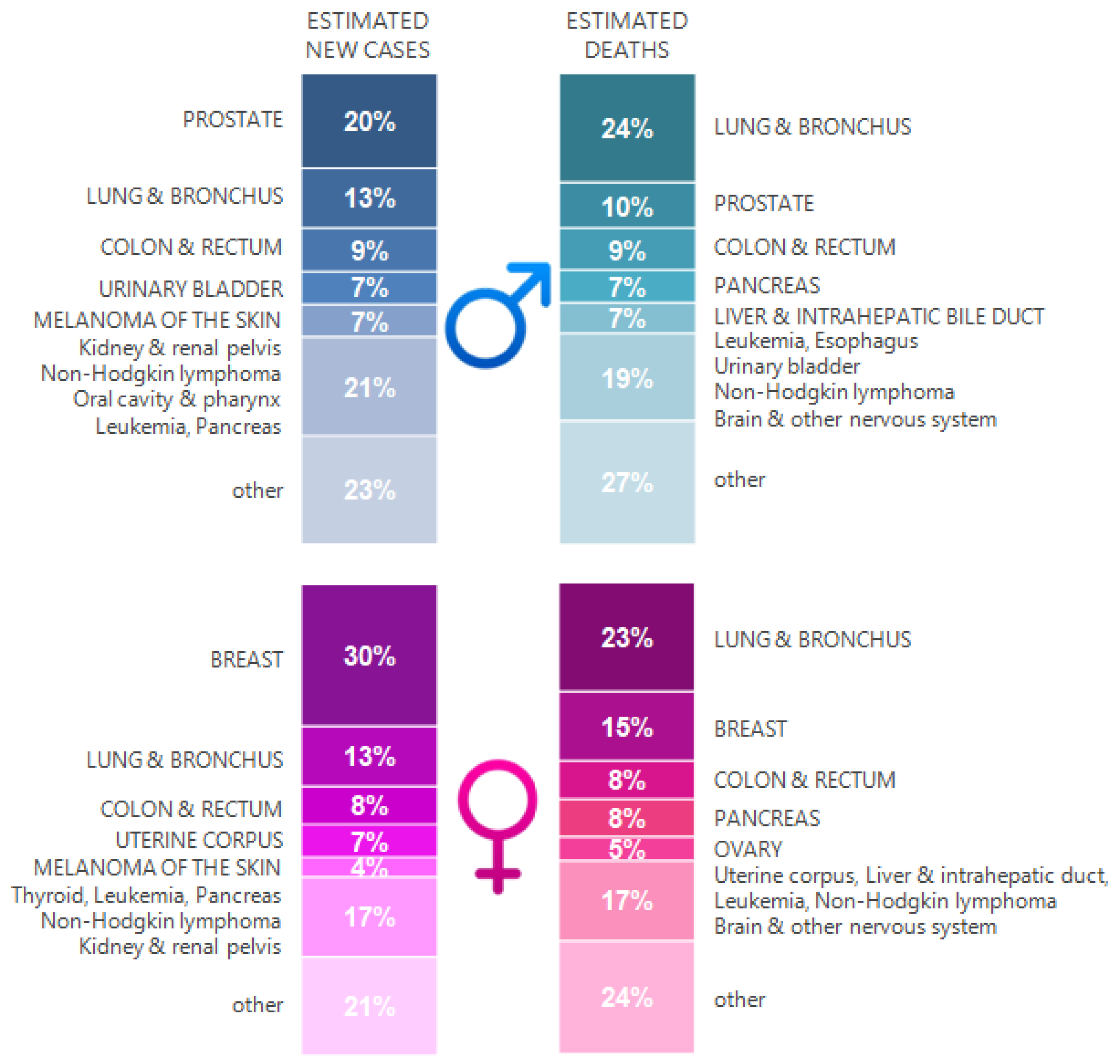
Figure 1. Lung cancer against the background of other cancer types with the highest estimated new cases and deaths by sex, United States, 2020. The estimates are rounded to the nearest 10 and exclude basal cell carcinoma, squamous cell carcinoma of the skin, and carcinoma in situ except from bladder cancer. The ranking is based on modeled forecasts and may differ from the most recent data observed (based on [8]).
Ijms 22 08332 g001
Figure 2. The dual nature of the relationship between mycobacteria and cancer. The risk of tumor development in chronic M.tb infections is believed to be the result of multiple host effector mechanisms at various stages of oncogenesis. On the other hand, the influence of M. bovis BCG on the immune system prevents the development of neoplastic processes and/or increases the effectiveness of cancer therapy.
Ijms 22 08332 g002
Publisher’s Note: MDPI stays neutral with regard to jurisdictional claims in published maps and institutional affiliations.