
Biofilms in Diabetic Foot Ulcers: Impact, Risk Factors and Control Strategies

 ,
,  ,
,  and
and 
Abstract
:1. Introduction
2. Epidemiology and Risk Factors of Diabetic Foot Ulcers
3. Social and Economic Burden of Diabetic Foot Ulcers
4. Etiopathogenesis of Diabetic Foot Ulcers
4.1. Peripheral Neuropathy
4.2. Peripheral Arterial Disease
5. Preventive Measures for Diabetic Foot Ulcers
6. Management and Treatment of Diabetic Foot Ulcers
7. The Role of Biofilms in Diabetic Foot Ulcers
7.1. Biofilm-Associated Diabetic Foot Ulcer Infections
7.1.1. Clinical Profile
7.1.2. Diagnosis and Treatment Using Conventional Methods
7.1.3. Unconventional Therapeutic Strategies
8. Concluding Remarks and Challenges
Supplementary Materials
Author Contributions
Funding
Conflicts of Interest
References
- American Diabetes Association. Diagnosis and Classification of Diabetes Mellitus. Diabetes Care 2013, 37, S81–S90. [Google Scholar] [CrossRef] [Green Version]
- Mukhtar, Y.; Galalain, A.M.; Yunusa, U.M. A Modern Overview on Diabetes Mellitus: A Chronic Endocrine Disorder. Eur. J. Biol. 2019, 4, 1–14. [Google Scholar]
- Katsarou, A.; Gudbjörnsdottir, S.; Rawshani, A.; Dabelea, D.; Bonifacio, E.; Anderson, B.J.; Jacobsen, L.; Schatz, D.A.; Lernmark, Å. Type 1 diabetes mellitus. Nat. Rev. Dis. Prim. 2017, 3, nrdp201716. [Google Scholar] [CrossRef]
- Chatterjee, S.; Khunti, K.; Davies, M.J. Type 2 diabetes. Lancet 2017, 389, 2239–2251. [Google Scholar] [CrossRef]
- Coustan, D.R. Gestational Diabetes Mellitus. Clin. Chem. 2013, 59, 1310–1321. [Google Scholar] [CrossRef] [PubMed] [Green Version]
- WHO. Diabetes. 2020. Available online: https://www.who.int/health-topics/diabetes#tab=tab_1 (accessed on 23 March 2021).
- Wild, S.; Roglic, G.; Green, A.; Sicree, R.; King, H. Estimates for the year 2000 and projections for 2030. Diabetes Care 2004, 27, 1047–1053. [Google Scholar] [CrossRef] [PubMed] [Green Version]
- IDF. IDF Diabetes Atlas, 9th ed.; International Diabetes Federation: Brussels, Belgium, 2019. [Google Scholar]
- Forlee, M. What is the diabetic foot? The rising prevalence of diabetes worldwide will mean an increasing prevalence of complications such as those of the extremities. CME 2011, 29, 4–8. [Google Scholar]
- Icks, A.; Scheer, M.; Morbach, S.; Genz, J.; Haastert, B.; Giani, G.; Glaeske, G.; Hoffmann, F. Time-Dependent Impact of Diabetes on Mortality in Patients After Major Lower Extremity Amputation: Survival in a population-based 5-year cohort in Germany. Diabetes Care 2011, 34, 1350–1354. [Google Scholar] [CrossRef] [PubMed] [Green Version]
- Kerr, M. Foot Care for People with Diabetes: The Economic Case for Change. NHS Diabetes 2012-03. 2012. Available online: http://www.diabetes.org.uk/Documents/%0Anhs-diabetes/footcare/footcare-for-people-with-diabetes.pdf (accessed on 23 March 2021).
- Barshes, N.R.; Sigireddi, M.; Wrobel, J.S.; Mahankali, A.; Robbins, J.M.; Kougias, P.; Armstrong, D.G. The system of care for the diabetic foot: Objectives, outcomes, and opportunities. Diabet. Foot Ankle 2013, 4, 21847. [Google Scholar] [CrossRef]
- WHO. Global Report on Diabetes; Geneva World Health Organization Press: Geneva, Switzerland, 2016. [Google Scholar]
- Noor, S.; Zubair, M.; Ahmad, J. Diabetic foot ulcer—A review on pathophysiology, classification and microbial etiology. Diabetes Metab. Syndr. Clin. Res. Rev. 2015, 9, 192–199. [Google Scholar] [CrossRef]
- Yazdanpanah, L. Literature review on the management of diabetic foot ulcer. World J. Diabetes 2015, 6, 37–53. [Google Scholar] [CrossRef] [PubMed]
- Johannesson, A.; Larsson, G.-U.; Ramstrand, N.; Turkiewicz, A.; Wiréhn, A.-B.; Atroshi, I. Incidence of Lower-Limb Amputation in the Diabetic and Nondiabetic General Population: A 10-year population-based cohort study of initial unilateral and contralateral amputations and reamputations. Diabetes Care 2009, 32, 275–280. [Google Scholar] [CrossRef] [Green Version]
- Liu, C.; You, J.; Zhu, W.; Chen, Y.; Li, S.; Zhu, Y.; Ji, S.; Wang, Y.; Li, H.; Li, L.; et al. The COVID-19 Outbreak Negatively Affects the Delivery of Care for Patients with Diabetic Foot Ulcers. Diabetes Care 2020, 43, e125–e126. [Google Scholar] [CrossRef]
- Boulton, A. Diabetic Foot Disease during the COVID-19 Pandemic. Medicina 2021, 57, 97. [Google Scholar] [CrossRef]
- Caruso, P.; Longo, M.; Signoriello, S.; Gicchino, M.; Maiorino, M.I.; Bellastella, G.; Chiodini, P.; Giugliano, D.; Esposito, K. Diabetic Foot Problems During the COVID-19 Pandemic in a Tertiary Care Center: The Emergency Among the Emergencies. Diabetes Care 2020, 43, e123–e124. [Google Scholar] [CrossRef]
- Martin, J.M.; Zenilman, J.M.; Lazarus, G.S. Molecular Microbiology: New Dimensions for Cutaneous Biology and Wound Healing. J. Investig. Dermatol. 2010, 130, 38–48. [Google Scholar] [CrossRef] [Green Version]
- Geerlings, S.E. Immune dysfunction in patients with diabetes mellitus (DM). FEMS Immunol. Med. Microbiol. 1999, 26, 259–265. [Google Scholar] [CrossRef]
- Lipsky, B.A.; Berendt, A.R.; Deery, H.G.; Embil, J.M.; Joseph, W.S.; Karchmer, A.W.; Lefrock, J.L.; Lew, D.P.; Mader, J.T.; Norden, C.; et al. Diagnosis and Treatment of Diabetic Foot Infections. Clin. Infect. Dis. 2004, 39, 885–910. [Google Scholar] [CrossRef]
- Pouget, C.; Dunyach-Remy, C.; Pantel, A.; Schuldiner, S.; Sotto, A.; Lavigne, J.-P. Biofilms in Diabetic Foot Ulcers: Significance and Clinical Relevance. Microorganisms 2020, 8, 1580. [Google Scholar] [CrossRef] [PubMed]
- Versey, Z.; Nizer, W.S.D.C.; Russell, E.; Zigic, S.; DeZeeuw, K.G.; Marek, J.E.; Overhage, J.; Cassol, E. Biofilm-Innate Immune Interface: Contribution to Chronic Wound Formation. Front. Immunol. 2021, 12. [Google Scholar] [CrossRef] [PubMed]
- Stewart, P.S.; Costerton, J.W. Antibiotic resistance of bacteria in biofilms. Lancet 2001, 358, 135–138. [Google Scholar] [CrossRef]
- Stewart, P.S. Mechanisms of antibiotic resistance in bacterial biofilms. Int. J. Med. Microbiol. 2002, 292, 107–113. [Google Scholar] [CrossRef]
- Fux, C.; Costerton, J.; Stewart, P.; Stoodley, P. Survival strategies of infectious biofilms. Trends Microbiol. 2005, 13, 34–40. [Google Scholar] [CrossRef]
- Price, B.L.; Morley, R.; Bowling, F.L.; Lovering, A.M.; Dobson, C. Susceptibility of monomicrobial or polymicrobial biofilms derived from infected diabetic foot ulcers to topical or systemic antibiotics in vitro. PLoS ONE 2020, 15, e0228704. [Google Scholar] [CrossRef] [Green Version]
- IDF. IDF Diabetes Atlas, 8th ed.; International Diabetes Federation: Brussels, Belgium, 2017. [Google Scholar]
- Zhang, P.; Lu, J.; Jing, Y.; Tang, S.; Zhu, D.; Bi, Y. Global epidemiology of diabetic foot ulceration: A systematic review and meta-analysis. Ann. Med. 2017, 49, 106–116. [Google Scholar] [CrossRef] [PubMed]
- Armstrong, D.G.; Wrobel, J.; Robbins, J.M. Guest Editorial: Are diabetes-related wounds and amputations worse than cancer? Int. Wound J. 2007, 4, 286–287. [Google Scholar] [CrossRef] [PubMed]
- Van Acker, K.; Oleen-Burkey, M.; De Decker, L.; Vanmaele, R.; Van Schil, P.; Matricali, G.; Dys, H.; De Leeuw, I. Cost and resource utilization for prevention and treatment of foot lesions in a diabetic foot clinic in Belgium. Diabetes Res. Clin. Pr. 2000, 50, 87–95. [Google Scholar] [CrossRef]
- Tapp, R.; Zimmet, P.Z.; Harper, C.A.; De Courten, M.P.; Balkau, B.; Mccarty, D.J.; Taylor, H.; Welborn, T.A.; Shaw, J.E. Diabetes Care in an Australian Population: Frequency of screening examinations for eye and foot complications of diabetes. Diabetes Care 2004, 27, 688–693. [Google Scholar] [CrossRef] [PubMed] [Green Version]
- Driver, V.R.; Fabbi, M.; Lavery, L.; Gibbons, G. The costs of diabetic foot: The economic case for the limb salvage team. J. Vasc. Surg. 2010, 52, 17S–22S. [Google Scholar] [CrossRef] [PubMed] [Green Version]
- CORDIS. Device for Prophylaxis and Treatment of Diabetic Foot Ulcers for Hospital and Home Use. Final Report Summary. 2014. Available online: https://cordis.europa.eu/project/id/286709/reporting (accessed on 25 January 2020).
- Li, X.; Huang, W.; Zheng, X.; Chang, S.; Liu, C.; Cheng, Q.; Zhu, S. Synergistic in vitro effects of indocyanine green and ethylenediamine tetraacetate-mediated antimicrobial photodynamic therapy combined with antibiotics for resistant bacterial biofilms in diabetic foot infection. Photodiagn. Photodyn. Ther. 2019, 25, 300–308. [Google Scholar] [CrossRef] [PubMed]
- Ekmektzoglou, K.A.; Zografos, G.C. A concomitant review of the effects of diabetes mellitus and hypothyroidism in wound healing. World J. Gastroenterol. 2006, 12, 2721–2729. [Google Scholar] [CrossRef] [PubMed]
- Monteiro-Soares, M.; Boyko, E.; Ribeiro, J.; Dinis-Ribeiro, M. Predictive factors for diabetic foot ulceration: A systematic review. Diabetes/Metab. Res. Rev. 2012, 28, 574–600. [Google Scholar] [CrossRef] [PubMed]
- Reiber, G.E.; Vileikyte, L.; Boyko, E.; Del Aguila, M.; Smith, D.G.; Lavery, L.A.; Boulton, A.J. Causal pathways for incident lower-extremity ulcers in patients with diabetes from two settings. Diabetes Care 1999, 22, 157–162. [Google Scholar] [CrossRef] [PubMed]
- Bowering, C.K. Diabetic foot ulcers. Pathophysiology, assessment, and therapy. Can. Fam. Physician Med. Fam. Can. 2001, 47, 1007–1016. [Google Scholar]
- Pereira, S.G.; Moura, J.; Carvalho, E.; Empadinhas, N. Microbiota of Chronic Diabetic Wounds: Ecology, Impact, and Potential for Innovative Treatment Strategies. Front. Microbiol. 2017, 8, 1791. [Google Scholar] [CrossRef]
- Vuorisalo, S.; Venermo, M.; Lepantalo, M. Treatment of Diabetic Foot Ulcer. J. Cardiovasc. Surg. 2009, 50, 2009. [Google Scholar]
- Boulton, A.J.M. The diabetic foot: A global view. Diabetes/Metab. Res. Rev. 2000, 16, S2–S5. [Google Scholar] [CrossRef]
- Clayton, W.; Elasy, T.A. A Review of the Pathophysiology, Classification, and Treatment of Foot Ulcers in Diabetic Patients. Clin. Diabetes 2009, 27, 52–58. [Google Scholar] [CrossRef] [Green Version]
- Allan, J.; Munro, W.; Figgins, E. Foot deformities within the diabetic foot and their influence on biomechanics. Prosthet. Orthot. Int. 2016, 40, 182–192. [Google Scholar] [CrossRef] [PubMed]
- Pendsey, S. Understanding diabetic foot. Int. J. Diabetes Dev. Ctries. 2010, 30, 75–79. [Google Scholar] [CrossRef] [Green Version]
- Vinik, A.I.; Maser, R.E.; Mitchell, B.; Freeman, R. Diabetic Autonomic Neuropathy. Diabetes Care 2003, 26, 1553–1579. [Google Scholar] [CrossRef] [Green Version]
- Alexiadou, K.; Doupis, J. Management of Diabetic Foot Ulcers. Diabetes Ther. 2012, 3, 1–15. [Google Scholar] [CrossRef] [PubMed] [Green Version]
- Boulton, A.J.M.; Armstrong, D.G.; Albert, S.F.; Frykberg, R.G.; Hellman, R.; Kirkman, M.S.; Lavery, L.; LeMaster, J.W.; Mills, J.; Mueller, M.J.; et al. Comprehensive Foot Examination and Risk Assessment: A Report of the Task Force of the Foot Care Interest Group of the American Diabetes Association, with endorsement by the American Association of Clinical Endocrinologists. Phys. Ther. 2008, 88, 1437–1443. [Google Scholar] [CrossRef] [PubMed] [Green Version]
- Huijberts, M.S.; Schaper, N.; Schalkwijk, C.G. Advanced glycation end products and diabetic foot disease. Diabetes/Metab. Res. Rev. 2008, 24 (Suppl. 1), S19–S24. [Google Scholar] [CrossRef]
- Zochodne, D.W. Diabetic polyneuropathy: An update. Curr. Opin. Neurol. 2008, 21, 527–533. [Google Scholar] [CrossRef] [PubMed]
- Paraskevas, K.I.; Baker, D.M.; Pompella, A.; Mikhailidis, D.P. Does Diabetes Mellitus Play a Role in Restenosis and Patency Rates Following Lower Extremity Peripheral Arterial Revascularization? A Critical Overview. Ann. Vasc. Surg. 2008, 22, 481–491. [Google Scholar] [CrossRef]
- Armstrong, D.G.; Lavery, L. Diabetic foot ulcers: Prevention, diagnosis and classification. Am. Fam. Physician 1998, 57, 1325–1332. [Google Scholar]
- Wild, T.; Rahbarnia, A.; Kellner, M.; Sobotka, L.; Eberlein, T. Basics in nutrition and wound healing. Nutrition 2010, 26, 862–866. [Google Scholar] [CrossRef]
- Schaper, N.C.; van Netten, J.J.; Apelqvist, J.; Bus, S.A.; Hinchliffe, R.J.; Lipsky, B.A.; IWGDF Editorial Board. Practical Guidelines on the prevention and management of diabetic foot disease (IWGDF 2019 update). Diabetes/Metab. Res. Rev. 2020, 36, e3266. [Google Scholar] [CrossRef] [PubMed] [Green Version]
- Iraj, B.; Khorvash, F.; Ebneshahidi, A.; Askari, G. Prevention of Diabetic Foot Ulcer. Int. J. Prev. Med. 2013, 4, 373. [Google Scholar]
- Shankhdhar, L.K.; Shankhdhar, K.; Shankhdhar, U.; Shankhdhar, S. Offloading A Diabetic Foot Ulcer In The Developing World. Podiatry Today 2015, 28, 18–24. [Google Scholar]
- Kim, P.J.; Steinberg, J.S. Wound Care: Biofilm and Its Impact on the Latest Treatment Modalities for Ulcerations of the Diabetic Foot. Semin. Vasc. Surg. 2012, 25, 70–74. [Google Scholar] [CrossRef] [PubMed]
- Boulton, J.M.; Cavanagh, P.R.; Rayman, G. The Foot in Diabetes, 4th ed.; Wiley: Hoboken, NJ, USA, 2006. [Google Scholar]
- Smith, L.; Plehwe, W.; McGill, M.; Genev, N.; Yue, D.; Turtle, J. Foot Bearing Pressure in Patients with Unilateral Diabetic Foot Ulcers. Diabet. Med. 1989, 6, 573–575. [Google Scholar] [CrossRef] [PubMed]
- Armstrong, D.G.; Boulton, A.J. Activity Monitors: Should We Begin Dosing Activity as We Dose a Drug? J. Am. Podiatr. Med. Assoc. 2001, 91, 152–153. [Google Scholar] [CrossRef] [PubMed]
- Crews, R. Diabetes: Improving foot care compliance. Low. Extrem. Rev. Mag. 2009, 1, 25–30. [Google Scholar]
- Cavanagh, P.R.; Bus, S.A. Off-loading the diabetic foot for ulcer prevention and healing. J. Vasc. Surg. 2010, 52, 37S–43S. [Google Scholar] [CrossRef] [Green Version]
- Michelson, J.D. Treatment of diabetic ulcers by total contact casting. Oper. Tech. Orthop. 1994, 4, 190–195. [Google Scholar] [CrossRef]
- Wu, S.C.; Driver, V.R.; Wrobel, J.S.; Armstrong, D.G. Foot ulcers in the diabetic patient, prevention and treatment. Vasc. Health Risk Manag. 2007, 3, 65–76. [Google Scholar]
- McIntosh, C.; Kelly, L. Importance of wound debridement in management of diabetic foot ulcers: Case report. Wound Essent. 2009, 4, 122–125. [Google Scholar]
- Burns, J.; Begg, L. Optimizing the offloading properties of the total contact cast for plantar foot ulceration. Diabet. Med. 2011, 28, 179–185. [Google Scholar] [CrossRef] [PubMed]
- Caravaggi, C.; Faglia, E.; De Giglio, R.; Mantero, M.; Quarantiello, A.; Sommariva, E.; Gino, M.; Pritelli, C.; Morabito, A. Effectiveness and safety of a nonremovable fiberglass off-bearing cast versus a therapeutic shoe in the treatment of neuropathic foot ulcers: A randomized study. Diabetes Care 2000, 23, 1746–1751. [Google Scholar] [CrossRef] [PubMed] [Green Version]
- Armstrong, D.G.; Nguyen, H.C.; Lavery, L.A.; Van Schie, C.H.; Boulton, A.J.; Harkless, L.B. Off-Loading the Diabetic Foot Wound: A randomized clinical trial. Diabetes Care 2001, 24, 1019–1022. [Google Scholar] [CrossRef] [Green Version]
- Doupis, J.; Veves, A. Classification, diagnosis, and treatment of diabetic foot ulcers. Wounds 2008, 20, 117–126. [Google Scholar] [PubMed]
- Snyder, R.J.; Hanft, J.R. Diabetic foot ulcers--effects on QOL, costs, and mortality and the role of standard wound care and advanced-care therapies. Ostomy Wound Manag. 2009, 55, 28–38. [Google Scholar]
- Sahu, B.; Prusty, A.; Tudu, B. Total contact casting versus traditional dressing in diabetic foot ulcers. J. Orthop. Surg. 2018, 26. [Google Scholar] [CrossRef] [PubMed] [Green Version]
- Davis, S.C.; Martinez, L.; Kirsner, R. The diabetic foot: The importance of biofilms and wound bed preparation. Curr. Diabetes Rep. 2006, 6, 439–445. [Google Scholar] [CrossRef]
- Anghel, E.L.; DeFazio, M.V.; Barker, J.C.; Janis, J.; Attinger, C.E. Current Concepts in Debridement. Plast. Reconstr. Surg. 2016, 138, 82S–93S. [Google Scholar] [CrossRef]
- Kim, P.J.; E Attinger, C.; Bigham, T.; Hagerty, R.; Platt, S.; Anghel, E.; Steinberg, J.S.; Evans, K. Clinic-based Debridement of Chronic Ulcers Has Minimal Impact on Bacteria. Wounds 2018, 30, 114–119. [Google Scholar] [PubMed]
- Lebrun, E.; Tomic-Canic, M.; Kirsner, R.S. The role of surgical debridement in healing of diabetic foot ulcers. Wound Repair Regen. 2010, 18, 433–438. [Google Scholar] [CrossRef] [PubMed]
- CADTH. Debridement Procedures for Managing Diabetic Foot Ulcers: A Review of Clinical Effectiveness, Cost-Effectiveness, and Guidelines; Canadian Agency for Drugs and Technologies in Health: Toronto, ON, Canada, 2014. [Google Scholar]
- Lipsky, B.A. Empirical therapy for diabetic foot infections: Are there clinical clues to guide antibiotic selection? Clin. Microbiol. Infect. 2007, 13, 351–353. [Google Scholar] [CrossRef] [Green Version]
- Ramakant, P.; Verma, A.K.; Misra, R.; Prasad, K.N.; Chand, G.; Mishra, A.; Agarwal, G.; Mishra, S.K. Changing microbiological profile of pathogenic bacteria in diabetic foot infections: Time for a rethink on which empirical therapy to choose? Diabetologia 2011, 54, 58–64. [Google Scholar] [CrossRef] [Green Version]
- Lipsky, B.A.; Berendt, A.R.; Cornia, P.B.; Pile, J.C.; Peters, E.J.G.; Armstrong, D.G.; Deery, H.G.; Embil, J.M.; Joseph, W.S.; Karchmer, A.W.; et al. 2012 Infectious Diseases Society of America Clinical Practice Guideline for the Diagnosis and Treatment of Diabetic Foot Infections. Clin. Infect. Dis. 2012, 54, e132–e173. [Google Scholar] [CrossRef] [Green Version]
- Lipsky, B.A.; Aragón-Sánchez, J.; Diggle, M.; Embil, J.M.; Kono, S.; Lavery, L.; Senneville, É.; Urbančič-Rovan, V.; van Asten, S.; Peters, E.; et al. IWGDF guidance on the diagnosis and management of foot infections in persons with diabetes. Diabetes/Metab. Res. Rev. 2016, 32, 45–74. [Google Scholar] [CrossRef] [Green Version]
- Charles, P.G.; Uçkay, I.; Kressmann, B.; Emonet, S.; Lipsky, B.A. The role of anaerobes in diabetic foot infections. Anaerobe 2015, 34, 8–13. [Google Scholar] [CrossRef]
- Uçkay, I.; Aragón-Sánchez, J.; Lew, D.; Lipsky, B.A. Diabetic foot infections: What have we learned in the last 30 years? Int. J. Infect. Dis. 2015, 40, 81–91. [Google Scholar] [CrossRef] [Green Version]
- Dhivya, S.; Padma, V.V.; Santhini, E. Wound dressings—A review. BioMedicine 2015, 5, 1–5. [Google Scholar] [CrossRef]
- Hilton, J.R.; Williams, D.T.; Beuker, B.; Miller, D.R.; Harding, K.G. Wound Dressings in Diabetic Foot Disease. Clin. Infect. Dis. 2004, 39, S100–S103. [Google Scholar] [CrossRef] [PubMed] [Green Version]
- Atiyeh, B.S.; Ioannovich, J.; Al-Amm, C.; A El-Musa, K. Management of Acute and Chronic Open Wounds: The Importance of Moist Environment in Optimal Wound Healing. Curr. Pharm. Biotechnol. 2002, 3, 179–195. [Google Scholar] [CrossRef]
- Jones, V. Selecting a dressing for the diabetic foot: Factors to consider. Diabet. Foot 1998, 48–52. [Google Scholar]
- Kavitha, K.V. Choice of wound care in diabetic foot ulcer: A practical approach. World J. Diabetes 2014, 5, 546–556. [Google Scholar] [CrossRef] [PubMed]
- Barnes, R.C. Point: Hyperbaric Oxygen Is Beneficial for Diabetic Foot Wounds. Clin. Infect. Dis. 2006, 43, 188–192. [Google Scholar] [CrossRef]
- Alavi, A.; Sibbald, R.G.; Mayer, D.; Goodman, L.; Botros, M.; Armstrong, D.G.; Woo, K.; Boeni, T.; Ayello, E.A.; Kirsner, R.S. Diabetic foot ulcers. J. Am. Acad. Dermatol. 2014, 70, 21.e1–21.e24. [Google Scholar] [CrossRef] [PubMed]
- Broussard, C.L. Hyperbaric oxygenation and wound healing. J. Vasc. Nurs. 2004, 22, 42–48. [Google Scholar] [CrossRef] [PubMed]
- Tan, T.; Shaw, E.J.; Siddiqui, F.; Kandaswamy, P.; Barry, P.W.; Baker, M.; on behalf of the Guideline Development Group. Inpatient management of diabetic foot problems: Summary of NICE guidance. BMJ 2011, 342, d1280. [Google Scholar] [CrossRef] [PubMed]
- Hasan, M.Y.; Teo, R.; Nather, A. Negative-pressure wound therapy for management of diabetic foot wounds: A review of the mechanism of action, clinical applications, and recent developments. Diabet. Foot Ankle 2015, 6, 27618. [Google Scholar] [CrossRef] [Green Version]
- Ubbink, D.T.; Westerbos, S.J.; Evans, D.; Land, L.; Vermeulen, H. Topical negative pressure for treating chronic wounds. Cochrane Database Syst. Rev. 2008, CD001898. [Google Scholar] [CrossRef]
- Vikatmaa, P.; Juutilainen, V.; Kuukasjärvi, P.; Malmivaara, A. Negative Pressure Wound Therapy: A Systematic Review on Effectiveness and Safety. Eur. J. Vasc. Endovasc. Surg. 2008, 36, 438–448. [Google Scholar] [CrossRef] [PubMed] [Green Version]
- Bello, Y.M.; Falabella, A.F.; Eaglstein, W.H. Tissue-Engineered Skin. Am. J. Clin. Dermatol. 2001, 2, 305–313. [Google Scholar] [CrossRef] [PubMed]
- Kim, P.J.; Heilala, M.; Steinberg, J.S.; Weinraub, G.M. Bioengineered Alternative Tissues and Hyperbaric Oxygen in Lower Extremity Wound Healing. Clin. Podiatr. Med. Surg. 2007, 24, 529–546. [Google Scholar] [CrossRef] [PubMed]
- Teng, Y.J.; Li, Y.P.; Wang, J.W.; Yang, K.H.; Zhang, Y.C.; Wang, Y.J.; Tian, J.H.; Ma, B.; Yan, X. Bioengineered skin in diabetic foot ulcers. Diabetes Obes. Metab. 2010, 12, 307–315. [Google Scholar] [CrossRef]
- Barrientos, S.; Stojadinovic, O.; Golinko, M.S.; Brem, H.; Tomic-Canic, M. PERSPECTIVE ARTICLE: Growth factors and cytokines in wound healing. Wound Repair Regen. 2008, 16, 585–601. [Google Scholar] [CrossRef] [PubMed]
- Wieman, T.J.; Smiell, J.M.; Su, Y. Efficacy and Safely of a Topical Gel Formulation of Recombinant Human Platelet-Derived Growth Factor-BB (Becaplermin) in Patients With Chronic Neuropathic Diabetic Ulcers: A phase III randomized placebo-controlled double-blind study. Diabetes Care 1998, 21, 822–827. [Google Scholar] [CrossRef] [PubMed]
- Bennett, S.P.; Griffiths, G.D.; Schor, A.M.; Leese, G.P.; Schor, S.L. Growth factors in the treatment of diabetic foot ulcers. BJS 2003, 90, 133–146. [Google Scholar] [CrossRef]
- Steed, D.L. Clinical Evaluation of Recombinant Human Platelet-Derived Growth Factor for the Treatment of Lower Extremity Ulcers. Plast. Reconstr. Surg. 2006, 117, 143S–149S. [Google Scholar] [CrossRef]
- Papanas, D.; Maltezos, E. Benefit-Risk Assessment of Becaplermin in the Treatment of Diabetic Foot Ulcers. Drug Saf. 2010, 33, 455–461. [Google Scholar] [CrossRef]
- Johani, K.; Malone, M.; Jensen, S.; Gosbell, I.; Dickson, H.; Hu, H.; Vickery, K. Microscopy visualisation confirms multi-species biofilms are ubiquitous in diabetic foot ulcers. Int. Wound J. 2017, 14, 1160–1169. [Google Scholar] [CrossRef]
- Zhao, G.; Usui, M.L.; Lippman, S.I.; James, G.A.; Stewart, P.; Fleckman, P.; Olerud, J.E. Biofilms and Inflammation in Chronic Wounds. Adv. Wound Care 2013, 2, 389–399. [Google Scholar] [CrossRef] [Green Version]
- Johnson, T.R.; Gómez, B.I.; McIntyre, M.K.; Dubick, M.A.; Christy, R.J.; Nicholson, S.E.; Burmeister, D.M. The Cutaneous Microbiome and Wounds: New Molecular Targets to Promote Wound Healing. Int. J. Mol. Sci. 2018, 19, 2699. [Google Scholar] [CrossRef] [Green Version]
- Eming, S.A.; Martin, P.; Tomic-canic, M.; Park, H.; Medicine, R. HHS Public Access. Sci. Transl. Med. 2014, 6, 1–36. [Google Scholar] [CrossRef]
- Redel, H.; Gao, Z.; Li, H.; Alekseyenko, A.V.; Zhou, Y.; Perez-Perez, G.I.; Weinstock, G.; Sodergren, E.; Blaser, M.J. Quantitation and Composition of Cutaneous Microbiota in Diabetic and Nondiabetic Men. J. Infect. Dis. 2013, 207, 1105–1114. [Google Scholar] [CrossRef] [Green Version]
- Gardiner, M.; Vicaretti, M.; Sparks, J.; Bansal, S.; Bush, S.; Liu, M.; Darling, A.; Harry, E.; Burke, C.M. A longitudinal study of the diabetic skin and wound microbiome. PeerJ 2017, 5, e3543. [Google Scholar] [CrossRef]
- Park, J.-U.; Oh, B.; Lee, J.P.; Choi, M.-H.; Lee, M.-J.; Kim, B.-S. Influence of Microbiota on Diabetic Foot Wound in Comparison with Adjacent Normal Skin Based on the Clinical Features. BioMed Res. Int. 2019, 2019, 7459236-10. [Google Scholar] [CrossRef]
- Duerden, B.I. Virulence Factors in Anaerobes. Clin. Infect. Dis. 1994, 18, S253–S259. [Google Scholar] [CrossRef]
- Dowd, S.E.; Wolcott, R.D.; Sun, Y.; McKeehan, T.; Smith, E.; Rhoads, D. Polymicrobial Nature of Chronic Diabetic Foot Ulcer Biofilm Infections Determined Using Bacterial Tag Encoded FLX Amplicon Pyrosequencing (bTEFAP). PLoS ONE 2008, 3, e3326. [Google Scholar] [CrossRef] [PubMed]
- Gardner, S.E.; Hillis, S.L.; Heilmann, K.; Segre, J.A.; Grice, E.A. The Neuropathic Diabetic Foot Ulcer Microbiome Is Associated With Clinical Factors. Diabetes 2012, 62, 923–930. [Google Scholar] [CrossRef] [Green Version]
- Citron, D.M.; Goldstein, E.J.C.; Merriam, C.V.; Lipsky, B.A.; Abramson, M.A. Bacteriology of Moderate-to-Severe Diabetic Foot Infections and In Vitro Activity of Antimicrobial Agents. J. Clin. Microbiol. 2007, 45, 2819–2828. [Google Scholar] [CrossRef] [Green Version]
- Donlan, R.M.; Costerton, J.W. Biofilms: Survival Mechanisms of Clinically Relevant Microorganisms. Clin. Microbiol. Rev. 2002, 15, 167–193. [Google Scholar] [CrossRef] [Green Version]
- Hall-Stoodley, L.; Costerton, J.W.; Stoodley, P. Bacterial biofilms: From the Natural environment to infectious diseases. Nat. Rev. Genet. 2004, 2, 95–108. [Google Scholar] [CrossRef]
- Parsek, M.R.; Singh, P.K. Bacterial Biofilms: An Emerging Link to Disease Pathogenesis. Annu. Rev. Microbiol. 2003, 57, 677–701. [Google Scholar] [CrossRef]
- Percival, S.L.; Hill, K.E.; Williams, D.; Hooper, S.J.; Thomas, D.; Costerton, J.W. A review of the scientific evidence for biofilms in wounds. Wound Repair Regen. 2012, 20, 647–657. [Google Scholar] [CrossRef]
- Berlanga, M.; Guerrero, R. Living together in biofilms: The microbial cell factory and its biotechnological implications. Microb. Cell Factories 2016, 15, 1–11. [Google Scholar] [CrossRef] [Green Version]
- Mah, T.-F.C.; O’Toole, G.A. Mechanisms of biofilm resistance to antimicrobial agents. Trends Microbiol. 2001, 9, 34–39. [Google Scholar] [CrossRef]
- Soucy, S.M.; Huang, J.; Gogarten, J.P. Horizontal gene transfer: Building the web of life. Nat. Rev. Genet. 2015, 16, 472–482. [Google Scholar] [CrossRef]
- Leid, J.G.; Shirtliff, M.E.; Costerton, J.W.; Stoodley, P. Human Leukocytes Adhere to, Penetrate, and Respond to Staphylococcus aureus Biofilms. Infect. Immun. 2002, 70, 6339–6345. [Google Scholar] [CrossRef] [Green Version]
- Yamada, K.J.; Kielian, T. Biofilm-Leukocyte Cross-Talk: Impact on Immune Polarization and Immunometabolism. J. Innate Immun. 2018, 11, 280–288. [Google Scholar] [CrossRef]
- Høiby, N.; Bjarnsholt, T.; Moser, C.; Bassi, G.; Coenye, T.; Donelli, G.; Hall-Stoodley, L.; Holá, V.; Imbert, C.; Kirketerp-Møller, K.; et al. ESCMID∗ guideline for the diagnosis and treatment of biofilm infections 2014. Clin. Microbiol. Infect. 2015, 21, S1–S25. [Google Scholar] [CrossRef] [Green Version]
- Santos, R.; Veiga, A.S.; Tavares, L.; Castanho, M.; Oliveira, M. Bacterial biofilms in diabetic foot ulcers: Potential alternative therapeutics. In Microbial Biofilms—Importance and Applications; Dhanasekaran, D., Thajuddin, N., Eds.; IntechOpen: London, UK, 2016; pp. 251–269. [Google Scholar]
- Ali, L.; Khambaty, F.; Diachenko, G. Investigating the suitability of the Calgary Biofilm Device for assessing the antimicrobial efficacy of new agents. Bioresour. Technol. 2006, 97, 1887–1893. [Google Scholar] [CrossRef]
- James, G.A.; Swogger, E.; Wolcott, R.; Pulcini, E.D.; Secor, P.; Sestrich, J.; Costerton, J.W.; Stewart, P. Biofilms in chronic wounds. Wound Repair Regen. 2008, 16, 37–44. [Google Scholar] [CrossRef]
- Costerton, J.W.; Stewart, P.; Greenberg, E. Bacterial Biofilms: A Common Cause of Persistent Infections. Science 1999, 284, 1318–1322. [Google Scholar] [CrossRef] [PubMed] [Green Version]
- Phillips, P.; Wolcott, R.; Fletcher, J.; Schultz, G. Biofilms made easy. Wounds Int. 2010, 3, 6. [Google Scholar]
- Mittal, M.; Siddiqui, M.R.; Tran, K.; Reddy, S.P.; Malik, A.B. Reactive Oxygen Species in Inflammation and Tissue Injury. Antioxid. Redox Signal. 2014, 20, 1126–1167. [Google Scholar] [CrossRef] [Green Version]
- Fazli, M.M.; Bjarnsholt, T.; Kirketerp-Møller, K.; Jørgensen, A.; Andersen, C.B.; Givskov, M.; Tolker-Nielsen, T. Quantitative analysis of the cellular inflammatory response against biofilm bacteria in chronic wounds. Wound Repair Regen. 2011, 19, 387–391. [Google Scholar] [CrossRef] [PubMed]
- Günther, S.; Koch, C.; Hübschmann, T.; Röske, I.; Müller, R.A.; Bley, T.; Harms, H.; Müller, S. Correlation of Community Dynamics and Process Parameters As a Tool for the Prediction of the Stability of Wastewater Treatment. Environ. Sci. Technol. 2011, 46, 84–92. [Google Scholar] [CrossRef]
- Yager, D.R.; Zhang, L.-Y.; Liang, H.-X.; Diegelmann, R.F.; Cohen, I.K. Wound Fluids from Human Pressure Ulcers Contain Elevated Matrix Metalloproteinase Levels and Activity Compared to Surgical Wound Fluids. J. Investig. Dermatol. 1996, 107, 743–748. [Google Scholar] [CrossRef] [Green Version]
- Trengove, N.J.; Stacey, M.C.; Macauley, S.; Bennett, N.; Gibson, J.; Burslem, F.; Murphy, G.; Schultz, G. Analysis of the acute and chronic wound environments: The role of proteases and their inhibitors. Wound Repair Regen. 1999, 7, 442–452. [Google Scholar] [CrossRef] [PubMed]
- Trengove, N.J.; Bielefeldt-Ohmann, H.; Stacey, M.C. Mitogenic activity and cytokine levels in non-healing and healing chronic leg ulcers. Wound Repair Regen. 2000, 8, 13–25. [Google Scholar] [CrossRef]
- Lobmann, R.; Ambrosch, A.; Schultz, G.; Waldmann, K.; Schiweck, S.; Lehnert, H. Expression of matrix-metalloproteinases and their inhibitors in the wounds of diabetic and non-diabetic patients. Diabetologia 2002, 45, 1011–1016. [Google Scholar] [CrossRef]
- Chang, M.; Nguyen, T.T. Strategy for Treatment of Infected Diabetic Foot Ulcers. Acc. Chem. Res. 2021, 54, 1080–1093. [Google Scholar] [CrossRef] [PubMed]
- Parnham, A.; Bousfield, C. The influence of matrix metalloproteases and biofilm on chronic wound healing: A discussion. Br. J. Community Nurs. 2018, 23, S22–S29. [Google Scholar] [CrossRef]
- Kharazmi, A.; Høiby, N.; Döring, G.; Valerius, N.H. Pseudomonas aeruginosa exoproteases inhibit human neutrophil chemiluminescence. Infect. Immun. 1984, 44, 587–591. [Google Scholar] [CrossRef] [PubMed] [Green Version]
- Jensen, P.Ø.; Bjarnsholt, T.; Phipps, R.; Rasmussen, T.B.; Calum, H.; Christoffersen, L.; Moser, C.; Williams, P.; Pressler, T.; Givskov, M.; et al. Rapid necrotic killing of polymorphonuclear leukocytes is caused by quorum-sensing-controlled production of rhamnolipid by Pseudomonas aeruginosa. Microbiology 2007, 153, 1329–1338. [Google Scholar] [CrossRef] [Green Version]
- Baltimore, R.S.; Christie, C.D.C.; Smith, G.J.W. Immunohistopathologic Localization of Pseudomonas aeruginosa in Lungs from Patients with Cystic Fibrosis: Implications for the Pathogenesis of Progressive Lung Deterioration. Am. Rev. Respir. Dis. 1989, 140, 1650–1661. [Google Scholar] [CrossRef]
- Ramirez, T.; Shrestha, A.; Kishen, A. Inflammatory potential of monospecies biofilm matrix components. Int. Endod. J. 2019, 52, 1020–1027. [Google Scholar] [CrossRef] [PubMed]
- Stashenko, P. The role of immune cytokines in the pathogenesis of periapical lesions. Dent. Traumatol. 1990, 6, 89–96. [Google Scholar] [CrossRef]
- Lawrence, J.; Swerhone, G.; Kuhlicke, U.; Neu, T. In situ evidence for microdomains in the polymer matrix of bacterial microcolonies. Can. J. Microbiol. 2007, 53, 450–458. [Google Scholar] [CrossRef]
- Zubair, M.; Malik, A.; Ahmad, J.; Rizvi, M.; Farooqui, K.J.; Rizvi, M.W. A study of biofilm production by gram-negative organisms isolated from diabetic foot ulcer patients. Biol. Med. 2011, 3, 147–157. [Google Scholar]
- Malik, A.; Mohammad, Z.; Ahmad, J. The diabetic foot infections: Biofilms and antimicrobial resistance. Diabetes Metab. Syndr. Clin. Res. Rev. 2013, 7, 101–107. [Google Scholar] [CrossRef]
- Vatan, A.; Saltoglu, N.; Yemisen, M.; Balkan, I.I.; Surme, S.; Demiray, T.; Mete, B.; Tabak, F. Association between biofilm and multi/extensive drug resistance in diabetic foot infection. Int. J. Clin. Pract. 2018, 72, e13060. [Google Scholar] [CrossRef]
- Pugazhendhi, S.; Dorairaj, A.P. Appraisal of Biofilm Formation in Diabetic Foot Infections by Comparing Phenotypic Methods With the Ultrastructural Analysis. J. Foot Ankle Surg. 2018, 57, 309–315. [Google Scholar] [CrossRef]
- Banu, A.; Hassan, M.M.N.; Rajkumar, J.; Srinivasa, S. Spectrum of bacteria associated with diabetic foot ulcer and biofilm formation: A prospective study. Australas. Med. J. 2015, 8, 280–285. [Google Scholar] [CrossRef]
- Mottola, C.; Mendes, J.J.; Cristino, J.M.; Cavaco-Silva, P.; Tavares, L.; De Oliveira, M.N.D. Polymicrobial biofilms by diabetic foot clinical isolates. Folia Microbiol. 2016, 61, 35–43. [Google Scholar] [CrossRef]
- Magana, M.; Sereti, C.; Ioannidis, A.; Mitchell, C.A.; Ball, A.R.; Magiorkinis, E.; Chatzipanagiotou, S.; Hamblin, M.R.; Hadjifrangiskou, M.; Tegos, G.P. Options and Limitations in Clinical Investigation of Bacterial Biofilms. Clin. Microbiol. Rev. 2018, 31, e00084-16. [Google Scholar] [CrossRef] [Green Version]
- Boulton, A.J.; Armstrong, D.G.; Hardman, M.J.; Malone, M.; Embil, J.M.; Attinger, C.E.; Lipsky, B.A.; Aragón-Sánchez, J.; Li, H.K.; Schultz, G.; et al. Diagnosis and Management of Diabetic Foot Infections; American Diabetes Association: Arlington, VA, USA, 2020. [Google Scholar]
- Bjarnsholt, T.; Jensen, P.Ø.; Fiandaca, M.J.; Pedersen, J.; Hansen, C.R.; Andersen, C.B.; Pressler, T.; Givskov, M.; Høiby, N. Pseudomonas aeruginosa biofilms in the respiratory tract of cystic fibrosis patients. Pediatr. Pulmonol. 2009, 44, 547–558. [Google Scholar] [CrossRef] [PubMed]
- Cataldo, M.A.; Petrosillo, N.; Cipriani, M.; Cauda, R.; Tacconelli, E. Prosthetic joint infection: Recent developments in diagnosis and management. J. Infect. 2010, 61, 443–448. [Google Scholar] [CrossRef]
- Corvec, S.; Portillo, M.E.; Pasticci, B.M.; Borens, O.; Trampuz, A. Epidemiology and New Developments in the Diagnosis of Prosthetic Joint Infection. Int. J. Artif. Organs 2012, 35, 923–934. [Google Scholar] [CrossRef]
- Malic, S.; Hill, K.E.; Hayes, A.; Percival, S.; Thomas, D.; Williams, D. Detection and identification of specific bacteria in wound biofilms using peptide nucleic acid fluorescent in situ hybridization (PNA FISH). Microbiology 2009, 155, 2603–2611. [Google Scholar] [CrossRef] [PubMed] [Green Version]
- Choe, H.-S.; Son, S.-W.; Choi, H.-A.; Kim, H.-J.; Ahn, S.-G.; Bang, J.-H.; Lee, S.-J.; Lee, J.-Y.; Cho, Y.-H.; Lee, S.-S. Analysis of the distribution of bacteria within urinary catheter biofilms using four different molecular techniques. Am. J. Infect. Control 2012, 40, e249–e254. [Google Scholar] [CrossRef]
- Donelli, G.; Vuotto, C. Biofilm-based infections in long-term care facilities. Future Microbiol. 2014, 9, 175–188. [Google Scholar] [CrossRef]
- Schultz, G.; Bjarnsholt, T.; James, G.A.; Leaper, D.J.; McBain, A.; Malone, M.; Stoodley, P.; Swanson, T.; Tachi, M.; Wolcott, R.D.; et al. Consensus guidelines for the identification and treatment of biofilms in chronic nonhealing wounds. Wound Repair Regen. 2017, 25, 744–757. [Google Scholar] [CrossRef] [PubMed]
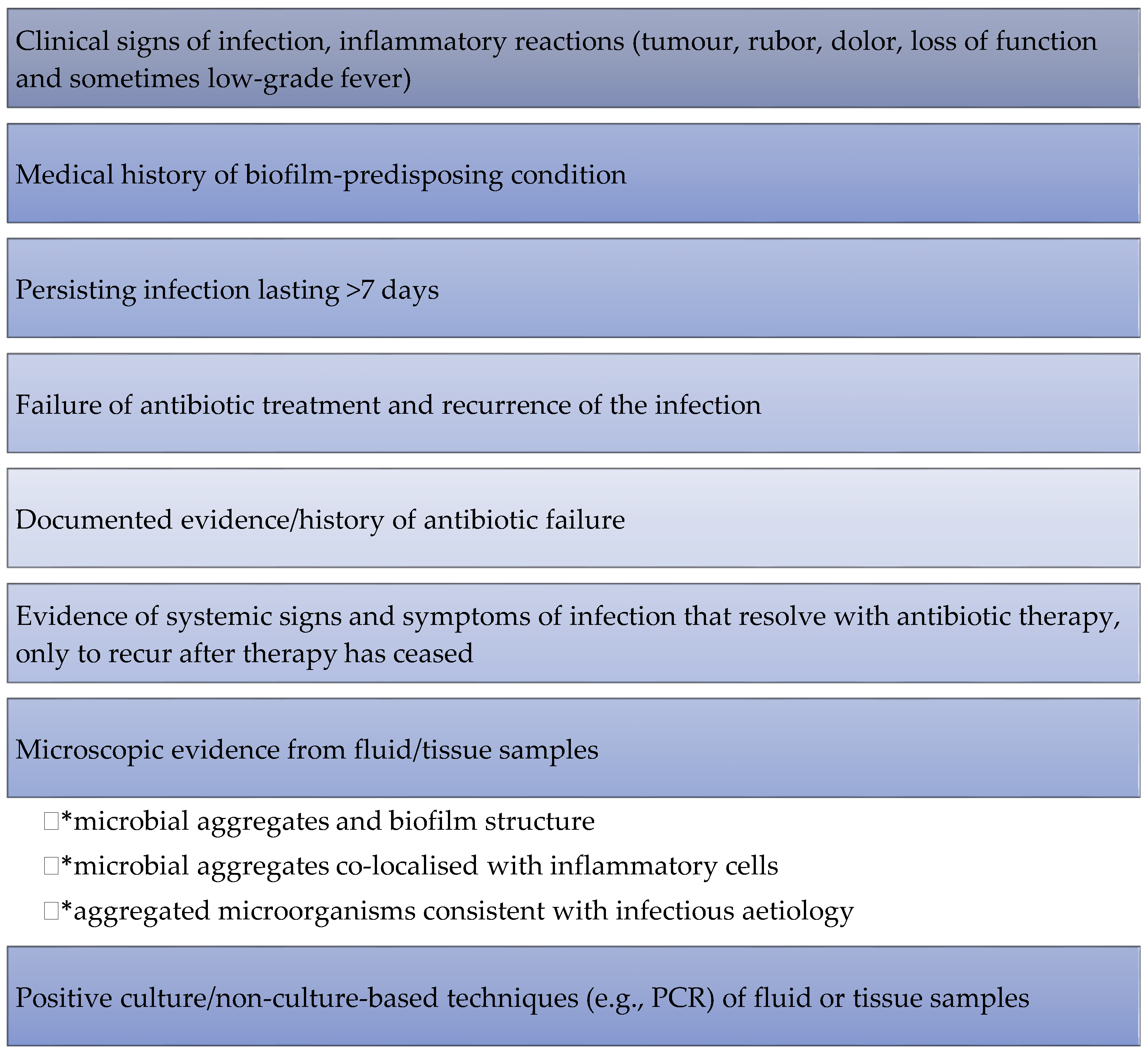
- Metcalf, D.; Bowler, P.; Hurlow, J. A clinical algorithm for wound biofilm identification. J. Wound Care 2014, 23, 137–142. [Google Scholar] [CrossRef] [PubMed] [Green Version]
- Percival, S.; Vuotto, C.; Donelli, G.; Lipsky, B.A. Biofilms and Wounds: An Identification Algorithm and Potential Treatment Options. Adv. Wound Care 2015, 4, 389–397. [Google Scholar] [CrossRef]
- Gompelman, M.; van Asten, S.; Peters, E. Update on the Role of Infection and Biofilms in Wound Healing. Plast. Reconstr. Surg. 2016, 138, 61S–70S. [Google Scholar] [CrossRef]
- Geraghty, T.; LaPorta, G. Current health and economic burden of chronic diabetic osteomyelitis. Expert Rev. Pharm. Outcomes Res. 2019, 19, 279–286. [Google Scholar] [CrossRef]
- Gawande, P.V.; Leung, K.P.; Madhyastha, S. Antibiofilm and Antimicrobial Efficacy of DispersinB®-KSL-W Peptide-Based Wound Gel Against Chronic Wound Infection Associated Bacteria. Curr. Microbiol. 2014, 68, 635–641. [Google Scholar] [CrossRef]
- Gomes, A.; Bessa, L.J.; Fernandes, I.; Ferraz, R.; Mateus, N.; Gameiro, P.; Teixeira, C.; Gomes, P. Turning a Collagenesis-Inducing Peptide Into a Potent Antibacterial and Antibiofilm Agent Against Multidrug-Resistant Gram-Negative Bacteria. Front. Microbiol. 2019, 10, 1915. [Google Scholar] [CrossRef] [Green Version]
- Roche, E.D.; Woodmansey, E.J.; Yang, Q.; Gibson, D.; Zhang, H.; Schultz, G.S. Cadexomer iodine effectively reduces bacterial biofilm in porcine wounds ex vivo and in vivo. Int. Wound J. 2019, 16, 674–683. [Google Scholar] [CrossRef]
- Sampathkumar, S.J.; Srivastava, P.; Ramachandran, S.; Sivashanmugam, K.; Gothandam, K.M. Lutein: A potential antibiofilm and antiquorum sensing molecule from green microalga Chlorella pyrenoidosa. Microb. Pathog. 2019, 135, 103658. [Google Scholar] [CrossRef]
- Miller, M.; Rogers, J.C.; Badham, M.A.; Cadenas, L.; Brightwell, E.; Adams, J.; Tyler, C.; Sebahar, P.R.; Haussener, T.J.; Reddy, H.R.K.; et al. Examination of a first-in-class bis-dialkylnorspermidine-terphenyl antibiotic in topical formulation against mono and polymicrobial biofilms. PLoS ONE 2020, 15, e0234832. [Google Scholar] [CrossRef]
- Sanpinit, S.; Yincharoen, K.; Jindamanee, C.; Jobthin, S.; Limsuwan, S.; Kunworarath, N.; Jaisamut, P.; Chokpaisarn, J.; Voravuthikunchai, S.P.; Chusri, S. Antibacterial properties of Ya-Samarn-Phlae (YaSP): A pilot study on diabetic patients with chronic ulcers. J. Herb. Med. 2020, 23, 100381. [Google Scholar] [CrossRef]
- Silva, V.; Peirone, C.; Amaral, J.S.; Capita, R.; Alonso-Calleja, C.; Marques-Magallanes, J.A.; Martins, Â.; Carvalho, Á.; Maltez, L.; Pereira, J.E.; et al. High Efficacy of Ozonated Oils on the Removal of Biofilms Produced by Methicillin-Resistant Staphylococcus aureus (MRSA) from Infected Diabetic Foot Ulcers. Molecules 2020, 25, 3601. [Google Scholar] [CrossRef]
- Sharma, A.; Gupta, P.; Kumar, R.; Bhardwaj, A. dPABBs: A Novel in silico Approach for Predicting and Designing Anti-biofilm Peptides. Sci. Rep. 2016, 6, 1–13. [Google Scholar] [CrossRef] [PubMed]
- Dean, S.N.; Bishop, B.M.; Van Hoek, M.L. Natural and synthetic cathelicidin peptides with anti-microbial and anti-biofilm activity against Staphylococcus aureus. BMC Microbiol. 2011, 11, 114. [Google Scholar] [CrossRef] [PubMed] [Green Version]
- Seleem, M.; Thangamani, S.; Nepal, M.; Chmielewski, J. Antibacterial activity and therapeutic efficacy of Fl-PRPRPL-5, a cationic amphiphilic polyproline helix, in a mouse model of staphylococcal skin infection. Drug Des. Dev. Ther. 2015, ume 9, 5749–5754. [Google Scholar] [CrossRef] [Green Version]
- Pletzer, D.; Coleman, S.R.; Hancock, R.E. Anti-biofilm peptides as a new weapon in antimicrobial warfare. Curr. Opin. Microbiol. 2016, 33, 35–40. [Google Scholar] [CrossRef] [PubMed] [Green Version]
- Bechinger, B.; Gorr, S.-U. Antimicrobial Peptides: Mechanisms of Action and Resistance. J. Dent. Res. 2017, 96, 254–260. [Google Scholar] [CrossRef] [Green Version]
- Chung, P.Y.; Khanum, R. Antimicrobial peptides as potential anti-biofilm agents against multidrug-resistant bacteria. J. Microbiol. Immunol. Infect. 2017, 50, 405–410. [Google Scholar] [CrossRef]
- de Breij, A.; Riool, M.; Cordfunke, R.A.; Malanovic, N.; de Boer, L.; Koning, R.I.; Ravensbergen, E.; Franken, M.; van der Heijde, T.; Boekema, B.K.; et al. The antimicrobial peptide SAAP-148 combats drug-resistant bacteria and biofilms. Sci. Transl. Med. 2018, 10, eaan4044. [Google Scholar] [CrossRef] [Green Version]
- Kim, M.K.; Kang, N.H.; Ko, S.J.; Park, J.; Park, E.; Shin, D.W.; Kim, S.H.; Lee, S.H.; Lee, J.I.; Ha, E.G.; et al. Antibacterial and Antibiofilm Activity and Mode of Action of Magainin 2 against Drug-Resistant Acinetobacter baumannii. Int. J. Mol. Sci. 2018, 19, 3041. [Google Scholar] [CrossRef] [Green Version]
- de Alteriis, E.; Lombardi, L.; Falanga, A.; Napolano, M.; Galdiero, S.; Siciliano, A.; Carotenuto, R.; Guida, M. Polymicrobial antibiofilm activity of the membranotropic peptide gH625 and its analogue. Microb. Pathog. 2018, 125, 189–195. [Google Scholar] [CrossRef] [PubMed]
- Kim, M.K.; Kang, H.K.; Ko, S.J.; Hong, M.J.; Bang, J.K.; Seo, C.H.; Park, Y. Mechanisms driving the antibacterial and antibiofilm properties of Hp1404 and its analogue peptides against multidrug-resistant Pseudomonas aeruginosa. Sci. Rep. 2018, 8, 1763. [Google Scholar] [CrossRef] [PubMed] [Green Version]
- Galdiero, E.; Lombardi, L.; Falanga, A.; Libralato, G.; Guida, M.; Carotenuto, R. Biofilms: Novel Strategies Based on Antimicrobial Peptides. Pharmaceutics 2019, 11, 322. [Google Scholar] [CrossRef] [Green Version]
- Gomes, D.; Santos, R.; Soares, R.S.; Reis, S.; Carvalho, S.; Rego, P.; Peleteiro, M.C.; Tavares, L.; Oliveira, M. Pexiganan in Combination with Nisin to Control Polymicrobial Diabetic Foot Infections. Antibiotics 2020, 9, 128. [Google Scholar] [CrossRef] [PubMed] [Green Version]
- Yasir, M.; Willcox, M.D.P.; Dutta, D. Action of Antimicrobial Peptides against Bacterial Biofilms. Materials 2018, 11, 2468. [Google Scholar] [CrossRef] [Green Version]
- Beaudoin, T.; Stone, T.A.; Glibowicka, M.; Adams, C.; Yau, Y.; Ahmadi, S.; Bear, C.E.; Grasemann, H.; Waters, V.; Deber, C.M. Activity of a novel antimicrobial peptide against Pseudomonas aeruginosa biofilms. Sci. Rep. 2018, 8, 1–12. [Google Scholar] [CrossRef]
- Overhage, J.; Campisano, A.; Bains, M.; Torfs, E.C.W.; Rehm, B.H.A.; Hancock, R.E.W. Human Host Defense Peptide LL-37 Prevents Bacterial Biofilm Formation. Infect. Immun. 2008, 76, 4176–4182. [Google Scholar] [CrossRef] [PubMed] [Green Version]
- De La Fuente-Núñez, C.; Korolik, V.; Bains, M.; Nguyen, U.; Breidenstein, E.B.M.; Horsman, S.; Lewenza, S.; Burrows, L.; Hancock, R. Inhibition of Bacterial Biofilm Formation and Swarming Motility by a Small Synthetic Cationic Peptide. Antimicrob. Agents Chemother. 2012, 56, 2696–2704. [Google Scholar] [CrossRef] [Green Version]
- Singh, P.K.; Parsek, M.R.; Greenberg, E.; Welsh, M.J. A component of innate immunity prevents bacterial biofilm development. Nat. Cell Biol. 2002, 417, 552–555. [Google Scholar] [CrossRef]
- Picioreanu, C.; Kreft, J.-U.; Klausen, M.; Haagensen, J.A.J.; Tolker-Nielsen, T.; Molin, S. Microbial motility involvement in biofilm structure formation—A 3D modelling study. Water Sci. Technol. 2007, 55, 337–343. [Google Scholar] [CrossRef]
- Braeken, K.; Moris, M.; Daniels, R.; Vanderleyden, J.; Michiels, J. New horizons for (p)ppGpp in bacterial and plant physiology. Trends Microbiol. 2006, 14, 45–54. [Google Scholar] [CrossRef]
- Potrykus, K.; Cashel, M. (p)ppGpp: Still Magical? Annu. Rev. Microbiol. 2008, 62, 35–51. [Google Scholar] [CrossRef] [Green Version]
- Aberg, A.; Shingler, V.; Balsalobre, C. (p)ppGpp regulates type 1 fimbriation of Escherichia coli by modulating the expression of the site-specific recombinase FimB. Mol. Microbiol. 2006, 60, 1520–1533. [Google Scholar] [CrossRef]
- de la Fuente-Núñez, C.; Reffuveille, F.; Mansour, S.C.; Reckseidler-Zenteno, S.L.; Hernández, D.; Brackman, G.; Coenye, T.; Hancock, R.E. D-Enantiomeric Peptides that Eradicate Wild-Type and Multidrug-Resistant Biofilms and Protect against Lethal Pseudomonas aeruginosa Infections. Chem. Biol. 2015, 22, 196–205. [Google Scholar] [CrossRef]
- De La Fuente-Núñez, C.; Reffuveille, F.; Haney, E.F.; Straus, S.; Hancock, R. Broad-Spectrum Anti-biofilm Peptide That Targets a Cellular Stress Response. PLOS Pathog. 2014, 10, e1004152. [Google Scholar] [CrossRef] [Green Version]
- Ribeiro, S.; De La Fuente-Núñez, C.; Baquir, B.; Faria-Junior, C.; Franco, O.L.; Hancock, R. Antibiofilm Peptides Increase the Susceptibility of Carbapenemase-Producing Klebsiella pneumoniae Clinical Isolates to β-Lactam Antibiotics. Antimicrob. Agents Chemother. 2015, 59, 3906–3912. [Google Scholar] [CrossRef] [Green Version]
- Reffuveille, F.; De La Fuente-Núñez, C.; Mansour, S.; Hancock, R.E.W. A Broad-Spectrum Antibiofilm Peptide Enhances Antibiotic Action against Bacterial Biofilms. Antimicrob. Agents Chemother. 2014, 58, 5363–5371. [Google Scholar] [CrossRef] [PubMed] [Green Version]
- Thawal, N.D.; Yele, A.B.; Sahu, P.; Chopade, B.A. Effect of a Novel Podophage AB7-IBB2 on Acinetobacter baumannii Biofilm. Curr. Microbiol. 2012, 65, 66–72. [Google Scholar] [CrossRef]
- Alves, D.R.; Gaudion, A.; Bean, J.E.; Esteban, P.P.; Arnot, T.; Harper, D.R.; Kot, W.; Hansen, L.H.; Enright, M.; Jenkins, A.T.A. Combined Use of Bacteriophage K and a Novel Bacteriophage To Reduce Staphylococcus aureus Biofilm Formation. Appl. Environ. Microbiol. 2014, 80, 6694–6703. [Google Scholar] [CrossRef] [Green Version]
- Mendes, J.J.; Leandro, C.; Mottola, C.; Barbosa, R.; Silva, F.A.; Oliveira, M.; Vilela, C.L.; Cristino, J.M.; Górski, A.; Pimentel, M.; et al. In vitro design of a novel lytic bacteriophage cocktail with therapeutic potential against organisms causing diabetic foot infections. J. Med. Microbiol. 2014, 63, 1055–1065. [Google Scholar] [CrossRef]
- Liu, Y.; Mi, Z.; Niu, W.; An, X.; Yuan, X.; Liu, H.; Wang, Y.; Feng, Y.; Huang, Y.; Zhang, X.; et al. Potential of a lytic bacteriophage to disrupt Acinetobacter baumannii biofilms in vitro. Future Microbiol. 2016, 11, 1383–1393. [Google Scholar] [CrossRef]
- Yuan, Y.; Qu, K.; Tan, D.; Li, X.; Wang, L.; Cong, C.; Xiu, Z.; Xu, Y. Isolation and characterization of a bacteriophage and its potential to disrupt multi-drug resistant Pseudomonas aeruginosa biofilms. Microb. Pathog. 2019, 128, 329–336. [Google Scholar] [CrossRef]
- Łusiak-Szelachowska, M.; Weber-Dąbrowska, B.; Górski, A. Bacteriophages and Lysins in Biofilm Control. Virol. Sin. 2020, 35, 125–133. [Google Scholar] [CrossRef]
- Clokie, M.R.J.; Millard, A.D.; Letarov, A.V.; Heaphy, S. Phages in nature. Bacteriophage 2011, 1, 31–45. [Google Scholar] [CrossRef] [Green Version]
- Harper, D.R.; Parracho, H.M.R.T.; Walker, J.; Sharp, R.J.; Hughes, G.; Werthén, M.; Lehman, S.M.; Morales, S. Bacteriophages and Biofilms. Antibiotics 2014, 3, 270–284. [Google Scholar] [CrossRef]
- Taha, O.A.; Connerton, P.L.; Connerton, I.; El-Shibiny, A. Bacteriophage ZCKP1: A Potential Treatment for Klebsiella pneumoniae Isolated From Diabetic Foot Patients. Front. Microbiol. 2018, 9, 2127. [Google Scholar] [CrossRef]
- Pires, D.P.; Melo, L.; Boas, D.V.; Sillankorva, S.; Azeredo, J. Phage therapy as an alternative or complementary strategy to prevent and control biofilm-related infections. Curr. Opin. Microbiol. 2017, 39, 48–56. [Google Scholar] [CrossRef] [PubMed] [Green Version]
- Alves, D.R.; Esteban, P.P.; Kot, W.; Bean, J.; Arnot, T.; Hansen, L.; Enright, M.; Jenkins, T. A novel bacteriophage cocktail reduces and disperses Pseudomonas aeruginosa biofilms under static and flow conditions. Microb. Biotechnol. 2015, 9, 61–74. [Google Scholar] [CrossRef] [Green Version]
- Rahman, M.; Kim, S.; Kim, S.M.; Seol, S.Y.; Kim, J. Characterization of induced Staphylococcus aureus bacteriophage SAP-26 and its anti-biofilm activity with rifampicin. Biofouling 2011, 27, 1087–1093. [Google Scholar] [CrossRef]
- Chaudhry, W.N.; Concepción-Acevedo, J.; Park, T.; Andleeb, S.; Bull, J.J.; Levin, B.R. Synergy and Order Effects of Antibiotics and Phages in Killing Pseudomonas aeruginosa Biofilms. PLoS ONE 2017, 12, e0168615. [Google Scholar] [CrossRef]
- Akturk, E.; Oliveira, H.; Santos, S.B.; Costa, S.; Kuyumcu, S.; Melo, L.D.R.; Azeredo, J. Synergistic Action of Phage and Antibiotics: Parameters to Enhance the Killing Efficacy Against Mono and Dual-Species Biofilms. Antibiotics 2019, 8, 103. [Google Scholar] [CrossRef] [Green Version]
- Borges, A.; Abreu, A.C.; Dias, C.; Saavedra, M.J.; Borges, F.; Simões, M. New Perspectives on the Use of Phytochemicals as an Emergent Strategy to Control Bacterial Infections Including Biofilms. Molecules 2016, 21, 877. [Google Scholar] [CrossRef]
- Borges, A.; Simões, L.; Saavedra, M.J.; Simões, M. The action of selected isothiocyanates on bacterial biofilm prevention and control. Int. Biodeterior. Biodegrad. 2014, 86, 25–33. [Google Scholar] [CrossRef] [Green Version]
- Kot, B.; Wierzchowska, K.; Grużewska, A.; Lohinau, D. The effects of selected phytochemicals on biofilm formed by five methicillin-resistant Staphylococcus aureus. Nat. Prod. Res. 2018, 32, 1299–1302. [Google Scholar] [CrossRef]
- Vipin, C.; Mujeeburahiman, M.; Ashwini, P.; Arun, A.; Rekha, P. Anti-biofilm and cytoprotective activities of quercetin against Pseudomonas aeruginosa isolates. Lett. Appl. Microbiol. 2019, 68, 464–471. [Google Scholar] [CrossRef]
- Simões, M.; Bennett, R.N.; Rosa, E. Understanding antimicrobial activities of phytochemicals against multidrug resistant bacteria and biofilms. Nat. Prod. Rep. 2009, 26, 746–757. [Google Scholar] [CrossRef]
- Abreu, A.C.; McBain, A.; Simões, M. Plants as sources of new antimicrobials and resistance-modifying agents. Nat. Prod. Rep. 2012, 29, 1007–1021. [Google Scholar] [CrossRef] [PubMed]
- Borges, A.; Saavedra, M.J.; Simões, M. Insights on antimicrobial resistance, biofilms and the use of phytochemicals as new antimicrobial agents. Curr. Med. Chem. 2015, 22, 2590–2614. [Google Scholar] [CrossRef] [Green Version]
- Borges, A.; Maria, J.; Simoes, M. The activity of ferulic and gallic acids in biofilm prevention and control of pathogenic bacteria. Biofouling 2012, 28, 755–767. [Google Scholar] [CrossRef]
- Monte, J.; Abreu, A.C.; Borges, A.; Simões, L.C.; Simões, M. Antimicrobial Activity of Selected Phytochemicals against Escherichia coli and Staphylococcus aureus and Their Biofilms. Pathogens 2014, 3, 473–498. [Google Scholar] [CrossRef] [Green Version]
- Ouyang, J.; Sun, F.; Feng, W.; Sun, Y.; Qiu, X.; Xiong, L.; Liu, Y.; Chen, Y. Quercetin is an effective inhibitor of quorum sensing, biofilm formation and virulence factors in Pseudomonas aeruginosa. J. Appl. Microbiol. 2016, 120, 966–974. [Google Scholar] [CrossRef] [PubMed] [Green Version]
- Ülkür, E.; Oncul, O.; Karagoz, H.; Yeniz, E.; Çeliköz, B. Comparison of silver-coated dressing (Acticoat™), chlorhexidine acetate 0.5% (Bactigrass®), and fusidic acid 2% (Fucidin®) for topical antibacterial effect in methicillin-resistant Staphylococci-contaminated, full-skin thickness rat burn wounds. Burns 2005, 31, 874–877. [Google Scholar] [CrossRef]
- Paddle-Ledinek, J.E.; Nasa, Z.; Cleland, H.J. Effect of Different Wound Dressings on Cell Viability and Proliferation. Plast. Reconstr. Surg. 2006, 117, 110S–118S. [Google Scholar] [CrossRef] [PubMed]
- Buch, P.J.; Chai, Y.; Goluch, E.D. Treating Polymicrobial Infections in Chronic Diabetic Wounds. Clin. Microbiol. Rev. 2019, 32. [Google Scholar] [CrossRef] [Green Version]
- Smith, A.W. Biofilms and antibiotic therapy: Is there a role for combating bacterial resistance by the use of novel drug delivery systems? Adv. Drug Deliv. Rev. 2005, 57, 1539–1550. [Google Scholar] [CrossRef]
- Taraszkiewicz, A.; Fila, G.; Grinholc, M.; Nakonieczna, J. Innovative Strategies to Overcome Biofilm Resistance. BioMed Res. Int. 2012, 2013, 1–13. [Google Scholar] [CrossRef] [Green Version]
- Martin, C.; Low, W.L.; Gupta, A.; Amin, M.; Radecka, I.; Britland, S.; Raj, P.D.; Kenward, K. Strategies for Antimicrobial Drug Delivery to Biofilm. Curr. Pharm. Des. 2014, 21, 43–66. [Google Scholar] [CrossRef]
- Shah, S.; Gaikwad, S.; Nagar, S.; Kulshrestha, S.; Vaidya, V.; Nawani, N.; Pawar, S. Biofilm inhibition and anti-quorum sensing activity of phytosynthesized silver nanoparticles against the nosocomial pathogen Pseudomonas aeruginosa. Biofouling 2019, 35, 34–49. [Google Scholar] [CrossRef] [Green Version]
- Mohanta, Y.K.; Biswas, K.; Jena, S.K.; Hashem, A.; Allah, E.F.A.; Mohanta, T.K. Anti-biofilm and Antibacterial Activities of Silver Nanoparticles Synthesized by the Reducing Activity of Phytoconstituents Present in the Indian Medicinal Plants. Front. Microbiol. 2020, 11, 1143. [Google Scholar] [CrossRef] [PubMed]
- Martinez-Gutierrez, F.; Boegli, L.; Agostinho, A.; Sánchez, E.M.; Bach, H.; Ruiz, F.; James, G. Anti-biofilm activity of silver nanoparticles against different microorganisms. Biofouling 2013, 29, 651–660. [Google Scholar] [CrossRef] [PubMed]
- Appapalam, S.T.; Paul, B.; Arockiasamy, S.; Panchamoorthy, R. Phytofabricated silver nanoparticles: Discovery of antibacterial targets against diabetic foot ulcer derived resistant bacterial isolates. Mater. Sci. Eng. C 2020, 117, 111256. [Google Scholar] [CrossRef]
- Serpe, L.; Giuntini, F. Sonodynamic antimicrobial chemotherapy: First steps towards a sound approach for microbe inactivation. J. Photochem. Photobiol. B Biol. 2015, 150, 44–49. [Google Scholar] [CrossRef] [PubMed]
- Abrahamse, H.; Hamblin, M.R. New photosensitizers for photodynamic therapy. Biochem. J. 2016, 473, 347–364. [Google Scholar] [CrossRef] [Green Version]
- Pantò, F.; Adamo, L.; Giordano, C.; Licciardello, C. Efficacy and safety of photodynamic therapy with RLP068 for diabetic foot ulcers: A review of the literature and clinical experience. Drugs Context 2020, 9, 1–7. [Google Scholar] [CrossRef]
- De Melo, W.C.M.; Avci, P.; De Oliveira, M.N.; Gupta, A.; Vecchio, D.; Sadasivam, M.; Chandran, R.; Huang, Y.; Yin, R.; Perussi, L.R.; et al. Photodynamic inactivation of biofilm: Taking a lightly colored approach to stubborn infection. Expert Rev. Anti-Infect. Ther. 2013, 11, 669–693. [Google Scholar] [CrossRef] [Green Version]
- Barra, F.; Roscetto, E.; Soriano, A.A.; Vollaro, A.; Postiglione, I.; Pierantoni, G.M.; Palumbo, G.; Catania, M.R. Photodynamic and Antibiotic Therapy in Combination to Fight Biofilms and Resistant Surface Bacterial Infections. Int. J. Mol. Sci. 2015, 16, 20417–20430. [Google Scholar] [CrossRef] [PubMed]
- Di Poto, A.; Sbarra, M.S.; Provenza, G.; Visai, L.; Speziale, P. The effect of photodynamic treatment combined with antibiotic action or host defence mechanisms on Staphylococcus aureus biofilms. Biomaterials 2009, 30, 3158–3166. [Google Scholar] [CrossRef] [PubMed]
- Brown, M.R.W.; Richards, R.M.E. Effect of Ethylenediamine Tetraacetate on the Resistance of Pseudomonas aeruginosa to Antibacterial Agents. Nature 1965, 207, 1391–1393. [Google Scholar] [CrossRef]
- Bertoloni, G.; Rossi, F.; Valduga, G.; Jori, G.; Van Lier, J.; Bertolini, G. Photosensitizing activity of water- and lipid-soluble phthalocyanines on Escherichia coli. FEMS Microbiol. Lett. 1990, 71, 149–155. [Google Scholar] [CrossRef] [PubMed]
- Finnegan, S.; Percival, S. EDTA: An Antimicrobial and Antibiofilm Agent for Use in Wound Care. Adv. Wound Care 2015, 4, 415–421. [Google Scholar] [CrossRef] [Green Version]

| Severity | Associated Pathogen(s) | Additional Factor(s) | Antibiotic(s) |
|---|---|---|---|
| Mild (topical or oral antibacterial agents) | Staphylococcus aureus (MSSA) Streptococcus spp. | No complicating features | First-generation cephalosporin, nafcillin, ampicillin/sulbactam, amoxicillin/clavulanate, clindamycin |
| β-lactam allergy or intolerance | Clindamycin, levofloxacin, moxifloxacin, doxycycline | ||
| Recent antibiotic exposure | Levofloxacin, moxifloxacin, second- or third-generation cephalosporin | ||
| MRSA | Clindamycin, doxycycline, trimethoprim/sulfamethoxazole | ||
| Moderate (oral or initial parenteral antibacterial agents) or Severe (parenteral antibacterial agents) | MSSA Streptococcus spp. Enterobacteriaceae obligate anaerobes | No complicating features | Second- or third-generation cephalosporin, aminoglycoside |
| Recent antibiotic exposure | Third-generation cephalosporin, aminoglycoside, ertapenem, piperacillin/tazobactam, cefepime | ||
| Pseudomonas aeruginosa | Piperacillin/tazobactam, cefepime, imipenem, meropenem | ||
| MRSA Enterobacteriaceae obligate anaerobes | Vancomycinc plus one of the following: ceftazidime, cefepime, piperacillin/tazobactam, aztreonam, or a carbapenem | ||
| ESBL, MDR Gram-negative | Piperacillin/tazobactam plus one of the following: aminoglycoside, or a carbapenem |
| Dressing Classes | Advantage(s) | Disadvantage(s) |
|---|---|---|
| Tulle | Good, moist environment | Be careful not to dry |
| Low-adherence | Hypoallergenic; Inexpensive; Moist environment | Minimal absorbency |
| Polyurethane films | Water-proof dressing; Comfortable; Transparent (allows wound monitoring) | Facilitates maceration |
| Hydrocolloids | Absorbent; Can be left for several days; Aids autolysis | Avoid use on infected wounds; Facilitates maceration; Unpleasant odour |
| Hydrogels | Good absorbent; Aids autolysis; Donate liquid | Avoid use on infected wounds; Facilitates maceration |
| Foams | Thermal insulation; Good absorbent | Can adhere to wound; Occasional dermatitis due to the adhesive |
| Alginates | Highly absorbent; Bacteriostatic; Haemostatic; Useful in cavities | Require wetting before removal |
| Iodine preparations | Antiseptic; Moderately absorbent | Iodine allergy; Discolours wounds; Avoid in cases of thyroid disease or pregnancy |
| Silver-impregnated | Antiseptic; Absorbent | Cost |
| Study 1 | Study 2 | Study 3 | Study 4 | |
|---|---|---|---|---|
| n * | 57 | 162 | - | 160 |
| Biofilm + | 44 | 110 | 115 | - |
| Gender (male) | 32 | 76 | - | 82 |
| Age distribution | 44.6 ± 7.3 | - | 62.7 | - |
| >40 years | 64.8% | 70.0% | - | - |
| Diabetes duration (average year) | 14.9 ± 2.6 | - | 16.0 ± 0.0 | - |
| Ulcer duration | 39.6 ± 2.6 | - | 48.2 ± 42.3 | - |
| >1 month | 52.6% | 75.0% | - | |
| Hospital stay | ||||
| >1 month | 59.5% | 75.0% | - | - |
| Amputation | 24.5% | 80.4% | 42.0% | |
| Ulcer size | ||||
| <4 cm2 | 64.9% | 69.3% | - | 89.0% |
| Comorbidities | ||||
| Hypertension | 72.7% | 80.4% | 34.0% | 16.0% |
| Nephropathy | 77.1% | 77.7% | 37.0% | 25.0% |
| Retinopathy | 68.7% | 52.4% | 3.0% | 22.0% |
| Neuropathy | 89.4% | 57.3% | 34.0% | 18.0% |
| Osteomyelitis | 88.8% | 65.0% | - | - |
| Status | ||||
| Death | 3.5% | 72.2% | 38.0% | - |
| Reference | |
|---|---|
| Gram-negative | |
| Enterobacteriaceae | [147] |
| Escherichia coli | [145,146,149,151,152] |
| Klebsiella spp. | [152] |
| Klebsiella pneumoniae | [145,146,151] |
| Klebsiella oxytoca | [145,146,149] |
| Pseudomonas spp. | [150] |
| Pseudomonas aeruginosa | [145,146,147,149,151,152] |
| Proteus spp. | [149] |
| Proteus vulgaris | [145,146] |
| Proteus mirabilis | [146] |
| Acinetobacter spp. | [145,146,150,151] |
| Acinetobacter baumani | [147] |
| Morganella morganii | [145,146] |
| Vibrio spp. | [152] |
| Citrobacter spp. | [149,151] |
| Gram-positive | |
| Coryneform spp. | [146] |
| Corynebacterium spp. | [150] |
| Beta-haemolytic Streptococcus | [146,151] |
| Coagulase-negative Staphylococcus spp. | [146] |
| Staphylococcus spp. | [150,152] |
| Staphylococcus aureus | [146,147,149,151] |
| MRSA | [149] |
| Enterococcus spp. | [147,150] |
| Enterococcus faecalis | [146] |
Publisher’s Note: MDPI stays neutral with regard to jurisdictional claims in published maps and institutional affiliations. |
© 2021 by the authors. Licensee MDPI, Basel, Switzerland. This article is an open access article distributed under the terms and conditions of the Creative Commons Attribution (CC BY) license (https://creativecommons.org/licenses/by/4.0/).
Share and Cite
Afonso, A.C.; Oliveira, D.; Saavedra, M.J.; Borges, A.; Simões, M. Biofilms in Diabetic Foot Ulcers: Impact, Risk Factors and Control Strategies. Int. J. Mol. Sci. 2021, 22, 8278. https://doi.org/10.3390/ijms22158278
Afonso AC, Oliveira D, Saavedra MJ, Borges A, Simões M. Biofilms in Diabetic Foot Ulcers: Impact, Risk Factors and Control Strategies. International Journal of Molecular Sciences. 2021; 22(15):8278. https://doi.org/10.3390/ijms22158278
Chicago/Turabian StyleAfonso, Ana C., Diana Oliveira, Maria José Saavedra, Anabela Borges, and Manuel Simões. 2021. "Biofilms in Diabetic Foot Ulcers: Impact, Risk Factors and Control Strategies" International Journal of Molecular Sciences 22, no. 15: 8278. https://doi.org/10.3390/ijms22158278
APA StyleAfonso, A. C., Oliveira, D., Saavedra, M. J., Borges, A., & Simões, M. (2021). Biofilms in Diabetic Foot Ulcers: Impact, Risk Factors and Control Strategies. International Journal of Molecular Sciences, 22(15), 8278. https://doi.org/10.3390/ijms22158278