Review

Autoimmune Responses in Oncology: Causes and Significance

by
Halin Bareke
1,2,
Pablo Juanes-Velasco
2,
Alicia Landeira-Viñuela
2,
Angela-Patricia Hernandez
2,
Juan Jesús Cruz
3,
Lorena Bellido
3,
Emilio Fonseca
3,
Alfonssina Niebla-Cárdenas
4,
Enrique Montalvillo
2,
Rafael Góngora
2 and
Manuel Fuentes
2,5,*
1
Department of Pharmaceutical Biotechnology, Faculty of Pharmacy, Institute of Health Sciences, Marmara University, Istanbul 34722, Turkey
2
Department of Medicine and General Cytometry Service-Nucleus, CIBERONC CB16/12/00400, Cancer Research Centre (IBMCC/CSIC/USAL/IBSAL), 37007 Salamanca, Spain
3
Medical Oncology Service, Hospital Universitario de Salamanca-IBSAL, 37007 Salamanca, Spain
4
Department of Nursing and Physiotherapy, Faculty of Nursing and Physiotherapy, University of Salamanca, 37007 Salamanca, Spain
5
Proteomics Unit, Cancer Research Center (IBMCC/CSIC/USAL/IBSAL), 37007 Salamanca, Spain
*
Author to whom correspondence should be addressed.
Int. J. Mol. Sci. 2021, 22(15), 8030; https://doi.org/10.3390/ijms22158030
Submission received: 29 June 2021 / Revised: 19 July 2021 / Accepted: 23 July 2021 / Published: 27 July 2021
(This article belongs to the Special Issue Immunological Responses to Cancer Therapy)

Abstract

:
Specific anti-tumor immune responses have proven to be pivotal in shaping tumorigenesis and tumor progression in solid cancers. These responses can also be of an autoimmune nature, and autoantibodies can sometimes be present even before the onset of clinically overt disease. Autoantibodies can be generated due to mutated gene products, aberrant expression and post-transcriptional modification of proteins, a pro-immunogenic milieu, anti-cancer treatments, cross-reactivity of tumor-specific lymphocytes, epitope spreading, and microbiota-related and genetic factors. Understanding these responses has implications for both basic and clinical immunology. Autoantibodies in solid cancers can be used for early detection of cancer as well as for biomarkers of prognosis and treatment response. High-throughput techniques such as protein microarrays make parallel detection of multiple autoantibodies for increased specificity and sensitivity feasible, affordable, and quick. Cancer immunotherapy has revolutionized cancer treatments and has made a considerable impact on reducing cancer-associated morbidity and mortality. However, immunotherapeutic interventions such as immune checkpoint inhibition can induce immune-related toxicities, which can even be life-threatening. Uncovering the reasons for treatment-induced autoimmunity can lead to fine-tuning of cancer immunotherapy approaches to evade toxic events while inducing an effective anti-tumor immune response.

1. Introduction

The self- vs. non-self-discrimination was long considered to be the sole determinant for immune activation and tolerance. As a safeguard mechanism against autoimmunity, the immune system was thought to be prevented from turning its destructive power toward itself by only recognizing non-self antigens. Since cancer cells are formed from the body’s own cells, the immune system has been deemed to be powerless against these treacherous, but nevertheless self-cells. However, this paradigm fails to account for many aspects of immune responses including the apparent lack of immune reactions to the commensal microorganisms found ubiquitously in the body. The self vs. non-self dichotomy has, then, been revisited with the harmful vs. harmless distinction. The immune system is trained to distinguish between harmful and harmless molecules (albeit, they are mostly self-antigens) through central and peripheral tolerance, the microbes encountered very early in life, and whether the milieu is pro-immunogenic or anti-immunogenic [1]. This shift in paradigm has created the possibility for the existence of anti-tumor immune responses against harmful self-cells. The elevated risk of cancer in immunosuppressed individuals and in immunocompromised severe combined immunodeficiency (SCID) mice corroborates the importance of the immune response in cancer [2]. Under the right pro-immunogenic conditions, the immune cells can, indeed, respond to tumor cells. However, autoimmune responses can concomitantly arise during those anti-tumor responses.
Reinvigorating the immune system against tumor cells in the form of oncological immunotherapy has dominated the cancer therapy field in the last decade [3]. The immune system’s hand is weakened in the fight against a tumor by poor antigenicity of tumor-associated antigens (TAAs), inefficient antigen presentation in the deficit of co-stimulatory molecules, T cell exhaustion, and the immunosuppressive tumor microenvironment (TME) [4]. Among immunotherapeutic approaches, releasing the ‘brakes’ on immune cells to enable their attack on a tumor mass using immune checkpoint inhibitors (ICI) has shown remarkable clinical results in different types of solid cancers, such as renal cell carcinoma and advanced melanoma [5]. However, ICI, as well as some other immunotherapy approaches such as cytokine administration, can stimulate the immune system non-specifically, and hence, they can also trigger the activation of self-reactive lymphocytes. Therefore, immune-related adverse effects (irAEs) such as skin lesions, colitis, and thyroiditis might occur due to an autoimmune attack, limiting the clinical benefit of these treatments [6]. As these therapies are finding their place in the clinic rather quickly, evading irAEs and stratifying patients at a risk of developing immune toxicities are becoming particularly important. To this end, it is important to delineate the mechanistic underpinnings of these autoimmune attacks.
As the path from cancer to the autoimmune response is not only caused by therapeutical treatment, other paths, especially due to the ‘self’ nature of the cancer cells, co-exist with the same autoimmune outcome. In fact, there is a high concentration of self-antigens released by conventional therapies in most solid tumors as well as some TAAs that closely resemble self-antigens, leading to autoantigen cross reactivity. Cancer and the autoimmune response can both be conceptualized under the umbrella of ‘immune dysregulation’. In the case of cancers, the immune system fails to clear the altered dangerous ‘self’ cells in a case of ‘abortive autoimmune response’ [7]. During the autoimmune response, the immune system launches a response on innocuous components of self-cells. Even though these two immune imbalances appear to be the opposite of each other, they can still continuously feed and bear each other. The constant attack on the self-cells and the chronic inflammation seen in autoimmunity can promote tumorigenesis through multiple mechanisms including increased cell division and DNA-damaging oxidative stress [8]. Given also the plasticity of lymphocytes, as exemplified by the conversion of inflammatory Th17 cells into suppressor regulatory T cells (Tregs), the two extremes are closer than anticipated in a normal immune response. Furthermore, from a functional point of view, autoantibodies (aAbs) from the autoimmune response may sometimes actively promote tumorigenesis and tumor progression [9]. However, in some tumors such as melanoma, the presence of aAbs can signal an effective anti-tumor response and be associated with better disease outcomes [10]. Moreover, it is well-described that autoantibodies are present even before the onset of overt cancer. In some cases, there might be autoimmune manifestations in the form of paraneoplastic syndromes in the early stages of cancer [11]. If these autoimmune responses are detected quickly, they can help with life-saving early diagnosis of cancer. Recent studies, including one from our research team, have also raised the possibility that the autoantibody profiles in cancer can be used as biomarkers for diagnosis and discrimination between metastatic and non-metastatic disease [12].
Overall, understanding the profile and the mechanism of the autoimmune response observed in solid tumors would shed light on basic mechanisms of immune tolerance, would help to refine cancer immunotherapy approaches to minimize immune-related toxicities, and might yield much-needed cancer biomarkers for early cancer diagnosis and accurate prognosis. Herein, we will focus solely on the observed autoimmune responses in solid malignant tumors. Hematological malignancies warrant their own dedicated discussion regarding the links between autoimmunity and these malignancies because both the malignant cells and the cells involved in autoimmune responses are the same cell population, namely lymphocytes. In the following sections, the anti-tumoral immune responses and autoimmune responses, mainly in the form of aAbs and irAEs, that are detected in solid cancer patients, as well as their possible causes and significance, will be discussed.

2. Immune Tolerance and Anti-Tumoral Immunity

Due to the self-nature of cancer cells, central and peripheral immune tolerance mechanisms reduce both the chance of occurrence and the effectiveness of anti-tumor immunity. These mechanisms are in place to prevent the maturation and activation of self-reactive lymphocytes. Central tolerance takes place in the primary lymphoid organs, namely the bone marrow and the thymus. In the thymus, thymocytes present a variety of self-antigens from the tissues all over the body to the developing T cells thanks to the thymic-specific expression of certain genes such as AIRE [13]. Those T cells that recognize self-peptides are negatively selected in the thymus by undergoing apoptosis during thymic education. Similarly, in the bone marrow, developing B cells that bind to self-antigens either edit their B cell receptors (BCR) to change receptor specificity or die by apoptosis [14].
The self-reactive lymphocytes that have survived central tolerance and reached maturation are ‘subdued’ in periphery by regulatory cells such as CD4+CD25+FOXP3+ Tregs, by anergy induction (functional unresponsiveness), by lack of co-stimulation, and by induction of cell death (i.e., deletion). However, even in a normal physiological state, central and peripheral tolerance is far from perfect, and self-reactive T and B cells are found in healthy individuals [15]. Therefore, rather than eliminating all of the self-reactive lymphocytes, these mechanisms might only reduce their number and raise their activation threshold. Under certain conditions such as a high concentration of self-antigen and a highly proinflammatory cytokine milieu, these autoreactive lymphocytes might assemble a specific immune response, including in cancer settings.
Evading immune response is required for the cancer cells’ survival, and thus, immune evasion has been recognized as one of the hallmarks of cancer [16]. Tumor mass has to establish an ‘immune-privileged’ site for itself by adopting tactics that are similar to natural immune privilege mechanisms including the expression of immunosuppressive cytokines, downregulation of major histocompatibility complex (MHC) molecules, induction of lymphocyte apoptosis by molecules such as immune checkpoint molecules, PD-L1, prevention of T cell stimulation by another immune checkpoint protein, CTLA-4, recruitment of Tregs and other immunosuppressive cell populations such as myeloid-derived suppressor cells (MDSC), and inhibition of effector leukocyte infiltration [17]. The major suppressive lymphocytes, CD4+CD25+FOXP3+ Tregs, can suppress effector T cell activity by inducing cytolysis by granzyme B/perforin and apoptosis through death receptors, sweeping the IL-2 from the environment, secreting immunoregulatory cytokines such as IL-10 and transforming growth factor-β (TGF-β), inhibiting the stimulatory function of DCs, and by the generation of extracellular adenosine [18]. The importance of Tregs in cancer has been revealed through the prognostic significance of Treg presence in solid cancers. In a meta-analysis that included 15,512 cancer cases with 17 different types of solid cancer, high FOXP3+ Treg-density in tumor samples was associated with a lower overall survival rate in the pooled data [19]. Another immunoregulator cell population, MDSCs, are myeloid-derived immature cells that can suppress T cells via several mechanisms including cell contact, tryptophan deprivation by indoleamine 2,3-dioxygenase (IDO) expression, L-arginine depletion by arginase, Treg recruitment, and reactive oxygen species (ROS) generation [20]. A meta-analysis that included 1864 cancer patients showed that MDSC was a bad prognostic factor that shortened overall survival [21]. These data support the importance of anti-tumor responses in shaping the disease course in cancer, and demonstrate how the cell populations that act as safeguards against autoimmunity can hamper effective anti-tumoral immune responses.
The immune system can recognize cancer cells through TAAs or by self-reactive lymphocytes. Therefore, the lines between autoimmune response and cancer immunity are blurred such that the immune tolerance might be compromised during anti-tumoral immune responses. TAAs can be neoantigens that include aberrantly expressed or modified self-antigens, or oncofetal antigens. The players that counteract the immunosuppressive TME are mainly the components of cellular immunity. They include tumor-lysing populations such as natural killer (NK) cells and CD8+ cytotoxic T lymphocytes (CTLs). Lysing of the tumor cells by these lymphocytes releases a high concentration of self-antigens and proinflammatory molecules that can potentially trigger autoimmune responses. IFN-γ secreted by NK cells also promotes dendritic cell maturation and secretion of IL-12. Mature dendritic cells are pivotal in presenting both endogenous and exogenous (by cross presentation) TAAs to the CD8+ T cells and providing the right costimulatory signals (i.e., signal 2) for T cell activation [22]. In the CD4+ T cell compartment, Th1 polarization that activates M1 macrophages is important in killing cancer cells. M1 macrophages, in turn, promote the recruitment of CTLs and Th1 cells. Accordingly, the presence of tumor-infiltrating lymphocytes (TIL) is usually correlated with a better prognosis. As an illustrative example, a meta-analysis of 43 studies on the association between TIL and colorectal cancer prognosis showed that high TIL numbers and CD3+ T cell density were associated with disease-free survival as well as overall survival [23].
Compared to T cells, much less is known about tumor-infiltrating B cells (TIBs). Their importance in the immune control of cancer remains to be fully uncovered. TIBs might promote T effector function by cytokines or by antigen presentation. In addition, antibodies produced by TIBs can bind to their cognate antigens on tumor cells, modifying the function of the antigens. They can also act as an opsonin, activate the classical pathway of complement, or induce antibody-dependent cellular cytotoxicity (ADCC) [24]. TIBs could also have a suppressive effect through subduing T cell responses by recruiting Tregs or by secreting immunomodulatory cytokines such as IL10 or TGF-β [25]. A meta-analysis that included 19 solid cancers found that CD20+ TIBs were mostly associated with a good prognosis; however, specific markers for different subtypes of B cells are required to shed more light on this data [24]. In the same study, the prognostic value of the plasma cells was found to be less straightforward. When IgG4 was used as a plasma cell marker, it was associated with negative outcomes in two out of three cancer types (gastric and pancreatic ductal adenocarcinoma) studied [24]. When the immunoglobulin kappa-light chain was used as a plasma cell marker, the presence of plasma cells was mostly associated with better outcomes, albeit in different cancers than cited above.
B cell-associated autoimmune responses are noted frequently in the serum of patients with solid cancers. Furthermore, a study has shown that 84% of breast cancer tissue contains autoantibodies secreted by TIBs [26]. Therefore, autoantibodies are found both in peripheral blood and in situ. Knowing more about cancer-associated B cell responses and the antigen specificity of antibodies, including aAbs, is important because antibodies are stable and easy to assay, as opposed to T cell functional assays, and can help in early diagnosis or the stratification of patients for treatment choice or prognosis as well as helping in the development of new cancer immunotherapy approaches.

3. Origins of Autoimmune Responses in Oncology

The precipitating factors for autoimmune responses in cancer can be multifactorial and include a combination of host genetic factors, the inflammatory milieu in the host, the nature of TAAs, and the effects of cancer therapy interventions (Figure 1).

3.1. Shared Genetic Factors

Solid tumors and autoimmune diseases both involve a genetic component; thus, a question arises as to whether there is a shared genetic component between tumorigenesis and autoimmune susceptibility. The shared genetic component between autoimmunity and cancer could possibly be in the form of loss-of-function and gain-of-function gene mutations as well as single nucleotide polymorphisms (SNPs). These changes can be in protein-encoding genes that are important both for cancer and autoimmunity progression such as apoptosis.
The evasion of apoptosis is one of the hallmarks of cancer, and many factors, including mutations in anti-apoptotic and proapoptotic genes, enable this evasion to take place. The role of apoptosis in autoimmunity is more complex, and it can be involved in the evasion of central and peripheral tolerance mechanisms by self-reactive lymphocytes. Hence, such genetic factors can promote the survival of autoreactive lymphocytes by sparing them from deletion during negative selection and from activation-induced cell death. Therefore, genetic alterations that lead to reduced apoptosis can link cancer and autoimmune responses. TP53 is a prototypical proapoptotic gene that is mutated in many different solid tumors [27]. The protein encoded by this gene, p53, is pivotal in DNA repair, cell cycle arrest, and the induction of apoptosis in the case of excess DNA damage to conserve genomic stability. Hence, mutations that inactivate p53 promote genomic instability, which is also a hallmark of cancer and enables the tumor to adapt quickly to survive multiple assaults, both from treatments and the immune response. aAb responses against this protein have also been detected in some forms of cancer including pancreatic cancer [28]. Interestingly, mutations in TP53 have been shown to increase autoimmune susceptibility in multiple strains of mice [29,30]. When T cells are deficient in p53, the reduction of FOXP3+ Tregs has been observed, suggesting a possible link between p53 and Treg induction [18,28]. In human studies, it has been shown that rheumatoid arthritis (RA) patients have lower p53 expression and elevated Th17 numbers, suggesting that in Treg/Th17 plasticity, p53 can shift the balance toward Tregs [31]. SNPs in TP53 are also associated with a higher risk for some autoimmune diseases, such as inflammatory bowel disease [32]. A recent study that investigated the effect of p53 peptides on the peripheral blood mononuclear cells of type I diabetes patients showed that even though p53 peptides increased CD8+ Treg numbers, they also increased T effector cells [33]. These data raise the possibility that p53 and autoimmune responses might not be linked via an immune mechanism or that p53 is involved in the initial stages of pathophysiology [33]. Interestingly, aAbs against the negative regulator of p53, MDM2, have been detected in the serums of patients with lung cancer and autoimmune diseases, namely systemic lupus erythematosus (SLE) and Sjogren’s syndrome [34,35,36]. Furthermore, these aAbs have been suggested as biomarkers for both cancer and autoimmune diseases [37,38]. The similar aAb profile between autoimmune diseases and cancer for the p53 pathway and the reduced function of p53 observed in both types of diseases can thus point to a shared, gene-based factor.
Akt (protein kinase B, PKB) is a serine/threonine protein kinase and a key mediator in the phosphoinositide 3-kinase (PI3K) pathway. Akt promotes proliferation and migration while suppressing apoptosis. Not surprisingly, its levels are increased in many different types of solid cancers including breast and pancreatic cancers [39]. The importance of the PI3K pathway is underscored by the fact that the gene coding for a subunit of PI3K, PIK3CA, was found to be the second-most-commonly mutated gene after TP53 in a study of 12 different cancer types in the Cancer Genome Atlas [27]. Akt is a protooncogene, and when the genomic data from patient samples of 32 different cancer cell types were analyzed, AKT1 was found to be mutated in 1% and amplified in 3% of 11,219 analyzed cases [40]. The pro-survival effects of Akt might also help lymphocytes evade central and peripheral tolerance. In a study where transgenic mice had T cells expressing Akt under the control of human CD2 promoter, both B and T cells accumulated in the lymph nodes and spleen, and T cells had a higher activation state with resistance to Fas-mediated apoptosis, and the mice exhibited systemic autoimmunity [41]. PI3K signaling has also been shown to inhibit in vitro Foxp3 expression and Treg differentiation in mice [42]. Higher activity by Akt in mouse Tregs due to the selective knock out of the negative regulator, PTEN, led to lymphoproliferative disease, renal failure, and the inability to resolve autoimmune encephalomyelitis, all of which indicated lower Treg activity [43]. A study on humans also showed that, pemphigus vulgaris patients had higher mRNA levels for the components of PI3K pathway, including Akt and its phosphorylated form, and a higher Th2/CD4+ T cell ratio than the controls [44]. Overall, the activation of this pathway confers a pro-survival advantage to cancer cells and effector T cells, whereas it decreases Treg differentiation, showing yet another pathway that is involved in both autoimmunity and solid cancers.
Another molecule that promotes cell survival is Bcl-2. Bcl-2 is a part of the intrinsic apoptotic pathway and is located on the mitochondrial membrane [45]. Bcl-2 inhibits apoptosis induced by the other BCL-2 family members, Bax and Bak, and hence, it is important for cell survival. Bcl-2 is mutated in some solid cancers such as skin cancer and lung adenocarcinoma [46]. It has been shown that Bcl-2 overexpression protects various cancer cells from apoptosis [47]. Certain Bcl-2 genotypes have also been shown to be associated with lupus [48]. The role of anti-apoptotic Bcl-2 in peripheral tolerance was shown when Bcl-2 overexpression prevented the apoptosis of ovalbumin (OVA)-reactive CD8+ T cells in transgenic mice where OVA was a self-antigen [49]. Consequently, genetic events that lead to Bcl-2 overexpression can promote both cancer and autoimmune responses.
The expression of genes can also be altered by epigenetic changes. Epigenetics refers to the reversible, and sometimes heritable, changes to DNA and/or chromatin that can affect gene expression without altering the nucleotide sequence. These epigenetic modifications can be in many forms including DNA methylation patterns and histone modifications [50]. Epigenetic processes are pivotal for many other processes including cell differentiation, proliferation, and survival. Enzymes such as DNA methyltransferases (DNMT), histone methyltransferases (HMT), and histone deacetylases (HDACs) create modification patterns that alter levels of gene expression, as exemplified by the trimethylation of Lysine 4 on histone 3 (H3K4 3me) that leads to transcription activation [51]. The epigenome changes drastically in cancer settings, affecting the DNA, RNA and histone components [52]. These changes affect the transcriptional status of genes and overall chromosomal stability [52]. Chronic inflammation is considered one of the crossroads between autoimmunity and cancer. In a meta-analysis of epigenome-wide association studies (EWAS) on the methylation of DNA and C-reactive protein (CRP) levels (as an indicator of low-grade inflammation), it was shown that the methylation patterns of many CpG island sites were associated with CRP levels for people of both European and African-American ethnicity [53]. Chronic inflammation can also induce epigenetic changes such as the aberrant hypermethylation of CpG islands, which lead to the transcriptional inactivation of tumor suppressor genes [54]. As the scope of this review is primarily the road from solid cancers to the autoimmune response, it is of note that aAbs against nucleosomes are found in autoimmune diseases. In particular, 88% of SLE patients had anti-nucleosome antibodies [55]. It has been put forth that the apoptotic epigenetic signature of nucleosomes increases their immunogenicity [52]. During cancer treatment, the induction of massive apoptosis by various treatment regimens might, thus, prompt the generation of anti-nucleosome aAbs.
MHC molecules are crucial for antigen presentation and hence, for T cell activation. MHC gene loci are highly polymorphic, with HLA-B being the most polymorphic locus in the human genome. MHC molecules dictate the epitopes that are presented to T cells. It has been suggested that some MHC molecules can present epitopes with close resemblance to self-peptides, which may lead to the activation of autoreactive T cells. Variants in the MHC loci are also strongly associated with many different autoimmune diseases, such as the strong HLA-B27 association of ankylosing spondylitis [56]. Given the highly polymorphic nature of the MHC loci and their importance in antigen presentation, such an association is not surprising, even though the direct mechanistic link has not yet been clearly elucidated. MHC alleles have also emerged as important in the autoimmune responses precipitated by ICI. Interestingly, the type I diabetes risk-associated MHC class 2 allele, HLA-DR4, is more prevalent in patients who develop diabetes as an irAE in anti- PD-1 and PD-L1 therapy than what is normally found in the US Caucasian population [57]. More studies are required to identify the reasons for this association, yet in a multiple hit theory for autoimmunity, the ICI might be another hit that precipitates autoimmune responses in genetically susceptible individuals.

3.2. Microbiota

Recent advances in -omics, including metagenomics, have revealed the pivotal role of microbiota in health and disease. Consequently, the human body cannot be regarded separately from its microbiota. Microbiota contain commensal bacteria, viruses, protozoans, fungi, and archaea that mainly colonize the mucosal surfaces and the skin. Interactions among the members of the microbiota and with the host have the potential to shape the course of infections and the immune tolerance landscape of the host.
Microbiota, especially gut microbiota, act as physical and biochemical barriers for the immune system and as shapers of the inflammatory response through training the immune system. The immunological effects of microbiota are thought to be mediated by microbial metabolites as well as immune components such as cytokines and ‘gut-trained’ immune cells [58]. As an example, short-chain fatty acids (SCFA) such as butyrate produced by intestinal microbiota are important in tolerance induction by promoting Treg differentiation [59]. Gut dysbiosis, defined as an imbalance in the gut microbial community, has been linked to many diseases including Crohn’s disease, RA, and respiratory diseases [60,61].
Microbiota are also important in shaping immune-based responses in cancer. Antibiotics can cause dysbiosis and disrupt the commensal microbiota. The importance of microbiota in the ICI treatment response was highlighted when administering antibiotics before ICI treatment reduced the survival benefit from ICI in renal cell carcinoma [62]. The over-representation of Akkermansia muciniphila species in the gut microbiota has been shown to be significantly associated with favorable outcomes upon ICI in renal cell carcinoma and non-small cell lung carcinoma patients [4]. This favorable outcome was associated with higher interferon gamma (IFN-ɣ) release by Th1 in the presence of A. muciniphila [4]. Primary resistance to anti PD-1 therapy is an important problem, and biomarkers associated with treatment response are under intense investigation. The microbiota content has emerged as an important predictor of treatment response [4]. Recently, a clinical trial was conducted wherein fecal microbiota transplant (FMT) was performed on metastatic melanoma patients who were refractory to anti PD-1 therapy [63]. FMT involves the introduction of the normal flora found in the stool of a donor into the colon of a recipient to transform their gut microbiota. In this study, patients who responded to anti-PD-1 therapy and were disease-free were used as donors, and it was shown that ICI resistance was reversed in 6 out of 15 patients due to the change in the gut [63].
The phyla of bacteria found in the gut microbiota also seem to protect from or promote the induction of irAEs as a result of ICI therapy. It was shown that having more bacteria from the Bacteroidetes phylum protected from colitis in CTLA-4 therapy, whereas Faecalibacterium raised the risk of ipilimumab treatment-associated colitis [64].
The role of the microbiota in the link between anti-tumor immunity and autoimmune responses could be multifaceted. The microbial communities have been deemed ‘extended self’ cells, and they can shape the antigens to which the immune system is tolerant [65]. Toll-like receptor 2 (TLR-2), which is one of the main pattern recognition receptors, upon engagement with polysaccharide A antigen from the Bacteroidetes phylum, promotes an anti-inflammatory environment by inducing Tregs and secretion of IL-10. In this way, commensal bacteria can establish a symbiotic existence with the human host. The relative abundance of some species as well as changes in abundance can change the tolerance landscape. Changes in the microbial communities can lead to immune responses with potential cross-reactivity with self-antigens, as exemplified by Th17 activation against some specific bacterial antigens with cross-reactivity to the myelin leading to demyelination [65]. A similar molecular mimicry between microbial peptides and self-antigens has been shown for type II collagen and peptides from certain members of the microbiota [66]. Microbial enzymes such as transglutaminases can also aberrantly modify human proteins, rendering them immunogenic, which can induce both anti-tumor immune and autoimmune responses [67].
Furthermore, the microbial communities can shape the polarization of immune cells, especially CD4+ T cells, as well as the type of immune cells recruited for the immune response. IL-17-secreting Th17 cells have emerged as important in mucosal immunity and in keeping the members of the microbiota under control to prevent overgrowth. Tregs and Th17 cells require a common cytokine, TGF-β, for their polarization from CD4+ T cells. These two helper T cell subtypes can transdifferentiate based on the cytokine milieu. As an example, FOXP3+ Tregs can acquire a Th17 phenotype in the presence of IL-6 and IL-23 [65]. Therefore, immune activation through Th17 and immune suppression are not far from each other, and it has also been demonstrated that tumor-infiltrating Th17 cells can be converted to Tregs in immune-suppressive TME [65]. In mouse models, microbiota species that skew T helper differentiation toward Th17 have shown to worsen autoimmunity, and in turn, IL17A-deficient mice are protected from experimental autoimmune encephalomyelitis due to changes in gut microflora [65,68] In terms of cancer, IL-17 has been shown to have both pro- and anti- tumorigenic effects based on the cancer type. On one hand, IL-17 can induce angiogenesis and pro-tumorigenic leukocyte recruitment and can be associated with decreased survival in some cancers, such as colon cancer. On the other hand, in melanoma, high Th17-related cytokine levels in serum are associated with progression-free survival with ipilimumab [65]. It can be speculated that in tumors where IL-17 is pro-tumorigenic, and the microbiota support Th17, more autoimmune responses can be seen upon tumorigenesis.

3.3. Current Onco-Immunotherapies Associated with Autoimmune Responses

The breakthrough in cancer therapies in the last decade has been through cancer immunotherapy. Cancer immunotherapy approaches have shown remarkable clinical efficiency and have quickly received approval for some solid and hematological malignancies. However, this treatment modality can also induce severe side effects. Most of these side effects are immune-mediated toxicities, also known as irAEs.
The causes of irAEs are multifactorial. Since the aim in cancer immunotherapy is to increase immune activation against cancer cells, the resultant immunostimulatory milieu can activate the autoreactive lymphocytes. Furthermore, on-target toxicities wherein the antibodies or T cells target the TAAs on normal tissues can arise. Immunotherapy can also induce epitope spreading, wherein responses are raised against additional antigens to the originally targeted ones, which might include autoantigens. A by-stander effect, wherein responses to self-antigens are evoked during an immune attack directed to another target in the vicinity, can also promote treatment-associated autoimmune responses in solid cancers.
As will be discussed in the next section, the characterized autoimmune responses during tumorigenesis and tumor progression are mainly aAb-based. However, in irAEs, we observe the involvement of both autoreactive B and T lymphocytes in the form of humoral and cellular autoimmunity, a profile that is more akin to autoimmune diseases. T cell involvement has been also underscored by the association between an increase in T cell repertoire and irAE development [62,69]. However, unlike autoimmune diseases, irAEs are usually self-limiting, and they resolve upon the cessation of treatment [70]. In terms of rheumatic irAEs, aAbs such as anti-rheumatoid factor (RF) and anti-cyclic citrullinated peptide (CCP), commonly observed and used as diagnostic markers in rheumatoid diseases, are not usually observed in the sera of cancer patients [70]. Similarly, aAbs against pancreatic islet antigens are frequently present in autoimmune type I diabetes; however, they are detected less often in patients that develop diabetes as a result of ICI. Autoimmune diseases also show a strong sex bias, but in inflammatory arthritis due to ICI, gender distribution was shown to be equal [71]. Therefore, there are similarities and differences between autoimmune diseases and irAE profiles. In some cases, preexisting autoimmunity such as the anti-acetylcholine receptor antibodies seen in thymoma can also increase the risk of irAEs [69]. It is thus probable that autoimmune responses could sometimes be due to the occult autoimmunity, whereas in other instances, they are generated ‘de novo’ during cancer formation and treatment.

3.3.1. ICI-Induced Autoimmune Responses

ICI has exhibited profound success and found its place in cancer treatments for various cancer types including melanoma, non-small cell lung cancer (NSCLC), and bladder cancer. Immune checkpoint molecules normally limit immune activation to prevent excessive immune-mediated damage and restore immune homeostasis. They are also important in maintaining peripheral tolerance. CTLA-4 and PD-1/PD-L1 work at different stages of T cell activation. CTLA-4 normally binds to CD80 and CD86 molecules on the antigen-presenting cells, limiting the binding of costimulatory CD28 molecules and depriving the T cells of the signal 2 required for their activation. PD-1/PD-L1, on the other hand, works more downstream and in the periphery by inducing the apoptosis of already-activated T cells. PD-1/PD-L1-based ICI can, thus, enable the function of non-exhausted effector cells and reverse the unresponsiveness of exhausted effectors [72]. Therefore, ICI works either by increasing the co-stimulation of T cells by CTLA-4-blocking monoclonal antibodies (e.g., ipilimumab) or by inhibiting the induced death of effector T cells via PD-1 (pembrolizumab and nivolumab) and/or PD-L1 (atezolizumab, avelumab) blockage. By blocking these molecules during ICI to favor anti-tumor immune responses, the breaks on the self-reactive T cells can also be lifted, leading to an immune response against normal cells (Figure 2) [73].
irAEs are noted in 60% of patients that are given ICI [74]. irAEs include, but are not limited to, dermatitis, hypophysitis, pneumonitis, pancreatitis, hepatitis, type I diabetes, colitis, and encephalitis [64]. These immune-based toxicities usually develop sometime after the initiation of treatment, which may even be after a year [75]. irAEs induced by anti-CTLA4 and anti-PD-1/PD-L1 immunotherapies can show differences in frequency. For example, colitis and diarrhea as irAE are seen more often with anti-CTLA4 therapy than with anti-PD-1/PD-L1 therapy [71]. Hypothyroidism is also seen more frequently with the former therapy; however, it has been observed most frequently in the combinatorial therapy that includes both of the checkpoint molecules [71]. These differing irAEs can be due to the different mechanisms of negative regulation normally imposed by these molecules. For example, it has been shown that anti-CTLA-4 therapy can lower the number of CTLA-4+ Tregs by ADCC, so this ICI might not only work through increasing co-stimulation, but also by directly decreasing the suppressor populations [17].
Another reason for irAE development could be the differential expression of these molecules on the body’s normal tissues, as exemplified by CTLA-4 expression in hypophyseal cells, which can lead to anti-CTLA4 related hypophysitis, as well as PD-L1 expression on pancreatic islet cells, which can induce type I diabetes upon PD-L1 targeting [69].
Combining these two effective approaches through the co-administration of CTLA-4 and PD-1/PD-L1 ICI increases the severity of irAEs and can lead to treatment discontinuation in one-third of patients [69]. This shows, once again, the importance of teasing apart the mechanistic link between the ICI and irAEs to fine tune treatments to avoid autoimmune-related toxicity and treatment discontinuation.

3.3.2. Autoimmunity in Other Onco-Immunotherapy Approaches

In addition to ICI, cancer immunotherapy can include adoptive T cell transfer to increase the number of anti-tumor effector cells, cytokine administration and DC vaccines to stimulate the effector cells, and cancer vaccines, both in the form of peptides and nucleic acids, to present TAAs) to the effector cells.
Soluble components of the immune system are potent in shaping lymphocyte activation, polarization, and function. IL-2, a potent T cell activator, and interferon alpha (IFN-α) administration are used in solid cancers including melanoma, renal cell carcinoma, and colorectal cancer. These cytokine treatments trigger T cell activation and effector function non-discriminately. For example, pernicious anemia was observed upon IFN-α administration in mid-gut carcinoid tumors [73]. Vitiligo is an autoimmune hypopigmentation phenomenon due to an immune attack on melanocytes. Vitiligo development as a result of autoimmune attack to melanocytes upon IL-2 administration has also been detected and has been correlated with good treatment response [73]. IrAEs can, thus, be due to collateral damage from inflammation and immune activation upon cytokine therapy [62].
In adoptive T cell therapy, the lack of activated effector T cells in vivo is made up for by stimulating patient-derived T cells ex vivo and reinfusing them to patients [60]. To give a competitive advantage to the transferred T cells, lymphodepletion can also be carried out. However, such interventions can increase the risk of on-target autoimmune toxicities, as exemplified by ocular attacks to melanin-expressing cells and vitiligo, in melanoma adoptive immunotherapy [73]. Chimeric Antigen Receptor (CAR) T cell therapy takes adoptive immunotherapy to the next level by custom producing T-cell receptors (TCR) with a desired specificity. CAR T cell technology counteracts the lack of naturally occurring antigen-specific T cells by using gene modification to create TCRs. Currently, there are four Food and Drug Administration (FDA)-approved, CD19-specific CAR T cell therapies, but they are only for hematological malignancies. In studies using carbonic anhydrase IX-specific CAR T cells in renal cell carcinoma, grade 3–4 liver toxicities were observed, indicating the irAE problem in CAR T cell therapies for solid cancers. [76]. A very recent study used CAR technology to engineer macrophages that target HER-2 positive cancers, and these macrophages phagocytosed the tumor cells and presented antigens to T cells [77]. The upcoming clinical trials will shed light on the efficiency and the irAE risk of this approach. As exemplified by this case, the repertoire of cancer immunotherapy is increasing at a fast pace, and thus, irAEs are becoming a higher-priority problem to tackle.

3.4. Conventional Onco-Therapies

In addition to immunotherapy, conventional chemotherapy can also lead to autoimmune responses. Chemotherapy often targets the cell cycle to counteract uncontrolled cell growth in cancer. These approaches target all of the dividing cells nonspecifically and create massive amounts of apoptosis. The release of self-antigens during apoptosis, especially in immunogenic cell death (ICD), can create an immunogenic milieu and increase the number of peptides available for self-reactive lymphocytes, which have low avidity. In contrast to conventional apoptosis, in ICD, an immune response is generated upon cell death, which is usually associated with endoplasmic reticulum (ER) stress. In fact, one of the mechanisms of action of some chemotherapeutic agents is through ICD [78]. As an example of autoimmunity induced by chemotherapeutics, bleomycin, which induces DNA damage, can cause skin sclerosis in cancer patients [79]. Furthermore, lupus- and RA-like syndromes can also be seen with the chemotherapeutic agents and aromatase inhibitors used in estrogen receptor-positive breast cancer [70,79,80].
Likewise, in radiation therapy, the localized killing of cancer cells by ionizing radiation can stimulate systemic immune responses, as seen in the abscopal effect. The abscopal effect refers to the resolution of lesions away from the site targeted by the radiotherapy, a phenomenon that is considered to be mediated by immune activation [81]. This effect points to the immunostimulatory environment created by radiation therapy, which can aid in the evasion of immune tolerance. In terms of autoimmunity, radiation therapy has been shown to trigger new localized scleroderma in patients with SS [79].

3.5. Changes in Self-Antigens

As discussed in the previous subsections, aAb generation is aided by the ‘adjuvant’ effect of conventional chemotherapeutic approaches, the breaking of peripheral tolerance by cancer immunotherapy interventions, or tumor-related inflammation [82]. The targets of tumor-associated aAbs can be self-antigens that are mutated or truncated, are aberrantly expressed (in time, place, and amount), or that have different post-translational modification (PTM) patterns. Different solid cancers can also share some common aAb repertoires. This could be due to common changes in protein structure and levels across different cancers as well as the antigenic potential of certain peptides.
  • aAbs against mutated proteins: aAbs against a commonly mutated gene product, p53, have been observed in several solid cancer types including colorectal, ovarian, lung, and breast cancer [82,83,84,85]. aAbs against some other frequently mutated proteins in cancer that have a role in cell cycle, such as c-myc and cyclin B1, are also found in some patients with solid cancers, including ovarian and lung cancer [82,86]. It is of note that these aAbs can also be observed in SLE, which is an auto-immune pathology [86]. Given the importance of apoptosis in both cancer and SLE, these aAbs might have pathophysiological importance [87].
  • aAbs against proteins with aberrant PTM: Aberrant or modified PTMs could also induce an autoimmune response by increasing the amount of and the affinity for the presented self-peptides [88]. PTMs are a diverse set of modifications, including phosphorylation, acetylation, SUMOylation, and O-glycosylation. Glycosylation is particularly important in cell recognition, adhesion, and motility. Mucin-1 (Muc-1) is a common TAA in epithelial cancers due to its aberrant glycosylation [89]. Muc-1 aAbs have been detected with prognostic significance in lung cancer, among others [90]. In terms of other PTMs, amino acids such as aspartic acid residues can be converted to isoaspartyl residues that create neo-epitopes [91]. In oncoproteomics, state-of-the-art, high-throughput, high-content, highly reproducible, and robust screening approaches such as nucleic acid programmable protein arrays (NAPPA) and reverse phase protein arrays (RPPA), as well as mass spectroscopy techniques, are being employed to further define the aberrant PTM landscape of cancers (cancer PTMome) and its associated antibodies in cancer [88,92].
  • aAbs against cancer testis antigens and oncofetal proteins: These proteins could also be immunogenic because of their aberrant expression in terms of location and stage of life. These antigens are normally only expressed during embryonic life and are not found in adult somatic cells. They can be re-expressed in tumor cells via processes such as DNA methylation, histone modification, or mi-RNA regulation [93]. Several examples include important TAAs such as MAGE-A1 and NY-ESO-1, which can induce immune responses in melanoma and lung cancer, respectively [94]. NY-ESO-1 expression is normally restricted to germline and embryological cells; however, it gets re-expressed in a wide range of tumors including esophageal squamous cell carcinoma, breast, lung, and prostate cancers [94]. Additionally, the presence of NY-ESO-1 aAbs was found to be a good biomarker for a better response to anti-PD-1 therapy in NSCLC [95]. Of clinical application importance, in a study where anti-NY-ESO-1 aAbs were analyzed by enzyme-linked immunosorbent assay (ELISA), aAbs were present in 7–31% of cases of esophageal cancer, lung cancer, hepatocellular carcinoma, gastric cancer, colorectal cancer, prostate cancer, and breast cancer; however, none of the healthy controls had these aAbs, making aAbs against this antigen a highly specific potential cancer biomarker [96]. A recent study that used protein microarrays to screen for the presence of 30 aAbs in nasopharyngeal cancer patients also showed that NY-ESO-1 (along with cyclin B1, survivin, and IMP3) could serve as a biomarker for the detection of this type of cancer [97]. A commercial product using NY-ESO-1, among others, is being used for early diagnosis of lung cancer in high-risk individuals. Furthermore, in protein assays using lung cancer analytes (PAULA’s test), this aAb is being assayed together with three tumor antigens for early detection of NSCLC.
  • aAbs against proteins with altered expression levels: Examples of aAbs against the proteins that are expressed at aberrant levels are survivin as well as heat shock proteins. Survivin is a protein that inhibits apoptosis via caspase inhibition through its interacting partners [98]. This molecule also inhibits autophagy, another process that has emerged as pivotal both in cancer and autoimmune disorders. Autophagy has important physiological roles such as fine-tuning protein levels and preventing the accumulation of damaged cellular components [99]. Therefore, faults in autophagy can lead to the accumulation of damaged and/or altered proteins, which can induce autoimmune responses. Autophagy can also promote genetic instability, which provides the leeway for cancer cells to counteract treatment or immune attacks. As a protein that helps in both the evasion of apoptosis as well as autophagy, survivin is overexpressed in many solid cancers including lung and breast cancer. It is also an important molecule for the T cell receptor formation of thymocytes and differentiation into effector and memory T cells [100]. aAbs against survivin are found in both chronic hepatitis and liver carcinoma patients, pointing at a shared target molecule for both types of diseases [101].
  • Heat shock proteins (HSPs) are expressed in cellular stress situations to help the cell in coping with the demand imposed by the stressor. HSPs are usually chaperones that aid in increased protein translation and/or correct folding of misfolded proteins. HSPs have anti-apoptotic properties and can help cancer cells evade apoptosis. Accordingly, they are overexpressed in a wide range of cancers, and this overexpression is a bad prognostic marker for some cancers [102,103]. Moreover, these molecules are shown to be involved in ICD. In ICD, damage-associated molecular patterns (DAMPs) are released, which ‘notify’ the immune system of the presence of harm and the need for immune responses. HSPs that are released into the extracellular environment can act as DAMPs and can induce immune responses, mainly through the activation of dendritic cells [102]. Hence, HSP 70 and 90 are being tested as cancer vaccines in breast cancer, renal carcinoma, etc. [102]. aAbs against various HSPs including HSP 70–90 have been consistently detected in cancer patients [86]. These aAbs are also observed in a wide range of autoimmune diseases including SLE and RA [86]. Anti-HSP90 aAbs in breast cancer patients are associated with a bad prognosis [103]. It was shown that an aAb against a single HSP was present in between 8%–40% of cancer patients as opposed to 1.6–25% of healthy subjects [103]. Anti-HSP aAbs are also found in some aAb panels that are being investigated for the diagnosis of various cancers including NSCLC, hepatocellular carcinoma, and prostate cancer [103].

4. The Significance of Autoimmune Responses in Solid Cancers

4.1. The Significance of Autoantibodies

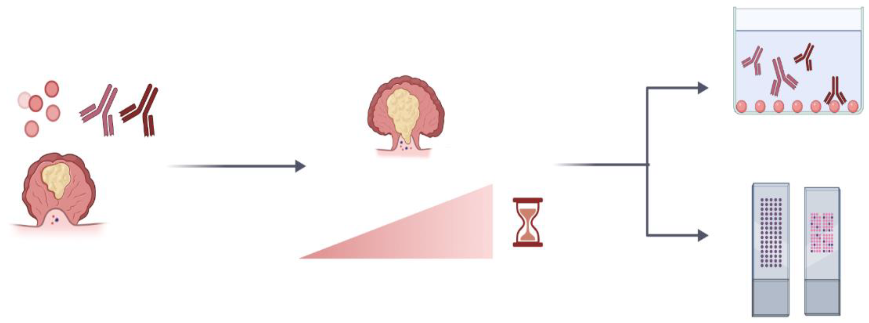
aAbs can sometimes be detected months before the onset of cancer, potentially signaling an early ‘tug of war’ between the immune system and emerging cancer cells. Hence, assessing the presence of autoantibodies seems useful in the early detection of cancer. Because tumors are highly heterogeneous, the use of aAbs as biomarkers would probably be harnessed in panels that containe a combination of aAbs and/or autoantigens, rather than single aAbs, to give optimum sensitivity and specificity. Thanks to high-throughput techniques such as protein microarrays, screening, validating, and using these panels are feasible, affordable, and quick. Currently, panels of aAbs are being tested for early cancer diagnoses such as lung cancer diagnosis [83] (Table 1). Furthermore, there are already commercial blood-based tests that either exclusively screen for a panel of aAbs, such as Early CDT-lung for lung cancer diagnosis, or combine an autoantibody panel with serum protein biomarkers, such as Videssa Breast for breast cancer diagnosis.
Characterizing the targets, triggers, and effects of the aAbs could also be important for providing information on the immunogenicity profile of self-epitopes to guide the design of peptide-based cancer vaccines [7,91]. Furthermore, if these aAbs are shown to have any anti-tumor actions, they can be integrated into cancer immunotherapy techniques.
One of the fundamental questions about aAbs in a tumor is whether they serve any function for/against cancer. It could be that only tumor cells that can evade or even use these autoantibodies to their benefit are selected for in the tumor mass. aAbs against a chaperone from the HSP 70 family, GRP78, are in fact shown to bind to the aberrantly membrane-expressed form of this chaperone. GRP78 can induce an unfolded protein response and prostate cell proliferation [9]. A similar, tumor-promoting role has been suggested for aAb against an estrogen receptor, where the aAb acted as an agonist to promote breast cancer cell proliferation [109].
On the other hand, the presence of aAbs can also be an indicator of a tumor-decimating immune response. Accordingly, in the Muc-1 aAb study mentioned above, the levels of aAbs were correlated with longer survival in lung cancer patients [90]. Anti-dsDNA aAbs in colorectal cancer were also associated with better outcomes [91]. Similarly, the presence of anti-nuclear antibodies (ANAs) was associated with better survival in stage III NSCLC [91]. ANAs can bind to their exposed antigens during apoptosis and might opsonize those cells, which might facilitate anti-tumor immune responses [91]. aAbs against DNA topoisomerase I were also associated with better overall survival rates in stage I, II, and IV NSCLC [110]. Another study showed that aAbs to human DNA topoisomerase I were biomarkers for favorable prognosis in breast cancer, and this antibody induced ADCC in the in vitro studies with ER+ and triple-negative human breast cancer cell lines [111]. This study is important because it demonstrates the direct link between the aAbs and anti-tumor immune responses. Another study has also shown that the presence of an aAb to topoisomerase I, as detected by ELISA in the absence of survivin-expressing cancer cells (in the peripheral blood), is associated with longer survival in endometrial cancer as compared to patients without aAbs but with survivin-positive cancer cells in the peripheral blood [112].
The presence of aAbs can also be associated with the mutational load in tumoral cells. It was shown that the higher the mutational load of the tumor (‘hot tumors’), the better the response to ICI [113]. A plausible explanation for this observation is the formation of neoantigens in hot tumors, which can then be recognized by effector T cells without the constraints of central tolerance faced by self-antigens [114]. The presence of aAbs can thus be an indirect indicator of higher mutational load, clarifying the link between the presence of some aAbs and treatment response.
In addition to the prognostic and tumor-centered importance of aAbs in cancer, another obvious question about these aAbs is whether they cause autoimmune damage to the other structures of the body via various mechanisms. This damage can arise when the immune system tries to clear the immune complexes through the Fc receptors present on the phagocytic cells or when the complement cascade is activated via these immunoglobulins, among other mechanisms. Clustering of some autoimmune diseases with cancers can be associated with destructive autoimmune responses. Systemic sclerosis (scleroderma) is an autoimmune disease that presents with widespread vasculopathy and fibrotic changes. Interestingly, the individuals that have aAbs against the RNA polymerase III subunit can have temporal clustering of scleroderma and cancer [115]. The question arises whether these aAbs lead to a scleroderma-inducing autoimmune attack. Interestıngly, this phenomenon is not detected in the presence of topoisomerase 1 or centromere protein B aAbs [115]. The same study showed that there were somatic mutations or loss of heterozygosity in 6 out of 8 patients in the gene coding for RNA polymerase III (POL3RA), pointing at the mutated protein as the cause of aAbs. A similar occurrence was seen in dermatomyositis, where the presence of anti-NXP2 and anti-transcription intermediary factor-1 gamma aAb correlated with cancer development right before or after dermatomyositis onset [79,116]. Furthermore, the progress of the dermatomyositis paralleled the progress of cancer, with co-relapses or co-resolutions [79]. It can be speculated that these aAbs could be the remnants of an early, failed anti-tumor immune response that led to an autoimmune pathology [7].
Another autoimmune attack in cancer is seen in paraneoplastic syndrome. This syndrome is a rare occurrence (approximately 1 in 10,000 patients with cancer) and shows symptoms not directly attributable to the tumor presence [11]. Paraneoplastic syndrome, as its name suggests, can occur before or right after the clinically overt cancer. Paraneoplastic syndromes are thought to be mediated by the tumoral secretion of functional molecules such as hormones and/or immune attacks due to the cross-reactivity of the anti-tumoral immune responses [11]. The latter is especially important in neurologic paraneoplastic syndromes, with Lambert-Eaton myasthenic syndrome (LEMS) occurring in 1% of patients with small-cell lung cancer [117]. Classic neurological paraneoplastic syndromes also include encephalitis and cerebellar ataxia [118]. The targets of aAbs in neurological paraneoplastic syndromes usually include onco-neuronal antigens such as Hu antigen or anti-amphiphysin [117]. The presence of anti-Hu aAbs and paraneoplastic neuropathies and encephalopathies are associated with better response to therapy, pointing to a potential cross-reactivity between neuronal antigens and TAAs [64].
Overall, aAbs have many advantages as tools for the early diagnosis of various solid cancers (Figure 3). Their biological and prognostic capabilities are probably dependent on their specificity, their immunological properties, and the immune milieu of the body and cancer.

4.2. The Significance of irAEs

As mentioned before, irAEs can limit the clinical utility of ICIs, leading to treatment discontinuation due to severe toxicities. Therefore, the biggest challenge in cancer immunotherapy is to prevent trading one evil for another in the form of autoimmunity. Thus, while eliminating cancer using immune components, destructive and potentially lethal autoimmunity should ideally be avoided. Steroid administration to counteract irAE does not seem to reduce the treatment efficiency of ICI, giving hope that different cell populations are involved in these two events, and irAEs are not obligate side effects of ICI.
Autoimmunity induced by ICI can also be associated with a good treatment response similar to some autoantibodies mentioned before, as it is often observed in melanoma. After ipilimumab treatment, Melan-A-specific cytotoxic T cells increase in peripheral blood, in tumor tissue, and in autoimmunity-induced skin rashes [73]. Vitiligo has been shown that it is associated with progression-free survival and overall survival in patients with advanced melanoma receiving immunotherapy, pointing to lymphocytes that are cross-reacting to melanoma and normal melanocytes [71]. Cutaneous irAEs (vitiligo, pruritus, nonspecific macular rash, etc.) are also observed frequently. When an analysis was carried out through grouping all cutaneous irAEs, they were shown to be associated with better therapeutic response and overall survival upon immunotherapy [74]. Furthermore, the presence of irAEs after nivolumab treatment was positively associated with overall survival in 143 melanoma patients [64]. A similar trend has also been observed in NSCLC, where the presence of irAEs after nivolumab and pembrolizumab regimens correlated with a good treatment response and progression-free survival [62,64]. These data might suggest that in some cases, the presence of irAEs is an indicator of the inflammatory response and heightened immune reactivity against tumors.

5. Conclusions and Perspectives

Autoimmune responses observed in tumor settings and solid cancer immunotherapy are complex. They can arise due to genetic changes, microbiota-related factors, TAAs, epitope spreading, the imbalance of antitumor immunity, and immune toxicity. It should also be noted that the infection history of the host and, in the specific case of virus-related cancers such as HPV-related cervical cancer, viral factors and anti-viral immunity can also potentially play a role in these responses.
Autoimmune responses in the form of aAbs can serve as stable, easy to assay, and specific biomarkers for cancer diagnosis that only require a small volume of patient samples. Advances in high-throughput proteomics in the form of protein microarrays, SEREX, and NAPPA arrays enable the screening of large cohorts of patients in parallel in a relatively short time. This could also lead to the use of aAb panels to discriminate among different tumor stages and grades, for assessing treatment responses, and for shaping treatment choices. Large prospective studies that are standardized in terms of design, the methods of detection, cut-points, and case characteristics are also important to be able to compare results across studies.
In addition to serum, the analysis of aAbs in body fluids other than peripheral blood, such as cerebrospinal fluid for glioma, can also expand the clinical utility of aAbs in clinical practice [119]. Furthermore, investigating classes of antibodies other than IgG such as IgA, especially in cancers such as colon cancer where mucosal immunity is altered, can shed more light on the autoimmune phenomenon in cancer settings.
It is of note that the relationship between autoimmunity and cancer is bidirectional, and certain autoimmune diseases such as SLE, RA, and SS increase the risk of cancers. This effect is probably mediated by chronic inflammation and the treatments involved in these diseases. The cancer to autoimmune response link, however, is mediated mainly by the autoantigens in the tumor cells, epitope spreading, and the treatment-induced, inflammatory tumor environment. However, clearly elucidating the root causes of this link would also shed light on the general concepts of immune tolerance as well as basic immunology in autoimmune diseases.
The need to understand autoimmune responses in cancer is more pressing than ever with the more widespread use of cancer immunotherapy approaches in the clinic, as irAEs are the major roadblock to their widespread use. A very recent study in a xenoplant, triple-negative breast cancer mouse model showed that targeting CD6 via an antibody decreased tumor growth and led to the activation of CD8+ and NK cells, whereas the same antibody dampened autoimmunity in mice [120]. In this way, selective activation of only anti-tumor effectors seemed possible. Similar efforts in the field would help in the fine-tuning of cancer immunotherapy approaches to evade or minimize irAEs while inducing an effective anti-tumor immune response to maximize the clinical benefits of immunotherapy.

Author Contributions

H.B. and M.F. designed the manuscript outline. H.B. conducted the literature search and designed and prepared text, figures, and tables. H.B. conducted the adjustment of the content and language edition. All the authors (P.J.-V., A.L.-V., A.-P.H., J.J.C., L.B., E.F., A.N.-C., E.M., R.G., M.F.) participated in writing, discussing, and preparing the manuscript. All authors have read and agreed to the published version of the manuscript.

Funding

We gratefully acknowledge financial support from the Spanish Health Institute Carlos III (ISCIII) for grants FIS PI14/01538, FIS PI17/01930, and CB16/12/00400. We also acknowledge Fondos FEDER (EU), Junta Castilla-León (COVID-19 grant COV20EDU/00187), and Fundación Solórzano FS/38-2017. The Proteomics Unit belongs to ProteoRed, PRB3-ISCIII, supported by grant PT17/0019/0023 of the PE I + D + I 2017–2020 and funded by ISCIII and FEDER. A. Landeira-Viñuela is supported by the VIII Centenario-USAL PhD Program. P. Juanes-Velasco is supported by the JCYL PhD Program ‘JCYL Nos Impulsa’ and scholarship JCYL-EDU/601/2020.

Institutional Review Board Statement

Not applicable.

Informed Consent Statement

Not applicable.

Data Availability Statement

Not applicable.

Conflicts of Interest

The authors declare no conflict of interest.

References

  1. Min, B.; Legge, K.L.; Pack, C.; Zaghouani, H. Neonatal Exposure to a Self-Peptide–Immunoglobulin Chimera Circumvents the Use of Adjuvant and Confers Resistance to Autoimmune Disease by a Novel Mechanism Involving Interleukin 4 Lymph Node Deviation and Interferon γ–Mediated Splenic Anergy. J. Exp. Med. 1998, 188, 2007–2017. [Google Scholar] [CrossRef] [Green Version]
  2. Burkholder, B.; Huang, R.-Y.; Burgess, R.; Luo, S.; Jones, V.S.; Zhang, W.; Lv, Z.-Q.; Gao, C.-Y.; Wang, B.-L.; Zhang, Y.-M.; et al. Tumor-Induced Perturbations of Cytokines and Immune Cell Networks. Biochim. Et Biophys. Acta (BBA) Rev. Cancer 2014, 1845, 182–201. [Google Scholar] [CrossRef] [Green Version]
  3. Farkona, S.; Diamandis, E.P.; Blasutig, I.M. Cancer Immunotherapy: The Beginning of the End of Cancer? BMC Med. 2016, 14, 73. [Google Scholar] [CrossRef] [Green Version]
  4. Routy, B.; Le Chatelier, E.; Derosa, L.; Duong, C.P.M.; Alou, M.T.; Daillère, R.; Fluckiger, A.; Messaoudene, M.; Rauber, C.; Roberti, M.P.; et al. Gut Microbiome Influences Efficacy of PD-1–Based Immunotherapy against Epithelial Tumors. Science 2018, 359, 91–97. [Google Scholar] [CrossRef] [PubMed] [Green Version]
  5. Li, B.; Chan, H.L.; Chen, P. Immune Checkpoint Inhibitors: Basics and Challenges. CMC 2019, 26, 3009–3025. [Google Scholar] [CrossRef] [PubMed]
  6. Song, P.; Zhang, D.; Cui, X.; Zhang, L. Meta-analysis of Immune-related Adverse Events of Immune Checkpoint Inhibitor Therapy in Cancer Patients. Thorac. Cancer 2020, 11, 2406–2430. [Google Scholar] [CrossRef]
  7. Andersen, M.H. Cancer and Autoimmunity. Semin. Immunopathol. 2017, 39, 241–243. [Google Scholar] [CrossRef]
  8. Coussens, L.M.; Werb, Z. Inflammation and Cancer. Nature 2002, 420, 860–867. [Google Scholar] [CrossRef] [PubMed]
  9. Al-Hashimi, A.A.; Lebeau, P.; Majeed, F.; Polena, E.; Lhotak, Š.; Collins, C.A.F.; Pinthus, J.H.; Gonzalez-Gronow, M.; Hoogenes, J.; Pizzo, S.V.; et al. Autoantibodies against the Cell Surface–Associated Chaperone GRP78 Stimulate Tumor Growth via Tissue Factor. J. Biol. Chem. 2017, 292, 21180–21192. [Google Scholar] [CrossRef] [PubMed] [Green Version]
  10. Ram, M.; Shoenfeld, Y. Harnessing Autoimmunity (Vitiligo) to Treat Melanoma: A Myth or Reality? Ann. N. Y. Acad. Sci. 2007, 1110, 410–425. [Google Scholar] [CrossRef] [PubMed]
  11. Pelosof, L.C.; Gerber, D.E. Paraneoplastic Syndromes: An Approach to Diagnosis and Treatment. Mayo Clin. Proc. 2010, 85, 838–854. [Google Scholar] [CrossRef] [Green Version]
  12. González-González, M.; Sayagués, J.M.; Muñoz-Bellvís, L.; Pedreira, C.E.; de Campos, M.L.R.; García, J.; Alcázar, J.A.; Braz, P.F.; Galves, B.L.; González, L.M.; et al. Tracking the Antibody Immunome in Sporadic Colorectal Cancer by Using Antigen Self-Assembled Protein Arrays. Cancers 2021, 13, 2718. [Google Scholar] [CrossRef] [PubMed]
  13. Kyewski, B.; Klein, L. A central role for central tolerance. Annu. Rev. Immunol. 2006, 24, 571–606. [Google Scholar] [CrossRef]
  14. Nemazee, D. Mechanisms of Central Tolerance for B Cells. Nat. Rev. Immunol. 2017, 17, 281–294. [Google Scholar] [CrossRef]
  15. Andersen, M.H. Anti-Regulatory T Cells. Semin Immunopathol. 2017, 39, 317–326. [Google Scholar] [CrossRef] [PubMed]
  16. Hanahan, D.; Weinberg, R.A. Hallmarks of Cancer: The Next Generation. Cell 2011, 144, 646–674. [Google Scholar] [CrossRef] [PubMed] [Green Version]
  17. Wing, J.B.; Tanaka, A.; Sakaguchi, S. Human FOXP3+ Regulatory T Cell Heterogeneity and Function in Autoimmunity and Cancer. Immunity 2019, 50, 302–316. [Google Scholar] [CrossRef] [PubMed] [Green Version]
  18. Arce-Sillas, A.; Álvarez-Luquín, D.D.; Tamaya-Domínguez, B.; Gomez-Fuentes, S.; Trejo-García, A.; Melo-Salas, M.; Cárdenas, G.; Rodríguez-Ramírez, J.; Adalid-Peralta, L. Regulatory T Cells: Molecular Actions on Effector Cells in Immune Regulation. J. Immunol. Res. 2016, 2016, 1–12. [Google Scholar] [CrossRef] [Green Version]
  19. Shang, B.; Liu, Y.; Jiang, S.; Liu, Y. Prognostic Value of Tumor-Infiltrating FoxP3+ Regulatory T Cells in Cancers: A Systematic Review and Meta-Analysis. Sci. Rep. 2015, 5, 15179. [Google Scholar] [CrossRef] [Green Version]
  20. Boros, P.; Ochando, J.; Zeher, M. Myeloid Derived Suppressor Cells and Autoimmunity. Hum. Immunol. 2016, 77, 631–636. [Google Scholar] [CrossRef] [Green Version]
  21. Ai, L.; Mu, S.; Wang, Y.; Wang, H.; Cai, L.; Li, W.; Hu, Y. Prognostic Role of Myeloid-Derived Suppressor Cells in Cancers: A Systematic Review and Meta-Analysis. BMC Cancer 2018, 18, 1220. [Google Scholar] [CrossRef] [Green Version]
  22. Wculek, S.K.; Cueto, F.J.; Mujal, A.M.; Melero, I.; Krummel, M.F.; Sancho, D. Dendritic Cells in Cancer Immunology and Immunotherapy. Nat. Rev. Immunol. 2020, 20, 7–24. [Google Scholar] [CrossRef]
  23. Idos, G.E.; Kwok, J.; Bonthala, N.; Kysh, L.; Gruber, S.B.; Qu, C. The Prognostic Implications of Tumor Infiltrating Lymphocytes in Colorectal Cancer: A Systematic Review and Meta-Analysis. Sci. Rep. 2020, 10, 3360. [Google Scholar] [CrossRef] [PubMed]
  24. Wouters, M.C.A.; Nelson, B.H. Prognostic Significance of Tumor-Infiltrating B Cells and Plasma Cells in Human Cancer. Clin. Cancer Res. 2018, 24, 6125–6135. [Google Scholar] [CrossRef] [Green Version]
  25. Tsou, P.; Katayama, H.; Ostrin, E.J.; Hanash, S.M. The Emerging Role of B Cells in Tumor Immunity. Cancer Res. 2016, 76, 5597–5601. [Google Scholar] [CrossRef] [PubMed] [Green Version]
  26. Garaud, S.; Zayakin, P.; Buisseret, L.; Rulle, U.; Silina, K.; de Wind, A.; Van den Eyden, G.; Larsimont, D.; Willard-Gallo, K.; Linē, A. Antigen Specificity and Clinical Significance of IgG and IgA Autoantibodies Produced in Situ by Tumor-Infiltrating B Cells in Breast Cancer. Front. Immunol. 2018, 9, 2660. [Google Scholar] [CrossRef] [PubMed]
  27. Kandoth, C.; McLellan, M.D.; Vandin, F.; Ye, K.; Niu, B.; Lu, C.; Xie, M.; Zhang, Q.; McMichael, J.F.; Wyczalkowski, M.A.; et al. Mutational Landscape and Significance across 12 Major Cancer Types. Nature 2013, 502, 333–339. [Google Scholar] [CrossRef] [PubMed] [Green Version]
  28. Fierabracci, A.; Pellegrino, M. The Double Role of P53 in Cancer and Autoimmunity and Its Potential as Therapeutic Target. IJMS 2016, 17, 1975. [Google Scholar] [CrossRef] [Green Version]
  29. Leech, M.; Xue, J.R.; Dacumos, A.; Hall, P.; Santos, L.; Yang, Y.; Li, M.; Kitching, A.R.; Morand, E.F. The Tumour Suppressor Gene P53 Modulates the Severity of Antigen-Induced Arthritis and the Systemic Immune Response: P53 and the Immune Response. Clin. Exp. Immunol. 2008, 152, 345–353. [Google Scholar] [CrossRef]
  30. Okuda, Y.; Okuda, M.; Bernard, C.C.A. Regulatory Role of P53 in Experimental Autoimmune Encephalomyelitis. J. Neuroimmunol. 2003, 135, 29–37. [Google Scholar] [CrossRef]
  31. Park, J.-S.; Lim, M.-A.; Cho, M.-L.; Ryu, J.-G.; Moon, Y.-M.; Jhun, J.-Y.; Byun, J.-K.; Kim, E.-K.; Hwang, S.-Y.; Ju, J.H.; et al. P53 Controls Autoimmune Arthritis via STAT-Mediated Regulation of the Th17 Cell/Treg Cell Balance in Mice: P53 Induces Treg Cells and Controls Autoimmune Arthritis. Arthritis Rheum. 2013, 65, 949–959. [Google Scholar] [CrossRef] [PubMed]
  32. Volodko, N. TP53 Codon 72 Arg/Arg Polymorphism Is Associated with a Higher Risk for Inflammatory Bowel Disease Development. WJG 2015, 21, 10358. [Google Scholar] [CrossRef] [PubMed]
  33. Pellegrino, M.; Traversi, G.; Arena, A.; Cappa, M.; Rosado, M.M.; Andreani, M.; Delfino, D.V.; Moretti, F.; Fierabracci, A. Effect of P53 Activation through Targeting MDM2/MDM4 Heterodimer on T Regulatory and Effector Cells in the Peripheral Blood of Type 1 Diabetes Patients. PLoS ONE 2020, 15, e0228296. [Google Scholar] [CrossRef]
  34. Liu, Y.; Dai, L.; Liu, W.; Shi, G.; Zhang, J. Autoantibody to MDM2: A Potential Serological Marker of Systemic Lupus Erythematosus. J. Immunol. Res. 2015, 2015, 1–6. [Google Scholar] [CrossRef] [Green Version]
  35. Liu, Y.; Liao, X.; Wang, Y.; Chen, S.; Sun, Y.; Lin, Q.; Shi, G. Autoantibody to MDM2: A Potential Serological Marker of Primary Sjogren’s Syndrome. Oncotarget 2017, 8, 14306–14313. [Google Scholar] [CrossRef]
  36. Li, P.; Shi, J.-X.; Dai, L.-P.; Chai, Y.-R.; Zhang, H.-F.; Kankonde, M.; Kankonde, P.; Yu, B.-F.; Zhang, J.-Y. Serum Anti-MDM2 and Anti-c-Myc Autoantibodies as Biomarkers in the Early Detection of Lung Cancer. OncoImmunology 2016, 5, e1138200. [Google Scholar] [CrossRef] [Green Version]
  37. Himoto, T.; Yoneyama, H.; Kurokohchi, K.; Inukai, M.; Masugata, H.; Goda, F.; Haba, R.; Watanabe, S.; Senda, S.; Masaki, T. Clinical Significance of Autoantibodies to P53 Protein in Patients with Autoimmune Liver Diseases. Can. J. Gastroenterol. 2012, 26, 125–129. [Google Scholar] [CrossRef]
  38. Li, Y.; Karjalainen, A.; Koskinen, H.; Hemminki, K.; Vainio, H.; Shnaidman, M.; Ying, Z.; Pukkala, E.; Brandt-Rauf, P.W. P53 Autoantibodies Predict Subsequent Development of Cancer. Int. J. Cancer 2005, 114, 157–160. [Google Scholar] [CrossRef]
  39. Revathidevi, S.; Munirajan, A.K. Akt in Cancer: Mediator and More. Semin. Cancer Biol. 2019, 59, 80–91. [Google Scholar] [CrossRef]
  40. Zhang, Y.; Kwok-Shing Ng, P.; Kucherlapati, M.; Chen, F.; Liu, Y.; Tsang, Y.H.; de Velasco, G.; Jeong, K.J.; Akbani, R.; Hadjipanayis, A.; et al. A Pan-Cancer Proteogenomic Atlas of PI3K/AKT/MTOR Pathway Alterations. Cancer Cell 2017, 31, 820–832.e3. [Google Scholar] [CrossRef] [Green Version]
  41. Parsons, M.J.; Jones, R.G.; Tsao, M.-S.; Odermatt, B.; Ohashi, P.S.; Woodgett, J.R. Expression of Active Protein Kinase B in T Cells Perturbs Both T and B Cell Homeostasis and Promotes Inflammation. J. Immunol. 2001, 167, 42–48. [Google Scholar] [CrossRef] [Green Version]
  42. Sauer, S.; Bruno, L.; Hertweck, A.; Finlay, D.; Leleu, M.; Spivakov, M.; Knight, Z.A.; Cobb, B.S.; Cantrell, D.; O’Connor, E.; et al. T Cell Receptor Signaling Controls Foxp3 Expression via PI3K, Akt, and MTOR. Proc. Natl. Acad. Sci. USA 2008, 105, 7797–7802. [Google Scholar] [CrossRef] [PubMed] [Green Version]
  43. Huynh, A.; DuPage, M.; Priyadharshini, B.; Sage, P.T.; Quiros, J.; Borges, C.M.; Townamchai, N.; Gerriets, V.A.; Rathmell, J.C.; Sharpe, A.H.; et al. Control of PI(3) Kinase in Treg Cells Maintains Homeostasis and Lineage Stability. Nat. Immunol. 2015, 16, 188–196. [Google Scholar] [CrossRef]
  44. Lai, K.; Zhang, W.; Li, S.; Zhang, Z.; Xie, S.; Xu, M.; Li, C.; Zeng, K. MTOR Pathway Regulates the Differentiation of Peripheral Blood Th2/Treg Cell Subsets in Patients with Pemphigus Vulgaris. Acta Biochim. Biophys. Sin. 2021, 53, 438–445. [Google Scholar] [CrossRef]
  45. Tischner, D.; Woess, C.; Ottina, E.; Villunger, A. Bcl-2-Regulated Cell Death Signalling in the Prevention of Autoimmunity. Cell Death Dis. 2010, 1, e48. [Google Scholar] [CrossRef] [PubMed]
  46. The AACR Project GENIE Consortium AACR Project GENIE: Powering Precision Medicine through an International Consortium. Cancer Discov. 2017, 7, 818–831. [CrossRef] [Green Version]
  47. Wong, R.S. Apoptosis in Cancer: From Pathogenesis to Treatment. J. Exp. Clin. Cancer Res. 2011, 30, 87. [Google Scholar] [CrossRef] [Green Version]
  48. Mehrian, R.; Quismorio, F.P.; Strassmann, G.; Stimmler, M.M.; Horwitz, D.A.; Kitridou, R.C.; Gauderman, W.J.; Morrison, J.; Brautbar, C.; Jacob, C.O. Synergistic Effect between IL-10 and Bcl-2 Genotypes in Determining Susceptibility to Systemic Lupus Erythematosus. Arthritis Rheum 1998, 41, 596–602. [Google Scholar] [CrossRef]
  49. Davey, G.M.; Kurts, C.; Miller, J.F.A.P.; Bouillet, P.; Strasser, A.; Brooks, A.G.; Carbone, F.R.; Heath, W.R. Peripheral Deletion of Autoreactive CD8 T Cells by Cross Presentation of Self-Antigen Occurs by a Bcl-2–Inhibitable Pathway Mediated by Bim. J. Exp. Med. 2002, 196, 947–955. [Google Scholar] [CrossRef] [Green Version]
  50. Fabbri, M.; Calin, G.A. Epigenetics and miRNAs in Human Cancer. In Advances in Genetics; Elsevier: Amsterdam, The Netherlands, 2010; Volume 70, pp. 87–99. ISBN 978-0-12-380866-0. [Google Scholar]
  51. Park, S.; Kim, G.W.; Kwon, S.H.; Lee, J. Broad Domains of Histone H3 Lysine 4 Trimethylation in Transcriptional Regulation and Disease. FEBS J. 2020, 287, 2891–2902. [Google Scholar] [CrossRef] [Green Version]
  52. Portela, A.; Esteller, M. Epigenetic Modifications and Human Disease. Nat. Biotechnol. 2010, 28, 1057–1068. [Google Scholar] [CrossRef]
  53. Ligthart, S.; Marzi, C.; Aslibekyan, S.; Mendelson, M.M.; Conneely, K.N.; Tanaka, T.; Colicino, E.; Waite, L.L.; Joehanes, R.; Guan, W.; et al. DNA Methylation Signatures of Chronic Low-Grade Inflammation Are Associated with Complex Diseases. Genome Biol. 2016, 17, 255. [Google Scholar] [CrossRef] [PubMed] [Green Version]
  54. Kinugawa, Y.; Uehara, T.; Sano, K.; Matsuda, K.; Maruyama, Y.; Kobayashi, Y.; Nakajima, T.; Hamano, H.; Kawa, S.; Higuchi, K.; et al. Methylation of Tumor Suppressor Genes in Autoimmune Pancreatitis. Pancreas 2017, 46, 614–618. [Google Scholar] [CrossRef]
  55. Pradhan, V.; Patwardhan, M.; Ghosh, K. Anti-Nucleosome Antibodies as a Disease Marker in Systemic Lupus Erythematosus and Its Correlation with Disease Activity and Other Autoantibodies. Indian J. Dermatol. Venereol. Leprol. 2010, 76, 145. [Google Scholar] [CrossRef]
  56. Matzaraki, V.; Kumar, V.; Wijmenga, C.; Zhernakova, A. The MHC Locus and Genetic Susceptibility to Autoimmune and Infectious Diseases. Genome Biol. 2017, 18, 76. [Google Scholar] [CrossRef] [PubMed]
  57. Stamatouli, A.M.; Quandt, Z.; Perdigoto, A.L.; Clark, P.L.; Kluger, H.; Weiss, S.A.; Gettinger, S.; Sznol, M.; Young, A.; Rushakoff, R.; et al. Collateral Damage: Insulin-Dependent Diabetes Induced With Checkpoint Inhibitors. Diabetes 2018, 67, 1471–1480. [Google Scholar] [CrossRef] [Green Version]
  58. Ruff, W.E.; Greiling, T.M.; Kriegel, M.A. Host–Microbiota Interactions in Immune-Mediated Diseases. Nat. Rev. Microbiol. 2020, 18, 521–538. [Google Scholar] [CrossRef]
  59. Tanoue, T.; Atarashi, K.; Honda, K. Development and Maintenance of Intestinal Regulatory T Cells. Nat. Rev. Immunol. 2016, 16, 295–309. [Google Scholar] [CrossRef]
  60. Itzhaki, O.; Levy, D.; Zikich, D.; Treves, A.J.; Markel, G.; Schachter, J.; Besser, M.J. Adoptive T-Cell Transfer in Melanoma. Immunotherapy 2013, 5, 79–90. [Google Scholar] [CrossRef]
  61. Dumas, A.; Bernard, L.; Poquet, Y.; Lugo-Villarino, G.; Neyrolles, O. The Role of the Lung Microbiota and the Gut-Lung Axis in Respiratory Infectious Diseases. Cell. Microbiol. 2018, 20, e12966. [Google Scholar] [CrossRef] [Green Version]
  62. Young, A.; Quandt, Z.; Bluestone, J.A. The Balancing Act between Cancer Immunity and Autoimmunity in Response to Immunotherapy. Cancer Immunol. Res. 2018, 6, 1445–1452. [Google Scholar] [CrossRef] [Green Version]
  63. Davar, D.; Dzutsev, A.K.; McCulloch, J.A.; Rodrigues, R.R.; Chauvin, J.-M.; Morrison, R.M.; Deblasio, R.N.; Menna, C.; Ding, Q.; Pagliano, O.; et al. Fecal Microbiota Transplant Overcomes Resistance to Anti-PD-1 Therapy in Melanoma Patients. Science 2021, 371, 595–602. [Google Scholar] [CrossRef]
  64. Khan, S.; Gerber, D.E. Autoimmunity, Checkpoint Inhibitor Therapy and Immune-Related Adverse Events: A Review. Semin. Cancer Biol. 2020, 64, 93–101. [Google Scholar] [CrossRef] [PubMed]
  65. Brevi, A.; Cogrossi, L.L.; Grazia, G.; Masciovecchio, D.; Impellizzieri, D.; Lacanfora, L.; Grioni, M.; Bellone, M. Much More Than IL-17A: Cytokines of the IL-17 Family Between Microbiota and Cancer. Front. Immunol. 2020, 11, 565470. [Google Scholar] [CrossRef] [PubMed]
  66. Zhang, X.; Chen, B.; Zhao, L.; Li, H. The Gut Microbiota: Emerging Evidence in Autoimmune Diseases. Trends Mol. Med. 2020, 26, 862–873. [Google Scholar] [CrossRef] [PubMed]
  67. Sakkas, L.I.; Bogdanos, D.P. Multiple Hit Infection and Autoimmunity: The Dysbiotic Microbiota–ACPA Connection in Rheumatoid Arthritis. Curr. Opin. Rheumatol. 2018, 30, 403–409. [Google Scholar] [CrossRef] [PubMed]
  68. Regen, T.; Isaac, S.; Amorim, A.; Núñez, N.G.; Hauptmann, J.; Shanmugavadivu, A.; Klein, M.; Sankowski, R.; Mufazalov, I.A.; Yogev, N.; et al. IL-17 Controls Central Nervous System Autoimmunity through the Intestinal Microbiome. Sci. Immunol. 2021, 6, eaaz6563. [Google Scholar] [CrossRef]
  69. Dhodapkar, K.M. Autoimmune Complications of Cancer Immunotherapy. Curr. Opin. Immunol. 2019, 61, 54–59. [Google Scholar] [CrossRef]
  70. Cappelli, L.C.; Shah, A.A. The Relationships between Cancer and Autoimmune Rheumatic Diseases. Best Pract. Res. Clin. Rheumatol. 2020, 34, 101472. [Google Scholar] [CrossRef]
  71. Ladak, K.; Bass, A.R. Checkpoint Inhibitor-Associated Autoimmunity. Best Pract. Res. Clin. Rheumatol. 2018, 32, 781–802. [Google Scholar] [CrossRef]
  72. Cubas, R.; Khan, Z.; Gong, Q.; Moskalenko, M.; Xiong, H.; Ou, Q.; Pai, C.; Rodriguez, R.; Cheung, J.; Chan, A.C. Autoimmunity Linked Protein Phosphatase PTPN22 as a Target for Cancer Immunotherapy. J. Immunother. Cancer 2020, 8, e001439. [Google Scholar] [CrossRef] [PubMed]
  73. Amos, S.M.; Duong, C.P.M.; Westwood, J.A.; Ritchie, D.S.; Junghans, R.P.; Darcy, P.K.; Kershaw, M.H. Autoimmunity Associated with Immunotherapy of Cancer. Blood 2011, 118, 499–509. [Google Scholar] [CrossRef]
  74. Sibaud, V. Dermatologic Reactions to Immune Checkpoint Inhibitors: Skin Toxicities and Immunotherapy. Am. J. Clin. Dermatol. 2018, 19, 345–361. [Google Scholar] [CrossRef] [PubMed]
  75. Kostine, M.; Chiche, L.; Lazaro, E.; Halfon, P.; Charpin, C.; Arniaud, D.; Retornaz, F.; Blanco, P.; Jourde-Chiche, N.; Richez, C.; et al. Opportunistic Autoimmunity Secondary to Cancer Immunotherapy (OASI): An Emerging Challenge. La Rev. De Médecine Interne 2017, 38, 513–525. [Google Scholar] [CrossRef]
  76. Lamers, C.H.J.; Sleijfer, S.; Vulto, A.G.; Kruit, W.H.J.; Kliffen, M.; Debets, R.; Gratama, J.W.; Stoter, G.; Oosterwijk, E. Treatment of Metastatic Renal Cell Carcinoma With Autologous T-Lymphocytes Genetically Retargeted Against Carbonic Anhydrase IX: First Clinical Experience. JCO 2006, 24, e20–e22. [Google Scholar] [CrossRef]
  77. Klichinsky, M.; Ruella, M.; Shestova, O.; Lu, X.M.; Best, A.; Zeeman, M.; Schmierer, M.; Gabrusiewicz, K.; Anderson, N.R.; Petty, N.E.; et al. Human Chimeric Antigen Receptor Macrophages for Cancer Immunotherapy. Nat. Biotechnol. 2020, 38, 947–953. [Google Scholar] [CrossRef] [PubMed]
  78. Gebremeskel, S.; Johnston, B. Concepts and Mechanisms Underlying Chemotherapy Induced Immunogenic Cell Death: Impact on Clinical Studies and Considerations for Combined Therapies. Oncotarget 2015, 6, 41600–41619. [Google Scholar] [CrossRef] [Green Version]
  79. Egiziano, G.; Bernatsky, S.; Shah, A.A. Cancer and Autoimmunity: Harnessing Longitudinal Cohorts to Probe the Link. Best Pract. Res. Clin. Rheumatol. 2016, 30, 53–62. [Google Scholar] [CrossRef] [Green Version]
  80. Valencia, J.C.; Egbukichi, N.; Erwin-Cohen, R.A. Autoimmunity and Cancer, the Paradox Comorbidities Challenging Therapy in the Context of Preexisting Autoimmunity. J. Interferon Cytokine Res. 2019, 39, 72–84. [Google Scholar] [CrossRef]
  81. Ashrafizadeh, M.; Farhood, B.; Eleojo Musa, A.; Taeb, S.; Rezaeyan, A.; Najafi, M. Abscopal Effect in Radioimmunotherapy. Int. Immunopharmacol. 2020, 85, 106663. [Google Scholar] [CrossRef] [PubMed]
  82. Fortner, R.T.; Damms-Machado, A.; Kaaks, R. Systematic Review: Tumor-Associated Antigen Autoantibodies and Ovarian Cancer Early Detection. Gynecol. Oncol. 2017, 147, 465–480. [Google Scholar] [CrossRef]
  83. Mu, Y.; Xie, F.; Sun, T. Clinical Value of Seven Autoantibodies Combined Detection in the Diagnosis of Lung Cancer. J. Clin. Lab. Anal. 2020, 34. [Google Scholar] [CrossRef]
  84. Wang, H.; Li, X.; Zhou, D.; Huang, J. Autoantibodies as Biomarkers for Colorectal Cancer: A Systematic Review, Meta-Analysis, and Bioinformatics Analysis. Int. J. Biol. Markers 2019, 34, 334–347. [Google Scholar] [CrossRef] [Green Version]
  85. Pagaza-Straffon, C.; Marchat, L.A.; Herrera, L.; Díaz-Chávez, J.; Avante, M.G.; Rodríguez, Y.P.; Arreola, M.C.; López-Camarillo, C. Evaluation of a Panel of Tumor-Associated Antigens in Breast Cancer. CBM 2020, 27, 207–211. [Google Scholar] [CrossRef]
  86. Bei, R. The Crossroads between Cancer Immunity and Autoimmunity Antibodies to Self Antigens. Front. Biosci. 2017, 22, 1289–1329. [Google Scholar] [CrossRef]
  87. Herkel, J.; Mimran, A.; Erez, N.; Kam, N.; Lohse, A.W.; Märker-Hermann, E.; Rotter, V.; Cohen, I.R. Autoimmunity to the P53 Protein Is a Feature of Systemic Lupus Erythematosus (SLE) Related to Anti-DNA Antibodies. J. Autoimmun. 2001, 17, 63–69. [Google Scholar] [CrossRef] [Green Version]
  88. Atak, A.; Mukherjee, S.; Jain, R.; Gupta, S.; Singh, V.A.; Gahoi, N.K.P.M.; Srivastava, S. Protein Microarray Applications: Autoantibody Detection and Posttranslational Modification. Proteomics 2016, 16, 2557–2569. [Google Scholar] [CrossRef]
  89. Nath, S.; Mukherjee, P. MUC1: A Multifaceted Oncoprotein with a Key Role in Cancer Progression. Trends Mol. Med. 2014, 20, 332–342. [Google Scholar] [CrossRef] [Green Version]
  90. Hirasawa, Y.; Kohno, N.; Yokoyama, A.; Kondo, K.; Hiwada, K.; Miyake, M. Natural Autoantibody to MUC1 Is a Prognostic Indicator for Non–Small Cell Lung Cancer. Am. J. Respir. Crit. Care Med. 2000, 161, 589–594. [Google Scholar] [CrossRef]
  91. Bei, R.; Masuelli, L.; Palumbo, C.; Modesti, M.; Modesti, A. A Common Repertoire of Autoantibodies Is Shared by Cancer and Autoimmune Disease Patients: Inflammation in Their Induction and Impact on Tumor Growth. Cancer Lett. 2009, 281, 8–23. [Google Scholar] [CrossRef] [PubMed]
  92. Murray, H.C.; Dun, M.D.; Verrills, N.M. Harnessing the Power of Proteomics for Identification of Oncogenic, Druggable Signalling Pathways in Cancer. Expert Opin. Drug Discov. 2017, 12, 431–447. [Google Scholar] [CrossRef]
  93. Macdonald, I.K.; Parsy-Kowalska, C.B.; Chapman, C.J. Autoantibodies: Opportunities for Early Cancer Detection. Trends Cancer 2017, 3, 198–213. [Google Scholar] [CrossRef]
  94. Thomas, R.; Al-Khadairi, G.; Roelands, J.; Hendrickx, W.; Dermime, S.; Bedognetti, D.; Decock, J. NY-ESO-1 Based Immunotherapy of Cancer: Current Perspectives. Front. Immunol. 2018, 9, 947. [Google Scholar] [CrossRef]
  95. Ohue, Y.; Kurose, K.; Karasaki, T.; Isobe, M.; Yamaoka, T.; Futami, J.; Irei, I.; Masuda, T.; Fukuda, M.; Kinoshita, A.; et al. Serum Antibody Against NY-ESO-1 and XAGE1 Antigens Potentially Predicts Clinical Responses to Anti–Programmed Cell Death-1 Therapy in NSCLC. J. Thorac. Oncol. 2019, 14, 2071–2083. [Google Scholar] [CrossRef] [Green Version]
  96. Oshima, Y.; Shimada, H.; Yajima, S.; Nanami, T.; Matsushita, K.; Nomura, F.; Kainuma, O.; Takiguchi, N.; Soda, H.; Ueda, T.; et al. NY-ESO-1 Autoantibody as a Tumor-Specific Biomarker for Esophageal Cancer: Screening in 1969 Patients with Various Cancers. J. Gastroenterol. 2016, 51, 30–34. [Google Scholar] [CrossRef]
  97. Shi, Q.-P.; Wang, X.; Liu, Z.-X.; Zhang, J.-J.; Wang, Z.-Y. Autoantibody Signatures as a Biomarker Panel for the Detection of Nasopharyngeal Carcinoma. Arch. Med. Res. 2021, S0188440921000424. [Google Scholar] [CrossRef]
  98. Wheatley, S.P.; Altieri, D.C. Survivin at a Glance. J. Cell Sci. 2019, 132, jcs223826. [Google Scholar] [CrossRef] [PubMed] [Green Version]
  99. Mizushima, N.; Levine, B. Autophagy in Human Diseases. N. Engl. J. Med. 2020, 383, 1564–1576. [Google Scholar] [CrossRef]
  100. Gravina, G.; Wasén, C.; Garcia-Bonete, M.J.; Turkkila, M.; Erlandsson, M.C.; Töyrä Silfverswärd, S.; Brisslert, M.; Pullerits, R.; Andersson, K.M.; Katona, G.; et al. Survivin in Autoimmune Diseases. Autoimmun. Rev. 2017, 16, 845–855. [Google Scholar] [CrossRef]
  101. Pan, X.; Gao, Y.; Liu, J.; Liu, C.; Xia, Y. Progress in Studies on Autoantibodies against Tumor-Associated Antigens in Hepatocellular Carcinoma. Transl. Cancer Res. 2016, 5, 845–859. [Google Scholar] [CrossRef]
  102. Das, J.K.; Xiong, X.; Ren, X.; Yang, J.-M.; Song, J. Heat Shock Proteins in Cancer Immunotherapy. J. Oncol. 2019, 2019, 1–9. [Google Scholar] [CrossRef] [Green Version]
  103. Shi, L.; Chevolot, Y.; Souteyrand, E.; Laurenceau, E. Autoantibodies against Heat Shock Proteins as Biomarkers for the Diagnosis and Prognosis of Cancer. CBM 2017, 18, 105–116. [Google Scholar] [CrossRef] [Green Version]
  104. Chapman, C.J.; Healey, G.F.; Murray, A.; Boyle, P.; Robertson, C.; Peek, L.J.; Allen, J.; Thorpe, A.J.; Hamilton-Fairley, G.; Parsy-Kowalska, C.B.; et al. EarlyCDT®-Lung Test: Improved Clinical Utility through Additional Autoantibody Assays. Tumor Biol. 2012, 33, 1319–1326. [Google Scholar] [CrossRef] [Green Version]
  105. Ma, Y.; Wang, X.; Qiu, C.; Qin, J.; Wang, K.; Sun, G.; Jiang, D.; Li, J.; Wang, L.; Shi, J.; et al. Using Protein Microarray to Identify and Evaluate Autoantibodies to Tumor-associated Antigens in Ovarian Cancer. Cancer Sci. 2021, 112, 537–549. [Google Scholar] [CrossRef]
  106. Anderson, K.S.; Cramer, D.W.; Sibani, S.; Wallstrom, G.; Wong, J.; Park, J.; Qiu, J.; Vitonis, A.; LaBaer, J. Autoantibody Signature for the Serologic Detection of Ovarian Cancer. J. Proteome Res. 2015, 14, 578–586. [Google Scholar] [CrossRef] [Green Version]
  107. Garranzo-Asensio, M.; Guzmán-Aránguez, A.; Povedano, E.; Ruiz-Valdepeñas Montiel, V.; Poves, C.; Fernandez-Aceñero, M.J.; Montero-Calle, A.; Solís-Fernández, G.; Fernandez-Diez, S.; Camps, J.; et al. Multiplexed Monitoring of a Novel Autoantibody Diagnostic Signature of Colorectal Cancer Using HaloTag Technology-Based Electrochemical Immunosensing Platform. Theranostics 2020, 10, 3022–3034. [Google Scholar] [CrossRef]
  108. Zaenker, P.; Lo, J.; Pearce, R.; Cantwell, P.; Cowell, L.; Lee, M.; Quirk, C.; Law, H.; Gray, E.; Ziman, M. A Diagnostic Autoantibody Signature for Primary Cutaneous Melanoma. Oncotarget 2018, 9, 30539–30551. [Google Scholar] [CrossRef] [PubMed] [Green Version]
  109. Ortona, E.; Pierdominici, M.; Berstein, L. Autoantibodies to Estrogen Receptors and Their Involvement in Autoimmune Diseases and Cancer. J. Steroid Biochem. Mol. Biol. 2014, 144, 260–267. [Google Scholar] [CrossRef]
  110. Wu, W.; Yie, S.; Ye, S.; Xie, K.; Zhang, J.; Cao, M.; Chen, J.; He, X.; Ma, X.; Zhang, J. An Autoantibody Against Human DNA-Topoisomerase I Is a Novel Biomarker for Non-Small Cell Lung Cancer. Ann. Thorac. Surg. 2018, 105, 1664–1670. [Google Scholar] [CrossRef] [Green Version]
  111. He, X.; Jiang, X.; Yie, K.Y.-X.; Chen, J.; Zhang, J.; Yie, S. An Autoantibody against a 48-Kd Fragment of Human DNA-Topoiomerase I in Breast Cancer: Implication for Diagnosis and Prognosis, and Antibody-Dependent Cellular Cytotoxicity in Vitro. Cell. Immunol. 2020, 347, 104007. [Google Scholar] [CrossRef]
  112. Jiang, X.; Yao, Z.; He, X.; Zhang, J.; Xie, K.; Chen, J.; Cao, M.; Zhang, J.; Yie, S. Clinical Significance of Plasma Anti-TOPO48 Autoantibody and Blood Survivin-Expressing Circulating Cancer Cells in Patients with Early Stage Endometrial Carcinoma. Arch. Gynecol. Obstet 2019, 299, 229–237. [Google Scholar] [CrossRef]
  113. Samstein, R.M.; Lee, C.-H.; Shoushtari, A.N.; Hellmann, M.D.; Shen, R.; Janjigian, Y.Y.; Barron, D.A.; Zehir, A.; Jordan, E.J.; Omuro, A.; et al. Tumor Mutational Load Predicts Survival after Immunotherapy across Multiple Cancer Types. Nat. Genet. 2019, 51, 202–206. [Google Scholar] [CrossRef]
  114. Kakimi, K.; Karasaki, T.; Matsushita, H.; Sugie, T. Advances in Personalized Cancer Immunotherapy. Breast Cancer 2017, 24, 16–24. [Google Scholar] [CrossRef]
  115. Joseph, C.G.; Darrah, E.; Shah, A.A.; Skora, A.D.; Casciola-Rosen, L.A.; Wigley, F.M.; Boin, F.; Fava, A.; Thoburn, C.; Kinde, I.; et al. Association of the Autoimmune Disease Scleroderma with an Immunologic Response to Cancer. Science 2014, 343, 152–157. [Google Scholar] [CrossRef] [Green Version]
  116. Best, M.; Molinari, N.; Chasset, F.; Vincent, T.; Cordel, N.; Bessis, D. Use of Anti-Transcriptional Intermediary Factor-1 Gamma Autoantibody in Identifying Adult Dermatomyositis Patients with Cancer: A Systematic Review and Meta-Analysis. Acta Derm Venerol. 2019, 99, 256–262. [Google Scholar] [CrossRef] [Green Version]
  117. Rosenfeld, M.R.; Dalmau, J. Paraneoplastic Neurologic Syndromes. Neurologic. Clin. 2018, 36, 675–685. [Google Scholar] [CrossRef]
  118. Zekeridou, A.; Majed, M.; Heliopoulos, I.; Lennon, V.A. Paraneoplastic Autoimmunity and Small-cell Lung Cancer: Neurological and Serological Accompaniments. Thorac. Cancer 2019, 10, 1001–1004. [Google Scholar] [CrossRef] [PubMed] [Green Version]
  119. Gahoi, N.; Syed, P.; Choudhary, S.; Epari, S.; Moiyadi, A.; Varma, S.G.; Gandhi, M.N.; Srivastava, S. A Protein Microarray-Based Investigation of Cerebrospinal Fluid Reveals Distinct Autoantibody Signature in Low and High-Grade Gliomas. Front. Oncol. 2020, 10, 543947. [Google Scholar] [CrossRef] [PubMed]
  120. Ruth, J.H.; Gurrea-Rubio, M.; Athukorala, K.S.; Rasmussen, S.M.; Weber, D.P.; Randon, P.M.; Gedert, R.J.; Lind, M.E.; Amin, M.A.; Campbell, P.L.; et al. CD6 Is a Target for Cancer Immunotherapy. JCI Insight 2021, 6, e145662. [Google Scholar] [CrossRef]
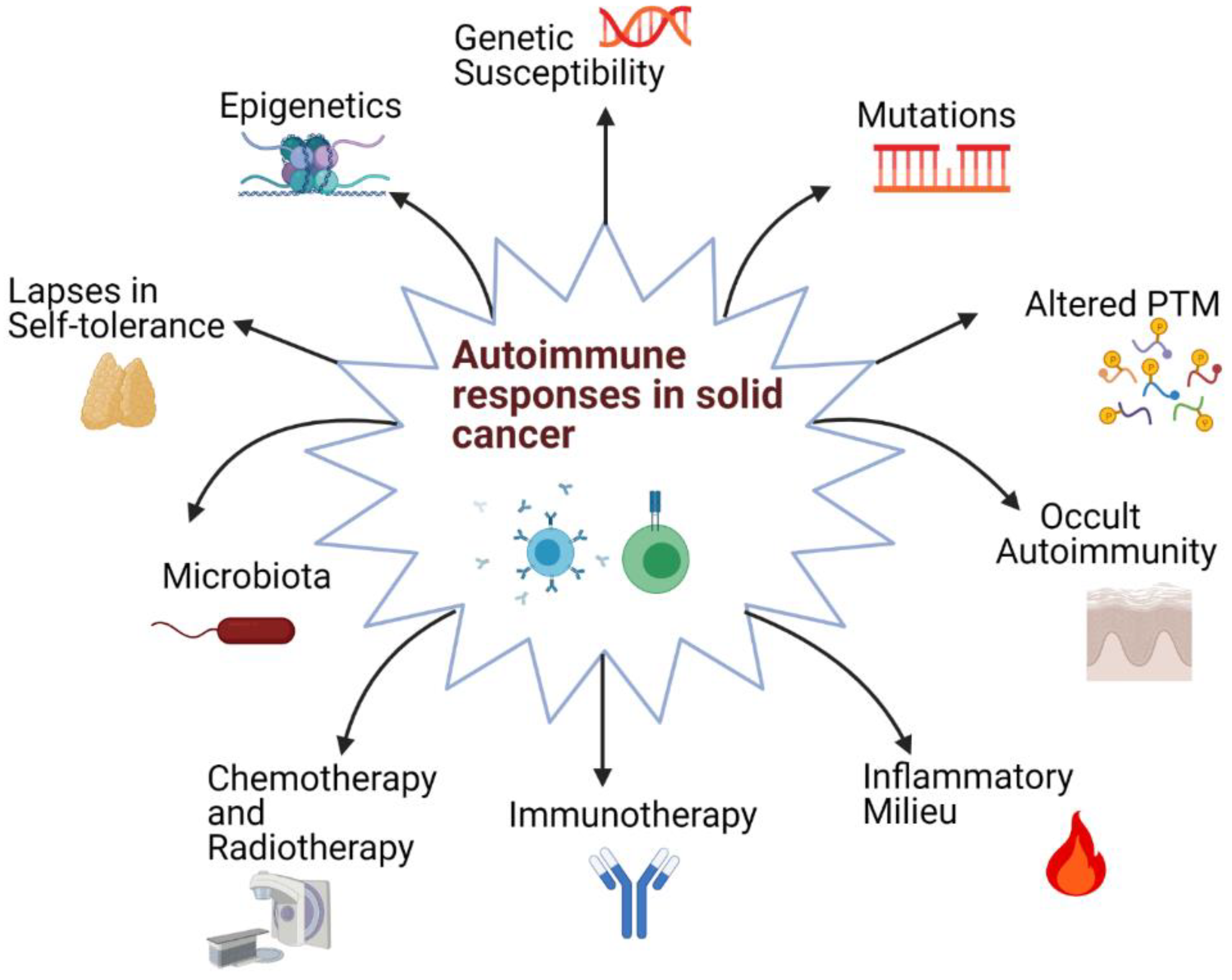
Figure 1. The probable causes of autoimmune responses observed in solid cancers. Various factors ranging from mutations to therapy-induced autoimmunity can lead to autoimmune responses. The multifactorial nature of this phenomenon can contribute to the extent of variation observed in autoimmune responses in patients (PTM: Post-translational modification).
Ijms 22 08030 g001
Figure 2. The breaking of immune tolerance in ICI therapy. (A) ICI can lift the inhibitory signal for the activation of self-reactive lymphocytes (left panel) or prevent the apoptosis of effector self-reactive lymphocytes (right panel). (B) Expression of immune checkpoint molecules on healthy cells can lead to the destruction of self-cells by antibody-mediated mechanisms such as activation of the classical complement pathway and antibody-dependent cellular cytotoxicity (ADCC). (C) The effective immune response against the tumor cells as a result of ICI can lead to high amounts of cell death, which results in the release of many self-antigens in a pro-inflammatory milieu. This can enable the activation of self-reactive lymphocytes, which normally have low avidity and affinity to the cognate self-antigen and a high activation threshold.
Ijms 22 08030 g002
Figure 3. Advantages of autoantibodies as tumor biomarkers. Autoantibodies can be present at very early stages of disease to help in life-saving early diagnosis of cancer. They are stable molecules which facilitates their detection. Autoantibodies can easily be assayed by conventional and widespread techniques. The parallel detection of multiple autoantibodies is also possible with protein microarrays to increase specificity and sensitivity.
Ijms 22 08030 g003
Table 1. Autoantibody panels being tested for diagnostic use in various solid cancers. (ELISA: enzyme-linked immunosorbent assay).
Type of CancerComparison GroupAntibody PanelMethod UsedSensitivity (%)Specificity (%)Reference
Breast cancerBreast cancer patients vs. healthy donorsp53/PRDX6/c-Myc/Hsp70/Nm23ELISA34100[85]
Lung cancerPatients with recent diagnosis of lung cancer vs. healthy controlsp53, NY-ESO-1, CAGE, GBU4–5, MAGE A4, SOX2, and Hu-DELISA (Early CDT-Lung)4193[104]
Lung cancerLung cancer patients vs. healthy controls
and lung benign disease group
p53,PGP9.5, SOX2, GAGE7, GBU4–5, MAGE A1, and CAGEELISA25.491.7[83]
Ovarian cancerEarly (stage I-II) stage ovarian cancer patients vs. healthy controlsp53, GNAS, and NPM1ELISA5786[105]
Late-stage (stage III–IV) ovarian cancer patients vs. healthy controls4986
Ovarian cancerOvarian cancer patients vs. healthy controlsp53, PTPRA, and PTGFRLuminex bead assay23.398.3[106]
Colorectal cancerColorectal cancer patients vs. healthy individuals and breast and lung cancer patientsp53, GTF2B, MAPKAPK3, PIM1, PKN1, SRC, STK4, and SULF1Luminex bead assay and
electrochemical immunosensing by HaloTag fusion
76.098.6[107]
MelanomaEarly stage melanoma patients vs. healthy controlsp53, ZBTB7B, PRKCH, PCTK1, PQBP1, UBE2V1, IRF4, MAPK8_tv2, MSN, and TPM1Protein microarray7984[108]
Nasopharyngeal carcinomaNasopharyngeal cancer patients vs. healthy individualscyclin B1, NY-ESO-1, survivin and IMP3Protein microarray5486[97]
Publisher’s Note: MDPI stays neutral with regard to jurisdictional claims in published maps and institutional affiliations.