Article

Detection of Germline Variants in 450 Breast/Ovarian Cancer Families with a Multi-Gene Panel Including Coding and Regulatory Regions

by
Chiara Guglielmi
1,
Rosa Scarpitta
2,
Gaetana Gambino
3,
Eleonora Conti
1,
Francesca Bellè
4,
Mariella Tancredi
1,
Tiziana Cervelli
4,
Elisabetta Falaschi
1,
Cinzia Cosini
1,
Paolo Aretini
5,
Caterina Congregati
6,
Marco Marino
7,
Margherita Patruno
8,
Brunella Pilato
8,
Francesca Spina
9,
Luisa Balestrino
9,
Elena Tenedini
7,
Ileana Carnevali
10,
Laura Cortesi
11,
Enrico Tagliafico
7,
Maria Grazia Tibiletti
10,
Stefania Tommasi
8,
Matteo Ghilli
12,
Caterina Vivanet
9,
Alvaro Galli
4,* and
Maria Adelaide Caligo
1,*
add Show full author list remove Hide full author list
1
SOD Molecular Genetics, University Hospital of Pisa, 56126 Pisa, Italy
2
Division of Pathology, University of Pisa, 56126 Pisa, Italy
3
Department of Clinical and Experimental Medicine, University of Pisa, 56126 Pisa, Italy
4
Functional Genetics and Genomics Laboratory, Institute of Clinical Physiology, IFC-CNR, 56127 Pisa, Italy
5
Section of Oncological Genomics, Fondazione Pisana per la Scienza, 56017 Pisa, Italy
6
Division of Internal Medicine, University Hospital of Pisa, 56126 Pisa, Italy
7
Department of Life Sciences, University of Modena and Reggio Emilia, 41125 Modena, Italy
8
IRCCS Istituto Tumori “Giovanni Paolo II”, 70124 Bari, Italy
9
SC Medical Genetics, ASSL Cagliari, 09126 Cagliari, Italy
10
Ospedale di Circolo ASST Settelaghi, 21100 Varese, Italy
11
Department of Oncology, Haematology and Respiratory Diseases, University Hospital of Modena, 41124 Modena, Italy
12
Breast Cancer Center, University Hospital, 56126 Pisa, Italy
*
Authors to whom correspondence should be addressed.
Int. J. Mol. Sci. 2021, 22(14), 7693; https://doi.org/10.3390/ijms22147693
Submission received: 12 June 2021 / Revised: 10 July 2021 / Accepted: 14 July 2021 / Published: 19 July 2021
(This article belongs to the Special Issue Molecular Genetics of Cancer)

Abstract

:
With the progress of sequencing technologies, an ever-increasing number of variants of unknown functional and clinical significance (VUS) have been identified in both coding and non-coding regions of the main Breast Cancer (BC) predisposition genes. The aim of this study is to identify a mutational profile of coding and intron-exon junction regions of 12 moderate penetrance genes (ATM, BRIP1, CDH1, CHEK2, NBN, PALB2, PTEN, RAD50, RAD51C, RAD51D, STK11, TP53) in a cohort of 450 Italian patients with Hereditary Breast/Ovarian Cancer Syndrome, wild type for germline mutation in BRCA1/2 genes. The analysis was extended to 5′UTR and 3′UTR of all the genes listed above and to the BRCA1 and BRCA2 known regulatory regions in a subset of 120 patients. The screening was performed through NGS target resequencing on the Illumina platform MiSeq. 8.7% of the patients analyzed is carriers of class 5/4 coding variants in the ATM (3.6%), BRIP1 (1.6%), CHEK2 (1.8%), PALB2 (0.7%), RAD51C (0.4%), RAD51D (0.4%), and TP53 (0.2%) genes, while variants of uncertain pathological significance (VUSs)/class 3 were identified in 9.1% of the samples. In intron-exon junctions and in regulatory regions, variants were detected respectively in 5.1% and in 32.5% of the cases analyzed. The average age of disease onset of 44.4 in non-coding variant carriers is absolutely similar to the average age of disease onset in coding variant carriers for each proband’s group with the same cancer type. Furthermore, there is not a statistically significant difference in the proportion of cases with a tumor onset under age of 40 between the two groups, but the presence of multiple non-coding variants in the same patient may affect the aggressiveness of the tumor and it is worth underlining that 25% of patients with an aggressive tumor are carriers of a PTEN 3′UTR-variant. This data provides initial information on how important it might be to extend mutational screening to the regulatory regions in clinical practice.

1. Introduction

Breast cancer (BC) is the most common malignant tumor in women representing 25% of the total number of cancer cases in women with an annual increase of 793,700 new cases and 197,600 deaths only in developed countries [1,2].
The presence of germline mutations in the BRCA1 and BRCA2 genes significantly increases the risk of developing breast and ovarian cancer [3,4,5], however only 25% of the risk of familial cancer can be attributed to pathogenetic mutations localized in the coding regions of these two genes. Most of the risk is probably due to a combination of high, moderate, and low risk variants which may also include non-coding variants located in regulatory regions [4].
In clinical practice, during the past years, the mutational analysis in patients with Hereditary Breast and Ovarian Cancer (HBOC) has been mostly limited to the coding regions and to the intron-exon junctions of BRCA1 and BRCA2, precluding the identification of mutations in the non-coding and/or regulatory regions and in other genes that can confer a high or moderate risk to the disease. Under these conditions, mutation screening is negative in 80% of the cases analyzed [6].
Segregation analysis generated no evidence for further dominant genes with a risk penetrance profile comparable to BRCA1 or BRCA2 and this has been corroborated by linkage studies [7,8]. Over the years, efforts have been made to identify new intermediate-penetrance predisposition genes, although it is difficult to predict how many they might be and what proportion of the total familial risk is attributable to them. The known intermediate-penetrance genes are all involved in DNA repair or have a role in BRCA1 or BRCA2 pathway, so these genes represent plausible candidates and are the most intensively investigated [9]. In addition, in recent years, the interest in investigating the effect that regulatory variants could have on cancer risk has increased [10].
Generally, gene expression is controlled at various levels by key regulatory elements located in promoters, introns, in “long-range enhancers” and in Untranslated Regions (5′and 3′ UTR). This is also true for BRCA1 and BRCA2; in fact, it has been shown that the activity of the 5′UTR and of the BRCA1/2 exon-flanking regions is controlled by a self-regulation mechanism and by an increasing number of transcription factors that act as activators or repressors. Interestingly, germline variants of 5′UTR causing a significant change in promoter activity have been identified in such regions [11,12,13].
Similarly, germline variants localized in consensus regions for “RNA binding protein” and miRNA located in 3′UTR of BRCA1 [14,15] and PTEN [16,17] have been associated with breast cancer risk.
Therefore, germline regulatory variants can be associated with a deregulation of the transcriptional activity and modify the physiological expression both of BRCA genes and of other genes that can affect the risk of breast cancer [13].
With the progression of DNA sequencing technologies, it is possible to analyze in each patient multiple genomic regions by simultaneously screening multiple target genes.
An ever-increasing number of variants of unknown functional and clinical significance (VUS) have been identified in both coding and non-coding regions. Therefore, it is absolutely necessary to establish their risk level in carrier patients, for more effective genetic counselling.
In 2008, the International Agency for Research on Cancer (IARC) proposed to classify the missense variants on the basis of their probable pathogenicity, calculated by multifactorial models, in five risk classes: Class 1 (non-pathogenic or of no clinical significance), Class 2 (likely non-pathogenic or of little clinical significance), Class 3 (uncertain), Class 4 (likely pathogenic), and Class 5 (definitely pathogenic) [18].
Recently, the indications of the American College of Medical Genetics (ACMG) have been increasingly followed by several clinicians and research groups. They have recommended a five-tier classification system where variants can be classified as: ‘Pathogenic’, ‘Likely pathogenic’, ‘Uncertain significance’, ‘Likely benign’, and ‘Benign’ [19].
In this work we used this classification that, among those available, is the most complete and is based on an algorithm that takes into account data on penetrance, clinical information of carriers, and scientific evidence.
To classify VUSs, alternative methods have also been developed. They do not directly evaluate the role of the variant in the development of cancer, but the structural and functional modifications of the proteins of interest by providing indirect evidence of the pathogenicity of the variant [20,21]. Both in vitro functional assays and in silico predictors can be used [20].
Recently the international consortium ENIGMA (Evidence-based Network for the Interpretation of Germline Mutant Alleles) has become in charge of reviewing and comparing all the available and useful functional tests in order to define the functional meaning of a variant and to contribute to the interpretation of clinical data [22,23,24,25].
In this study involving five Italian hospital centers (Pisa, Cagliari, Varese, Modena, and Bari), a genomic screening was conducted on 450 Breast and/or Ovarian Cancer patients with cancer family history in at least two generations and wild type for germinal mutations in the coding regions and/or for large rearrangements in BRCA1/2 genes.
The analysis was carried out by Next Generation Sequencing (NGS) using a panel that covers the coding regions and the intron-exon junctions of 10 genes mainly involved in DNA repair pathways: ATM, BRIP1, CDH1, CHECK2, PALB2, PTEN, RAD51C, RAD51D, STK11, TP53. For 120 of these patients, the screening was extended to RAD50 and NBN genes and to 5′UTR and 3′UTR of all the genes listed above including the known regulatory regions of BRCA1 and BRCA2. We hypothesized that variants located in the regulatory regions of the main BC predisposition genes can contribute to tumor development in a manner comparable to the variants located in coding regions. To support this hypothesis, we compared the average age of disease onset between the coding and the non-coding variant carriers with the same cancer type. Moreover, we analyzed the proportion of cases with a tumor onset under the age of 40 between the two groups and we verified any variant association in more aggressive tumours.

2. Results

2.1. Summary Overview of Germline Variants Found

Mutational screening was performed for 450 patients in the coding regions and 50 bp flanking intronic sequences of 10 genes: ATM, BRIP1, CDH1, CHECK2, PALB2, PTEN, RAD51C, RAD51D, STK11, TP53. For 120 of these patients, the analysis was extended to two additional genes, RAD50 and NBN, and to the regulatory regions of BRCA1 and BRCA2, 5′UTR, and 3′UTR for all 12 genes listed above.
We identified 88 coding variants in 91 patients distributed as follows:
-
16 class 4 (Likely-pathogenic) and 19 class 5 (Pathogenic) variants in 39 patients (8.7%)
-
37 class 3 (VUS) in 41 patients (9.1%)
-
16 Likely-benign variants in 19 patients (4.2%), but all of them potentially classifiable as VUS, according to ACMG classification, because of interpretation conflicts.
In the intron-exon junctions, 13 variants in 23 patients were identified (5.1%).
The extension of the screening in 120 patients to the known regulatory regions of BRCA1/2 and to 5′UTR, 3′UTR for all the genes analyzed, allowed us to identify another 51 variants in 39 patients (32.5%).

2.2. Coding Variants

A total of 88 coding variants were identified: 35 Pathogenic or Likely-pathogenic variants, 37 VUS, and 16 Likely-benign variants.
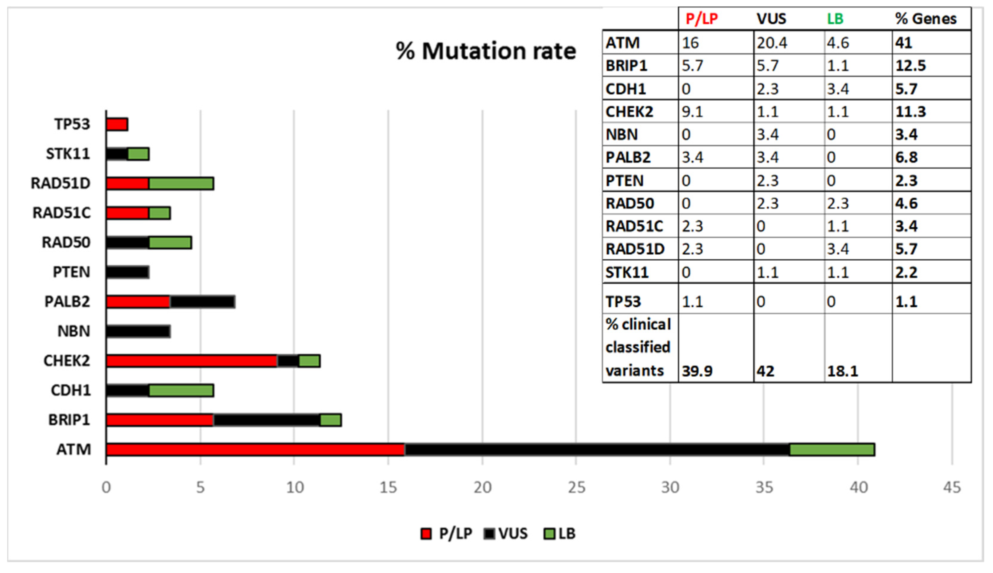
Thirty out of 35 Pathogenic or Likely-pathogenic identified coding variants were encoded in dbSNP and 27 were already known in literature or reported in databases such as IARC, LOVD, MGeND, ClinVar Miner with more clinical information. Five variants have never been registered: c.8368delA in ATM, c.507delT and c.1430delC in CHEK2, c.1565dupC in PALB2, c.397_398delCA in RAD51C. The 35 class 4 and 5 variants are distributed in the following genes: 14 in ATM, 5 in BRIP1, 8 in CHEK2, 3 in PALB2, 2 in RAD51C, 2 in RAD51D, and 1 in TP53.
Thirty-four out of 37 VUS identified were encoded in dbSNP and 25 of these were already known and reported in databases previously cited. The following 3 VUSs were completely new: c.2317A > G in ATM, c.3029A > G in BRIP1 and c.17_18insGCG in PTEN. Eighteen of the 37 VUSs identified are in ATM, 5 in BRIP1, 2 in CDH1, 1 in CHEK2, 3 in NBN, 3 in PALB2, 2 in PTEN, 2 in RAD50, and 1 in STK11.
Fifteen out of 16 Likely-benign variants found had a dbSNP entrie and 12 of these were encoded in databases. One variant had never been registered (c.252T > C in RAD51D). The 16 Likely-benign variants are distributed as follows: 4 in ATM, 1 in BRIP1, 3 in CDH1, 1 in CHEK2, 2 in RAD50, 1 in RAD51C, 3 in RAD51D, and 1 in STK11.
Figure 1 shows the percentage of distributions of the variants stratified by gene, according to the current ACMG classification. As shown in Figure 1 ATM is the gene with the highest mutation rate.
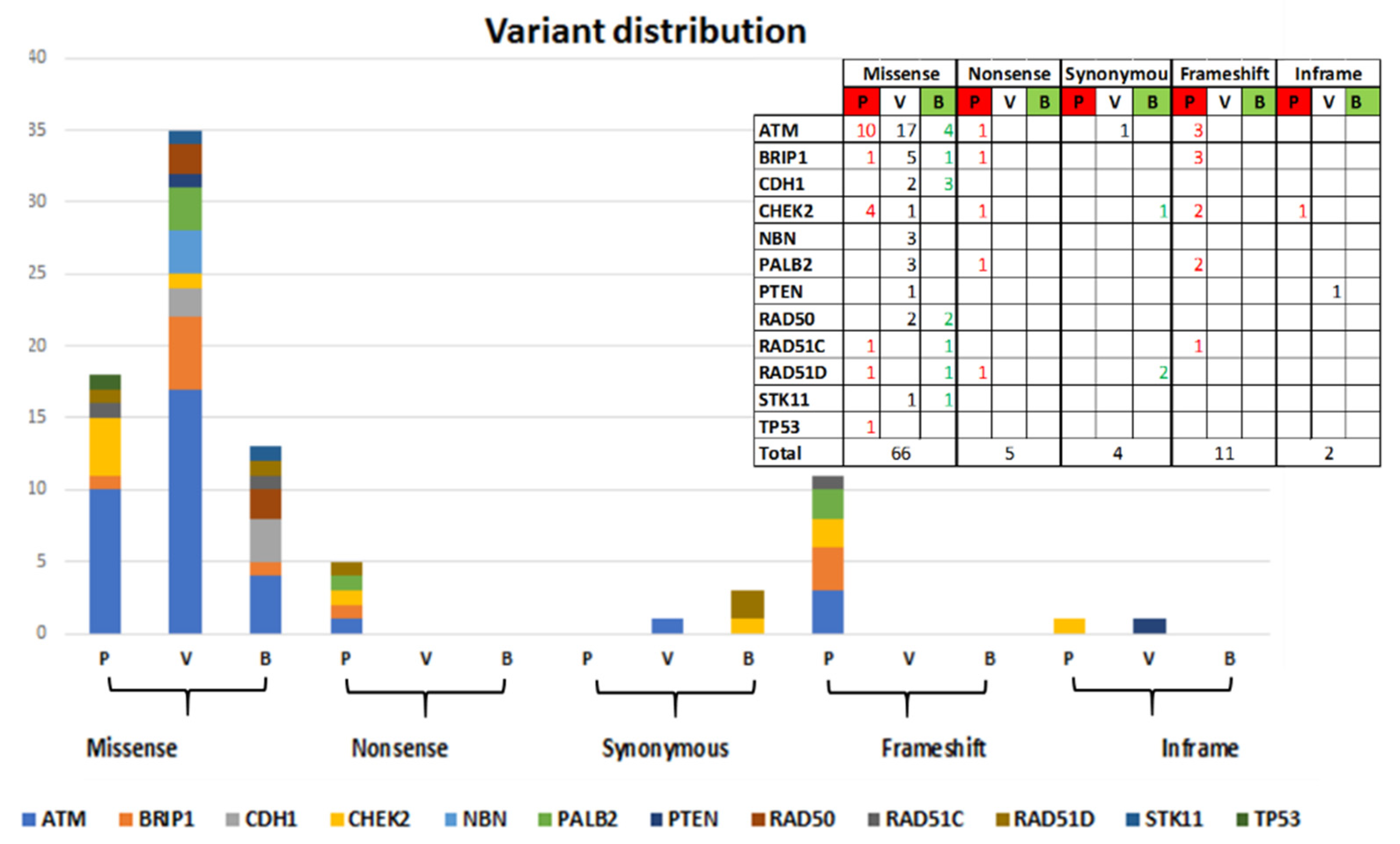
Sixty-six out of 88 identified variants are missense variants (of which 18 Pathogenic and Likely-pathogenic, 35 VUS and 13 Likely-benign), 5 Nonsense variants (all Pathogenic), 4 Synonymous variants (1 VUS and 3 Likely-benign), 11 frameshift variants (all Pathogenic), and 2 in-frame variants (1VUS and 1 Pathogenic). The distribution by gene is shown in Figure 2.
The predictive analysis on the effect of amino acid change on the function of the protein was conducted with PROVEAN and Polyphen2 software, as described in Materials and Methods. Specifically, the information for each identified variant is shown in Table S1 in Supplementary Materials, where it is also reported whether the variant has already been identified and registered in database by other groups.

2.3. Demographics and Clinical Features of Patiens with Coding Variants

The average age of tumor diagnosis for 91 patients with coding variants is 44. 8.8% (8) of the patients have a bilateral BC, 2.2% (2) have both breast and ovarian cancer, 4.4% (4) have ovarian cancer, 2.2% (2) have multiple tumors (in addition to breast cancers) and the remaining percentage, 82.4% (75) of total cases, has a BC. Among the latter patients, 16% (12) have Triple-Negative Breast Cancer (TNBC).
Their family histories are distributed as follows: 48.3% (44) have Hereditary Breast Cancer (HBC), 12.1% (11) have Hereditary Breast and Ovarian Cancer (HBOC), 18.7% (17) have Early Onset Breast Cancer (EOBC), and 20.9% (19) have other Hereditary Breast Cancer Syndromes (HBCS) (Table 1).
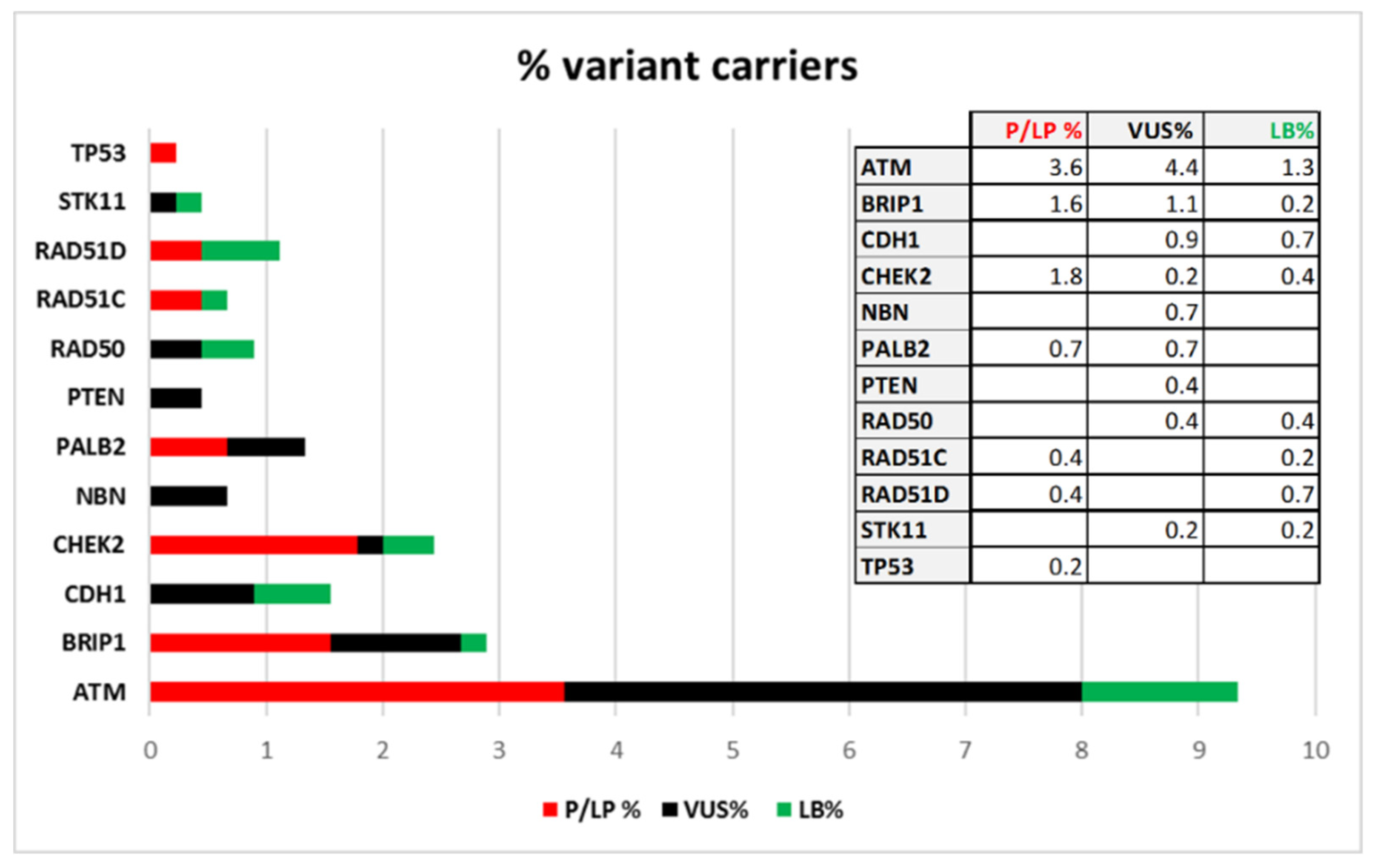
Thirty-nine patients are carriers of variants classified as “Pathogenic” or “Likely-pathogenic”, 41 patients are carriers of VUS, while “Likely-benign” variants have been identified in 19 patients. If we consider the total number of patients screened in the coding regions (450) the percentage of class 4 and 5 variant carriers is 8.7% and it is distributed in this way: 3.6% have variants in ATM, 1.6% in BRIP1, 1.8% in CHEK2, 0.7% in PALB2, 0.4% in RAD51C, 0.4% in RAD51D, and 0.2% in TP53. In Figure 3 the frequencies of patients with Pathogenic and Likely-pathogenic, VUS and Likely-benign variants are reported.
Among the 75 patients with BC, we identified 82 variants of which 30 Pathogenic and Likely-pathogenic, broken down as follows: 13 in ATM gene, 5 in BRIP1, 2 in PALB2, 6 in CHEK2, 2 in RAD51C, 1 in RAD51D and 1 in TP53. In these same patients we identified 38 VUS: 15 in ATM, 6 in BRIP1, 4 in CDH1, 3 in NBN, 3 in PALB2, 2 in CHEK2, 2 in PTEN, 1 in RAD50, 1 in RAD51D and 1 in STK11. The 14 remaining variants found are classified as Likely-benign and distributed as follows: 6 in ATM, 3 in RAD51D, 2 in CDH1 and 3 respectively in BRIP1, RAD50, and RAD51C genes.
Among the four cases of patients with ovarian cancer, we found a Pathogenic variant in RAD51C, 2 VUSs, respectively in ATM and RAD50 genes, and a Likely-benign variant in STK11.
Among the 12 cases of multiple tumors in coding variant carriers (9 cases of bilateral breast cancer, 2 of breast and ovarian cancer, and 1 case of breast tumor associated with pancreatic cancer), 9 have Pathogenic or Likely-pathogenic variants, 1 VUS and 2 Likely-benign variants. The statistical analysis has found a significant association of these cases with Pathogenic variant carriers (χ2-test, p < 0.01) in comparison with the cases of single tumors.
Three out of nine patients with Pathogenic variants have variants in ATM, 4 in CHEK2, 1 in BRIP1 and 1 in PALB2 (Table 2). Three of these nine variants are new variants never reported in dbSNP. One case has a VUS in ATM gene and two patients with multiple tumors have Likely-benign variants in CDH1 and CHEK2 genes. Frequency of coding variants in CHEK2 gene is significant in these patients (Fisher test, p < 0.05).
By analyzing cases of more aggressive tumors such as the 12 Triple-negative breast cancer patients (TNBC) (Table 3), all with G2 or G3 infiltrating ductal carcinoma, we identified 6 Pathogenic and Likely-pathogenic variants: 1 in ATM, 1 in BRIP1, 2 in CHEK2, 1 in RAD51D and 1 in TP53. In 6 patients, 4 VUSs (3 in ATM and 1 in PTEN never recorded before) and 2 Likely-benign variants were identified. The latter all have a conserved evolutionary profile (GERP > 3.8) and a conflict of interpretation according to Varsome and ClinVar.

2.4. Non-Coding and/or Regulatory Variants

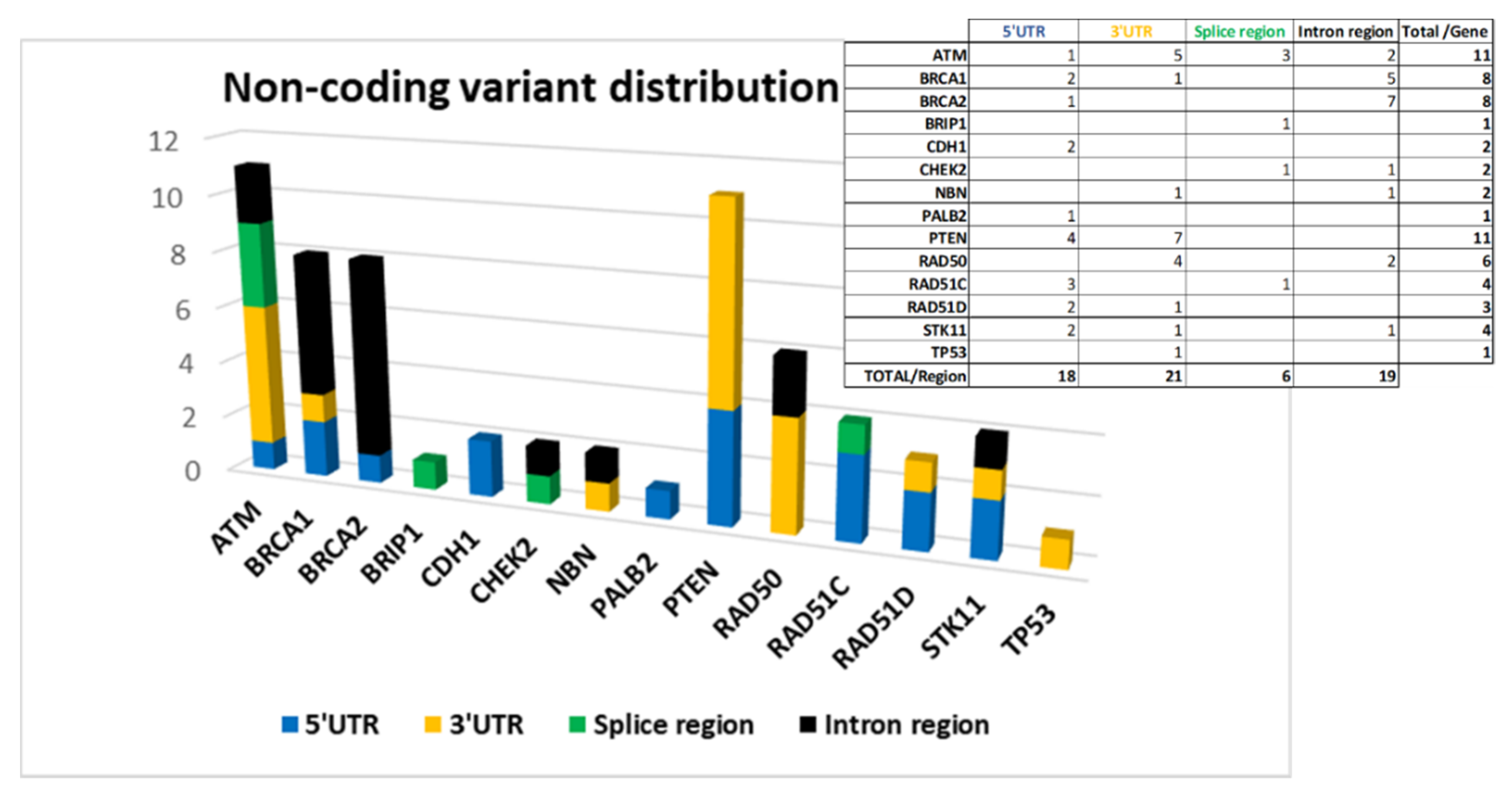
The screening of non-coding regions has allowed us to identify 64 variants. Forty-six of these have a dbSNP code and only 24 are known variants already registered in databases by other groups.
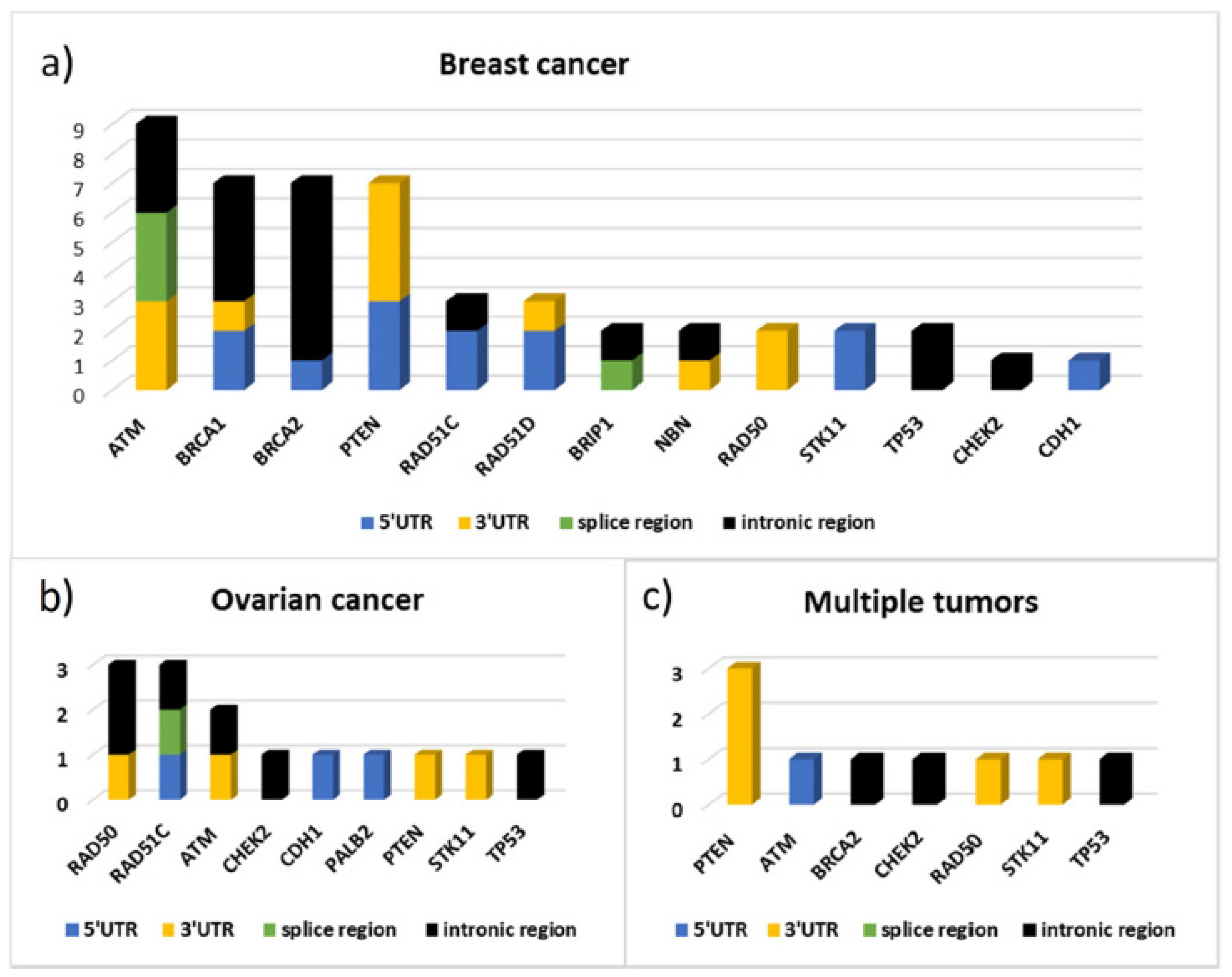
The 64 variants are distributed as follows: 18 are located at 5′UTR, 21 at 3′UTR, 6 in splice site regions, and 19 in intron regions.
Figure 4 shows the distribution of the variants by gene divided according to their location.
ATM, BRCA1, BRCA2, and PTEN are among the genes in which we have found more non-coding variants (Figure 4).
The variant putative effect on gene expression, was carried out by using different software (described in Materials and Methods) depending on variant position. The results are reported in Table S2a–d in Supplementary Materials.
An unexpectedly high number of variants in 5′ UTR of PTEN (4) and RAD51C (3) was found and, according to our analysis, all of them are localized in TF binding regions (Table S2a/Supplementary Materials).
The in-silico analysis performed by ‘TFbind’ and ‘Promo’ software, has identified TF consensus sequences in the regions where the 5′UTR of BRCA1 (c.-676T > A) and BRCA2 (c.-1193C > T) variants are localized (Table S2a/Supplementary Materials).
In 3′UTR, the greatest contribution is due to variants located in ATM, PTEN, and RAD50 genes (Figure 4) on which putative miRNA-binding regions have been identified by ‘TargetScanHuman’ and ‘miRDB’ software (Table S2b/Supplementary Materials).
In splicing regions, we identified six variants and only one, c.904+5G > T, was predicted to inactivate the wild-type donor site in the intron 6 of the RAD51C gene, and therefore is classified by the ACMG criteria as ‘Pathogenic’.
Three variants were in ATM but only the c.7788+ 8G > T, localized in intron 52, was predicted by ‘Human Splicing Finder’ as a possible modifier of an exonic ESE site with a potential alteration of splicing. The remaining 2 in CHEK2 and BRIP1 genes were predicted neutral on splicing (Table S2c/Supplementary materials).
In the remaining intronic regions, variants in ATM, BRCA1, BRCA2, CHEK2, NBN, RAD50, STK11, and TP53 genes were identified. The possible presence of enhancer regions, compatible with bonds to transcription factors, was analyzed by the software mentioned above. The four variants identified in intron 2 of BRCA1 (c.81-3510_81-3505delCTTTTT; c.81-3790G > C; c.81-4100A > C; c.80 + 247C > T) deserve attention. In fact, all four variants identified in this study map in two conserved enhancer regions, that have already been demonstrated to be involved in gene expression regulation (Table S2d/Supplementary Materials) [26].
In conclusion, we found only one Pathogenic variant in the RAD51C gene, whereas the large majority were classified as VUSs (50), 2 as Likely-benign and 11 as Benign variants.
The high number of VUSs is due to the fact that very few studies have analyzed these non-coding regions, and for this reason the classification is based only on in silico prediction.

2.5. Deep Intronic Regions

The panel of 9071 probes used was designed to optimize coverage of exonic regions, 50 bp flanking each intron-exon junction, 5′UTR, 3′UTR of all the genes of interest and the known regulatory regions of BRCA1 and BRCA2. However, the position of the probes managed to cover additional regions which led us to identify another nine variants in intronic regions (all with a coverage ≥ 50X) that proved to be interesting for the predictions obtained by ‘TFbind’ and ‘Promo’ software and for the information obtained from rVarBase and ENCODE (Table S2e/Supplementary Materials). Two variants are in the intron 61 of ATM, one in the intron 16 of BRIP1, two in intron 2 respectively of RAD51C and RAD51D genes and four variants are in the intron 1 of TP53. Eight out of nine variants are classified as VUS and four are not registered in dbSNP.

2.6. Demographics and Clinical Features of Patiens with Non-Coding Variants

Non-coding variants were found in 64 patients (excluding two cases of co-occurrence of Pathogenic coding variants). According to this data, in these patients there is a significant increase in both ovarian cancer (χ2-test, p < 0.05) and HBOC families (χ2-test, p < 0.01), compared to patients with variants in coding regions (Table 1/Table 4).
The mean age of tumor onset for these patients is 44.4. If we compare the coding and non-coding variant carriers in each proband group with the same kind of cancer, the average age of disease onset is completely comparable (Table 1/Table 4). In particular, if we compare the 64 non-coding variant carriers to the 39 Pathogenic coding variant carriers, there is not a statistically significant difference in the proportion of cases with a tumor onset under age 40 (Table 5, χ2-test, p > 0.05).
Figure 5 shows the distribution by gene and by gene location of the variants identified respectively in 43 patients with BC, in 12 patients with ovarian cancer, and in 9 cases of patients with multiple tumors (seven with bilateral cancer, one with breast and ovarian cancer, and one with breast and pancreatic cancer). From this data emerges the high frequency of variants identified in the ATM gene, as well as in BRCA1 and BRCA2, in patients with BC and the high contribution of variants localized in the 3′UTR of PTEN in patients with multiple tumors. In particular, three out of nine variants found in these patients are localized in PTEN 3′UTR and the bioinformatics predictions found, for all three, consensus sequences compatible with miRNA binding sites (Figure 5, Table S2b/Supplementary Materials). In patients with ovarian cancer, instead, variants were identified mainly in the RAD50 and RAD51C genes.
Among the 12 cases of TNBC, all with G2 or G3 infiltrating ductal carcinoma, 4 variants out of 19 identified are on the PTEN gene: one located at 5′UTR and the others at 3′UTR. Other interesting variants, for the type of feedback we obtained through prediction analysis, are the variants in the 5′UTR of RAD51D and STK11 genes. The complete list of variants found in these patients is shown in Table 6. Four variants are in intronic regions of BRCA2. Furthermore, among all the non-coding variants filtered in this work, the only variant in the RAD51C gene classified as “Pathogenic” by ACMG criteria was found in one of these patients.
Twelve patients with multiple variants in non-coding regions were filtered to assess whether the presence of multiple mutations could affect tumor pathogenicity. Ten out of 12 filtered patients have BC and 6 of these have TNBC. According to our data, there is a statistically significant association between TNBC and multiple variants in non-coding regions (χ2-test, p < 0.05). In fact, 50% of BC patients with multiple variants in non-coding regions have TNBC, while, among the 52 BC non-coding variants carriers, only 12 (23.1%) have TNBC (χ2-test, p < 0.05; Table 7).

3. Discussion

This study regards the analysis of coding regions and 50 bp flanking intron–exon junction of 10 BC predisposition genes in a cohort of 450 HBOC patients. For a portion of these patients (120) the screening was extended to additional two genes (NBN and RAD50), to the 5′UTR and 3′UTR of each gene analyzed and to the known regulatory regions of BRCA1 and BRCA2.Eighty eight coding variants were identified in 91/450 patients (20.2%): 35 of these were classified as pathogenic (class 5) or likely-pathogenic (class 4) and found in 39 patients (8.7%), 37 were classified as VUSs in 41 patients (9.1%) and 16 as “Likely-benign” in 19 patients (4.2%).
ATM was the more mutated gene (16 carriers), followed by CHEK2 (8 carriers), BRIP1 (7 carriers), PALB2 (3 carriers). RAD51C and RAD51D were found mutated in 2 patients/each and TP53 in one patient only.
Thirteen variants in the intron-exon junctions were identified in 23/450 patients (5.1%) and 51 non-coding variants in 39 out of 120 patients analyzed for this purpose (32.5%).
Our results support what was recently published by the Breast Cancer Association Consortium in a larger and more geographically heterogeneous population consisting of 60,466 women. With a panel of 34 putative susceptibility genes, a significant association between the increased risk of breast cancer and truncating mutations in ATM, CHEK2, PALB2, BARD1, RAD51C, RAD51D, and TP53 was identified [27]. Similar results were found through a screening of 65,057 white woman with breast cancer where a high or moderately increased risk of disease was found in relation with pathogenic mutations in ATM, CHEK2, PALB2, and RAD51D [28].
ATM, CHEK2, PALB2, and TP53 were confirmed as breast cancer predisposition genes also by the German Consortium for Hereditary Breast and Ovarian Cancer, through a screening of eight cancer predisposition genes conducted on 5589 breast cancer patients negative for pathogenic BRCA1/2 mutations [29]. An American retrospective study performed on 35,000 women who underwent genetic testing through a 25-gene hereditary cancer panel found the same correlation [30].
The analysis of non-coding regions reports a consistent number of variants never registered previously (18/64 variants).
In details, in the 5′UTR 2 variants were found, one in BRCA1 (c.-676t > a) and one in BRCA2 (c.-1193c > t). Both were registered in BRCA-exchange and LOVD database as VUSs. The in-silico analysis, conducted by ‘TFbind’ and ‘Promo Prediction’, has identified putative consensus sequences for Elk-1, NF-AT1, STAT4 transcription factors, for the first variant, and NRF2, GATA1/2, USF for the second.
Whereas the 5′UTR and 3′UTR of the BRCA1/2 genes have been two of the most intensely investigated non–coding regions over the last few years [12,13,31,32,33,34], this is the first fully comprehensive study of 5′and 3′UTRs of so called “minor genes for breast/ovarian cancer susceptibility” and a number of variants were also identified in the 5′UTRs: 4 in the PTEN gene and 3 in the RAD51C gene and the in-silico predictions obtained regarding their involvement in TF binding are interesting (Table S2a in Supplementary Materials).
In the 3′UTRs of genes other than BRCA1/2, the greatest contribution was due to variants located in the ATM (5), PTEN (7) and RAD50 (4) genes. The use of software such as ‘TargetScanHuman’ and ‘miRDB’ has made it possible to identify in those regions binding sites for miRNAs (Table S2b in Supplementary Materials).
The variants localized in splice regions mainly affect the ATM (3) gene and, in particular the c.7788+8G >T variant, localized in intron 52 of ATM, could modify an exonic ESE site (Table S2c in Supplementary Materials). Another variant of interest is the c.904+5G >T of RAD51C, classified by ACMG criteria as ‘Pathogenic’.
According to our data, variants in the intronic regions have been identified mainly in the BRCA1 (5), BRCA2 (7), ATM (2), and RAD50 (2) genes.
Four out of 5 BRCA1 variants are localized in intron 2 (c.81-3510_81-3505delCTTTTT; c.81-3790G > C; c.81-4100A > C; c.80+247C > T) and the in-silico predictive analysis led us to identify TF binding sites in those regions (Table S2d/Supplementary Materials). Those variants deserve to be studied in depth through a functional assay because in BRCA1 intron 2, two conserved enhancer regions (CNS1 and CNS2) are localized and involved in the regulation of gene expression [26].
Eleven out of 12 intronic variants found in BRCA1/2 genes could be classified as ‘deep intronic’. The deep intronic regions of the BRCA genes are intensely been studied in the recent years. These investigations have led to the identification of variants capable to modify the expression level or the functionality of the BRCA1 protein [35].
Finally, we incidentally found two new variants in deep intronic regions of the ATM and TP53 genes in two patients with TNBC (due to the fact that additional regions that were not of our interest were included in the panel design). The variants are respectively in intron 61 of ATM (c.8850+389G > A) and intron 1 of TP53(c.-29+475C > A) and are both predicted as TF binding regions. This result underlines how important is to broaden screening to deeper intronic regions (not only in BRCA genes).
By analyzing the characteristics of patients for the type of variants identified, distinguishing between coding variant carriers versus non-coding variant carriers, we can make some interesting considerations.
The average age of breast cancer onset in coding variants carriers is 44 and most of them have breast cancer only family history as shown in Table 1.
A possible association of ATM-BRIP1-PALB2 and a significant association of CHEK2 pathogenic/likely-pathogenic coding variants with multiple tumors, including bilateral breast cancer, was observed. These data are in agreement with the findings of German Consortium for Hereditary Breast and Ovarian Cancer, that demonstrated a significant association of CHEK2 deleterious variants with bilateral breast cancer [29].
Among the 12 cases of TNBC patients, 6 were carriers of pathogenic coding variants: 2 in CHEK2 and the remaining 4 in the ATM, BRIP1, RAD51D and TP53 genes respectively (Table 3).
Studies conducted on a larger number of TNBC patients have come to concordant results regarding the association of these cases with pathogenic variants in the PALB2, BRIP1, RAD51C, and RAD51D genes [30,36,37,38]. Our results partially agree with these data, having identified pathogenic variants in TNBC patients with ATM, RAD51D, BRIP1, and TP53 variants. It is likely that the analysis of a higher number of TNBC cases could have led us to a greater degree of concordance.
However, it is certainly very interesting that 50% of the TNBC cases analyzed had pathogenic coding variants: it is a higher proportion compared to that obtained in other cohorts of TNBC patients filtered in the population solely on the basis of their tumor phenotype [36,37]. It is probable that the higher percentage registered in our cases depends on the double filtering, based not only on patient tumor phenotype but also on the patient breast cancer family history.
It is known that some TNBCs BRCAWT show the so-called ‘BRCAness’ phenotype, exhibiting clinical and pathological properties similar to BRCA-mutated tumors such as the homologous recombination deficiency and the sensitivity to DNA damaging agents [39,40]. Therefore, our data could be of interest at a therapeutic level. In fact, the data supports what we have already reported in Spugnesi et al. [38] where germline mutations in DNA repair genes, in BRCA1/2 WT tumors, were associated with a group of TNBC patients who responded to anthracyclines/taxanes neoadjuvant therapy. It is becoming increasingly evident that genes involved in DNA repair could become response biomarkers for DNA-damaging therapy in these patients [41].
Although we cannot make any consideration about the variants identified in ovarian cancer, having only analyzed four patients with ovarian cancer and two with breast and ovarian cancer, the detection of pathogenic variants in RAD51C and ATM is in agreement with what is reported in a case-control study performed on 2051 women with ovarian cancer. The authors of this paper identified an increased risk for ovarian cancer associated with MSH6, RAD51C, TP53, and ATM [42].
Analyzing the characteristics of non-coding variants carriers, we recorded an average age of disease onset of 44.4 absolutely similar to the average age of disease onset in coding variant carriers for each proband’s group with the same cancer type. Furthermore, there is not a statistically significant difference in the proportion of cases with a tumor onset under the age of 40 between the two groups (χ2-test, p > 0.05; Table 5).
A statistically significant increase in cases of ovarian cancer and of HBOC families in non-coding variant carriers compared to coding variant carriers was found (Table 1/Table 4). This is an interesting observation but very preliminary considering the limited number of samples analyzed, and it needs to be further investigated.
One of the results that emerges clearly when analyzing the characteristics of non-coding variant carriers, is the relationship between variants in 3′UTR of PTEN and multiple tumors or TNBC tumor phenotype. This result is in agreement with what has already been described in previous studies in which the deregulation of PTEN expression, via binding sites for RNA-binding-protein and miRNA located in 3′UTR, was associated with aggressive phenotype and poor outcome for BC patients [16,17]. Moreover, four out of seven BRCA2 deep intronic variants identified (c.9257-3610G > A, c.681+697C > T, c.317-1021A > G, c.317-512A > T) have been found in TNBC patients.
Finally, by analyzing the 12 patients carrying multiple non-coding variants we observed a statistically significant association with a TNBC phenotype (χ2-test, p < 0.05).
This work has the merit of having highlighted certain variants never recorded in the coding and regulatory regions which are worthy of further study. It also underlines the importance, increasingly evident in recent years, of also screening the regulatory regions in patients with clearly familial tumors with no relevant variation in the coding sequences of the main breast/ovarian cancer predisposition genes. Certainly, in the future it will be necessary to perform a segregation analysis in order to understand the real involvement of certain genomic regions in breast and ovarian cancer predisposition. This work surely could help to pave the way for other in-depth projects on the functional meaning of the variants identified, by providing, thanks to the many notes obtained, a lot of useful information for a better understanding of their clinical significance.

4. Materials and Methods

4.1. Selection of Patients Included in the Study

We selected a total of 450 patients eligible for this study who underwent genetic counseling between 2001 and 2019 at five different Italian centers distributed as follows:
-
163 patients from University Hospital of Pisa
-
210 patients from Azienda Tutela Salute Sardegna, ASST of Cagliari
-
55 patients from Ospedale di Circolo, ASST-Sette Laghi of Varese
-
13 patients from University Hospital of Modena
-
9 patients from IRCCS Istituto Tumori “Giovanni Paolo II” of Bari
All patients signed an informed consent. The eligibility criteria included:
-
family history of BC in at least two generations: (a) two or more BC cases in first degree relatives, one of which diagnosed before age 40 (b) one ovarian cancer case associated with a BC in a first degree relative at any age;
-
a priori risk of being carriers of BRCA1/2 mutation >10% calculated with BRCAPRO (http//www4.utsouthwestern.edu/breast-health/cagene, accessed on March 2017–December 2019 ) and BOADICEA (https://pluto.srl.cam.ac.uk/cgi-bin/BD2/v2/bd.cgi, accessed on March 2017–December 2019) software;
-
to be wild type for point mutations and large rearrangements in the BRCA1/2 coding regions.

4.2. Library Preparation and NGS Sequencing

DNA was extracted from blood samples. Genomic DNA screening was carried out with a NGS target resequencing approach on the Illumina MiSeq machine. 450 patients were analyzed with a custom panel optimized to sequence the coding regions and 50 bp flanking each intron-exon junction of 10 genes involved in breast and ovarian cancer predisposition: ATM (NM_000051.3), BRIP1 (NM_032043.2), CDH1 (NM_004360.3), CHECK2 (NM_007194.3), PALB2 (NM_024675.3), PTEN (NM_000314.6), RAD51C (NM_058216.2), RAD51D (NM_002878.3), STK11 (NM_000455.4), TP53 (NM_000546.5). For a subset of 120 patients (43 from Pisa, 55 from Varese, 13 from Modena and 9 from Bari), the screening was also extended to NBN (NM_002485.5) and RAD50 (NM_005732) genes, including 5′UTR and 3′UTR of each gene and the known regulatory regions of BRCA1 (NM_007294.4) and BRCA2 (NM_000059) [12,13,14,15,26]. To amplify the target sequences, a panel of 9071 probes was designed using SureSelect DNA Advanced Design Wizard in order to ensure the total coverage of 178.155 kb corresponding to 99% of the entire region of interest (Supplementary Materials Table S2a,b). Library preparation was performed according to SureSelectQXT Target Enrichment for Illumina Multiplexed Sequencing protocol (SureSelectQXT Reagent Kit, Agilent Technologies, Santa Clara, CA, USA). To assess library DNA quantity and quality after fragmentation and adaptor-tag addiction phase and after amplification of the indexed library, we used the Agilent 2100 Bioanalyzer. Following purification and quantification of pooled libraries, we sequenced them by 2 × 100 bp paired-end reads on a Miseq (Illumina, San Diego, CA, USA) instrument. Sequence reads allocated to each individual were aligned to the human reference sequence (hg19) using Agilent Sure Call (ver.4.1.1). All the regions considered for the analysis had a coverage ≥50X and a Quality Score (QS) ≥30. Sanger sequencing was performed to confirm the identified variants in lymphocyte DNA.

4.3. Bioinformatic Annotation and Prioritization of the Identified Variants

Each identified variant was evaluated in terms of Minor Allele Frequency (MAF): reference allelic frequency is that of total population reported in 1000 Genomes (http://www.internationalgenome.org, accessed on January 2020) and in gnomAD (https://gnomad.broadinstitute.org, accessed on January 2020). All variants with a MAF >1% were excluded from further analysis as they were considered common polymorphisms. To speed up the reading of the variants and their filtering on the basis of the known annotations, we used BaseSpace Variant Interpreter platform (https://variantinterpreter.informatics.illumina.com, accessed on January 2020). All variants were also evaluated in term of evolutionary conservation profile (GERP score) [43]. The clinical significance of each variant was annotated considering information from ClinVar (https://www.ncbi.nlm.nih.gov/clinvar, accessed on August 2020) [44] and according to ACMG (American College of Medical Genetics) criteria [19,45] also used by Varsome (https://varsome.com, accessed on August 2020) [46]. The latter helped us mostly to summarize all the known information and the pathogenicity predictions of the identified variants.
For the coding variant, we annotated the possible impact of an amino acid substitution on the protein structure and function obtained by PolyPhen2 (http://genetics.bwh.harvard.edu/pph2, accessed on May 2020) and PROVEAN (http://provean.jcvi.org/index.php, accessed on May 2020) software [47,48]. For the non-coding variants, in order to obtain information about chromatin states of the surrounding regions, methylation and acetylation status and hypersensitivity to DNAseI, we used rVarBase (http://rv.psych.ac.cn, accessed on February 2020) and ENCODE (https://www.encodeproject.org, accessed on February 2020) database [49,50,51].
To predict possible TF binding site in 5′UTR and in intronic enhancer regions, we emploied three different software: Promo (http://alggen.lsi.upc.es/cgi-bin/promo_v3/promo/promoinit.cgi?dirDB=TF_8.3, accessed on February 2020) [52], Tfbind (http://tfbind.hgc.jp/) and Lasagna-Search2.0 (https://biogrid-lasagna.engr.uconn.edu/lasagna_search/index.php, accessed on February 2020) [53]. The presence of miRNA binding site in 3′UTR variants, was evaluated by TargetScanHuman (http://www.targetscan.org/vert_72, accessed on February 2020) and miRDB (http://www.mirdb.org, accessed on February 2020) [54,55]. Finally, in order to identify consensus regions for potential splice sites, we made use of Human Splicing Finder (http://www.umd.be/HSF, accessed on February 2020) [56]. All the programs used to obtain predictions about the variants found are free software and they are summarized in Table 8.
All this information allowed us to have more complete annotations of the variants, making it possible to suggest future functional tests, especially those never reported in literature or in databases such as ENIGMA (https://enigmaconsortium.org, accessed on December 2020), BRCA Exchange (https://brcaexchange.org, accessed on December 2020), LOVD (https://www.lovd.nl, accessed on December 2020), MgeND (https://mgend.med.kyoto-u.ac.jp, accessed on December 2020), and ClinVar Miner (https://clinvarminer.genetics.utah.edu, accessed on December 2020). For each variant, the dbSNP (https://www.ncbi.nlm.nih.gov/snp, accessed on January 2021) encoding was also checked.

4.4. Statistical Analysis

Tumor type percentages, family distribution, and age at diagnosis in coding and non-coding variant carriers were compared by χ2-test. p < 0.05% was considered statistically significant. The same test was used to estimate the effect of Pathogenic, VUS, and Likely Benign variants on clinical characteristics of coding variant carriers and to evaluate the presence of multiple variants in more aggressive tumors in non-coding variant carriers.
The gene association with tumor aggressiveness was performed through the Fisher’s test. The results were considered significant with a threshold of p < 0.05%. All analyses were performed with the free software Social Science Statistics (https://www.socscistatistics.com, accessed on January 2021).

Supplementary Materials

The following are available online at https://www.mdpi.com/article/10.3390/ijms22147693/s1.

Author Contributions

Conceptualization, M.A.C. and A.G.; methodology, C.G., R.S., G.G., E.C., M.T., F.B., T.C., E.F., and C.C. (Cinzia Cosini); Software, C.G., P.A., and E.C.; Validation, C.G. and E.C.; Investigation C.V., F.S., L.B., S.T., M.G.T., E.T. (Enrico Tagliafico), L.C., I.C., C.C. (Caterina Congregati), M.M., M.P., B.P., and M.G.; Data curation, C.G., E.C., M.T., and P.A.; Writing—original draft preparation, C.G. and M.A.C.; Writing—review and editing S.T., M.G.T., E.T. (Enrico Tagliafico), E.T. (Elena Tenedini), L.C., I.C., and T.C.; Supervision, A.G. and M.A.C. All authors have read and agreed to the published version of the manuscript.

Funding

This research was funded by FONDAZIONE PISA, grant number no. 127/16 (Italy).

Institutional Review Board Statement

The study was approved by Ethics Committee of Area Vasta Nord Ovest (CEAVNO-protocol code: 13616-date of approval: 10 October 2019).

Informed Consent Statement

Informed consent was obtained from all subjects involved in the study.

Acknowledgments

The authors would like to acknowledge all laboratory technicians of the Molecular Genetics Section AOUP.

Conflicts of Interest

The authors declare no conflict of interest.

References

  1. Torre, L.A.; Bray, F.; Siegel, R.L.; Ferlay, J.; Lortet-Tieulent, J.; Jemal, A. Global cancer statistics, 2012. CA Cancer J. Clin. 2015, 65, 87–108. [Google Scholar] [CrossRef] [Green Version]
  2. Bray, F.; Ferlay, J.; Soerjomataram, I.; Siegel, R.L.; Torre, L.A.; Jemal, A. Global cancer statistics 2018: GLOBOCAN estimates of incidence and mortality worldwide for 36 cancers in 185 countries. CA Cancer J. Clin. 2018, 68, 394–424. [Google Scholar] [CrossRef] [PubMed] [Green Version]
  3. Mavaddat, N.; Peock, S.; Frost, D.; Ellis, S.; Platte, R.; Fineberg, E.; Evans, D.G.; Izatt, L.; Eeles, R.A.; Adlard, J.; et al. Cancer risks for BRCA1 and BRCA2 mutation carriers: Results from prospective analysis of EMBRACE. J. Nat. Cancer Inst. 2013, 105, 812–822. [Google Scholar] [CrossRef] [PubMed] [Green Version]
  4. Nielsen, F.C.; van Overeem Hansen, T.; Sorensen, C.S. Hereditary breast and ovarian cancer: New genes in confined pathways. Nat. Rev. Cancer 2016, 16, 599–612. [Google Scholar] [CrossRef] [PubMed]
  5. Lord, C.J.; Ashworth, A. BRCAness revisited. Nat. Rev. Cancer 2016, 16, 110–120. [Google Scholar] [CrossRef] [PubMed]
  6. Campeau, P.M.; Foulkes, W.D.; Tischkowitz, M.D. Hereditary breast cancer: New genetic developments, new therapeutic avenues. Hum. Genet. 2008, 124, 31–42. [Google Scholar] [CrossRef] [PubMed]
  7. Antoniou, A.C.; Pharoah, P.D.; McMullan, G.; Day, N.E.; Ponder, B.A.; Easton, D. Evidence for further breast cancer susceptibility genes in addition to BRCA1 and BRCA2 in a population-based study. Genet. Epidemiol. 2001, 21, 1–18. [Google Scholar] [CrossRef]
  8. Bonadona, V.; Lasset, C. Inherited predisposition to breast cancer: After the BRCA1 and BRCA2 genes, what next? Bull. Cancer 2003, 90, 587–594. [Google Scholar]
  9. Turnbull, C.; Rahman, N. Genetic Predisposition to Breast Cancer: Past, Present, and Future. Hum. Genet. 2008, 9, 321–345. [Google Scholar] [CrossRef] [PubMed]
  10. Diederichs, S.; Bartsch, L.; Berkmann, J.C.; Fröse, K.; Heitmann, J.; Hoppe, C.; Iggena, D.; Jazmati, D.; Karschnia, P.; Linsenmeier, M.; et al. The dark matter of the cancer genome: Aberrations in regulatory elements, untranslated regions, splice sites, non-coding RNA and synonymous mutations. EMBO Mol. Med. 2016, 8, 442–457. [Google Scholar] [CrossRef]
  11. De Siervi, A.; De Luca, P.; Byun, J.S.; Di, L.J.; Fufa, T.; Haggerty, C.M.; Vazquez, E.; Moiola, C.; Longo, D.L.; Gardner, K. Transcriptional autoregulation by BRCA1. Cancer Res. 2010, 70, 532–542. [Google Scholar] [CrossRef] [PubMed] [Green Version]
  12. Wang, J.; Bian, C.; Li, J.; Couch, F.J.; Wu, K.; Zhao, R.C. Poly(ADP-ribose) polymerase-1 down-regulates BRCA2 expression through the BRCA2 promoter. J. Biol. Chem. 2008, 283, 36249–36256. [Google Scholar] [CrossRef] [Green Version]
  13. Burke, L.J.; Sevcik, J.; Gambino, G.; Tudini, E.; Mucaki, E.J.; Shirley, B.C.; Whiley, P.; Parsons, M.T.; Leeneer, K.D.; Gutiérrez-Enríquez, S.; et al. BRCA1 and BRCA2 5’ noncoding region variants identified in breast cancer patients alter promoter activity and protein binding. Hum. Mutat. 2018, 39, 2025–2039. [Google Scholar] [CrossRef] [PubMed]
  14. Lheureux, S.; Lambert, B.; Krieger, S.; Legros, A.; Vaur, D.; Denoyelle, C.; Berthet, P.; Poulain, L.; Hardouin, A. Two novel variants in the 3’UTR of the BRCA1 gene in familial breast and/or ovarian cancer. Breast Cancer Res. Treat. 2011, 125, 885–891. [Google Scholar] [CrossRef] [PubMed]
  15. Brewster, B.L.; Rossiello, F.; French, J.D.; Edwards, S.L.; Wong, M.; Wronski, A.; Whiley, P.; Waddell, N.; Chen, X.; Bove, B.; et al. Identification of fifteen novel germline variants in the BRCA1 3’UTR reveals a variant in a breast cancer case that introduces a functional miR-103 target site. Hum. Mutat. 2012, 33, 1665–1675. [Google Scholar] [CrossRef] [PubMed]
  16. Zhou, X.J.; Wu, J.; Shi, L.; Li, X.-X.; Zhu, L.; Sun, X.; Qian, J.-Y.; Wang, Y.; Wei, J.-F.; Ding, Q. PTEN expression is upregulated by a RNA-binding protein RBM38 via enhancing its mRNA stability in breast cancer. J. Exp. Clin. Cancer Res. 2017, 36, 149. [Google Scholar] [CrossRef] [Green Version]
  17. Fang, H.; Xie, J.; Zhang, M.; Zhao, Z.; Wan, Y.; Yao, Y. miRNA-21 promotes proliferation invasion of triple-negative breast cancer cells through targeting, P.T.E.N. Am. J. Transl. Res. 2017, 9, 953–961. [Google Scholar]
  18. Plon, S.E.; Eccles, D.M.; Easton, D.; Foulkes, W.D.; Genuardi, M.; Greenblatt, M.S.; Hogervorst, F.B.L.; Hoogerbrugge, N.; Spurdle, A.B.; Tavtigian, S.V.; et al. Sequence variant classification and reporting: Recommendations for improving the interpretation of cancer susceptibility genetic test results. Hum. Mutat. 2008, 29, 1282–1291. [Google Scholar] [CrossRef] [Green Version]
  19. Tavtigian, S.V.; Greenblatt, M.S.; Harrison, S.M.; Nussbaum, R.L.; Prabhu, S.A.; Boucher, K.M.; Biesecker, L.G. ClinGen Sequence Variant Interpretation Working Group. Modeling the ACMG/AMP variant classification guidelines as a Bayesian classification framework. Genet. Med. 2018, 20, 1054–1060. [Google Scholar] [CrossRef] [Green Version]
  20. Iversen, E.S., Jr.; Couch, F.J.; Goldgar, D.E.; Tavtigian, S.V.; Monteiro, A.N. A computational method to classify variants of uncertain significance using functional assay data with application to BRCA1. Cancer Epidemiol. Biomark. Prev. 2001, 20, 1078–1088. [Google Scholar] [CrossRef] [Green Version]
  21. Parsons, M.T.; Tudini, E.; Li, H.; Hahnen, E.; Wappenschmidt, B.; Feliubadaló, L.; Aalfs, C.M.; Agata, S.; Aittomäki, K.; Alducci, E.; et al. Large scale multifactorial likelihood quantitative analysis of BRCA1 and BRCA2 variants: An ENIGMA resource to support clinical variant classification. Hum. Mutat. 2019, 40, 1557–1578. [Google Scholar] [CrossRef] [Green Version]
  22. Millot, G.A.; Carvalho, M.A.; Caputo, S.M.; Vreeswijk, M.P.; Brown, M.A.; Webb, M.; Rouleau, E.; Neuhausen, S.L.; Hansen, T.V.O.; Galli, A.; et al. A guide for functional analysis of BRCA1 variants of uncertain significance. Hum. Mutat. 2012, 33, 1526–1537. [Google Scholar] [CrossRef] [Green Version]
  23. Guidugli, L.; Carreira, A.; Caputo, S.M.; Ehlen, A.; Galli, A.; Monteiro, A.N.A.; Neuhausen, S.L.; Hansen, T.V.O.; Couch, F.J.; Vreeswijk, M.P.G.; et al. Functional assays for analysis of variants of uncertain significance in BRCA2. Hum. Mutat. 2014, 35, 151–164. [Google Scholar] [CrossRef] [Green Version]
  24. Jhuraney, A.; Velkova, A.; Johnson, R.C.; Kessing, B.; Carvalho, R.S.; Whiley, P.; Spurdle, A.B.; Vreeswijk, M.P.G.; Caputo, S.M.; Millot, G.A.; et al. BRCA1 Circos: A visualisation resource for functional analysis of missense variants. J. Med. Genet. 2015, 52, 224–230. [Google Scholar] [CrossRef]
  25. Lindor, N.M.; Guidugli, L.; Wang, X.; Vallée, M.P.; Monteiro, A.N.A.; Tavtigian, S.; Goldgar, D.E.; Couch, F.J. A review of a multifactorial probability-based model for classification of BRCA1 and BRCA2 variants of uncertain significance (VUS). Hum. Mutat. 2012, 33, 8–21. [Google Scholar] [CrossRef] [PubMed] [Green Version]
  26. Wardrop, S.L.; kConFab Investigators; Browna, M.A. Identification of two evolutionarily conserved and functional regulatory elements in intron 2 of the human BRCA1 gene. Genomics 2005, 86, 316–328. [Google Scholar] [CrossRef] [PubMed]
  27. Breast Cancer Association Consortium; Dorling, L.; Carvalho, S.; Allen, J.; González-Neira, A.; Luccarini, C.; Wahlström, C.; Pooley, K.A.; Parsons, M.T.; Fortuno, C.; et al. Breast Cancer Risk Genes-Association Analysis in More than 113,000 Women. N. Engl. J. Med. 2021, 384, 428–439. [Google Scholar] [PubMed]
  28. Couch, F.J.; Shimelis, H.; Hu, C.; Hart, S.N.; Polley, E.C.; Na, J.; Hallberg, E.; Moore, R.; Thomas, A.; Lilyquist, J.; et al. Associations Between Cancer Predisposition Testing Panel Genes and Breast Cancer. JAMA Oncol. 2017, 3, 1190–1196. [Google Scholar] [CrossRef] [Green Version]
  29. Hauke, J.; Horvath, J.; Groß, E.; Gehrig, A.; Honisch, E.; Hackmann, K.; Schmidt, G.; Arnold, N.; Faust, U.; Sutter, C.; et al. Gene panel testing of 5589 BRCA1/2-negative index patientswith breast cancer in a routine diagnostic setting: Results ofthe German Consortium for Hereditary Breast and OvarianCancer. Cancer Med. 2018, 7, 1349–1358. [Google Scholar] [CrossRef]
  30. Buys, S.S.; Sandbach, J.F.; Gammon, A.; Patel, G.; Kidd, G.; Brown, K.L.; Sharma, L.; Saam, J.; Lancaster, J.; Daly, M.B. A Study of Over 35,000 Women With Breast Cancer Tested With a 25-Gene Panel of Hereditary Cancer Genes. Cancer 2017, 123, 1721–1730. [Google Scholar] [CrossRef] [Green Version]
  31. Santana Dos Santos, E.; Caputo, S.M.; Castera, L.; Gendrot, M.; Briaux, A.; Breault, M.; Krieger, S.; Rogan, P.K.; Mucaki, E.J.; Burke, L.J.; et al. Assessment of the functional impact of germline BRCA1/2 variants located in non-coding regions in families with breast and/or ovarian cancer predisposition. Breast Cancer Res. Treat. 2018, 168, 311–325. [Google Scholar] [CrossRef] [PubMed]
  32. Evans, D.G.R.; van Veen, E.M.; Byers, H.J.; Wallace, A.J.; Ellingford, J.; Beaman, G.M.; Santoyo-Lopez, J.; Aitman, T.J.; Eccles, D.; Lalloo, F.; et al. A Dominantly Inherited 5’ UTR Variant Causing Methylation-Associated Silencing of BRCA1 as a Cause of Breast and Ovarian Cancer. Am. J. Hum. Genet. 2018, 103, 213–220. [Google Scholar] [CrossRef] [Green Version]
  33. Laner, A.; Benet-Pages, A.; Neitzel, B.; Holinski-Feder, E. Analysis of 3297 individuals suggests that the pathogenic germline 5’-UTR variant BRCA1 c.-107A > T is not common in south-east Germany. Fam. Cancer 2020, 19, 211–213. [Google Scholar] [CrossRef] [PubMed]
  34. Yoshikawa, Y.; Kozuma, H.; Morimatsu, M.; Sugawara, K.; Orino, K. Reduced translation efficiency due to novel splicing variants in 5’ untranslated region and identification of novel cis-regulatory elements in canine and human BRCA2. BMC Mol. Cell Biol. 2021, 22, 2. [Google Scholar] [CrossRef] [PubMed]
  35. Montalban, G.; Bonache, S.; Moles-Fernández, A.; Gisbert-Beamud, A.; Tenés, A.; Bach, V.; Carrasco, E.; López-Fernández, A.; Stjepanovic, N.; Balmaña, J.; et al. Screening of BRCA1/2 deep intronic regions by targeted gene sequencing identifies the first germline BRCA1 variant causing pseudoexon activation in a patient with breast/ovarian cancer. J. Med. Genet. 2019, 56, 63–74. [Google Scholar] [CrossRef]
  36. Shimelis, H.; LaDuca, H.; Hu, C.; Hart, S.N.; Na, J.; Thomas, A.; Akinhanmi, M.; Moore, R.; Brauch, H.; Cox, A.; et al. Triple-Negative Breast Cancer Risk Genes Identified byMultigene Hereditary Cancer Panel Testing. J. Natl. Cancer Inst. 2018, 110, 855–862. [Google Scholar] [CrossRef]
  37. Zanti, M.; Loizidou, M.A.; Michailidou, K.; Pirpa, P.; Machattou, C.; Marcou, Y.; Kyriakou, F.; Kakouri, E.; Tanteles, G.A.; Spanou, E.; et al. NGS Panel Testing of Triple-Negative Breast Cancer Patients in Cyprus: A Study of BRCA-Negative Cases. Cancers 2020, 12, 3140. [Google Scholar] [CrossRef]
  38. Spugnesi, L.; Gabriele, M.; Scarpitta, R.; Tancredi, M.; Maresca, L.; Gambino, G.; Collavoli, A.; Aretini, P.; Bertolini, I.; Salvadori, B.; et al. Germline mutations in DNA repair genes may predict neoadjuvant therapy response in triple negative breast patients. Genes Chromosom. Cancer 2016, 55, 915–924. [Google Scholar] [CrossRef]
  39. Silver, D.P.; Richardson, A.L.; Eklund, A.C.; Wang, Z.C.; Szallasi, Z.; Li, Q.; Juul, N.; Leong, C.-O.; Calogrias, D.; Buraimoh, A.; et al. Efficacy of neoadjuvant cisplatin in triple-negative breast cancer. J. Clin. Oncol. 2010, 28, 1145–1153. [Google Scholar] [CrossRef]
  40. Byrski, T.; Dent, R.; Blecharz, P.; Foszczynska-Kloda, M.; Gronwald, J.; Huzarski, T.; Cybulski, C.; Marczyk, E.; Chrzan, R.; Eisen, A.; et al. Results of a phase II open-label, non-randomized trial of cisplatin chemotherapy in patients with BRCA1-positive metastatic breast cancer. Breast Cancer Res. 2012, 14, R110. [Google Scholar] [CrossRef] [Green Version]
  41. Priyanka, S. Biology and Management of Patients With Triple-Negative Breast Cancer. Oncologist 2016, 21, 1050–1062. [Google Scholar]
  42. Lu, H.-M.; Li, S.; Black, M.H.; Lee, S.; Hoiness, R.; Wu, S.; Mu, W.; Huether, R.; Chen, J.; Sridhar, S.; et al. Association of Breast and Ovarian Cancers With Predisposition Genes Identified by Large-Scale Sequencing. JAMA Oncol. 2019, 5, 122. [Google Scholar] [CrossRef] [PubMed]
  43. Cooper, G.M.; Stone, E.A.; Asimenos, G.; NISC Comparative Sequencing Program; Green, E.D.; Batzoglou, S. Arend Sidow Distribution and intensity of constraint in mammalian genomic sequence. Genome Res. 2005, 15, 901–913. [Google Scholar] [CrossRef] [Green Version]
  44. Landrum, M.J.; Lee, J.M.; Benson, M.; Brown, G.; Chao, C.; Chitipiralla, S.; Gu, B.; Hart, J.; Hoffman, D.; Hoover, J.; et al. ClinVar: Public archive of interpretations of clinically relevant variants. Nucleic Acids Res. 2016, 44, D862–D868. [Google Scholar] [CrossRef] [PubMed] [Green Version]
  45. Maxwell, K.N.; Hart, S.N.; Vijai, J.; Schrader, K.A.; Slavin, T.P.; Thomas, T.; Wubbenhorst, B.; Ravichandran, V.; Moore, R.M.; Hu, C.; et al. Evaluation of ACMG-Guideline-Based Variant Classification of Cancer Susceptibility and Non-Cancer-Associated Genes in Families Affected by Breast Cancer. Am. J. Hum. Genet. 2016, 98, 801–817. [Google Scholar] [CrossRef] [PubMed] [Green Version]
  46. Kopanos, C.; Tsiolkas, V.; Kouris, A.; Chapple, C.E.; Aguilera, M.A.; Meyer, R.; Massouras, A. VarSome: The human genomic variant search engine. Bioinformatics 2019, 35, 1978–1980. [Google Scholar] [CrossRef]
  47. Adzhubei, I.; Jordan, D.M.; Sunyaev, S.R. Predicting functional effect of human missense mutations using PolyPhen-2. Curr. Protoc. Hum. Genet. 2013. [Google Scholar] [CrossRef] [Green Version]
  48. Choi, Y.; Chan, A.P. PROVEAN web server: A tool to predict the functional effect of amino acid substitutions and indels. Bioinformatics 2015, 31, 2745–2747. [Google Scholar] [CrossRef] [Green Version]
  49. Guo, L.; Du, Y.; Qu, S.; Wang, J. rVarBase: An updated database for regulatory features of human variants. Nucleic Acids Res. 2016, 44, D888–D893. [Google Scholar] [CrossRef] [Green Version]
  50. Ward, L.D.; Kellis, M. Interpreting noncoding genetic variation in complex traits and human disease. Nat. Biotechnol. 2012, 30, 1095–1106. [Google Scholar] [CrossRef] [PubMed] [Green Version]
  51. Rojano, E.; Seoane, P.; Ranea JA, G.; Perkins, J.R. Regulatory variants: From detection to predicting impact. Brief Bioinform. 2018, 20, 1639–1654. [Google Scholar] [CrossRef] [Green Version]
  52. Farré, D.; Roset, R.; Huerta, M.; Adsuara, J.E.; Roselló, L.; Albà, M.M.; Messeguer, X. Identification of patterns in biological sequences at the ALGGEN server: PROMO and MALGEN. Nucleic Acids Res. 2003, 31, 3651–3653. [Google Scholar] [CrossRef] [PubMed] [Green Version]
  53. Lee, C.; Huang, C.H. LASAGNA-Search 2.0: Integrated transcription factor binding site search and visualization in a browser. Bioinformatics 2014, 30, 1923–1925. [Google Scholar] [CrossRef] [Green Version]
  54. Agarwal, V.; Bell, G.W.; Nam, J.; Bartel, D.P. Predicting effective microRNA target sites in mammalian mRNAs. eLife 2015, 4, e05005. [Google Scholar] [CrossRef] [PubMed]
  55. Yuhao, C.; Xiaowei, W. miRDB: An online database for prediction of functional microRNA targets. Nucleic Acids Res. 2020, 48, D127–D131. [Google Scholar]
  56. Ohno, K.; Takeda, J.I.; Masuda, A. Rules and tools to predict the splicing effects of exonic and intronic mutations. Wiley Interdiscip. Rev. RNA 2018, 9. [Google Scholar] [CrossRef]
Figure 1. Percentage of identified coding variants distributed by gene and by clinical classification. P/LP = pathogenic/likely pathogenic, VUS = variant of uncertain significance, LB = likely benign.
Ijms 22 07693 g001
Figure 2. Distribution of 88 variants by effect and by clinical classification (P = Pathogenic/Likely-pathogenic, V = VUS, B = Likely-benign).
Ijms 22 07693 g002
Figure 3. Percentages of coding-variant carriers distributed by gene and by clinical classification out of the total of 450 cases screened.
Ijms 22 07693 g003
Figure 4. Non-coding variant distribution by gene and by gene region (5′UTR, 3′UTR, Splice regions, and Intron regions).
Ijms 22 07693 g004
Figure 5. Variant distribution by gene and by gene region (5′UTR, 3′UTR, Splice regions and Intron regions) respectively in patients with breast cancer (a), ovarian cancer (b), and with multiple tumors (c).
Ijms 22 07693 g005
Table 1. Coding variant carrier characteristics: percentage distribution of probands according to the cancer they are affected by and to their cancer family history. The number of patients is shown in brackets. The average age at disease onset is reported for each proband kind of cancer (for patients with bilateral breast cancer and multiple tumors, the age of onset of initial tumor has been considered).
Coding Variant Carrier Characteristics
Patient number91
Average age of cancer onset44
Proband cancer kindAverage age at onset
Breast cancer82.4% (75)42.8
Bilateral Breast Cancer8.8% (8)45.7
Ovarian cancer4.4% (4)47.7
Breast and ovarian cancer2.2% (2)49.5
Breast/Ovarian Cancer and other tumors2.2% (2)54.5
Proband cancer history
HBC48.3% (44)
HBOC12.1% (11)
EOBC18.7% (17)
Other HBCS20.9% (19)
Table 2. List of variants found among the 12 cases of patients with multiple tumors in coding-variant carriers. The table reports the protein change of variant, the clinical classification (P = Pathogenic, LP = Likely-pathogenic, VUS = Variant of Unknown Clinical Significance, and LB = Likely-benign), the variant frequency among these 12 cases, the patient cancer type in which they were detected and the eventual database registration. The variants are reported in HGVS nomenclature.
Variants in 12 Cases of Multiple Tumors
VariantProtein ChangeGeneClassificationN.patientsTumordbSNPDatabase
c.3894dupT p.Ala1299CysfsTer3ATMP1Bil. Breastrs587781823ClinVar Miner
c.8368delA p.Arg2790AspfsTer16ATMP1Bil. Breast
c.3284G > A p.Arg1095LysATMLP1Br/Ov rs587781815LOVD
c.2237_2240delTCAA p.Ile746AsnfsTer12BRIP1P1Bil.Breastrs587782726MGeND
c.1312G > T p.Val438PheCHEK2LP1Bil.Breastrs58778017LOVD
c.483_485delAGA p.Glu204delCHEK2LP1Bil.Breast/Pancr.rs587782008MGeND
c.507delT p.Asp169GlufsTer5CHEK2LP1Br/Ov
c.1430delC p.Thr477LysfsTer7CHEK2LP1Bil.Breast
c.1451T > A p.Leu484TerPALB2P1Bil.Breastrs786203714ClinVar Miner
c.1516G > T p.Gly506CysATMVUS1Bil.Breastrs587779816LOVD
c.131G > T p.Arg44LeuCDH1LB1Bil.Breastrs1375178645
c.132C > T p.Ser44=CHEK2LB1Bil.Breastrs199715101ClinVar Miner
Table 3. List of variants identified in 12 cases of patients with Triple-Negative Breast cancer (TNBC). The table also reports the protein change for each variant, their clinical classification (P = Pathogenic, LP = Likely-pathogenic, VUS = Variant of Unknown clinical Significance, LB = Likely-benign), their frequency identified in these patients and whether they have been previously recorded in the database LOVD. The variants are reported in HGVS nomenclature.
Variants in 12 Cases of TNBC
VariantProtein ChangeGeneClassificationN.patientsdbSNPDatabase
c.6067G > A p.Gly2023ArgATMP1rs11212587LOVD
c.2392C > T p.Arg798TerBRIP1P1rs137852986LOVD
c.409C > T p.Arg137TerCHEK2P1rs730881701LOVD
c.1118A > G p.Gln373ArgCHEK2LP1rs894075046LOVD
c.694C > T p.Arg232TerRAD51DP1rs587780104LOVD
c.817C > G p.Arg273GlyTP53P1rs121913343LOVD
c.1272T > C p.Pro424=ATMVUS1rs35578748LOVD
c.4703A > G p.His1568ArgATMVUS1rs368830730LOVD
c.4060C > A p.Pro1354ThrATMVUS1rs145119475LOVD
c.17_18insGCG p.Lys6_Glu7insArgPTENVUS1
c.2119T > Cp.Ser707ProATMLB1rs4986761LOVD
c.1810C > T p.Pro604SerATMLB1rs2227922LOVD
Table 4. Non-coding variant carrier characteristics: percentage distribution of probands according to cancer type and cancer family history. The number of patients is shown in brackets. The average age at disease onset is reported for each proband cancer type (for patients with bilateral breast cancer and multiple tumors, the age of onset of initial tumor has been considered).
Non-Coding Variant Carrier Characteristics
Patient number64
Average age of cancer onset44.4
Proband cancer kindAverage age at onset
Breast cancer67.2% (43)43.9
Bilateral Breast Cancer10.9% (7)43.1
Ovarian cancer18.7% (12)44
Breast and ovarian cancer1.6% (1)48
Breast/Ovarian Cancer and other tumors 1.6% (1)49
Proband cancer history
HBC53.1% (34)
HBOC29.7% (19)
EOBC4.7% (3)
Other HBCS12.5% (8)
Table 5. Age of tumor onset: comparison between Pathogenic coding variant carriers and non-coding variant carriers.
Pathogenic Coding Variant CarriersNon-Coding Variant Carriers
≤401522
>402442
Total3964
Table 6. Variants identified in the 12 patients with TNBC among the non-coding-variant carriers. The table shows the variant position in the gene (ss = splice site), the clinical classification (P = Pathogenic, VUS = Variant of Unknown clinical Significance, B = Benign), the frequency of each variant and any database registration. The variants are reported in HGVS nomenclature and those highlighted with the same color belong to the same patient.
Variants in 12 Cases of TNBC
VariantGenePositionClassificationPatientdbSNPDatabase
c.-323C > TPTEN5’UTRVUSVarese 36
c.-116C > TRAD51D5’UTRVUSVarese 36
c.*1122_*1123insT PTEN3’UTRVUSVarese 36
c.9257-3610G > A BRCA2Intron 24VUSVarese 36rs17077519|BRCAexchange
c.-239C > T RAD51D5’UTRLBVarese 55rs550346401
c.681+697C > T BRCA2Intron 8VUSVarese 55rs11571632BRCAexchange
c.-636C > T STK115’UTRVUSPisa 1489rs981125953
c.*1674 > G RAD503’UTRVUSPisa 1489
c.*26A > G ATM3’UTRVUSPisa 2348rs1247328981
c.*724dupT NBN3’UTRVUSVarese 10rs564878448
c.*4606_*4609delGCTT PTEN3’UTRBBari 1
c.*1152C > T PTEN3’UTRBVarese 17rs552354954
c.7788+8G > T ATMIntron 52ssVUSBari 4rs112775908LOVD
c.317-1021A > GBRCA2Intron 3VUS Bari 4rs11571604BRCAexchange
c.8850+389G > A ATMIntron 61LBVarese 14
c.317-512A > T BRCA2Intron 3VUS Varese 14
c.320-5T > C CHEK2Intron 24ssVUSPisa 2280 LOVD
c.904+5G > T RAD51CIntron 6ssLPPisa 1564rs587782702LOVD
c.-29+475C > A TP53Intron 1VUS Varese 20
Table 7. Patients with multiple non-coding variants, age at disease diagnosis and tumor characteristics. The table also reports the cases of co-occurrence of VUS and Likely-benign coding variants with multiple non-coding variants in the same patient. The variants are reported in HGVS nomenclature.
PatientsNon-Coding VariantsCoding VariantsCancerAge at DiagnosisHistological CharacteristicsTNBC
Pisa 710c.-1193C > T BRCA2; c.-408T > A BRCA1 Breast57infiltrating ductal carcinoma
Pisa 1489c.*1674A > G RAD50; c.-636C > T STK11 Breast50infiltrating ductal carcinomaV
Bari 4c.7788+8G > T ATM, c.317-1021A > G BRCA1c.2074G > A CDH1, c.17_18insGCG PTENBreast37infiltrating ductal carcinomaV
Varese 7c.-138G > C PALB2, c.-29+622G > C TP53c.1094G > A RAD50Ovary48serous carcinoma
Varese 8c.81-4100A > C BRCA1, c.*114A > G RAD50c.4060C > A ATMBreast45infiltrating ductal carcinoma
Varese 14c.8850+389G > A ATM, c.317-512A > T BRCA2 Breast41infiltrating ductal carcinomaV
Varese 36c.9257-3610G>A BRCA2, c.-323C > T PTEN, c.*1122_*1123insT PTEN, c.-116C > T RAD51D Breast44infiltrating ductal carcinomaV
Varese 43c.560+19_560+22delTACT ATM, c.904+5G > T RAD51C Ovary49serous carcinoma
Varese 45c.*2842A > G ATM, c.*750A > G BRCA1, c.*106G > A RAD51Dc.1810C > T ATM, c.6226A > G ATMBreast46infiltrating ductal carcinoma
Varese 51c.8850+689A > C ATM, c.*537A > C ATM Breast34infiltrating ductal carcinoma
Varese 55c.681+697C > T BRCA2,c.-239C > T RAD51Dc.1810C > T ATMBreast45infiltrating ductal carcinomaV
Pisa 2582c.-24A>C ATM, c.*261C>T STK11 Bil.Breast46infiltrating ductal carcinoma
Table 8. Tools used for variant analysis, annotations and prioritization.
Coding Variant Annotations Non-Coding Variant Annotations
Variant information from human genomic data (Variant Interpreter, Varsome)Variant information from human genomic data (Variant Interpreter, Varsome)
Relationships among human variants and phenotypes, clinical significance (ClinVar, ACMG criteria-Varsome)Regulatory annotations, chromatin states of the surrounding regions, methylation and acetylation status, hypersensitivity to the DNAseI (rVarBase, Encode)
Possible impact of an amino acid substitution on the structure and function of a human protein (PholyPhen2, PROVEAN)TF binding sites ( PromoPrediction, TF Bind, Lasagna)
miRNA binding sites (TargetScan Human, miRDB)
Consensus regions for potential splice sites (Human Splicing Finder)
Publisher’s Note: MDPI stays neutral with regard to jurisdictional claims in published maps and institutional affiliations.

