Review

Intraductal Papillary Mucinous Carcinoma Versus Conventional Pancreatic Ductal Adenocarcinoma: A Comprehensive Review of Clinical-Pathological Features, Outcomes, and Molecular Insights

1
Department of Hepato-Gastroenterology and Digestive Oncology, Pitié Salpêtrière Hospital, APHP, 47-83 Boulevard de l’Hôpital, 75013 Paris, France
2
Department of Digestive and Oncologic Surgery, Ambroise Paré Hospital, AP-HP, 9 Avenue Charles de Gaulle, 92104 Boulogne-Billancourt, France
3
UFR des Sciences de la Santé Simone Veil, Versailles St-Quentin-en-Yvelines/Paris Saclay University, 78180 Montigny-le-Bretonneux, France
4
Institut National de la Santé et de la Recherche Médicale-Centre de Recherche Biomédicale Bichat Beaujon (CRI)/INSERM U1149, 92110 Clichy, France
5
Department of Pathology, Beaujon Hospital, AP-HP, 100 Boulevard du Général Leclerc, 92110 Clichy, France
6
Sorbonne University, UPMC University, 75006 Paris, France
7
Departement of Genetics, Pitié Salpétrière Hospital, AP-HP, 47-83 Boulevard de l’Hôpital, 75013 Paris, France
8
INSERM UMRS_938, Microsatellite Instability and Cancer, Saint-Antoine Research Center, SIRIC CURAMUS, Sorbonne University, 75012 Paris, France
9
Department of Pathology, Saint-Antoine Hospital, AP-HP, 184 Rue du Faubourg Saint-Antoine, 75012 Paris, France
*
Author to whom correspondence should be addressed.
Int. J. Mol. Sci. 2021, 22(13), 6756; https://doi.org/10.3390/ijms22136756
Submission received: 5 June 2021 / Revised: 18 June 2021 / Accepted: 20 June 2021 / Published: 23 June 2021
(This article belongs to the Special Issue Pancreatic Ductal Adenocarcinoma: Precursors and Variants)

Abstract

:
Intraductal papillary mucinous neoplasms (IPMN) are common and one of the main precursor lesions of pancreatic ductal adenocarcinoma (PDAC). PDAC derived from an IPMN is called intraductal papillary mucinous carcinoma (IPMC) and defines a subgroup of patients with ill-defined specificities. As compared to conventional PDAC, IPMCs have been associated to clinical particularities and favorable pathological features, as well as debated outcomes. However, IPMNs and IPMCs include distinct subtypes of precursor (gastric, pancreato-biliary, intestinal) and invasive (tubular, colloid) lesions, also associated to specific characteristics. Notably, consistent data have shown intestinal IPMNs and associated colloid carcinomas, defining the “intestinal pathway”, to be associated with less aggressive features. Genomic specificities have also been uncovered, such as mutations of the GNAS gene, and recent data provide more insights into the mechanisms involved in IPMCs carcinogenesis. This review synthetizes available data on clinical-pathological features and outcomes associated with IPMCs and their subtypes. We also describe known genomic hallmarks of these lesions and summarize the latest data about molecular processes involved in IPMNs initiation and progression to IPMCs. Finally, potential implications for clinical practice and future research strategies are discussed.

1. Introduction

Pancreatic ductal adenocarcinoma (PDAC) is one of the most lethal cancers, with a rising incidence in developed countries, and is projected to become the third and second leading cause of cancer-related deaths by 2025 in Europe and by 2030 in the United States, respectively [1,2]. Despite significant improvements in management of this malignancy over the last decade, its prognosis remains dramatically poor, with five-year survival rate of <10% for all-stages combined [3,4].
Pre-malignant pancreatic lesions include pancreatic intraepithelial neoplasia (PanIN), intraductal papillary mucinous neoplasms (IPMN), and mucinous cystic neoplasms. IPMNs are grossly visible (typically > 5 mm) intraductal epithelial neoplasms of mucin-producing cells, first recognized by the World Health Organization (WHO) in 1996 [5]. IPMNs, that arise in the main pancreatic duct and/or their branches, are common lesions, with an estimated prevalence of 3–6% in the general population and more than 10% in older adults [6,7,8]. These tumors represent the most common pancreatic cystic neoplasms undergoing resection [9,10]. Depending on the morphological features and mucin expression profile of the epithelial component, IPMNs are divided into gastric, intestinal, and pancreato-biliary (PB) subtypes [11]. Intraductal oncocytic papillary neoplasms, considered as a fourth subtype over the past decades, carry distinct genomic and morphological features. These are recognized a distinct entity apart from IPMNs since the 2019 WHO classification of tumors of the digestive system [12], and will not be discussed hereafter. These lesions have the potential for malignant transformation, justifying surveillance protocols and, in some cases, surgical resection, the modalities and indications of which have been reviewed elsewhere [13].
When IPMN progresses to an invasive PDAC, it is referred to as “IPMN with an invasive carcinoma” or “intraductal papillary mucinous carcinoma” (IPMC). IPMC account for about 10% of resected pancreatic cancers of ductal origin [14,15,16,17,18] and define a subgroup of patients with ill-defined specificities. As compared to conventional PDAC (cPDAC), IPMC has been reported to harbor specific characteristics with respect to its clinical and pathological features, as well as debated clinical outcomes. Although molecular specificities of these lesions have been uncovered, understanding the molecular process that drives the initiation and/or progression of IMPC remains extremely limited.
In this review, we comprehensively summarize available data regarding the specificities of resected IPMCs in terms of clinical presentation, pathological features, and survival outcomes. We also describe the molecular characteristics of IPMCs and discuss the more recent insights into the mechanisms of their onset and progression.

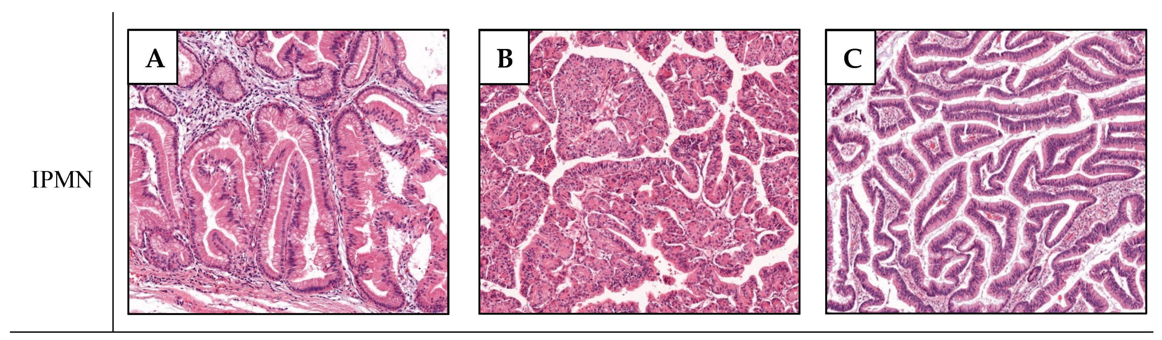
2. Non-Invasive IPMNs and Associated Carcinomas

In the clinical setting, IPMNs are classified according to their macroscopic features. Based on imaging findings, three subtypes, i.e., main duct IPMN (MD-IPMN), branch duct IPMN (BD-IPMN), and mixed type IPMN are recognized. MD-IPMN is characterized by segmental or diffuse dilatation of the main pancreatic duct (MPD) of more than 5 mm without other causes of obstruction, while BD-IPMN presents as a pancreatic cyst of more than 5 mm that communicates with the MPD. Mixed type IPMNs meet both these criteria [19].
Microscopically, IPMNs are characterized by a proliferation of columnar mucin-producing cells, predominantly papillary with fibrovascular cores, or rarely flat, arising in the main pancreatic duct or branch ducts. The papillae range from microscopic folds of neoplastic epithelium to grossly visible finger-like projections. The intraductal nature of these neoplasms can be appreciated by their involvement of the branching duct system. The lesions may be focal, multifocal, or diffuse. IPMNs are classified according to a two-tier grading system (low-grade or high-grade), based on the highest degree of cytoarchitectural atypia in the epithelium. The former intermediate-grade category is now included in the low-grade group [11]. Furthermore, three pathological subtypes are described based on the predominant architectural and cell differentiation pattern. According to the latter, tumor cells express different types of mucins. Four types of mucins (MUC1, MUC2, MUC5AC, and MUC6) are used to classify the different histological subtypes. Gastric IPMNs are the most common, accounting for 50–60%, and usually present as BD-IPMN with low-grade dysplasia. This subtype is characterized by a positive MUC5AC staining and the absence of both MUC1 and MUC2 expression (MUC6 staining may be positive or not). The intestinal subtype is the second most common (20–30%) that typically occurs in the MPD, with high-grade dysplasia in about 50% of cases. The immunohistochemical profile of intestinal IPMNs is defined by MUC5AC and MUC2 positive staining and MUC1 and MUC6 negative staining. Finally, the PB is a less common subtype (10–15%). Most PB-type IPMNs harbor high-grade dysplasia and express MUC5AC, MUC1, and MUC6, but not MUC2 [11,20]. Besides this classification, it has long been suggested that gastric and PB subtypes might represent the same entity with different grades of dysplasia, and recent data suggest that intestinal IPMNs might also derive from gastric lesions by clonal evolution [21,22]. Although all three subtypes of IPMN have the potential to progress to invasive carcinoma, PB and intestinal subtypes are estimated to carry a higher risk of tumor progression. Indeed, an invasive component is found in 60–70% of resected PB and in 30–40% of intestinal IPMNs, but only in 15% of resected gastric IPMNs [19,23,24,25].
The invasive component of IPMCs is either of the tubular (ductal) or the colloid type. The tubular type is the most common and presents a conventional ductal morphology with neoplastic cells arranged in small tubular glands that infiltrate a desmoplastic stroma. This type of carcinoma is preferentially associated with either PB or gastric-type IPMN [11]. In 25–30% of patients [26,27,28,29], the invasive component is of colloid type and is characterized by extensive stromal pools of acellular mucin either lined by neoplastic epithelial cells or containing floating neoplastic epithelial cells in more than 80% of the tumor [11]. In contrast with tubular carcinomas, indistinguishable from PDAC arising from PanIN lesions, colloid carcinomas are believed to derive almost exclusively from IPMNs of the intestinal phenotype [20,26,30,31,32].
In IPMCs, the carcinoma arises in topological relationship with the IPMN. It is worthy to note that a concomitant invasive carcinoma can also be observed in the setting of IPMN. In this latter case, the carcinoma is not contiguous with the IPMN (almost always of the branch duct type) and is typically a tubular adenocarcinoma. Emerging data, discussed hereafter, suggest that these concomitant invasive carcinomas may be biologically and prognostically distinct from IPMCs. Hence, thorough sampling of pancreatic tissue is recommended to determine the relationship between the invasive carcinoma and the IPMN [19]. The pathological aspects of the different precursor and invasive subtypes are summarized in Figure 1.

3. Clinical Presentation of IPMCs

Unlike patients with cPDAC, those with IPMC are older at diagnosis (a difference of 1–5 years) and have a higher proportion of lesions located in the pancreatic tail (8% to 34%), thereby justifying the higher rate of distal pancreatectomies [26,30,33,34,35,36]. A review of data from studies of resected IPMC (Table 1) showed that 63–90% of patients present with pre-operative symptoms. Woo et al. [37] demonstrated a significantly higher proportion of asymptomatic patients in the IPMC cohort when directly compared with cPDAC cohort (28% vs. 11%, p = 0.013). Just like cPDAC, abdominal pain, weight loss, and jaundice were the most frequent symptoms, with the reported frequencies of 43–56%, 33–53%, and 28–38%, respectively (Table 1). However, controversy still exists about the association of IPMC with acute pancreatitis with reported incidence varying from 3% to 24% [38,39,40,41].

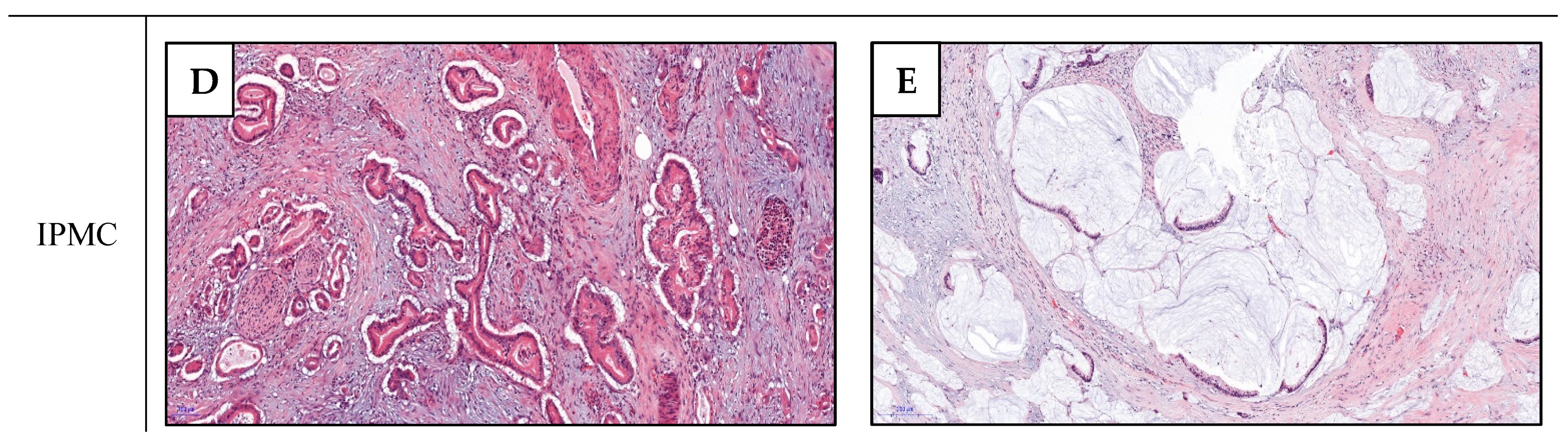
4. Pathological Features and Survival of IPMCs

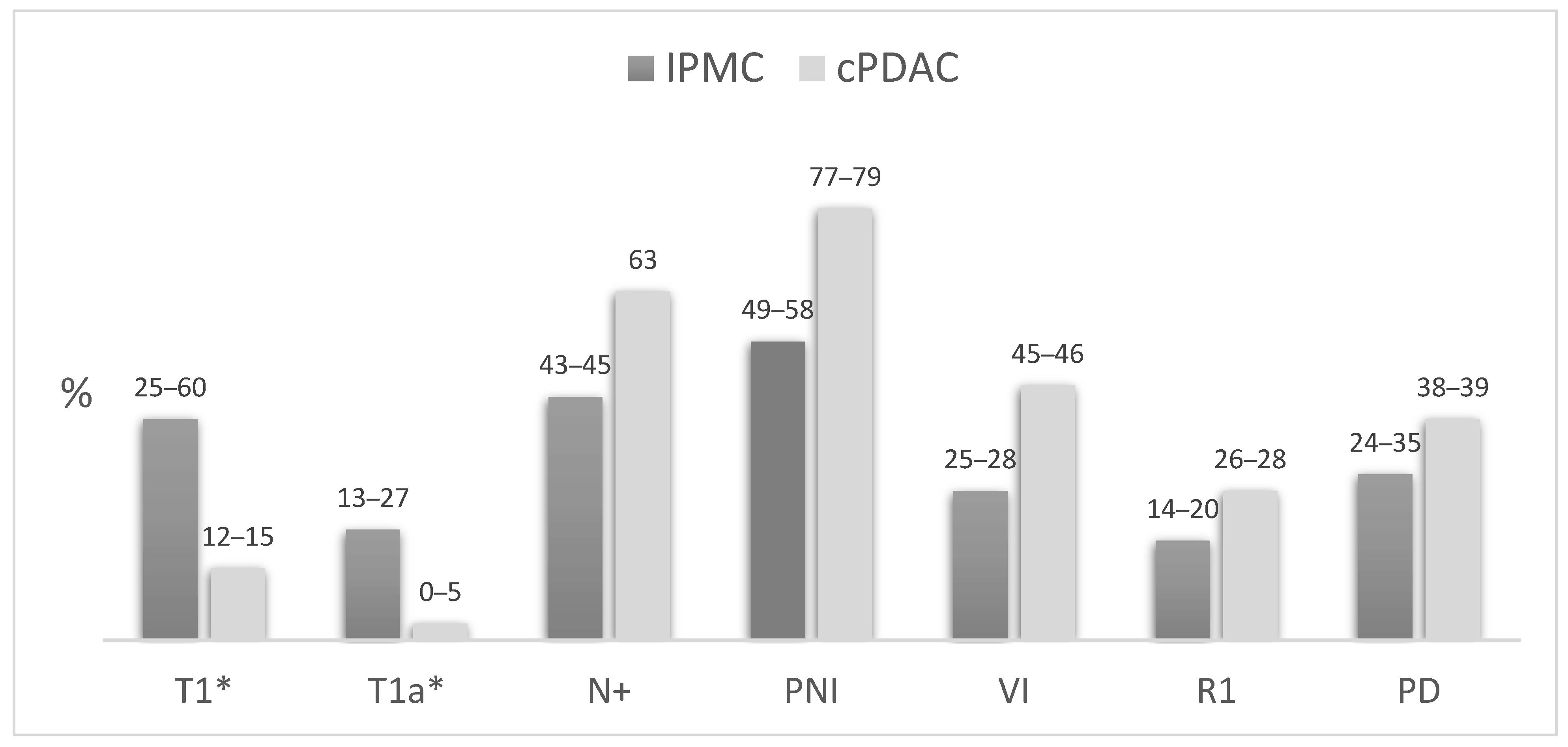
4.1. IPMC Versus cPDAC

In 2014, Koh et al. [36] performed the first meta-analysis comparing the pathological features of IPMC and cPDAC. They found IPMC to have a lower likelihood of T3 and T4 tumors, a significantly lower rate of nodal metastasis, and less TNM stage II and III tumors. Other adverse features such as incomplete resection (R1), perineural invasion, and vascular invasion were also less frequent in IPMC. A recent meta-analysis has confirmed these data and also found IPMC to be associated with a better differentiation of the tumor [43]. Of note, these data must be interpreted with caution knowing that the distinction between IPMC and PDAC concomitant with an IPMN was not reliably assessed. The principal differences between the main pathological features of IPMC and cPDAC are summarized in Figure 2.
IPMC shows a particularly high frequency of tumors with a minimal invasive component often referred to as “minimally invasive IPMN”. This subset has long been defined with a great variability of diagnostic criteria, based on morphological characteristics, tumor size, or the percentage of the invasive component [16,24,44,45]. In order to allow reproducible and comparable classifications, the 2012 international consensus guidelines [19] recommended a size-based only approach with substaging of the T1 category into T1a (<5 mm), T1b (5 mm–1 cm), and T1c (1–2 cm) lesions, while abandoning the non-specific “minimally invasive” term. Thus, according to the AJCC TNM 8th edition, T1 lesions account for 25% to 60% of IPMC and T1a for 13% to 27% [17,22,30,44,46,47,48]. Contrarily, cPDAC are categorized as T1 in only 12–14% of cases, among which less than 5% are T1a [49,50,51,52] (Figure 2).
Because of its association with favorable pathologic features, the better prognosis of IPMC compared with cPDAC has long been a matter of debate. The first reports of resected IPMC documented favorable outcomes with the five-year overall survival (OS) rates of 40% to 60%, which were significantly superior to that of unmatched comparative cPDAC cohorts (about 20%) [53,54]. The question whether this difference, besides pathological differences, could reflect a distinct tumor behavior resulted in matched studies comparing IPMCs to cPDAC, and the data were conflicting. Two studies, using age and tumor stage as matching parameters, reported a trend towards a superior five-year OS in IPMC patients (36% and 31% versus 21% and 24% in patients with cPDAC) [55,56]. However, this difference reached only statistical significance in the study of Maire et al. [56]. Using a more accurate matching process, based on a previously described nomogram for resected pancreatic cancer [57], Yopp et al. [42] found a significantly superior survival in IPMC (the 5-year OS of 68% versus 23% in cPDAC). However, this study did not include vascular invasion and perineural invasion, which are two main accepted prognostic factors for resected cPDAC [58,59], in matching parameters. Contrarily, in the study by Duconseil et al. [60], patients were matched by perineural invasion, tumor stage, lymph node ratio, and margin status and no statistical differences in survival were observed. Of six studies evaluating survival using multivariate regression analysis, five reported a significantly better OS in IPMC patients [16,30,33,35,61] and one, by Poultsides et al. [26], found no evidence of a survival difference. Nonetheless, these studies pointed toward a survival advantage for IPMC limited to early-stage tumors, frequently stage I or node negative [16,26,30,33,35,61]. Similarly, Poultsides et al. [26] showed a substantial survival difference only in the absence of any adverse pathological factors such as poor tumor differentiation, involved margin, vascular invasion, or perineural invasion.
Altogether, these data leave unanswered the question of whether IPMC may have an inherently favorable prognosis. Moreover, we cannot exclude that the reported differences result mainly from an earlier diagnosis. Several reasons may explain the earlier presentation of IPMC such as the presence of a large benign component associated with a smaller invasive component leading to more frequently fortuitous diagnosis or earlier symptoms leading to prompt exploration. It should be noted that available data mostly come from specialized centers in which the proportion of asymptomatic patients with fortuitous diagnosis of cystic lesions might be overestimated. Although these hypotheses should apply equally to all IPMC lesions, further discrepancies of pathologic findings and survival outcomes are observed among different subgroups of IPMC.

4.2. Colloid Versus Tubular IPMC

All of eight studies investigating colloid type and tubular type IPMC for the main pathological characteristics (Table 2) reported significant or almost significant trend towards less nodal metastasis in colloid carcinomas (16–29% versus 37–63% in tubular type). In addition, perineural, vascular and lymphatic invasion, poor differentiation, and margin involvement were reported in lower frequencies in colloid tumors (Table 2). Of note, two studies also reported an overrepresentation of colloid carcinomas among “minimally invasive IPMNs” [24,44].
Although possibly biased by these unbalanced pathological features, several studies reported better outcomes for colloid type IPMC than for tubular type, with a five-year OS ranging from 57–87% to 24–55% [26,30,40,42,46]. Recently, Rodrigues et al. found the survival advantage of colloid type over tubular type carcinomas to remain significant after multivariate analysis [48]. Furthermore, when separately compared to cPDAC, colloid type IPMCs were associated with significantly better outcomes, while no difference was observed between tubular type IPMC and cPDAC [16,30].
Altogether, these data highlight the importance of distinguishing pathological subtypes when comparing IPMC to cPDAC. Indeed, it could be hypothesized that only colloid IPMC is a more indolent entity, associated with both favorable pathological features and a significantly better prognosis truly reflective of a lower invasive potential. Contrarily, tubular IPMC could represent a subgroup closer, if not identical, to cPDAC that present a similar evolutionary profile.

5. Molecular Features

5.1. Mutationnal Landscape

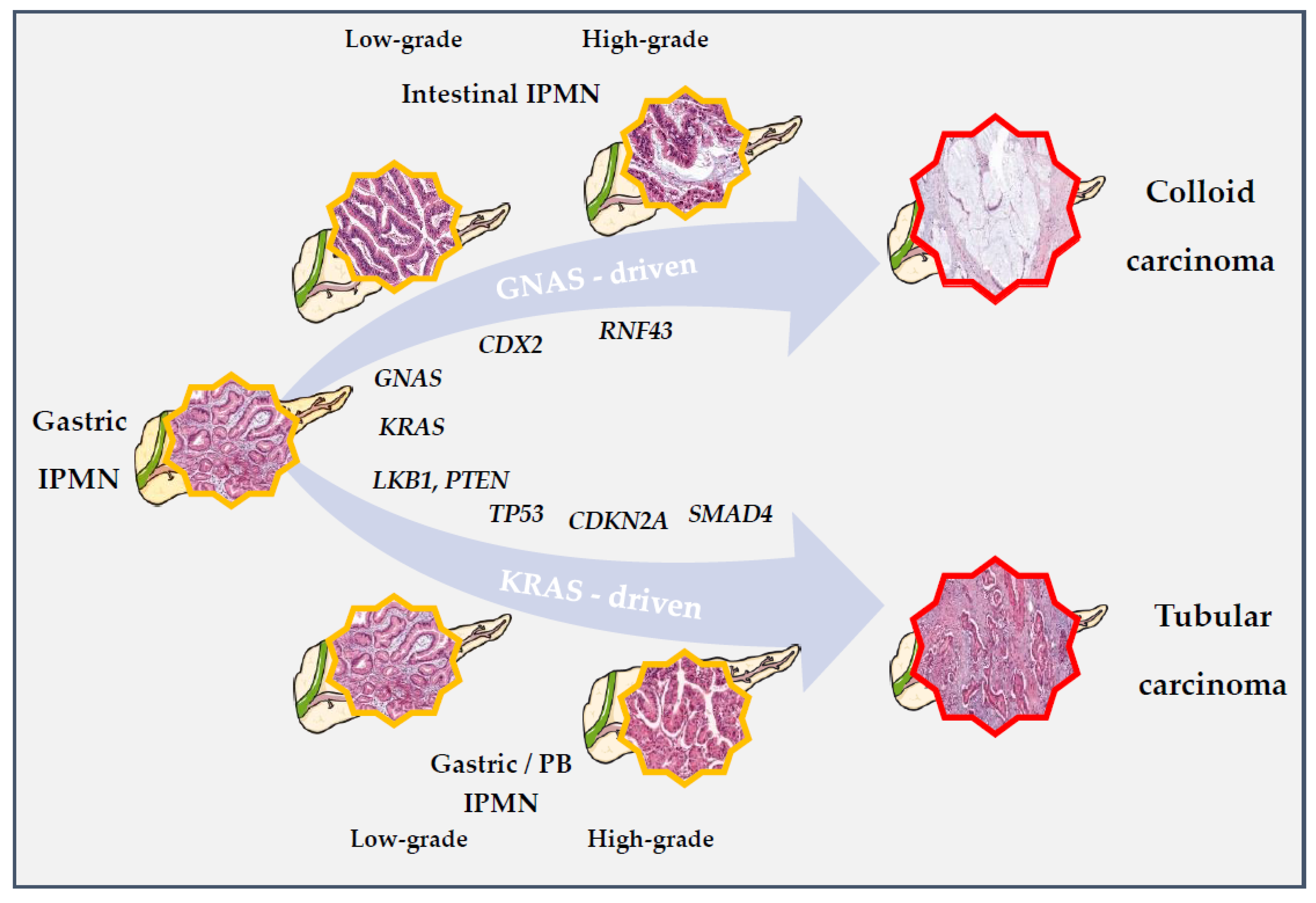
5.1.1. GNAS and KRAS

Mutations of the proto-oncogene GNAS have been shown to be highly specific of IPMNs as these are not found in any other type of pancreatic neoplasia [62,63,64,65,66,67,68]. In IPMNs, these mutations are almost exclusively found at codon 201, the most frequently observed being R201H and R201C, while R201S, R201Y or Q227L mutations were rarely described [68,69,70]. GNAS mutations alter the structure of GTPase domain in the α-stimulatory subunit of the G protein (Gsα) and vastly decrease GTPase activity. As a consequence, Gsα fails to hydrolyze GTP and release phosphoric acid, remaining in activated status, which constantly stimulates downstream molecules. This is responsible for the activation of the cyclic adenosine monophosphate (cAMP) and protein kinase A (PKA) pathway [71]. The cAMP-PKA signaling has been associated to tumor-promoting effects in several cancer types via the activation of diverse downstream targets [72]. In particular, cAMP-PKA signaling has been shown to drive tumorigenesis and promote cell proliferation and migration in breast cancer through the activation of Src, PI3K/AKT, and GSK3/βcathenin pathways [73,74,75]. In pancreatic carcinogenesis, salt inducible kinases (SIK) were recently identified as critical tumor suppressors inhibited by GNAS signaling [76].
Although present in all IPMN subtypes, GNAS mutations are significantly associated with the intestinal subtype (approximately 75% of cases) [77] and invasive colloid type lesions (found in 80–90% versus 15–30% in tubular carcinomas) [18,64,70]. Consistent with the favorable features and outcomes associated to intestinal IPMN and colloid type carcinomas, some studies reported a better survival or lower neural invasion [67,69,70] in GNAS-mutated patients. Interestingly, Felsenstein et al. [18] reported a higher prevalence of R201C mutations among colloid lesions (73%) and of R201H in tubular carcinomas (75%).
In a meta-analysis reported by Lee et al. [77], which included 11 studies, the pooled prevalence of GNAS mutations in IPMN cases was 56%, irrespective of the presence or absence of an associated carcinoma, with similar mutation rates among the different grades of dysplasia in non-invasive IPMNs. In the same work, no significant difference was reported between non-invasive IPMNs and IPMCs [77]. However, fewer patients were included for the latter comparison and the results of several studies suggest a lower prevalence of GNAS mutations among invasive lesions, ranging from 19% to 61% (Table 3). Noteworthy, the studies reporting 50% or more GNAS mutated IPMC were also those that included notably high proportions of intestinal type and/or colloid carcinomas.
KRAS mutations, present in more than 90% of cPDAC [62], are also found in about 65% of IPMCs and are associated with the gastric subtype [77] (Table 3). KRAS mutations results in a constitutively-active GTPase that locks the protein in its GTP-bound and results in its constitutive interaction with downstream signaling pathways. Activating mutation of the KRAS oncogene is involved in numerous cellular processes by interconnected regulation of signaling pathways such as PI3K/AKT/mTOR or Raf-MEK-ERK, which have been thoroughly rewieved elsewhere [78]. Deregulation of these pathways results in increased cell growth, and prevention of apoptosis [79]. Mutations at codon 12, mainly G12D and G12V (30–40% each), are the most prevalent in IPMCs, while G12R and G12C mutations are rare (about 5% each) [69,70,80]. KRAS mutations are more prevalent in tubular IPMC than in colloid carcinomas (80–90% versus 30–50%) [65,66,67,68,70]. However, no report has shown the association between KRAS mutations and survival [67,69].
KRAS and GNAS mutations are not mutually exclusive since double-mutant IPMCs are found in 25–35% of cases [68,70]. GNAS only mutated tumors are almost all of the intestinal subtype and GNAS mutated colloid carcinomas harbor way less KRAS mutations than GNAS mutated tubular lesions (40–50% versus 80%) [67,68,70]. Both GNAS and KRAS mutations are found at similar rates across all grades of dysplasia [77], suggesting that they represent the very early events in the carcinogenesis of IPMNs underlying the initiation of these neoplasms in most cases. Hence, these mutational events are thought to play a critical role in the diverging evolution between GNAS mutated lesions of intestinal subtype (and commonly wild-type KRAS) evolving into a colloid carcinoma and KRAS-mutated lesions of the gastric type evolving into tubular invasive lesions similar to cPDAC.

5.1.2. Other Mutations

Inactivating mutations of RNF43, involved in cell cycle control through the negative regulation of the Wnt signaling pathway [81,82], have also been reported in about 20% of IPMNs [77]. Unlike KRAS and GNAS, no definite hotspot exists for mutations of RNF43 (nonsense mutations, missense mutations, or frameshift mutations that lead to a decrease or loss of function) [83,84]. The RNF43 mutations might represent a later event as found mainly in high-grade lesions (20–75% versus 0–10% in low-grade IPMNs) [66,83]. Moreover, they are also significantly associated with GNAS mutations and the intestinal subtype, suggesting a synergistic role of these two events, particularly in the progression of the intestinal pathway [69,70,84,85]. However, RNF43 mutations might be involved mainly in the low grade—high grade transition since these alterations does not seem to be selected during progression to an invasive carcinoma [86,87]. Otherwise, mutations of KLF4 have recently been reported as a new driver gene, found in more than 50% of low-grade IPMNs but in only 15% of high grade lesions, suggesting a specific role in early stages of tumorigenesis [88].
Other main alterations found in cPDAC such as TP53, SMAD4, and CDKN2A seem also frequent in IPMC particularly of the tubular type, with more than 50% of TP53-mutated tumors and about 20–40% of SMAD4 and CDKN2A alterations, but are much less common in colloid carcinomas [18,87]. Furthermore, although these are almost absent in the vast majority of low-grade IPMNs, both mutations are more prevalent in high-grade and invasive lesions, suggesting their late onset in IPMN carcinogenesis and their role in the progression to invasive lesions rather than in initiation [66,68,70]. Figure 3 summarizes the main genomic features of IPMNs and IPMCs.

5.2. Carcinogenesis

5.2.1. Murine Models of IPMN Initiation

Despite the specific association of IPMNs with early GNAS alteration, the results of murine experiments reported by two studies [76,89] have shown GNAS mutations alone to be insufficient to induce the occurrence of IPMN-like lesions. This observation calls into question the hypothesis of a strictly GNAS-mediated carcinogenesis pathway. However, the association of KRAS and GNAS mutations led to the occurrence of mouse IPMN lesions [76,89]. In another work by Ideno et al. [90], the induction of GNAS mutation in adult mice with constitutive KRAS mutant background produced the same results. Notably, despite the association of GNAS mutations to intestinal type IPMNs in humans, the mouse IPMN lesions observed in those models were reminiscent of gastric and PB type IPMNs without any MUC2 expression, whether of modeling R201H or R201C GNAS alterations. Moreover, no invasive component was observed with associated KRAS and GNAS mutations, but the addition of P53, P16, or SMAD4 mutations led to the development of invasive lesions [76,90]. Among these, no colloid carcinomas were observed, but GNAS mutated PDACs showed better differentiation than controls [90].
Collectively, data coming from murine models suggest a weak oncogenic potential for isolated GNAS mutations and a synergistic effect of combined RAS and GNAS alterations on induction of IPMN formation. The progression to invasive carcinoma seems to require the later implementation of other mutational events. Furthermore, the GNAS signal does not seem sufficient to initiate the differentiation toward the intestinal pathway. In a recent study including 60 intestinal-type IPMN specimens, Omori et al. [85] observed a histological transition from gastric-type epithelia to intestinal-type epithelia in 48 cases (80%). This transition was driven by CDX2, which expression seems to precede the acquisition of intestinal features and MUC2 expression [85]. The authors suggested that intestinal type IPMNs might develop from gastric-type lesions after induction of CDX2, which could be related to the GNAS signaling and additional molecular events including RNF43 and ß-catenin.
Beyond tumor initiation, contradicting roles of the GNAS signaling have been reported on tumor growth and maintenance, which may be influenced by the timing of different mutational events. By activating the GNAS signaling on an already established KRAS-induced PDAC model, Ideno et al. [90] showed an attenuation of growth and invasiveness, related to an epithelial tumor differentiation. Contrarily, in the setting of lesions induced by concurrent KRAS and GNAS mutations, Patra et al. [76] highlighted a critical role of the GNAS signaling for tumor growth and maintenance. These data suggest a unique molecular program for tumors developed with GNAS mutation as an initiating event. Moreover, oncogenic GNAS signaling in the latter model was associated with a broad rewiring of lipid metabolism distinct from that of KRAS-driven pancreatic cancer [76].
In other murine models, the association of KRAS mutations with other mutations such as LKB1 or PTEN, in the absence of GNAS mutations, made it possible to induce the development of IPMN-like lesions. These possibly represent some alternative molecular pathways involved in the non-intestinal pathway [91,92]. Figure 4 presents hypothetical pathways of IPMNs initiation and progression.

5.2.2. IPMN to IPMC Sequence

PDAC Derived from and Concomitant with an IPMN

To analyze the progression pattern of IPMN lesions, two categories of IPMN-related PDACs need to be distinguished, carcinomas derived from an IPMN, and PDAC concomitant with an IPMN [93]. These entities have been well defined by Yamagushi et al. [34] in 2011, using the topological relationship and the presence of a histological transition between the two lesions. Based on these criteria, 67% of 183 cases in their initial work were classified as carcinomas derived from an IPMN, 17% as concomitant carcinomas, and 16% with undetermined relationship to the associated IPMN. Noteworthy, all colloid carcinomas were derived from IPMNs. Although concomitant carcinomas might be thought to represent a coincidental collusion of both cPDAC and IPMN, these lesions had favorable pathological and survival features significantly distinct from cPDAC and closer to IPMN-derived lesions. Subsequent studies characterizing these concomitant carcinomas highlighted an association with branch-duct and gastric type IPMNs and the lower prevalence of GNAS mutations [94,95,96].

Pathways of Progression

The genetic relatedness between precursor and invasive lesions has been shown to be highly correlated to the morphological classification. In the work by Hosoda et al. [64], 93% of genetically related IPMC-IPMN couples were classified as carcinomas derived from an IPMN, while 75% of cases with discrepant genetic results were either concomitant carcinomas or carcinomas of undetermined relationship. All colloid carcinomas were genetically related to their associated precursor lesion, confirming their exclusive association with IPMNs.
The relevance of this distinction has also been evidenced by Omori et al. [86]. In their work, all IPMN-derived PDACs harbored a concordant mutational profile of GNAS and KRAS between the precursor and invasive components. In addition, mutations in P16, P53, or SMAD4 were accumulated in invasive lesions indicating progression through an accumulation of such alterations. The authors called this pattern of progression as the “sequential” subtype. Conversely, half of concomitant PDACs harbored a clearly distinct mutational profile and were called “de novo” subtype as developed independently from the related IPMN. The remaining lesions were named the “branch-off” subtype. These lesions shared the identical KRAS mutations and common sequence variation or methylation profiles demonstrating a common origin and suggesting the development of both PDACs and IPMNs from the same founder clone. The classification proposed by Omori et al. [86] showed a clinical interest, with a significantly better disease-free survival for the branch-off subtype compared to the de-novo subtype. Importantly, the mutational analysis of other precursor lesions in pancreata of these patients revealed a higher burden of GNAS mutant clones in those of the sequential subtype. These results were also consistent with those demonstrated by Felsenstein et al. [18], showing a substantial proportion of IPMCs with a distinct mutational status compared to the associated precursor, and invasive lesions with a partially shared genetic profile.
Overall, these data demonstrate a direct transition from IPMNs to invasive PDACs in some cases. These are well identified by the IPMN-derived morphological category and by the association with GNAS mutations and include all cases of colloid carcinomas. A significant proportion of concomitant cases is also genetically related to the IPMN but seems to arise from a common founder clone. The shared clonal origin might explain the observed pathological and clinical proximity of this category with IPMN-derived PDACs.

5.2.3. Tumor Heterogeneity

Recent research, however, highlighted a high molecular heterogeneity among a single IPMN lesion, challenging the interpretation of previous genetic characterization of IPMNs and genetic relatedness studies. Fischer et al. [97] demonstrated a striking heterogeneity of mutational profiles among 20 IPMN lesions using multi-region sequencing. Most lesions harbored multiple mutations within the same driver genes and many IPMNs did not have any mutation present across every region. Low-grade IPMNs had the greatest heterogeneity among early driver genes KRAS and GNAS, with up to six distinct mutations among the same lesion. High-grade IPMNs were characterized by a common early driver mutation and high heterogeneity of late occurring mutations such as RNF43 or P53. Accordingly, a recent study used whole exome sequencing to analyze the evolution from precursor lesions to IPMC [87], confirming the presence of multiple clones with independent evolution among a single IPMN. Multiple instances of clear driver mutations, such as RNF43, limited to the non-invasive component, were observed, suggesting unique selective processes at different time points in tumorigenesis, such that mutations selected in the precancerous lesion are not selected for (or are even selected against) in the invasive cancer. Furthermore, multiple separate invasion events could be identified in one IPMN [87].
These data challenge the traditional model of pancreatic tumor progression by demonstrating the polyclonal origin of some IPMNs. At early stages, IPMNs contain multiple independent clones harboring distinct driver mutations in KRAS and GNAS; a single clone can further acquire additional mutations leading to clonal expansion and progression. In addition, the spatial heterogeneity among IPMN lesions shows that one-region sequencing approaches cannot accurately capture the complete genetic landscape of these lesions, which calls into question the accuracy of previous data on mutation prevalence and genetic relatedness.

6. Therapeutic Implications

6.1. Adjuvant Therapy

At this day, no specific therapeutic strategy for IPMC is recommended. These patients are managed following guidelines proposed for cPDAC, with adjuvant therapy recommended in all patients after resection, regardless of T, N, and R status. Preferred regimens are modified FOLFIRINOX in ECOG 0–1 patients, and gemcitabine monotherapy, 5-FU, or gemcitabine plus capecitabine in those not eligible for FOLFIRINOX [98,99]. There is only limited evidence of the beneficial effect of adjuvant therapy in resected IPMC and this is mostly derived from the results of phase III clinical trials using pooled data of cPDAC and IPMC [100,101]. The distinct features and outcomes associated to IPMC raise the question about the interest of a specific therapeutic strategy for these patients.
Retrospective comparisons of survivals in IPMC patients with or without adjuvant therapy are inevitably biased by the non-randomized attribution of post-operative treatments and the consequent imbalances in tumor characteristics. In five studies that we analyzed and summarized in Table 4, multivariate analysis to mitigate this bias was used. Of note, these included patients over a long period of time since the late 1990s, during which treatment guidelines have evolved considerably. Four studies reported on patients receiving adjuvant chemotherapy, with gemcitabine being the most widely used agent and 30% to 69% receiving additional chemoradiation [33,48,102,103]. The fifth included only patients who received 5-FU based chemoradiation followed by 5-FU based chemotherapy alone [104]. In the study by Rodrigues et al. [48], no beneficial effects of adjuvant gemcitabine-based chemotherapy in the cohort of 103 IPMC neither in any subgroup were observed. The remaining studies reported a significant survival benefit limited to the subgroup of node-positive patients [33,102,103,104]. Likewise, the beneficial effect of adjuvant therapy in resected IPMC has also been reported in the presence of other adverse pathologic features such as stage II-IV disease, tubular histology, and poorly differentiated or margin positive tumors [25,33,104].
These data, although not reporting on currently recommended treatment modalities, suggest that adjuvant therapy might be indicated in patients after resection for IPMC that present only adverse pathologic features, while those patients who have early-stage tumors without node metastasis do not seem to benefit from it. Moreover, tubular histology has been linked to adjuvant therapy benefit [105]. Noteworthy, a recent work by Shaib et al. [106] also questioned the relevance of adjuvant therapy in T1a/bN0 PDACs, for which no survival benefit was observed. Prospective studies dedicated to IPMC are needed in the future to better address the question of a specific adjuvant strategy, with particular attention paid to the histological subtype.

6.2. Future Therapeutic Strategies

Better characterization of the molecular pathways specifically involved in IPMC will provide opportunities for the development of targeted therapeutic strategies for these tumors. In this way, the intestinal pathway probably represents a truly distinct entity to focus on. GNAS mutations, which are the only molecular specificity identified to date, might guide the developments of effective treatments. Downstream effectors of the cAMP-PKA pathway such as SIKs can represent potential targets [107]. In addition, the specific metabolic features of GNAS mutated tumors represent other potential research subjects. Nevertheless, the presence of a GNAS mutation alone cannot be considered sufficient to correctly identify tumors with a distinct molecular program and a better understanding of the distinct oncogenic processes involved is still necessary to accurately select patients. The yet unknown factors associated with the intestinal pathway induction might in the near future help to guide the development of targeted strategies. Moreover, new markers of the intestinal pathway have been recently reported such as GPA33 [108], which is already evaluated in colorectal cancer for targeted cytotoxic delivery or photoimmunotherapy [109,110].
Furthermore, evaluating the prevalence of already stablished relevant alterations in cPDAC among IPMCs, such as microsatellite instability (MSI) [111], could provide further opportunities for precision oncology in this subgroup. Importantly, Lupinacci et al. [14] found a significantly higher prevalence (6.9% vs. 1.4%) of the MSI phenotype in IPMC patients, and colloid carcinomas, known to arise exclusively with IPMNs, are significantly associated with microsatellite instability in PDAC [112]. Numerous approaches are investigated to expand the use of immunotherapy beyond MSI patients, including checkpoint blockade or vaccine therapy [113], and the identification of specific immune features associated with IPMCs might also lead to targeted strategies. Otherwise, the prevalence of BRCA mutations or BRCAness phenotype, associated to known or suspected therapeutic implications [114,115], has not been reliably assessed in IPMCs. However, Skaro et al. [116] showed a significant proportion of IPMC patients harboring germline mutations other than BRCA involving genes related to DNA damage repair (ATM, FANC family, PALB2, BRIP1, NBN). In addition, somatic mutations of ARID1A and ATM have been reported in about 10% and 10–15% of IPMCs, respectively [18,70,87].

7. Conclusions

IPMCs account for about 10% of resected PDACs and represent a subgroup of tumors with specific features. Two histologic types are to be recognized: (1) colloid IPMCs that present with distinct morphology, favorable pathologic features, and significantly better outcomes, and (2) tubular IPMCs, morphologically indistinguishable from cPDACs with whom they share most of pathological and clinical features. The question of a distinct prognosis in tubular IPMC, therefore, is still unanswered.
Consistent with this dichotomy, molecular data confirm a distinct mutational profile of intestinal type and related colloid carcinomas, characterized mainly by the higher prevalence of GNAS mutations and lower rate of KRAS alterations. Altogether, the available data support the idea of a specific oncogenic intestinal pathway, correlated with GNAS mutations and yet unknown initiating events, leading to a clearly distinct molecular program and less-aggressive tumors. Contrarily, tubular IPMCs probably arise from the common KRAS-driven carcinogenesis pathway. Growing insights into the mechanisms of initiation and progression of IPMNs evidenced both a direct precursor to invasive lesion sequence related to the intestinal pathway and alternative mechanisms with a separate onset of IPMN and PDAC from a common origin.
As an entirely distinct entity, IPMC might offer opportunities for specific therapeutic options in the future. The role of adjuvant therapy in early-stage IPMC and/or tumors with colloid histology remains an unsolved issue and requires dedicated prospective studies. The specific molecular features associated with the intestinal pathway could also lead to targeted therapeutic options, but further characterization of the molecular pathways involved will be needed to refine patient selection.

Author Contributions

L.M., R.M.L., and M.S. contributed to the design of the review; L.M. and R.M.L. acquired and extracted the data.; L.M. prepared the manuscript; R.M.L., M.S., J.C., J.-B.B., and F.C. participated in the writing and reviewing of the manuscript. All authors have read and agreed to the published version of the manuscript.

Funding

This research received no external funding.

Institutional Review Board Statement

Not applicable.

Informed Consent Statement

Not applicable.

Data Availability Statement

Not applicable.

Acknowledgments

The authors thank Magdalena Benetkiewicz for editorial assistance.

Conflicts of Interest

The authors declare no conflict of interest.

References

  1. Ferlay, J.; Partensky, C.; Bray, F. More Deaths from Pancreatic Cancer than Breast Cancer in the EU by 2017. Acta Oncol. 2016, 55, 1158–1160. [Google Scholar] [CrossRef]
  2. Rahib, L.; Smith, B.D.; Aizenberg, R.; Rosenzweig, A.B.; Fleshman, J.M.; Matrisian, L.M. Projecting Cancer Incidence and Deaths to 2030: The Unexpected Burden of Thyroid, Liver, and Pancreas Cancers in the United States. Cancer Res. 2014, 74, 2913–2921. [Google Scholar] [CrossRef] [Green Version]
  3. Siegel, R.L.; Miller, K.D.; Jemal, A. Cancer Statistics, 2019. CA Cancer J. Clin. 2019, 69, 7–34. [Google Scholar] [CrossRef] [Green Version]
  4. Bengtsson, A.; Andersson, R.; Ansari, D. The Actual 5-Year Survivors of Pancreatic Ductal Adenocarcinoma Based on Real-World Data. Sci. Rep. 2020, 10, 16425. [Google Scholar] [CrossRef] [PubMed]
  5. Histological Typing of Tumours of the Exocrine Pancreas, 2nd ed.; Klöppel, G.; Weltgesundheitsorganisation (Eds.) International Histological Classification of Tumours; Corr. Printing; Springer: Berlin, Germany, 1998; ISBN 978-3-540-60280-4. [Google Scholar]
  6. Ricci, C.; Migliori, M.; Imbrogno, A.; Mazzotta, E.; Felicani, C.; Serra, C.; Bergonzoni, B.; Calculli, L.; Casadei, R. Prevalence of Asymptomatic Intraductal Papillary Mucinous Neoplasms in Healthy and Ill Populations Detected by Ultrasonography: A Single-Center Study of 6353 Outpatients. Pancreas 2019, 48, 113–120. [Google Scholar] [CrossRef]
  7. Laurent, L.; Vullierme, M.; Rebours, V.; Maire, F.; Hentic, O.; Francoz, C.; Durand, F.; Ruszniewski, P.; Lévy, P. Estimation of the Prevalence of Intraductal Papillary Mucinous Neoplasm of the Pancreas in the French Population through Patients Waiting for Liver Transplantation. United Eur. Gastroenterol. J. 2017, 5, 499–503. [Google Scholar] [CrossRef] [PubMed] [Green Version]
  8. Baiocchi, G.L.; Portolani, N.; Grazioli, L.; Mazza, G.; Gheza, F.; Bartoli, M.; Vanzetti, E.; Giulini, S.M. Management of Pancreatic Intraductal Papillary Mucinous Neoplasm in an Academic Hospital (2005Y2010). Pancreas 2013, 42, 5. [Google Scholar] [CrossRef] [PubMed]
  9. Gaujoux, S.; Brennan, M.F.; Gonen, M.; D’Angelica, M.I.; DeMatteo, R.; Fong, Y.; Schattner, M.; DiMaio, C.; Janakos, M.; Jarnagin, W.R.; et al. Cystic Lesions of the Pancreas: Changes in the Presentation and Management of 1424 Patients at a Single Institution over a 15-Year Time Period. J. Am. Coll. Surg. 2011, 212, 590–600. [Google Scholar] [CrossRef] [Green Version]
  10. Valsangkar, N.P.; Morales-Oyarvide, V.; Thayer, S.P.; Ferrone, C.R.; Wargo, J.A.; Warshaw, A.L.; Fernández-del Castillo, C. 851 Resected Cystic Tumors of the Pancreas: A 33-Year Experience at the Massachusetts General Hospital. Surgery 2012, 152, S4–S12. [Google Scholar] [CrossRef] [Green Version]
  11. Lokuhetty, D.; Organisation Mondiale de la Santé; Centre International de Recherche sur le Cancer. WHO Classification of Tumours; World Health Organization: Geneva, Switzerland, 2019; ISBN 978-92-832-4499-8. [Google Scholar]
  12. Nagtegaal, I.D.; Odze, R.D.; Klimstra, D.; Paradis, V.; Rugge, M.; Schirmacher, P.; Washington, K.M.; Carneiro, F.; Cree, I.A.; The WHO Classification of Tumours Editorial Board. The 2019 WHO Classification of Tumours of the Digestive System. Histopathology 2020, 76, 182–188. [Google Scholar] [CrossRef] [Green Version]
  13. Keane, M.G.; Afghani, E. A Review of the Diagnosis and Management of Premalignant Pancreatic Cystic Lesions. J. Clin. Med. 2021, 10, 1284. [Google Scholar] [CrossRef]
  14. Lupinacci, R.M.; Goloudina, A.; Buhard, O.; Bachet, J.-B.; Maréchal, R.; Demetter, P.; Cros, J.; Bardier-Dupas, A.; Collura, A.; Cervera, P.; et al. Prevalence of Microsatellite Instability in Intraductal Papillary Mucinous Neoplasms of the Pancreas. Gastroenterology 2018, 154, 1061–1065. [Google Scholar] [CrossRef]
  15. Lewis, R.; Drebin, J.A.; Callery, M.P.; Fraker, D.; Kent, T.S.; Gates, J.; Vollmer, C.M. A Contemporary Analysis of Survival for Resected Pancreatic Ductal Adenocarcinoma. HPB 2013, 15, 49–60. [Google Scholar] [CrossRef] [Green Version]
  16. Waters, J.A.; Schnelldorfer, T.; Aguilar-Saavedra, J.R.; Chen, J.-H.; Yiannoutsos, C.T.; Lillemoe, K.D.; Farnell, M.B.; Sarr, M.G.; Schmidt, C.M. Survival after Resection for Invasive Intraductal Papillary Mucinous Neoplasm and for Pancreatic Adenocarcinoma: A Multi-Institutional Comparison According to American Joint Committee on Cancer Stage. J. Am. Coll. Surg. 2011, 213, 275–283. [Google Scholar] [CrossRef]
  17. Murakami, Y.; Uemura, K.; Sudo, T.; Hayashidani, Y.; Hashimoto, Y.; Nakashima, A.; Sueda, T. Invasive Intraductal Papillary-Mucinous Neoplasm of the Pancreas: Comparison with Pancreatic Ductal Adenocarcinoma. J. Surg. Oncol. 2009, 100, 13–18. [Google Scholar] [CrossRef]
  18. Felsenstein, M.; Noë, M.; Masica, D.L.; Hosoda, W.; Chianchiano, P.; Fischer, C.G.; Lionheart, G.; Brosens, L.A.A.; Pea, A.; Yu, J.; et al. IPMNs with Co-Occurring Invasive Cancers: Neighbours but Not Always Relatives. Gut 2018, 67, 1652–1662. [Google Scholar] [CrossRef]
  19. Tanaka, M.; Fernández-del Castillo, C.; Adsay, V.; Chari, S.; Falconi, M.; Jang, J.-Y.; Kimura, W.; Levy, P.; Pitman, M.B.; Schmidt, C.M.; et al. International Consensus Guidelines 2012 for the Management of IPMN and MCN of the Pancreas. Pancreatology 2012, 12, 183–197. [Google Scholar] [CrossRef]
  20. Furukawa, T.; Klöppel, G.; Volkan Adsay, N.; Albores-Saavedra, J.; Fukushima, N.; Horii, A.; Hruban, R.H.; Kato, Y.; Klimstra, D.S.; Longnecker, D.S.; et al. Classification of Types of Intraductal Papillary-Mucinous Neoplasm of the Pancreas: A Consensus Study. Virchows Arch. 2005, 447, 794–799. [Google Scholar] [CrossRef] [PubMed]
  21. Noë, M.; Brosens, L.A.A. Gastric- and Intestinal-Type IPMN: Two of a Kind? Virchows Arch. 2020, 477, 17–19. [Google Scholar] [CrossRef]
  22. Adsay, N.V.; Conlon, K.C.; Zee, S.Y.; Brennan, M.F.; Klimstra, D.S. Intraductal Papillary-Mucinous Neoplasms of the Pancreas: An Analysis of in Situ and Invasive Carcinomas in 28 Patients. Cancer 2002, 94, 62–77. [Google Scholar] [CrossRef] [PubMed]
  23. Furukawa, T.; Hatori, T.; Fujita, I.; Yamamoto, M.; Kobayashi, M.; Ohike, N.; Morohoshi, T.; Egawa, S.; Unno, M.; Takao, S.; et al. Prognostic Relevance of Morphological Types of Intraductal Papillary Mucinous Neoplasms of the Pancreas. Gut 2011, 60, 509–516. [Google Scholar] [CrossRef]
  24. Kang, M.J.; Lee, K.B.; Jang, J.-Y.; Kwon, W.; Park, J.W.; Chang, Y.R.; Kim, S.-W. Disease Spectrum of Intraductal Papillary Mucinous Neoplasm With an Associated Invasive Carcinoma: Invasive IPMN Versus Pancreatic Ductal Adenocarcinoma-Associated IPMN. Pancreas 2013, 42, 1267–1274. [Google Scholar] [CrossRef] [PubMed]
  25. Marchegiani, G.; Mino-Kenudson, M.; Ferrone, C.R.; Morales-Oyarvide, V.; Warshaw, A.L.; Lillemoe, K.D.; Castillo, C.F. Patterns of Recurrence After Resection of IPMN: Who, When, and How? Ann. Surg. 2015, 262, 1108–1114. [Google Scholar] [CrossRef] [PubMed]
  26. Poultsides, G.A.; Reddy, S.; Cameron, J.L.; Hruban, R.H.; Pawlik, T.M.; Ahuja, N.; Jain, A.; Edil, B.H.; Iacobuzio-Donahue, C.A.; Schulick, R.D.; et al. Histopathologic Basis for the Favorable Survival after Resection of Intraductal Papillary Mucinous Neoplasm-Associated Invasive Adenocarcinoma of the Pancreas. Ann. Surg. 2010, 251, 470–476. [Google Scholar] [CrossRef] [PubMed] [Green Version]
  27. Rezaee, N.; Barbon, C.; Zaki, A.; He, J.; Salman, B.; Hruban, R.H.; Cameron, J.L.; Herman, J.M.; Ahuja, N.; Lennon, A.M.; et al. Intraductal Papillary Mucinous Neoplasm (IPMN) with High-Grade Dysplasia Is a Risk Factor for the Subsequent Development of Pancreatic Ductal Adenocarcinoma. HPB (Oxford) 2016, 18, 236–246. [Google Scholar] [CrossRef] [PubMed] [Green Version]
  28. Hirono, S.; Shimizu, Y.; Ohtsuka, T.; Kin, T.; Hara, K.; Kanno, A.; Koshita, S.; Hanada, K.; Kitano, M.; Inoue, H.; et al. Recurrence Patterns after Surgical Resection of Intraductal Papillary Mucinous Neoplasm (IPMN) of the Pancreas; a Multicenter, Retrospective Study of 1074 IPMN Patients by the Japan Pancreas Society. J. Gastroenterol. 2020, 55, 86–99. [Google Scholar] [CrossRef]
  29. Morales-Oyarvide, V.; Mino-Kenudson, M.; Ferrone, C.R.; Warshaw, A.L.; Lillemoe, K.D.; Sahani, D.V.; Pergolini, I.; Attiyeh, M.A.; Al Efishat, M.; Rezaee, N.; et al. Intraductal Papillary Mucinous Neoplasm of the Pancreas in Young Patients: Tumor Biology, Clinical Features, and Survival Outcomes. J. Gastrointest. Surg. 2018, 22, 226–234. [Google Scholar] [CrossRef]
  30. Mino-Kenudson, M.; Fernandez-del Castillo, C.; Baba, Y.; Valsangkar, N.P.; Liss, A.S.; Hsu, M.; Correa-Gallego, C.; Ingkakul, T.; Perez Johnston, R.; Turner, B.G.; et al. Prognosis of Invasive Intraductal Papillary Mucinous Neoplasm Depends on Histological and Precursor Epithelial Subtypes. Gut 2011, 60, 1712–1720. [Google Scholar] [CrossRef] [Green Version]
  31. Sadakari, Y.; Ohuchida, K.; Nakata, K.; Ohtsuka, T.; Aishima, S.; Takahata, S.; Nakamura, M.; Mizumoto, K.; Tanaka, M. Invasive Carcinoma Derived from the Nonintestinal Type Intraductal Papillary Mucinous Neoplasm of the Pancreas Has a Poorer Prognosis than That Derived from the Intestinal Type. Surgery 2010, 147, 812–817. [Google Scholar] [CrossRef]
  32. Yamada, S.; Fujii, T.; Shimoyama, Y.; Kanda, M.; Nakayama, G.; Sugimoto, H.; Koike, M.; Nomoto, S.; Fujiwara, M.; Nakao, A.; et al. Clinical Implication of Morphological Subtypes in Management of Intraductal Papillary Mucinous Neoplasm. Ann. Surg. Oncol. 2014, 21, 2444–2452. [Google Scholar] [CrossRef]
  33. McMillan, M.T.; Lewis, R.S.; Drebin, J.A.; Teitelbaum, U.R.; Lee, M.K.; Roses, R.E.; Fraker, D.L.; Vollmer, C.M. The Efficacy of Adjuvant Therapy for Pancreatic Invasive Intraductal Papillary Mucinous Neoplasm (IPMN): Adjuvant Therapy for Invasive IPMN. Cancer 2016, 122, 521–533. [Google Scholar] [CrossRef] [PubMed] [Green Version]
  34. Yamaguchi, K.; Kanemitsu, S.; Hatori, T.; Maguchi, H.; Shimizu, Y.; Tada, M.; Nakagohri, T.; Hanada, K.; Osanai, M.; Noda, Y.; et al. Pancreatic Ductal Adenocarcinoma Derived From IPMN and Pancreatic Ductal Adenocarcinoma Concomitant With IPMN. Pancreas 2011, 40, 571–580. [Google Scholar] [CrossRef]
  35. Wasif, N.; Bentrem, D.J.; Farrell, J.J.; Ko, C.Y.; Hines, O.J.; Reber, H.A.; Tomlinson, J.S. Invasive Intraductal Papillary Mucinous Neoplasm versus Sporadic Pancreatic Adenocarcinoma: A Stage-Matched Comparison of Outcomes. Cancer 2010, 116, 3369–3377. [Google Scholar] [CrossRef] [Green Version]
  36. Koh, Y.-X.; Chok, A.-Y.; Zheng, H.-L.; Tan, C.-S.; Goh, B.K.P. Systematic Review and Meta-Analysis Comparing the Surgical Outcomes of Invasive Intraductal Papillary Mucinous Neoplasms and Conventional Pancreatic Ductal Adenocarcinoma. Ann. Surg. Oncol. 2014, 21, 2782–2800. [Google Scholar] [CrossRef]
  37. Woo, S.M.; Ryu, J.K.; Lee, S.H.; Yoo, J.W.; Park, J.K.; Kim, Y.-T.; Yoon, Y.B. Survival and Prognosis of Invasive Intraductal Papillary Mucinous Neoplasms of the Pancreas: Comparison With Pancreatic Ductal Adenocarcinoma. Pancreas 2008, 36, 50–55. [Google Scholar] [CrossRef]
  38. Hirono, S.; Kawai, M.; Okada, K.; Miyazawa, M.; Shimizu, A.; Kitahata, Y.; Ueno, M.; Yanagisawa, A.; Yamaue, H. Factors Associated With Invasive Intraductal Papillary Mucinous Carcinoma of the Pancreas. JAMA Surg. 2017, 152, e165054. [Google Scholar] [CrossRef] [PubMed]
  39. Turrini, O.; Waters, J.A.; Schnelldorfer, T.; Lillemoe, K.D.; Yiannoutsos, C.T.; Farnell, M.B.; Sarr, M.G.; Schmidt, C.M. Invasive Intraductal Papillary Mucinous Neoplasm: Predictors of Survival and Role of Adjuvant Therapy. HPB 2010, 12, 447–455. [Google Scholar] [CrossRef] [Green Version]
  40. Sohn, T.A.; Yeo, C.J.; Cameron, J.L.; Hruban, R.H.; Fukushima, N.; Campbell, K.A.; Lillemoe, K.D. Intraductal Papillary Mucinous Neoplasms of the Pancreas: An Updated Experience. Ann. Surg. 2004, 239, 788–799. [Google Scholar] [CrossRef] [PubMed]
  41. Partelli, S.; Castillo, C.F.-D.; Bassi, C.; Mantovani, W.; Thayer, S.P.; Crippa, S.; Ferrone, C.R.; Falconi, M.; Pederzoli, P.; Warshaw, A.L.; et al. Invasive Intraductal Papillary Mucinous Carcinomas of the Pancreas: Predictors of Survival and the Role of Lymph Node Ratio. Ann. Surg. 2010, 251, 477–482. [Google Scholar] [CrossRef] [PubMed] [Green Version]
  42. Yopp, A.C.; Katabi, N.; Janakos, M.; Klimstra, D.S.; D’Angelica, M.I.; DeMatteo, R.P.; Fong, Y.; Brennan, M.F.; Jarnagin, W.R.; Allen, P.J. Invasive Carcinoma Arising in Intraductal Papillary Mucinous Neoplasms of the Pancreas: A Matched Control Study with Conventional Pancreatic Ductal Adenocarcinoma. Ann. Surg. 2011, 253, 968–974. [Google Scholar] [CrossRef] [PubMed]
  43. Aronsson, L.; Bengtsson, A.; Torén, W.; Andersson, R.; Ansari, D. Intraductal Papillary Mucinous Carcinoma versus Pancreatic Ductal Adenocarcinoma: A Systematic Review and Meta-Analysis. Int. J. Surg. 2019, 71, 91–99. [Google Scholar] [CrossRef] [PubMed]
  44. Nara, S.; Shimada, K.; Kosuge, T.; Kanai, Y.; Hiraoka, N. Minimally Invasive Intraductal Papillary-Mucinous Carcinoma of the Pancreas: Clinicopathologic Study of 104 Intraductal Papillary-Mucinous Neoplasms. Am. J. Surg. Pathol. 2008, 32, 243–255. [Google Scholar] [CrossRef] [PubMed]
  45. Jang, J.-Y.; Hwang, D.W.; Kim, M.A.; Kang, M.-J.; Lim, C.S.; Lee, S.E.; Kim, S.-W. Analysis of Prognostic Factors and a Proposed New Classification for Invasive Papillary Mucinous Neoplasms. Ann. Surg. Oncol. 2011, 18, 644–650. [Google Scholar] [CrossRef] [PubMed]
  46. D’Angelica, M.; Brennan, M.F.; Suriawinata, A.A.; Klimstra, D.; Conlon, K.C. Intraductal Papillary Mucinous Neoplasms of the Pancreas: An Analysis of Clinicopathologic Features and Outcome. Ann. Surg. 2004, 239, 400–408. [Google Scholar] [CrossRef]
  47. Winter, J.M.; Jiang, W.; Basturk, O.; Mino-Kenudson, M.; Fong, Z.V.; Tan, W.P.; Lavu, H.; Vollmer, C.M.; Furth, E.E.; Haviland, D.; et al. Recurrence and Survival After Resection of Small Intraductal Papillary Mucinous Neoplasm-Associated Carcinomas (≤20-Mm Invasive Component): A Multi-Institutional Analysis. Ann. Surg. 2016, 263, 793–801. [Google Scholar] [CrossRef] [Green Version]
  48. Rodrigues, C.; Hank, T.; Qadan, M.; Ciprani, D.; Mino-Kenudson, M.; Weekes, C.D.; Ryan, D.P.; Clark, J.W.; Allen, J.N.; Hong, T.S.; et al. Impact of Adjuvant Therapy in Patients with Invasive Intraductal Papillary Mucinous Neoplasms of the Pancreas. Pancreatology 2020, 20, 722–728. [Google Scholar] [CrossRef]
  49. Schlitter, A.M.; Jesinghaus, M.; Jäger, C.; Konukiewitz, B.; Muckenhuber, A.; Demir, I.E.; Bahra, M.; Denkert, C.; Friess, H.; Kloeppel, G.; et al. PT but Not PN Stage of the 8th TNM Classification Significantly Improves Prognostication in Pancreatic Ductal Adenocarcinoma. Eur. J. Cancer 2017, 84, 121–129. [Google Scholar] [CrossRef]
  50. Park, M.Y.; Shin, S.H.; Song, K.B.; Hwang, D.; Lee, J.H.; Lee, Y.-J.; Kim, S.C. Validation of the Eighth Edition of the American Joint Committee on Cancer Staging System and Proposal of an Improved Staging System for Pancreatic Ductal Adenocarcinoma. Ann. Hepato-Biliary-Pancreat. Surg. 2019, 23, 46. [Google Scholar] [CrossRef] [Green Version]
  51. Shin, D.W.; Lee, J.; Kim, J.; Woo, S.M.; Lee, W.J.; Han, S.-S.; Park, S.-J.; Choi, K.S.; Cha, H.S.; Yoon, Y.-S.; et al. Validation of the American Joint Committee on Cancer 8th Edition Staging System for the Pancreatic Ductal Adenocarcinoma. Eur. J. Surg. Oncol. 2019, 45, 2159–2165. [Google Scholar] [CrossRef]
  52. Kwon, W.; Park, T.; He, J.; Higuchi, R.; Son, D.; Lee, S.Y.; Kim, J.; Byun, Y.; Kim, H.; Kim, S.-W.; et al. Is the New T1 Category as Defined in the Eighth Edition of the AJCC Pancreatic Cancer Staging System an Improvement? J. Gastrointest. Surg. 2020, 24, 262–269. [Google Scholar] [CrossRef]
  53. Sohn, T.A.; Yeo, C.J.; Cameron, J.L.; Iacobuzio-Donahue, C.A.; Hruban, R.H.; Lillemoe, K.D. Intraductal Papillary Mucinous Neoplasms of the Pancreas: An Increasingly Recognized Clinicopathologic Entity. Ann. Surg. 2001, 234, 313–322. [Google Scholar] [CrossRef]
  54. Shimada, K.; Sakamoto, Y.; Sano, T.; Kosuge, T.; Hiraoka, N. Invasive Carcinoma Originating in an Intraductal Papillary Mucinous Neoplasm of the Pancreas: A Clinicopathologic Comparison with a Common Type of Invasive Ductal Carcinoma. Pancreas 2006, 32, 281–287. [Google Scholar] [CrossRef]
  55. Schnelldorfer, T. Experience With 208 Resections for Intraductal Papillary Mucinous Neoplasm of the Pancreas. Arch. Surg. 2008, 143, 639. [Google Scholar] [CrossRef] [Green Version]
  56. Maire, F. Prognosis of Malignant Intraductal Papillary Mucinous Tumours of the Pancreas after Surgical Resection. Comparison with Pancreatic Ductal Adenocarcinoma. Gut 2002, 51, 717–722. [Google Scholar] [CrossRef]
  57. Brennan, M.F.; Kattan, M.W.; Klimstra, D.; Conlon, K. Prognostic Nomogram for Patients Undergoing Resection for Adenocarcinoma of the Pancreas. Ann. Surg. 2004, 240, 293–298. [Google Scholar] [CrossRef] [PubMed]
  58. Crippa, S.; Pergolini, I.; Javed, A.A.; Honselmann, K.C.; Weiss, M.J.; Di Salvo, F.; Burkhart, R.; Zamboni, G.; Belfiori, G.; Ferrone, C.R.; et al. Implications of Perineural Invasion on Disease Recurrence and Survival After Pancreatectomy for Pancreatic Head Ductal Adenocarcinoma. Ann. Surg. 2021. Publish Ahead of Print. [Google Scholar] [CrossRef]
  59. Epstein, J.D.; Kozak, G.; Fong, Z.V.; He, J.; Javed, A.A.; Joneja, U.; Jiang, W.; Ferrone, C.R.; Lillemoe, K.D.; Cameron, J.L.; et al. Microscopic Lymphovascular Invasion Is an Independent Predictor of Survival in Resected Pancreatic Ductal Adenocarcinoma. J. Surg. Oncol. 2017, 116, 658–664. [Google Scholar] [CrossRef] [PubMed]
  60. Duconseil, P.; Périnel, J.; Autret, A.; Adham, M.; Sauvanet, A.; Chiche, L.; Mabrut, J.-Y.; Tuech, J.-J.; Mariette, C.; Régenet, N.; et al. Resectable Invasive IPMN versus Sporadic Pancreatic Adenocarcinoma of the Head of the Pancreas: Should These Two Different Diseases Receive the Same Treatment? A Matched Comparison Study of the French Surgical Association (AFC). Eur. J. Surg. Oncol. 2017, 43, 1704–1710. [Google Scholar] [CrossRef] [PubMed]
  61. Yamada, S.; Fujii, T.; Hirakawa, A.; Takami, H.; Suenaga, M.; Hayashi, M.; Niwa, Y.; Hattori, N.; Iwata, N.; Kanda, M.; et al. Comparison of the Survival Outcomes of Pancreatic Cancer and Intraductal Papillary Mucinous Neoplasms. Pancreas 2018, 47, 974–979. [Google Scholar] [CrossRef] [PubMed]
  62. Witkiewicz, A.K.; McMillan, E.A.; Balaji, U.; Baek, G.; Lin, W.-C.; Mansour, J.; Mollaee, M.; Wagner, K.-U.; Koduru, P.; Yopp, A.; et al. Whole-Exome Sequencing of Pancreatic Cancer Defines Genetic Diversity and Therapeutic Targets. Nat. Commun. 2015, 6, 6744. [Google Scholar] [CrossRef] [PubMed]
  63. Furukawa, T.; Kuboki, Y.; Tanji, E.; Yoshida, S.; Hatori, T.; Yamamoto, M.; Shibata, N.; Shimizu, K.; Kamatani, N.; Shiratori, K. Whole-Exome Sequencing Uncovers Frequent GNAS Mutations in Intraductal Papillary Mucinous Neoplasms of the Pancreas. Sci. Rep. 2011, 1, 161. [Google Scholar] [CrossRef] [PubMed] [Green Version]
  64. Hosoda, W.; Sasaki, E.; Murakami, Y.; Yamao, K.; Shimizu, Y.; Yatabe, Y. GNAS Mutation Is a Frequent Event in Pancreatic Intraductal Papillary Mucinous Neoplasms and Associated Adenocarcinomas. Virchows Arch. 2015, 466, 665–674. [Google Scholar] [CrossRef] [PubMed]
  65. Wu, J.; Matthaei, H.; Maitra, A.; Dal Molin, M.; Wood, L.D.; Eshleman, J.R.; Goggins, M.; Canto, M.I.; Schulick, R.D.; Edil, B.H.; et al. Recurrent GNAS Mutations Define an Unexpected Pathway for Pancreatic Cyst Development. Sci. Transl. Med. 2011, 3, 92ra66. [Google Scholar] [CrossRef] [PubMed] [Green Version]
  66. Amato, E.; dal Molin, M.; Mafficini, A.; Yu, J.; Malleo, G.; Rusev, B.; Fassan, M.; Antonello, D.; Sadakari, Y.; Castelli, P.; et al. Targeted Next-Generation Sequencing of Cancer Genes Dissects the Molecular Profiles of Intraductal Papillary Neoplasms of the Pancreas: IPMN Molecular Profile. J. Pathol. 2014, 233, 217–227. [Google Scholar] [CrossRef]
  67. Gaujoux, S.; Parvanescu, A.; Cesaretti, M.; Silve, C.; Bieche, I.; Rebours, V.; Lévy, P.; Sauvanet, A.; Cros, J. GNAS but Not Extended RAS Mutations Spectrum Are Associated with a Better Prognosis in Intraductal Pancreatic Mucinous Neoplasms. Ann. Surg. Oncol. 2019, 26, 2640–2650. [Google Scholar] [CrossRef] [PubMed]
  68. Kuboki, Y.; Shimizu, K.; Hatori, T.; Yamamoto, M.; Shibata, N.; Shiratori, K.; Furukawa, T. Molecular Biomarkers for Progression of Intraductal Papillary Mucinous Neoplasm of the Pancreas. Pancreas 2015, 44, 227–235. [Google Scholar] [CrossRef]
  69. Chang, X.Y.; Wu, Y.; Jiang, Y.; Wang, P.Y.; Chen, J. RNF43 Mutations in IPMN Cases: A Potential Prognostic Factor. Gastroenterol. Res. Pract. 2020, 2020, 1–10. [Google Scholar] [CrossRef] [PubMed]
  70. Tan, M.C.; Basturk, O.; Brannon, A.R.; Bhanot, U.; Scott, S.N.; Bouvier, N.; LaFemina, J.; Jarnagin, W.R.; Berger, M.F.; Klimstra, D.; et al. GNAS and KRAS Mutations Define Separate Progression Pathways in Intraductal Papillary Mucinous Neoplasm-Associated Carcinoma. J. Am. Coll. Surg. 2015, 220, 845–854. [Google Scholar] [CrossRef] [Green Version]
  71. O’Hayre, M.; Vázquez-Prado, J.; Kufareva, I.; Stawiski, E.W.; Handel, T.M.; Seshagiri, S.; Gutkind, J.S. The Emerging Mutational Landscape of G Proteins and G-Protein-Coupled Receptors in Cancer. Nat. Rev. Cancer 2013, 13, 412–424. [Google Scholar] [CrossRef]
  72. Zhang, H.; Kong, Q.; Wang, J.; Jiang, Y.; Hua, H. Complex Roles of CAMP–PKA–CREB Signaling in Cancer. Exp. Hematol. Oncol. 2020, 9. [Google Scholar] [CrossRef]
  73. Jin, X.; Zhu, L.; Cui, Z.; Tang, J.; Xie, M.; Ren, G. Elevated Expression of GNAS Promotes Breast Cancer Cell Proliferation and Migration via the PI3K/AKT/Snail1/E-Cadherin Axis. Clin. Transl. Oncol. 2019, 21, 1207–1219. [Google Scholar] [CrossRef]
  74. Beristain, A.G.; Molyneux, S.D.; Joshi, P.A.; Pomroy, N.C.; Di Grappa, M.A.; Chang, M.C.; Kirschner, L.S.; Privé, G.G.; Pujana, M.A.; Khokha, R. PKA Signaling Drives Mammary Tumorigenesis through Src. Oncogene 2015, 34, 1160–1173. [Google Scholar] [CrossRef]
  75. Wang, Z.; Li, Y.; Xiao, Y.; Lin, H.-P.; Yang, P.; Humphries, B.; Gao, T.; Yang, C. Integrin A9 Depletion Promotes β-Catenin Degradation to Suppress Triple Negative Breast Cancer Tumor Growth and Metastasis. Int. J. Cancer 2019, 145, 2767–2780. [Google Scholar] [CrossRef]
  76. Patra, K.C.; Kato, Y.; Mizukami, Y.; Widholz, S.; Boukhali, M.; Revenco, I.; Grossman, E.A.; Ji, F.; Sadreyev, R.I.; Liss, A.S.; et al. Mutant GNAS Drives Pancreatic Tumourigenesis by Inducing PKA-Mediated SIK Suppression and Reprogramming Lipid Metabolism. Nat. Cell Biol. 2018, 20, 811–822. [Google Scholar] [CrossRef] [PubMed]
  77. Lee, J.-H.; Kim, Y.; Choi, J.-W.; Kim, Y.-S. KRAS, GNAS, and RNF43 Mutations in Intraductal Papillary Mucinous Neoplasm of the Pancreas: A Meta-Analysis. SpringerPlus 2016, 5, 1172. [Google Scholar] [CrossRef] [PubMed] [Green Version]
  78. Jonckheere, N.; Vasseur, R.; Van Seuningen, I. The Cornerstone K-RAS Mutation in Pancreatic Adenocarcinoma: From Cell Signaling Network, Target Genes, Biological Processes to Therapeutic Targeting. Crit. Rev. Oncol./Hematol. 2017, 111, 7–19. [Google Scholar] [CrossRef]
  79. Maertens, O.; Cichowski, K. An Expanding Role for RAS GTPase Activating Proteins (RAS GAPs) in Cancer. Adv. Biol. Regul. 2014, 55, 1–14. [Google Scholar] [CrossRef]
  80. Chang, X.Y.; Wu, Y.; Li, Y.; Wang, J.; Chen, J. Intraductal Papillary Mucinous Neoplasms of the Pancreas: Clinical Association with KRAS. Mol. Med. Rep. 2018. [Google Scholar] [CrossRef] [PubMed]
  81. Koo, B.-K.; Spit, M.; Jordens, I.; Low, T.Y.; Stange, D.E.; van de Wetering, M.; van Es, J.H.; Mohammed, S.; Heck, A.J.R.; Maurice, M.M.; et al. Tumour Suppressor RNF43 Is a Stem-Cell E3 Ligase That Induces Endocytosis of Wnt Receptors. Nature 2012, 488, 665–669. [Google Scholar] [CrossRef]
  82. Jiang, X.; Hao, H.-X.; Growney, J.D.; Woolfenden, S.; Bottiglio, C.; Ng, N.; Lu, B.; Hsieh, M.H.; Bagdasarian, L.; Meyer, R.; et al. Inactivating Mutations of RNF43 Confer Wnt Dependency in Pancreatic Ductal Adenocarcinoma. Proc. Natl. Acad. Sci. USA 2013, 110, 12649–12654. [Google Scholar] [CrossRef] [PubMed] [Green Version]
  83. Wu, J.; Jiao, Y.; Dal Molin, M.; Maitra, A.; de Wilde, R.F.; Wood, L.D.; Eshleman, J.R.; Goggins, M.G.; Wolfgang, C.L.; Canto, M.I.; et al. Whole-Exome Sequencing of Neoplastic Cysts of the Pancreas Reveals Recurrent Mutations in Components of Ubiquitin-Dependent Pathways. Proc. Natl. Acad. Sci. USA 2011, 108, 21188–21193. [Google Scholar] [CrossRef] [Green Version]
  84. Sakamoto, H.; Kuboki, Y.; Hatori, T.; Yamamoto, M.; Sugiyama, M.; Shibata, N.; Shimizu, K.; Shiratori, K.; Furukawa, T. Clinicopathological Significance of Somatic RNF43 Mutation and Aberrant Expression of Ring Finger Protein 43 in Intraductal Papillary Mucinous Neoplasms of the Pancreas. Mod. Pathol. 2015, 28, 261–267. [Google Scholar] [CrossRef] [PubMed]
  85. Omori, Y.; Ono, Y.; Kobayashi, T.; Motoi, F.; Karasaki, H.; Mizukami, Y.; Makino, N.; Ueno, Y.; Unno, M.; Furukawa, T. How Does Intestinal-Type Intraductal Papillary Mucinous Neoplasm Emerge? CDX2 Plays a Critical Role in the Process of Intestinal Differentiation and Progression. Virchows Arch. 2020, 477, 21–31. [Google Scholar] [CrossRef] [PubMed]
  86. Omori, Y.; Ono, Y.; Tanino, M.; Karasaki, H.; Yamaguchi, H.; Furukawa, T.; Enomoto, K.; Ueda, J.; Sumi, A.; Katayama, J.; et al. Pathways of Progression From Intraductal Papillary Mucinous Neoplasm to Pancreatic Ductal Adenocarcinoma Based on Molecular Features. Gastroenterology 2019, 156, 647–661. [Google Scholar] [CrossRef] [PubMed] [Green Version]
  87. Noë, M.; Niknafs, N.; Fischer, C.G.; Hackeng, W.M.; Beleva Guthrie, V.; Hosoda, W.; Debeljak, M.; Papp, E.; Adleff, V.; White, J.R.; et al. Genomic Characterization of Malignant Progression in Neoplastic Pancreatic Cysts. Nat. Commun. 2020, 11. [Google Scholar] [CrossRef] [PubMed]
  88. Fujikura, K.; Hosoda, W.; Felsenstein, M.; Song, Q.; Reiter, J.G.; Zheng, L.; Beleva Guthrie, V.; Rincon, N.; Dal Molin, M.; Dudley, J.; et al. Multiregion Whole-Exome Sequencing of Intraductal Papillary Mucinous Neoplasms Reveals Frequent Somatic KLF4 Mutations Predominantly in Low-Grade Regions. Gut 2021, 70, 928–939. [Google Scholar] [CrossRef]
  89. Taki, K.; Ohmuraya, M.; Tanji, E.; Komatsu, H.; Hashimoto, D.; Semba, K.; Araki, K.; Kawaguchi, Y.; Baba, H.; Furukawa, T. GNASR201H and KrasG12D Cooperate to Promote Murine Pancreatic Tumorigenesis Recapitulating Human Intraductal Papillary Mucinous Neoplasm. Oncogene 2016, 35, 2407–2412. [Google Scholar] [CrossRef] [PubMed]
  90. Ideno, N.; Yamaguchi, H.; Ghosh, B.; Gupta, S.; Okumura, T.; Steffen, D.J.; Fisher, C.G.; Wood, L.D.; Singhi, A.D.; Nakamura, M.; et al. GNASR201C Induces Pancreatic Cystic Neoplasms in Mice That Express Activated KRAS by Inhibiting YAP1 Signaling. Gastroenterology 2018, 155, 1593–1607. [Google Scholar] [CrossRef] [PubMed]
  91. Kopp, J.L.; Dubois, C.L.; Schaeffer, D.F.; Samani, A.; Taghizadeh, F.; Cowan, R.W.; Rhim, A.D.; Stiles, B.L.; Valasek, M.; Sander, M. Loss of Pten and Activation of Kras Synergistically Induce Formation of Intraductal Papillary Mucinous Neoplasia From Pancreatic Ductal Cells in Mice. Gastroenterology 2018, 154, 1509–1523.e5. [Google Scholar] [CrossRef]
  92. Collet, L.; Ghurburrun, E.; Meyers, N.; Assi, M.; Pirlot, B.; Leclercq, I.A.; Couvelard, A.; Komuta, M.; Cros, J.; Demetter, P.; et al. Kras and Lkb1 Mutations Synergistically Induce Intraductal Papillary Mucinous Neoplasm Derived from Pancreatic Duct Cells. Gut 2020, 69, 704–714. [Google Scholar] [CrossRef]
  93. Tanaka, M.; Fernández-del Castillo, C.; Kamisawa, T.; Jang, J.Y.; Levy, P.; Ohtsuka, T.; Salvia, R.; Shimizu, Y.; Tada, M.; Wolfgang, C.L. Revisions of International Consensus Fukuoka Guidelines for the Management of IPMN of the Pancreas. Pancreatology 2017, 17, 738–753. [Google Scholar] [CrossRef] [PubMed]
  94. Sahora, K.; Crippa, S.; Zamboni, G.; Ferrone, C.; Warshaw, A.L.; Lillemoe, K.; Mino-Kenudson, M.; Falconi, M.; Fernandez-del Castillo, C. Intraductal Papillary Mucinous Neoplasms of the Pancreas with Concurrent Pancreatic and Periampullary Neoplasms. Eur. J. Surg. Oncol. (EJSO) 2016, 42, 197–204. [Google Scholar] [CrossRef] [PubMed]
  95. Ideno, N.; Ohtsuka, T.; Kono, H.; Fujiwara, K.; Oda, Y.; Aishima, S.; Ito, T.; Ishigami, K.; Tokunaga, S.; Ohuchida, K.; et al. Intraductal Papillary Mucinous Neoplasms of the Pancreas With Distinct Pancreatic Ductal Adenocarcinomas Are Frequently of Gastric Subtype. Ann. Surg. 2013, 258, 141–151. [Google Scholar] [CrossRef] [PubMed] [Green Version]
  96. Ideno, N.; Ohtsuka, T.; Matsunaga, T.; Kimura, H.; Watanabe, Y.; Tamura, K.; Aso, T.; Aishima, S.; Miyasaka, Y.; Ohuchida, K.; et al. Clinical Significance of GNAS Mutation in Intraductal Papillary Mucinous Neoplasm of the Pancreas with Concomitant Pancreatic Ductal Adenocarcinoma. Pancreas 2015, 44, 311–320. [Google Scholar] [CrossRef] [PubMed]
  97. Fischer, C.G.; Beleva Guthrie, V.; Braxton, A.M.; Zheng, L.; Wang, P.; Song, Q.; Griffin, J.F.; Chianchiano, P.E.; Hosoda, W.; Niknafs, N.; et al. Intraductal Papillary Mucinous Neoplasms Arise From Multiple Independent Clones, Each With Distinct Mutations. Gastroenterology 2019, 157, 1123–1137. [Google Scholar] [CrossRef] [Green Version]
  98. Tempero, M.A.; Malafa, M.P.; Chiorean, E.G.; Czito, B.; Scaife, C.; Narang, A.K.; Fountzilas, C.; Wolpin, B.M.; Al-Hawary, M.; Asbun, H.; et al. NCCN Guidelines Insights: Pancreatic Adenocarcinoma, Version 1.2019: Featured Updates to the NCCN Guidelines. J. Natl. Compr. Cancer Netw. 2019, 17, 202–210. [Google Scholar] [CrossRef] [Green Version]
  99. Neuzillet, C.; Gaujoux, S.; Williet, N.; Bachet, J.-B.; Bauguion, L.; Colson Durand, L.; Conroy, T.; Dahan, L.; Gilabert, M.; Huguet, F.; et al. Pancreatic Cancer: French Clinical Practice Guidelines for Diagnosis, Treatment and Follow-up (SNFGE, FFCD, GERCOR, UNICANCER, SFCD, SFED, SFRO, ACHBT, AFC). Dig. Liver Dis. 2018, 50, 1257–1271. [Google Scholar] [CrossRef]
  100. Conroy, T.; Hammel, P.; Hebbar, M.; Ben Abdelghani, M.; Wei, A.C.; Raoul, J.-L.; Choné, L.; Francois, E.; Artru, P.; Biagi, J.J.; et al. FOLFIRINOX or Gemcitabine as Adjuvant Therapy for Pancreatic Cancer. N. Engl. J. Med. 2018, 379, 2395–2406. [Google Scholar] [CrossRef]
  101. Neoptolemos, J.P.; Palmer, D.H.; Ghaneh, P.; Psarelli, E.E.; Valle, J.W.; Halloran, C.M.; Faluyi, O.; O’Reilly, D.A.; Cunningham, D.; Wadsley, J.; et al. Comparison of Adjuvant Gemcitabine and Capecitabine with Gemcitabine Monotherapy in Patients with Resected Pancreatic Cancer (ESPAC-4): A Multicentre, Open-Label, Randomised, Phase 3 Trial. Lancet 2017, 389, 1011–1024. [Google Scholar] [CrossRef]
  102. Mungo, B.; Croce, C.; Oba, A.; Ahrendt, S.; Gleisner, A.; Friedman, C.; Schulick, R.D.; Del Chiaro, M. Controversial Role of Adjuvant Therapy in Node-Negative Invasive Intraductal Papillary Mucinous Neoplasm. Ann. Surg. Oncol. 2020. [Google Scholar] [CrossRef]
  103. Caponi, S.; Vasile, E.; Funel, N.; De Lio, N.; Campani, D.; Ginocchi, L.; Lucchesi, M.; Caparello, C.; Lencioni, M.; Cappelli, C.; et al. Adjuvant Chemotherapy Seems Beneficial for Invasive Intraductal Papillary Mucinous Neoplasms. Eur. J. Surg. Oncol. (EJSO) 2013, 39, 396–403. [Google Scholar] [CrossRef] [PubMed]
  104. Swartz, M.J.; Hsu, C.C.; Pawlik, T.M.; Winter, J.; Hruban, R.H.; Guler, M.; Schulick, R.D.; Cameron, J.L.; Laheru, D.A.; Wolfgang, C.L.; et al. Adjuvant Chemoradiotherapy After Pancreatic Resection for Invasive Carcinoma Associated With Intraductal Papillary Mucinous Neoplasm of the Pancreas. Int. J. Radiat. Oncol. Biol. Phys. 2010, 76, 839–844. [Google Scholar] [CrossRef] [Green Version]
  105. Marchegiani, G.; Andrianello, S.; Dal Borgo, C.; Secchettin, E.; Melisi, D.; Malleo, G.; Bassi, C.; Salvia, R. Adjuvant Chemotherapy Is Associated with Improved Postoperative Survival in Specific Subtypes of Invasive Intraductal Papillary Mucinous Neoplasms (IPMN) of the Pancreas: It Is Time for Randomized Controlled Data. HPB 2019, 21, 596–603. [Google Scholar] [CrossRef]
  106. Shaib, W.L.; Narayan, A.S.; Switchenko, J.M.; Kane, S.R.; Wu, C.; Akce, M.; Alese, O.B.; Patel, P.R.; Maithel, S.K.; Sarmiento, J.M.; et al. Role of Adjuvant Therapy in Resected Stage IA Subcentimeter (T1a/T1b) Pancreatic Cancer: Adjuvant Therapy for Subcentimeter PDAC. Cancer 2019, 125, 57–67. [Google Scholar] [CrossRef] [Green Version]
  107. Chen, F.; Chen, L.; Qin, Q.; Sun, X. Salt-Inducible Kinase 2: An Oncogenic Signal Transmitter and Potential Target for Cancer Therapy. Front. Oncol. 2019, 9, 18. [Google Scholar] [CrossRef] [PubMed]
  108. Katsukura, N.; Watanabe, S.; Shirasaki, T.; Hibiya, S.; Kano, Y.; Akahoshi, K.; Tanabe, M.; Kirimura, S.; Akashi, T.; Kitagawa, M.; et al. Intestinal Phenotype Is Maintained by Atoh1 in the Cancer Region of Intraductal Papillary Mucinous Neoplasm. Cancer Sci. 2021, 112, 932–944. [Google Scholar] [CrossRef] [PubMed]
  109. Wei, D.; Tao, Z.; Shi, Q.; Wang, L.; Liu, L.; She, T.; Yi, Q.; Wen, X.; Liu, L.; Li, S.; et al. Selective Photokilling of Colorectal Tumors by Near-Infrared Photoimmunotherapy with a GPA33-Targeted Single-Chain Antibody Variable Fragment Conjugate. Mol. Pharm. 2020, 17, 2508–2517. [Google Scholar] [CrossRef] [PubMed]
  110. Li, Y.; Gao, Y.; Gong, C.; Wang, Z.; Xia, Q.; Gu, F.; Hu, C.; Zhang, L.; Guo, H.; Gao, S. A33 Antibody-Functionalized Exosomes for Targeted Delivery of Doxorubicin against Colorectal Cancer. Nanomedicine 2018, 14, 1973–1985. [Google Scholar] [CrossRef] [PubMed]
  111. Marabelle, A.; Le, D.T.; Ascierto, P.A.; Di Giacomo, A.M.; De Jesus-Acosta, A.; Delord, J.-P.; Geva, R.; Gottfried, M.; Penel, N.; Hansen, A.R.; et al. Efficacy of Pembrolizumab in Patients With Noncolorectal High Microsatellite Instability/Mismatch Repair–Deficient Cancer: Results From the Phase II KEYNOTE-158 Study. JCO 2020, 38, 1–10. [Google Scholar] [CrossRef] [PubMed]
  112. Luchini, C.; Brosens, L.A.A.; Wood, L.D.; Chatterjee, D.; Shin, J.I.; Sciammarella, C.; Fiadone, G.; Malleo, G.; Salvia, R.; Kryklyva, V.; et al. Comprehensive Characterisation of Pancreatic Ductal Adenocarcinoma with Microsatellite Instability: Histology, Molecular Pathology and Clinical Implications. Gut 2021, 70, 148–156. [Google Scholar] [CrossRef] [PubMed]
  113. Carpenter, E.; Nelson, S.; Bednar, F.; Cho, C.; Nathan, H.; Sahai, V.; di Magliano, M.P.; Frankel, T.L. Immunotherapy for Pancreatic Ductal Adenocarcinoma. J. Surg. Oncol. 2021, 123, 751–759. [Google Scholar] [CrossRef]
  114. Golan, T.; Hammel, P.; Reni, M.; Van Cutsem, E.; Macarulla, T.; Hall, M.J.; Park, J.-O.; Hochhauser, D.; Arnold, D.; Oh, D.-Y.; et al. Maintenance Olaparib for Germline BRCA-Mutated Metastatic Pancreatic Cancer. N. Engl. J. Med. 2019, 381, 317–327. [Google Scholar] [CrossRef] [PubMed]
  115. Byrum, A.K.; Vindigni, A.; Mosammaparast, N. Defining and Modulating ‘BRCAness’. Trends Cell Biol. 2019, 29, 740–751. [Google Scholar] [CrossRef] [PubMed]
  116. Skaro, M.; Nanda, N.; Gauthier, C.; Felsenstein, M.; Jiang, Z.; Qiu, M.; Shindo, K.; Yu, J.; Hutchings, D.; Javed, A.A.; et al. Prevalence of Germline Mutations Associated With Cancer Risk in Patients With Intraductal Papillary Mucinous Neoplasms. Gastroenterology 2019, 156, 1905–1913. [Google Scholar] [CrossRef] [PubMed]
Figure 1. IPMN and IPMC subtypes. Pathological aspects of the three non-invasive IPMN subtypes: the gastric type (A) forms tall columnar cells with basally oriented nuclei and pale mucinous cytoplasm reminiscent of gastric foveolar epithelium; the pancreato-biliary type (B) has complex arborizing and interconnecting papillae composed of cuboidal cells with amphophilic cytoplasm, enlarged nuclei and prominent nucleoli; the intestinal type (C) forms villous papillae composed of tall columnar cells with cigar-shaped enlarged nuclei and basophilic cytoplasm with variable amount of apical mucin; IPMC are either of the tubular type (D) or colloid type (E), described above.
Ijms 22 06756 g001aIjms 22 06756 g001b
Figure 2. Comparison of pathological features of IPMC and cPDAC. Pooled data from Koh et al. [36] and Aronsson et al. [43]. * AJCC TNM 8th edition; PNI: perineural invasion; VI: vascular invasion; R1: resection margin positive for invasive carcinoma; PD: poor differentiation.
Ijms 22 06756 g002
Figure 3. Estimated prevalence of the main genomic alterations in IPMNs and IPMCs, according to the grade of dysplasia and carcinoma subtypes. LG: Low-grade dysplasia; HG: high-grade dysplasia; NA: not available.
Ijms 22 06756 g003
Figure 4. Hypothetic pathways for IPMN initiation and progression to IPMC. In available murine models, KRAS appears critical to IPMN’s initiation in both the intestinal and gastric-PB pathways. The early association of KRAS and GNAS is supposed to correlate with the differentiation toward the intestinal subtype, associated to CDX2 expression and RNF43 alterations. In these lesions, GNAS signaling is supposed to drive carcinogenesis and induce a unique molecular program (in which KRAS mutations may be later selected against), leading to the occurrence of colloid type carcinomas with less aggressive features. Contrarily, other mutations such as PTEN or LKB1 (among others) might, in association with KRAS, induce the differentiation toward the non-intestinal pathway relying on a classical KRAS-driven carcinogenesis. Like cPDAC, common mutations such as TP53, CDKN2A, or SMAD4 accumulate during progression of these lesions, resulting in a tubular IPMC almost identical to cPDAC. GNAS mutations may occur also in this subset but at later stages, without taking over KRAS induced processes.
Ijms 22 06756 g004
Table 1. Summary of studies presenting data on symptoms in large cohorts (n > 50) of resected IPMCs.
Author, Year [Ref.]EthnicityNAny Symptom (%)Abdominal Pain (%)Weight Loss (%)Jaundice
(%)
AP
(%)
Hirono 2017 [38]Asian9663---23
Rezaee 2016 [27]Caucasian18380464338-
Marchegiani 2015 [25]Caucasian8466----
Mino 2011 [30]Caucasian6172433828-
Yopp 2011 [42]Caucasian59885339--
Partelli 2010 [41]Caucasian104875153333
Turrini 2010 [39]Caucasian989056333423
Sohn 2004 [40]Caucasian52-54443312
AP: acute pancreatitis.
Table 2. Pathological features of colloid versus tubular type IPMC.
Author, Year [Ref.]N% ColloidN+PNIVILIPDR1
Tubular/Colloid
Poultsides 2010 [26]1272659/29 *69/4842/7 *-28/11 *18/0
Yopp 2011 [42]594149/1717/1720/12-17/1214/4
Mino 2011 [30]543041/2768/2526/629/1916/19-
Waters 2011 [16]1132449/29-----
Rezaee 2016 [27]1832463/20 *65/36 *40/9 *---
Morales 2018 [29]4093356/20 *-----
Hirono 2020 [28]1972937/16 *-----
Rodrigues 2020 [48]973442/2453/27 *--19/1318/9
* p < 0.05; N+: nodal invasion; PNI: perineural invasion; VI: vascular invasion; LI: lymphatic invasion; PD: poor differentiation; R1: resection margin positive for invasive carcinoma.
Table 3. Studies evaluating GNAS and KRAS mutations in IPMC +/- IPMN (tissue analysis, >10 IPMC cases).
Author, Year [Ref.]EthnicityDetection MethodN TotalIntestinal IPMN (%)N IPMCColloid (%)GNAS MutationKRAS Mutation
IPMN vs. IPMC (%)
Chang 2020
[69]
AsianSequencing614828NA63 vs. 6152 vs. 64
Gaujoux 2019 [67]CaucasianPCR15941312244 vs. 1953 vs. 68
Tan 2015
[70]
CaucasianSequencing38473850NA vs. 50NA vs. 61
Hosoda 2015 [64]AsianSequencing, PCR9130302064 vs. 3759 vs. 77
Kuboki 2015
[68]
AsianSequencing17233494350 vs. 4154 vs. 59
Furukawa 2011 [63]AsianSequencing1183447NA42 vs. 2845 vs. 38
NA: not available.
Table 4. Main studies evaluating adjuvant therapy in IPMC patients using multivariate analysis.
Author, Year [Ref.]N IPMCN AT (%)AT TypeSurvival Benefit
GlobalN−N+Other Subgroups
Mungo 2020 [102] 492225 (48)AC+/−RT-NoYes-
Rodrigues 2020 [48]10334 (33)AC+/−RTNoNoNo-
McMillan 2016 [33]1220541 (44)AC+/−RTYesNoYesStage II-IV, PD
Caponi 2013 [103]6433 (52)AC+/−RTYesNoYes-
Swartz 2010 [104]7040 (57)CRTYes-YesR1
AT: adjuvant therapy; AC: adjuvant chemotherapy; RT: radiotherapy; CRT: chemoradiotherapy; PD: poor differentiation; R1: resection margin positive for invasive carcinoma.
Publisher’s Note: MDPI stays neutral with regard to jurisdictional claims in published maps and institutional affiliations.

