Review

The Journey of DDR1 and DDR2 Kinase Inhibitors as Rising Stars in the Fight Against Cancer

1
College of Pharmacy, Dongguk University-Seoul, Goyang 10326, Korea
2
Department of Pharmaceutical Organic Chemistry, Faculty of Pharmacy, Mansoura University, Mansoura 35516, Egypt
*
Author to whom correspondence should be addressed.
Int. J. Mol. Sci. 2021, 22(12), 6535; https://doi.org/10.3390/ijms22126535
Submission received: 21 May 2021 / Revised: 7 June 2021 / Accepted: 13 June 2021 / Published: 18 June 2021
(This article belongs to the Special Issue Activity, Function and Druggability of Cancer-Related Enzymes)

Abstract

:
Discoidin domain receptor (DDR) is a collagen-activated receptor tyrosine kinase that plays critical roles in regulating essential cellular processes such as morphogenesis, differentiation, proliferation, adhesion, migration, invasion, and matrix remodeling. As a result, DDR dysregulation has been attributed to a variety of human cancer disorders, for instance, non-small-cell lung carcinoma (NSCLC), ovarian cancer, glioblastoma, and breast cancer, in addition to some inflammatory and neurodegenerative disorders. Since the target identification in the early 1990s to date, a lot of efforts have been devoted to the development of DDR inhibitors. From a medicinal chemistry perspective, we attempted to reveal the progress in the development of the most promising DDR1 and DDR2 small molecule inhibitors covering their design approaches, structure-activity relationship (SAR), biological activity, and selectivity.

1. Introduction

Discoidin domain receptor (DDR), discovered in the early 1990s, belongs to a family of the transmembrane receptor tyrosine kinases (RTKs) which acts as a hub for signal transduction initiation. A discoidin motif (DS) which encompasses the collagen-binding site is a specific structural feature that distinguishes the human DDRs (DDR1 (CD167a) and DDR2 (CD167b)) from other RTKs. While DDR1 has five isoforms (DDR1a, DDR1b, DDR1c, DDR1d, and DDR1e) different in the extent of glycosylation, protein interactions, expression patterns, phosphorylation, as well as functions, DDR2 has only a single isoform to date. DDR1a, b, and c are found to be kinase-active, while DDR1d and e are kinase domain-deficient receptors with unknown function. It is well-established that ligands of typical RTKs are peptide-like growth factors, on the other hand, DDR activation is controlled by numerous types of triple-helical collagens. DDR1 is mainly expressed in epithelial cells of different tissues while DDR2 is found in mesenchymal cells including fibroblasts, myofibroblasts, smooth muscle cells, and chondrocytes. DDR plays a key role in the production and degradation processes of collagen and the essential cellular processes such as proliferation, differentiation, adhesion, in addition to matrix remodeling [1,2,3].
Collective evidence suggests that dysregulation of DDR is attributed to different human disorders, such as cancer, fibrosis, atherosclerosis, neurodegeneration, and other inflammatory disorders. Accordingly, DDR has been considered as novel potential molecular target, mainly for drug development of cancer. Many DDR inhibitors have been disclosed highlighting the promising potential of DDR inhibition as a novel therapeutic strategy [4,5]. From a medicinal chemistry perspective, this review offers an updated overview on the development journey of the most promising DDR small molecule inhibitors including design, structure-activity relationship (SAR), biological activity, and selectivity.

2. Structure and Activation of DDR Kinase

Similar to the classic RTKs, which share high structural features, the molecular structure of DDR consists of three main domains: an extracellular binding one, a transmembrane domain (TM), and an intracellular domain (Figure 1). In the extracellular part of DDR, there are two main components: a unique DS motif and a DS-like domain responsible for the collagen binding. The TM domain consists of an extracellular juxtamembrane (JM) region possessing phosphorylatable tyrosine amino acids (the docking site for DDR binding proteins), and a TM helix which arbitrates collagen independent receptor dimerization. The intracellular domain contains an intracellular JM region and a catalytic tyrosine kinase domain which controls the intrinsic enzymatic activity [6]. The kinase domains of DDR1 and DDR2 were discovered to have a high degree of sequence and morphological homology with those of Abl and c-Kit kinases [7]. Glycine-rich loop, A-loop, DFG motif, and C-helix are among the loops or motifs found in the DDR1 kinase domain, which are also found in other RTKs [8]. However, DDR1 and DDR2 are activated by different forms of collagen rather than soluble growth factors, unlike other RTKs [9]. Collagen binding activates the tyrosine kinase Src to phosphorylate tyrosines in the DDR activation loop. Accordingly, the activation of kinase domain in DDR autophosphorylates numerous additional tyrosines in the JM region, which subsequently leads to the recruitment of downstream adaptor proteins to regulate cell behavior. In comparison to ligand-induced dimerization in other RTKs, intrinsic DDR dimerization occurs without recognizing the ligand [10]. Furthermore, while typical RTK activation is triggered in just a few seconds after binding to a ligand, DDR phosphorylation is noticeably slow; several hours are needed to get full activation [9,11]. An additional insulin-like growth factor (IIGF) system-based activation model of DDR was recently discovered. In a collagen-independent manner, insulin, IGF1, and IGF2 were found to upregulate DDR1 expression and phosphorylation [3,12].

3. Biological Role of DDR

Both DDR1 and DDR2 are essential regulatory factors for organ development and physiological function [9,14,15,16,17,18]. In addition to their important roles in cell proliferation and differentiation, DDRs were also found to have rules in cell migration, invasion, and adhesion [10,19,20,21]. DDR1 has an essential role in the biogenesis of multiple organs, for example, DDR1-knockout mice were found to be shorter than their littermates and to have a lactational defect in pregnant females. Multiple reproductive disorders, including infertility due to abnormal embryo implantation and abnormal mammary gland growth, were also discovered in DDR1-null mice [22,23]. In addition to extreme auditory function loss and progressive morphological changes, they displayed abnormalities in kidney and inner ear architecture [24]. DDR1 deficiency impairs adhesion and migration abilities [25]; it was reported that DDR1a is an important factor for the promotion of leukocyte migration in three-dimensional collagen lattices [20]. DDR1 mediated activated T cells were also found to bind to collagen, which enhanced T cell migration [26].
DDR2 was reported to be involved in skeletetogenesis since it was found to be important for chondrocyte proliferation [27,28]; In DDR2-null mice, skeletal disorders such as the shortening of long bones and abnormal growth of flat bones have recently been reported [29]. Another study by Kano et al., showed the critical role of DDR2 signaling in the maintenance of male spermatogenesis [30]. Skin wound-healing disorders were also observed in DDR2-knockout mice, which were primarily caused by decreased skin fibroblast proliferation and abnormal extracellular matrix remodeling [21]. Furthermore, a link has been found between DDR2 deletion or mis-sense mutation and autosomal recessive growth disorders such as Smallie (Slie) and human spondylo-meta-epiphyseal dysplasia, which is characterized by short limbs and irregular calcifications (SMED-SL) [31,32,33,34,35]. Overexpression of DDR2 promotes the proliferation and invasion of hepatic stellate cells mediated by matrix metalloproteinase-2 (MMP-2) [36]. It was confirmed that DDR2 is necessary for normal fibroblast spreading and migration, regardless of the presence of adhesion ligands or collagen activation [37]. Studies also suggested that DDR2 function is essential for the membrane dynamics that control the mechanical attachment of fibroblasts to the 3D collagen matrices [38]. DDR2 reduction was also found to increase the population of CD8+ T cells as well as the sensitivity to anti–programmed cell death protein 1 (PD-1) therapy [39].

4. Role of DDR in Cancer

DDR1 and DDR2 dysregulation has been linked to multiple forms of cancer. Many studies have shown that elevated DDR expression levels and/or mutations can be found in a number of cancer cell lines as well as primary tumor tissues including lung [40,41,42,43,44,45,46,47], pancreas [48], prostate [49], breast [50,51], brain [52,53], ovary [54,55], liver [56], and others [57,58,59,60,61,62,63]. DDR1 was found to be a prognostic marker for non-small-cell lung carcinoma (NSCLC) patients. A clinicopathological parameter analysis in NSCLC patients presented a significant connection between DDR1 overexpression and lymph node metastasis [64]. A recent study by Reger de Moura et al., demonstrated that siRNA-mediated downregulation of DDR1 suppressed melanoma cell malignancy, migration, invasion, and survival [65]. DDR1 protein was also found to be expressed in 63% of serous ovarian cancer tissue, but not in normal ovarian surface epithelium [55]. Ma et al., also found an important role of DDR1 in glioblastoma cell invasion and epithelial-mesenchymal transition (EMT) [66]. According to a recent study by Hur et al., DDR1 expression was found in 50.5% of gastric cancer tissues [67]. Furthermore, a combination of DDR1 and Notch signaling inhibitors has been shown to be an effective treatment option for patients with K-Ras-mutant lung adenocarcinoma [68]. DDR1 was found to control triple-negative breast cancer growth by modulating tumor-infiltrating CD4+ and CD8+ T cells [69]. There is also strong evidence indicating that DDR2 could be a potential biomarker and a molecular target for a variety of cancer disorders. For instance, DDR2 overexpression was reported to contribute to NSCLC [44,46,64], thyroid carcinoma [70,71], Hodgkin’s lymphoma [72,73], nasopharyngeal carcinomas [57,71], prostate cancer, as well as to head and neck squamous cell carcinoma [74,75]. DDR2 contributes to breast cancer metastasis by stabilizing the SNAIL1 protein, according to mode of action studies reported by Zhang et al. [76]. DDR2 has also been shown to be a favorable independent predictor of recurrence and outcome in primary breast cancers. [77,78]. In addition to the essential roles of the wild type of DDR in cancer pathology and prognosis, various mutations of DDR1 and/or DDR2 have also been reported in numerous types of cancer cells, for instance, G1486T(DDR1) and A496S(DDR1) in lung cancer [44], N502S(DDR1), A533S(DDR1), and A803V(DDR1) in acute myeloid leukemia (AML) [79,80], and S768R(DDR2) in squamous cell carcinoma [81].
DDRs also play a role in cancer growth by controlling how tumor cells interact with their surrounding collagen matrix [5]. This role of DDRs becomes more prominent when considering their role as extracellular matrix receptors. The extracellular matrix (ECM) confers structural properties to tissues around the tumor, as well as regulating cell proliferation, survival, migration, and invasion [82]. The physiological interactions between tumor cells and their immediate microenvironment, represented by the extracellular matrix, are disrupted in metastatic cancers. As a key component of the tumor extracellular matrix, type I collagen shows high density and distorted architecture in malignant cancer, linking it to tumor formation and metastasis [83]. Therefore, the discovery of DDRs as collagen receptors represents a new target in the regulation of tumor progression [84,85,86,87,88,89].

5. Role of DDR in Inflammation and Neurodegenerative Disorders

Aside from the main role of DDR1 and DDR2 in human cancer, they are also involved in other disorders such as inflammation, tissue fibrosis and atherosclerosis, and neurodegenerative diseases [5,90,91,92]. A study by Matsuyama et al., reported that DDR1 not only stimulated inflammatory factor secretion, but it also enhanced the effects of other stimuli including proinflammatory cytokines or bacterial products [93]. Another study found that DDR1 null mice’s renal cortical slices had a blunted chemokine response to lipopolysaccharide (LPS), as well as significant defense against LPS-induced mortality, implying that DDR1 is an essential mediator of inflammation [94]. Gross et al., demonstrated that DDR1 was expressed in glomerular epithelial cells (podocytes) where its loss or downregulation reduced the total amount of transforming growth factor-β1 (TGFβ) and connective tissue growth factor (CTGF) within the kidney. Furthermore, in inherited type IV collagen disease, loss of DDR1 expression in the kidney delayed renal fibrosis and inflammation [95]. DDR1 deletion was also found to be effective in reducing bleomycin-induced lung inflammation and pulmonary fibrosis [96]. In various renal disease models, such as hypertensive nephropathy and glomerular nephritis, inhibition of DDR expression was found to prevent the production of renal inflammation and fibrosis, as well as maintain renal structure. Reduced DDR2 has also been shown to slow the progression of osteoarthritis in knee joints [97]. These findings indicated that DDR inhibition may be a promising new treatment option for inflammatory diseases.
Although the function of DDRs in neurodegeneration is unclear, they have been found to be upregulated in Alzheimer’s and Parkinson’s diseases (AD and PD) [98]. A study by Zhu et al., reported DDR1 to be found in the central nervous system (CNS) and has been linked to the regulation of microglial activity and MMPs, as well as the degradation of the blood–brain barrier (BBB) [99]. Using multiple models of neurodegeneration and DDR1 knockout mice, some potent and preferential DDR1 inhibitors were able to reduce neurotoxic protein levels in vitro and in vivo [4]. In a mouse model confronted with α-synuclein, partial or full deletion or inhibition of DDR1 improved autophagy and decreased inflammation and neurotoxic proteins. Some potent DDR1 inhibitors such as nilotinib and LCB-03-0110 were also found to lower amyloid-β (Aβ), hyperphosphorylated tau (p-tau), and α-synuclein levels in the CNS while increasing dopamine levels [100,101,102,103,104,105,106]. Thus, DDR1 inhibition, which succeeded to decrease neurotoxic proteins and inflammation was demonstrated as a potential therapeutic approach in neurodegeneration.

6. Small Molecule DDR Kinase Inhibitors

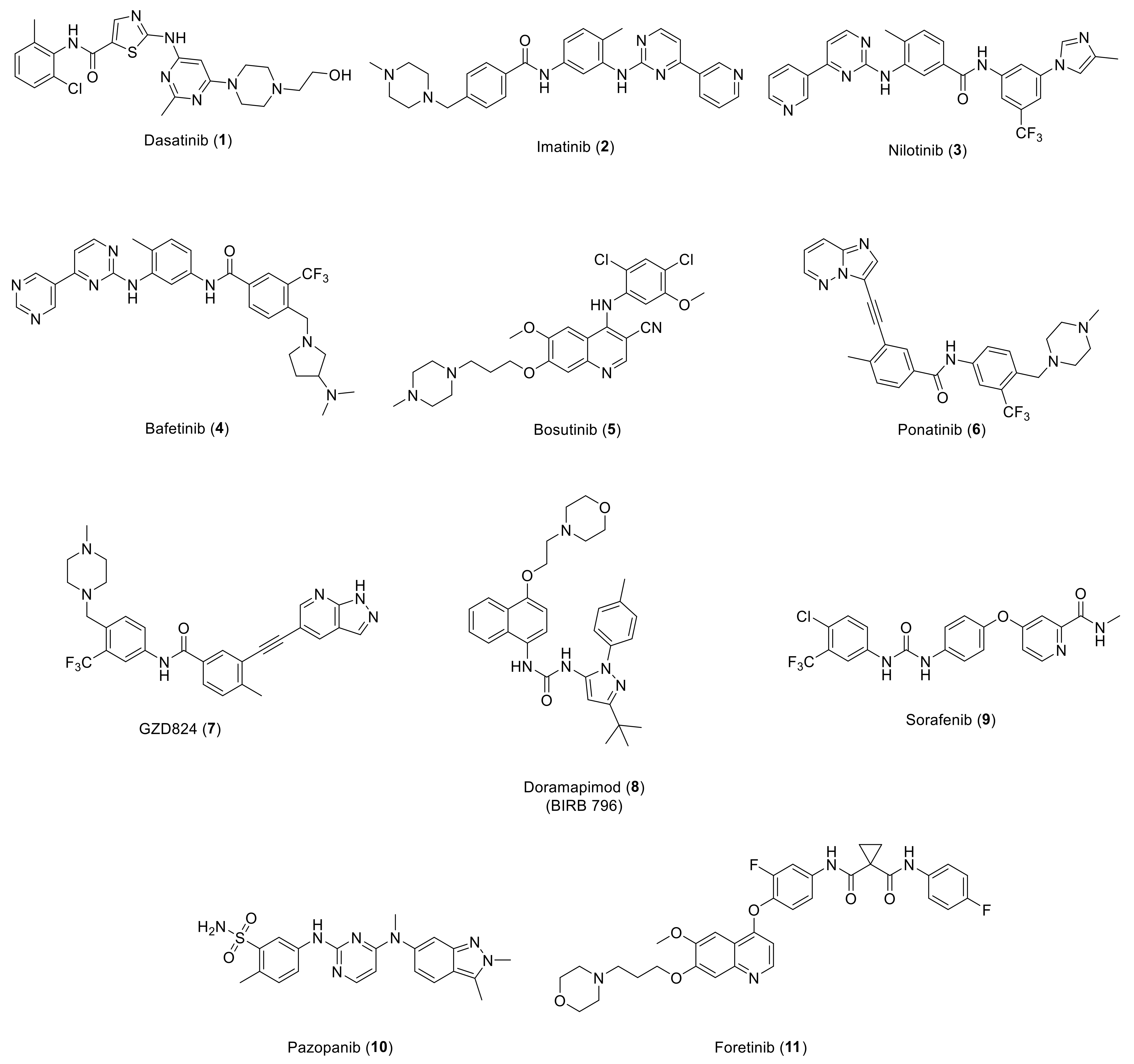
Overexpression of various types of RTKs is found in different types of cancer, which encouraged medicinal chemists worldwide to develop numerous RTKs inhibitors [107,108,109,110,111,112,113,114,115,116,117,118,119,120]. Among these efforts, several DDR kinase inhibitors have been discovered so far, some of them have been demonstrated to possess a promising therapeutic potential. With the aim to reveal the developmental journey of small molecule DDR inhibitors, we selected the most promising potent and/or selective small molecules to present. However, most of the reported DDR1/2 inhibitors have a broad inhibition over several kinases with limited potency. Since the majority of kinase inhibitors bind within the ATP binding region, unintended inhibitor off-target binding can occur due to high structural homology across kinase ATP binding pockets. As a result, a number of Bcr-Abl inhibitors (Figure 2) including dasatinib (1), imatinib (2), and nilotinib (3) were found to inhibit DDR1b kinase and DDR2 with IC50 values of 0.5, 337, 43 nM, and 1.4, 675, 55 nM, respectively [7]. Further research revealed that these three compounds inhibited collagen-induced autophosphorylation of DDR1b and suppressed monocyte chemoattractant protein-1 (MCP-1) release in monocytic cells in a DDR1 dependent manner [7]. A comprehensive drug-protein interaction profiles for compounds 13 confirmed their strong DDR inhibitory effects via a global chemical proteomics approach [121]. Another study on lung cancer cells harboring “gain-of-function” mutations of DDR2 reported compound 1 to demonstrate a highly promising therapeutic efficacy [46]. Moreover, a number of squamous cell carcinoma (SQCC) patients with S768R mutation of DDR2 had substantial tumor shrinkage following treatment with compound 1 [81,122,123,124]. Other type II Bcr-Abl inhibitors, for instance, bafetinib (4), bosutinib (5), ponatinib (6), and GZD824 (7), have also been shown to potently inhibit DDR [125,126,127,128]. The X-ray structures of DDR1 cocrystals containing ligands 2 and 6 show that the compounds bind to inactive DDR1 in a type II binding mode close to that of Bcr-Abl kinase [8]. Numerous other kinase inhibitors were found to show non-selective inhibitory activity over DDR (Figure 2) [129]; doramapimod (BIRB 796, 8), a potent p38 MAPK inhibitor, was reported to bind to DDR1 and DDR2 in highly potent nanomolar range (Kd = 1.9 and 33 nM, respectively). Sorafenib (9), a B-Raf/ VEGFR dual inhibitor approved for advanced hepatocellular carcinoma, was found to bind to DDR1 and DDR2 with Kd values of 1.5 and 6.6 nM, respectively. It was also reported that pazopanib (VEGFR inhibitor, 10) was able to bind to DDR2 with Kd value of 57 nM. Foretinib, another c-Met/VEGFR-2 dual inhibitor (11), was shown to potently bind to DDR1with a Kd value of 0.2 nM.
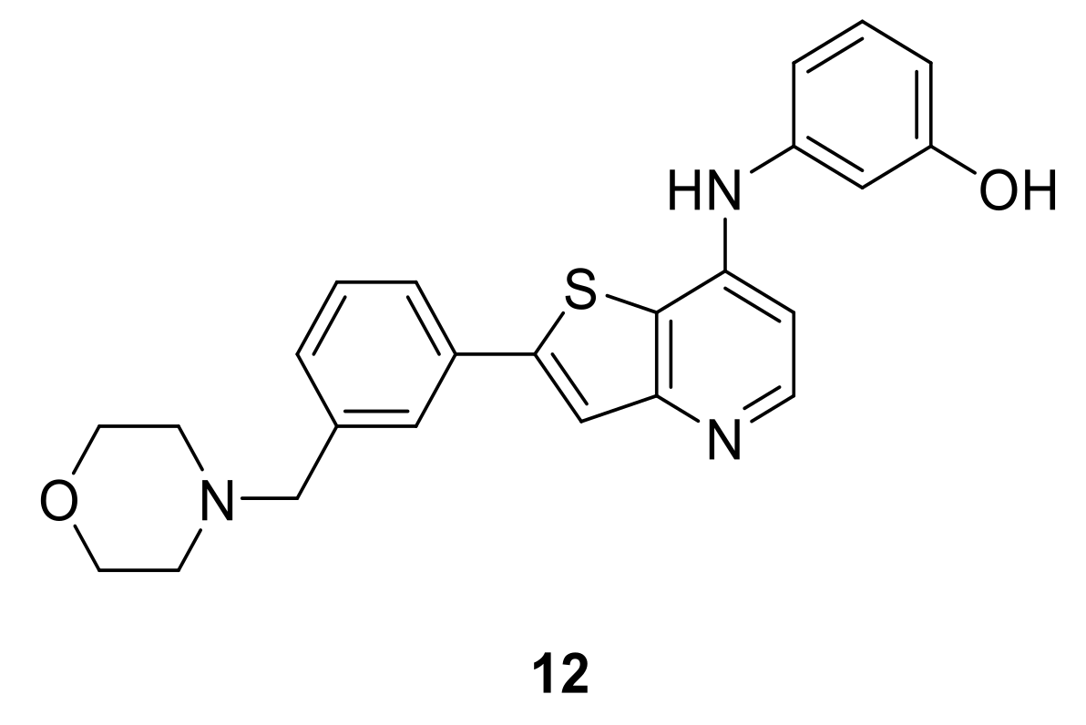
LCB 03-0110 (12, Figure 3) is a thienopyridine derivative that inhibits many tyrosine kinases, including the c-Src family, Btk, VEGFR-2, and DDR kinases, all of which have been implicated in fibroblast and macrophage activation. DDR2 active form was inhibited by LCB 03-0110 at IC50 value of 6 nM, while the non-activated form was inhibited at IC50 value of 145 nM, indicating that LCB 03-0110 is more inhibitory to the active form. The kinetics assay of LCB 03-0110 against the active DDR2 tyrosine kinase revealed that inhibition is ATP-competitive. In a cell-based assay, LCB 03-0110 suppressed DDR1 and DDR2 induced autophosphorylation in HEK293 cells designed to overexpress either HEK293-DDR1b or HEK293-DDR2 with IC50 values of 164 and 171 nM, respectively. Moreover, LCB 03-0110 inhibited more than 90% of 20 tyrosine kinases at 10 µM in a kinase panel assay against 60 kinases, suggesting LCB 03-0110 as a multiple tyrosine kinase inhibitor. In addition, LCB 03-0110 was reported to suppress the proliferation and migration of primary dermal fibroblasts, as well as inhibiting cell migration, and nitric oxide, iNOS, COX-2, and TNF-α synthesis, suggesting its role as a novel anti-fibro-inflammatory agent via suppressing fibro-inflammation by concurrently targeting activated fibroblasts and macrophages [130].
Gray et al., from Harvard Medical School designed a general pharmacophore model for type II kinase inhibitors to develop a library of potential kinase inhibitors [131] which led to the discovery of the potent and selective DDR1 inhibitor DDR1-IN-1 (13, Figure 4A) [132,133]. It was found that DDR1-IN-1 was able to bind to DDR1 in the DFG-out conformation and inhibited DDR1 autophosphorylation in cells at submicromolar concentrations with promising selectivity as assessed over a panel of 451 kinases. While DDR1-IN-1 showed IC50 values of 105 nM against DDR1 and 413 nM against DDR2, its analog DDR1-IN-2 (14, Figure 4A) demonstrated IC50 values of 47 and 145 nM against DDR1 and DDR2, respectively. Despite the high potency of DDR1-IN-2 over DDR1, it also potently inhibits a number of additional kinases. The corrected X-ray cocrystal structure of DDR1-IN-1 with DDR1 kinase (PDB: 4CKR) confirmed the presumed type II binding mode (Figure 4B) where electron density analysis showed that the indolin-2-one group is flipped, forming only a single hydrogen bond to Met704. The DDR1 crystal structure with a modeled G707A mutation indicated the presence of a potential clash between the side-chain methyl group of alanine and the azaindole ring of DDR1-IN-2 (Figure 4C) and predicted that Thr701 (gatekeeper) has the potential to form a hydrogen bond with nitrogen between the “Head” and “Linker” region of DDR1-IN-2, clarifying its ability to more potently inhibit DDR1 relative to DDR1-IN-1. In a mechanistic study comparing the inhibition of DDR1 by different kinase inhibitors by Canning et al., imatinib (2) and ponatinib (6) were able to bind potently to both the DDR and ABL kinases, while DDR1-IN-1 appears to fail to satisfy the hydrophobic interactions of the ABL P-loop, implying a structural basis for its DDR1 selectivity [8].
Takeshi et al., identified some benzamide and quinazolindione analogs as novel DDR1 inhibitors [134,135] (Figure 5). Compound 15 was found to exhibit a high inhibitory activity over DDR1 kinase (IC50 = 0.097 µM) and suppressed the proliferation in U2OS cells with DDR1 overexpression with an IC50 value of 0.44 µM. The conformationally restricted quinazolindione analog 16 was also able to inhibit the kinase activity of DDR1 with a more potent IC50 value (0.043 μM). Interestingly, both compounds exhibited promising in vivo anticancer activity.
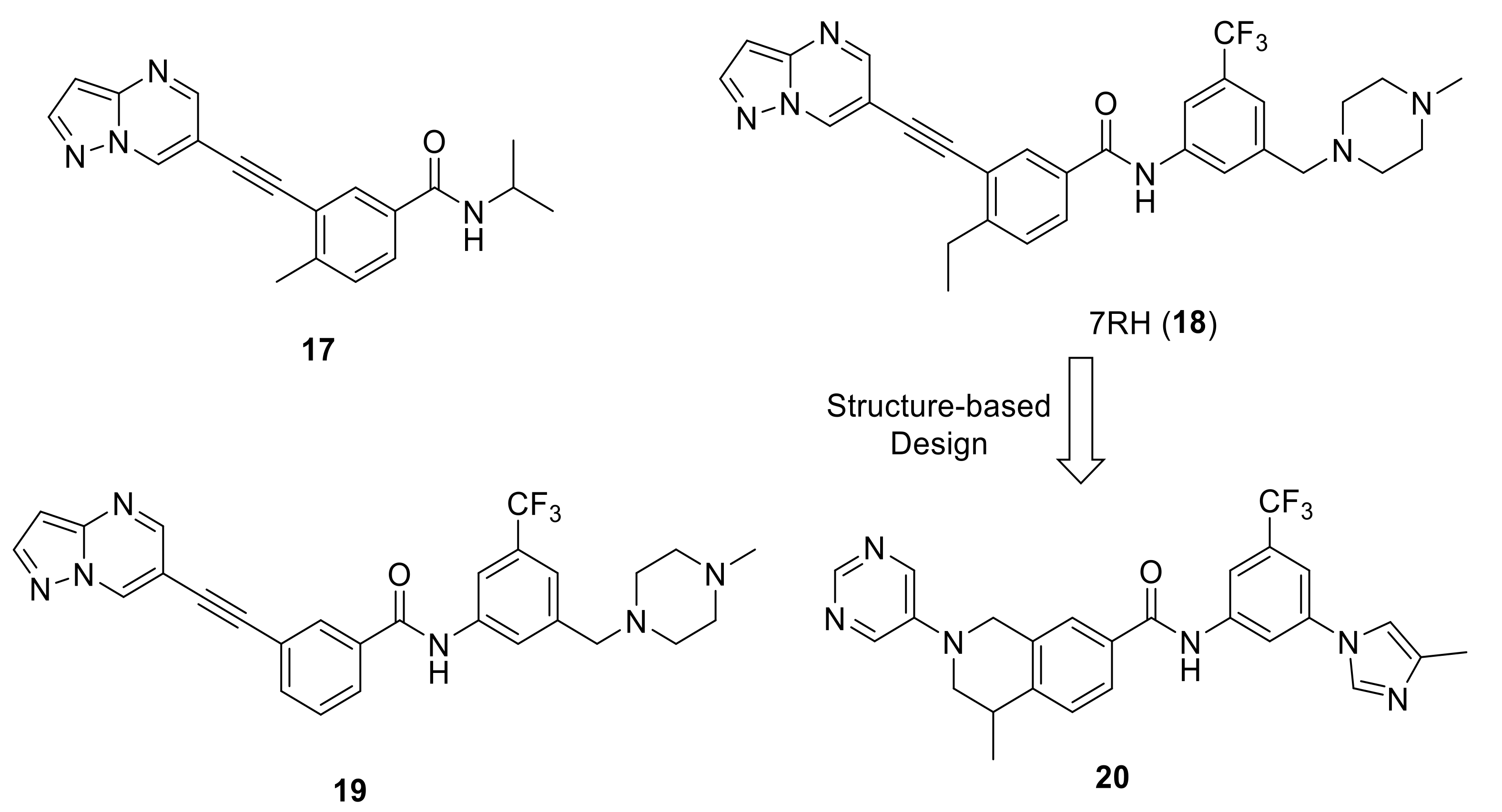
In 2013, Gao et al., reported a new series of 3-(2-(pyrazolo[1,5-a]pyrimidin-6-yl)ethynyl)benzamides as selective DDR1 inhibitors [136]. Compound 17 (Figure 6) was selected as the lead molecule for extensive structural optimization since it demonstrated a promising selective DDR1 inhibition (IC50 = 39.6 nM). All the synthesized derivatives were tested for their inhibitory activity against four kinases (DDR1, DDR2, Bcl-Abl and c-Kit). Among all, only compounds 18 (7RH) and 19 (Figure 6) showed potent selective activity against DDR1 with IC50 values of 6.8 and 7.0 nM, respectively. Compound 7RH was able to interact with the ATP-binding site of DDR1 with a Kd value of 0.6 nM. It also showed high selectivity for DDR1 upon its screening over a large kinase panel of 456 kinases. Both compounds (18 and 19) demonstrated low IC50 values over NCI-H23 NSCLC cell line expressing high level of DDR1 and promising oral bioavailability (67.4% and 56.2%, respectively). Three years later, Lu et al., reported the antitumor activity of 7RH alone or in combination with dasatinib in nasopharyngeal carcinoma (NPC) [137]. 7RH demonstrated cytotoxicity in CNE2, HONE1, CNE1 and SUNE1 (NPC cell lines) with IC50 values of 1.97, 3.71, 2.06, and 3.95 µM, respectively. It also reduced protein expression levels, induced cell cycle arrest and apoptosis of CNE2 cells, and inhibited NPC cell adhesion at different concentrations. 7RH and dasatinib were found to exhibit a synergistic inhibitory effect over human NPC cell proliferation. 7RH, dasatinib and 7RH + dasatinib groups showed 27, 28 and 33% in vivo tumor growth inhibitory rates, respectively.
Using compound 7RH (18) as a lead compound, a structure-based design of new tetrahydroisoquinoline-7-carboxamide derivatives as selective DDR1 inhibitors was reported by Wang et al. [138]. Among 11 target derivatives assessed for their inhibitory activity against two kinases (DDR1 and Abl1), compound 20 (Figure 6) was the most promising compound over DDR1 (IC50 = 9.4 nM and Kd = 4.7 nM). Moreover, remarkable pharmacokinetic (PK) properties were demonstrated with an oral bioavailability of 66.8%. To examine the target specificity of compound 20, a kinase selectivity profiling analysis was performed at 1.0 M against a panel of 468 kinases (including 403 nonmutated kinases). The outcomes demonstrated an outstanding target selectivity. In addition, it exhibited an inhibitory effect of DDR1-mediated signaling in a concentration dependent manner in PHLF (Primary Human Lung Fibroblast) and showed a blocking effect in BLM-induced lung fibrosis. The high potency along with the unique target specificity of compound 20 presents a great opportunity for biological target investigations and new drug discovery programs.
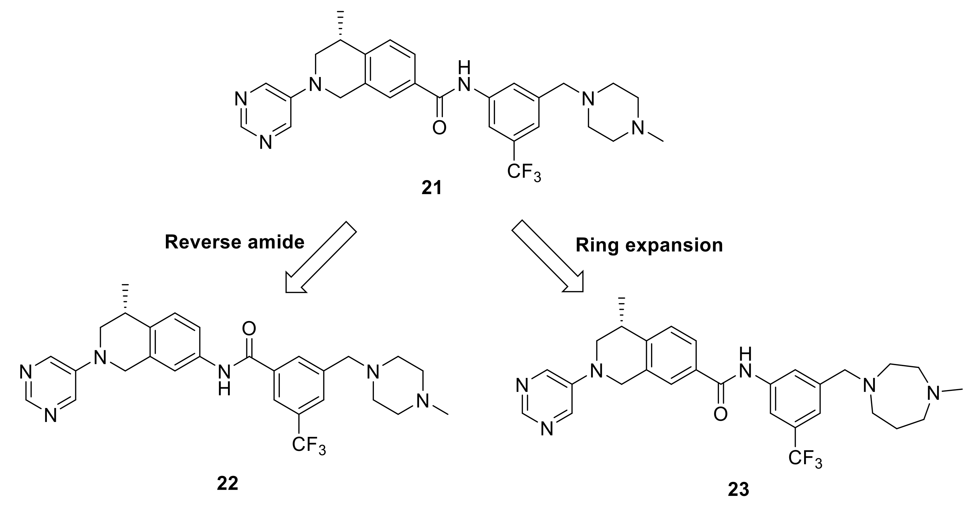
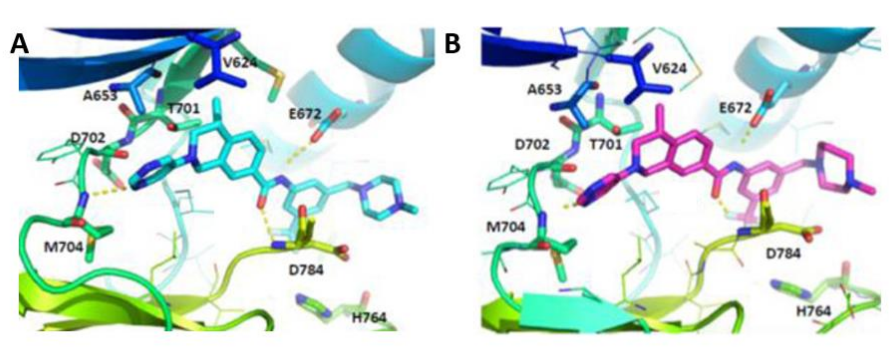
As a continuation of their research, Wang et al., optimized the tetrahydroisoquinoline-7-carboxamide-based scaffold [139]. Compound 21 [138] (Figure 7) was chosen as a lead molecule for optimization followed by an extensive SAR investigation. It was found that the trifluoromethylphenyl group in 21 was able to bind to the hydrophobic pocket formed by the DFG-out conformation of DDR1 (Figure 8A). The replacement of the trifluoromethylphenyl group with cyclopropyl or cyclohexyl substitutes, or its removal resulted in compounds with an obvious loss of potency, indicating that a lipophilic substitution at this position is critical to retain the potent DDR1 inhibition. The modeling studies also revealed that the amide linker in 21 bound deeply with Glu672 in the C-helix and Asp784 in the DFG motif by forming two pairs of hydrogen bonds. With the aim to evaluate of the contribution of these hydrogen bonds to DDR1 inhibition, compound 22 (Figure 7) with a reverse amide was made and evaluated. It turned out that compound 22 displayed a three-fold less potency than that of 21. The reversed amide linker forced the trifluoromethylphenyl group to moderately rotate away from the C-helix, leading to the longer distances between amide moiety and the corresponding Glu672 and Asp784 residues with values of 2.5 and 2.3 Å. Accordingly, the hydrogen bond between 22 with the corresponding amino acids might be weaker than that of 21, which explains the potency loss of compound 22.
The most active compound in this series (23, Figure 7) with 1-methylhomopiperazinemethyl substituent was discovered to tightly bind to DDR1 protein (Figure 8B) with a Kd value of 2.2 nM and IC50 value of 6.6 nM. Compound 23 demonstrated a promising target specificity and suppressed LPS-induced interleukin-6 (IL-6) and TNF-α release in a dose-dependent manner suggesting it as a promising in vivo anti- inflammatory agent. Furthermore, it also inhibited the LPS-induced increase in total cell number and total protein concentration in bronchial alveolar lavage fluid (BALF). Accordingly, compound 23 was found not only to be a potent DDR1 inhibitor, but also a potential lead candidate for anti-inflammatory drug discovery.
Wang et al. also recently described a new series of dual DDR1/2 inhibitors (some of which are illustrated in Figure 9, 2428) based on their starting lead 7RH (18). A series of 18 compounds were obtained as selective dual DDR1/2 inhibitors [140]. Among them, compound 28 turned out to be one of the most specific DDR1/2 dual inhibitors to-date, with IC50 values of 9.4 and 20.4 nM and Kd values of 7.9 and 8.0 nM, respectively. It also exhibited a dose-dependently inhibitory effect on LPS-induced IL-6 release in mouse primary peritoneal macrophages (MPMs) and attenuated the lung inflammation in LPS-treated mice. It is worthy to mention that this study was the first in vivo investigation to find selective dual inhibitors (DDR1 and DDR2) as novel anti-inflammatory candidates.
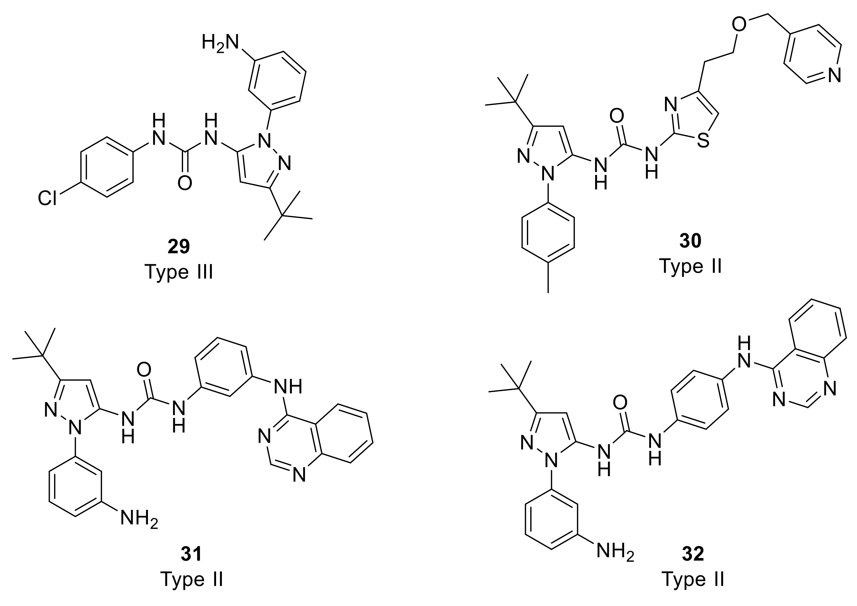
Richters et al. designed a fluorescence-based direct binding assay for the identification of DDR2 type II and III DFG-out binders [141]. While a total number of 852 compounds from internal library were screened, seven candidates including compound 29 (Figure 10) belonging to type III-like inhibitors were found to bind tightly to DDR2 with Kd values of 0.015–0.702 μM. However, they exhibited moderate IC50 values of 0.183–4.70 μM. On the other hand, type II-like analogs (30, 31, and 32, Figure 10) showed excellent binding effects with Kd values of 0.020–0.063 μM and potent IC50 values of 0.008–0.029 μM. Type II-like molecules also exhibited inhibitory effect on DDR1 with IC50 values 0.039–0235 μM. In addition, the inhibitory effect of compound 32 against the gatekeeper mutations was found to be conserved, for instance, it exhibited an IC50 value of 0.002 μM against DDR2 (T654M). Accordingly, compound 32 was suggested to offer a future approach towards the development of novel inhibitors that can overcome the potential resistant variants of DDR.
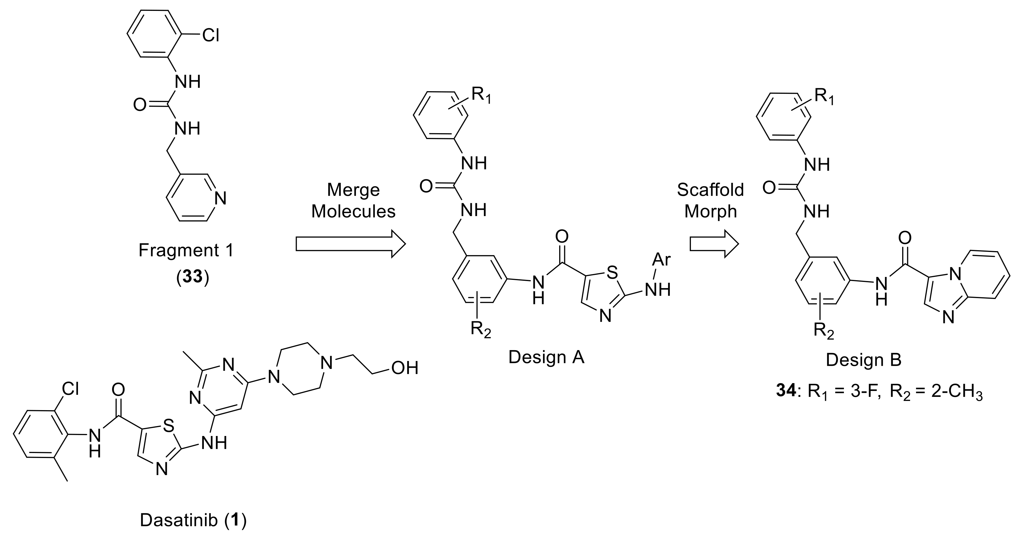
Murray et al., in Astex Pharmaceuticals Co. screened ~1500 small molecules from a fragment library against DDR1 using a protein thermal shift assay. They discovered several compounds that were able to bind at the hinge or in the back pocket associated with the DFG-out conformation of DDR1, such as the multitargeted kinase inhibitor dasatinib (1). Compound 33 (Fragment 1, Figure 11) possessing a chlorophenyl moiety placed in the back region and a pyridyl group in the selectivity pocket proximal to the small gatekeeper residue (Thr701 in DDR1/2, Figure 12A), was selected as the starting fragment for the computational tool, AstexMerge. Accordingly, design A (Figure 11) was suggested by AstexMerge, based on the superimposition with dasatinib (Figure 12B). Further modification of design A by replacing the thiazole hinge binder of dasatinib with imidazopyridine led to design B (Figure 11). Using this fragment-based drug design, novel DDR1/2 inhibitors were generated [142].
All resulting compounds exhibited potent DDR2 inhibition activity in cells and displayed promising PK properties. The SAR study showed that the imidazopyridine motif improved DDR2 affinity and inclusion of methyl group in the selective pocket region in design B demonstrated a 30-fold increase in affinity. The best compound (34, Figure 11) showed an IC50 value of 3.3 nM towards DDR2 and an IC50 = 54% at 1.5 nM towards DDR1. Compound 34 inhibited a number of tyrosine kinases and was particularly effective against c-kit (IC50 = 19 nM) and significantly reduced basal and collagen I-stimulated DDR2 phosphorylation in both wild type DDR2- and mutant DDR2-expressing HEK293 cells. The binding mode of 34 superimposed on the starting fragment 33 inside DDR1 active site (Figure 12C) showed that the imidazopyridine was able to form essential hydrogen bonds with the hinge region.
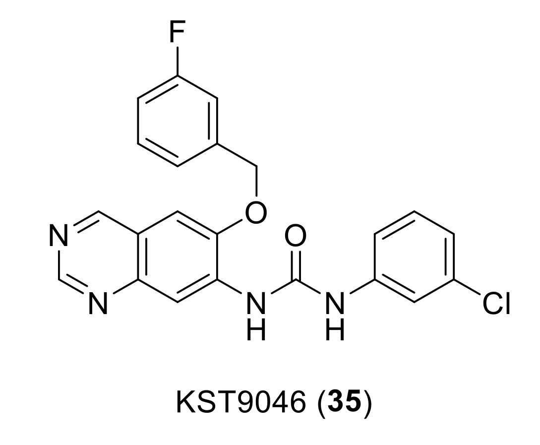
Compound KST9046 (35, Figure 13) is a recently reported DDR1 inhibitor with 6,7-disubstituted quinazoline-urea scaffold discovered by Elkamhawy et al., from Korea Institute of Science and Technology (KIST) [143]. Since 6,7-disubstitution of quinazoline ring is not common for kinase activity, a unique selectivity for DDR1 was found when the compound was checked over a large panel of 347 different kinases; the screening results showed a remarkable selective inhibitory activity over DDR1 kinase with IC50 of 4.38 µM. KST9046 was identified as a possible type III inhibitor for DDR1 kinase with an allosteric mode of interaction, which could explain its selectivity. A broad-spectrum activity of compound 35 with GI50 ranging from 1.3 to 3.9 µM over the 60 cell-line tumor panel of National Cancer Institute (NCI). Moreover, a promising low toxicity profile against four different isoforms of CYP450 was presented. Thus, compound 35 was reported as a promising lead for the development of broad-spectrum DDR1 selective antiproliferative candidates.
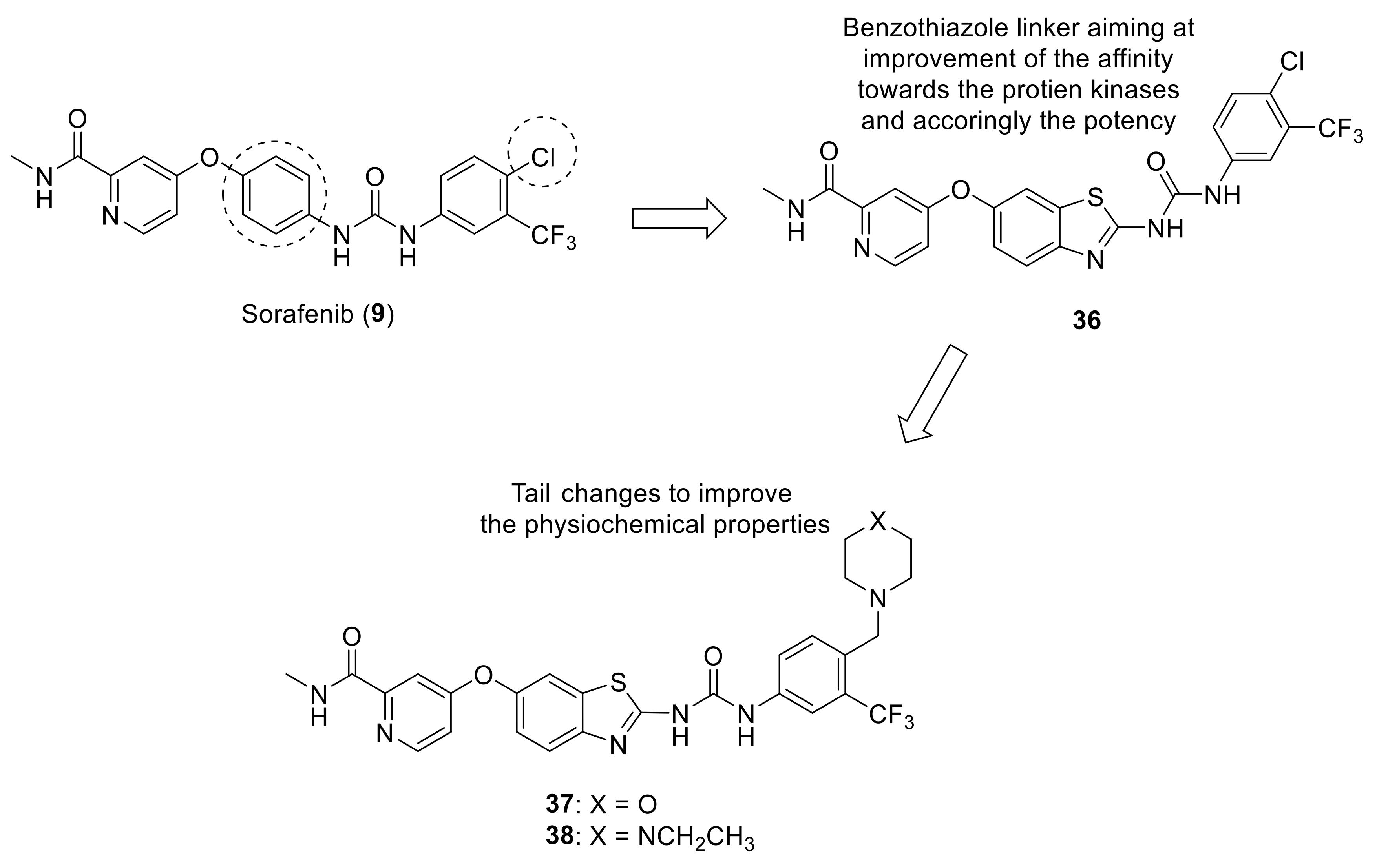
Recent studies from KIST by El-Damasy et al., attempted modifications to sorafenib (9) in order to increase its activity as a multi-target kinase inhibitor and derive more potent analogs. Using structure-based design strategy, the central phenyl linker of sorafenib was replaced with either quinoline moiety [144] or benzothiazole scaffold (36, Figure 14) [145] while maintaining the other structural features. This resulted in a remarkable increase in the cellular antiproliferative potency as well as favorable inhibitory activity toward B-Raf (V600E) and C-Raf kinases. However, the physiochemical properties needed further improvement, which was carried out through replacing the small lipophilic chlorine atom with either (morpholin-1-yl)methyl (37, Figure 14) or (4-ethylpiperazin-1-yl)methyl moieties (38, Figure 14) [146]. These two compounds were evaluated for their antitumor effects over a panel of 60 human cancer cell lines where they exhibited promising antiproliferative effects over numerous cell lines such as HL-60 (leukemia), HOP-92 (non-small cell lung cancer), HCT-116 (colon cancer), SF-5339 (CNS cancer) and SK-MEL-28 (melanoma). Both compounds were also subjected to further testing against a panel of 50 oncogenic kinases, where compound 38 potently inhibited 23 kinases including ABL-1, LYN, DDR1, and DDR2. Additionally, when compound 38 was tested against the human foreskin fibroblast (HFF-1) normal cell line, it demonstrated a relatively safe cytotoxicity profile (10.36 ± 0.19% inhibition). Based on these data, the promising inhibitory activity of compound 38 over DDR1 coupled with its safe cytotoxicity presented an ideal candidate for further development to enhance its selectivity against DDRs.
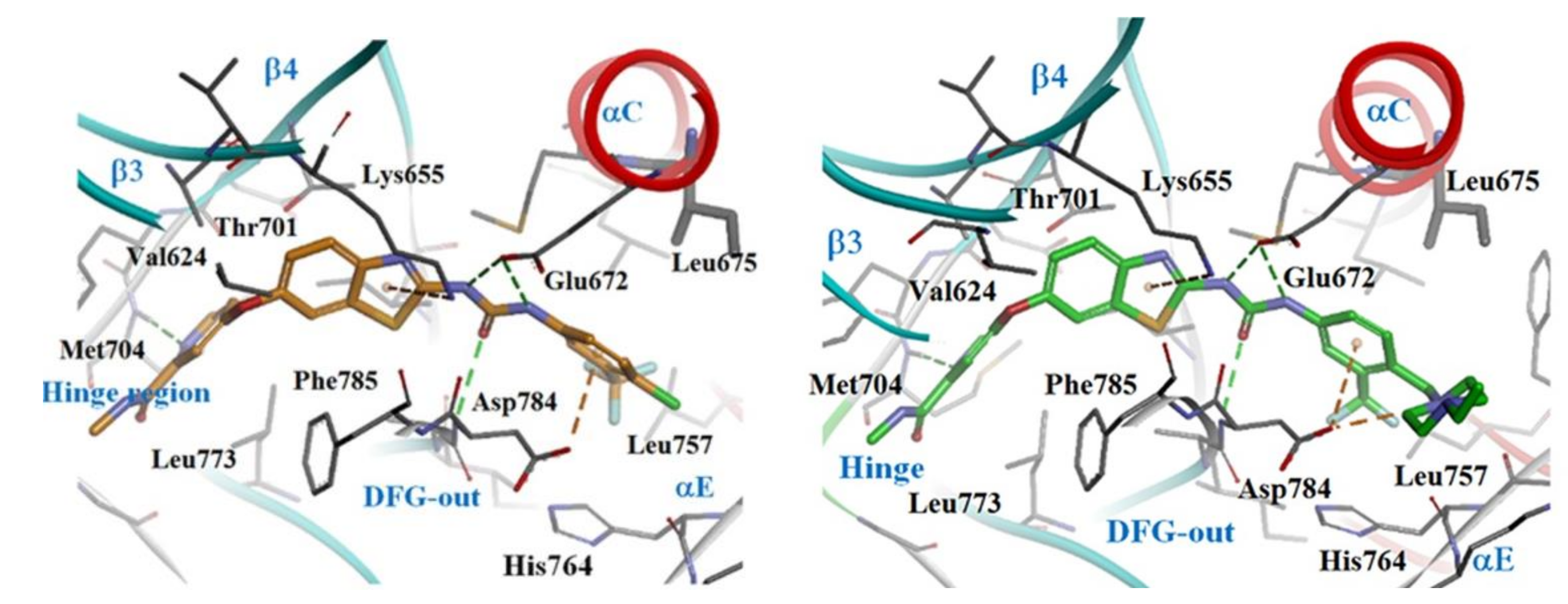
To understand the elicited kinase inhibitory activities of both compounds (36 and 38), a molecular docking study was carried out as demonstrated in Figure 15. The hydrogen bond formed by both compound 36 and 38 with the Met704 residue in the kinase hinge region via their pyridine nitrogen into the ATP binding site of DDR1 was found to be a large contributing factor to their activity, making the pyridine moiety essential for activity. Similarly, the urea moiety proved to be essential for activity through its ability to form three hydrogen bonds with Asp784 and Glu672. Additionally, the m-trifluoromethylphenyl group of 36 and 38 was predicted to be essential for activity as well through its ability to form hydrophobic interactions with the backbone Asp784 residue in the DFG region.
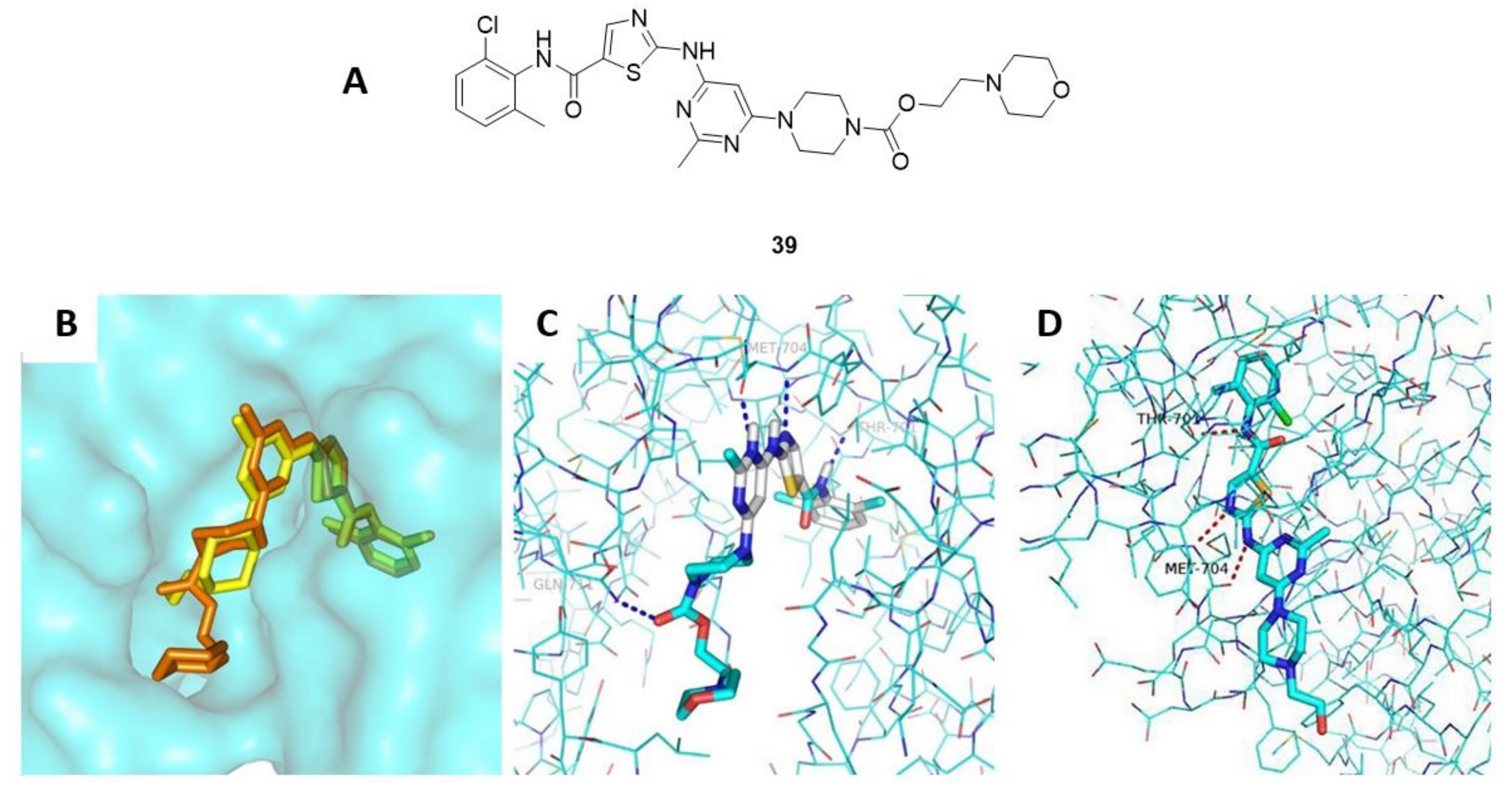
In another study, Liu et al., synthesized novel dasatinib derivatives with potent DDR1 and DDR2 inhibitory activities [147]. One of the synthesized compounds, compound 39 (Figure 16A), compared with the parental dasatinib (1), demonstrated a considerably superior inhibitory potency over both DDRs and K562 cell lines (IC50 values of 2.26 ± 0.46 nM for DDR1, 7.04 ± 2.90 nM for DDR2, and 0.125 ± 0.017 nM for K562 cell line). Upon performing a comparative docking study for both the parental dasatinib (1) and compound 39 over DDR1 kinase (Figure 16B–D), they had almost the same estimated binding free energies of −9.7 and −9.6 kcal/mol, respectively. This suggested that the addition of the morpholine ring may have improved PK of the parent dasatinib compound leading to increased biological activity. Due to the potent activity of compound 39, it could also be a promising therapeutic agent itself for additional assessments in a variety of disease models related to pathological DDR1 or DDR2 activities.
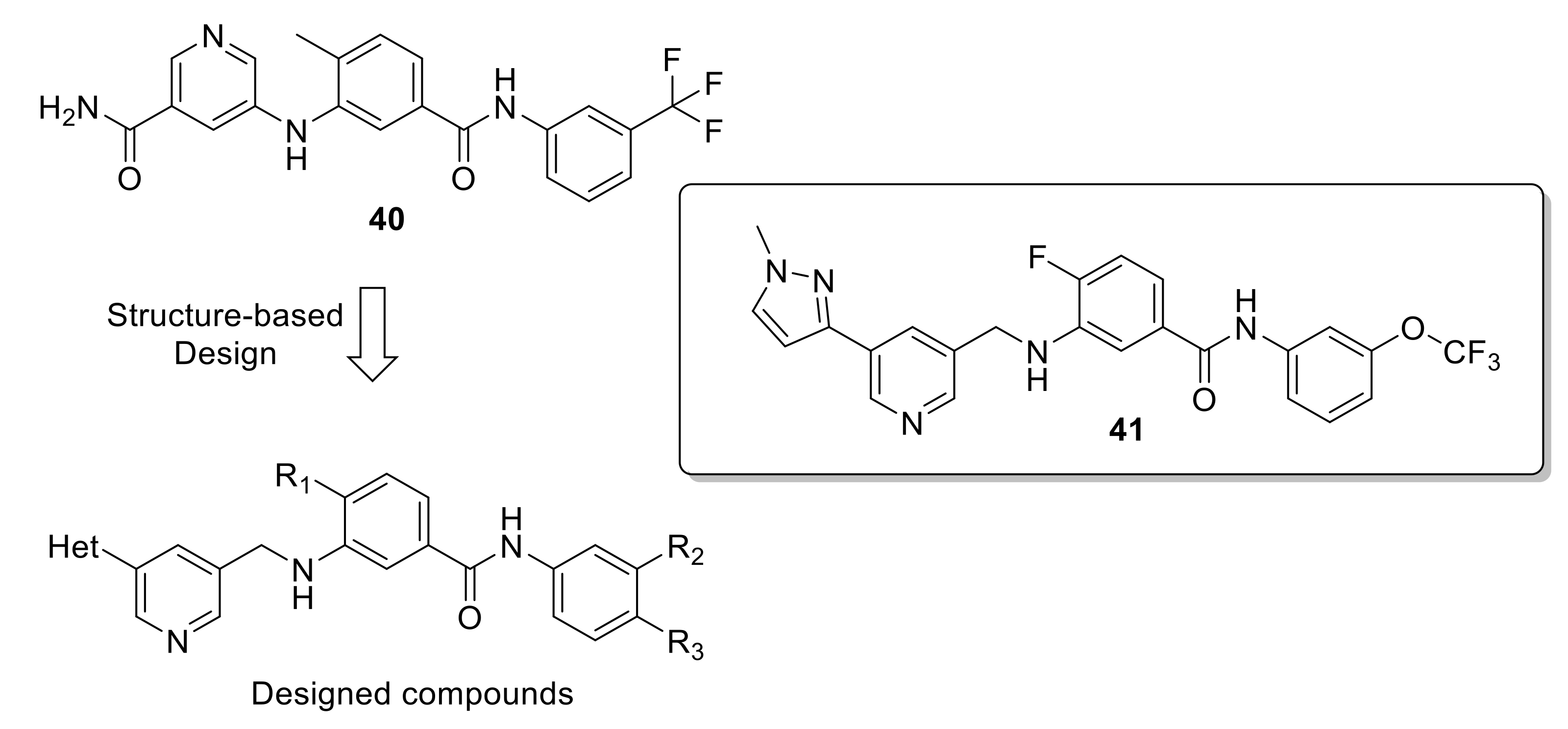
Jeffries et al., also attempted to synthesize a selective DDR1 inhibitor [148] through using compound 40 (Figure 17) as starting lead, which was initially identified as a selective DDR2 inhibitor (IC50 = 18.6 nM), although it inhibited DDR1 with an IC50 value of 12.4 nM [149]. Despite the high potency of compound 40 against DDR1, it demonstrated a poor drug metabolism and PK profile, as well as cytotoxicity at low concentrations. Therefore, Jeffries et al., modified its chemical structure as illustrated in Figure 17. However, these efforts failed to meet its original goal of synthesizing a selective DDR1 inhibitor. Instead, they produced compound 41 (Figure 17) which was proved to be a dual DDR1/2 inhibitor, with improved potency compared to compound 40 (DDR1 IC50 = 4.67 nM, DDR2 IC50 = 7.39 nM). This coupled with the fact that compound 41 showed enhanced binding and cell-based potency, augmented physiochemical and PK properties, make it an ideal candidate for further development.
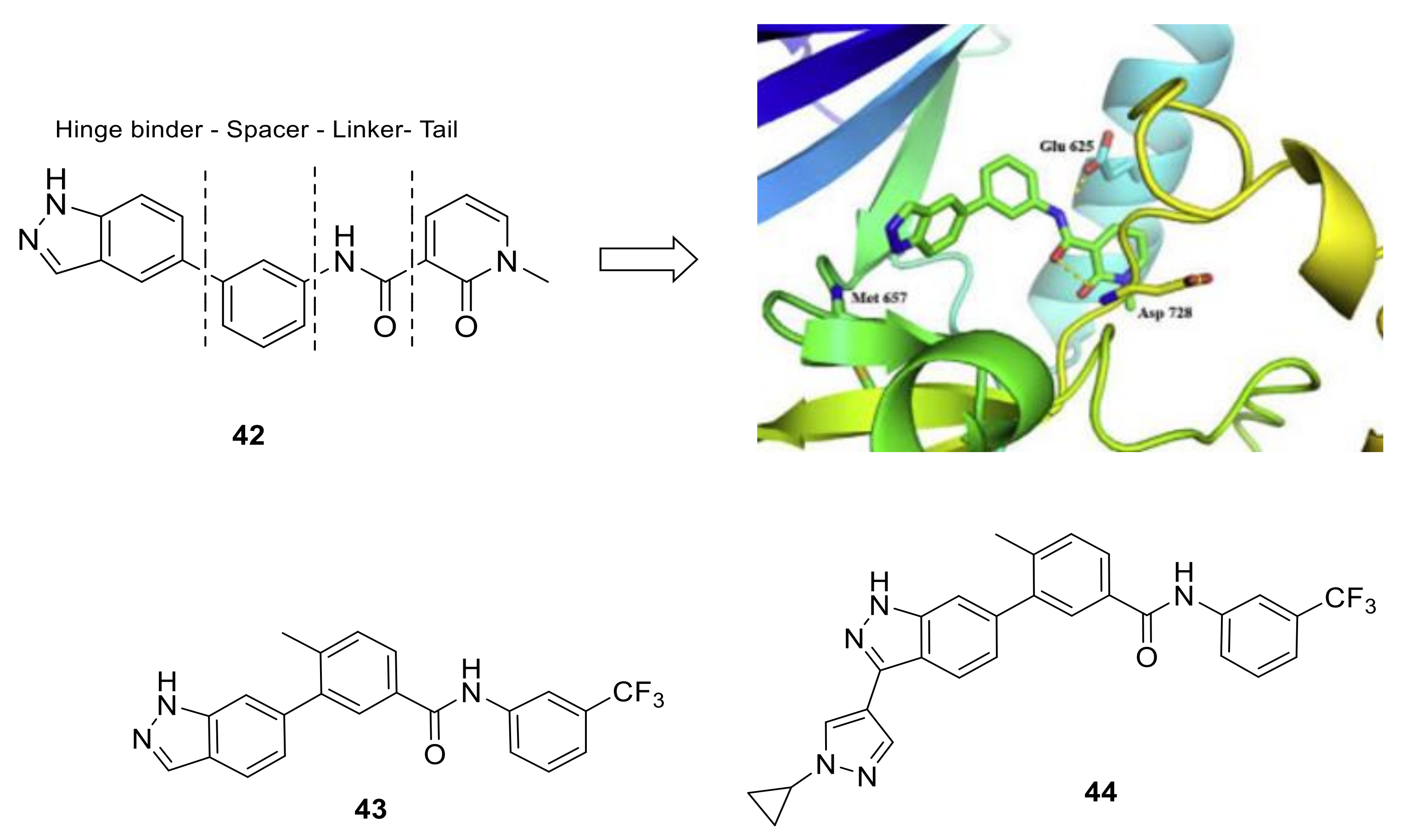
Wang et al., discovered and optimized a novel series of 3-substituted indazole analogs as non-selective kinase inhibitors for lung squamous cell carcinoma [150]. Hit compound 42 (Figure 18), found by in-house library, showed moderate inhibitory activity in DDR2 inhibition assay. Through the docking prediction, compound 42 combined with hinge segment of DDR2 as a typical Type-II DFG-out kinase inhibitor (Figure 18) where nitrogen atoms in the indazole ring and the hinge segment of DDR2 formed essential hydrogen bonds. In addition, the interaction with nearby residues Glu625 and Asp728 has been enhanced by the amide group. When SAR optimization was carried out over the four parts of compound 42 (Hinge binder, Spacer, Linker and Cap part, Figure 18), compound 43 (Figure 18) was obtained with an IC50 value of 1.5 ± 0.1 nM against DDR2. A further optimization afforded four compounds with enhanced biological activity, among them, compound 44 (Figure 18) exhibited remarkable PK properties with a good exposure and moderate half-life of 3.05 h. In addition, 44 also showed a certain selectivity in in vitro enzymatic screen assays. Compound 44 also inhibited DDR2 phosphorylation and suppressed tumor growth with tumor growth inhibition rates (TGI) of 82.8% in mice bearing NCI-H2286 (DDR-dependent cell line) at doses of 10 mg/kg for 10 consecutive days. According to this study, compound 44 may encourage further research on drug development of lung squamous cell carcinoma.
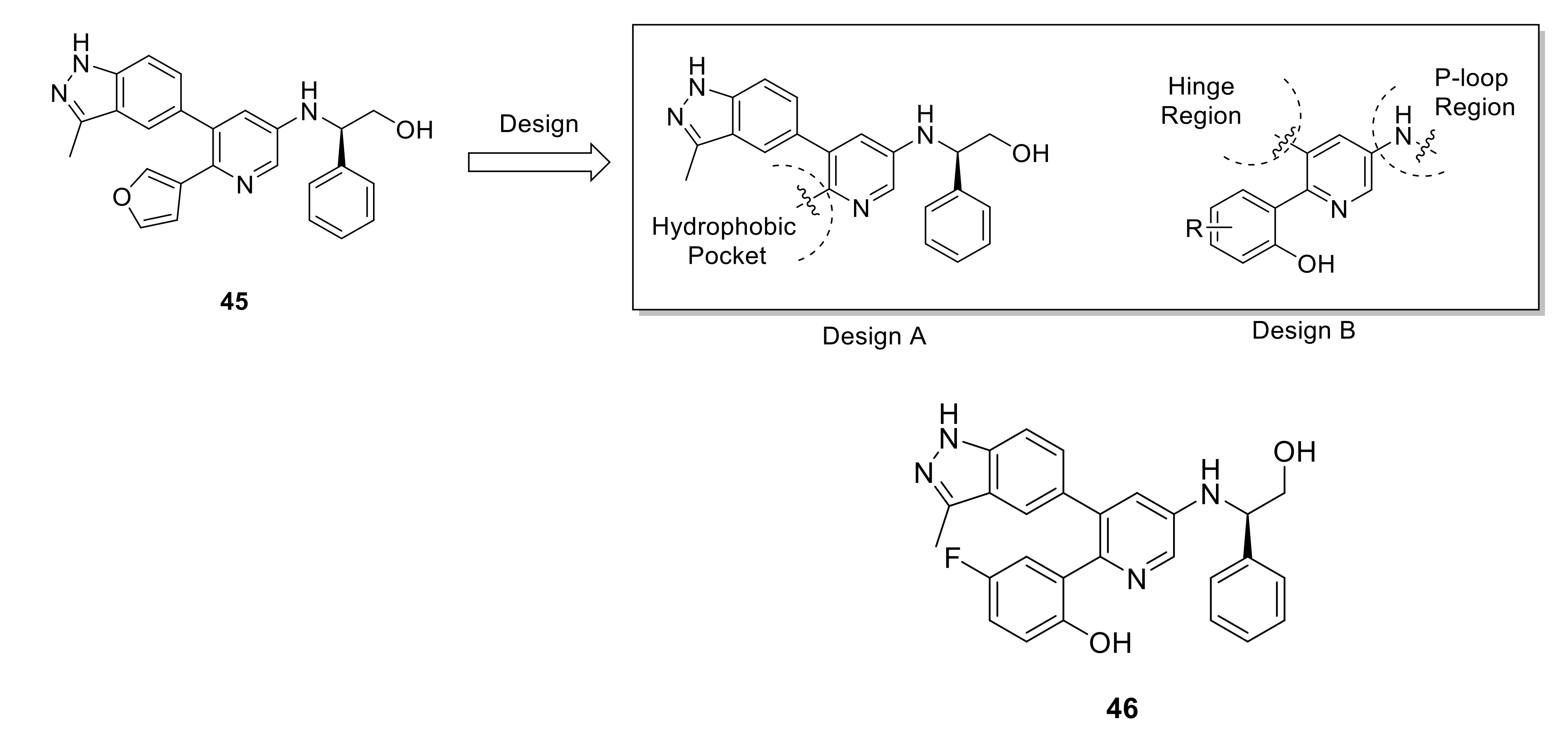
Zhu et al., reported a novel series of pyridin-3-amine derivatives as protein kinase inhibitors for NSCLC treatment [151]. As demonstrated in Figure 19, hit optimization of compound 45 led to SAR exploration of the new pyridine-3-amine derivatives. Among all synthesized compounds, compound 46 demonstrated nanomolar IC50 values for several NSCLC-related oncogene kinases including FGFR1–3 (18.0, 1.6, and 27.5 nM, respectively), RET (0.2 nM), KDR (1.7 nM), EGFR/T790M/L858R (6.0 nM), DDR2 (0.8 nM), and ALK 101.1 nM). In cell assay over SNU16 cell line, it exhibited an IC50 value of 24.8 nM. Furthermore, compound 46 demonstrated a strong antitumor efficacy (TGI = 66.1%) in NCI-H1581 NSCLC xenografts with a favorable PK profile in vivo. Despite the low selectivity ratio of compound 46, its high potency against DDR2 kinase and its good PK properties are two main factors that could be taken into consideration for further development of novel DDR2 inhibitors based on the pyridin-3-amine scaffold.
Using a functional signature-based ontology map approach to identify the signaling pathway/molecular target of natural products, Hu et al., discovered a family of alkaloid natural products (discoipyrroles A–D, 4750, Figure 20) from Bacillus hunanensis possessing strong inhibitory activity against DDR2-dependent migration of BR5 fibroblasts [152]. Discoipyrroles A–D exhibited potent and selective cytotoxicity toward DDR2 mutant lung cancer cell lines with a range of IC50 values of 120–400 nM. Three years later, due to their potent activity against DDR2, Zhang et al., attempted various methods to synthesize these naturally occurring compounds [153].
Recently, while Zhu et al., discovered a new series of 2-amino-2,3-dihydro-1H-indene-5-carboxamides as selective DDR1 inhibitors using a structure-based design strategy based on compound 20 [154], their efforts generated compound 51 (Figure 21) which showed a unique ability to bind to the ATP-binding site of DDR1 with a Kd value of 5.9 nM and an IC50 value of 14.9 nM. In addition, compared to 20, compound 51 had slightly lower potencies against all off-target kinases and effectively inhibited collagen-induced activation of DDR1 as well as its downstream signaling proteins. Compound 51 also inhibited DDR1-mediated cadherin switching, suppressed colony development of pancreatic cancer cells, exhibited good PK properties, and displayed a promising therapeutic activity through oral administration in orthotropic syngeneic pancreatic cancer models.
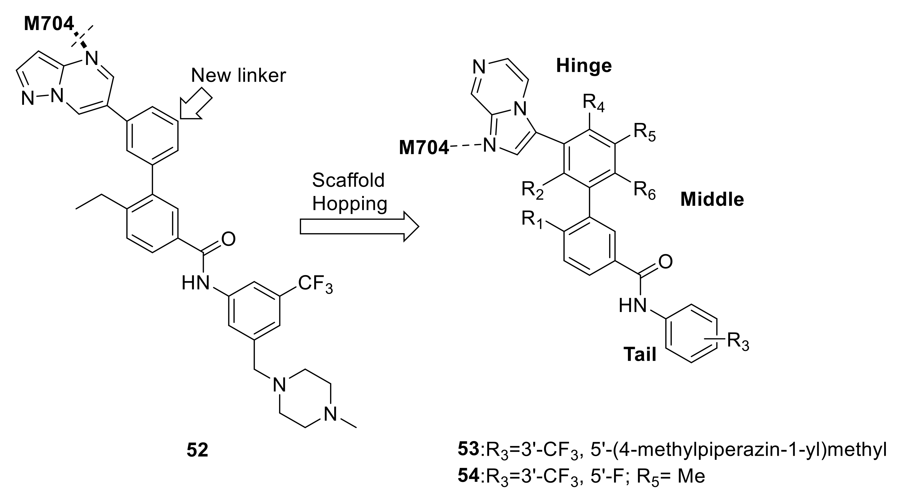
Mo et al., also recently designed a new carboxamide series as selective DDR1 inhibitors via incorporating a novel substituted phenyl linker in place of the alkyne group in compound 7RH (18) [155]. Although the preliminary design led to a higher selectivity for DDR1 kinase by compound 52 (Figure 22), there was a 50-fold loss of activity against DDR1. Modeling studies suggested that the reason for the activity loss was that compound 52 was not able to fit tightly into the DDR1 ATP binding pocket and could not achieve the essential hydrogen bond with Met704. Thus, imidazo[1,2-a]pyrazine moiety was used to replace the pyrazolo[3,4-b]pyridine to restore the key hydrogen bond with Met704. Further optimizations led to compound 53 (Figure 22) which exhibited excellent inhibitory activity against DDR1 with an IC50 value of 9.6 nM, but its selectivity against other homologous kinases was decreased. Modeling simulations showed that compound 53 bound to DDR1 with a classical type II binding mode. The best compound (54, Figure 22) displayed potent inhibitory activity against DDR1 with an IC50 value of 23.8 nM with an extraordinary target selectivity. Moreover, the in vivo study revealed that compound 54 suppressed colony formation, migration, and invasion of H1299 NSCLC.
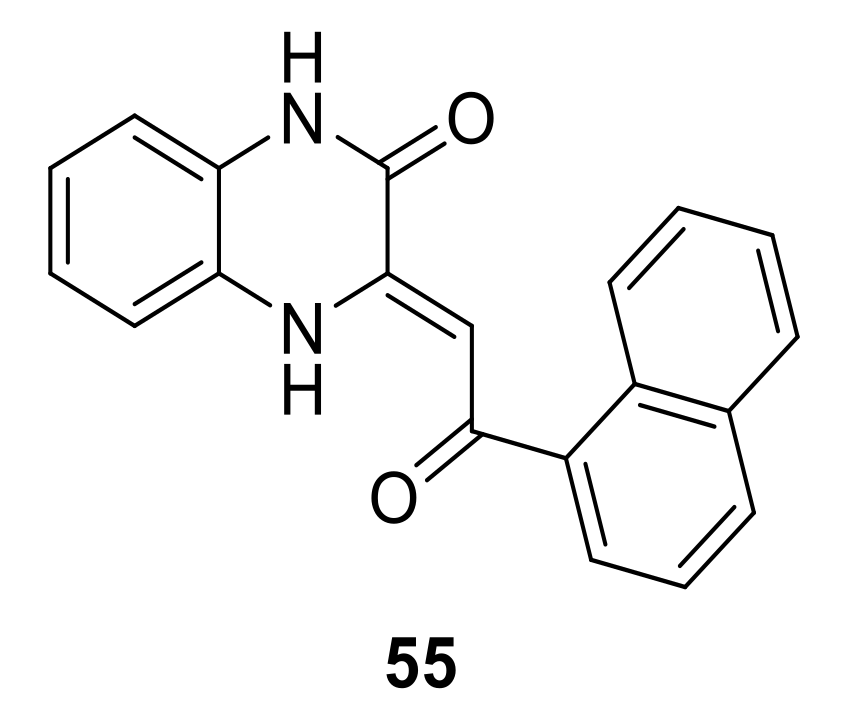
During the research for development of c-Jun N-terminal kinase 3 (JNK3) new inhibitors, Dou et al., discovered that a potent JNK3 inhibitor (55, Figure 23) possessing a 3,4-dihydroquinoxalin-2(1H)-one core structure displayed an interesting DDR1 inhibition activity (IC50 = 0.16 µM) [156]. The modeling study of compound 55 into the active site of DDR1 indicated that the oxygen atom in the quinoxaline core and hydrogen atom of NH formed two hydrogen bonds to the NH and backbone carbonyl of Met704. Thus, further optimization of compound 55 may lead to the development of more potent and selective quinoxaline-based DDR1 inhibitors.
Lately, Dong et al., reported the synthesis and evaluation of new 4-amino-1H-pyrazolo[3,4-d]pyrimidin derivatives as DDR1 inhibitors [157]. Compound 56 (Figure 24) which showed the most potent DDR1 inhibitory activity (IC50 value = 0.044 µM) exhibited potent inhibition against cell proliferation in HCT-116 and MDA-MB-231 cell lines (IC50 = 4.00 and 3.36 μM respectively). The conducted modeling studies (Figure 24) revealed that it was able to bind into DDR1 kinase active site via four hydrogen bonds, two π-π stacked interactions, electrostatic interactions, and various Van der Waals interactions.
Another series of potent and selective DDR1 inhibitors were synthesized and reported by Richter et al. They employed a parallel DNA encoded library screening against DDR1 and DDR2 to uncover a series of small molecules which exhibited a potent selectivity for DDR1 over DDR2. Figure 25 depicts the structural optimization process utilized to reveal compound 59 (DDR1 IC50 = 0.029 µM and DDR2 IC50 = 1.9 µM) starting from the lead compound 57 (DDR1 IC50 = 1.4 µM and DDR2 IC50 > 167 µM) followed by compound 58 (DDR1 IC50 = 0.016 µM and DDR2 IC50 = 6.3 µM). Compound 59 possessed a remarkable kinome selectivity coupled with promising in vitro safety profile, PK, and physicochemical properties. Richter et al., through a series of structural modifications not only managed to enhance the potency and selectivity of the final compound 59 but also increase the metabolic stability of the synthesized compound, with a clearance rate of 10 µL/min/mg in both mouse and human [158].

7. Conclusions

Throughout this review, we discussed the biological roles DDRs, their relation to different human diseases, mainly for cancer, and the medicinal chemistry approaches used in the journey of development of DDR1 and DDR2 kinase inhibitors since the discovery of these targets. Since great efforts have been made in this area of research, a number of selective and potent DDR1 and DDR2 inhibitors have been reported and discussed in this review. Furthermore, the available molecular docking studies allowed us to predict that a potent DDR1 inhibitor must contain certain structural features. One such essential structural feature is an amide linker that facilitates the formation of a hydrogen bond with GLU672 and an additional hydrogen bond with the backbone NH of ASP784. A moiety able to form hydrogen bonding with the “gate keeper” amino acid THR071 is another essential feature a potent and selective DDR1 inhibitor must possess. The ability of the synthesized compound to form a hydrogen bonding with MET704 as well as containing a hydrophobic moiety in its “tail” are two structural features that are predicted to further increase the binding affinity of any synthesized molecule with the DDR1 active site residue.
In addition to the classical methods of drug design, many researchers are currently attempting to apply new strategies to develop DDR inhibitors; for instance, ligand-based pharmacophore mapping [159], integrative transcriptome meta-analysis [160], and deep learning [161,162,163,164]. However, given the fact that the pathological roles of DDR1 and DDR2 are not yet fully understood, further chemical biology research is highly needed to gain further understanding of these vital kinase targets. As discussed, several potent small molecule inhibitors were found in literature, for example, compounds 34, 39, and 40 (IC50 < 5 nM), however, major concerns related to selectivity, PK properties, mutation resistance, and safety of the promising inhibitors needs to be addressed. Since no selective DDR1 or DDR2 inhibitor has been moved into clinical investigation to date, we believe that further medicinal chemistry research aiming at SAR improvement of the most promising compounds highlighted in this review as well as design of DDR1 and DDR2 allosteric modulators targeting pockets out of DDR conserved kinase domain may play a significant role to develop more potent, selective, and safe inhibitors. Furthermore, since many of the discussed small molecule DDR inhibitors were found to share the same “skeleton”, a 3D QSAR study utilizing their known IC50 values would be key to the synthesis of new structurally modified DDR inhibitors with higher selectivity and potency.

Author Contributions

Conceptualization, A.E. and K.L.; literature review, A.E., draft preparation, A.E.; writing—review and editing, A.E., Q.L., H.N., J.W., G.Q., and K.L. All authors have read and agreed to the published version of the manuscript.

Funding

This work was supported by the national Research Foundation of Korea (NRF) grant funded by the Korea government (MSIT) (No. NRF-2018R1A5A2023127).

Conflicts of Interest

The authors declare no conflict of interest.

References

  1. Fridman, R.; Agarwal, G. New Concepts on the Interactions of Discoidin Domain Receptors with Collagen. Biochim. Biophys. Acta BBA Mol. Cell Res. 2019, 1866, 118527. [Google Scholar] [CrossRef] [PubMed]
  2. Henriet, E.; Sala, M.; Abou Hammoud, A.; Tuariihionoa, A.; Di Martino, J.; Ros, M.; Saltel, F. Multitasking discoidin domain receptors are involved in several and specific hallmarks of cancer. Cell Adh. Migr. 2018, 12, 363–377. [Google Scholar] [CrossRef] [PubMed] [Green Version]
  3. Yeh, Y.-C.; Lin, H.-H.; Tang, M.-J. Dichotomy of the function of DDR1 in cells and disease progression. Biochim. Biophys. Acta BBA Mol. Cell Res 2019, 1866, 118473. [Google Scholar] [CrossRef] [PubMed]
  4. Fowler, A.J.; Hebron, M.; Balaraman, K.; Shi, W.; Missner, A.A.; Greenzaid, J.D.; Chiu, T.L.; Ullman, C.; Weatherdon, E.; Duka, V.; et al. Discoidin Domain Receptor 1 is a therapeutic target for neurodegenerative diseases. Hum. Mol. Genet. 2020, 29, 2882–2898. [Google Scholar] [CrossRef]
  5. Guo, J.; Zhang, Z.; Ding, K. A patent review of discoidin domain receptor 1 (DDR1) modulators (2014-present). Expert Opin. Ther. Pat. 2020, 30, 341–350. [Google Scholar] [CrossRef]
  6. Carafoli, F.; Mayer, M.C.; Shiraishi, K.; Pecheva, M.A.; Chan, L.Y.; Nan, R.; Leitinger, B.; Hohenester, E. Structure of the discoidin domain receptor 1 extracellular region bound to an inhibitory Fab fragment reveals features important for signaling. Structure 2012, 20, 688–697. [Google Scholar] [CrossRef] [Green Version]
  7. Day, E.; Waters, B.; Spiegel, K.; Alnadaf, T.; Manley, P.W.; Buchdunger, E.; Walker, C.; Jarai, G. Inhibition of collagen-induced discoidin domain receptor 1 and 2 activation by imatinib, nilotinib and dasatinib. Eur. J. Pharmacol. 2008, 599, 44–53. [Google Scholar] [CrossRef]
  8. Canning, P.; Tan, L.; Chu, K.; Lee, S.W.; Gray, N.S.; Bullock, A.N. Structural mechanisms determining inhibition of the collagen receptor DDR1 by selective and multi-targeted type II kinase inhibitors. J. Mol. Biol. 2014, 426, 2457–2470. [Google Scholar] [CrossRef] [Green Version]
  9. Leitinger, B. Discoidin domain receptor functions in physiological and pathological conditions. Int. Rev. Cell Mol. Biol. 2014, 310, 39–87. [Google Scholar] [CrossRef] [Green Version]
  10. Fu, H.-L.; Valiathan, R.R.; Arkwright, R.; Sohail, A.; Mihai, C.; Kumarasiri, M.; Mahasenan, K.V.; Mobashery, S.; Huang, P.; Agarwal, G.; et al. Discoidin domain receptors: Unique receptor tyrosine kinases in collagen-mediated signaling. J. Biol. Chem. 2013, 288, 7430–7437. [Google Scholar] [CrossRef] [Green Version]
  11. Chappell, W.H.; Candido, S.; Abrams, S.L.; Akula, S.M.; Steelman, L.S.; Martelli, A.M.; Ratti, S.; Cocco, L.; Cervello, M.; Montalto, G.; et al. Influences of TP53 and the anti-aging DDR1 receptor in controlling Raf/MEK/ERK and PI3K/Akt expression and chemotherapeutic drug sensitivity in prostate cancer cell lines. Aging 2020, 12, 10194–10210. [Google Scholar] [CrossRef]
  12. Vella, V.; Malaguarnera, R. The Emerging Role of Insulin Receptor Isoforms in Thyroid Cancer: Clinical Implications and New Perspectives. Intern. J. Mol. Sci. 2018, 19, 3814. [Google Scholar] [CrossRef] [Green Version]
  13. Gao, Y.; Zhou, J.; Li, J. Discoidin domain receptors orchestrate cancer progression: A focus on cancer therapies. Cancer Sci. 2020. [Google Scholar] [CrossRef]
  14. Valiathan, R.R.; Marco, M.; Leitinger, B.; Kleer, C.G.; Fridman, R. Discoidin domain receptor tyrosine kinases: New players in cancer progression. Cancer Metastasis Rev. 2012, 31, 295–321. [Google Scholar] [CrossRef] [Green Version]
  15. Vogel, W.F.; Abdulhussein, R.; Ford, C.E. Sensing extracellular matrix: An update on discoidin domain receptor function. Cell. Signal. 2006, 18, 1108–1116. [Google Scholar] [CrossRef]
  16. Iwai, L.K.; Luczynski, M.T.; Huang, P.H. Discoidin domain receptors: A proteomic portrait. Cell. Mol. Life Sci. CMLS 2014, 71, 3269–3279. [Google Scholar] [CrossRef]
  17. Borza, C.M.; Pozzi, A. Discoidin domain receptors in disease. Matrix Biol. J. Inter. Soc. Matrix Biol. 2014, 34, 185–192. [Google Scholar] [CrossRef]
  18. Kothiwale, S.; Borza, C.M.; Lowe, E.W., Jr.; Pozzi, A.; Meiler, J. Discoidin domain receptor 1 (DDR1) kinase as target for structure-based drug discovery. Drug Discov. Today 2015, 20, 255–261. [Google Scholar] [CrossRef] [Green Version]
  19. Yeh, Y.C.; Lin, H.H.; Tang, M.J. A tale of two collagen receptors, integrin β1 and discoidin domain receptor 1, in epithelial cell differentiation. Am. J. Physiol. Cell Physiol. 2012, 303, C1207–C1217. [Google Scholar] [CrossRef] [Green Version]
  20. Kamohara, H.; Yamashiro, S.; Galligan, C.; Yoshimura, T. Discoidin domain receptor 1 isoform-a (DDR1alpha) promotes migration of leukocytes in three-dimensional collagen lattices. FASEB J. Off. Publ. Fed. Am. Soc. Exp. Biol. 2001, 15, 2724–2726. [Google Scholar] [CrossRef]
  21. Cario, M. DDR1 and DDR2 in skin. Cell Adh. Migr. 2018, 12, 386–393. [Google Scholar] [CrossRef]
  22. Vogel, W.F.; Aszódi, A.; Alves, F.; Pawson, T. Discoidin domain receptor 1 tyrosine kinase has an essential role in mammary gland development. Mol. Cell. Biol. 2001, 21, 2906–2917. [Google Scholar] [CrossRef] [Green Version]
  23. Faraci-Orf, E.; McFadden, C.; Vogel, W.F. DDR1 signaling is essential to sustain Stat5 function during lactogenesis. J. Cell. Biochem. 2006, 97, 109–121. [Google Scholar] [CrossRef]
  24. Meyer zum Gottesberge, A.M.; Gross, O.; Becker-Lendzian, U.; Massing, T.; Vogel, W.F. Inner ear defects and hearing loss in mice lacking the collagen receptor DDR1. Lab. Investig. J. Tech. Met. Pathol. 2008, 88, 27–37. [Google Scholar] [CrossRef] [Green Version]
  25. Dorison, A.; Dussaule, J.C.; Chatziantoniou, C. The Role of Discoidin Domain Receptor 1 in Inflammation, Fibrosis and Renal Disease. Nephron 2017, 137, 212–220. [Google Scholar] [CrossRef] [Green Version]
  26. Chetoui, N.; El Azreq, M.A.; Boisvert, M.; Bergeron, M.; Aoudjit, F. Discoidin domain receptor 1 expression in activated T cells is regulated by the ERK MAP kinase signaling pathway. J. Cell. Biochem. 2011, 112, 3666–3674. [Google Scholar] [CrossRef]
  27. Li, S.W.; Prockop, D.J.; Helminen, H.; Fässler, R.; Lapveteläinen, T.; Kiraly, K.; Peltarri, A.; Arokoski, J.; Lui, H.; Arita, M.; et al. Transgenic mice with targeted inactivation of the Col2 alpha 1 gene for collagen II develop a skeleton with membranous and periosteal bone but no endochondral bone. Genes Dev. 1995, 9, 2821–2830. [Google Scholar] [CrossRef] [Green Version]
  28. Yang, C.; Li, S.W.; Helminen, H.J.; Khillan, J.S.; Bao, Y.; Prockop, D.J. Apoptosis of chondrocytes in transgenic mice lacking collagen II. Exp. Cell Res. 1997, 235, 370–373. [Google Scholar] [CrossRef] [PubMed]
  29. Labrador, J.P.; Azcoitia, V.; Tuckermann, J.; Lin, C.; Olaso, E.; Mañes, S.; Brückner, K.; Goergen, J.L.; Lemke, G.; Yancopoulos, G.; et al. The collagen receptor DDR2 regulates proliferation and its elimination leads to dwarfism. EMBO Rep. 2001, 2, 446–452. [Google Scholar] [CrossRef] [PubMed] [Green Version]
  30. Kano, K.; Kitamura, A.; Matsuwaki, T.; Morimatsu, M.; Naito, K. Discoidin domain receptor 2 (DDR2) is required for maintenance of spermatogenesis in male mice. Mol. Reprod. Dev. 2010, 77, 29–37. [Google Scholar] [CrossRef] [PubMed]
  31. Borochowitz, Z.; Langer, L.O., Jr.; Gruber, H.E.; Lachman, R.; Katznelson, M.B.; Rimoin, D.L. Spondylo-meta-epiphyseal dysplasia (SMED), short limb-hand type: A congenital familial skeletal dysplasia with distinctive features and histopathology. Am. J. Med. Genet. 1993, 45, 320–326. [Google Scholar] [CrossRef]
  32. Kano, K.; Marín de Evsikova, C.; Young, J.; Wnek, C.; Maddatu, T.P.; Nishina, P.M.; Naggert, J.K. A novel dwarfism with gonadal dysfunction due to loss-of-function allele of the collagen receptor gene, Ddr2, in the mouse. Mol. Endocrin. 2008, 22, 1866–1880. [Google Scholar] [CrossRef] [Green Version]
  33. Bargal, R.; Cormier-Daire, V.; Ben-Neriah, Z.; Le Merrer, M.; Sosna, J.; Melki, J.; Zangen, D.H.; Smithson, S.F.; Borochowitz, Z.; Belostotsky, R.; et al. Mutations in DDR2 gene cause SMED with short limbs and abnormal calcifications. Am. J. Hum. Genet. 2009, 84, 80–84. [Google Scholar] [CrossRef] [Green Version]
  34. Ali, B.R.; Xu, H.; Akawi, N.A.; John, A.; Karuvantevida, N.S.; Langer, R.; Al-Gazali, L.; Leitinger, B. Trafficking defects and loss of ligand binding are the underlying causes of all reported DDR2 missense mutations found in SMED-SL patients. Hum. Mol. Genet. 2010, 19, 2239–2250. [Google Scholar] [CrossRef]
  35. Al-Kindi, A.; Kizhakkedath, P.; Xu, H.; John, A.; Sayegh, A.A.; Ganesh, A.; Al-Awadi, M.; Al-Anbouri, L.; Al-Gazali, L.; Leitinger, B.; et al. A novel mutation in DDR2 causing spondylo-meta-epiphyseal dysplasia with short limbs and abnormal calcifications (SMED-SL) results in defective intra-cellular trafficking. BMC Med. Genet. 2014, 15, 42. [Google Scholar] [CrossRef] [Green Version]
  36. Olaso, E.; Ikeda, K.; Eng, F.J.; Xu, L.; Wang, L.H.; Lin, H.C.; Friedman, S.L. DDR2 receptor promotes MMP-2-mediated proliferation and invasion by hepatic stellate cells. J. Clin. Investig. 2001, 108, 1369–1378. [Google Scholar] [CrossRef]
  37. Herrera-Herrera, M.L.; Quezada-Calvillo, R. DDR2 plays a role in fibroblast migration independent of adhesion ligand and collagen activated DDR2 tyrosine kinase. Biochem. Biophys. Res. Commun. 2012, 429, 39–44. [Google Scholar] [CrossRef]
  38. Kim, D.; You, E.; Min, N.Y.; Lee, K.H.; Kim, H.K.; Rhee, S. Discoidin domain receptor 2 regulates the adhesion of fibroblasts to 3D collagen matrices. Int. J. Mol. Med. 2013, 31, 1113–1118. [Google Scholar] [CrossRef] [Green Version]
  39. Tu, M.M.; Lee, F.Y.F.; Jones, R.T.; Kimball, A.K.; Saravia, E.; Graziano, R.F.; Coleman, B.; Menard, K.; Yan, J.; Michaud, E.; et al. Targeting DDR2 enhances tumor response to anti-PD-1 immunotherapy. Sci. Adv. 2019, 5, eaav2437. [Google Scholar] [CrossRef] [Green Version]
  40. Yang, S.H.; Baek, H.A.; Lee, H.J.; Park, H.S.; Jang, K.Y.; Kang, M.J.; Lee, D.G.; Lee, Y.C.; Moon, W.S.; Chung, M.J. Discoidin domain receptor 1 is associated with poor prognosis of non-small cell lung carcinomas. Oncol. Rep. 2010, 24, 311–319. [Google Scholar] [CrossRef] [Green Version]
  41. Miao, L.; Zhu, S.; Wang, Y.; Li, Y.; Ding, J.; Dai, J.; Cai, H.; Zhang, D.; Song, Y. Discoidin domain receptor 1 is associated with poor prognosis of non-small cell lung cancer and promotes cell invasion via epithelial-to-mesenchymal transition. Med. Oncol. 2013, 30, 626. [Google Scholar] [CrossRef]
  42. Valencia, K.; Ormazábal, C.; Zandueta, C.; Luis-Ravelo, D.; Antón, I.; Pajares, M.J.; Agorreta, J.; Montuenga, L.M.; Martínez-Canarias, S.; Leitinger, B.; et al. Inhibition of collagen receptor discoidin domain receptor-1 (DDR1) reduces cell survival, homing, and colonization in lung cancer bone metastasis. Clin. Cancer Res. Off. J. Am. Assoc. Cancer Res. 2012, 18, 969–980. [Google Scholar] [CrossRef] [Green Version]
  43. Rikova, K.; Guo, A.; Zeng, Q.; Possemato, A.; Yu, J.; Haack, H.; Nardone, J.; Lee, K.; Reeves, C.; Li, Y.; et al. Global survey of phosphotyrosine signaling identifies oncogenic kinases in lung cancer. Cell 2007, 131, 1190–1203. [Google Scholar] [CrossRef] [Green Version]
  44. Ding, L.; Getz, G.; Wheeler, D.A.; Mardis, E.R.; McLellan, M.D.; Cibulskis, K.; Sougnez, C.; Greulich, H.; Muzny, D.M.; Morgan, M.B.; et al. Somatic mutations affect key pathways in lung adenocarcinoma. Nature 2008, 455, 1069–1075. [Google Scholar] [CrossRef]
  45. Davies, H.; Hunter, C.; Smith, R.; Stephens, P.; Greenman, C.; Bignell, G.; Teague, J.; Butler, A.; Edkins, S.; Stevens, C.; et al. Somatic mutations of the protein kinase gene family in human lung cancer. Cancer Res. 2005, 65, 7591–7595. [Google Scholar] [CrossRef] [Green Version]
  46. Hammerman, P.S.; Sos, M.L.; Ramos, A.H.; Xu, C.; Dutt, A.; Zhou, W.; Brace, L.E.; Woods, B.A.; Lin, W.; Zhang, J.; et al. Mutations in the DDR2 kinase gene identify a novel therapeutic target in squamous cell lung cancer. Cancer Discov. 2011, 1, 78–89. [Google Scholar] [CrossRef] [Green Version]
  47. Wasinski, B.; Sohail, A.; Bonfil, R.D.; Kim, S.; Saliganan, A.; Polin, L.; Bouhamdan, M.; Kim, H.C.; Prunotto, M.; Fridman, R. Discoidin Domain Receptors, DDR1b and DDR2, Promote Tumour Growth within Collagen but DDR1b Suppresses Experimental Lung Metastasis in HT1080 Xenografts. Sci. Rep. 2020, 10, 2309. [Google Scholar] [CrossRef]
  48. Couvelard, A.; Hu, J.; Steers, G.; O’Toole, D.; Sauvanet, A.; Belghiti, J.; Bedossa, P.; Gatter, K.; Ruszniewski, P.; Pezzella, F. Identification of potential therapeutic targets by gene-expression profiling in pancreatic endocrine tumors. Gastroenterology 2006, 131, 1597–1610. [Google Scholar] [CrossRef] [PubMed]
  49. Shimada, K.; Nakamura, M.; Ishida, E.; Higuchi, T.; Yamamoto, H.; Tsujikawa, K.; Konishi, N. Prostate cancer antigen-1 contributes to cell survival and invasion though discoidin receptor 1 in human prostate cancer. Cancer Sci. 2008, 99, 39–45. [Google Scholar] [CrossRef] [PubMed]
  50. Barker, K.T.; Martindale, J.E.; Mitchell, P.J.; Kamalati, T.; Page, M.J.; Phippard, D.J.; Dale, T.C.; Gusterson, B.A.; Crompton, M.R. Expression patterns of the novel receptor-like tyrosine kinase, DDR, in human breast tumours. Oncogene 1995, 10, 569–575. [Google Scholar] [PubMed]
  51. Neuhaus, B.; Bühren, S.; Böck, B.; Alves, F.; Vogel, W.F.; Kiefer, F. Migration inhibition of mammary epithelial cells by Syk is blocked in the presence of DDR1 receptors. Cell. Mol. Life Sci. CMLS 2011, 68, 3757–3770. [Google Scholar] [CrossRef]
  52. Weiner, H.L.; Rothman, M.; Miller, D.C.; Ziff, E.B. Pediatric brain tumors express multiple receptor tyrosine kinases including novel cell adhesion kinases. Pediatric Neurosurg. 1996, 25, 64–71. [Google Scholar] [CrossRef]
  53. Weiner, H.L.; Huang, H.; Zagzag, D.; Boyce, H.; Lichtenbaum, R.; Ziff, E.B. Consistent and selective expression of the discoidin domain receptor-1 tyrosine kinase in human brain tumors. Neurosurgery 2000, 47, 1400–1409. [Google Scholar] [CrossRef]
  54. Heinzelmann-Schwarz, V.A.; Gardiner-Garden, M.; Henshall, S.M.; Scurry, J.; Scolyer, R.A.; Davies, M.J.; Heinzelmann, M.; Kalish, L.H.; Bali, A.; Kench, J.G.; et al. Overexpression of the cell adhesion molecules DDR1, Claudin 3, and Ep-CAM in metaplastic ovarian epithelium and ovarian cancer. Clin. Cancer Res. Off. J. Am. Assoc. Cancer Res. 2004, 10, 4427–4436. [Google Scholar] [CrossRef] [Green Version]
  55. Quan, J.; Yahata, T.; Adachi, S.; Yoshihara, K.; Tanaka, K. Identification of receptor tyrosine kinase, discoidin domain receptor 1 (DDR1), as a potential biomarker for serous ovarian cancer. Int. J. Mol. Sci. 2011, 12, 971–982. [Google Scholar] [CrossRef] [Green Version]
  56. Shen, Q.; Cicinnati, V.R.; Zhang, X.; Iacob, S.; Weber, F.; Sotiropoulos, G.C.; Radtke, A.; Lu, M.; Paul, A.; Gerken, G.; et al. Role of microRNA-199a-5p and discoidin domain receptor 1 in human hepatocellular carcinoma invasion. Mol. Cancer 2010, 9, 227. [Google Scholar] [CrossRef] [Green Version]
  57. Hidalgo-Carcedo, C.; Hooper, S.; Chaudhry, S.I.; Williamson, P.; Harrington, K.; Leitinger, B.; Sahai, E. Collective cell migration requires suppression of actomyosin at cell-cell contacts mediated by DDR1 and the cell polarity regulators Par3 and Par6. Nat. Cell Biol. 2011, 13, 49–58. [Google Scholar] [CrossRef] [Green Version]
  58. Matada, G.S.P.; Das, A.; Dhiwar, P.S.; Ghara, A. DDR1 and DDR2: A review on signaling pathway and small molecule inhibitors as an anticancer agent. Med. Chem. Res. 2021, 30, 535–551. [Google Scholar] [CrossRef]
  59. Gadiya, M.; Chakraborty, G. Signaling by discoidin domain receptor 1 in cancer metastasis. Cell Adh. Migr. 2018, 12, 315–323. [Google Scholar] [CrossRef]
  60. Chen, Y.L.; Tsai, W.H.; Ko, Y.C.; Lai, T.Y.; Cheng, A.J.; Shiah, S.G.; Hsiao, J.R.; Chang, J.Y.; Lin, S.F. Discoidin Domain Receptor-1 (DDR1) is Involved in Angiolymphatic Invasion in Oral Cancer. Cancers 2020, 12, 841. [Google Scholar] [CrossRef] [Green Version]
  61. Baltes, F.; Caspers, J.; Henze, S.; Schlesinger, M.; Bendas, G. Targeting Discoidin Domain Receptor 1 (DDR1) Signaling and Its Crosstalk with β(1)-integrin Emerges as a Key Factor for Breast Cancer Chemosensitization upon Collagen Type 1 Binding. Int. J. Mol. Sci. 2020, 21, 4956. [Google Scholar] [CrossRef] [PubMed]
  62. Reger de Moura, C.; Prunotto, M.; Sohail, A.; Battistella, M.; Jouenne, F.; Marbach, D.; Lebbé, C.; Fridman, R.; Mourah, S. Discoidin Domain Receptors in Melanoma: Potential Therapeutic Targets to Overcome MAPK Inhibitor Resistance. Front. Oncol. 2020, 10, 1748. [Google Scholar] [CrossRef]
  63. Lafitte, M.; Sirvent, A.; Roche, S. Collagen Kinase Receptors as Potential Therapeutic Targets in Metastatic Colon Cancer. Front. Oncol. 2020, 10, 125. [Google Scholar] [CrossRef]
  64. Ford, C.E.; Lau, S.K.; Zhu, C.Q.; Andersson, T.; Tsao, M.S.; Vogel, W.F. Expression and mutation analysis of the discoidin domain receptors 1 and 2 in non-small cell lung carcinoma. Br. J. Cancer 2007, 96, 808–814. [Google Scholar] [CrossRef]
  65. Reger de Moura, C.; Battistella, M.; Sohail, A.; Caudron, A.; Feugeas, J.P.; Podgorniak, M.P.; Pages, C.; Mazouz Dorval, S.; Marco, O.; Menashi, S.; et al. Discoidin domain receptors: A promising target in melanoma. Pigment Cell Melanoma Res. 2019, 32, 697–707. [Google Scholar] [CrossRef]
  66. Ma, Y.-S.; Wu, Z.-J.; Bai, R.-Z.; Dong, H.; Xie, B.-X.; Wu, X.-H.; Hang, X.-S.; Liu, A.-N.; Jiang, X.-H.; Wang, G.-R.; et al. DRR1 promotes glioblastoma cell invasion and epithelial-mesenchymal transition via regulating AKT activation. Cancer Lett. 2018, 423, 86–94. [Google Scholar] [CrossRef]
  67. Hur, H.; Ham, I.-H.; Lee, D.; Jin, H.; Aguilera, K.Y.; Oh, H.J.; Han, S.-U.; Kwon, J.E.; Kim, Y.-B.; Ding, K.; et al. Discoidin domain receptor 1 activity drives an aggressive phenotype in gastric carcinoma. BMC Cancer 2017, 17, 87. [Google Scholar] [CrossRef] [PubMed] [Green Version]
  68. Ambrogio, C.; Nadal, E.; Villanueva, A.; Gómez-López, G.; Cash, T.P.; Barbacid, M.; Santamaría, D. KRAS-driven lung adenocarcinoma: Combined DDR1/Notch inhibition as an effective therapy. ESMO Open 2016, 1, e000076. [Google Scholar] [CrossRef] [PubMed]
  69. Zhong, X.; Zhang, W.; Sun, T. DDR1 promotes breast tumor growth by suppressing antitumor immunity. Oncol. Rep. 2019, 42, 2844–2854. [Google Scholar] [CrossRef]
  70. Rodrigues, R.; Roque, L.; Espadinha, C.; Pinto, A.; Domingues, R.; Dinis, J.; Catarino, A.; Pereira, T.; Leite, V. Comparative genomic hybridization, BRAF, RAS, RET, and oligo-array analysis in aneuploid papillary thyroid carcinomas. Oncol. Rep. 2007, 18, 917–926. [Google Scholar] [CrossRef] [PubMed]
  71. Squire, J.A.; Bayani, J.; Luk, C.; Unwin, L.; Tokunaga, J.; MacMillan, C.; Irish, J.; Brown, D.; Gullane, P.; Kamel-Reid, S. Molecular cytogenetic analysis of head and neck squamous cell carcinoma: By comparative genomic hybridization, spectral karyotyping, and expression array analysis. Head Neck 2002, 24, 874–887. [Google Scholar] [CrossRef]
  72. Renné, C.; Willenbrock, K.; Küppers, R.; Hansmann, M.L.; Bräuninger, A. Autocrine- and paracrine-activated receptor tyrosine kinases in classic Hodgkin lymphoma. Blood 2005, 105, 4051–4059. [Google Scholar] [CrossRef] [Green Version]
  73. Willenbrock, K.; Küppers, R.; Renné, C.; Brune, V.; Eckerle, S.; Weidmann, E.; Bräuninger, A.; Hansmann, M.L. Common features and differences in the transcriptome of large cell anaplastic lymphoma and classical Hodgkin’s lymphoma. Haematologica 2006, 91, 596–604. [Google Scholar]
  74. Yan, Z.; Jin, S.; Wei, Z.; Huilian, H.; Zhanhai, Y.; Yue, T.; Juan, L.; Jing, L.; Libo, Y.; Xu, L. Discoidin domain receptor 2 facilitates prostate cancer bone metastasis via regulating parathyroid hormone-related protein. Biochim. Biophys. Acta BBA Mol. Basis Dis. 2014, 1842, 1350–1363. [Google Scholar] [CrossRef] [PubMed] [Green Version]
  75. Xu, J.; Lu, W.; Zhang, S.; Zhu, C.; Ren, T.; Zhu, T.; Zhao, H.; Liu, Y.; Su, J. Overexpression of DDR2 contributes to cell invasion and migration in head and neck squamous cell carcinoma. Cancer Biol. Ther. 2014, 15, 612–622. [Google Scholar] [CrossRef] [Green Version]
  76. Zhang, K.; Corsa, C.A.; Ponik, S.M.; Prior, J.L.; Piwnica-Worms, D.; Eliceiri, K.W.; Keely, P.J.; Longmore, G.D. The collagen receptor discoidin domain receptor 2 stabilizes SNAIL1 to facilitate breast cancer metastasis. Nat. Cell Biol. 2013, 15, 677–687. [Google Scholar] [CrossRef]
  77. Ren, T.; Zhang, J.; Zhang, J.; Liu, X.; Yao, L. Increased expression of discoidin domain receptor 2 (DDR2): A novel independent prognostic marker of worse outcome in breast cancer patients. Med. Oncol. 2013, 30, 397. [Google Scholar] [CrossRef]
  78. Ren, T.; Zhang, W.; Liu, X.; Zhao, H.; Zhang, J.; Zhang, J.; Li, X.; Zhang, Y.; Bu, X.; Shi, M.; et al. Discoidin domain receptor 2 (DDR2) promotes breast cancer cell metastasis and the mechanism implicates epithelial-mesenchymal transition programme under hypoxia. J. Pathol. 2014, 234, 526–537. [Google Scholar] [CrossRef]
  79. Loriaux, M.M.; Levine, R.L.; Tyner, J.W.; Fröhling, S.; Scholl, C.; Stoffregen, E.P.; Wernig, G.; Erickson, H.; Eide, C.A.; Berger, R.; et al. High-throughput sequence analysis of the tyrosine kinome in acute myeloid leukemia. Blood 2008, 111, 4788–4796. [Google Scholar] [CrossRef] [Green Version]
  80. Tomasson, M.H.; Xiang, Z.; Walgren, R.; Zhao, Y.; Kasai, Y.; Miner, T.; Ries, R.E.; Lubman, O.; Fremont, D.H.; McLellan, M.D.; et al. Somatic mutations and germline sequence variants in the expressed tyrosine kinase genes of patients with de novo acute myeloid leukemia. Blood 2008, 111, 4797–4808. [Google Scholar] [CrossRef] [Green Version]
  81. Pitini, V.; Arrigo, C.; Di Mirto, C.; Mondello, P.; Altavilla, G. Response to dasatinib in a patient with SQCC of the lung harboring a discoid-receptor-2 and synchronous chronic myelogenous leukemia. Lung Cancer 2013, 82, 171–172. [Google Scholar] [CrossRef] [PubMed]
  82. Vogel, W. Discoidin domain receptors: Structural relations and functional implications. FASEB J. 1999, 13, S77–S82. [Google Scholar] [CrossRef] [PubMed]
  83. Castro-Sanchez, L.; Soto-Guzman, A.; Guaderrama-Diaz, M.; Cortes-Reynosa, P.; Salazar, E.P. Role of DDR1 in the gelatinases secretion induced by native type IV collagen in MDA-MB-231 breast cancer cells. Clin. Exp. Metastasis 2011, 28, 463–477. [Google Scholar] [CrossRef] [PubMed]
  84. Walker, C.; Mojares, E.; Del Río Hernández, A. Role of Extracellular Matrix in Development and Cancer Progression. Int. J. Mol. Sci. 2018, 19, 3028. [Google Scholar] [CrossRef] [Green Version]
  85. Sadoughi, F.; Mirsafaei, L.; Dana, P.M.; Hallajzadeh, J.; Asemi, Z.; Mansournia, M.A.; Montazer, M.; Hosseinpour, M.; Yousefi, B. The role of DNA damage response in chemo- and radio-resistance of cancer cells: Can DDR inhibitors sole the problem? DNA Rep. 2021, 101, 103074. [Google Scholar] [CrossRef]
  86. Katoueezadeh, M.; Pilehvari, N.; Fatemi, A.; Hassanshahi, G.; Torabizadeh, S.A. Inhibition of DNA damage response pathway using combination of DDR pathway inhibitors and radiation in treatment of acute lymphoblastic leukemia cells. Future Oncol. 2021. [Google Scholar] [CrossRef]
  87. Nokin, M.J.; Darbo, E.; Travert, C.; Drogat, B.; Lacouture, A.; San José, S.; Cabrera, N.; Turcq, B.; Prouzet-Mauleon, V.; Falcone, M.; et al. Inhibition of DDR1 enhances in vivo chemosensitivity in KRAS-mutant lung adenocarcinoma. JCI Insight 2020, 5. [Google Scholar] [CrossRef]
  88. Majo, S.; Auguste, P. The Yin and Yang of Discoidin Domain Receptors (DDRs): Implications in Tumor Growth and Metastasis Development. Cancers 2021, 13, 1725. [Google Scholar] [CrossRef]
  89. Mehta, V.; Chander, H.; Munshi, A. Complex roles of discoidin domain receptor tyrosine kinases in cancer. Clin. Trans. Oncol. Off. Publ. Fed. Span. Oncol. Soc. Natl. Cancer Inst. Mex. 2021. [Google Scholar] [CrossRef]
  90. Li, Y.; Lu, X.; Ren, X.; Ding, K. Small molecule discoidin domain receptor kinase inhibitors and potential medical applications. J. Med. Chem. 2015, 58, 3287–3301. [Google Scholar] [CrossRef]
  91. Ferri, N.; Carragher, N.O.; Raines, E.W. Role of discoidin domain receptors 1 and 2 in human smooth muscle cell-mediated collagen remodeling: Potential implications in atherosclerosis and lymphangioleiomyomatosis. Am. J. Pathol 2004, 164, 1575–1585. [Google Scholar] [CrossRef]
  92. Sannomiya, Y.; Kaseda, S.; Kamura, M.; Yamamoto, H.; Yamada, H.; Inamoto, M.; Kuwazuru, J.; Niino, S.; Shuto, T.; Suico, M.A.; et al. The role of discoidin domain receptor 2 in the renal dysfunction of alport syndrome mouse model. Ren. Fail. 2021, 43, 510–519. [Google Scholar] [CrossRef]
  93. Matsuyama, W.; Wang, L.; Farrar, W.L.; Faure, M.; Yoshimura, T. Activation of discoidin domain receptor 1 isoform b with collagen up-regulates chemokine production in human macrophages: Role of p38 mitogen-activated protein kinase and NF-kappa B. J. Immunol. 2004, 172, 2332–2340. [Google Scholar] [CrossRef] [Green Version]
  94. Flamant, M.; Placier, S.; Rodenas, A.; Curat, C.A.; Vogel, W.F.; Chatziantoniou, C.; Dussaule, J.C. Discoidin domain receptor 1 null mice are protected against hypertension-induced renal disease. J. Am. Soc. Nephrol. JASN 2006, 17, 3374–3381. [Google Scholar] [CrossRef]
  95. Gross, O.; Girgert, R.; Beirowski, B.; Kretzler, M.; Kang, H.G.; Kruegel, J.; Miosge, N.; Busse, A.C.; Segerer, S.; Vogel, W.F.; et al. Loss of collagen-receptor DDR1 delays renal fibrosis in hereditary type IV collagen disease. Matrix Biol. J. Int. Soc. Matrix Biol. 2010, 29, 346–356. [Google Scholar] [CrossRef]
  96. Avivi-Green, C.; Singal, M.; Vogel, W.F. Discoidin domain receptor 1-deficient mice are resistant to bleomycin-induced lung fibrosis. Am. J. Respir. Crit. Care Med. 2006, 174, 420–427. [Google Scholar] [CrossRef]
  97. Xu, L.; Servais, J.; Polur, I.; Kim, D.; Lee, P.L.; Chung, K.; Li, Y. Attenuation of osteoarthritis progression by reduction of discoidin domain receptor 2 in mice. Arthritis Rheum. 2010, 62, 2736–2744. [Google Scholar] [CrossRef] [Green Version]
  98. Hebron, M.; Peyton, M.; Liu, X.; Gao, X.; Wang, R.; Lonskaya, I.; Moussa, C.E.-H. Discoidin domain receptor inhibition reduces neuropathology and attenuates inflammation in neurodegeneration models. J. Neuroimmunol. 2017, 311, 1–9. [Google Scholar] [CrossRef]
  99. Zhu, M.; Xing, D.; Lu, Z.; Fan, Y.; Hou, W.; Dong, H.; Xiong, L.; Dong, H. DDR1 may play a key role in destruction of the blood-brain barrier after cerebral ischemia-reperfusion. Neurosci. Res. 2015, 96, 14–19. [Google Scholar] [CrossRef]
  100. Pagan, F.L.; Hebron, M.L.; Wilmarth, B.; Torres-Yaghi, Y.; Lawler, A.; Mundel, E.E.; Yusuf, N.; Starr, N.J.; Anjum, M.; Arellano, J. Nilotinib effects on safety, tolerability, and potential biomarkers in Parkinson disease: A phase 2 randomized clinical trial. JAMA Neurol. 2020, 77, 309–317. [Google Scholar] [CrossRef] [Green Version]
  101. Pagan, F.L.; Hebron, M.L.; Wilmarth, B.; Torres-Yaghi, Y.; Lawler, A.; Mundel, E.E.; Yusuf, N.; Starr, N.J.; Arellano, J.; Howard, H.H.; et al. Pharmacokinetics and pharmacodynamics of a single dose Nilotinib in individuals with Parkinson’s disease. Pharmaco. Res. Perspect. 2019, 7, e00470. [Google Scholar] [CrossRef] [PubMed]
  102. Hebron, M.L.; Javidnia, M.; Moussa, C.E. Tau clearance improves astrocytic function and brain glutamate-glutamine cycle. J. Neurol. Sci. 2018, 391, 90–99. [Google Scholar] [CrossRef] [PubMed]
  103. Pagan, F.; Hebron, M.; Valadez, E.H.; Torres-Yaghi, Y.; Huang, X.; Mills, R.R.; Wilmarth, B.M.; Howard, H.; Dunn, C.; Carlson, A.; et al. Nilotinib Effects in Parkinson’s disease and Dementia with Lewy bodies. J. Parkinson Dis. 2016, 6, 503–517. [Google Scholar] [CrossRef] [Green Version]
  104. Lonskaya, I.; Hebron, M.L.; Selby, S.T.; Turner, R.S.; Moussa, C.E. Nilotinib and bosutinib modulate pre-plaque alterations of blood immune markers and neuro-inflammation in Alzheimer’s disease models. Neuroscience 2015, 304, 316–327. [Google Scholar] [CrossRef]
  105. Lonskaya, I.; Hebron, M.L.; Desforges, N.M.; Schachter, J.B.; Moussa, C.E. Nilotinib-induced autophagic changes increase endogenous parkin level and ubiquitination, leading to amyloid clearance. J. Mol. Med. 2014, 92, 373–386. [Google Scholar] [CrossRef]
  106. Hebron, M.L.; Lonskaya, I.; Moussa, C.E. Nilotinib reverses loss of dopamine neurons and improves motor behavior via autophagic degradation of α-synuclein in Parkinson’s disease models. Hum. Mol. Geneti. 2013, 22, 3315–3328. [Google Scholar] [CrossRef] [Green Version]
  107. Yamaoka, T.; Kusumoto, S.; Ando, K.; Ohba, M.; Ohmori, T. Receptor Tyrosine Kinase-Targeted Cancer Therapy. J. Mol. Sci. 2018, 19, 3491. [Google Scholar] [CrossRef] [Green Version]
  108. Zhao, Y.; Bilal, M.; Raza, A.; Khan, M.I.; Mehmood, S.; Hayat, U.; Hassan, S.T.S.; Iqbal, H.M.N. Tyrosine kinase inhibitors and their unique therapeutic potentialities to combat cancer. Int. J. Biol. Macromol. 2021, 168, 22–37. [Google Scholar] [CrossRef] [PubMed]
  109. Kim, G.; Ko, Y.T. Small molecule tyrosine kinase inhibitors in glioblastoma. Arch. Pharmacol. Res. 2020, 43, 385–394. [Google Scholar] [CrossRef]
  110. Liu, T.; Song, S.; Wang, X.; Hao, J. Small-molecule inhibitors of breast cancer-related targets: Potential therapeutic agents for breast cancer. Eur. J. Med. Chem. 2021, 210, 112954. [Google Scholar] [CrossRef]
  111. Sun, X.; Xu, S.; Yang, Z.; Zheng, P.; Zhu, W. Epidermal growth factor receptor (EGFR) tyrosine kinase inhibitors for the treatment of non-small cell lung cancer: A patent review (2014-present). Expert Opin. Ther. Pat. 2021, 31, 223–238. [Google Scholar] [CrossRef]
  112. Sabbah, D.A.; Hajjo, R.; Sweidan, K. Review on Epidermal Growth Factor Receptor (EGFR) Structure, Signaling Pathways, Interactions, and Recent Updates of EGFR Inhibitors. Curr. Top. Med. Chem. 2020, 20, 815–834. [Google Scholar] [CrossRef]
  113. Kannaiyan, R.; Mahadevan, D. A comprehensive review of protein kinase inhibitors for cancer therapy. Expert Rev. AntiCancer Ther. 2018, 18, 1249–1270. [Google Scholar] [CrossRef]
  114. Bhullar, K.S.; Lagarón, N.O.; McGowan, E.M.; Parmar, I.; Jha, A.; Hubbard, B.P.; Rupasinghe, H.P.V. Kinase-targeted cancer therapies: Progress, challenges and future directions. Mol. Cancer 2018, 17, 48. [Google Scholar] [CrossRef]
  115. Nada, H.; Elkamhawy, A.; Lee, K. Structure Activity Relationship of Key Heterocyclic Anti-Angiogenic Leads of Promising Potential in the Fight against Cancer. Molecules 2021, 26, 553. [Google Scholar] [CrossRef]
  116. Elkamhawy, A.; Paik, S.; Hassan, A.H.E.; Lee, Y.S.; Roh, E.J. Hit discovery of 4-amino-N-(4-(3-(trifluoromethyl)phenoxy)pyrimidin-5-yl)benzamide: A novel EGFR inhibitor from a designed small library. Bioorg. Chem. 2017, 75, 393–405. [Google Scholar] [CrossRef]
  117. Elkamhawy, A.; Farag, A.K.; Viswanath, A.N.; Bedair, T.M.; Leem, D.G.; Lee, K.T.; Pae, A.N.; Roh, E.J. Targeting EGFR/HER2 tyrosine kinases with a new potent series of 6-substituted 4-anilinoquinazoline hybrids: Design, synthesis, kinase assay, cell-based assay, and molecular docking. Bioorg. Med. Chem. Lett. 2015, 25, 5147–5154. [Google Scholar] [CrossRef]
  118. Elkamhawy, A.; Al-Sanea, M.M.; Song, C.; Sim, T.; Roh, E.J. Design and Synthesis of New [1,2,3]Triazolo[4,5-d]pyrimidine Derivatives as Potential Antiproliferative Agents. Bull. Korean Chem. Soc. 2015, 36, 1863–1873. [Google Scholar] [CrossRef]
  119. Nakada, M.; Kita, D.; Teng, L.; Pyko, I.V.; Watanabe, T.; Hayashi, Y.; Hamada, J.I. Receptor Tyrosine Kinases: Principles and Functions in Glioma Invasion. Adv. Exp. Med. Biol. 2020, 1202, 151–178. [Google Scholar] [CrossRef]
  120. Bhanumathy, K.; Balagopal, A.; Vizeacoumar, F.S.; Vizeacoumar, F.J.; Freywald, A.; Giambra, V. Protein Tyrosine Kinases: Their Roles and Their Targeting in Leukemia. Cancers 2021, 13, 184. [Google Scholar] [CrossRef]
  121. Rix, U.; Hantschel, O.; Dürnberger, G.; Remsing Rix, L.L.; Planyavsky, M.; Fernbach, N.V.; Kaupe, I.; Bennett, K.L.; Valent, P.; Colinge, J.; et al. Chemical proteomic profiles of the BCR-ABL inhibitors imatinib, nilotinib, and dasatinib reveal novel kinase and nonkinase targets. Blood 2007, 110, 4055–4063. [Google Scholar] [CrossRef]
  122. Haura, E.B.; Tanvetyanon, T.; Chiappori, A.; Williams, C.; Simon, G.; Antonia, S.; Gray, J.; Litschauer, S.; Tetteh, L.; Neuger, A.; et al. Phase I/II study of the Src inhibitor dasatinib in combination with erlotinib in advanced non-small-cell lung cancer. J. Clin. Oncol. Off. J. Am. Soc. Clin. Oncol. 2010, 28, 1387–1394. [Google Scholar] [CrossRef] [Green Version]
  123. Johnson, F.M.; Bekele, B.N.; Feng, L.; Wistuba, I.; Tang, X.M.; Tran, H.T.; Erasmus, J.J.; Hwang, L.L.; Takebe, N.; Blumenschein, G.R.; et al. Phase II study of dasatinib in patients with advanced non-small-cell lung cancer. J. Clin. Oncol. Off. J. Am. Soc. Clin. Oncol. 2010, 28, 4609–4615. [Google Scholar] [CrossRef] [Green Version]
  124. Johnson, M.L.; Riely, G.J.; Rizvi, N.A.; Azzoli, C.G.; Kris, M.G.; Sima, C.S.; Ginsberg, M.S.; Pao, W.; Miller, V.A. Phase II trial of dasatinib for patients with acquired resistance to treatment with the epidermal growth factor receptor tyrosine kinase inhibitors erlotinib or gefitinib. J. Thor. Oncol. Off. Publ. Int. Assoc. Study Lung Cancer 2011, 6, 1128–1131. [Google Scholar] [CrossRef] [Green Version]
  125. Bantscheff, M.; Eberhard, D.; Abraham, Y.; Bastuck, S.; Boesche, M.; Hobson, S.; Mathieson, T.; Perrin, J.; Raida, M.; Rau, C.; et al. Quantitative chemical proteomics reveals mechanisms of action of clinical ABL kinase inhibitors. Nat. Biotechnol. 2007, 25, 1035–1044. [Google Scholar] [CrossRef]
  126. Rix, U.; Remsing Rix, L.L.; Terker, A.S.; Fernbach, N.V.; Hantschel, O.; Planyavsky, M.; Breitwieser, F.P.; Herrmann, H.; Colinge, J.; Bennett, K.L.; et al. A comprehensive target selectivity survey of the BCR-ABL kinase inhibitor INNO-406 by kinase profiling and chemical proteomics in chronic myeloid leukemia cells. Leukemia 2010, 24, 44–50. [Google Scholar] [CrossRef]
  127. Ren, X.; Pan, X.; Zhang, Z.; Wang, D.; Lu, X.; Li, Y.; Wen, D.; Long, H.; Luo, J.; Feng, Y.; et al. Identification of GZD824 as an orally bioavailable inhibitor that targets phosphorylated and nonphosphorylated breakpoint cluster region-Abelson (Bcr-Abl) kinase and overcomes clinically acquired mutation-induced resistance against imatinib. J. Med. Chem. 2013, 56, 879–894. [Google Scholar] [CrossRef] [PubMed]
  128. Fowler, A.J.; Hebron, M.; Missner, A.A.; Wang, R.; Gao, X.; Kurd-Misto, B.T.; Liu, X.; Moussa, C.E. Multikinase Abl/DDR/Src Inhibition Produces Optimal Effects for Tyrosine Kinase Inhibition in Neurodegeneration. Drugs R D 2019, 19, 149–166. [Google Scholar] [CrossRef] [PubMed] [Green Version]
  129. Davis, M.I.; Hunt, J.P.; Herrgard, S.; Ciceri, P.; Wodicka, L.M.; Pallares, G.; Hocker, M.; Treiber, D.K.; Zarrinkar, P.P. Comprehensive analysis of kinase inhibitor selectivity. Nat. Biotechnol. 2011, 29, 1046–1051. [Google Scholar] [CrossRef]
  130. Sun, X.; Phan, T.N.; Jung, S.H.; Kim, S.Y.; Cho, J.U.; Lee, H.; Woo, S.H.; Park, T.K.; Yang, B.S. LCB 03-0110, a novel pan-discoidin domain receptor/c-Src family tyrosine kinase inhibitor, suppresses scar formation by inhibiting fibroblast and macrophage activation. J. Pharmacol. Exp. Ther. 2012, 340, 510–519. [Google Scholar] [CrossRef] [PubMed]
  131. Okram, B.; Nagle, A.; Adrián, F.J.; Lee, C.; Ren, P.; Wang, X.; Sim, T.; Xie, Y.; Wang, X.; Xia, G.; et al. A general strategy for creating “inactive-conformation” abl inhibitors. Chem. Biol. 2006, 13, 779–786. [Google Scholar] [CrossRef] [Green Version]
  132. Kim, H.G.; Tan, L.; Weisberg, E.L.; Liu, F.; Canning, P.; Choi, H.G.; Ezell, S.A.; Wu, H.; Zhao, Z.; Wang, J.; et al. Discovery of a potent and selective DDR1 receptor tyrosine kinase inhibitor. ACS Chem. Biol. 2013, 8, 2145–2150. [Google Scholar] [CrossRef] [Green Version]
  133. Kim, H.-G.; Tan, L.; Weisberg, E.L.; Liu, F.; Canning, P.; Choi, H.-G.; Ezell, S.; Zhao, Z.; Wu, H.; Wang, J.; et al. Correction to Discovery of a Potent and Selective DDR1 Receptor Tyrosine Kinase Inhibitor. ACS Chem. Biol. 2014, 9, 840. [Google Scholar] [CrossRef]
  134. Murata, T.; Kawada, H.; Niizuma, S.; Hara, S.; Hada, K.; Shimada, H.; Tanaka, H.; Mio, T. Preparation of quinazolinedione derivatives as discoidin domain receptor 1 (DDR1) inhibitors. Patent No. WO201316 1853, 2013. [Google Scholar]
  135. Murata, T.; Niizuma, S.; Hara, S.; Kawada, H.; Hada, K.; Shimada, H.; Tanaka, H.; Nakanishi, Y. Preparation of benzamide derivatives as discoidin domain receptor 1 (DDR1) inhibitors. Patent No. WO201316 1851, 2013. [Google Scholar]
  136. Gao, M.; Duan, L.; Luo, J.; Zhang, L.; Lu, X.; Zhang, Y.; Zhang, Z.; Tu, Z.; Xu, Y.; Ren, X.; et al. Discovery and Optimization of 3-(2-(Pyrazolo[1,5-a]pyrimidin-6-yl)ethynyl)benzamides as Novel Selective and Orally Bioavailable Discoidin Domain Receptor 1 (DDR1) Inhibitors. J. Med. Chem. 2013, 56, 3281–3295. [Google Scholar] [CrossRef]
  137. Lu, Q.P.; Chen, W.D.; Peng, J.R.; Xu, Y.D.; Cai, Q.; Feng, G.K.; Ding, K.; Zhu, X.F.; Guan, Z. Antitumor activity of 7RH, a discoidin domain receptor 1 inhibitor, alone or in combination with dasatinib exhibits antitumor effects in nasopharyngeal carcinoma cells. Oncol. Lett. 2016, 12, 3598–3608. [Google Scholar] [CrossRef] [Green Version]
  138. Wang, Z.; Bian, H.; Bartual, S.G.; Du, W.; Luo, J.; Zhao, H.; Zhang, S.; Mo, C.; Zhou, Y.; Xu, Y.; et al. Structure-Based Design of Tetrahydroisoquinoline-7-carboxamides as Selective Discoidin Domain Receptor 1 (DDR1) Inhibitors. J. Med. Chem. 2016, 59, 5911–5916. [Google Scholar] [CrossRef] [Green Version]
  139. Wang, Z.; Zhang, Y.; Bartual, S.G.; Luo, J.; Xu, T.; Du, W.; Xun, Q.; Tu, Z.; Brekken, R.A.; Ren, X.; et al. Tetrahydroisoquinoline-7-carboxamide Derivatives as New Selective Discoidin Domain Receptor 1 (DDR1) Inhibitors. ACS Med. Chem. Lett. 2017, 8, 327–332. [Google Scholar] [CrossRef]
  140. Wang, Z.; Zhang, Y.; Pinkas, D.M.; Fox, A.E.; Luo, J.; Huang, H.; Cui, S.; Xiang, Q.; Xu, T.; Xun, Q.; et al. Design, Synthesis, and Biological Evaluation of 3-(Imidazo[1,2-a]pyrazin-3-ylethynyl)-4-isopropyl-N-(3-((4-methylpiperazin-1-yl)methyl)-5-(trifluoromethyl)phenyl)benzamide as a Dual Inhibitor of Discoidin Domain Receptors 1 and 2. J. Med. Chem. 2018, 61, 7977–7990. [Google Scholar] [CrossRef]
  141. Richters, A.; Nguyen, H.D.; Phan, T.; Simard, J.R.; Grütter, C.; Engel, J.; Rauh, D. Identification of Type II and III DDR2 Inhibitors. J. Med. Chem. 2014, 57, 4252–4262. [Google Scholar] [CrossRef]
  142. Murray, C.W.; Berdini, V.; Buck, I.M.; Carr, M.E.; Cleasby, A.; Coyle, J.E.; Curry, J.E.; Day, J.E.H.; Day, P.J.; Hearn, K.; et al. Fragment-Based Discovery of Potent and Selective DDR1/2 Inhibitors. ACS Med. Chem. Lett. 2015, 6, 798–803. [Google Scholar] [CrossRef] [Green Version]
  143. Elkamhawy, A.; Park, J.-e.; Cho, N.-C.; Sim, T.; Pae, A.N.; Roh, E.J. Discovery of a broad spectrum antiproliferative agent with selectivity for DDR1 kinase: Cell line-based assay, kinase panel, molecular docking, and toxicity studies. J. Enzym. Inhib. Med. Chem. 2016, 31, 158–166. [Google Scholar] [CrossRef] [PubMed] [Green Version]
  144. El-Damasy, A.K.; Seo, S.H.; Cho, N.C.; Kang, S.B.; Pae, A.N.; Kim, K.S.; Keum, G. Design, synthesis, in-vitro antiproliferative activity and kinase profile of new picolinamide based 2-amido and ureido quinoline derivatives. Eur. J. Med. Chem. 2015, 101, 754–768. [Google Scholar] [CrossRef] [PubMed]
  145. El-Damasy, A.K.; Lee, J.H.; Seo, S.H.; Cho, N.C.; Pae, A.N.; Keum, G. Design and synthesis of new potent anticancer benzothiazole amides and ureas featuring pyridylamide moiety and possessing dual B-Raf(V600E) and C-Raf kinase inhibitory activities. Eur. J. Med. Chem. 2016, 115, 201–216. [Google Scholar] [CrossRef] [PubMed]
  146. El-Damasy, A.K.; Cho, N.C.; Nam, G.; Pae, A.N.; Keum, G. Discovery of a Nanomolar Multikinase Inhibitor (KST016366): A New Benzothiazole Derivative with Remarkable Broad-Spectrum Antiproliferative Activity. ChemMedChem 2016, 11, 1587–1595. [Google Scholar] [CrossRef]
  147. Liu, L.; Hussain, M.; Luo, J.; Duan, A.; Chen, C.; Tu, Z.; Zhang, J. Synthesis and biological evaluation of novel dasatinib analogues as potent DDR1 and DDR2 kinase inhibitors. Chem. Biol. Drug Des. 2017, 89, 420–427. [Google Scholar] [CrossRef]
  148. Jeffries, D.E.; Borza, C.M.; Blobaum, A.L.; Pozzi, A.; Lindsley, C.W. Discovery of VU6015929: A Selective Discoidin Domain Receptor 1/2 (DDR1/2) Inhibitor to Explore the Role of DDR1 in Antifibrotic Therapy. ACS Med. Chem. Lett. 2020, 11, 29–33. [Google Scholar] [CrossRef]
  149. Terai, H.; Tan, L.; Beauchamp, E.M.; Hatcher, J.M.; Liu, Q.; Meyerson, M.; Gray, N.S.; Hammerman, P.S. Characterization of DDR2 Inhibitors for the Treatment of DDR2 Mutated Nonsmall Cell Lung Cancer. ACS Chem. Biol. 2015, 10, 2687–2696. [Google Scholar] [CrossRef]
  150. Wang, Q.; Dai, Y.; Ji, Y.; Shi, H.; Guo, Z.; Chen, D.; Chen, Y.; Peng, X.; Gao, Y.; Wang, X.; et al. Discovery and optimization of a series of 3-substituted indazole derivatives as multi-target kinase inhibitors for the treatment of lung squamous cell carcinoma. Eur. J. Med. Chem. 2019, 163, 671–689. [Google Scholar] [CrossRef]
  151. Zhu, W.; Chen, H.; Wang, Y.; Wang, J.; Peng, X.; Chen, X.; Gao, Y.; Li, C.; He, Y.; Ai, J.; et al. Design, Synthesis, and Pharmacological Evaluation of Novel Multisubstituted Pyridin-3-amine Derivatives as Multitargeted Protein Kinase Inhibitors for the Treatment of Non-Small Cell Lung Cancer. J. Med. Chem. 2017, 60, 6018–6035. [Google Scholar] [CrossRef]
  152. Hu, Y.; Potts, M.B.; Colosimo, D.; Herrera-Herrera, M.L.; Legako, A.G.; Yousufuddin, M.; White, M.A.; MacMillan, J.B. Discoipyrroles A–D: Isolation, Structure Determination, and Synthesis of Potent Migration Inhibitors from Bacillus hunanensis. J. Am. Chem. Soc. 2013, 135, 13387–13392. [Google Scholar] [CrossRef] [Green Version]
  153. Zhang, Y.; Banwell, M.G.; Carr, P.D.; Willis, A.C. Modular Total Syntheses of the Alkaloids Discoipyrroles A and B, Potent Inhibitors of the DDR2 Signaling Pathway. Org. Lett. 2016, 18, 704–707. [Google Scholar] [CrossRef]
  154. Zhu, D.; Huang, H.; Pinkas, D.M.; Luo, J.; Ganguly, D.; Fox, A.E.; Arner, E.; Xiang, Q.; Tu, Z.-C.; Bullock, A.N.; et al. 2-Amino-2,3-dihydro-1H-indene-5-carboxamide-Based Discoidin Domain Receptor 1 (DDR1) Inhibitors: Design, Synthesis, and in Vivo Antipancreatic Cancer Efficacy. J. Med. Chem. 2019, 62, 7431–7444. [Google Scholar] [CrossRef]
  155. Mo, C.; Zhang, Z.; Li, Y.; Huang, M.; Zou, J.; Luo, J.; Tu, Z.-C.; Xu, Y.; Ren, X.; Ding, K.; et al. Design and Optimization of 3′-(Imidazo[1,2-a]pyrazin-3-yl)-[1,1′-biphenyl]-3-carboxamides as Selective DDR1 Inhibitors. ACS Med. Chem. Lett. 2020, 11, 379–384. [Google Scholar] [CrossRef]
  156. Dou, X.; Huang, H.; Jiang, L.; Zhu, G.; Jin, H.; Jiao, N.; Zhang, L.; Liu, Z.; Zhang, L. Rational modification, synthesis and biological evaluation of 3,4-dihydroquinoxalin-2(1H)-one derivatives as potent and selective c-Jun N-terminal kinase 3 (JNK3) inhibitors. Eur. J. Med. Chem. 2020, 201, 112445. [Google Scholar] [CrossRef]
  157. Dong, R.; Zhou, X.; Wang, M.; Li, W.; Zhang, J.-Y.; Zheng, X.; Tang, K.-X.; Sun, L.-P. Discovery of 4-amino-1H-pyrazolo[3,4-d]pyrimidin derivatives as novel discoidin domain receptor 1 (DDR1) inhibitors. Bioorg. Med. Chem. 2021, 29, 115876. [Google Scholar] [CrossRef]
  158. Richter, H.; Satz, A.L.; Bedoucha, M.; Buettelmann, B.; Petersen, A.C.; Harmeier, A.; Hermosilla, R.; Hochstrasser, R.; Burger, D.; Gsell, B.; et al. DNA-Encoded Library-Derived DDR1 Inhibitor Prevents Fibrosis and Renal Function Loss in a Genetic Mouse Model of Alport Syndrome. ACS Chem. Biol. 2019, 14, 37–49. [Google Scholar] [CrossRef]
  159. Vanajothi, R.; Hemamalini, V.; Jeyakanthan, J.; Premkumar, K. Ligand-based pharmacophore mapping and virtual screening for identification of potential discoidin domain receptor 1 inhibitors. J. Biomol. Struc. Dyn. 2020, 38, 2800–2808. [Google Scholar] [CrossRef]
  160. Chen, C.; Deng, J.; Yu, X.; Wu, F.; Men, K.; Yang, Q.; Zhu, Y.; Liu, X.; Jiang, Q. Identification of novel inhibitors of DDR1 against idiopathic pulmonary fibrosis by integrative transcriptome meta-analysis, computational and experimental screening. Mol. BioSyst. 2016, 12, 1540–1551. [Google Scholar] [CrossRef]
  161. Zhavoronkov, A.; Ivanenkov, Y.A.; Aliper, A.; Veselov, M.S.; Aladinskiy, V.A.; Aladinskaya, A.V.; Terentiev, V.A.; Polykovskiy, D.A.; Kuznetsov, M.D.; Asadulaev, A.; et al. Deep learning enables rapid identification of potent DDR1 kinase inhibitors. Nat. Biotechnol. 2019, 37, 1038–1040. [Google Scholar] [CrossRef] [PubMed]
  162. Crunkhorn, S. Deep learning identifies DDR1 kinase inhibitors. Nat. Rev. Drug Discov. 2019, 18, 826. [Google Scholar] [CrossRef] [PubMed]
  163. Yoshimori, A.; Asawa, Y.; Kawasaki, E.; Tasaka, T.; Matsuda, S.; Sekikawa, T.; Tanabe, S.; Neya, M.; Natsugari, H.; Kanai, C. Design and Synthesis of DDR1 Inhibitors with a Desired Pharmacophore Using Deep Generative Models. ChemMedChem 2021, 16, 955–958. [Google Scholar] [CrossRef] [PubMed]
  164. Jeon, W.; Kim, D. Autonomous molecule generation using reinforcement learning and docking to develop potential novel inhibitors. Sci. Rep. 2020, 10, 22104. [Google Scholar] [CrossRef] [PubMed]
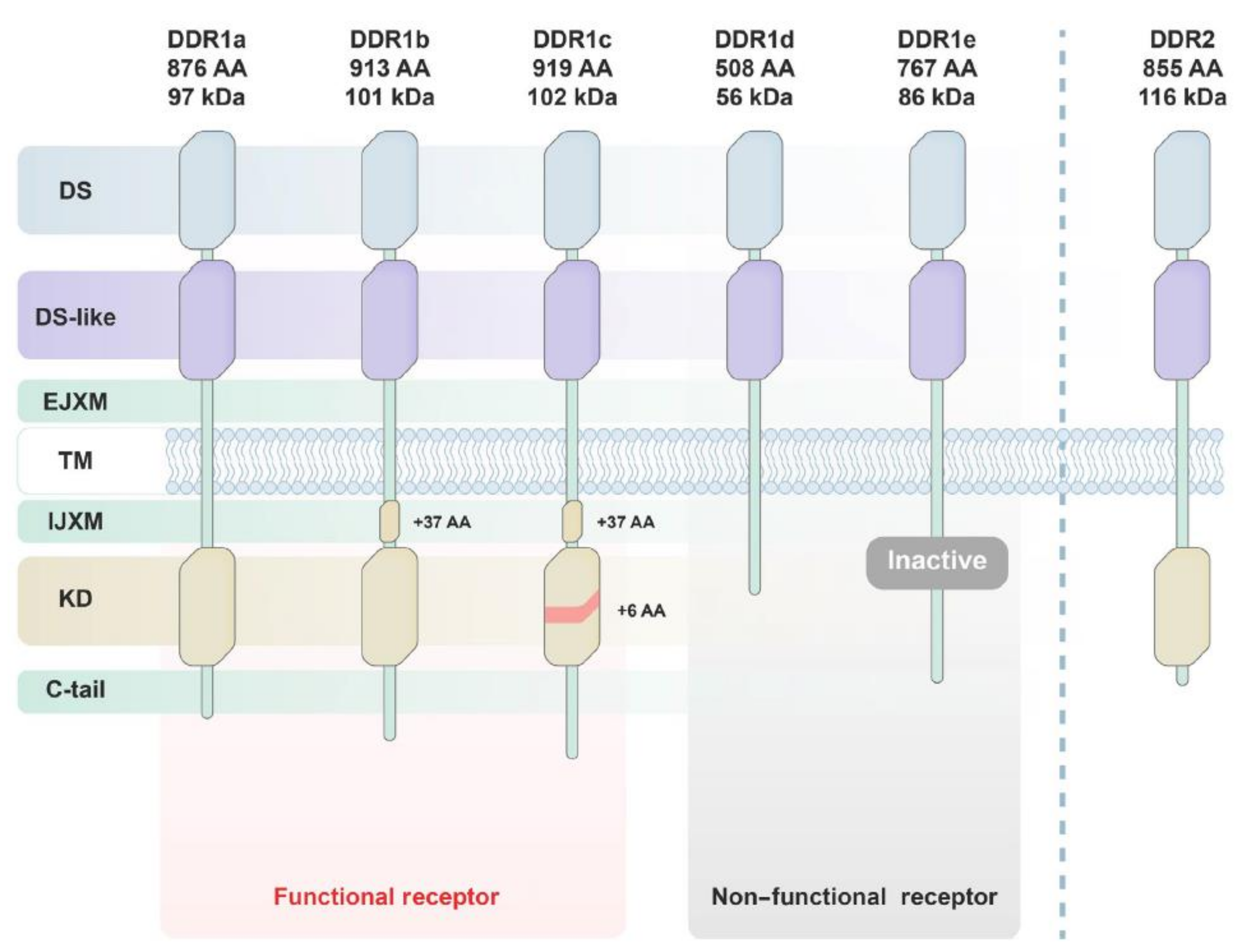
Figure 1. Structures and subtypes of DDR1 and DDR2. Reprinted from with permission Ref. [13].
Ijms 22 06535 g001
Figure 2. Chemical structures of compounds 111.
Ijms 22 06535 g002
Figure 3. Chemical structure of compound 12.
Ijms 22 06535 g003
Figure 4. (A) Chemical structures of compounds 13 and 14; (B) X-ray co-crystal structure of compound 13 with DDR1 kinase. Reprinted with permission from Ref. [133]; (C) Compound 14 docking model into the DDR1 G707A mutation. Reprinted with permission from Ref. [132].
Ijms 22 06535 g004aIjms 22 06535 g004b
Figure 5. Restricted conformation strategy and chemical structures of compounds 15 and 16.
Ijms 22 06535 g005
Figure 6. Chemical structures of DDR1inhibitors 17, 18 (7RH), 19, and 20.
Ijms 22 06535 g006
Figure 7. Drug design approaches and chemical structures of compounds 2123.
Ijms 22 06535 g007
Figure 8. Docking models of compounds 21 (A) and 23 (B) into DDR1 binding pocket. Reprinted from Ref. [139].
Ijms 22 06535 g008
Figure 9. Chemical structures of compound 2428 designed based on 7RH (18) as a lead molecule.
Ijms 22 06535 g009
Figure 10. Chemical structures of type II and III DFG-out binders for DDR2 (2932).
Ijms 22 06535 g010
Figure 11. Schematic of the design process leading to discovery of compound 34. Reprinted with permission from Ref. [142].
Ijms 22 06535 g011
Figure 12. (A) Fragment 1 (33) in DDR1 binding pocket; (B) Cocomplex of dasatinib (1) and fragment 1 (33) (C) Cocomplex of compounds 33 and 34. Reprinted with permission from Ref. [142].
Ijms 22 06535 g012
Figure 13. Chemical structure of compound KST9046 (35).
Ijms 22 06535 g013
Figure 14. Structure-based design strategy of compounds 3638 derived from sorafenib (9).
Ijms 22 06535 g014
Figure 15. Docking models of compounds 36 (orange) and 38 (green) into DFG-out conformation of DDR1. Reprinted with permission from Ref. [146].
Ijms 22 06535 g015
Figure 16. (A) Chemical structure of compound 39; (B) Both compounds 39 (orange) and dasatinib (1) (yellow) overlayed in the active site residue of DDR1 kinase (PDB ID: 5BVW); (C) docking of compound 39 within DDR1 active site (D) Docking of dasatinib (1) within DDR1 active site. Reprinted with permission from Ref. [147].
Ijms 22 06535 g016
Figure 17. Structural modifications to compound 40 and chemical structure of compound 41.
Ijms 22 06535 g017
Figure 18. Chemical structures of compounds 4244 and docking model of compound 42 into DDR2 active site. Reprinted with permission from Ref. [150].
Ijms 22 06535 g018
Figure 19. Structural-based design and chemical structures of compounds 45 and 46. Reprinted with permission from Ref. [151].
Ijms 22 06535 g019
Figure 20. Chemical structures of natural alkaloids discoipyrroles A–D (4750). Reprinted with permission from Ref. [153].
Ijms 22 06535 g020
Figure 21. Structural-based design of compound 20 to afford compound 51. Reprinted from Ref. [154].
Ijms 22 06535 g021
Figure 22. Structural-based design of compound 5254. Reprinted with permission from Ref. [155].
Ijms 22 06535 g022
Figure 23. Chemical structure of compound 55.
Ijms 22 06535 g023
Figure 24. Chemical structure of compound 56 and its docking model to DDR1 active site. Reprinted with permission from Ref. [157].
Ijms 22 06535 g024
Figure 25. Chemical structure of compounds 5759.
Ijms 22 06535 g025
Publisher’s Note: MDPI stays neutral with regard to jurisdictional claims in published maps and institutional affiliations.