Review

Integration of Rap1 and Calcium Signaling

by
Ramoji Kosuru
1 and
Magdalena Chrzanowska
1,2,3,*
1
Versiti Blood Research Institute, Milwaukee, WI 53201, USA
2
Department of Pharmacology and Toxicology, Medical College of Wisconsin, PO Box 2178, Milwaukee, WI 53201-2178, USA
3
Cardiovascular Center, Medical College of Wisconsin, PO Box 2178, Milwaukee, WI 53201-2178, USA
*
Author to whom correspondence should be addressed.
Int. J. Mol. Sci. 2020, 21(5), 1616; https://doi.org/10.3390/ijms21051616
Submission received: 6 February 2020 / Revised: 24 February 2020 / Accepted: 25 February 2020 / Published: 27 February 2020
(This article belongs to the Special Issue Small GTPases 2022)

Abstract

:
Ca2+ is a universal intracellular signal. The modulation of cytoplasmic Ca2+ concentration regulates a plethora of cellular processes, such as: synaptic plasticity, neuronal survival, chemotaxis of immune cells, platelet aggregation, vasodilation, and cardiac excitation–contraction coupling. Rap1 GTPases are ubiquitously expressed binary switches that alternate between active and inactive states and are regulated by diverse families of guanine nucleotide exchange factors (GEFs) and GTPase-activating proteins (GAPs). Active Rap1 couples extracellular stimulation with intracellular signaling through secondary messengers—cyclic adenosine monophosphate (cAMP), Ca2+, and diacylglycerol (DAG). Much evidence indicates that Rap1 signaling intersects with Ca2+ signaling pathways to control the important cellular functions of platelet activation or neuronal plasticity. Rap1 acts as an effector of Ca2+ signaling when activated by mechanisms involving Ca2+ and DAG-activated (CalDAG-) GEFs. Conversely, activated by other GEFs, such as cAMP-dependent GEF Epac, Rap1 controls cytoplasmic Ca2+ levels. It does so by regulating the activity of Ca2+ signaling proteins such as sarcoendoplasmic reticulum Ca2+-ATPase (SERCA). In this review, we focus on the physiological significance of the links between Rap1 and Ca2+ signaling and emphasize the molecular interactions that may offer new targets for the therapy of Alzheimer’s disease, hypertension, and atherosclerosis, among other diseases.

Graphical Abstract

1. Discovery, Early and Classical Functions of Rap1: Ras Antagonism, Integrin Activation

Rap1, a 21 kDa monomeric G-protein, was discovered in 1989 by Noda and his coworkers in a screen for proteins able to suppress the oncogenic effect of K-Ras (one of the mutated Ras genes) [1]. Described as Kristen-ras-revertant-1 (Krev-1), the protein was found to have high similarity to Ras proteins [2]. Simultaneously, Pizen et al. characterized two proteins, Rap1 and Rap2, as Ras homologues and proposed that Rap1, identical to Krev-1, might function as an antagonist of Ras by competing for a common target, or mediating growth inhibitory signals independently of Ras [3,4]. Since then, many groups have reported that Rap1 antagonizes Ras signaling by trapping its effector proteins, serine/threonine kinase Raf, in an inactive complex [5]. However, much research has also demonstrated the functions of Rap1 independent of Ras.
The two highly conserved Rap1 isoforms, Rap1a and Rap1b, share 95% sequence identity, with a 50% sequence homology to Ras [3]. The basic structure of Rap1 is similar to Ras and consists of a catalytic domain made of a six-stranded central β-sheet (β1–β6) surrounded by five α-helices (α1–α5) and ten loops (L1–L10) [6,7]. The two regions of highest sequence similarity between Ras and Rap1 correspond to the switch 1 (amino acids 32–38) and switch 2 (amino acids 60–70) regions [7,8]. These regions adopt different conformations when bound to GTP (active) or GDP (inactive) and allow effector proteins to discriminate between the active and inactive form of small G protein. Despite the identical effector domains and a shared subset of effectors, many of Rap1’s biological functions are distinct from Ras, due to cellular and signaling differences in the utilization of the same effectors [9]. Furthermore, Rap1 controls cell adhesion by modulating the activity of adhesion receptors—integrins and cadherins—through specific interactions with its effectors: RAPL, Riam, AF-6, Krit1, Vav2, Tiam1, and Arap3, [9,10,11].
The kinetics of the GDP–GTP cycle is governed by diverse families of guanine exchange factors (GEFs) containing a Ras exchange motif (REM), a catalytic Cdc25 homology domain with nucleotide exchange activity, and additional regulatory domains which enable a wide variety of regulatory mechanisms (Table 1) [9,12]. Two of those families—CalDAG-GEFs, activated by Ca2+ and diacylglycerol (DAG), and Epac proteins, activated by cyclic adenosine monophosphate (cAMP)—are of particular importance for coordinating Rap1 and Ca2+ cross talk, and will be discussed in more detail in the following sections. In addition to regulation by GEFs, Rap1 undergoes a series of posttranslational modifications that determine its activity and cellular functions.

2. Posttranslational Modifications and Cellular Localization of Rap1

Rap1 is a soluble cytosolic protein that undergoes isoprenylation (geranylgeranylation), a covalent binding of geranylgeraniol to the –SH group of cysteine in the C-terminal CAAX motif (Cys-aliphatic residue-aliphatic residue-X amino acid sequence; X- usually Met, Gln, Ser or Leu). This posttranslational modification, combined with CAAX motif cystine carboxymethylation, enhances Rap1 hydrophobicity and facilitates its membrane localization [13,14,15]. Rap1 can also be modified by phosphorylation, which provides another layer of functional regulation, and is catalyzed by protein kinase A (PKA) [16,17]. PKA-mediated phosphorylation at serine 180 and serine 179 positions on Rap1a and Rap1b, respectively, promotes the direct binding of Rap1 to scaffold protein KSR (kinase suppressor of Ras) and enables the coupling of B-Raf to extracellular signal-regulated kinase-1 (ERK) and sustained ERK activation. Consequently, the phosphorylation of Rap1 has been implicated in the regulation of cell differentiation and growth [17,18].
Signaling events that interfere with the prenylation of Rap1 may decrease its membrane localization and, thus, interfere with cell–cell adhesion. For instance, adenosine A2B receptor-mediated signaling induces Rap1b (ser179 and ser180) phosphorylation and leads to decreased binding to chaperone protein small G-protein dissociation stimulator (SmgGDS). This signaling inhibits Rap1 prenylation and membrane localization and results in cell scattering [19,20].
Cyclic nucleotide phosphodiesterases (PDEs) are key regulators of cAMP signaling. A component of scaffolding complexes that contain A-kinase anchoring proteins such as: PKA, Epac, and adenylate cyclase [21], PDEs directly interact with prenylated Rap1 to control its function in various cell types. In vascular endothelial cells, interaction of PDE4D and Epac1 is critical for the integration into the VE-cadherin-based signaling complex and the coordination of cAMP-mediated vascular endothelial cell adhesion and permeability [22]. PDE6δ (retinal rod rhodopsin-sensitive cGMP 3′,5′-cyclic phosphodiesterase, subunit delta) interacts with prenylated Rap1 in neurons and interferes with its trafficking, thereby dissociating it from the cell membrane, which is where Rap1 promotes Ca2+ influx [23]. In this way, the inhibition of Rap1 interaction with PDE6δ has been proven to be beneficial in restraining disease-associated, abnormal Ca2+ influx and neuronal hyperactivity, and providing neuroprotection in models of Alzheimer’s disease [24].
In addition to localizing at the plasma membrane [25], Rap1 is present at other membranes, including Golgi apparatus and late endocytic compartments [26,27]. Specifically, the subcellular localization of Rap1 pools determines Rap1 coupling to its effectors and its susceptibility to GEF regulation. This has key functional significance for Rap1-regulated processes such as exocytosis [28,29] and integrin-mediated adhesion. Epac1 activity towards Rap1 depends on Rap1 subcellular localization; in effect, Epac activates the plasma membrane, but not perinuclear pools of Rap1. The activation of plasma membrane-localized Rap1 promotes ERK activation and granule secretion [30]. Ca2+ and DAG-dependent Rap1 GEF, CalDAG-GEFI, localization at plasma membrane is key for Rap1 activation and, subsequently, integrin activation in platelets [31]. All these Rap1 regulatory factors contribute to Rap1 and Ca2+ signaling cross-talk.

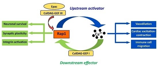
3. Ca2+ Signaling and Rap1

Ca2+ is a ubiquitous secondary messenger, responsible for controlling a myriad of key cell processes, including fertilization, proliferation, contraction, and neural signaling and learning, such as synaptic plasticity, neuronal survival, chemotaxis of immune cells, platelet aggregation, vasodilation, and cardiac excitation-contraction coupling [32,33,34,35,36]. In most cells under basal conditions, the cytosolic concentration of free Ca2+ is approximately 100 nM, which is 10,000 times less than that of extracellular Ca2+. Upon stimulation, intracellular Ca2+ levels rapidly, but transiently, rise to above 1 μM. However, sustained increases in intracellular free Ca2+ to the micromolar range are deleterious to cellular functions and the efficient lowering of intracellular Ca2+ via Ca2+ buffers and uptake by Ca2+ pumps is essential for preventing cell damage or death.
The rise in cytoplasmic Ca2+ levels can be generated either from intracellular stores or extracellular sources. Ca2+ release from internal stores is controlled by several channels, of which the inositol 1,4,5-trisphosphate (IP3) receptor-mediated Ca2+ release from endoplasmic reticulum is a universal and highly versatile mechanism [37,38]. Most of the drugs/agonists that act on the G protein-coupled receptor (GPCR) and tyrosine kinase receptors (TKR) utilize the IP3-mediated Ca2+ release pathway to promote their signal transduction [39]. Phospholipase C (PLC) activated downstream from GPCRs and TKRs, cleaves phosphatidylinositol 4,5 bisphosphate into IP3 and diacylglycerol (DAG). Liberated IP3 then binds to IP3 receptors present on endoplasmic reticulum to allow Ca2+ release, and increases cytoplasmic Ca2+ levels [39]. On the other hand, extracellular Ca2+ entry is regulated by several channels that open after the depletion of intracellular Ca2+, which include voltage-gated Ca2+ channels, receptor-operated Ca2+ channels and store-operated Ca2+ channels [40]. The combined action of intracellular Ca2+ release and extracellular Ca2+ entry is required to tightly control changes in the length and amplitude of Ca2+ fluxes to regulate multiple signaling pathways [37,38].
The universal mechanisms that lead to Ca2+ release also generate signals that activate Rap1. Multiple modalities of Rap1 GEF activation allow Rap1 to act as an effector, as well as an upstream regulator of Ca2+ signaling. Downstream from Ca2+ and DAG generated in parallel to the induction of the Ca2+ signal - Rap1 acts as one of Ca2+ signaling effectors. Activated by other GEFs, in particular by Epac in response to elevated cAMP, Rap1 controls Ca2+ signals. How these pathways intersect to exert multiple, tissue-specific effects is described in more detail below.

4. Rap1 Activators in Integration of Ca2+ Signaling

Rap1 signaling is remarkably complex, with cross-talk between multiple receptors and its interacting effector proteins [16,41,42,43]. Rap1 activity is controlled by several evolutionarily conserved families of GEFs, and, in particular, Ca2+ and DAG-activated CalDAG-GEFs and 3′ and 5′-cyclic adenosine monophosphate (cAMP)-activated Epacs. These two GEF families are of key importance for the cross-talk between Rap1 and Ca2+ signaling.

4.1. CalDAG-GEFs

The discovery of a Ca2+-binding GEF, CalDAG-GEFII, encoded by RASGRP1 gene, with an activity towards Ras, introduced an intriguing possibility of a cross-talk between the Ca2+ and Rap1 signaling pathways [44]. This possibility materialized when a second family member, CalDAG-GEFI (RASGRP2) was identified as a novel brain transcript, and was shown to activate Rap proteins [45,46]. Subsequently, other family members: CalDAG-GEFIII (RASGRP3) and CalDAG-GEFIV (RASGRP4) were identified as regulators of various Ras proteins (Table 1) in B-cells and mast cells [47,48]. Different CalDAG-GEF isoforms are present in most tissues, including the hematopoietic and neuronal cells where some of their functions have been characterized, and in blood vessels [45,49,50,51,52,53,54].
The four CalDAG-GEF family members (I, II, III and IV) share a similar structure containing conserved Cdc25 homology domain (catalytic site), a Ras exchange motif (REM), and two atypical EF hands involved in Ca2+ binding and release of autoinhibition involved in GEF activation (in case of CalDAG-GEFI) [55]. In addition, a C-terminal C1 motif that mediates lipid interactions is important for the localization and/or activation of CalDAG-GEFs. Except for CalDAG-GEFI, which contains atypical C1, the remaining CalDAG-GEFs contain typical C1 motifs with a high affinity for DAG. The differences in C1 domains contribute to the differential regulation of CalDAG-GEFs by Ca2+ and DAG [56,57,58] (Table 1). Both CalDAG-GEFII and CalDAG-GEFIII contain typical C1 domains with high affinity for DAG and translocate to the plasma membrane after treatment with DAG mimetic 12,13-tetradecanoyl phorbol acetate (TPA), but are insensitive to increased levels of Ca2+ [59,60,61,62]. In contrast, the atypical C1 domain of CalDAG-GEFI has low affinity for DAG [63], but high affinity for plasma membrane phosphoinositides PIP2 and PIP3. This atypical C1 domain is required for CalDAG-GEFI association with the plasma membrane [31].
The four CalDAG-GEF family members exhibit different GTPase specificities depending on the availability of Ca2+ and DAG (Table 1) [12,60]. For example, CalDAG-GEFI functions as a dual R-Ras/Rap1 activator and Ca2+ regulation plays a key role in determining its specificity. Since Ca2+ stimulates the Rap-exchange activity of CalDAG-GEFI, while inhibiting the Ras-exchange activity, a cytosolic Ca2+ signal effectively shifts the catalytic activity of CalDAG-GEFI from Rap to Ras GTPases [45,46]. CalDAG-GEFII specifically functions as a Ras and R-Ras activator while CalDAG-GEFIII activates several Ras GTPases, including Rap1, Rap2, Ras, and R-Ras [44,45,60]. Two CalDAG-GEFs act via Rap1 and the structural difference in the C1 domain determines the mechanism of their activation and signaling context. CalDAG-GEFI promotes Rap1 activation via Ca2+ while CalDAG-GEFIII mediates Rap1 activation via DAG. CalDAG-GEFI-Rap1 signaling is important in central nervous system (CNS) and platelet function. While the exact functions of DAG-activated CalDAG-GEFIII are less well understood, it is important in macrophage activation and has been linked with hypertension through GWAS studies [53].

4.2. Epac

Rap1 is an important mediator of cellular cAMP signaling [64]. Elevation in cAMP levels, resulting from adenylyl cyclase activation downstream from ligand-induced Gαs-coupled GPCRs stimulation, induces the activation of Rap1 GEFs and Epacs (exchange proteins directly activated by cAMP) [64,65,66]. Two members of Epac family, Epac1 and Epac2, catalyze the guanine nucleotide exchange on Ras GTPases, including Rap1 and Rap2 [66,67]. Epac1 is ubiquitously expressed in the CNS, heart, and other organs, including the kidney, spleen, pancreas, ovary, thyroid, adrenal glands, as well as the endothelium. Epac2 is predominantly expressed in the brain and the adrenal glands [66,67].
Epac1 and Epac2 share a similar structural organization, with the C-terminal catalytic GEF region and N-terminal regulatory region. The catalytic regions of Epacs possess a Ras exchange motif (REM domain), a Cdc25-homology catalytic domain that mediates the GEF activity for Rap GTPases and a RAS-association domain (RA domain), which translocates Epac2 to the plasma membrane. The regulatory region of Epac1 consists of a DEP domain (Dishevelled, Egl-10, and Pleckstrin) that is responsible for membrane anchoring, and a conserved cAMP-binding domain [66,68] (Table 1). In the unbound state, the cAMP-binding domain acts as an auto-inhibitory module for the catalytic Cdc25-homology domain. The binding of cAMP induces conformational changes in hinge helix and allows the regulatory region to move away from the catalytic region, thereby exposing the GEF domain to allow Rap1 binding [69]. Although they are similar in domain structure, Epac2 differs from Epac1 in the additional N-terminal cAMP-binding domain, which binds cAMP with a much lower affinity and is unable to induce GEF activity after cAMP binding [69,70].
Epac plays an important role in the regulation of Ca2+ homeostasis and Epac and Ca2+ signaling pathways crosstalk at multiple levels, converging on effectors like IP3 receptor and ryanodine receptor (RyR), mediating Ca2+ release, or sarcoendoplasmic reticulum Ca2+-ATPases (SERCA), mediating Ca2+ clearance, effectively forming a signaling network in non-excitable cells [71,72,73]. The signaling schemes include Epac acting as an inducer of Ca2+-induced Ca2+ release to mobilize intracellular Ca2+ levels, as found in the regulation of exocytosis in human pancreatic β-cells and INS-1 insulin-secreting cells [74,75]. Interestingly, Ca2+ can also modulate the Epac signaling pathway by activating the adenylyl cyclase to increase the production of cAMP levels [76]. It is important to note that downstream effects are dependent on the distinctive activation of subcellular pools of Rap1. While not all of Epac functions are mediated by Rap1 [30,64], the Epac/Rap1 axis and Ca2+ signaling intersect to regulate important functions in several tissues. Some of the mechanisms uncovered are described below.

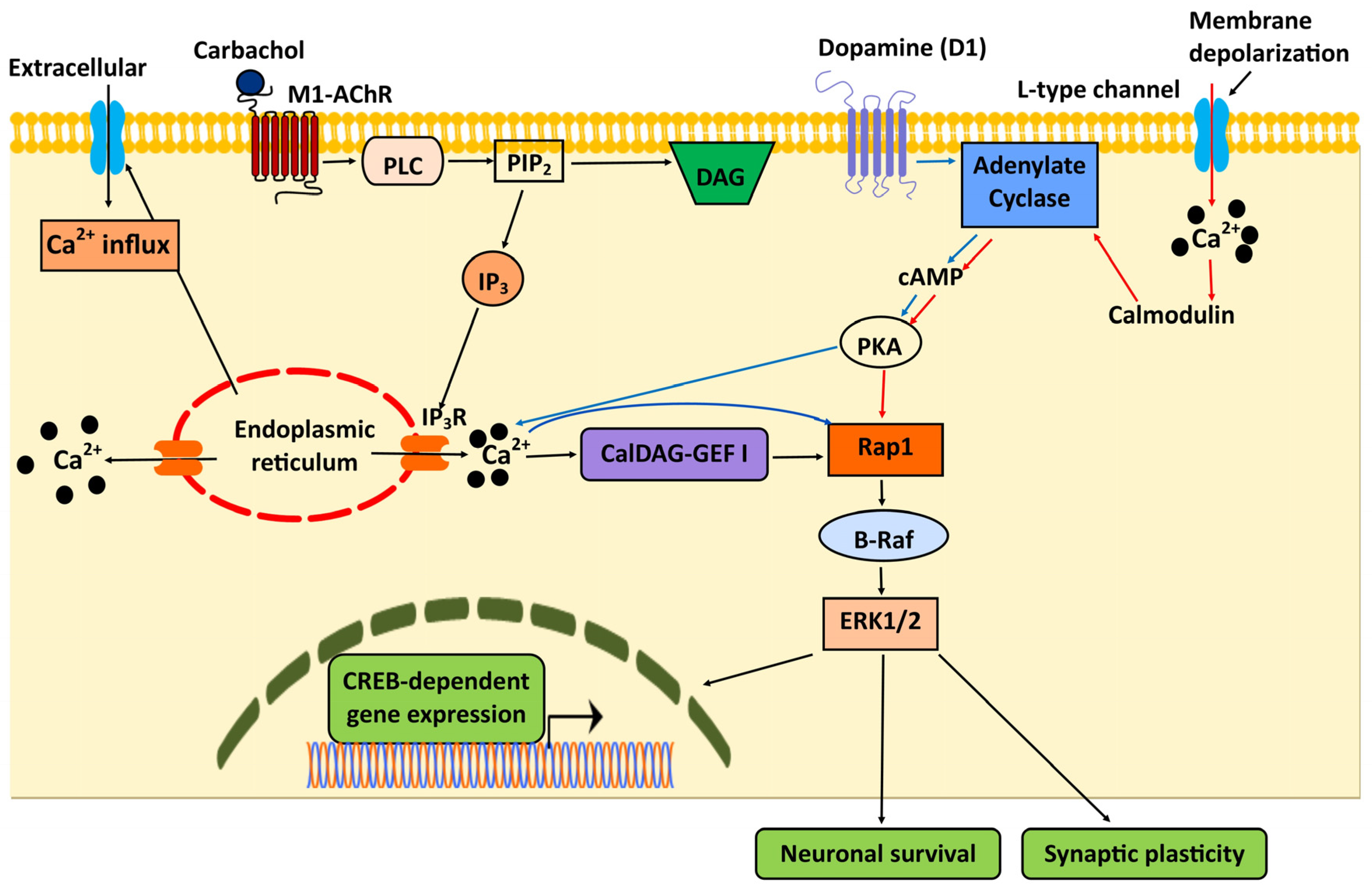
5. Integration of Rap1 and Ca2+ Signaling in the Central Nervous System (CNS)

Via the activation of the ERK signaling pathway, Rap1 is involved in a number of Ca2+-dependent processes, such as neuronal excitability, synaptic plasticity, long-term potentiation and gene transcription [77,78,79,80]. In most cases, the activation of Rap1, and the ensuing B-Raf1 and ERK activation, depends on agonist-induced Ca2+ influx leading to CalDAG-GEFI activation and the subsequent formation of CalDAG-GEFI/Rap1/B-Raf cassette to stimulate the ERK pathway [81] (Figure 1). The mechanisms that lead to Ca2+-dependent Rap1 activation vary in different neuronal cells depending on the source and magnitude of Ca2+ signal. In PC12 and hippocampal neurons, Rap1-ERK signaling is mediated by PKA activation upon depolarization-induced Ca2+ influx through L-type Ca2+ [79] (Figure 1). In primary striatal neurons, the dopamine D1 receptor-induced, PKA-mediated Ca2+ release activates the Rap1/B-Raf/ERK pathway to regulate cAMP-response element binding protein (CREB)-phosphorylation and gene expression [77] (Figure 1). This mechanism of Rap1 activation appears to be primed by PKA-induced Ca2+ release, but is not further induced by direct or indirect PKA- or protein kinase C-dependent phosphorylation [77]. Thus, Ca2+ and Rap1 signaling can intersect at multiple signaling modules.
Furthermore, the magnitude of intracellular Ca2+ activates different pools of Rap1 to mediate ERK signaling at spatially discrete, subcellular locations, which is essential for controlling spatially discrete processes underlying neuronal function and survival. For example, in resting neurons, steady-state levels of Ca2+ and cAMP drive the activation of the membrane-associated pool of Rap1-ERK signaling [78], which leads to a reduction in the A-type K+ channel Kv4.2 activity that controls the back-propagation of action potentials (or action potential repolarization) in hippocampal CA1 pyramidal neurons [82]. Conversely, in depolarizing neurons, Ca2+ influx stimulates the nuclear pool of Rap1-ERK signaling, which phosphorylates nuclear targets involved in the expression of hippocampal long term potentiation [78] and CREB-dependent gene transcription [83]. Thus, in these cellular scenarios, Rap1 is a mediator of Ca2+-signaling.
In addition to acting as an important mediator of Ca2+-mediated ERK signaling, Epac-mediated Rap1 activation controls Ca2+-dependent signaling events, such as resting membrane potential, glutamate release, and cortico-amygdala plasticity in neurons. In mouse cerebellar granule cells, the Epac-induced activation of Rap1 and p38 mitogen-activated protein kinase (MAPK) mobilizes intracellular Ca2+ release, facilitating the opening of large conductance Ca2+-activated K+ channels to modulate resting membrane potential and after-hyperpolarization [84]. In primary cortical neurons ERK1/2 and L-type, Ca2+ channels act as the downstream Rap1 effectors to mediate the suppression of glutamate release required for cortico-amygdala plasticity and fear learning. Rap1 deletion in these cells leads to increased axonal Ca2+ influx, ERK inhibition, and increased plasma membrane expression of L-type voltage-gated Ca2+ channels (Cav1.2 or Cav1.3), enabling the Ca2+-regulated glutamate release [80].
Interestingly, the modulation of prenylated Rap1 appears to play a role in controlling disease-associated Ca2+ aberrations and neuronal activity. The inhibition of Rap1 interaction with PDE6δ restrains disease-associated abnormal Ca2+ influx and neuronal hyperactivity and confers neuroprotection in models of Alzheimer’s disease [24]. Rap1 may be an important therapeutic target for the treatment of neuro-degenerative disorders associated with Ca2+ aberrations, such as Alzheimer’s disease.

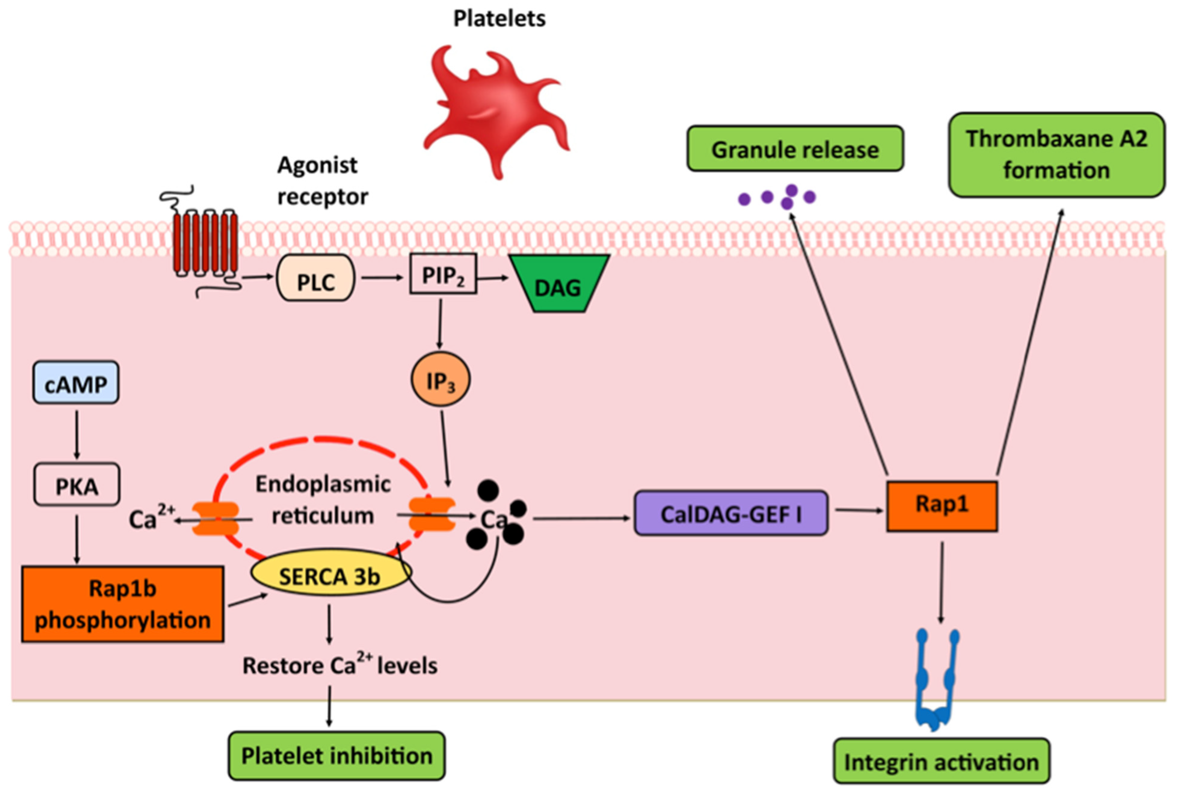
6. Platelets: Integrins and SERCA

Early studies demonstrated that an increase in cytosolic Ca2+ is required and sufficient for Rap1 activation in platelets [41]. Much research has implicated the CalDAG-GEFI–Rap1 signaling axis as a mediator of that activation [85] and recent studies revealed a Ca2+-dependent mechanism of CalDAG-GEFI, and downstream, Rap1 activation. In resting platelets, where cytosolic Ca2+ levels are low, CalDAG-GEFI remains in an auto-inhibited state. In response to agonist stimulation, elevated cytosolic Ca2+ binds to the EF hands and induces structural rearrangements that free the catalytic surface of CalDAG-GEFI to activate Rap1b [55]. Downstream from activated Rap1, its effector, Rap1-GTP-interacting adaptor molecule (RIAM), recruits talin to β3 integrin subunit and contributes to integrin activation [86,87]. While the functional significance of RIAM in αIIbβ3 integrin activation has not been fully validated in vivo [88,89], evidence points to CalDAG-GEFI’s importance in hemostasis. CalDAG-GEFI deficiency in platelets leads to delayed Ca2+-dependent rapid activation of Rap1 and a marked defect in platelet aggregation [49], similar to the phenotype of Rap1b knockout mice [90]. Human platelets expressing an inactive CalDAG-GEFI are defective at clot formation, which points to a fundamental role for CalDAG-GEFI–Rap1 signaling module in platelet Ca2+ homeostasis [91]. Importantly, Rap1 activation by CalDAG-GEFI is a critical signaling step linking Ca2+ signaling with integrin αIIbβ3 activation, thromboxane A2 formation, and granule release in platelets [49,92,93] (Figure 2).
In addition to acting as an effector of Ca2+-induced CalDAG-GEFI in platelet integrin activation, Rap1 has been implicated in the regulation of IP3-sensitive intracellular Ca2+ pools via the regulation of sarcoendoplasmic reticulum Ca2+-ATPases (SERCA) [94,95]. SERCA is a key Ca2+ pump that transports Ca2+ into ER, reducing cytosolic Ca2+ concentration, thereby controlling IP3-sensitive intracellular Ca2+ pools in platelets [96]. Studying pathological platelets obtained from congestive heart failure patients, Magnier et al., observed a decrease in the expression and phosphorylation of Rap1 that correlated with the reduced expression of 97 kDa SERCA in the platelets of congestive heart failure patients [94]. Later, Lacabaratz-Porret et al., demonstrated that this 97 kDa SERCA, a SERCA3b isoform, physically interacts with Rap1b protein, which suggests that SERCA 3b is a target of Rap1b [95].
The dynamic regulation of the interaction between Rap1b and SERCA 3b by cAMP-dependent phosphorylation of Rap1 may act to regulate a transition between platelet inhibition and activation [97,98]. Increased cAMP production leads to the phosphorylation of Rap1b and its subsequent dissociation from SERCA 3b protein results in the stimulation of its activity to enhance the filling state of SERCA-associated Ca2+ pool to induce platelet inhibition [95] (Figure 2). In diseased or hypertensive platelets, decreased cAMP leads to a decrease in phosphorylation of Rap1b and SERCA 3b activity. This results in a smaller SERCA-associated Ca2+ pool, thus decreasing IP3-sensitive Ca2+ release to promote platelet activation [95]. Thus, the interplay between SERCA 3b and Rap1-modulating phosphorylation may be clinically significant in cardiovascular pathology.

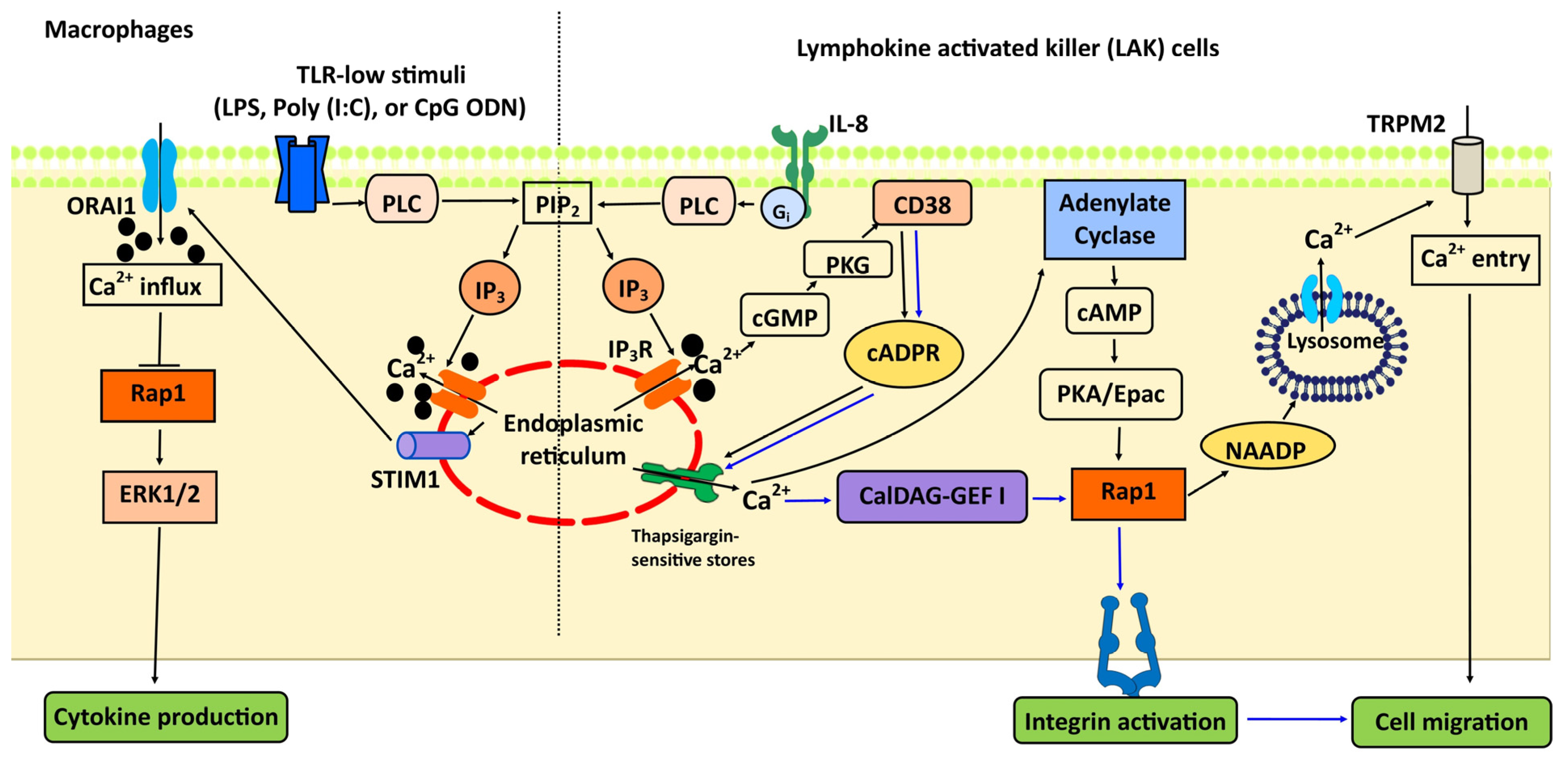
7. Rap1 and Ca2+ Signaling in the Immune System: TLR, Integrins, and Chemotaxis

Similarly to platelets, the activation of integrins is one of the best characterized Rap1 functions in leukocytes that intersects with Ca2+ signaling, with CalDAG-GEFI acting as Rap1 activator [99,100] and RIAM and talin as Rap1 effectors [101,102]. Integrin activation is fundamental for leukocyte migration, chemotaxis and trafficking, and cell adhesion [103,104].
CalDAG-GEFI deficiency in neutrophils impairs F-actin formation, E-selectin-dependent slow rolling, adhesion, and speed and the directionality of migration [100,105,106]. In vivo, CalDAG-GEF1 deficiency blocks TNFα-induced intravascular neutrophil adhesion and recruitment during sterile peritonitis [105]. Decreased CalDAG-GEF1/Rap1 signaling, as in cases of genetic deletion of RASGRP2 in mice [100] or loss of function mutations in humans [107,108], has been suggested as a cause of the rare leukocyte adhesion deficiency type III (LAD-III). However, mutations in Kindlin-3 were found to be causative of LAD-III [109,110], solving that controversy [111]. On the other hand, increased CalDAG-GEFI/Rap1 signaling is responsible for increased cell migration in chronic lymphocytic leukemia (CLL) downstream from acyclic ADP ribose hydrolase, CD38 [112]. Elevated expression of CD38 leads to elevated intracellular Ca2+ [113] and activates Rap1 via CalDAG-GEFI, subsequently leading to activation of integrin and facilitating CLL adhesion [112].
Once activated by CalDAG-GEFI or Epac, Rap1 controls chemotaxis and the trafficking of immune cells via additional, distinct mechanisms. In neutrophils, CalDAG-GEFI-activated Rap1 controls chemotaxis in an integrin-independent manner through a mechanism that involves actin cytoskeleton and cellular polarization [106]. In lymphokine-activated killer (LAK) cells, Epac–Rap1 activation downstream from endoplasmic reticulum Ca2+ release triggers the production of nicotinic acid adenine dinucleotide phosphate (NAADP) and enables Ca2+ release from lysosomal acidic organelles to stimulate long-lasting Ca2+ entry through transient receptor potential melastatin 2 (TRPM2) channels required for cell migration [114] (Figure 3).
In addition to the regulation of migration, Rap1 signaling is an important modulator of Ca2+-dependent regulation of toll-like receptor (TLR) signaling in immune cells [115,116]. The intensity of pathogenic TLR stimuli and the corresponding intensity of Ca2+ signal has been linked with differential activation signaling by Ras and Rap1 as well as a differential effect on ERK activation and cytokine production [116,117]. Induced by low-intensity TLR stimuli, low-intensity Ca2+ influx mediated by stromal interaction molecule 1 (STIM1) favors Rap1 inhibition and ERK activation, while high-intensity TLR stimuli trigger more intense Ca2+ influx, leading to Ras activation and cytokine production [116] (Figure 3). Interestingly, this effect is mediated by CalDAG-GEFIII, which limits TLR-mediated cytokine production by activating Rap1 and ERK in response to a low level of antagonists and, in vivo, limits the inflammatory response [117]. These findings underscore the importance of Rap1 in the modulation of the Ca2+-dependent, key aspects of the immune response.

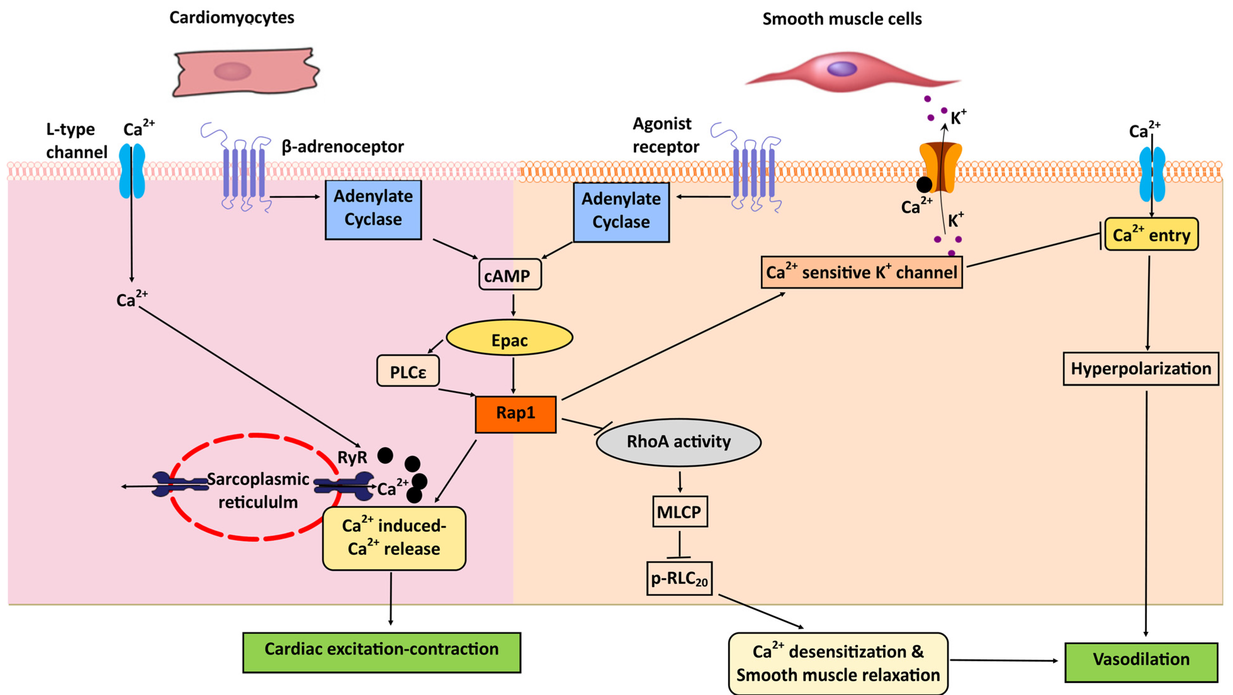
8. Heart: Excitation-Contraction Coupling; Cardiac Hypertrophy

Epac proteins play an important role in the regulation of cardiac physiology, with some of its functions being mediated by Ras family members other than Rap1 [64,118,119]. Nonetheless, the Epac–Rap axis intersects with cardiac Ca2+ signaling to regulate cardiac excitation–contraction coupling, with phosplipase Cε (PLCε) and CamKII as Epac effectors [64,71]. PLCε acts as an effector of both Epac and Rap GTPase Rap2b [120]. Importantly, by activating Rap via its Rap GEF activity, PLCε facilitates Ca2+-induced Ca2+ release (CICR) in adult ventricular cardiomyocytes. In response to β-adrenergic receptor (βAR) stimulation, Epac–Rap2b induce both Rap GEF activity and hydrolytic activity of PLCε. This leads to sustained Rap activation and the induction of PKCε and CamKII phosphorylation downstream from PIP2 breakdown. These signaling events lead to enhanced sarcoplasmic reticulum Ca2+-induced Ca2+ release (CICR) [121,122] (Figure 4). Thus, Epac–Rap modulates cardiac Ca2+ homeostasis through the regulation of CICR in cardiomyocytes. Although these studies identified Rap2b as the main isoform involved in the cardiac Ca2+ signaling, the role of Rap1 in this context is not known. Therefore, Rap1 isoform-specific knockout animal models will be useful to determine the in vivo role of Rap1 in cardiac Ca2+ signaling.
In addition to its role in cardiac physiology, Epac is also implicated in cardiac pathology. Epac stimulation induces cardiac hypertrophy via activation of IP3-induced intracellular Ca2+ rise, leading to the activation of the numerous Ca2+ sensitive hypertrophic proteins, including calcineurin, histone deacetylases, and nuclear factor of activated T cells (NFAT) [123,124]. In cardiomyocytes, Epac proteins function as signalosomes [64]—macromolecular complexes which consist of mAKAP (muscle A kinase-anchoring protein), protein kinase A, phosphodiesterase PDE4D3, ryanodine receptor, phosphatases PP2A, and calcineurin, and serve as signaling nodes in the Ca2+ signaling network [125]. While the significance of Epac for Ca2+ homeostasis and heart function is indisputable, the identity and function of its specific effectors, including Rap GTPases, in normal and pathologic conditions, remain to be fully elucidated.

9. Vascular Smooth Muscle Cells: Vasorelaxation

Rap1 plays an important role in the regulation of vascular tone. At least two distinct mechanisms connect Ca2+ and Epac-dependent Rap1 activation in vascular smooth muscle cells to control smooth muscle relaxation. The activation of Epac modulates Ca2+ sensitivity of the contractile proteins by a Rap1-dependent reduction in RhoA GTPase activity in several types of smooth muscle cell from airway, gut, and vascular tissues [126,127]. In these cells, Rap1 activation downstream of Epac leads to reduced RhoA activity. This induces a series of events, including decreased myosin regulatory light chain (RLC20) phosphorylation and the disinhibition of myosin light chain phosphatase (MLCP) activity, which leads to Ca2+-desensitization and relaxation of force in smooth muscle [126,127] (Figure 4). Consistently, Epac-induced vasorelaxation is decreased in Rap1b knockout vascular smooth muscle cells through inhibition of RhoA-mediated sensitization to Ca2+, medicated by decreased RLC20 phosphorylation [128]. Importantly, Rap1b deficiency led to the development of hypertension, in part via functional changes to vascular smooth muscle cells [128].
In addition to triggering signaling that modulates Ca2+ sensitivity of contractile proteins, Epac–Rap1 may act directly to regulate Ca2+ influx, thus inducing hyperpolarization of smooth muscle membrane and leading to vasorelaxation. Epac activation increases the activity of Ca2+ sparks from ryanodine receptors to open Ca2+-sensitive K+ channels (BKCa), inducing smooth muscle hyperpolarization. This, subsequently, leads to a decrease in the activity of voltage-gated Ca2+ channels, reducing Ca2+ influx and promoting vasorelaxation [129] (Figure 4). Altogether, these studies indicate that Rap1, by altering Ca2+ sensitivity of vascular smooth muscle cells, plays an important role in maintaining normal vascular contractile state and contributes to blood pressure regulation.

10. Endothelium: NO, Vasorelaxation, Vasoreactivity

Some of the best-described functions of Rap1 in the endothelium involve the dynamic regulation of endothelial junctions and the control of the endothelial and vascular barrier [11,130]. Acting via its effectors Rasip, Radil, and afadin (AF6), Rap1 facilitates interactions between adherens and tight junction components and, by regulating the activity of Rho small GTPases, orchestrates actin cytoskeletal rearrangements to enhance endothelial barrier [131,132,133,134,135]. While multiple Rap1 GEFs are involved in the dynamic regulation of the endothelial barrier [130], cAMP/Epac -activated Rap1 plays a particularly important role in lung vasculature in vivo by protecting against ventilator- or inflammation-induced lung injury [136,137]. In pulmonary endothelium in vitro, Rap1 geranylgeranylation has been linked with Ang II-mediated activation of Ca2+-activated tyrosine kinase Pyk2. This finding suggests the involvement of functional Rap1 in hypertension and vascular permeability [138]. However, the physiological significance of the Rap1 activation by AngII pathway is unknown [139,140]. While Ca2+ signal is an important regulator of endothelial permeability, particularly in lung edema [141], it is not currently known whether CalDAG-GEFs are involved or how Rap1 signaling may intersect with Ca2+ signaling. Despite that, a recently uncovered role in nitric oxide (NO) release is where Rap1 and Ca2+ signaling intersect and this is one of the most physiologically impactful functions of Rap1 [11].
The endothelial cell-specific deletion of Rap1 during development leads to an impaired vascular barrier and is not compatible with vascular maturation [142]. However, the deletion of both Rap1 isoforms after birth does not lead to increased vascular permeability in most vascular beds. Instead, it leads to severely attenuated NO release and impaired vasodilation resulting in hypertension [143,144]. Underlying this defect is the impaired activation of endothelial nitric oxide synthase (eNOS), in part due to defective sensing of shear-stress of flowing blood, which is a major physiological regulator of NO release [143]. The shear stress-induced activation of eNOS, largely regulated by eNOS phosphorylation [145], is impaired in Rap1-deficient endothelial cells [143]. The studies of the underlying mechanism revealed that Rap1 promotes the assembly of the endothelial junctional mechanosensing complex (comprised of PECAM-1, VE-Cadherin, and VEGFR2) and is critical for VEGFR2 transactivation and signaling to NO release [143]. In addition to promoting phosphorylation-dependent eNOS activation, Rap1 is required for Ca2+-dependent eNOS activation, as endothelium-specific deletion of Rap1 leads to a significant impairment of acetylcholine induced-Ca2+-dependent vasodilation and is sufficient to induce hypertension in vivo [128,143]. Conversely, (Epac-induced) Rap1 activation induces a sustained increase in cytosolic Ca2+, eNOS activity, and subsequent NO production contributing to endothelium-dependent vasorelaxation [129] (Figure 5). While the underlying mechanisms are still under investigation, these findings underscore the fundamental role of cross-talk between Rap1 and Ca2+ signaling in EC homeostasis.
In summary, endothelial Rap1 signaling plays an important role in the fundamental endothelial processes regulated by Ca2+ signaling—the dynamic regulation of endothelial barrier and release of NO. While the identity of some of the Rap1 effectors and GEFs regulating these processes is known, more research is needed to understand the exact underlying mechanisms. Such analysis may reveal novel therapeutic targets for treating diseases associated with endothelial dysfunction such as atherosclerosis, myocardial infarction and hypertension.

11. Conclusions and Future Perspectives

In two different signaling modalities, as an upstream activator and downstream effector, active Rap1 intersects with Ca2+ signaling to control important functions in the multiple tissues. The significance of Rap1/Ca2+ cross-talk is particularly evident in the CNS, where increased Ca2+ from external Ca2+ influx or internal Ca2+ release activates Rap1 to stimulate B-Raf/ERK signaling. This, in turn, controls neuronal function and gene expression [77,79]. However, it is not known how these Ca2+ signals, with different frequencies and amplitudes, activate Rap1 to induce ERK activation. Better understanding of the kinetics of Rap1 activity and identity of Rap1 activating GEFs involved in Ca2+ signaling is needed. To this end, novel tools [146] and model organisms [143] might help provide answers.
Studies of Rap1 in the endothelium, heart and platelets have revealed its novel effectors and functions important for tissue and organ homeostasis [95,122,128,129,143]. In the endothelium, eNOS has been identified as a novel Rap1 effector [143]. eNOS activity is regulated by phosphorylation events, particularly in response to shear-stress, but also depends on Ca2+ signaling elicited downstream from several GPCR-agonists [147,148,149]. Furthermore, store-operated Ca2+ entry is required for sustained eNOS activation in endothelial cells [150]. Interestingly, via distinct mechanisms, Rap1 intersects with Ca2+ signaling in smooth muscle cells, promoting their relaxation. Thus, in both tissues, Rap1 controls vascular tone [128]. CalDAG-GEFIII, a potential Rap1 activator, has been implicated in hypertension by genome-wide association studies [53]. However, its function in the endothelium has not been widely studied and has been reported only once to date [54].
It is becoming evident that Rap1 signaling is essential for the function of multiple organs, and that some key Rap1 functions rely on cross-talk with Ca2+ signals. A thorough characterization of Rap1 regulators, effectors, and the mechanisms connecting them in time and space may provide novel therapeutic strategies for several cardiovascular and neurological diseases.

Author Contributions

Writing—original draft preparation, R.K.; Writing—review and editing, M.C. All authors have read and agreed to the published version of the manuscript.

Funding

This work was supported by NIH grant HL111582.

Acknowledgments

We thank Shana Maker for proof-reading the manuscript.

Conflicts of Interest

The authors declare no conflict of interest. The funders had no role in the design of the study; in the collection, analyses, or interpretation of data; in the writing of the manuscript, or in the decision to publish the results.

Abbreviations

Ca2+Calcium
CalDAG-GEFCa2+ and DAG-activated GEF
cAMPcyclic adenosine monophosphate
CICRCa2+-induced Ca2+ release
CLLchronic lymphocytic leukemia
CNScentral nervous system
CREBcAMP-response element binding protein
eNOSendothelial nitric oxide synthase
Epacexchange protein activated by cAMP
ERKextracellular signal-regulated kinase-1
GAPsGTPase-activating proteins
GEFsguanine nucleotide exchange factors
GPCRG protein coupled receptor
IP3inositol 1,4,5-trisphosphate
MAPKmitogen-activated protein kinase
NOnitric oxide
PDEsphosphodiesterases
PIPphosphatidyl inositol phosphate
PKAprotein kinase A
PLCphospholipase C
REMRas exchange motif
RIAMRap1-GTP-interacting adaptor molecule
RLC20myosin regulatory light chain
RyRryanodine receptor
SERCAsarcoendoplasmic reticulum Ca2+-ATPase
TKRtyrosine kinase receptors
TLRToll-like receptor
VEGFR2vascular endothelial growth factor receptor 2

References

  1. Noda, M.; Kitayama, H.; Matsuzaki, T.; Sugimoto, Y.; Okayama, H.; Bassin, R.H.; Ikawa, Y. Detection of genes with a potential for suppressing the transformed phenotype associated with activated ras genes. Proc. Natl. Acad. Sci. USA 1989, 86, 162–166. [Google Scholar] [CrossRef] [PubMed] [Green Version]
  2. Kitayama, H.; Sugimoto, Y.; Matsuzaki, T.; Ikawa, Y.; Noda, M. A ras-related gene with transformation suppressor activity. Cell 1989, 56, 77–84. [Google Scholar] [CrossRef]
  3. Pizon, V.; Lerosey, I.; Chardin, P.; Tavitian, A. Nucleotide sequence of a human cDNA encoding a ras-related protein (rap1B). Nucleic Acids Res. 1988, 16, 7719. [Google Scholar] [CrossRef] [PubMed] [Green Version]
  4. Pizon, V.; Chardin, P.; Lerosey, I.; Olofsson, B.; Tavitian, A. Human cDNAs rap1 and rap2 homologous to the Drosophila gene Dras3 encode proteins closely related to ras in the ‘effector’ region. Oncogene 1988, 3, 201–204. [Google Scholar]
  5. Bos, J.L. All in the family? New insights and questions regarding interconnectivity of Ras, Rap1 and Ral. EMBO J. 1998, 17, 6776–6782. [Google Scholar] [CrossRef] [PubMed] [Green Version]
  6. Milburn, M.V.; Tong, L.; deVos, A.M.; Brunger, A.; Yamaizumi, Z.; Nishimura, S.; Kim, S.H. Molecular switch for signal transduction: Structural differences between active and inactive forms of protooncogenic ras proteins. Science 1990, 247, 939–945. [Google Scholar] [CrossRef] [Green Version]
  7. Noguchi, H.; Ikegami, T.; Nagadoi, A.; Kamatari, Y.O.; Park, S.Y.; Tame, J.R.; Unzai, S. The structure and conformational switching of Rap1B. Biochem. Biophys. Res. Commun. 2015, 462, 46–51. [Google Scholar] [CrossRef] [Green Version]
  8. van den Berghe, N.; Cool, R.H.; Wittinghofer, A. Discriminatory residues in Ras and Rap for guanine nucleotide exchange factor recognition. J. Biol. Chem. 1999, 274, 11078–11085. [Google Scholar] [CrossRef] [Green Version]
  9. Raaijmakers, J.H.; Bos, J.L. Specificity in Ras and Rap signaling. J. Biol. Chem. 2009, 284, 10995–10999. [Google Scholar] [CrossRef] [Green Version]
  10. Boettner, B.; Van Aelst, L. Control of cell adhesion dynamics by Rap1 signaling. Curr. Opin. Cell Biol. 2009, 21, 684–693. [Google Scholar] [CrossRef] [Green Version]
  11. Chrzanowska-Wodnicka, M. Rap1 in endothelial biology. Curr. Opin. Hematol. 2017, 24, 248–255. [Google Scholar] [CrossRef] [PubMed] [Green Version]
  12. Bos, J.L.; Rehmann, H.; Wittinghofer, A. GEFs and GAPs: Critical elements in the control of small G proteins. Cell 2007, 129, 865–877. [Google Scholar] [CrossRef] [PubMed] [Green Version]
  13. Karbstein, K. Role of GTPases in ribosome assembly. Biopolymers 2007, 87, 1–11. [Google Scholar] [CrossRef] [PubMed] [Green Version]
  14. Jamroz-Wisniewska, A.; Beltowski, J. Protein isoprenylation. Postepy Biochem. 2004, 50, 316–329. [Google Scholar]
  15. Cox, A.D.; Der, C.J.; Philips, M.R. Targeting RAS Membrane Association: Back to the Future for Anti-RAS Drug Discovery? Clin. Cancer Res. 2015, 21, 1819–1827. [Google Scholar] [CrossRef] [PubMed] [Green Version]
  16. Altschuler, D.L.; Peterson, S.N.; Ostrowski, M.C.; Lapetina, E.G. Cyclic AMP-dependent activation of Rap1b. J. Biol. Chem. 1995, 270, 10373–10376. [Google Scholar] [CrossRef] [Green Version]
  17. Altschuler, D.; Lapetina, E.G. Mutational analysis of the cAMP-dependent protein kinase-mediated phosphorylation site of Rap1b. J. Biol. Chem. 1993, 268, 7527–7531. [Google Scholar]
  18. Takahashi, M.; Li, Y.; Dillon, T.J.; Stork, P.J. Phosphorylation of Rap1 by cAMP-dependent Protein Kinase (PKA) Creates a Binding Site for KSR to Sustain ERK Activation by cAMP. J. Biol. Chem. 2017, 292, 1449–1461. [Google Scholar] [CrossRef] [Green Version]
  19. Ntantie, E.; Gonyo, P.; Lorimer, E.L.; Hauser, A.D.; Schuld, N.; McAllister, D.; Kalyanaraman, B.; Dwinell, M.B.; Auchampach, J.A.; Williams, C.L. An adenosine-mediated signaling pathway suppresses prenylation of the GTPase Rap1B and promotes cell scattering. Sci. Signal 2013, 6, ra39. [Google Scholar] [CrossRef] [Green Version]
  20. Wilson, J.M.; Prokop, J.W.; Lorimer, E.; Ntantie, E.; Williams, C.L. Differences in the Phosphorylation-Dependent Regulation of Prenylation of Rap1A and Rap1B. J. Mol. Biol. 2016, 428, 4929–4945. [Google Scholar] [CrossRef] [Green Version]
  21. Epstein, P.M. Different phosphodiesterases (PDEs) regulate distinct phosphoproteomes during cAMP signaling. Proc. Natl. Acad. Sci. USA 2017, 114, 7741–7743. [Google Scholar] [CrossRef] [PubMed] [Green Version]
  22. Rampersad, S.N.; Ovens, J.D.; Huston, E.; Umana, M.B.; Wilson, L.S.; Netherton, S.J.; Lynch, M.J.; Baillie, G.S.; Houslay, M.D.; Maurice, D.H. Cyclic AMP phosphodiesterase 4D (PDE4D) Tethers EPAC1 in a vascular endothelial cadherin (VE-Cad)-based signaling complex and controls cAMP-mediated vascular permeability. J. Biol. Chem. 2010, 285, 33614–33622. [Google Scholar] [CrossRef] [PubMed] [Green Version]
  23. Nancy, V.; Callebaut, I.; El Marjou, A.; de Gunzburg, J. The delta subunit of retinal rod cGMP phosphodiesterase regulates the membrane association of Ras and Rap GTPases. J. Biol. Chem. 2002, 277, 15076–15084. [Google Scholar] [CrossRef] [PubMed] [Green Version]
  24. Dumbacher, M.; Van Dooren, T.; Princen, K.; De Witte, K.; Farinelli, M.; Lievens, S.; Tavernier, J.; Dehaen, W.; Wera, S.; Winderickx, J.; et al. Modifying Rap1-signalling by targeting Pde6δ is neuroprotective in models of Alzheimer’s disease. Mol. Neurodegener. 2018, 13, 50. [Google Scholar] [CrossRef]
  25. Sakurai, A.; Fukuhara, S.; Yamagishi, A.; Sako, K.; Kamioka, Y.; Masuda, M.; Nakaoka, Y.; Mochizuki, N. MAGI-1 is required for Rap1 activation upon cell-cell contact and for enhancement of vascular endothelial cadherin-mediated cell adhesion. Mol. Biol. Cell 2006, 17, 966–976. [Google Scholar] [CrossRef] [Green Version]
  26. Beranger, F.; Goud, B.; Tavitian, A.; de Gunzburg, J. Association of the Ras-antagonistic Rap1/Krev-1 proteins with the Golgi complex. Proc. Natl. Acad. Sci. USA 1991, 88, 1606–1610. [Google Scholar] [CrossRef] [Green Version]
  27. Pizon, V.; Desjardins, M.; Bucci, C.; Parton, R.G.; Zerial, M. Association of Rap1a and Rap1b proteins with late endocytic/phagocytic compartments and Rap2a with the Golgi complex. J. Cell Sci. 1994, 107 Pt 6, 1661–1670. [Google Scholar]
  28. Maridonneau-Parini, I.; de Gunzburg, J. Association of rap1 and rap2 proteins with the specific granules of human neutrophils. Translocation to the plasma membrane during cell activation. J. Biol. Chem. 1992, 267, 6396–6402. [Google Scholar]
  29. Berger, G.; Quarck, R.; Tenza, D.; Levy-Toledano, S.; de Gunzburg, J.; Cramer, E.M. Ultrastructural localization of the small GTP-binding protein Rap1 in human platelets and megakaryocytes. Br. J. Haematol. 1994, 88, 372–382. [Google Scholar] [CrossRef]
  30. Wang, Z.; Dillon, T.J.; Pokala, V.; Mishra, S.; Labudda, K.; Hunter, B.; Stork, P.J. Rap1-mediated activation of extracellular signal-regulated kinases by cyclic AMP is dependent on the mode of Rap1 activation. Mol. Cell. Biol. 2006, 26, 2130–2145. [Google Scholar] [CrossRef] [Green Version]
  31. Sarker, M.; Goliaei, A.; Golesi, F.; Poggi, M.; Cook, A.; Khan, M.A.I.; Temple, B.R.; Stefanini, L.; Canault, M.; Bergmeier, W.; et al. Subcellular localization of Rap1 GTPase activator CalDAG-GEFI is orchestrated by interaction of its atypical C1 domain with membrane phosphoinositides. J. Thromb. Haemost. 2019. [Google Scholar] [CrossRef] [PubMed]
  32. Zündorf, G.; Reiser, G. Calcium dysregulation and homeostasis of neural calcium in the molecular mechanisms of neurodegenerative diseases provide multiple targets for neuroprotection. Antioxid Redox Signal 2011, 14, 1275–1288. [Google Scholar] [CrossRef] [PubMed] [Green Version]
  33. Olszak, I.T.; Poznansky, M.C.; Evans, R.H.; Olson, D.; Kos, C.; Pollak, M.R.; Brown, E.M.; Scadden, D.T. Extracellular calcium elicits a chemokinetic response from monocytes in vitro and in vivo. J. Clin. Investig. 2000, 105, 1299–1305. [Google Scholar] [CrossRef] [PubMed] [Green Version]
  34. Nesbitt, W.S.; Giuliano, S.; Kulkarni, S.; Dopheide, S.M.; Harper, I.S.; Jackson, S.P. Intercellular calcium communication regulates platelet aggregation and thrombus growth. J. Cell Biol. 2003, 160, 1151–1161. [Google Scholar] [CrossRef] [Green Version]
  35. Amberg, G.C.; Navedo, M.F. Calcium dynamics in vascular smooth muscle. Microcirculation 2013, 20, 281–289. [Google Scholar] [CrossRef] [PubMed] [Green Version]
  36. Eisner, D.A.; Caldwell, J.L.; Kistamas, K.; Trafford, A.W. Calcium and Excitation-Contraction Coupling in the Heart. Circ. Res. 2017, 121, 181–195. [Google Scholar] [CrossRef]
  37. Berridge, M.J.; Lipp, P.; Bootman, M.D. The versatility and universality of calcium signalling. Nat. Rev. Mol. Cell Biol. 2000, 1, 11–21. [Google Scholar] [CrossRef]
  38. Bootman, M.D.; Lipp, P.; Berridge, M.J. The organisation and functions of local Ca2+signals. J. Cell Sci. 2001, 114, 2213–2222. [Google Scholar]
  39. Werry, T.D.; Wilkinson, G.F.; Willars, G.B. Mechanisms of cross-talk between G-protein-coupled receptors resulting in enhanced release of intracellular Ca2+. Biochem. J. 2003, 374 Pt 2, 281–296. [Google Scholar] [CrossRef]
  40. Putney, J.W., Jr.; Broad, L.M.; Braun, F.J.; Lievremont, J.P.; Bird, G.S. Mechanisms of capacitative calcium entry. J. Cell Sci. 2001, 114 Pt 12, 2223–2229. [Google Scholar]
  41. Franke, B.; Akkerman, J.W.; Bos, J.L. Rapid Ca2+-mediated activation of Rap1 in human platelets. EMBO J. 1997, 16, 252–259. [Google Scholar] [CrossRef] [PubMed] [Green Version]
  42. McLeod, S.J.; Ingham, R.J.; Bos, J.L.; Kurosaki, T.; Gold, M.R. Activation of the Rap1 GTPase by the B cell antigen receptor. J. Biol. Chem. 1998, 273, 29218–29223. [Google Scholar] [CrossRef] [PubMed] [Green Version]
  43. Zwartkruis, F.J.; Wolthuis, R.M.; Nabben, N.M.; Franke, B.; Bos, J.L. Extracellular signal-regulated activation of Rap1 fails to interfere in Ras effector signalling. EMBO J. 1998, 17, 5905–5912. [Google Scholar] [CrossRef] [PubMed] [Green Version]
  44. Ebinu, J.O.; Bottorff, D.A.; Chan, E.Y.; Stang, S.L.; Dunn, R.J.; Stone, J.C. RasGRP, a Ras guanyl nucleotide-releasing protein with calcium- and diacylglycerol-binding motifs. Science 1998, 280, 1082–1086. [Google Scholar] [CrossRef]
  45. Kawasaki, H.; Springett, G.M.; Toki, S.; Canales, J.J.; Harlan, P.; Blumenstiel, J.P.; Chen, E.J.; Bany, I.A.; Mochizuki, N.; Ashbacher, A.; et al. A Rap guanine nucleotide exchange factor enriched highly in the basal ganglia. Proc. Natl. Acad. Sci. USA 1998, 95, 13278–13283. [Google Scholar] [CrossRef] [Green Version]
  46. Clyde-Smith, J.; Silins, G.; Gartside, M.; Grimmond, S.; Etheridge, M.; Apolloni, A.; Hayward, N.; Hancock, J.F. Characterization of RasGRP2, a plasma membrane-targeted, dual specificity Ras/Rap exchange factor. J. Biol. Chem. 2000, 275, 32260–32267. [Google Scholar] [CrossRef] [Green Version]
  47. Teixeira, C.; Stang, S.L.; Zheng, Y.; Beswick, N.S.; Stone, J.C. Integration of DAG signaling systems mediated by PKC-dependent phosphorylation of RasGRP3. Blood 2003, 102, 1414–1420. [Google Scholar] [CrossRef] [Green Version]
  48. Yang, Y.; Li, L.; Wong, G.W.; Krilis, S.A.; Madhusudhan, M.S.; Sali, A.; Stevens, R.L. RasGRP4, a new mast cell-restricted Ras guanine nucleotide-releasing protein with calcium- and diacylglycerol-binding motifs. Identification of defective variants of this signaling protein in asthma, mastocytosis, and mast cell leukemia patients and demonstration of the importance of RasGRP4 in mast cell development and function. J. Biol. Chem. 2002, 277, 25756–25774. [Google Scholar]
  49. Crittenden, J.R.; Bergmeier, W.; Zhang, Y.; Piffath, C.L.; Liang, Y.; Wagner, D.D.; Housman, D.E.; Graybiel, A.M. CalDAG-GEFI integrates signaling for platelet aggregation and thrombus formation. Nat. Med. 2004, 10, 982–986. [Google Scholar] [CrossRef]
  50. Reuther, G.W.; Lambert, Q.T.; Rebhun, J.F.; Caligiuri, M.A.; Quilliam, L.A.; Der, C.J. RasGRP4 is a novel Ras activator isolated from acute myeloid leukemia. J. Biol. Chem. 2002, 277, 30508–30514. [Google Scholar] [CrossRef] [Green Version]
  51. Roose, J.P.; Mollenauer, M.; Gupta, V.A.; Stone, J.; Weiss, A. A diacylglycerol-protein kinase C-RasGRP1 pathway directs Ras activation upon antigen receptor stimulation of T cells. Mol. Cell. Biol. 2005, 25, 4426–4441. [Google Scholar] [CrossRef] [PubMed] [Green Version]
  52. Limnander, A.; Depeille, P.; Freedman, T.S.; Liou, J.; Leitges, M.; Kurosaki, T.; Roose, J.P.; Weiss, A. STIM1, PKC-delta and RasGRP set a threshold for proapoptotic Erk signaling during B cell development. Nat. Immunol. 2011, 12, 425–433. [Google Scholar] [CrossRef] [Green Version]
  53. Yang, H.-C.; Liang, Y.-J.; Wu, Y.-L.; Chung, C.-M.; Chiang, K.-M.; Ho, H.-Y.; Ting, C.-T.; Lin, T.-H.; Sheu, S.-H.; Tsai, W.-C.; et al. Genome-wide association study of young-onset hypertension in the Han Chinese population of Taiwan. PLoS ONE 2009, 4, e5459. [Google Scholar] [CrossRef] [PubMed] [Green Version]
  54. Roberts, D.M.; Anderson, A.L.; Hidaka, M.; Swetenburg, R.L.; Patterson, C.; Stanford, W.L.; Bautch, V.L. A vascular gene trap screen defines RasGRP3 as an angiogenesis-regulated gene required for the endothelial response to phorbol esters. Mol. Cell. Biol. 2004, 24, 10515–10528. [Google Scholar] [CrossRef] [PubMed] [Green Version]
  55. Cook, A.A.; Deng, W.; Ren, J.; Li, R.; Sondek, J.; Bergmeier, W. Calcium-induced structural rearrangements release autoinhibition in the Rap-GEF CalDAG-GEFI. J. Biol. Chem. 2018, 293, 8521–8529. [Google Scholar] [CrossRef] [Green Version]
  56. Walker, S.A.; Cullen, P.J.; Taylor, J.A.; Lockyer, P.J. Control of Ras cycling by Ca2+. Febs Lett. 2003, 546, 6–10. [Google Scholar] [CrossRef] [Green Version]
  57. Cullen, P.J.; Lockyer, P.J. Integration of calcium and Ras signalling. Nat. Rev. Mol. Cell Biol. 2002, 3, 339–348. [Google Scholar] [CrossRef] [PubMed]
  58. Stone, J.C. Regulation and Function of the RasGRP Family of Ras Activators in Blood Cells. Genes Cancer 2011, 2, 320–334. [Google Scholar] [CrossRef]
  59. Bos, J.L.; de Rooij, J.; Reedquist, K.A. Rap1 signalling: Adhering to new models. Nat. Rev. Mol. Cell Biol. 2001, 2, 369–377. [Google Scholar] [CrossRef]
  60. Yamashita, S.; Mochizuki, N.; Ohba, Y.; Tobiume, M.; Okada, Y.; Sawa, H.; Nagashima, K.; Matsuda, M. CalDAG-GEFIII activation of Ras, R-ras, and Rap1. J. Biol. Chem. 2000, 275, 25488–25493. [Google Scholar] [CrossRef] [Green Version]
  61. Lorenzo, P.S.; Beheshti, M.; Pettit, G.R.; Stone, J.C.; Blumberg, P.M. The Guanine Nucleotide Exchange Factor RasGRP Is a High-Affinity Target for Diacylglycerol and Phorbol Esters. Mol. Pharmacol. 2000, 57, 840–846. [Google Scholar] [PubMed]
  62. Lorenzo, P.S.; Kung, J.W.; Bottorff, D.A.; Garfield, S.H.; Stone, J.C.; Blumberg, P.M. Phorbol Esters Modulate the Ras Exchange Factor RasGRP3. Cancer Res. 2001, 61, 943–949. [Google Scholar] [PubMed]
  63. Johnson, J.E.; Goulding, R.E.; Ding, Z.; Partovi, A.; Anthony, K.V.; Beaulieu, N.; Tazmini, G.; Cornell, R.B.; Kay, R.J. Differential membrane binding and diacylglycerol recognition by C1 domains of RasGRPs. Biochem. J. 2007, 406, 223–236. [Google Scholar] [CrossRef] [PubMed]
  64. Robichaux, W.G.R., III; Cheng, X. Intracellular cAMP Sensor EPAC: Physiology, Pathophysiology, and Therapeutics Development. Physiol. Rev. 2018, 98, 919–1053. [Google Scholar] [CrossRef] [PubMed]
  65. Yu, X.; Zhang, Q.; Zhao, Y.; Schwarz, B.J.; Stallone, J.N.; Heaps, C.L.; Han, G. Activation of G protein-coupled estrogen receptor 1 induces coronary artery relaxation via Epac/Rap1-mediated inhibition of RhoA/Rho kinase pathway in parallel with PKA. PLoS ONE 2017, 12, e0173085. [Google Scholar] [CrossRef] [Green Version]
  66. de Rooij, J.; Zwartkruis, F.J.T.; Verheijen, M.H.G.; Cool, R.H.; Nijman, S.M.B.; Wittinghofer, A.; Bos, J.L. Epac is a Rap1 guanine-nucleotide-exchange factor directly activated by cyclic AMP. Nature 1998, 396, 474–477. [Google Scholar] [CrossRef]
  67. Kawasaki, H.; Springett, G.M.; Mochizuki, N.; Toki, S.; Nakaya, M.; Matsuda, M.; Housman, D.E.; Graybiel, A.M. A Family of cAMP-Binding Proteins that Directly Activate Rap1. Science 1998, 282, 2275–2279. [Google Scholar] [CrossRef] [Green Version]
  68. Holz, G.G.; Kang, G.; Harbeck, M.; Roe, M.W.; Chepurny, O.G. Cell physiology of cAMP sensor Epac. J. Physiol. 2006, 577 Pt 1, 5–15. [Google Scholar] [CrossRef]
  69. Rehmann, H.; Arias-Palomo, E.; Hadders, M.A.; Schwede, F.; Llorca, O.; Bos, J.L. Structure of Epac2 in complex with a cyclic AMP analogue and RAP1B. Nature 2008, 455, 124–127. [Google Scholar] [CrossRef]
  70. de Rooij, J.; Rehmann, H.; van Triest, M.; Cool, R.H.; Wittinghofer, A.; Bos, J.L. Mechanism of regulation of the Epac family of cAMP-dependent RapGEFs. J. Biol. Chem. 2000, 275, 20829–20836. [Google Scholar] [CrossRef] [Green Version]
  71. Ruiz-Hurtado, G.; Morel, E.; Dominguez-Rodriguez, A.; Llach, A.; Lezoualc’h, F.; Benitah, J.P.; Gomez, A.M. Epac in cardiac calcium signaling. J. Mol. Cell. Cardiol. 2013, 58, 162–171. [Google Scholar] [CrossRef] [PubMed]
  72. Pereira, L.; Bare, D.J.; Galice, S.; Shannon, T.R.; Bers, D.M. β-Adrenergic induced SR Ca2+ leak is mediated by an Epac-NOS pathway. J. Mol. Cell. Cardiol. 2017, 108, 8–16. [Google Scholar] [CrossRef]
  73. Lezcano, N.; Mariángelo, J.I.E.; Vittone, L.; Wehrens, X.H.T.; Said, M.; Mundiña-Weilenmann, C. Early effects of Epac depend on the fine-tuning of the sarcoplasmic reticulum Ca2+ handling in cardiomyocytes. J. Mol. Cell. Cardiol. 2018, 114, 1–9. [Google Scholar] [CrossRef] [PubMed]
  74. Kang, G.; Joseph, J.W.; Chepurny, O.G.; Monaco, M.; Wheeler, M.B.; Bos, J.L.; Schwede, F.; Genieser, H.-G.; Holz, G.G. Epac-selective cAMP analog 8-pCPT-2′-O-Me-cAMP as a stimulus for Ca2+-induced Ca2+ release and exocytosis in pancreatic beta-cells. J. Biol. Chem. 2003, 278, 8279–8285. [Google Scholar] [CrossRef] [PubMed] [Green Version]
  75. Kang, G.; Chepurny, O.G.; Rindler, M.J.; Collis, L.; Chepurny, Z.; Li, W.-H.; Harbeck, M.; Roe, M.W.; Holz, G.G. A cAMP and Ca2+ coincidence detector in support of Ca2+-induced Ca2+ release in mouse pancreatic beta cells. J. Physiol. 2005, 566, 173–188. [Google Scholar] [CrossRef] [Green Version]
  76. Pratt, E.P.; Salyer, A.E.; Guerra, M.L.; Hockerman, G.H. Ca2+ influx through L-type Ca2+ channels and Ca2+-induced Ca2+ release regulate cAMP accumulation and Epac1-dependent ERK 1/2 activation in INS-1 cells. Mol. Cell. Endocrinol. 2016, 419, 60–71. [Google Scholar] [CrossRef] [PubMed] [Green Version]
  77. Zanassi, P.; Paolillo, M.; Feliciello, A.; Avvedimento, E.V.; Gallo, V.; Schinelli, S. cAMP-dependent protein kinase induces cAMP-response element-binding protein phosphorylation via an intracellular calcium release/ERK-dependent pathway in striatal neurons. J. Biol. Chem. 2001, 276, 11487–11495. [Google Scholar] [CrossRef] [Green Version]
  78. Morozov, A.; Muzzio, I.A.; Bourtchouladze, R.; Van-Strien, N.; Lapidus, K.; Yin, D.; Winder, D.G.; Adams, J.P.; Sweatt, J.D.; Kandel, E.R. Rap1 Couples cAMP Signaling to a Distinct Pool of p42/44MAPK Regulating Excitability, Synaptic Plasticity, Learning, and Memory. Neuron 2003, 39, 309–325. [Google Scholar] [CrossRef] [Green Version]
  79. Grewal, S.S.; Horgan, A.M.; York, R.D.; Withers, G.S.; Banker, G.A.; Stork, P.J. Neuronal calcium activates a Rap1 and B-Raf signaling pathway via the cyclic adenosine monophosphate-dependent protein kinase. J. Biol. Chem. 2000, 275, 3722–3728. [Google Scholar] [CrossRef] [Green Version]
  80. Subramanian, J.; Dye, L.; Morozov, A. Rap1 signaling prevents L-type calcium channel-dependent neurotransmitter release. J. Neurosci. Off. J. Soc. Neurosci. 2013, 33, 7245–7252. [Google Scholar] [CrossRef] [Green Version]
  81. Guo, F.F.; Kumahara, E.; Saffen, D. A CalDAG-GEFI/Rap1/B-Raf cassette couples M (1) muscarinic acetylcholine receptors to the activation of ERK1/2. J. Biol. Chem. 2001, 276, 25568–25581. [Google Scholar] [CrossRef] [Green Version]
  82. Kim, J.; Wei, D.-S.; Hoffman, D.A. Kv4 potassium channel subunits control action potential repolarization and frequency-dependent broadening in rat hippocampal CA1 pyramidal neurones. J. Physiol. 2005, 569, 41–57. [Google Scholar] [CrossRef]
  83. Grewal, S.S.; Fass, D.M.; Yao, H.; Ellig, C.L.; Goodman, R.H.; Stork, P.J. Calcium and cAMP signals differentially regulate cAMP-responsive element-binding protein function via a Rap1-extracellular signal-regulated kinase pathway. J. Biol. Chem. 2000, 275, 34433–34441. [Google Scholar] [CrossRef] [PubMed] [Green Version]
  84. Ster, J.; De Bock, F.; Guerineau, N.C.; Janossy, A.; Barrere-Lemaire, S.; Bos, J.L.; Bockaert, J.; Fagni, L. Exchange protein activated by cAMP (Epac) mediates cAMP activation of p38 MAPK and modulation of Ca2+-dependent K+ channels in cerebellar neurons. Proc. Natl. Acad. Sci. USA 2007, 104, 2519–2524. [Google Scholar] [CrossRef] [PubMed] [Green Version]
  85. Stefanini, L.; Bergmeier, W. CalDAG-GEFI and platelet activation. Platelets 2010, 21, 239–243. [Google Scholar] [CrossRef] [PubMed] [Green Version]
  86. Han, J.; Lim, C.J.; Watanabe, N.; Soriani, A.; Ratnikov, B.; Calderwood, D.A.; Puzon-McLaughlin, W.; Lafuente, E.M.; Boussiotis, V.A.; Shattil, S.J.; et al. Reconstructing and Deconstructing Agonist-Induced Activation of Integrin αIIbβ3. Curr. Biol. 2006, 16, 1796–1806. [Google Scholar] [CrossRef] [Green Version]
  87. Shattil, S.J.; Kim, C.; Ginsberg, M.H. The final steps of integrin activation: The end game. Nat. Rev. Mol. Cell Biol. 2010, 11, 288–300. [Google Scholar] [CrossRef] [Green Version]
  88. Calderwood, D.A. The Rap1-RIAM pathway prefers b2 integrins. Blood 2015, 126, 2658–2659. [Google Scholar] [CrossRef] [Green Version]
  89. Stritt, S.; Wolf, K.; Lorenz, V.; Vogtle, T.; Gupta, S.; Bosl, M.R.; Nieswandt, B. Rap1-GTP-interacting adaptor molecule (RIAM) is dispensable for platelet integrin activation and function in mice. Blood 2015, 125, 219–222. [Google Scholar] [CrossRef] [Green Version]
  90. Chrzanowska-Wodnicka, M.; Smyth, S.S.; Schoenwaelder, S.M.; Fischer, T.H.; White, G.C. 2nd, Rap1b is required for normal platelet function and hemostasis in mice. J. Clin. Investig. 2005, 115, 680–687. [Google Scholar] [CrossRef] [Green Version]
  91. Canault, M.; Ghalloussi, D.; Grosdidier, C.; Guinier, M.; Perret, C.; Chelghoum, N.; Germain, M.; Raslova, H.; Peiretti, F.; Morange, P.E.; et al. Human CalDAG-GEFI gene (RASGRP2) mutation affects platelet function and causes severe bleeding. J. Exp. Med. 2014, 211, 1349–1362. [Google Scholar] [CrossRef] [PubMed] [Green Version]
  92. Bergmeier, W.; Stefanini, L. Novel molecules in calcium signaling in platelets. J. Thromb. Haemost. 2009, 7, 187–190. [Google Scholar] [CrossRef] [PubMed]
  93. Stefanini, L.; Roden, R.C.; Bergmeier, W. CalDAG-GEFI is at the nexus of calcium-dependent platelet activation. Blood 2009, 114, 2506–2514. [Google Scholar] [CrossRef] [Green Version]
  94. Magnier, C.; Corvazier, E.; Aumont, M.C.; Le Jemtel, T.H.; Enouf, J. Relationship between Rap1 protein phosphorylation and regulation of Ca2+ transport in platelets: A new approach. Biochem. J. 1995, 310 Pt 2, 469–475. [Google Scholar] [CrossRef] [Green Version]
  95. Lacabaratz-Porret, C.; Corvazier, E.; Kovacs, T.; Bobe, R.; Bredoux, R.; Launay, S.; Papp, B.; Enouf, J. Platelet sarco/endoplasmic reticulum Ca2+ ATPase isoform 3b and Rap 1b: Interrelation and regulation in physiopathology. Biochem. J. 1998, 332, 173–181. [Google Scholar] [CrossRef] [PubMed] [Green Version]
  96. Bagur, R.; Hajnóczky, G. Intracellular Ca2+ Sensing: Its Role in Calcium Homeostasis and Signaling. Mol. Cell 2017, 66, 780–788. [Google Scholar] [CrossRef] [Green Version]
  97. Bobe, R.; Dally, S.; Chaabane, C.; Corvazier, E.; Polidano, E.; Bredoux, R.; Enouf, J. Platelet Ca2+ ATPases: Identification and regulation in hypertension. Curr. Hypertens. Rev. 2010, 6, 155–165. [Google Scholar] [CrossRef]
  98. Jeyaraj, S.C.; Unger, N.T.; Chotani, M.A. Rap1 GTPases: An emerging role in the cardiovasculature. Life Sci. 2011, 88, 645–652. [Google Scholar] [CrossRef] [Green Version]
  99. Ghandour, H.; Cullere, X.; Alvarez, A.; Luscinskas, F.W.; Mayadas, T.N. Essential role for Rap1 GTPase and its guanine exchange factor CalDAG-GEFI in LFA-1 but not VLA-4 integrin mediated human T-cell adhesion. Blood 2007, 110, 3682–3690. [Google Scholar] [CrossRef] [Green Version]
  100. Bergmeier, W.; Goerge, T.; Wang, H.W.; Crittenden, J.R.; Baldwin, A.C.; Cifuni, S.M.; Housman, D.E.; Graybiel, A.M.; Wagner, D.D. Mice lacking the signaling molecule CalDAG-GEFI represent a model for leukocyte adhesion deficiency type III. J. Clin. Investig. 2007, 117, 1699–1707. [Google Scholar] [CrossRef] [Green Version]
  101. Lee, H.S.; Lim, C.J.; Puzon-McLaughlin, W.; Shattil, S.J.; Ginsberg, M.H. RIAM activates integrins by linking talin to ras GTPase membrane-targeting sequences. J. Biol. Chem. 2009, 284, 5119–5127. [Google Scholar] [CrossRef] [Green Version]
  102. Lagarrigue, F.; Kim, C.; Ginsberg, M.H. The Rap1-RIAM-talin axis of integrin activation and blood cell function. Blood 2016, 128, 479–487. [Google Scholar] [CrossRef] [Green Version]
  103. Mitroulis, I.; Alexaki, V.I.; Kourtzelis, I.; Ziogas, A.; Hajishengallis, G.; Chavakis, T. Leukocyte integrins: Role in leukocyte recruitment and as therapeutic targets in inflammatory disease. Pharmacol. Ther. 2015, 147, 123–135. [Google Scholar] [CrossRef] [PubMed] [Green Version]
  104. Abram, C.L.; Lowell, C.A. The ins and outs of leukocyte integrin signaling. Annu. Rev. Immunol. 2009, 27, 339–362. [Google Scholar] [CrossRef] [PubMed] [Green Version]
  105. Stadtmann, A.; Brinkhaus, L.; Mueller, H.; Rossaint, J.; Bolomini-Vittori, M.; Bergmeier, W.; Van Aken, H.; Wagner, D.D.; Laudanna, C.; Ley, K.; et al. Rap1a activation by CalDAG-GEFI and p38 MAPK is involved in E-selectin-dependent slow leukocyte rolling. Eur. J. Immunol. 2011, 41, 2074–2085. [Google Scholar] [CrossRef] [PubMed] [Green Version]
  106. Carbo, C.; Duerschmied, D.; Goerge, T.; Hattori, H.; Sakai, J.; Cifuni, S.M.; White Ii, G.C.; Chrzanowska-Wodnicka, M.; Luo, H.R.; Wagner, D.D. Integrin-independent role of CalDAG-GEFI in neutrophil chemotaxis. J. Leukoc. Biol. 2010, 88, 313–319. [Google Scholar] [CrossRef] [Green Version]
  107. Pasvolsky, R.; Feigelson, S.W.; Kilic, S.S.; Simon, A.J.; Tal-Lapidot, G.; Grabovsky, V.; Crittenden, J.R.; Amariglio, N.; Safran, M.; Graybiel, A.M.; et al. A LAD-III syndrome is associated with defective expression of the Rap-1 activator CalDAG-GEFI in lymphocytes, neutrophils, and platelets. J. Exp. Med. 2007, 204, 1571–1582. [Google Scholar] [CrossRef]
  108. Kilic, S.S.; Etzioni, A. The clinical spectrum of leukocyte adhesion deficiency (LAD) III due to defective CalDAG-GEF1. J. Clin. Immunol. 2009, 29, 117–122. [Google Scholar] [CrossRef]
  109. Kuijpers, T.W.; Van Bruggen, R.; Kamerbeek, N.; Tool, A.T.J.; Hicsonmez, G.; Gurgey, A.; Karow, A.; Verhoeven, A.J.; Seeger, K.; Sanal, Ö.; et al. Natural history and early diagnosis of LAD-1/variant syndrome. Blood 2007, 109, 3529–3537. [Google Scholar] [CrossRef] [Green Version]
  110. Svensson, L.; Howarth, K.; McDowall, A.; Patzak, I.; Evans, R.; Ussar, S.; Moser, M.; Metin, A.; Fried, M.; Tomlinson, I.; et al. Leukocyte adhesion deficiency-III is caused by mutations in KINDLIN3 affecting integrin activation. Nat. Med. 2009, 15, 306–312. [Google Scholar] [CrossRef] [Green Version]
  111. Abram, C.L.; Lowell, C.A. Leukocyte adhesion deficiency syndrome: A controversy solved. Immunol. Cell Biol. 2009, 87, 440–442. [Google Scholar] [CrossRef] [PubMed] [Green Version]
  112. Mele, S.; Devereux, S.; Pepper, A.G.; Infante, E.; Ridley, A.J. Calcium-RasGRP2-Rap1 signaling mediates CD38-induced migration of chronic lymphocytic leukemia cells. Blood Adv. 2018, 2, 1551–1561. [Google Scholar] [CrossRef] [PubMed]
  113. Wei, W.; Graeff, R.; Yue, J. Roles and mechanisms of the CD38/cyclic adenosine diphosphate ribose/Ca2+ signaling pathway. World J. Biol. Chem. 2014, 5, 58–67. [Google Scholar] [CrossRef] [PubMed] [Green Version]
  114. Rah, S.Y.; Mushtaq, M.; Nam, T.S.; Kim, S.H.; Kim, U.H. Generation of cyclic ADP-ribose and nicotinic acid adenine dinucleotide phosphate by CD38 for Ca2+ signaling in interleukin-8-treated lymphokine-activated killer cells. J. Biol. Chem. 2010, 285, 21877–21887. [Google Scholar] [CrossRef] [PubMed] [Green Version]
  115. Ziegler, S.; Gartner, K.; Scheuermann, U.; Zoeller, T.; Hantzschmann, J.; Over, B.; Foermer, S.; Heeg, K.; Bekeredjian-Ding, I. Ca2+-related signaling events influence TLR9-induced IL-10 secretion in human B cells. Eur. J. Immunol. 2014, 44, 1285–1298. [Google Scholar] [CrossRef] [PubMed]
  116. Tang, S.; Chen, T.; Yang, M.; Wang, L.; Yu, Z.; Xie, B.; Qian, C.; Xu, S.; Li, N.; Cao, X.; et al. Extracellular calcium elicits feedforward regulation of the Toll-like receptor-triggered innate immune response. Cell Mol. Immunol. 2017, 14, 180–191. [Google Scholar] [CrossRef] [Green Version]
  117. Tang, S.; Chen, T.; Yu, Z.; Zhu, X.; Yang, M.; Xie, B.; Li, N.; Cao, X.; Wang, J. RasGRP3 limits Toll-like receptor-triggered inflammatory response in macrophages by activating Rap1 small GTPase. Nat. Commun. 2014, 5, 4657. [Google Scholar] [CrossRef]
  118. Fujita, T.; Umemura, M.; Yokoyama, U.; Okumura, S.; Ishikawa, Y. The role of Epac in the heart. Cell Mol. Life Sci. Cmls 2017, 74, 591–606. [Google Scholar] [CrossRef]
  119. Lezoualc’h, F.; Fazal, L.; Laudette, M.; Conte, C. Cyclic AMP Sensor EPAC Proteins and Their Role in Cardiovascular Function and Disease. Circ. Res. 2016, 118, 881–897. [Google Scholar] [CrossRef]
  120. Schmidt, M.; Evellin, S.; Weernink, P.A.; von Dorp, F.; Rehmann, H.; Lomasney, J.W.; Jakobs, K.H. A new phospholipase-C-calcium signalling pathway mediated by cyclic AMP and a Rap GTPase. Nat. Cell Biol. 2001, 3, 1020–1024. [Google Scholar] [CrossRef]
  121. Oestreich, E.A.; Malik, S.; Goonasekera, S.A.; Blaxall, B.C.; Kelley, G.G.; Dirksen, R.T.; Smrcka, A.V. Epac and phospholipase Cepsilon regulate Ca2+ release in the heart by activation of protein kinase Cepsilon and calcium-calmodulin kinase II. J. Biol. Chem. 2009, 284, 1514–1522. [Google Scholar] [CrossRef] [PubMed] [Green Version]
  122. Oestreich, E.A.; Wang, H.; Malik, S.; Kaproth-Joslin, K.A.; Blaxall, B.C.; Kelley, G.G.; Dirksen, R.T.; Smrcka, A.V. Epac-mediated activation of phospholipase C (epsilon) plays a critical role in beta-adrenergic receptor-dependent enhancement of Ca2+ mobilization in cardiac myocytes. J. Biol. Chem. 2007, 282, 5488–5495. [Google Scholar] [CrossRef] [PubMed] [Green Version]
  123. Morel, E.; Marcantoni, A.; Gastineau, M.; Birkedal, R.; Rochais, F.; Garnier, A.; Lompre, A.M.; Vandecasteele, G.; Lezoualc’h, F. cAMP-binding protein Epac induces cardiomyocyte hypertrophy. Circ. Res. 2005, 97, 1296–1304. [Google Scholar] [CrossRef]
  124. Metrich, M.; Laurent, A.C.; Breckler, M.; Duquesnes, N.; Hmitou, I.; Courillau, D.; Blondeau, J.P.; Crozatier, B.; Lezoualc’h, F.; Morel, E. Epac activation induces histone deacetylase nuclear export via a Ras-dependent signalling pathway. Cell. Signal. 2010, 22, 1459–1468. [Google Scholar] [CrossRef]
  125. Dodge-Kafka, K.L.; Kapiloff, M.S. The mAKAP signaling complex: Integration of cAMP, calcium, and MAP kinase signaling pathways. Eur. J. Cell Biol. 2006, 85, 593–602. [Google Scholar] [CrossRef]
  126. Zieba, B.J.; Artamonov, M.V.; Jin, L.; Momotani, K.; Ho, R.; Franke, A.S.; Neppl, R.L.; Stevenson, A.S.; Khromov, A.S.; Chrzanowska-Wodnicka, M.; et al. The cAMP-responsive Rap1 guanine nucleotide exchange factor, Epac, induces smooth muscle relaxation by down-regulation of RhoA activity. J. Biol. Chem. 2011, 286, 16681–16692. [Google Scholar] [CrossRef] [Green Version]
  127. Roscioni, S.S.; Maarsingh, H.; Elzinga, C.R.; Schuur, J.; Menzen, M.; Halayko, A.J.; Meurs, H.; Schmidt, M. Epac as a novel effector of airway smooth muscle relaxation. J. Cell. Mol. Med. 2011, 15, 1551–1563. [Google Scholar] [CrossRef] [Green Version]
  128. Lakshmikanthan, S.; Zieba Bartosz, J.; Ge, Z.-D.; Momotani, K.; Zheng, X.; Lund, H.; Artamonov Mykhaylo, V.; Maas Jason, E.; Szabo, A.; Zhang David, X.; et al. Rap1b in Smooth Muscle and Endothelium Is Required for Maintenance of Vascular Tone and Normal Blood Pressure. Arterioscler. Thromb. Vasc. Biol. 2014, 34, 1486–1494. [Google Scholar] [CrossRef] [Green Version]
  129. Roberts, O.L.; Kamishima, T.; Barrett-Jolley, R.; Quayle, J.M.; Dart, C. Exchange protein activated by cAMP (Epac) induces vascular relaxation by activating Ca2+-sensitive K+ channels in rat mesenteric artery. J. Physiol. 2013, 591, 5107–5123. [Google Scholar] [CrossRef]
  130. Pannekoek, W.-J.; Post, A.; Bos, J.L. Rap1 signaling in endothelial barrier control. Cell Adhes. Migr. 2014, 8, 100–107. [Google Scholar] [CrossRef] [Green Version]
  131. Cullere, X.; Shaw, S.K.; Andersson, L.; Hirahashi, J.; Luscinskas, F.W.; Mayadas, T.N. Regulation of vascular endothelial barrier function by Epac, a cAMP-activated exchange factor for Rap GTPase. Blood 2005, 105, 1950–1955. [Google Scholar] [CrossRef] [PubMed] [Green Version]
  132. Kooistra, M.R.; Corada, M.; Dejana, E.; Bos, J.L. Epac1 regulates integrity of endothelial cell junctions through VE-cadherin. Febs Lett. 2005, 579, 4966–4972. [Google Scholar] [CrossRef] [PubMed] [Green Version]
  133. Birukova, A.A.; Fu, P.; Wu, T.; Dubrovskyi, O.; Sarich, N.; Poroyko, V.; Birukov, K.G. Afadin controls p120-catenin-ZO-1 interactions leading to endothelial barrier enhancement by oxidized phospholipids. J. Cell. Physiol. 2012, 227, 1883–1890. [Google Scholar] [CrossRef] [PubMed] [Green Version]
  134. Birukova, A.A.; Tian, X.; Tian, Y.; Higginbotham, K.; Birukov, K.G. Rap-afadin axis in control of Rho signaling and endothelial barrier recovery. Mol. Biol. Cell 2013, 24, 2678–2688. [Google Scholar] [CrossRef] [PubMed]
  135. Pannekoek, W.-J.; Vliem, M.J.; Bos, J.L. Multiple Rap1 effectors control Epac1-mediated tightening of endothelial junctions. Small GTPases 2018, 1–8. [Google Scholar] [CrossRef] [Green Version]
  136. Birukova, A.A.; Burdette, D.; Moldobaeva, N.; Xing, J.; Fu, P.; Birukov, K.G. Rac GTPase is a hub for protein kinase A and Epac signaling in endothelial barrier protection by cAMP. Microvasc. Res. 2010, 79, 128–138. [Google Scholar] [CrossRef] [Green Version]
  137. Birukova, A.A.; Meng, F.; Tian, Y.; Meliton, A.; Sarich, N.; Quilliam, L.A.; Birukov, K.G. Prostacyclin post-treatment improves LPS-induced acute lung injury and endothelial barrier recovery via Rap1. Biochim. Biophys. Acta Mol. Basis Dis. 2015, 1852, 778–791. [Google Scholar] [CrossRef] [Green Version]
  138. Pacurari, M.; Kafoury, R.; Tchounwou, P.B.; Ndebele, K. The Renin-Angiotensin-aldosterone system in vascular inflammation and remodeling. Int. J. Inflamm. 2014, 2014, 689360. [Google Scholar] [CrossRef]
  139. Satoh, K.; Ichihara, K.; Landon, E.J.; Inagami, T.; Tang, H. 3-Hydroxy-3-methylglutaryl-CoA reductase inhibitors block calcium-dependent tyrosine kinase Pyk2 activation by angiotensin II in vascular endothelial cells. involvement of geranylgeranylation of small G protein Rap1. J. Biol. Chem. 2001, 276, 15761–15767. [Google Scholar] [CrossRef] [Green Version]
  140. Ohtsu, H.; Suzuki, H.; Nakashima, H.; Dhobale, S.; Frank, G.D.; Motley, E.D.; Eguchi, S. Angiotensin II signal transduction through small GTP-binding proteins: Mechanism and significance in vascular smooth muscle cells. Hypertension 2006, 48, 534–540. [Google Scholar] [CrossRef] [Green Version]
  141. Townsley, M.I. Permeability and calcium signaling in lung endothelium: Unpack the box. Pulm. Circ. 2018, 8. [Google Scholar] [CrossRef] [PubMed] [Green Version]
  142. Chrzanowska-Wodnicka, M.; White, G.C.; Quilliam, L.A.; Whitehead, K.J. Small GTPase Rap1 is essential for mouse development and formation of functional vasculature. PLoS ONE 2015, 10, e0145689. [Google Scholar] [CrossRef] [PubMed] [Green Version]
  143. Lakshmikanthan, S.; Zheng, X.; Nishijima, Y.; Sobczak, M.; Szabo, A.; Vasquez-Vivar, J.; Zhang, D.X.; Chrzanowska-Wodnicka, M. Rap1 promotes endothelial mechanosensing complex formation, NO release and normal endothelial function. EMBO Rep. 2015, 16, 628–637. [Google Scholar] [CrossRef] [PubMed] [Green Version]
  144. Lakshmikanthan, S.; Sobczak, M.; Li Calzi, S.; Shaw, L.; Grant, M.B.; Chrzanowska-Wodnicka, M. Rap1B promotes VEGF-induced endothelial permeability and is required for dynamic regulation of the endothelial barrier. J. Cell Sci. 2018, 131. [Google Scholar] [CrossRef] [PubMed] [Green Version]
  145. Sessa, W.C. eNOS at a glance. J. Cell Sci. 2004, 117 Pt 12, 2427–2429. [Google Scholar] [CrossRef] [Green Version]
  146. O’Shaughnessy, E.C.; Stone, O.J.; LaFosse, P.K.; Azoitei, M.L.; Tsygankov, D.; Heddleston, J.M.; Legant, W.R.; Wittchen, E.S.; Burridge, K.; Elston, T.C.; et al. Software for lattice light-sheet imaging of FRET biosensors, illustrated with a new Rap1 biosensor. J. Cell Biol. 2019, 218, 3153–3160. [Google Scholar] [CrossRef] [Green Version]
  147. Busse, R.; Mulsch, A. Calcium-dependent nitric oxide synthesis in endothelial cytosol is mediated by calmodulin. Febs Lett. 1990, 265, 133–136. [Google Scholar] [CrossRef] [Green Version]
  148. Fleming, I.; Busse, R. Signal transduction of eNOS activation. Cardiovasc. Res. 1999, 43, 532–541. [Google Scholar] [CrossRef] [Green Version]
  149. Michel, T.; Feron, O. Nitric oxide synthases: Which, where, how, and why? J. Clin. Investig. 1997, 100, 2146–2152. [Google Scholar] [CrossRef] [Green Version]
  150. Lin, S.; Fagan, K.A.; Li, K.X.; Shaul, P.W.; Cooper, D.M.; Rodman, D.M. Sustained endothelial nitric-oxide synthase activation requires capacitative Ca2+ entry. J. Biol. Chem. 2000, 275, 17979–17985. [Google Scholar] [CrossRef] [Green Version]
Figure 1. Rap1 and Ca2+ signaling in the central nervous system (CNS). Rap1/B-Raf/ERK is the main pathway controlling synaptic plasticity, gene expression and neuronal survival, and is induced by Ca2+ signal upstream from Rap1 activation. Muscarinic acetylcholine receptor (M1-AChR)-induced Ca2+ release and extracellular Ca2+ influx activate Rap1 via CalDAG-GEFI. Dopamine (D1)-induced cAMP/PKA activation followed by Ca2+ release from intracellular stores (blue arrows), results in CREB phosphorylation and gene expression. Membrane depolarization and Ca2+ influx via L-type Ca2+ channels activates cAMP/PKA/Rap1/B-Raf via calmodulin (red arrows), modulating synaptic plasticity and neuronal survival.
Ijms 21 01616 g001
Figure 2. Rap1 and Ca2+ cross-talk in platelet function. Downstream from agonist receptors, CalDAG-GEFI links the intracellular Ca2+ rise with Rap1 activation, promoting integrin activation, thromboxane A2 formation and granule release. Rap1b physical interaction with sarco/endoplasmic reticulum Ca2+ ATPase (SERCA 3b), regulated by phosphorylation, modulates Ca2+ re-uptake and platelet activation.
Ijms 21 01616 g002
Figure 3. Rap1 and Ca2+-cross-talk in immune cells. Ca2+ signal upstream from Rap1 either inhibits or activates it, eliciting different responses in immune system. In macrophages (to the left of the dotted line), low doses of TLR agonists trigger signaling which induces Ca2+ release from ER and oligomerization of stromal interaction molecule 1 (STIM1), leading to the opening of the plasma membrane Ca2+ release-activated Ca2+ channel protein (ORAI1) channels and Ca2+ influx, which subsequently causes Rap1 inhibition and ERK activation to induce cytokine production. In lymphokine activated killer (LAK) cells (to the right of the dotted line), CD38 activation, downstream from the IL8 receptor, induces cyclic ADP-ribose (cADPR) production, enhancing Ca2+ release from thapsigargin-sensitive stores (blue lines), which promotes the activation of adenylyl cyclase. Downstream, cAMP-activated Epac/Rap1 induce nicotinic acid adenine dinucleotide phosphate (NAADP) production, resulting in CICR through TRPM2 channel, promoting cell migration.
Ijms 21 01616 g003
Figure 4. Rap1 and Ca2+ signaling in cardiac and smooth muscle function. In cardiomyocytes, cAMP/Epac-activated Rap1 stimulates Ca2+-induced Ca2+ release (CICR) from sarcoplasmic reticulum stores through ryanodine receptors (RyR) to regulate excitation–contraction coupling. In smooth muscle cells, Rap1 inhibits RhoA activity and relieves the disinhibition of myosin light-chain phosphatase (MLCP), which decreases myosin regulatory light chain of myosin (RLC20) phosphorylation, promoting Ca2+ desensitization and smooth muscle relaxation. Furthermore, Rap1 activation induces smooth muscle hyperpolarization by decreasing Ca2+ entry through opening of endothelial Ca2+-sensitive K+ channel to promote vasodilation.
Ijms 21 01616 g004
Figure 5. Role of Rap1 in endothelial cell function. Rap1 promotes nitric oxide (NO) and endothelial function in response to Ca2+-dependent agonists and by sensing shear stress and promoting signaling to eNOS phosphorylation downstream from the endothelial junctional mechanosensing complex consisting of PECAM-1, VE-cadherin and VEGFR2. Upon cAMP increase, Epac-activated Rap1 induces a sustained increase in cytosolic Ca2+, eNOS activity and subsequent NO production and vasodilation.
Ijms 21 01616 g005
Table 1. Domain structure and specificity of Rap1 regulatory proteins.
Gene SymbolName of the ProteinMolecular Structure DomainsProtein LengthGEF Activity
RAPGEF3Epac1 Ijms 21 01616 i001881Rap1, Rap2
RAPGEF4Epac2 Ijms 21 01616 i0021011Rap2
RASGRP2CalDAG GEF-I Ijms 21 01616 i003609Rap1a>N-Ras
RASGRPCalDAG GEF-II Ijms 21 01616 i004797H-Ras, R-Ras
RASGRP3CalDAG GEF-III Ijms 21 01616 i005689H-Ras, R-Ras, M-Ras, Rap1a, Rap2a
RASGRP4CalDAG GEF-IV Ijms 21 01616 i006673H-Ras
REM: Ras-exchange motif; Cdc25: catalytic Cdc25 homology domain; EF:Ca2+ binding EF hand; C1: Diacylglycerol binding motif; cAMP: cyclic adenosine monophosphate binding domain; DEP:Dishevelled, Egl-10, Pleckstrin region; RA: Ras association domain.

