Review

Roles of Splicing Factors in Hormone-Related Cancer Progression

1
Division of Gene Regulation and Signal Transduction, Research Center for Genomic Medicine, Saitama Medical University, Hidaka, Saitama 350-1241, Japan
2
Department of Systems Aging Science and Medicine, Tokyo Metropolitan Institute of Gerontology, Itabashi-ku, Tokyo 173-0015, Japan
*
Author to whom correspondence should be addressed.
Int. J. Mol. Sci. 2020, 21(5), 1551; https://doi.org/10.3390/ijms21051551
Submission received: 8 February 2020 / Accepted: 20 February 2020 / Published: 25 February 2020

Abstract

:
Splicing of mRNA precursor (pre-mRNA) is a mechanism to generate multiple mRNA isoforms from a single pre-mRNA, and it plays an essential role in a variety of biological phenomena and diseases such as cancers. Previous studies have demonstrated that cancer-specific splicing events are involved in various aspects of cancers such as proliferation, migration and response to hormones, suggesting that splicing-targeting therapy can be promising as a new strategy for cancer treatment. In this review, we focus on the splicing regulation by RNA-binding proteins including Drosophila behavior/human splicing (DBHS) family proteins, serine/arginine-rich (SR) proteins and heterogeneous nuclear ribonucleoproteins (hnRNPs) in hormone-related cancers, such as breast and prostate cancers.

1. Introduction

Splicing of mRNA precursors (pre-mRNAs) is an essential mechanism in the posttranscriptional regulation of gene expression. Eukaryotic protein-coding genes are usually split into exons that are intervened by introns. Mature mRNAs are generated by splicing, removing the introns from pre-mRNAs. Pre-mRNA splicing is carried out by macromolecular complexes, designated as spliceosomes [1,2]. Pre-mRNA splicing often generates multiple mRNA isoforms from a single pre-mRNA through different exon/intron recognition patterns, or alternative splicing, which gives rise to the diversity of protein-coding sequences and cellular proteome [3,4]. In humans, transcripts from more than 90% of multi-exon genes undergo alternative splicing [5,6]. Furthermore, tissue- or cell-type-specific alternative splicing has been shown to be important for various biological processes such as tissue development and cell differentiation [7]. In addition, recent studies have shown that pre-mRNA splicing is also involved in various diseases including cancers. It has been revealed that the expressions and activities of splicing factors are different among various types of cancers, which result in distinct splicing patterns of multiple transcripts that contribute to particular disease pathophysiology, such as proliferation, migration and hormone responsiveness [8,9,10,11]. Therefore, the splicing factors dysregulated in cancers would be promising new targets for cancer management including prognosis, diagnosis, and therapy. In this review, we focus on the roles of splicing factors associated with hormone-related cancers, such as breast and prostate cancers.

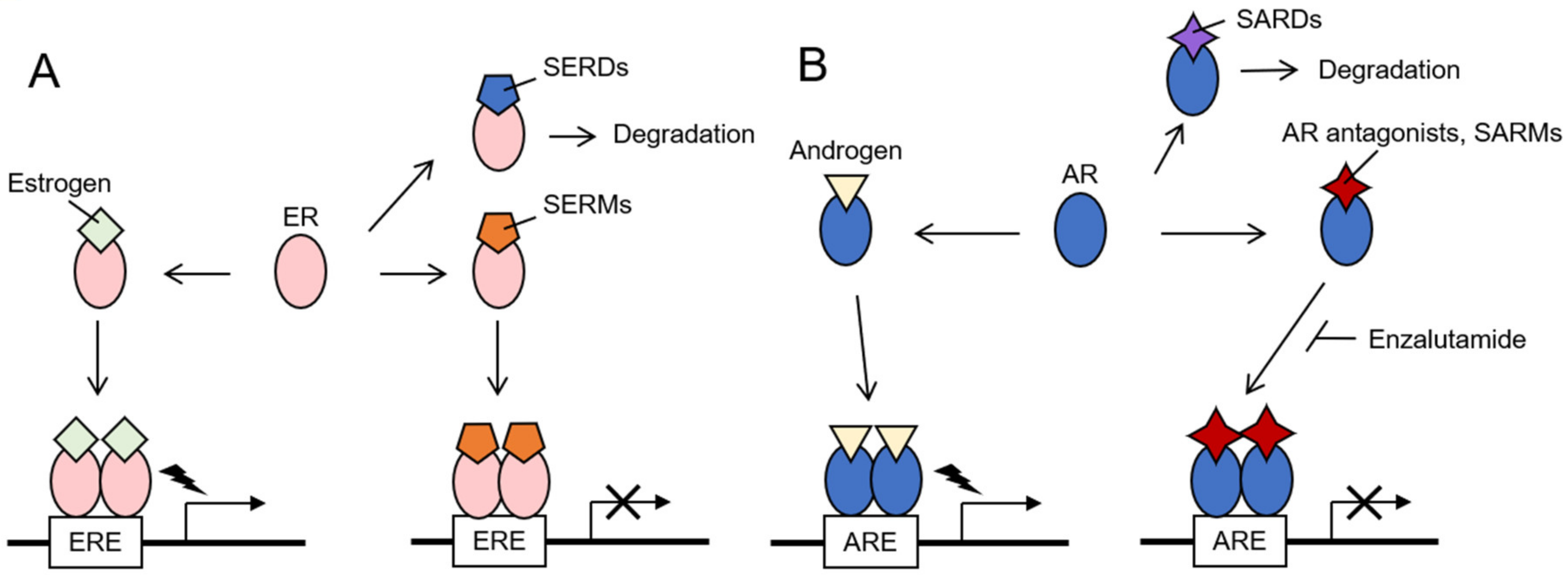
2. Hormone-Related Cancers

Several cancers including breast, endometrial, ovarian, prostate, testicular and thyroid cancers are categorized as hormone-related cancers, in which cell proliferation is driven by various endogenous and exogenous hormones [12]. Among women worldwide, breast cancer is the most commonly diagnosed cancer and the leading cause of cancer deaths. For men, prostate cancer is the second commonly diagnosed cancer, following lung cancer, and the fifth leading cause of cancer deaths worldwide. According to the GLOBOCAN 2018, which estimates the cancer incidence and mortality produced by the International Agency for Research on Cancer (IARC), the numbers of new cases of breast and prostate cancers in 2018 are estimated as 2,088,849 and 626,679, respectively, those of deaths due to breast and prostate cancers are estimated as 1,276,106 and 358,989, respectively [13]. The majority of breast cancers are primarily sensitive to sex steroid hormones, estrogen and progesterone, and the majority of prostate cancers are sensitive to another sex hormone, androgen [14,15,16,17]. The receptors for these sex hormones, namely, estrogen, progesterone and androgen receptors (ERs, PRs, and ARs, respectively) are ligand-dependent transcription factors. With their cognate ligands, these hormone receptors dimerize and associate with DNA through their DNA-binding domains. These hormone receptors form complexes with other transcription factors and co-regulators, such as the steroid receptor coactivator (SRC)/p160 family proteins and CREB binding protein (CBP)/p300, and they contribute to the transcriptional regulation of their target genes (Figure 1) [16,17,18,19,20,21].
Since sex hormone signaling pathways play essential roles in cancer pathophysiology; the therapies targeting the hormones and their receptors, or endocrine therapies, are the mainstays of breast and prostate cancer treatment [22,23,24,25,26,27,28,29]. For breast cancer treatment, drugs that suppress estrogen signaling or estrogen production were used for endocrine therapies. To suppress estrogen-mediated ER activation, the drugs such as selective estrogen receptor modulators (SERMs) and selective estrogen receptor degraders or down-regulators (SERDs) are used. Although both SERMs and SERDs compete with estrogen, they differently regulate ER signaling (Figure 1A). SERMs modulate the ER activity by affecting the interaction with co-factors, resulting in the changes of ER-targeted gene expression. SERMs like tamoxifen and raloxifene function as ER antagonists in breast cancer and are used for breast cancer therapy or prevention. In contrast, SERDs, such as fulvestrant, induce the destabilization of ER and abolish ER signaling [23]. Aromatase inhibitors and luteinizing hormone-releasing agonists are used to prevent the estrogen synthesis [22]. For prostate cancer treatment, orchiectomy, a surgery to eliminate testis, has been originally performed as androgen deprivation therapy for years [24,25]. For chemical castration, gonadotropin-releasing hormone agonists and antagonists are used to prevent the androgen synthesis. Abiraterone has been recently used for metastatic castration-resistant prostate cancer (CRPC) as a selective inhibitor for androgen biosynthesis by blocking the enzymatic activity of cytochrome P-450c17 (CYP17) [26]. As androgen receptor antagonists that prevent androgen–AR interaction, flutamide and bicalutamide have been used for years, and recently more potent AR antagonists, such as enzalutamide, has been applied to CRPC and reported to decrease the risk of cancer progression and death [27]. Selective androgen receptor modulators (SARMs) that function as AR antagonists in specific cell types including prostate cancer cells, and selective androgen receptor degraders or down-regulators (SARDs) are being studied for clinical application (Figure 1B) [28,29].
Although the endocrine therapies are initially successful, both breast and prostate cancers eventually acquire the therapy resistance. Endocrine therapy-resistant CRPC usually arises in the majority of prostate cancer patients with treatment within two to three years [30]. On the contrary, it has been reported that the distant recurrence of breast cancer occur in 20%–50% of the patients during the period of 20 years from the start of the endocrine therapy [31]. While the majority of breast cancers are sensitive to the endocrine therapy, patients with triple-negative breast cancer (TNBC) exhibit poor outcomes as the subtype lacks effective therapeutic targets including ER, PR and human epidermal growth factor receptor 2 (HER2)/ErbB2. Thus, novel strategies for breast and prostate cancers remain to be developed. Recent studies have indicated that the cancer-specific splicing events are closely associated with cancer progression, suggesting that splicing factors could be promising targets for cancer treatment [8,9,10]. Interestingly, alternative splicing of AR in prostate cancer is suggested to be important for acquiring the endocrine therapy resistance, as described in a later part.

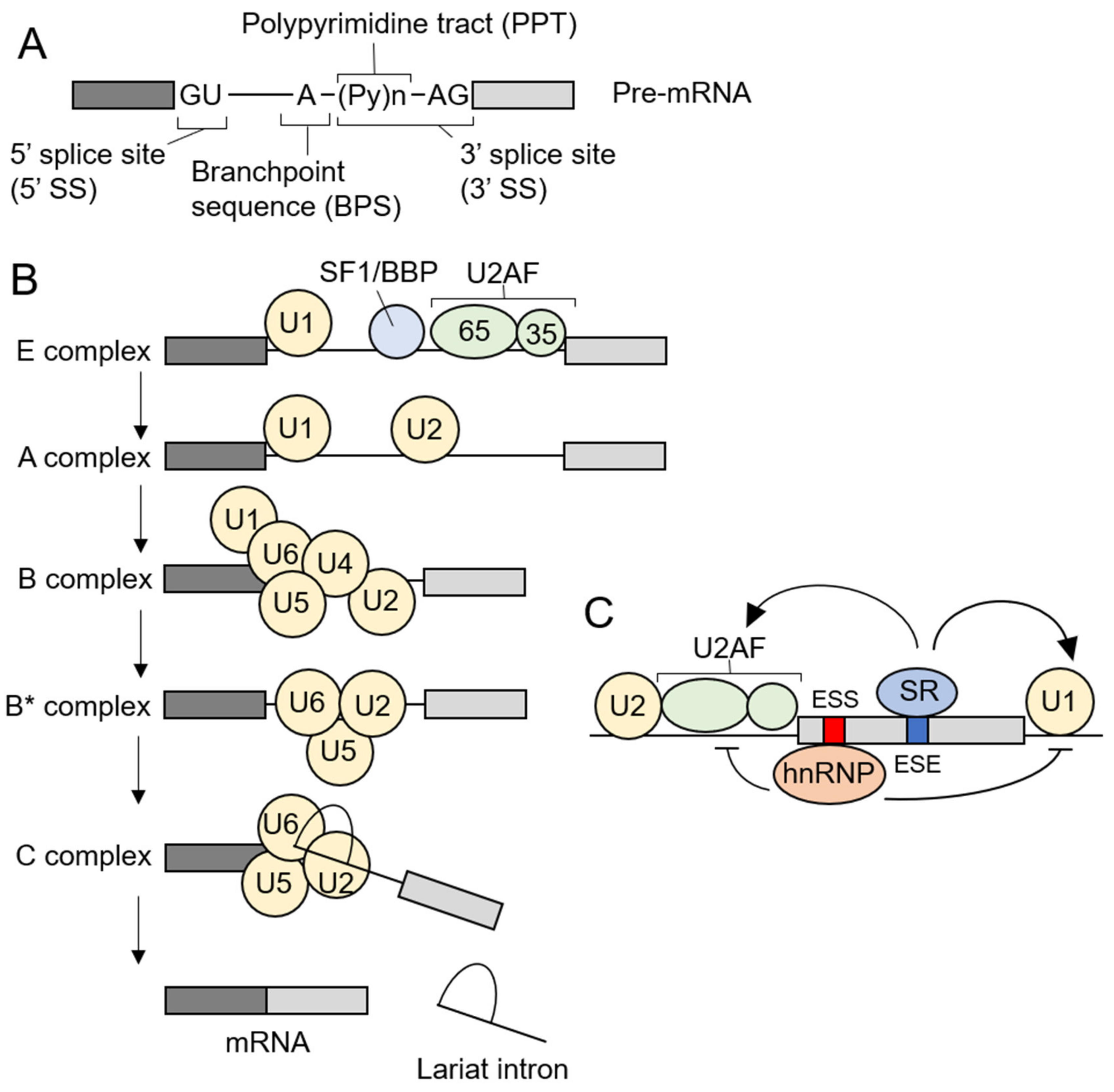
3. Pre-mRNA Splicing and Spliceosome Assembly

Pre-mRNA splicing is carried out by spliceosomes [1,2] and there are two types of spliceosomes in humans, known as major (U2-dependent) and minor (U12-dependent) spliceosomes. The major and minor spliceosomes recognize and excise the major (U2-type) or minor (U12-type) introns, respectively. The most human introns are the major introns, while the minor introns make up only ~0.35% of human introns and are contained in 700-800 genes, each of which basically has one minor intron and multiple major introns [32]. The major spliceosome consists of five small nuclear ribonucleoproteins (snRNPs) including U1, U2, U4, U5, and U6, and proteins called splicing factors. The assembly of the major spliceosomes requires the conserved sequences of the pre-mRNA, the 5’ splice site (5’ SS), the branchpoint sequence (BPS), and the 3’ splice site (3’ SS). The 3’ SS contains the continuous pyrimidine sequences termed polypyrimidine tract (PPT) and following conserved AG-dinucleotides (Figure 2A). In the early phase of the spliceosome assembly, U1 snRNP, splicing factor 1 (SF1)/branchpoint binding protein (BBP) and U2 snRNP auxiliary factor (U2AF) bind to the 5’ SS, BPS and the 3’ SS, respectively (E complex). U2AF is a heterodimer of U2AF65 and U2AF35, which recognize PPT or AG-dinucleotides of the 3’ SS, respectively. Next, U2 replaces SF1 to generate A complex. U2 snRNP consists of U2 small nuclear RNA (snRNA) and multiple proteins, including SF3b complex, and recognizes BPS through base-pairing between U2 snRNA and BPS. U4, U5 and U6 form a complex, U4/U6.U5 tri-snRNP, which is recruited to pre-mRNA to form B complex. After U1 and U4 exit, the major spliceosome becomes catalytically active (B* complex) and catalyzes two-sequential steps of splicing, resulting in ligation of exons and removal of lariat introns (Figure 2B) [1]. Although the 5’/3’ SS and BPS are required for pre-mRNA splicing, these sequences alone are often insufficient to exon/intron recognition. It has been revealed that the majority of pre-mRNAs possess the additional splicing regulatory sequences in exonic and intronic regions. The sequences that enhance the intron exclusion are termed exonic or intronic splicing enhancers (ESEs or ISEs), while the sequences that suppress the intron exclusion are called exonic or intronic splicing silencers (ESSs or ISSs) [3,4]. Splicing factors, such as serine/arginine-rich proteins (SR proteins) and heterogeneous nuclear ribonucleoproteins (hnRNPs), bind to these splicing regulatory elements and regulate the spliceosome assembly (Figure 2C). SR proteins are RNA-binding proteins characterized by having one or two RNA recognition motifs (RRMs) in their N-termini and a domain rich in arginine and serine (RS domain) in their C-termini. RS domains are involved in the interaction with other splicing factors including SR proteins [33,34]. SR proteins generally bind to ESEs and ISEs and facilitate intron exclusion through interaction with U1 and U2AF. On the other hand, hnRNPs typically bind to ESSs and ISSs to inhibit the intron exclusion. The functions of SR proteins and hnRNPs; however, are shown to be context-dependent [3,4].
In the minor spliceosomes, U11, U12, U4atac and U6atac replace U1, U2, U4 and U6, respectively. U5 snRNP is shared by both spliceosomes [32,35]. The minor introns have 5’/3’ SS and BPS, which have different sequences from those of the major introns. In contrast to the sequential recognition of the 5’ SS and BPS by U1 and U2, U11 and U12 form the U11/U12 di-snRNP complex, and cooperatively recognize these sequences. Although U2AF recognizes the 3’ SS in major introns, the 3’ SS in minor introns lacks PPT and is recognized by zinc finger CCCH-type, RNA binding motif and serine/arginine rich 2 (ZRSR2)/U2AF35-related protein (Urp). ZRSR2 was also shown to function in the major spliceosome, specifically required for the second step of splicing [36].

4. Alteration of Splicing Factors in Cancer

4.1. Somatic Mutations of Splicing Factors in Cancer

Recent whole-genome sequencing analyses have revealed that recurrent somatic mutations occur in some genes of splicing factors in cancers [8,37]. Mutations of splicing factors were initially identified in patients with several types of hematological malignancies, such as myelodysplastic syndromes (MDS) and related disorders (myelodysplasia) [38,39,40,41], as well as chronic lymphocytic leukemia (CLL) [42,43,44]. Especially, the majority of splicing factors that are frequently mutated in those cancers are components of the E/A complex and are involved in BPS and 3’ SS recognition, suggesting that the specific steps of the spliceosome assembly may have crucial roles in hematological malignancies. The most common mutations are found in splicing factor 3b subunit 1 (SF3B1), U2AF35, serine/arginine-rich splicing factor 2 (SRSF2) and ZRSR2 [8,37]. SF3B1 is a component of SF3b complex and SRSF2 is a member of SR proteins. In fact, some mutations in SF3B1, U2AF35 and SRSF2 have been demonstrated to affect alternative splicing [38,40,44,45,46,47,48,49,50,51,52], and SF3B1 and SRSF2 mutations are suggested to be adverse prognostic risks of CLL and of MDS, respectively [43,53,54,55,56]. Interestingly, the splicing factor mutations occur in a mutually-exclusive manner in myelodysplasia [38]. It may suggest synthetic lethality of those mutations. SF3B1 and U2AF35 mutations have been also found in the solid cancers. SF3B1 mutations have been identified in uveal melanoma (15%–29%), cutaneous melanoma (1%), pleural mesothelioma (2%), pancreatic ductal adenocarcinoma (3%), breast cancer (2%–4%), and prostate cancer (1.1%), while U2AF35 mutations were found in lung adenocarcinoma (3%) and prostate cancer (0.5%) [8,57].
Furthermore, mutations occur not only in protein components of the spliceosome. Recently, mutations in an RNA component of the spliceosome were discovered in several types of cancers. U1 snRNA is an RNA component of U1 snRNP and plays an essential role in the initial recognition of 5’ SS through base-pairing between the 5’ part of U1 snRNA and 5’ SS. It was reported that several bases of U1 snRNA are mutated in some cancers including CLL, hepatocellular carcinoma (HCC) and Sonic hedgehog (SHH) medulloblastomas [58,59]. Among these mutations, mutations of the third base of U1 snRNA demonstrated the changes of 5’ SS recognition. A to C or G mutation of the third base of U1 snRNA results in mis-splicing of several cancer-related genes such as CD44, Musashi RNA binding protein 2 (MSI2) and DNA polymerase delta 1, catalytic subunit (POLD1) in CLL and Patched 1 (PTCH1) and GLI family zinc finger 2 (GLI2) in SHH medulloblastomas, respectively [58,59]. In addition, these U1 snRNA mutations were suggested to be associated with the pathophysiology of CLL, HCC and SHH medulloblastoma. Thus, mutations of early spliceosome complexes are closely associated with the diverse types of cancers and initial steps of the spliceosome assembly could be promising targets for cancer treatment.

4.2. Deregulated Gene Expression of Splicing Factors in Cancer

Although recurrent somatic mutations of splicing factors are detected in the solid cancers, those mutations are generally rare. Instead, alteration of gene expression is frequently observed. Deregulated expression of some splicing factors such as DBHS family proteins and SR proteins induces cancer-specific splicing events and promotes cancer progression. However, whether the splicing factors function as oncogenes or tumor-suppressors is dependent on cancer types. For example, it was shown that serine/arginine-rich splicing factor 3 (SRSF3) promotes the progression of breast cancer but suppresses hepatic carcinogenesis [60,61]. Moreover, it has been implicated that the post-translational modification of splicing factors regulates cancer-specific splicing events. For instance, SR protein kinases (SRPKs) and CDC-like kinases (CLKs) phosphorylate and regulate the activity of SR proteins and control alternative splicing events [29].
In the following sections, we introduce the roles of DBHS family proteins, SR proteins and hnRNPs in breast and prostate cancers. In addition, we also discuss the functions of other splicing factors and splicing regulatory proteins in those cancers.

5. The Roles of Splicing Factors and Splicing Regulatory Proteins in Breast and Prostate Cancers

5.1. DBHS Family Proteins
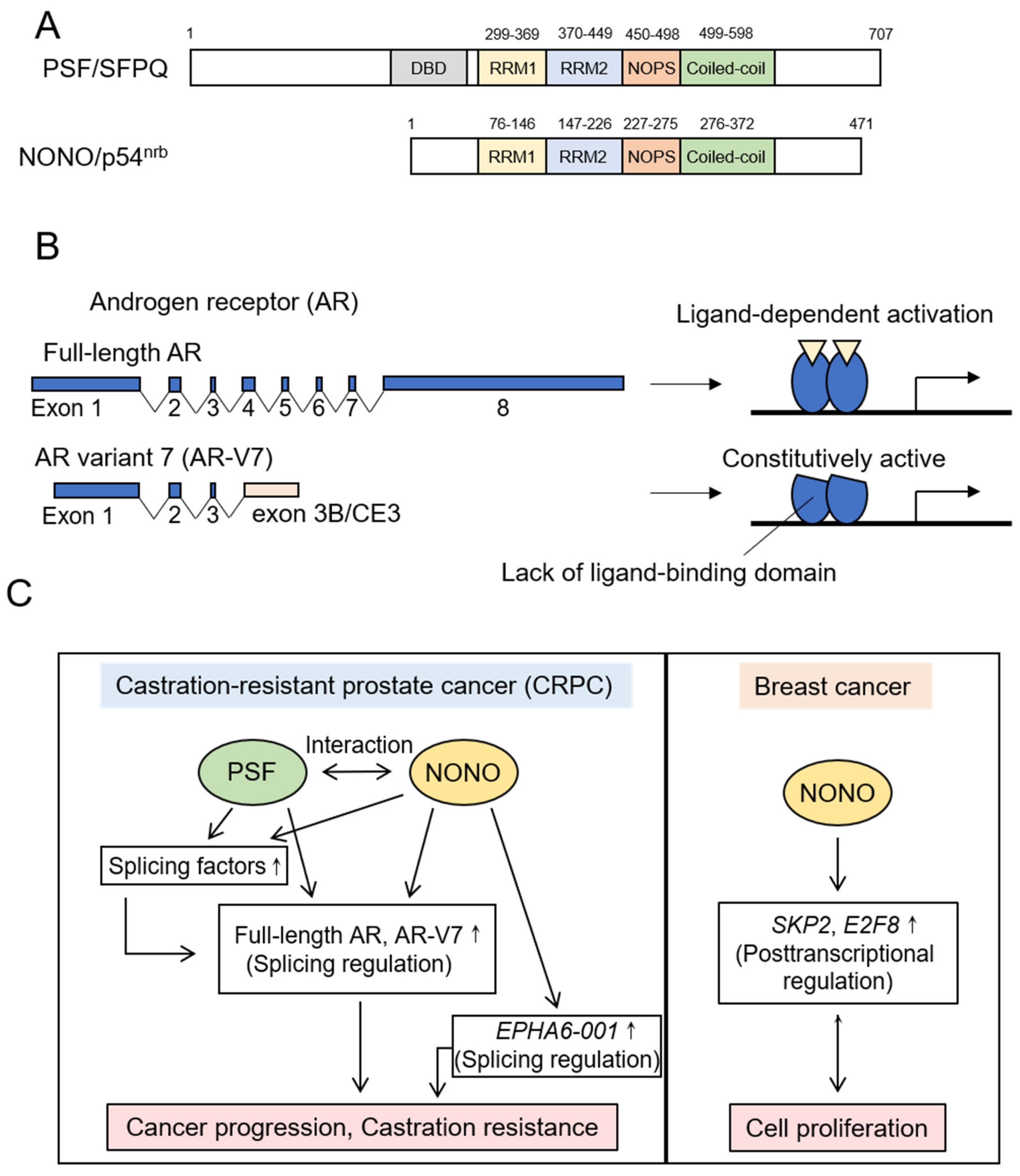
The DBHS family proteins are defined by a conserved core region termed the DBHS region. The DBHS region consists of tandem RNA recognition motifs (RRM1 and RRM2), a NonA/paraspeckle (NOPS) domain and a C-terminal coiled-coil domain (Figure 3A) [62]. In humans, there are three DBHS family members: PTB-associated splicing factor (PSF)/splicing factor proline/glutamine rich (SFPQ), Non-POU domain-containing octamer-binding protein (NONO)/nuclear RNA-binding protein, 54 kDa (p54nrb) and paraspeckle component 1 (PSPC1)/paraspeckle protein 1 (PSP1). These proteins form homodimer or heterodimer with other DBHS family proteins by reciprocal interactions between RRM2, NOPS and coiled-coil domains [62]. In addition, a recent study suggests that oligomerization of DBHS family proteins is mediated by the interaction between coiled-coil domains [63]. The DBHS family proteins can interact with proteins, RNA and DNA, and are involved in multiple cellular events such as pre-mRNA splicing, transcriptional activation and repression, DNA repair and the formation of a subnuclear ribonucleoprotein body, or paraspeckle [62]. It has recently been reported that PSF and NONO in breast and prostate cancers promote cancer progression by regulating cancer-specific splicing events. In the following sections, we describe recent findings on splicing regulation by PSF and NONO in both cancers.

5.1.1. PSF

As for PSF expression in prostate cancer, PSF expression is upregulated in prostate cancer cells compared to normal prostate epithelial cells [64]. In addition, PSF protein is highly expressed in a subset of tumor samples and higher expression of PSF correlates with cancer-specific survival after surgery and the prostate specific antigen (PSA)-free survival after endocrine therapy. Moreover, PSF mRNA is increased in metastatic and advanced prostate cancer clinical samples [64]. These data suggest that PSF plays important roles in the pathophysiology of prostate cancer. Consistently, PSF is demonstrated to be essential for the in vitro growth of hormone-refractory prostate cancer cells and the in vivo tumor growth in castrated mice as a CRPC model [64]. PSF binds to multiple transcripts and upregulates the stability of its target transcripts. Moreover, the main targets of PSF are transcripts of the spliceosome genes such as SF1 and splicing factor 3b subunit 2 (SF3B2). Therefore, PSF would control a broad range of cancer-specific splicing events by upregulating the expression of spliceosome genes, resulting in the activation of oncogenic pathways (Figure 3C) [64].
Importantly, PSF targets include an AR splice variant, AR-V7, which is closely linked to the acquirement of endocrine therapy resistance [64]. Among multiple AR splice variants, AR-V7 is the most common AR variant in CRPC [65,66,67,68]. Previous studies indicated that AR-V7 expression was detected in circulating tumor cells (CTCs) from CRPC patients and associated with endocrine therapy resistance using abiraterone [26] and enzalutamide [27]. Moreover, AR-V7-positive CTCs are associated with worse PSA response rates and shorter progression-free and overall survival rates. Therefore, AR-V7 may be promising for a potential target of diagnosis, prognosis and therapy in metastatic CRPC [69,70,71]. The full-length AR protein is encoded by AR isoform composed of eight exons and consists of four core domains: The N-terminal domain (NTD), the DNA binding domain (DBD), the hinge region and the C-terminal ligand binding domain (LBD). In contrast, AR-V7 protein is encoded by AR variant composed of exons 1, 2, and 3 and a cryptic exon termed exon 3B or cryptic exon 3 (CE3), which is generated by alternative use of the cryptic 3’ SS in intron 3 (Figure 3B). The resultant AR-V7 protein includes the NTD and DBD, but lacks the hinge region and LBD [72,73]. The AR-V7 protein is constitutively active and forms a heterodimer with the full-length AR protein, leading to the androgen-independent activation of canonical AR signaling. Moreover, the AR-V7 protein is indicated to form homodimer and regulate a unique set of genes [72,73].
PSF is suggested to regulate the splicing events that produce the full-length AR and AR-V7 variant. PSF forms a complex with NONO and other splicing factors including U2AF65, hnRNPU and DDX23, and promotes the expression of mRNAs encoding the full-length AR and AR-V7 (Figure 3C) [64]. Moreover, PSF upregulates the expression of SF3B2 that is a component of the SF3b complex, and associates with the splicing of AR pre-mRNA to generate AR-V7 variant as mentioned below [64]. In addition, transcripts of AR-regulated genes are PSF targets, suggesting that PSF plays a role in AR signaling. Thus, PSF may regulate the AR signaling pathway at multiple steps, which lead to prostate cancer progression [64].

5.1.2. NONO

It is recently indicated that high-NONO immunoreactivity associates with poor prognosis of breast cancer patients [74]. Public breast cancer databases also show that NONO mRNA expression is increased in invasive ductal breast cancer compared to normal breast samples, and higher expression of NONO mRNA associates with poor distant disease-free and overall survivals of the patients with all subtypes and ER-positive subtype of breast cancers [74]. In terms of prostate cancer, the expression of NONO is upregulated in CRPC and metastatic prostate cancers, and increased expression of NONO associates with poor prognosis of prostate cancer patients [64]. These data suggest that NONO plays a key role in the progression of both breast and prostate cancers [64,74].
A recent study reported that NONO in breast cancer regulates the expression of S-phase-associated kinase 2 (SKP2) and E2F transcription factor 8 (E2F8) at the post-transcriptional level, promoting breast cancer proliferation (Figure 3C) [74]. In this report, NONO knockdown decreased the expression level of exonic regions of these mRNAs, while hardly affected the expression of intronic regions. These data suggest that NONO may regulate the expression of SKP2 and E2F8 by controlling their splicing processes [74]. As for NONO functions in prostate cancer, it is indicated that NONO promotes the proliferation of prostate cancer cells and controls the splicing regulation, which upregulates the expression of mRNAs encoding the full-length AR and AR-V7 proteins in CRPC (Figure 3C). Moreover, NONO increases the expression of some splicing factors, which would affect the splicing events in CRPC [64]. In addition, NONO modulates alternative splicing of ephrin type-A receptor 6 (EPHA6) pre-mRNA in CRPC [75]. EPHA6 is an ephrin receptor reported to be overexpressed and to promote angiogenesis and metastasis in prostate cancer. NONO induces the production of a splice variant, EPHA6-001, which contributes to the growth of CRPC by unknown mechanisms [75].
Thus, recent findings have shown that the DBHS family proteins PSF and NONO regulate cancer-specific splicing events and promote breast and prostate cancer progression. Future studies will elucidate the precise molecular mechanisms of splicing regulation by these DBHS family proteins.

5.2. SR Proteins

In breast cancer, a member of SR proteins, SRSF1, has been shown to regulate various alternative splicing events of cancer-related genes and promote cancer progression. Recepteur d’origine nantais (Ron) is a tyrosine kinase receptor for macrophage-stimulating protein (MSP) and is involved in cell motility and matrix invasion. SRSF1 is suggested to interact with an exonic splicing enhancer (ESE) in exon 12 of Ron pre-mRNA and induce the skipping of exon 11 [75]. The resultant mRNA isoform is translated to a constitutively active isoform of Ron, ΔRon, which upregulates breast cancer cell motility [76]. Moreover, SRSF1 controls alternative splicing of myeloid cell leukemia-1 (Mcl-1) pre-mRNA [77]. Mcl-1 is a member of the B-cell lymphoma 2 (Bcl-2) family and regulates the apoptotic process. An Mcl-1 pre-mRNA contains three exons, and the inclusion or skipping of exon 2 results in the production of two functionally different isoforms, the pro-apoptotic Mcl-1S and the anti-apoptotic Mcl-1L. SRSF1 is indicated to upregulate the inclusion of exon 2 to produce Mcl-1L [77]. It is also suggested that SRSF1 may affect protein stability and translation of Mcl-1 [77]. In addition to Mcl-1, SRSF1 controls splicing events of another Bcl-2 family member, Bcl-2 interacting mediator of cell death (BIM)/Bcl-2 like 11 (BCL2L11), leading to the upregulation of splice variants, BIM γ1 and γ2 [78]. Although BIM possesses the BH3 domain that is necessary for the induction of apoptosis, BIM γ1 and γ2 lack the BH3 domain. Importantly, overexpression of BIM γ1, but not γ2, was reported to promote the growth and survival of MCF-10A mammary epithelial cell line cultured in Matrigel [78]. Therefore, splicing regulation of Mcl-1 and BIM by SRSF1 may contribute to breast cancer cell survival. Furthermore, SRSF1 regulates the expression of bridging integrator 1 (BIN1) and mitogen-activated protein kinase (MAPK) signal-integrating kinase 2 (Mnk2), which are tumor suppressor and kinase, respectively [78]. SRSF1 promotes the inclusion of exon 12a of BIN1 to generate an isoform, BIN+12a, which lacks tumor-suppressive activity. On the other hand, SRSF1 enhances the inclusion of exon 13b of Mnk2, generating the Mnk2b isoform that increases the phosphorylation of translational initiation factor eIF4E. Mnk2-mediated hyperactivation of eIF4E may promote the growth and survival of breast cancer. Thus, SRSF1 regulates various aspects of breast cancer such as cell proliferation and motility.
Meanwhile, it was reported that SRSF1 regulates splicing of AR pre-mRNA to generate AR-V7 in prostate cancer [79]. Moreover, SRSF1 regulates the production of cyclin D1 (CCND1) isoform CCND1b in prostate cancer [80]. It was previously reported that CCND1b was upregulated in prostate cancer compared to non-neoplastic tissue and could promote anchorage-independent growth and cell invasiveness [81,82]. A transcript encoding CCND1b is generated by the premature transcriptional termination in intron 4 and the partial retention of intron 4. CCND1b lacks exon 5-encoded amino acids and contains a novel C-terminal domain of unknown function. SRSF1 is suggested to bind to the CCND1 exon 4–intron 4 boundary and promote CCND1b expression [80]. Since the upregulation of CCND1b was also observed in breast cancer, SRSF1 may regulate CCND1b expression in the similar manner in breast and prostate cancers [83].
In addition to SRSF1, the roles of SRSF5 in breast and prostate cancers have been reported. In breast cancer, SRSF5 is shown to regulate alternative splicing of Mcl-1 pre-mRNA to produce Mcl-1L [77]. On the other hand, SRSF5 is indicated to be involved in pre-mRNA splicing of Kruppel-like factor 6 (KLF6) in prostate cancer [84,85]. KLF6 is a member of Kruppel-like zinc finger transcription factors and functions as a tumor suppressor. The allelic loss and somatic mutations of KLF6 were observed in patients with sporadic prostate cancer and a germline single nucleotide polymorphism (SNP) in KLF6 gene IVS1-27 G > A is shown to cause aberrant splicing to generate 3 splice variants, KLF6-SV1, -SV2 and -SV3. Moreover, KLF6-SV1 and -SV2 antagonize wild-type KLF6 function and promote cell proliferation through decreasing the expression of p21 [84,85]. It is suggested that IVS1-27 G > A polymorphism generates a novel binding site of SRSF5, which enhances alternative splicing of KLF6 in a dose-dependent manner [84].
Furthermore, the roles of other SR proteins, including SRSF3, SRSF4 and SRSF6, have been shown in breast cancer. It was reported that SRSF3, in cooperation with another splicing factor TAR DNA-binding protein 43 (TDP43), plays an important role in the growth and metastasis of TNBC tumors. SRSF3 and TDP43 enhance the skipping of exon 12 of partitioning defective 3 (PAR3) and the inclusion of exon 12 of NUMB, and these splicing alterations promote the growth and metastasis of TNBC tumors, respectively [60]. In addition, SRSF3 is indicated to regulate alternative splicing of HER2 [86]. SRSF3 enhances the removal of intron 15 and the skipping of exon 16 of HER2. These changes in HER2 splicing downregulate the p100 isoform and upregulate the Δ16HER2 isoform, the former is a secreted protein that inhibits cell proliferation and the latter is a constitutively active isoform, respectively. Therefore, SRSF3 may contribute to cell proliferation through modulating the production of HER2 isoforms [86].
SRSF4 and SRSF6, like SRSF1, were reported to play crucial roles in the proliferation of MCF-10A cells cultured in Matrigel [87]. SRSF4 and SRSF6 controlled alternative splicing events of genes that regulate transformation-associated processes and the expression of genes associated with cell proliferation, migration, cytoskeleton organization, polarity, cell signaling or cholesterol metabolism [87]. In addition, SRSF4 is suggested to be involved in cell death caused by cisplatin treatment possibly through the splicing regulation [88]. Thus, SR proteins regulate a broad range of cellular processes to promote breast and prostate cancer progression by controlling cancer-specific splicing events, suggesting that the expressions and activities of SR proteins are important for cancer progression.

5.3. hnRNPs

Previous studies suggested that hnRNPs play key roles in the production of AR-V7 in prostate cancer. A member of hnRNPs, hnRNPF, is shown to enhance the generation of AR-V7. It was reported that histone demethylase jumonji domain containing 1A (JMJD1A) recruits hnRNPF onto the region around the 5’ side of AR exon 3B, and JMJD1A and hnRNPF support the recruitment of splicing factors including U2AF65 to the 3’ SS in front of exon 3B, which leads to the inclusion of exon 3B to produce AR-V7 [89]. Moreover, another hnRNP family member hnRNPA1 is suggested to be involved in AR-V7 expression [90,91,92]. It was previously reported that hnRNPA1 is highly expressed in prostate tumors, compared to benign prostates, and upregulates the expression of AR-V7 [90]. Furthermore, it was shown that the downregulation of hnRNPA1 by quercetin, a flavonoid abundantly present in plants, concomitantly decreases the expression of AR-V7, resensitizing enzalutamide-resistant prostate cancer cells to enzalutamide treatment in mouse xenografts [91]. However, another study suggested that hnRNPA1 suppresses AR-V7 expression in prostate cancer cells [92]. This contradiction may be due to the difference of cell lines used in their studies, suggesting cell-type specific functions of hnRNPA1 in splicing regulation. In addition, hnRNPL is indicated to bind to intron 3 of AR and to control the inclusion of another cryptic exon termed exon 2b [93]. Although the significance of exon 2b inclusion is unknown, hnRNPL regulates alternative splicing of multiple genes and circular RNA formation, and promotes prostate cancer growth and survival [93,94].
In breast cancer, hnRNPs promote cell survival and tumor metastasis. It is suggested that hnRNPF, hnRNPH1 and hnRNPK enhance the production of Mcl-1L to inhibit cell death [95]. Moreover, hnRNPM was shown to regulate alternative splicing of CD44 [96]. CD44 is a cell surface adhesion receptor involved in epithelial to mesenchymal transition (EMT) processes [97], and hnRNPM upregulates alternative splicing of CD44, which results in EMT and metastasis at final stage [96]. In TNBC, it was reported that a substitution mutation M276I in a chromatin remodeling protein microrchidia family CW-type zinc finger 2 (MORC2) that promotes the interaction between MORC2 and hnRNPM and the hnRNPM-mediated CD44 splicing switch, enhancing invasion and metastasis of TNBC cells in immunodeficient mice [98].

5.4. Other Splicing Factors and Splicing Regulatory Proteins in Breast and Prostate Cancers

5.4.1. SF3b Complex

As mentioned above, the SF3b complex is a component of U2 snRNP and the mutations of the SF3b complex component SF3B1 have been reported in some cancers, including breast and prostate cancers [8,57]. Although less frequently than SF3B1, the mutations of SF3B3 and SF3B4, other components of the SF3b complex, are also observed in breast cancers (0.2% each) [99]. Regarding expressions of SF3b complex components, it was reported that SF3B1 and SF3B3 are upregulated in fulvestrant/tamoxifen-resistant breast cancer cells, and SF3B3 overexpression associates with poor relapse-free and overall survival in ER-positive breast cancer patients [100]. In addition, the elevated expression of another component of SF3b complex, PHD finger 5A (PHF5A)/SF3B7, is observed in breast tumors, and high PHF5A expression correlates with poor disease-free survival in breast cancer patients [101]. PHF5A is reported to upregulate the stability of SF3b complex and to modulate alternative splicing events of multiple genes involved in apoptotic processes, such as FAS-activated serine/threonine kinase (FASTK) that inhibits apoptosis [101]. These data suggest that SF3b complex may be a potential target of breast cancer treatment. On the other hand, another component of SF3b complex SF3B2 is increased in CRPC and prostate cancers with high expression of AR-V7 and associates with poor progression-free survival in prostate cancer patients [102]. In prostate cancer, SF3B2 directly binds to CE3 of AR and controls alternative splicing of AR to produce AR-V7. Interestingly, pladienolide B, an mRNA splicing inhibitor/modulator, decreases AR-V7 expression through modulating the SF3b complex and causes the regression of prostate cancer xenografts in castrated mice. Moreover, prostate cancer xenografts with SF3B2-overexpression are more sensitive to pladienolide B treatment [102]. SF3B1 is overexpressed in prostate tumors and associates with aggressive features of prostate cancers [103]. SF3B1 expression positively correlates with AR-V7 expression, and pladienolide B treatment decreases AR-V7 expression. In addition, pladienolide B treatment decreases the expression of In1-Ghrelin, an oncogenic splice variant of Ghrelin, suggesting that SF3B1 or the SF3b complex associates with alternative splicing of Ghrelin. Supporting these findings, pladienolide B treatment is indicated to suppress the proliferation, survival and migration of prostate cancer cells [103]. Therefore, SF3B2, SF3B1 and the SF3b complex may be good candidates of therapeutic targets, and small chemicals inhibiting/modulating the SF3b complex might be promising for prostate cancer treatment.

5.4.2. Src Associated in Mitosis of 68 kDa (Sam68)

Sam68 belongs to the signal transduction and activation of RNA metabolism (STAR) family of RNA-binding proteins. Sam68 can interact with multiple RNAs and proteins, and is involved in various steps of gene expression, such as transcription, pre-mRNA splicing and translation [104]. Previous studies demonstrated that Sam68 plays a crucial role in alternative splicing of cancer-related genes in breast and prostate cancers. For example, Sam68 enhances the inclusion of variable exon 5 of CD44, promoting the migration of both cancer cells [105,106]. In prostate cancer, transcriptional coactivator staphylococcal nuclease and tudor domain containing 1 (SND1) promotes the recruitment of Sam68 onto CD44 pre-mRNA, suggesting the significance of transcription-coupled RNA splicing events in cancer [106]. In addition, Sam68 upregulates AR-V7 and CCND1b expression by controlling alternative splicing of AR and CCND1 [107,108]. Besides splicing regulation, Sam68 is shown to promote the transcription of AR-V7 target genes possibly through the interaction between Sam68 and AR-V7 [108]. Although these Sam68 functions result in cancer progression, Sam68 upregulation can cause prostate cancer cell death partly through the splicing alteration of Bcl-2 family member BCL-X. There are two BCL-X isoforms generated by the alternative use of two 5’ SS regions in exon 2: anti-apoptotic BCL-XL and pro-apoptotic BCL-XS. Sam68 controls a splicing switch toward BCL-XS, leading to apoptosis [109]. Since Sam68 can regulate cancer progression positively or negatively, it is suggested that Sam68 activity may be strictly regulated in cancer. Previous studies demonstrated that post-translational modifications play essential roles in the regulation of Sam68 activity. Especially, the acetylation of Sam68 is suggested to enhance its activities of RNA binding and splicing regulation in breast and prostate cancers. It is indicated that an acetyltransferase CBP could regulate Sam68 acetylation [110], while histone deacetylase 6 (HDAC6) causes Sam68 deacetylation in cooperation with a nuclear matrix-associated protein, scaffold/matrix-associated region-binding protein 1 (SMAR1), in breast cancer [111]. With respect to HDAC6, it is suggested that HDAC6 interacts with ERα in an ER ligand-dependent manner and subsequently upregulates deacetylation of α-tubulin in breast cancer cells [112]. The deacetylation of α-tubulin is supposed to contribute to the migration of breast cancer cells and the growth of breast cancer xenografts in nude mice [112]. Thus, the HDAC6-mediated deacetylation process might play a pivotal role in controlling breast cancer progression.
On the other hand, in prostate cancer, a factor that binds to inducer of short transcripts protein 1 (FBI-1) is shown to interact with Sam68, and control Sam68-mediated alternative splicing of BCL-X through histone deacetylases, suggesting the importance of acetylation for Sam68 activity [109]. Targeting these Sam68 acetylation mechanisms may provide a novel therapeutic approach in breast and prostate cancers.

5.4.3. SR Protein Kinases

SR proteins are phosphorylated by multiple kinases including SRPKs and CDC-like kinases (CLKs). Phosphorylation of SR proteins is essential for their nuclear import and interactions with target RNAs and other splicing factors [34]. Considering the roles of SR proteins in cancer-specific splicing events, kinases that phosphorylate SR proteins could be promising targets for cancer treatment. SRPK1, a member of SRPK, is shown to be highly expressed in various cancers including breast and prostate cancers [113,114,115,116]. Among breast cancer cells, ER-negative breast cancer cells show higher expression of SRPK1, and knockdown of SRPK1 was reported to inhibit metastasis of ER-negative breast tumors in immunodeficient mice. Moreover, high expression of SRPK1 correlates with decreased metastasis-free survival of the patients with breast cancer and preferential metastasis to lung and brain [116]. These data suggest that SRPK1 play essential roles in breast tumor metastasis. Meanwhile, SRPK1 in prostate cancer is shown to promote angiogenesis [117]. Vascular endothelial growth factor (VEGF) is a key player in angiogenesis, and alternative splicing of VEGFA pre-mRNA generates two groups of VEGF isoforms by alternative use of two 3’ SS regions in exon 8 of VEGFA. The VEGF165 angiogenic isoform is generated by splicing using the proximal 3’ SS, while the VEGF165b anti-angiogenic isoform is produced by splicing using the distal 3’ SS. SRPK1 is indicated to promote the use of the proximal 3’ SS to upregulate VEGF165 expression, which results in angiogenesis and prostate tumor growth in nude mice [117]. Since alternative splicing of VEGFA was reported to be regulated by SR proteins such as SRSF1 and SRSF6 [118], SRPK1 may associate with the alternative splicing of VEGFA by controlling the phosphorylation states of these SR proteins. Moreover, SRPK1 is indicated to induce the phosphorylation of a splicing factor other than SR proteins. It was reported that elevated SRPK1 in breast cancer induces RNA binding motif 4 (RBM4) phosphorylation [119]. This phosphorylation causes the cytoplasmic accumulation of RBM4 and prevents RBM4-mediated alternative splicing of Mcl-1 and insulin receptor (IR). It is shown that RBM4 upregulates Mcl-1S expression and enhances the inclusion of exon 11 of IR to generate the IR-B pro-apoptotic isoform in normal tissue, while breast cancer cells decrease expression of Mcl-1S and IR-B through enhanced the phosphorylation of RBM4, acquiring apoptotic resistance [119]. In addition to SRPK1, another member of SRPKs, SRPK2, is suggested to promote the growth and metastasis of prostate cancer, although SRPK2 targets associated with these phenomena have not been identified [120].
Moreover, CLKs is demonstrated to regulate breast cancer progression. Previous studies using small molecule inhibitors of CLKs indicated that CLKs regulate global splicing patterns in breast cancer cells and promote breast cancer cell proliferation [121,122,123]. Alternative splicing of a tumor suppressor, p53, is regulated by CLKs, and inhibition of CLKs by a benzothiazole compound, TG003, causes the inclusion of exon 9β or exons 9β and 9γ to generate p53β and p53γ isoforms, respectively [121]. The expression of these isoforms is lost in about 60% of breast tumors [121]. A CLK-target, SRSF1 is involved in these splicing events, suggesting the importance of CLKs-SRSF1 in expression of p53 isoforms. p53β and p53γ activate the transcriptional activity of p53α isoform that is encoded by p53 mRNA excluding exons 9β and 9γ, and suppress the growth of breast cancer cells under the normal condition. However, they enhance the growth of TG003-treated breast cancer cells [121]. Although the mechanism of the dual functions of p53β and p53γ remains to be revealed, this result implies that splicing regulation of p53 may provide a new therapeutic strategy for breast cancer [121]. RNA interference-mediated silencing experiments indicated that a member of CLKs, CLK2, promotes cell proliferation, migration and invasion of breast cancer cells, and the growth of tumor xenograft in mice [124]. Silencing of CLK2 changes alternative splicing patterns of several EMT-related genes including enabled homolog (ENAH). This may be a part of the mechanism by which CLK2 regulates breast cancer progression [124].

6. Conclusions

It has become clear that somatic mutations and changes in expression levels of splicing factors play important roles in cancer pathophysiology. In this review, we focused on the roles of splicing factors and splicing regulatory proteins in breast and prostate cancers (Table 1). Accumulating evidence suggests that inhibition or modulation of these factors could be new therapies for breast and prostate cancers. For example, siRNAs and antisense oligonucleotides against mRNAs encoding these factors may be clinically applicable. Moreover, small molecule inhibitors of SRPKs and CLKs have been developed and were shown to inhibit cancer cell proliferation and promote apoptosis [125]. Interestingly, it was reported that intraperitoneal administration of SRPK inhibitor SPHINX suppresses the growth of prostate cancer xenografts in nude mice [117]. In addition, CLK1 inhibitor TG-003 and inhibitors of SRPKs and CLKs, called Cpd-1, Cpd-2 and Cpd-3, are shown to suppress the proliferation of breast cancer cells [121,122]. Thus, small molecule inhibitors of SRPKs and CLKs could be promising for the therapies of those cancers. In addition, small compounds inhibiting/modulating the SF3b complex might be potential candidates as therapeutic drugs for those cancers [100,101,102,103]. Further studies on cancer-specific splicing regulation will provide new therapeutic targets and advance the development of new drugs.

Author Contributions

Conceptualization, T.T. and S.I.; writing—original draft preparation, T.T.; writing—review and editing, Y.M., K.I., K.H.-I. and S.I.; supervision, S.I. All authors have read and agreed to the published version of the manuscript.

Funding

This study was supported by the Support Project of Strategic Research Center in Private Universities from the MEXT; grants from the Japan Society for the Promotion of Science (15K15353 to SI and 17H04205 to KH-I); and Practical Research for Innovative Cancer Control (JP18ck0106194 to KI) and Project for Cancer Research and Therapeutic Evolution (P-CREATE, JP18cm0106144 to SI) from the Japan Agency for Medical Research and Development (AMED).

Conflicts of Interest

The authors declare no conflicts of interest.

References

  1. Will, C.L.; Lührmann, R. Spliceosome structure and function. Cold Spring Harb. Perspect. Biol. 2011, 3, a003707. [Google Scholar] [CrossRef] [Green Version]
  2. Herzel, L.; Ottoz, D.S.M.; Alpert, T.; Neugebauer, K.M. Splicing and transcription touch base: Co-transcriptional spliceosome assembly and function. Nat. Rev. Mol. Cell Biol. 2017, 18, 637–650. [Google Scholar] [CrossRef]
  3. Lee, Y.; Rio, D.C. Mechanisms and Regulation of Alternative Pre-mRNA Splicing. Annu. Rev. Biochem. 2015, 84, 291–323. [Google Scholar] [CrossRef] [Green Version]
  4. De Conti, L.; Baralle, M.; Buratti, E. Exon and intron definition in pre-mRNA splicing. Wiley Interdiscip. Rev. RNA 2013, 4, 49–60. [Google Scholar] [CrossRef]
  5. Pan, Q.; Shai, O.; Lee, L.J.; Frey, B.J.; Blencowe, B.J. Deep surveying of alternative splicing complexity in the human transcriptome by high-throughput sequencing. Nat. Genet. 2008, 40, 1413–1415. [Google Scholar] [CrossRef]
  6. Wang, E.T.; Sandberg, R.; Luo, S.; Khrebtukova, I.; Zhang, L.; Mayr, C.; Kingsmore, S.F.; Schroth, G.P.; Burge, C.B. Alternative isoform regulation in human tissue transcriptomes. Nature 2008, 456, 470–476. [Google Scholar] [CrossRef] [Green Version]
  7. Baralle, F.E.; Giudice, J. Alternative splicing as a regulator of development and tissue identity. Nat. Rev. Mol. Cell Biol. 2017, 18, 437–451. [Google Scholar] [CrossRef]
  8. Anczuków, O.; Krainer, A.R. Splicing-factor alterations in cancers. RNA 2016, 22, 1285–1301. [Google Scholar] [CrossRef] [Green Version]
  9. Agrawal, A.A.; Yu, L.; Smith, P.G.; Buonamici, S. Targeting splicing abnormalities in cancer. Curr. Opin. Genet. Dev. 2018, 48, 67–74. [Google Scholar] [CrossRef]
  10. Song, X.; Zeng, Z.; Wei, H.; Wang, Z. Alternative splicing in cancers: From aberrant regulation to new therapeutics. Semin. Cell Dev. Biol. 2018, 75, 13–22. [Google Scholar] [CrossRef]
  11. Takayama, K.I. Splicing Factors Have an Essential Role in Prostate Cancer Progression and Androgen Receptor Signaling. Biomolecules 2019, 9, 131. [Google Scholar] [CrossRef] [Green Version]
  12. Henderson, B.E.; Feigelson, H.S. Hormonal carcinogenesis. Carcinogenesis 2000, 21, 427–433. [Google Scholar] [CrossRef]
  13. Bray, F.; Ferlay, J.; Soerjomataram, I.; Siegel, R.L.; Torre, L.A.; Jemal, A. Global cancer statistics 2018: GLOBOCAN estimates of incidence and mortality worldwide for 36 cancers in 185 countries. CA: Cancer J. Clin. 2018, 68, 394–424. [Google Scholar] [CrossRef] [Green Version]
  14. Hilton, H.N.; Clarke, C.L.; Graham, J.D. Estrogen and progesterone signalling in the normal breast and its implications for cancer development. Mol. Cell. Endocrinol. 2018, 466, 2–14. [Google Scholar] [CrossRef]
  15. Brisken, C. Progesterone signalling in breast cancer: A neglected hormone coming into the limelight. Nat. Rev. Cancer 2013, 13, 385–396. [Google Scholar] [CrossRef]
  16. Dai, C.; Heemers, H.; Sharifi, N. Androgen Signaling in Prostate Cancer. Cold Spring Harb. Perspect. Med. 2017, 7, a030452. [Google Scholar] [CrossRef] [Green Version]
  17. Sampson, N.; Neuwirt, H.; Puhr, M.; Klocker, H.; Eder, I.E. In vitro model systems to study androgen receptor signaling in prostate cancer. Endocr. Relat. Cancer 2013, 20, R49–R64. [Google Scholar] [CrossRef] [Green Version]
  18. Chmelar, R.; Buchanan, G.; Need, E.F.; Tilley, W.; Greenberg, N.M. Androgen receptor coregulators and their involvement in the development and progression of prostate cancer. Int. J. Cancer 2007, 120, 719–733. [Google Scholar] [CrossRef]
  19. Renoir, J.M.; Marsaud, V.; Lazennec, G. Estrogen receptor signaling as a target for novel breast cancer therapeutics. Biochem. Pharmacol. 2013, 85, 449–465. [Google Scholar] [CrossRef] [Green Version]
  20. Leonhardt, S.A.; Boonyaratanakornkit, V.; Edwards, D.P. Progesterone receptor transcription and non-transcription signaling mechanisms. Steroids 2003, 68, 761–770. [Google Scholar] [CrossRef]
  21. Gao, X.; Nawaz, Z. Progesterone receptors-animal models and cell signaling in breast cancer: Role of steroid receptor coactivators and corepressors of progesterone receptors in breast cancer. Breast Cancer Res. 2002, 4, 182–186. [Google Scholar] [CrossRef]
  22. Reinbolt, R.E.; Mangini, N.; Hill, J.L.; Levine, L.B.; Dempsey, J.L.; Singaravelu, J.; Koehler, K.A.; Talley, A.; Lustberg, M.B. Endocrine therapy in breast cancer: The neoadjuvant, adjuvant, and metastatic approach. Semin. Oncol. Nurs. 2015, 31, 146–155. [Google Scholar] [CrossRef]
  23. Patel, H.K.; Bihani, T. Selective estrogen receptor modulators (SERMs) and selective estrogen receptor degraders (SERDs) in cancer treatment. Pharmacol. Ther. 2018, 186, 1–24. [Google Scholar] [CrossRef]
  24. Damber, J.E. Endocrine therapy for prostate cancer. Acta. Oncol. 2005, 44, 605–609. [Google Scholar] [CrossRef]
  25. Sharifi, N.; Gulley, J.L.; Dahut, W.L. Androgen deprivation therapy for prostate cancer. JAMA 2005, 294, 238–244. [Google Scholar] [CrossRef]
  26. De Bono, J.S.; Logothetis, C.J.; Molina, A.; Fizazi, K.; North, S.; Chu, L.; Chi, K.N.; Jones, R.J.; Goodman, O.B.; Saad, F.; et al. Abiraterone and increased survival in metastatic prostate cancer. N. Engl. J. Med. 2011, 364, 1995–2005. [Google Scholar] [CrossRef]
  27. Hussain, M.; Fizazi, K.; Saad, F.; Rathenborg, P.; Shore, N.; Ferreira, U.; Ivashchenko, P.; Demirhan, E.; Modelska, K.; Phung, D.; et al. Enzalutamide in Men with Nonmetastatic, Castration-Resistant Prostate Cancer. N. Engl. J. Med. 2018, 378, 2465–2474. [Google Scholar] [CrossRef]
  28. Solomon, Z.J.; Mirabal, J.R.; Mazur, D.J.; Kohn, T.P.; Lipshultz, L.I.; Pastuszak, A.W. Selective Androgen Receptor Modulators: Current Knowledge and Clinical Applications. Sex. Med. Rev. 2019, 7, 84–94. [Google Scholar] [CrossRef]
  29. Narayanan, R.; Ponnusamy, S.; Miller, D.D. Destroying the androgen receptor (AR)-potential strategy to treat advanced prostate cancer. Oncoscience 2017, 4, 175–177. [Google Scholar]
  30. Chandrasekar, T.; Yang, J.C.; Gao, A.C.; Evans, C.P. Mechanisms of resistance in castration-resistant prostate cancer (CRPC). Transl. Androl. Urol. 2015, 4, 365–380. [Google Scholar]
  31. Pan, H.; Gray, R.; Braybrooke, J.; Davies, C.; Taylor, C.; McGale, P.; Peto, R.; Pritchard, K.I.; Bergh, J.; Dowsett, M.; et al. 20-Year Risks of Breast-Cancer Recurrence after Stopping Endocrine Therapy at 5 Years. N. Engl. J. Med. 2017, 377, 1836–1846. [Google Scholar] [CrossRef] [Green Version]
  32. Verma, B.; Akinyi, M.V.; Norppa, A.J.; Frilander, M.J. Minor spliceosome and disease. Semin. Cell Dev. Biol. 2018, 79, 103–112. [Google Scholar] [CrossRef]
  33. Howard, J.M.; Sanford, J.R. The RNAissance family: SR proteins as multifaceted regulators of gene expression. Wiley Interdiscip. Rev. RNA 2015, 6, 93–110. [Google Scholar] [CrossRef]
  34. Zhou, Z.; Fu, X.D. Regulation of splicing by SR proteins and SR protein-specific kinases. Chromosoma 2013, 122, 191–207. [Google Scholar] [CrossRef]
  35. Turunen, J.J.; Niemelä, E.H.; Verma, B.; Frilander, M.J. The significant other: Splicing by the minor spliceosome. Wiley Interdiscip. Rev. RNA 2013, 4, 61–76. [Google Scholar] [CrossRef] [Green Version]
  36. Shen, H.; Zheng, X.; Luecke, S.; Green, M.R. The U2AF35-related protein Urp contacts the 3’ splice site to promote U12-type intron splicing and the second step of U2-type intron splicing. Genes Dev. 2010, 24, 2389–2394. [Google Scholar] [CrossRef] [Green Version]
  37. Yoshida, K.; Ogawa, S. Splicing factor mutations and cancer. Wiley Interdiscip. Rev. RNA 2014, 5, 445–459. [Google Scholar] [CrossRef] [Green Version]
  38. Yoshida, K.; Sanada, M.; Shiraishi, Y.; Nowak, D.; Nagata, Y.; Yamamoto, R.; Sato, Y.; Sato-Otsubo, A.; Kon, A.; Nagasaki, M.; et al. Frequent pathway mutations of splicing machinery in myelodysplasia. Nature 2011, 478, 64–69. [Google Scholar] [CrossRef]
  39. Papaemmanuil, E.; Cazzola, M.; Boultwood, J.; Malcovati, L.; Vyas, P.; Bowen, D.; Pellagatti, A.; Wainscoat, J.S.; Hellstrom-Lindberg, E.; Gambacorti-Passerini, C.; et al. Somatic SF3B1 mutation in myelodysplasia with ring sideroblasts. N. Engl. J. Med. 2011, 365, 1384–1395. [Google Scholar] [CrossRef] [Green Version]
  40. Graubert, T.A.; Shen, D.; Ding, L.; Okeyo-Owuor, T.; Lunn, C.L.; Shao, J.; Krysiak, K.; Harris, C.C.; Koboldt, D.C.; Larson, D.E.; et al. Recurrent mutations in the U2AF1 splicing factor in myelodysplastic syndromes. Nat. Genet. 2011, 44, 53–57. [Google Scholar] [CrossRef] [Green Version]
  41. Visconte, V.; Makishima, H.; Jankowska, A.; Szpurka, H.; Traina, F.; Jerez, A.; O’Keefe, C.; Rogers, H.J.; Sekeres, M.A.; Maciejewski, J.P.; et al. SF3B1, a splicing factor is frequently mutated in refractory anemia with ring sideroblasts. Leukemia 2012, 26, 542–545. [Google Scholar] [CrossRef] [Green Version]
  42. Wang, L.; Lawrence, M.S.; Wan, Y.; Stojanov, P.; Sougnez, C.; Stevenson, K.; Werner, L.; Sivachenko, A.; DeLuca, D.S.; Zhang, L.; et al. SF3B1 and other novel cancer genes in chronic lymphocytic leukemia. N. Engl. J. Med. 2011, 365, 2497–2506. [Google Scholar] [CrossRef]
  43. Rossi, D.; Bruscaggin, A.; Spina, V.; Rasi, S.; Khiabanian, H.; Messina, M.; Fangazio, M.; Vaisitti, T.; Monti, S.; Chiaretti, S.; et al. Mutations of the SF3B1 splicing factor in chronic lymphocytic leukemia: Association with progression and fludarabine-refractoriness. Blood 2011, 118, 6904–6908. [Google Scholar] [CrossRef] [Green Version]
  44. Quesada, V.; Conde, L.; Villamor, N.; Ordóñez, G.R.; Jares, P.; Bassaganyas, L.; Ramsay, A.J.; Beà, S.; Pinyol, M.; Martínez-Trillos, A.; et al. Exome sequencing identifies recurrent mutations of the splicing factor SF3B1 gene in chronic lymphocytic leukemia. Nat. Genet. 2011, 44, 47–52. [Google Scholar] [CrossRef]
  45. Darman, R.B.; Seiler, M.; Agrawal, A.A.; Lim, K.H.; Peng, S.; Aird, D.; Bailey, S.L.; Bhavsar, E.B.; Chan, B.; Colla, S.; et al. Cancer-Associated SF3B1 Hotspot Mutations Induce Cryptic 3’ Splice Site Selection through Use of a Different Branch Point. Cell Rep. 2015, 13, 1033–1045. [Google Scholar] [CrossRef] [Green Version]
  46. DeBoever, C.; Ghia, E.M.; Shepard, P.J.; Rassenti, L.; Barrett, C.L.; Jepsen, K.; Jamieson, C.H.; Carson, D.; Kipps, T.J.; Frazer, K.A. Transcriptome sequencing reveals potential mechanism of cryptic 3’ splice site selection in SF3B1-mutated cancers. PLoS Comput. Biol. 2015, 11, e1004105. [Google Scholar] [CrossRef] [Green Version]
  47. Alsafadi, S.; Houy, A.; Battistella, A.; Popova, T.; Wassef, M.; Henry, E.; Tirode, F.; Constantinou, A.; Piperno-Neumann, S.; Roman-Roman, S.; et al. Cancer-associated SF3B1 mutations affect alternative splicing by promoting alternative branchpoint usage. Nat. Commun. 2016, 7, 10615. [Google Scholar] [CrossRef]
  48. Przychodzen, B.; Jerez, A.; Guinta, K.; Sekeres, M.A.; Padgett, R.; Maciejewski, J.P.; Makishima, H. Patterns of missplicing due to somatic U2AF1 mutations in myeloid neoplasms. Blood 2013, 122, 999–1006. [Google Scholar] [CrossRef] [Green Version]
  49. Brooks, A.N.; Choi, P.S.; de Waal, L.; Sharifnia, T.; Imielinski, M.; Saksena, G.; Pedamallu, C.S.; Sivachenko, A.; Rosenberg, M.; Chmielecki, J.; et al. A pan-cancer analysis of transcriptome changes associated with somatic mutations in U2AF1 reveals commonly altered splicing events. PLoS ONE 2014, 9, e87361. [Google Scholar] [CrossRef] [Green Version]
  50. Ilagan, J.O.; Ramakrishnan, A.; Hayes, B.; Murphy, M.E.; Zebari, A.S.; Bradley, P.; Bradley, R.K. U2AF1 mutations alter splice site recognition in hematological malignancies. Genome Res. 2015, 25, 14–26. [Google Scholar] [CrossRef]
  51. Okeyo-Owuor, T.; White, B.S.; Chatrikhi, R.; Mohan, D.R.; Kim, S.; Griffith, M.; Ding, L.; Ketkar-Kulkarni, S.; Hundal, J.; Laird, K.M.; et al. U2AF1 mutations alter sequence specificity of pre-mRNA binding and splicing. Leukemia 2015, 29, 909–917. [Google Scholar] [CrossRef] [Green Version]
  52. Shirai, C.L.; White, B.S.; Tripathi, M.; Tapia, R.; Ley, J.N.; Ndonwi, M.; Kim, S.; Shao, J.; Carver, A.; Saez, B.; et al. Mutant U2AF1-expressing cells are sensitive to pharmacological modulation of the spliceosome. Nat. Commun. 2017, 8, 14060. [Google Scholar] [CrossRef]
  53. Jeromin, S.; Weissmann, S.; Haferlach, C.; Dicker, F.; Bayer, K.; Grossmann, V.; Alpermann, T.; Roller, A.; Kohlmann, A.; Haferlach, T.; et al. SF3B1 mutations correlated to cytogenetics and mutations in NOTCH1, FBXW7, MYD88, XPO1 and TP53 in 1160 untreated CLL patients. Leukemia 2014, 28, 108–117. [Google Scholar] [CrossRef]
  54. Landau, D.A.; Tausch, E.; Taylor-Weiner, A.N.; Stewart, C.; Reiter, J.G.; Bahlo, J.; Kluth, S.; Bozic, I.; Lawrence, M.; Böttcher, S.; et al. Mutations driving CLL and their evolution in progression and relapse. Nature 2015, 526, 525–530. [Google Scholar] [CrossRef] [Green Version]
  55. Zheng, X.; Zhan, Z.; Naren, D.; Li, J.; Yan, T.; Gong, Y. Prognostic value of SRSF2 mutations in patients with de novo myelodysplastic syndromes: A meta-analysis. PLoS ONE 2017, 12, e0185053. [Google Scholar] [CrossRef] [Green Version]
  56. Arbab Jafari, P.; Ayatollahi, H.; Sadeghi, R.; Sheikhi, M.; Asghari, A. Prognostic significance of SRSF2 mutations in myelodysplastic syndromes and chronic myelomonocytic leukemia: A meta-analysis. Hematology 2018, 23, 778–784. [Google Scholar] [CrossRef] [Green Version]
  57. Armenia, J.; Wankowicz, S.A.M.; Liu, D.; Gao, J.; Kundra, R.; Reznik, E.; Chatila, W.K.; Chakravarty, D.; Han, G.C.; Coleman, I.; et al. The long tail of oncogenic drivers in prostate cancer. Nat. Genet. 2018, 50, 645–651. [Google Scholar] [CrossRef]
  58. Shuai, S.; Suzuki, H.; Diaz-Navarro, A.; Nadeu, F.; Kumar, S.A.; Gutierrez-Fernandez, A.; Delgado, J.; Pinyol, M.; López-Otín, C.; Puente, X.S.; et al. The U1 spliceosomal RNA is recurrently mutated in multiple cancers. Nature 2019, 574, 712–716. [Google Scholar] [CrossRef]
  59. Suzuki, H.; Kumar, S.A.; Shuai, S.; Diaz-Navarro, A.; Gutierrez-Fernandez, A.; De Antonellis, P.; Cavalli, F.M.G.; Juraschka, K.; Farooq, H.; Shibahara, I.; et al. Recurrent noncoding U1 snRNA mutations drive cryptic splicing in SHH medulloblastoma. Nature 2019, 574, 707–711. [Google Scholar] [CrossRef]
  60. Ke, H.; Zhao, L.; Zhang, H.; Feng, X.; Xu, H.; Hao, J.; Wang, S.; Yang, Q.; Zou, L.; Su, X.; et al. Loss of TDP43 inhibits progression of triple-negative breast cancer in coordination with SRSF3. Proc. Natl. Acad. Sci. USA 2018, 115, E3426–E3435. [Google Scholar] [CrossRef] [Green Version]
  61. Sen, S.; Langiewicz, M.; Jumaa, H.; Webster, N.J. Deletion of serine/arginine-rich splicing factor 3 in hepatocytes predisposes to hepatocellular carcinoma in mice. Hepatology 2015, 61, 171–183. [Google Scholar] [CrossRef] [PubMed]
  62. Knott, G.J.; Bond, C.S.; Fox, A.H. The DBHS proteins SFPQ, NONO and PSPC1: A multipurpose molecular scaffold. Nucleic Acids Res. 2016, 44, 3989–4004. [Google Scholar] [CrossRef] [PubMed]
  63. Lee, M.; Sadowska, A.; Bekere, I.; Ho, D.; Gully, B.S.; Lu, Y.; Iyer, K.S.; Trewhella, J.; Fox, A.H.; Bond, C.S. The structure of human SFPQ reveals a coiled-coil mediated polymer essential for functional aggregation in gene regulation. Nucleic Acids Res. 2015, 43, 3826–3840. [Google Scholar] [CrossRef]
  64. Takayama, K.I.; Suzuki, T.; Fujimura, T.; Yamada, Y.; Takahashi, S.; Homma, Y.; Suzuki, Y.; Inoue, S. Dysregulation of spliceosome gene expression in advanced prostate cancer by RNA-binding protein PSF. Proc. Natl. Acad. Sci. USA 2017, 114, 10461–10466. [Google Scholar] [CrossRef] [Green Version]
  65. Antonarakis, E.S.; Armstrong, A.J.; Dehm, S.M.; Luo, J. Androgen receptor variant-driven prostate cancer: Clinical implications and therapeutic targeting. Prostate Cancer Prostatic Dis. 2016, 19, 231–241. [Google Scholar] [CrossRef] [Green Version]
  66. Shao, C.; Yu, B.; Liu, Y. Androgen receptor splicing variant 7: Beyond being a constitutively active variant. Life Sci. 2019, 234, 116768. [Google Scholar] [CrossRef]
  67. Sprenger, C.C.; Plymate, S.R. The link between androgen receptor splice variants and castration-resistant prostate cancer. Horm. Cancer 2014, 5, 207–217. [Google Scholar] [CrossRef] [Green Version]
  68. Seisen, T.; Rouprêt, M.; Gomez, F.; Malouf, G.G.; Shariat, S.F.; Peyronnet, B.; Spano, J.P.; Cancel-Tassin, G.; Cussenot, O. A comprehensive review of genomic landscape, biomarkers and treatment sequencing in castration-resistant prostate cancer. Cancer Treat. Rev. 2016, 48, 25–33. [Google Scholar] [CrossRef]
  69. Bastos, D.A.; Antonarakis, E.S. CTC-derived AR-V7 detection as a prognostic and predictive biomarker in advanced prostate cancer. Expert Rev. Mol. Diagn. 2018, 18, 155–163. [Google Scholar] [CrossRef]
  70. Okegawa, T.; Ninomiya, N.; Masuda, K.; Nakamura, Y.; Tambo, M.; Nutahara, K. AR-V7 in circulating tumor cells cluster as a predictive biomarker of abiraterone acetate and enzalutamide treatment in castration-resistant prostate cancer patients. Prostate 2018, 78, 576–582. [Google Scholar] [CrossRef]
  71. Antonarakis, E.S.; Lu, C.; Luber, B.; Wang, H.; Chen, Y.; Zhu, Y.; Silberstein, J.L.; Taylor, M.N.; Maughan, B.L.; Denmeade, S.R.; et al. Clinical Significance of Androgen Receptor Splice Variant-7 mRNA Detection in Circulating Tumor Cells of Men With Metastatic Castration-Resistant Prostate Cancer Treated With First- and Second-Line Abiraterone and Enzalutamide. J. Clin. Oncol. 2017, 35, 2149–2156. [Google Scholar] [CrossRef] [PubMed]
  72. Wadosky, K.M.; Koochekpour, S. Androgen receptor splice variants and prostate cancer: From bench to bedside. Oncotarget 2017, 8, 18550–18576. [Google Scholar] [CrossRef] [PubMed] [Green Version]
  73. Ciccarese, C.; Santoni, M.; Brunelli, M.; Buti, S.; Modena, A.; Nabissi, M.; Artibani, W.; Martignoni, G.; Montironi, R.; Tortora, G.; et al. AR-V7 and prostate cancer: The watershed for treatment selection? Cancer Treat. Rev. 2016, 43, 27–35. [Google Scholar] [CrossRef] [PubMed]
  74. Iino, K.; Mitobe, Y.; Ikeda, K.; Takayama, K.I.; Suzuki, T.; Kawabata, H.; Suzuki, Y.; Horie-Inoue, K.; Inoue, S. RNA-binding protein NONO promotes breast cancer proliferation by post-transcriptional regulation of SKP2 and E2F8. Cancer Sci. 2020, 111, 148–159. [Google Scholar] [CrossRef] [Green Version]
  75. Yamamoto, R.; Osawa, T.; Sasaki, Y.; Yamamoto, S.; Anai, M.; Izumi, K.; Matsumura, Y.; Sakai, J.; Aburatani, H.; Mizokami, A.; et al. Overexpression of p54. Oncotarget 2018, 9, 10510–10524. [Google Scholar]
  76. Ghigna, C.; Giordano, S.; Shen, H.; Benvenuto, F.; Castiglioni, F.; Comoglio, P.M.; Green, M.R.; Riva, S.; Biamonti, G. Cell motility is controlled by SF2/ASF through alternative splicing of the Ron protooncogene. Mol. Cell 2005, 20, 881–890. [Google Scholar] [CrossRef]
  77. Gautrey, H.L.; Tyson-Capper, A.J. Regulation of Mcl-1 by SRSF1 and SRSF5 in cancer cells. PLoS ONE 2012, 7, e51497. [Google Scholar] [CrossRef]
  78. Anczuków, O.; Rosenberg, A.Z.; Akerman, M.; Das, S.; Zhan, L.; Karni, R.; Muthuswamy, S.K.; Krainer, A.R. The splicing factor SRSF1 regulates apoptosis and proliferation to promote mammary epithelial cell transformation. Nat. Struct. Mol. Biol. 2012, 19, 220–228. [Google Scholar] [CrossRef] [Green Version]
  79. Liu, L.L.; Xie, N.; Sun, S.; Plymate, S.; Mostaghel, E.; Dong, X. Mechanisms of the androgen receptor splicing in prostate cancer cells. Oncogene 2014, 33, 3140–3150. [Google Scholar] [CrossRef] [Green Version]
  80. Olshavsky, N.A.; Comstock, C.E.; Schiewer, M.J.; Augello, M.A.; Hyslop, T.; Sette, C.; Zhang, J.; Parysek, L.M.; Knudsen, K.E. Identification of ASF/SF2 as a critical, allele-specific effector of the cyclin D1b oncogene. Cancer Res. 2010, 70, 3975–3984. [Google Scholar] [CrossRef] [Green Version]
  81. Comstock, C.E.; Augello, M.A.; Benito, R.P.; Karch, J.; Tran, T.H.; Utama, F.E.; Tindall, E.A.; Wang, Y.; Burd, C.J.; Groh, E.M.; et al. Cyclin D1 splice variants: Polymorphism, risk, and isoform-specific regulation in prostate cancer. Clin. Cancer Res. 2009, 15, 5338–5349. [Google Scholar] [CrossRef] [PubMed] [Green Version]
  82. Kim, C.J.; Nishi, K.; Isono, T.; Okuyama, Y.; Tambe, Y.; Okada, Y.; Inoue, H. Cyclin D1b variant promotes cell invasiveness independent of binding to CDK4 in human bladder cancer cells. Mol. Carcinog. 2009, 48, 953–964. [Google Scholar] [CrossRef] [PubMed]
  83. Millar, E.K.; Dean, J.L.; McNeil, C.M.; O’Toole, S.A.; Henshall, S.M.; Tran, T.; Lin, J.; Quong, A.; Comstock, C.E.; Witkiewicz, A.; et al. Cyclin D1b protein expression in breast cancer is independent of cyclin D1a and associated with poor disease outcome. Oncogene 2009, 28, 1812–1820. [Google Scholar] [CrossRef] [PubMed] [Green Version]
  84. Narla, G.; Difeo, A.; Reeves, H.L.; Schaid, D.J.; Hirshfeld, J.; Hod, E.; Katz, A.; Isaacs, W.B.; Hebbring, S.; Komiya, A.; et al. A germline DNA polymorphism enhances alternative splicing of the KLF6 tumor suppressor gene and is associated with increased prostate cancer risk. Cancer Res. 2005, 65, 1213–1222. [Google Scholar] [CrossRef] [PubMed] [Green Version]
  85. Narla, G.; DiFeo, A.; Yao, S.; Banno, A.; Hod, E.; Reeves, H.L.; Qiao, R.F.; Camacho-Vanegas, O.; Levine, A.; Kirschenbaum, A.; et al. Targeted inhibition of the KLF6 splice variant, KLF6 SV1, suppresses prostate cancer cell growth and spread. Cancer Res. 2005, 65, 5761–5768. [Google Scholar] [CrossRef] [PubMed] [Green Version]
  86. Gautrey, H.; Jackson, C.; Dittrich, A.L.; Browell, D.; Lennard, T.; Tyson-Capper, A. SRSF3 and hnRNP H1 regulate a splicing hotspot of HER2 in breast cancer cells. RNA Biol. 2015, 12, 1139–1151. [Google Scholar] [CrossRef] [Green Version]
  87. Park, S.; Brugiolo, M.; Akerman, M.; Das, S.; Urbanski, L.; Geier, A.; Kesarwani, A.K.; Fan, M.; Leclair, N.; Lin, K.T.; et al. Differential Functions of Splicing Factors in Mammary Transformation and Breast Cancer Metastasis. Cell Rep. 2019, 29, 2672–2688. [Google Scholar] [CrossRef]
  88. Gabriel, M.; Delforge, Y.; Deward, A.; Habraken, Y.; Hennuy, B.; Piette, J.; Klinck, R.; Chabot, B.; Colige, A.; Lambert, C. Role of the splicing factor SRSF4 in cisplatin-induced modifications of pre-mRNA splicing and apoptosis. BMC Cancer 2015, 15, 227. [Google Scholar] [CrossRef] [Green Version]
  89. Fan, L.; Zhang, F.; Xu, S.; Cui, X.; Hussain, A.; Fazli, L.; Gleave, M.; Dong, X.; Qi, J. Histone demethylase JMJD1A promotes alternative splicing of AR variant 7 (AR-V7) in prostate cancer cells. Proc. Natl. Acad. Sci. USA 2018, 115, E4584–E4593. [Google Scholar] [CrossRef] [Green Version]
  90. Nadiminty, N.; Tummala, R.; Liu, C.; Lou, W.; Evans, C.P.; Gao, A.C. NF-κB2/p52:c-Myc:hnRNPA1 Pathway Regulates Expression of Androgen Receptor Splice Variants and Enzalutamide Sensitivity in Prostate Cancer. Mol. Cancer Ther. 2015, 14, 1884–1895. [Google Scholar] [CrossRef] [Green Version]
  91. Tummala, R.; Lou, W.; Gao, A.C.; Nadiminty, N. Quercetin Targets hnRNPA1 to Overcome Enzalutamide Resistance in Prostate Cancer Cells. Mol. Cancer Ther. 2017, 16, 2770–2779. [Google Scholar] [CrossRef] [PubMed] [Green Version]
  92. Zhang, Z.; Zhou, N.; Huang, J.; Ho, T.T.; Zhu, Z.; Qiu, Z.; Zhou, X.; Bai, C.; Wu, F.; Xu, M.; et al. Regulation of androgen receptor splice variant AR3 by PCGEM1. Oncotarget 2016, 7, 15481–15491. [Google Scholar] [CrossRef] [PubMed]
  93. Fei, T.; Chen, Y.; Xiao, T.; Li, W.; Cato, L.; Zhang, P.; Cotter, M.B.; Bowden, M.; Lis, R.T.; Zhao, S.G.; et al. Genome-wide CRISPR screen identifies HNRNPL as a prostate cancer dependency regulating RNA splicing. Proc. Natl. Acad. Sci. USA 2017, 114, E5207–E5215. [Google Scholar] [CrossRef] [PubMed] [Green Version]
  94. Zhou, X.; Li, Q.; He, J.; Zhong, L.; Shu, F.; Xing, R.; Lv, D.; Lei, B.; Wan, B.; Yang, Y.; et al. HnRNP-L promotes prostate cancer progression by enhancing cell cycling and inhibiting apoptosis. Oncotarget 2017, 8, 19342–19353. [Google Scholar] [CrossRef] [PubMed]
  95. Tyson-Capper, A.; Gautrey, H. Regulation of Mcl-1 alternative splicing by hnRNP F, H1 and K in breast cancer cells. RNA Biol. 2018, 15, 1448–1457. [Google Scholar] [CrossRef] [PubMed] [Green Version]
  96. Xu, Y.; Gao, X.D.; Lee, J.H.; Huang, H.; Tan, H.; Ahn, J.; Reinke, L.M.; Peter, M.E.; Feng, Y.; Gius, D.; et al. Cell type-restricted activity of hnRNPM promotes breast cancer metastasis via regulating alternative splicing. Genes Dev. 2014, 28, 1191–1203. [Google Scholar] [CrossRef] [Green Version]
  97. Xu, H.; Tian, Y.; Yuan, X.; Wu, H.; Liu, Q.; Pestell, R.G.; Wu, K. The role of CD44 in epithelial-mesenchymal transition and cancer development. Onco. Targets Ther. 2015, 8, 3783–3792. [Google Scholar]
  98. Zhang, F.L.; Cao, J.L.; Xie, H.Y.; Sun, R.; Yang, L.F.; Shao, Z.M.; Li, D.Q. Cancer-Associated MORC2-Mutant M276I Regulates an hnRNPM-Mediated CD44 Splicing Switch to Promote Invasion and Metastasis in Triple-Negative Breast Cancer. Cancer Res. 2018, 78, 5780–5792. [Google Scholar] [CrossRef] [Green Version]
  99. Maguire, S.L.; Leonidou, A.; Wai, P.; Marchiò, C.; Ng, C.K.; Sapino, A.; Salomon, A.V.; Reis-Filho, J.S.; Weigelt, B.; Natrajan, R.C. SF3B1 mutations constitute a novel therapeutic target in breast cancer. J. Pathol. 2015, 235, 571–580. [Google Scholar] [CrossRef] [Green Version]
  100. Gökmen-Polar, Y.; Neelamraju, Y.; Goswami, C.P.; Gu, X.; Nallamothu, G.; Janga, S.C.; Badve, S. Expression levels of SF3B3 correlate with prognosis and endocrine resistance in estrogen receptor-positive breast cancer. Mod. Pathol. 2015, 28, 677–685. [Google Scholar] [CrossRef] [Green Version]
  101. Zheng, Y.Z.; Xue, M.Z.; Shen, H.J.; Li, X.G.; Ma, D.; Gong, Y.; Liu, Y.R.; Qiao, F.; Xie, H.Y.; Lian, B.; et al. PHF5A Epigenetically Inhibits Apoptosis to Promote Breast Cancer Progression. Cancer Res. 2018, 78, 3190–3206. [Google Scholar] [CrossRef] [PubMed] [Green Version]
  102. Kawamura, N.; Nimura, K.; Saga, K.; Ishibashi, A.; Kitamura, K.; Nagano, H.; Yoshikawa, Y.; Ishida, K.; Nonomura, N.; Arisawa, M.; et al. SF3B2-Mediated RNA Splicing Drives Human Prostate Cancer Progression. Cancer Res. 2019, 79, 5204–5217. [Google Scholar] [CrossRef] [PubMed] [Green Version]
  103. Jiménez-Vacas, J.M.; Herrero-Aguayo, V.; Gómez-Gómez, E.; León-González, A.J.; Sáez-Martínez, P.; Alors-Pérez, E.; Fuentes-Fayos, A.C.; Martínez-López, A.; Sánchez-Sánchez, R.; González-Serrano, T.; et al. Spliceosome component SF3B1 as novel prognostic biomarker and therapeutic target for prostate cancer. Transl. Res. 2019, 212, 89–103. [Google Scholar] [CrossRef] [PubMed]
  104. Bielli, P.; Busà, R.; Paronetto, M.P.; Sette, C. The RNA-binding protein Sam68 is a multifunctional player in human cancer. Endocr. Relat. Cancer 2011, 18, R91–R102. [Google Scholar] [CrossRef] [PubMed] [Green Version]
  105. Huot, M.E.; Vogel, G.; Richard, S. Identification of a Sam68 ribonucleoprotein complex regulated by epidermal growth factor. J. Biol. Chem. 2009, 284, 31903–31913. [Google Scholar] [CrossRef] [Green Version]
  106. Cappellari, M.; Bielli, P.; Paronetto, M.P.; Ciccosanti, F.; Fimia, G.M.; Saarikettu, J.; Silvennoinen, O.; Sette, C. The transcriptional co-activator SND1 is a novel regulator of alternative splicing in prostate cancer cells. Oncogene 2014, 33, 3794–3802. [Google Scholar] [CrossRef] [Green Version]
  107. Paronetto, M.P.; Cappellari, M.; Busà, R.; Pedrotti, S.; Vitali, R.; Comstock, C.; Hyslop, T.; Knudsen, K.E.; Sette, C. Alternative splicing of the cyclin D1 proto-oncogene is regulated by the RNA-binding protein Sam68. Cancer Res. 2010, 70, 229–239. [Google Scholar] [CrossRef] [Green Version]
  108. Stockley, J.; Markert, E.; Zhou, Y.; Robson, C.N.; Elliott, D.J.; Lindberg, J.; Leung, H.Y.; Rajan, P. The RNA-binding protein Sam68 regulates expression and transcription function of the androgen receptor splice variant AR-V7. Sci. Rep. 2015, 5, 13426. [Google Scholar] [CrossRef]
  109. Bielli, P.; Busà, R.; Di Stasi, S.M.; Munoz, M.J.; Botti, F.; Kornblihtt, A.R.; Sette, C. The transcription factor FBI-1 inhibits SAM68-mediated BCL-X alternative splicing and apoptosis. EMBO Rep. 2014, 15, 419–427. [Google Scholar] [CrossRef] [Green Version]
  110. Babic, I.; Jakymiw, A.; Fujita, D.J. The RNA binding protein Sam68 is acetylated in tumor cell lines, and its acetylation correlates with enhanced RNA binding activity. Oncogene 2004, 23, 3781–3789. [Google Scholar] [CrossRef] [Green Version]
  111. Nakka, K.K.; Chaudhary, N.; Joshi, S.; Bhat, J.; Singh, K.; Chatterjee, S.; Malhotra, R.; De, A.; Santra, M.K.; Dilworth, F.J.; et al. Nuclear matrix-associated protein SMAR1 regulates alternative splicing via HDAC6-mediated deacetylation of Sam68. Proc. Natl. Acad. Sci. USA 2015, 112, E3374–E3383. [Google Scholar] [CrossRef] [PubMed] [Green Version]
  112. Azuma, K.; Urano, T.; Horie-Inoue, K.; Hayashi, S.; Sakai, R.; Ouchi, Y.; Inoue, S. Association of estrogen receptor alpha and histone deacetylase 6 causes rapid deacetylation of tubulin in breast cancer cells. Cancer Res. 2009, 69, 2935–2940. [Google Scholar] [CrossRef] [PubMed] [Green Version]
  113. Hayes, G.M.; Carrigan, P.E.; Beck, A.M.; Miller, L.J. Targeting the RNA splicing machinery as a novel treatment strategy for pancreatic carcinoma. Cancer Res. 2006, 66, 3819–3827. [Google Scholar] [CrossRef] [PubMed] [Green Version]
  114. Hayes, G.M.; Carrigan, P.E.; Miller, L.J. Serine-arginine protein kinase 1 overexpression is associated with tumorigenic imbalance in mitogen-activated protein kinase pathways in breast, colonic, and pancreatic carcinomas. Cancer Res. 2007, 67, 2072–2080. [Google Scholar] [CrossRef] [PubMed] [Green Version]
  115. Bullock, N.; Potts, J.; Simpkin, A.J.; Koupparis, A.; Harper, S.J.; Oxley, J.; Oltean, S. Serine-arginine protein kinase 1 (SRPK1), a determinant of angiogenesis, is upregulated in prostate cancer and correlates with disease stage and invasion. J. Clin. Pathol. 2016, 69, 171–175. [Google Scholar] [CrossRef] [PubMed] [Green Version]
  116. Van Roosmalen, W.; Le Dévédec, S.E.; Golani, O.; Smid, M.; Pulyakhina, I.; Timmermans, A.M.; Look, M.P.; Zi, D.; Pont, C.; de Graauw, M.; et al. Tumor cell migration screen identifies SRPK1 as breast cancer metastasis determinant. J. Clin. Invest. 2015, 125, 1648–1664. [Google Scholar] [CrossRef] [Green Version]
  117. Mavrou, A.; Brakspear, K.; Hamdollah-Zadeh, M.; Damodaran, G.; Babaei-Jadidi, R.; Oxley, J.; Gillatt, D.A.; Ladomery, M.R.; Harper, S.J.; Bates, D.O.; et al. Serine-arginine protein kinase 1 (SRPK1) inhibition as a potential novel targeted therapeutic strategy in prostate cancer. Oncogene 2015, 34, 4311–4319. [Google Scholar] [CrossRef] [Green Version]
  118. Stevens, M.; Oltean, S. Modulation of VEGF-A Alternative Splicing as a Novel Treatment in Chronic Kidney Disease. Genes (Basel) 2018, 9, 98. [Google Scholar] [CrossRef] [Green Version]
  119. Lin, J.C.; Lin, C.Y.; Tarn, W.Y.; Li, F.Y. Elevated SRPK1 lessens apoptosis in breast cancer cells through RBM4-regulated splicing events. RNA 2014, 20, 1621–1631. [Google Scholar] [CrossRef] [Green Version]
  120. Zhuo, Y.J.; Liu, Z.Z.; Wan, S.; Cai, Z.D.; Xie, J.J.; Song, S.D.; Wan, Y.P.; Hua, W.; Zhong, W.; Wu, C.L. Enhanced expression of SRPK2 contributes to aggressive progression and metastasis in prostate cancer. Biomed. Pharmacother. 2018, 102, 531–538. [Google Scholar] [CrossRef]
  121. Marcel, V.; Fernandes, K.; Terrier, O.; Lane, D.P.; Bourdon, J.C. Modulation of p53β and p53γ expression by regulating the alternative splicing of TP53 gene modifies cellular response. Cell Death Differ. 2014, 21, 1377–1387. [Google Scholar] [CrossRef] [PubMed] [Green Version]
  122. Araki, S.; Dairiki, R.; Nakayama, Y.; Murai, A.; Miyashita, R.; Iwatani, M.; Nomura, T.; Nakanishi, O. Inhibitors of CLK protein kinases suppress cell growth and induce apoptosis by modulating pre-mRNA splicing. PLoS ONE 2015, 10, e0116929. [Google Scholar] [CrossRef] [PubMed]
  123. Iwai, K.; Yaguchi, M.; Nishimura, K.; Yamamoto, Y.; Tamura, T.; Nakata, D.; Dairiki, R.; Kawakita, Y.; Mizojiri, R.; Ito, Y.; et al. Anti-tumor efficacy of a novel CLK inhibitor via targeting RNA splicing and MYC-dependent vulnerability. EMBO Mol. Med. 2018, 10, e8289. [Google Scholar] [CrossRef] [PubMed]
  124. Yoshida, T.; Kim, J.H.; Carver, K.; Su, Y.; Weremowicz, S.; Mulvey, L.; Yamamoto, S.; Brennan, C.; Mei, S.; Long, H.; et al. CLK2 Is an Oncogenic Kinase and Splicing Regulator in Breast Cancer. Cancer Res. 2015, 75, 1516–1526. [Google Scholar] [CrossRef] [Green Version]
  125. Czubaty, A.; Piekiełko-Witkowska, A. Protein kinases that phosphorylate splicing factors: Roles in cancer development, progression and possible therapeutic options. Int. J. Biochem. Cell Biol. 2017, 91, 102–115. [Google Scholar] [CrossRef]
Figure 1. Endocrine therapy targeting estrogen and androgen receptors. (A) Estrogen receptor (ER) dimerizes and forms a complex with co-regulators after binding to estrogen, leading to the transcriptional regulation of ER target genes. Selective estrogen receptor modulators (SERMs) and selective estrogen receptor degraders or down-regulators (SERDs) are utilized in endocrine therapy. SERMs modulate ER activity, whereas SERDs mediate the degradation of ER, abolishing ER signaling pathway. (B) Androgen binding and subsequent androgen receptor (AR)-mediated transcriptional regulation. AR antagonists and selective androgen receptor modulators (SARMs) compete with androgen and modulate AR activity. Selective androgen receptor degraders or down-regulators (SARDs) promote AR degradation. ERE—estrogen response element. ARE—androgen response element.
Ijms 21 01551 g001
Figure 2. Pre-mRNA splicing mediated by the major spliceosome. (A) The 5 ‘splice site (5’ SS), the branchpoint sequence (BPS) and the 3 ‘splice site (3’ SS) are required for the major spliceosome assembly. The 3’ SS contains polypyrimidine tract (PPT) and AG-dinucleotides. (B) Schema of the major spliceosome assembly. Each step of major spliceosome assembly is described in the text (3. Pre-mRNA splicing and spliceosome assembly). (C) A model of splicing regulation mediated by SR proteins and hnRNPs. SR proteins usually bind to splicing enhancers and support the recruitment of splicing factors such as U1 snRNP and U2AF. hnRNPs typically bind to splicing silencers and exert the effects opposite those of SR proteins. Pre-mRNA—mRNA precursor. U2AF—U2 snRNP auxiliary factor. SF1/BBP—splicing factor 1/branchpoint binding protein. ESE—exonic splicing enhancer. ESS—exonic splicing silencer. SR proteins—serine/arginine-rich proteins. hnRNPs—heterogeneous nuclear ribonucleoproteins.
Ijms 21 01551 g002
Figure 3. (A) Representation of domain structures of PTB-associated splicing factor (PSF)/ splicing factor proline/glutamine rich (SFPQ) and Non-POU domain-containing octamer-binding protein (NONO)/ nuclear RNA-binding protein, 54 kDa (p54nrb). PSF has the putative DNA-binding domain (DBD) N-terminal to RRM1 [62]. (B) Genomic structure of androgen receptor (AR) and alternative splicing of AR generating the AR-variant 7 (AR-V7) isoform. AR-V7 is a constitutively active form of AR that lacks its ligand-binding domain. (C) PSF and NONO function in prostate and breast cancers. PSF and NONO upregulate the expression of splicing factors and control the splicing of AR pre-mRNA to produce full-length AR and AR-V7 proteins, both of which promote castration-resistant prostate cancer (CRPC) development. NONO also modulates alternative splicing of ephrin type-A receptor 6 (EPHA6) in CRPC. In breast cancer, NONO regulates the expression of S-phase-associated kinase 2 (SKP2) and E2F transcription factor 8 (E2F8) through posttranscriptional mechanism, leading to the promotion of cell proliferation. DBD—DNA-binding domain. RRM—RNA recognition motif. NOPS domain—NonA/paraspeckle domain.
Ijms 21 01551 g003
Table 1. The functions of splicing factors and splicing-regulatory proteins in breast and prostate cancers.
Cancer TypeSplicing FactorTarget GenesEffects on Cancer ProgressionReferences
Breast cancerNONO/p54nrbSKP2 and E2F8Upregulating cell proliferation[74]
SRSF1RonActivating cell migration[76]
BIM, BIN1Upregulating cell proliferation
Inhibiting apoptosis
[78]
Mnk2 [78]
Mcl-1 [77]
SRSF3PAR3Activating cell migration and invasion[60]
NUMBUpregulate cell proliferation[60]
HER2Possibly upregulating cell proliferation[86]
SRSF4Cisplatin-induced splicing events including hnRNPDL and AMZ2Cisplatin-induced cell death[88]
SRSF4, 6Transformation-associated genesActivating cell migration and invasion[87]
hnRNPF, H1, KMcl-1Inhibiting apoptosis[95]
hnRNPH1HER2Possibly upregulating cell proliferation[86]
hnRNPMCD44Upregulating EMT and tumor metastasis[96,98]
SF3B3 Associating with poor relapse-free and overall survival in ER-positive breast cancer patients[100]
PHF5A/SF3B7Multiple genes involved in apoptosis including FASTKInhibiting apoptosis
Associating with poor disease-free survival in breast cancer patients
[101]
Sam68CD44Upregulating tumor metastasis[105]
SRPK1Mcl-1 and IRInhibiting apoptosis[119]
CLKsp53Regulating cell proliferation[121]
CLK2EMT-related genes including ENAHSuppressing cell migration and invasion
Upregulating cell proliferation
[123]
Prostate CancerPSF/SFPQAR, spliceosome genesUpregulating cell proliferation
Promoting CRPC progression
[64]
NONO/p54nrbAR, spliceosome genes, EPHA6Upregulating cell proliferation
Promoting CRPC progression
[64,75]
SRSF1AR [79]
CCND1 [80]
SRSF5KLF6Upregulating cell proliferation, migration, and invasion[84,85]
hnRNPA1AR [90,91,92]
hnRNPFARPossibly upregulating cell proliferation and inhibiting apoptosis[89]
hnRNPLVarious genes including AR and MYH10Upregulating cell proliferation
Inhibiting apoptosis
[93,94]
SF3B1AR, GhrelinUpregulating cell proliferation, survival, migration[103]
SF3B2ARPromoting CRPC progression[102]
Sam68CD44 [106]
CCND1 [107]
AR [108]
BCL-XPromoting apoptosis[109]
SRPK1VEGF-AUpregulating tumor growth via angiogenesis[117]
hnRNPDLhnRNPD-like. AMZ2archaelysin family metallopeptidase 2. MYH10Myosin Heavy Chain 10. EMT—epithelial to mesenchymal transition. CRPC—castration-resistant prostate cancer.

