Syntaxin 1: A Novel Robust Immunophenotypic Marker of Neuroendocrine Tumors

 ,
,
Abstract
1. Introduction
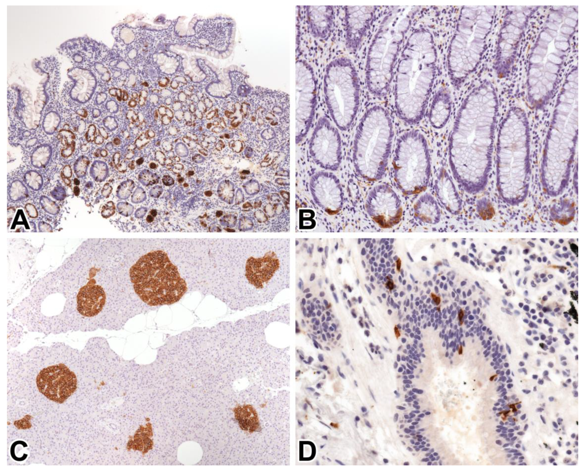
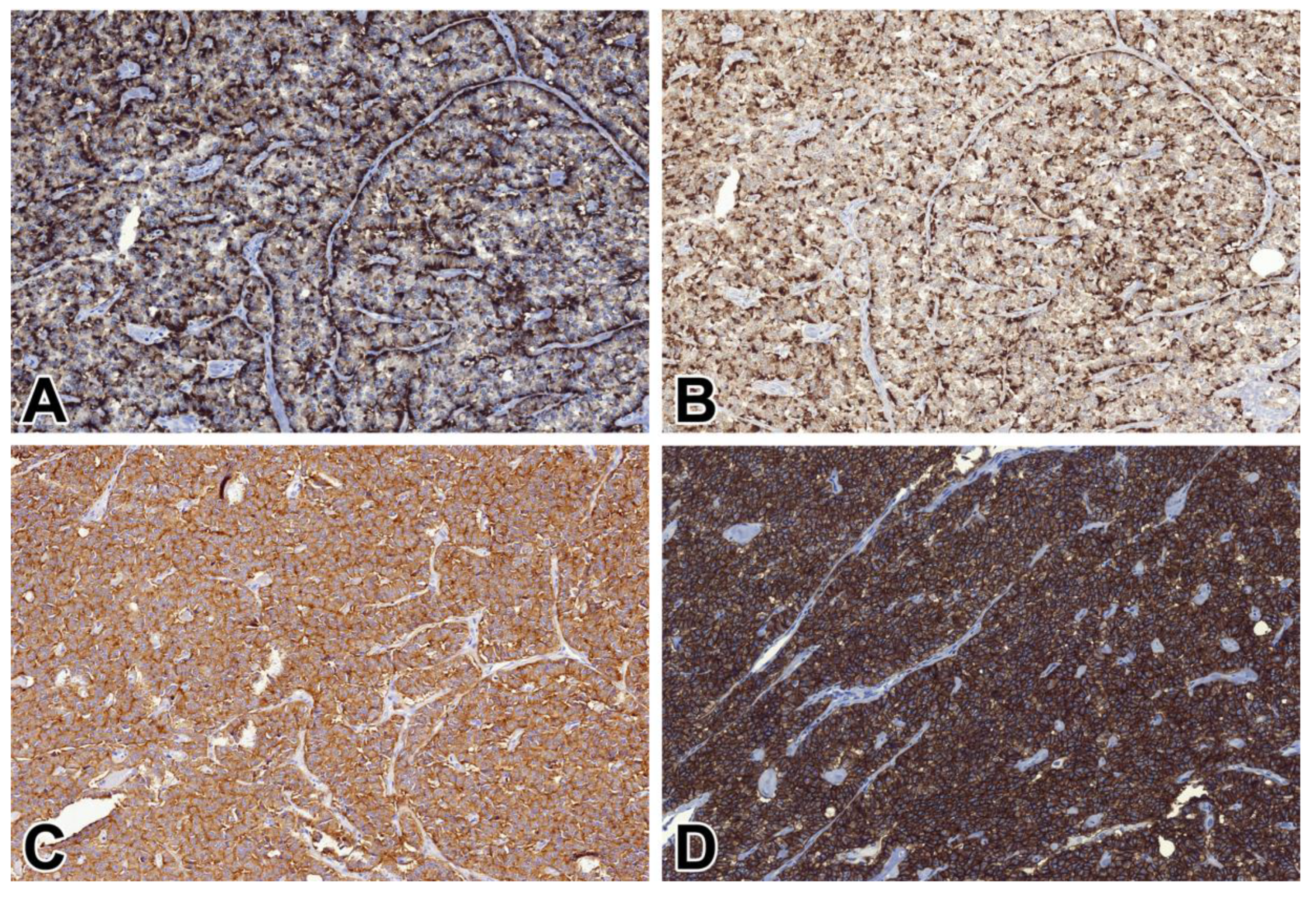
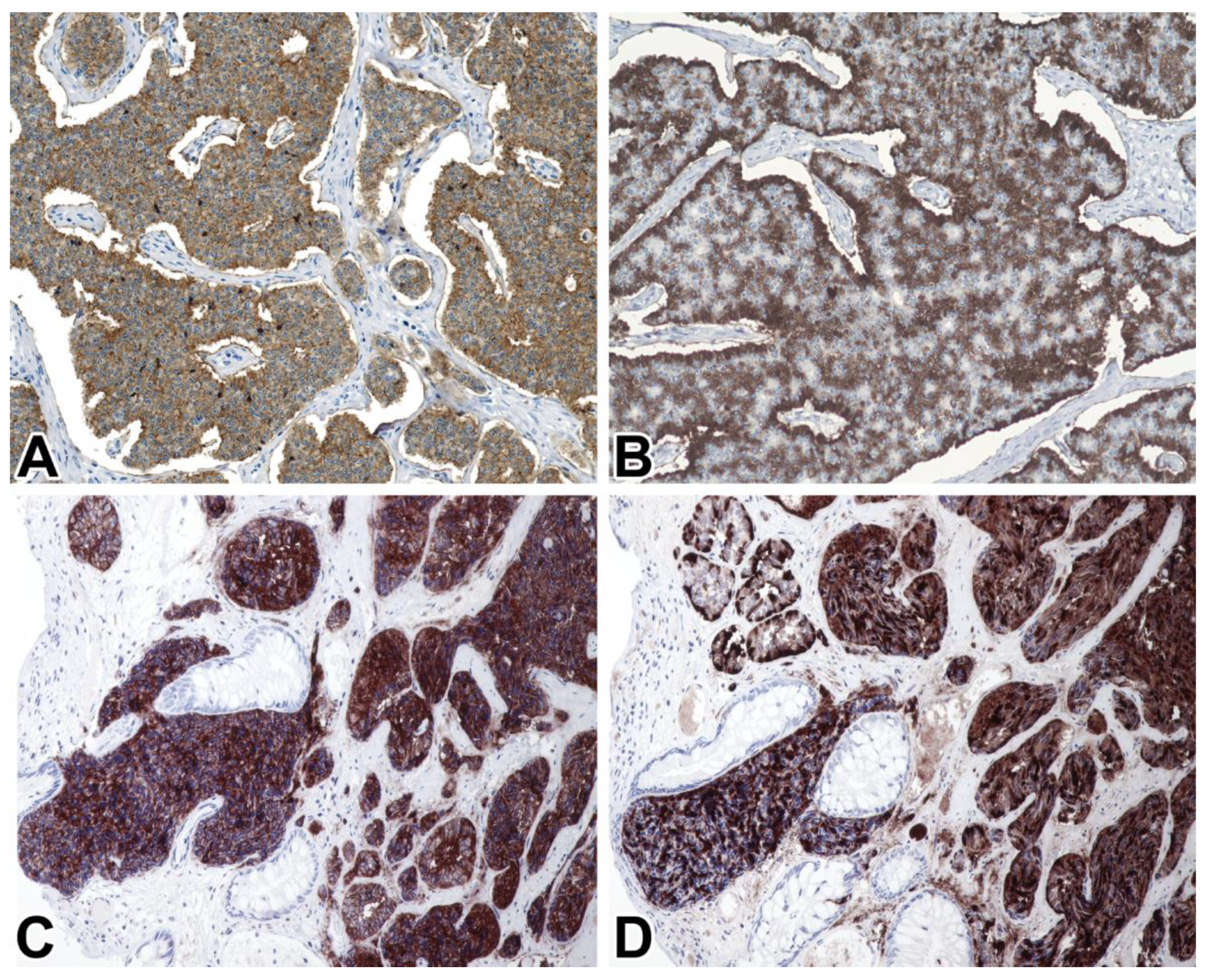
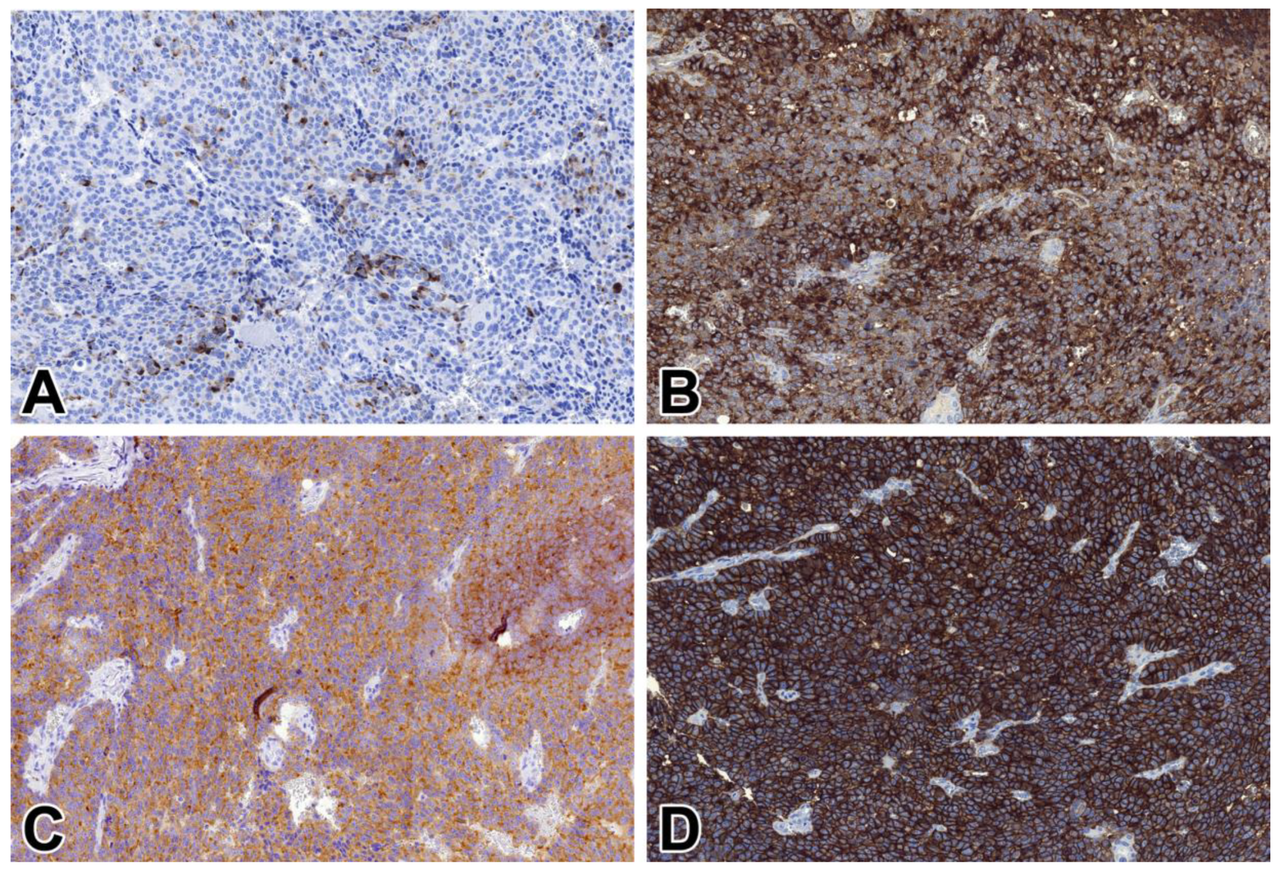
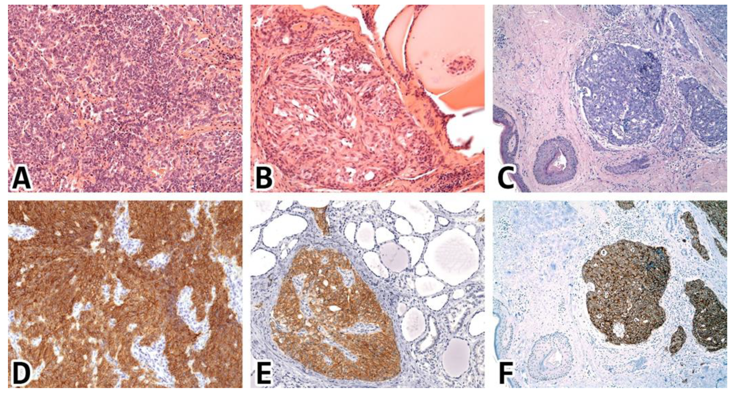
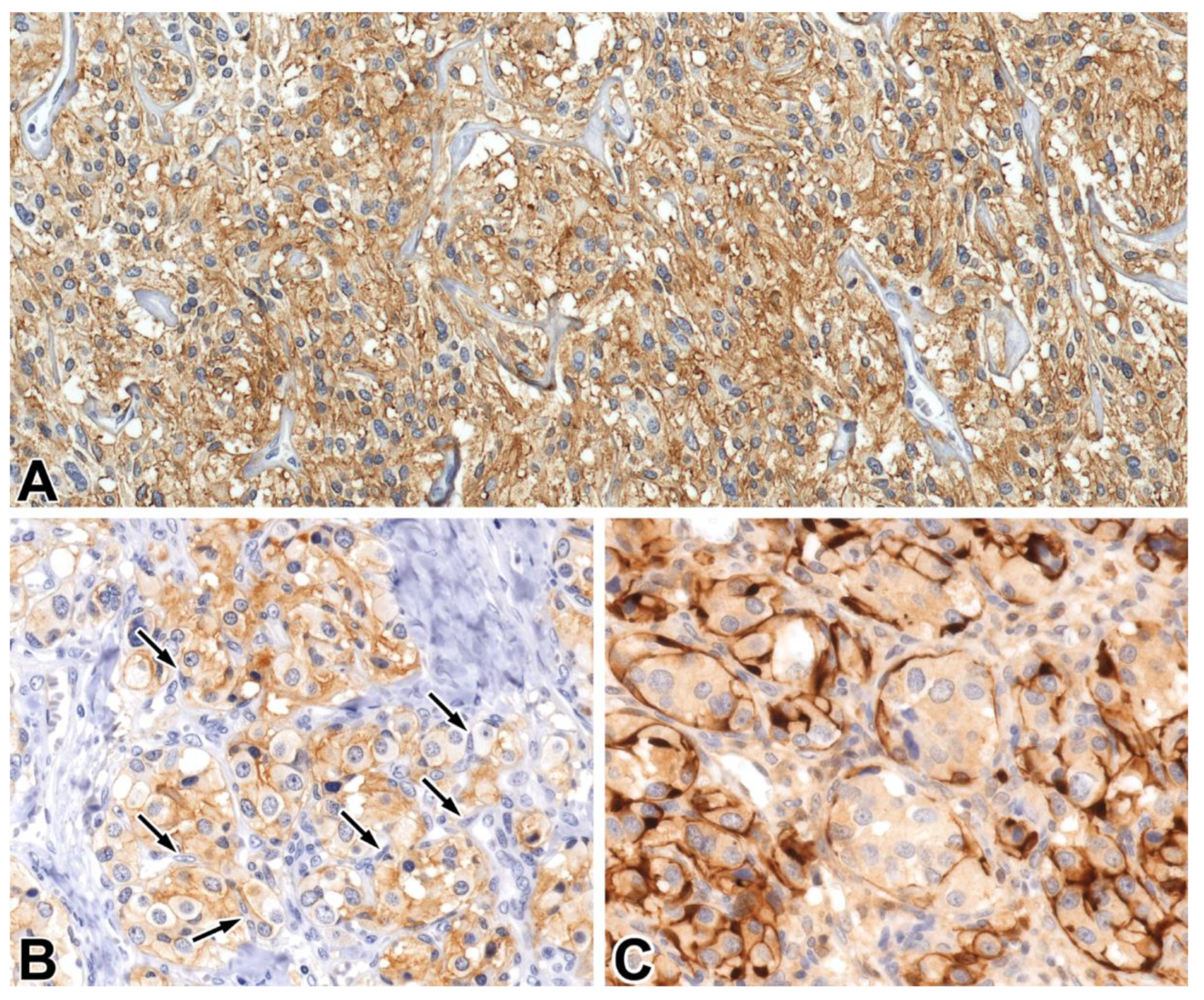
2. Results
3. Discussion
4. Materials and Methods
4.1. Samples Studied
4.2. Immunohistochemistry and Evaluation of Staining Patterns
5. Conclusions
Author Contributions
Funding
Acknowledgments
Conflicts of Interest
Abbreviations
| NE | Neuroendocrine |
| NEN | Neuroendocrine neoplasm |
| IHC | Immunohistochemistry |
| SYP | Synaptophysin |
| CHGA | Chromogranin-A |
| STX1 | Syntaxin 1 |
| NET | Neuroendocrine tumor |
| NEC | Neuroendocrine carcinoma |
| IARC | International Agency for Research on Cancer |
| WHO | World Health Organization |
| MiNEN | Mixed neuroendocrine-non neuroendocrine neoplasm |
| NSE | Neuron-specific enolase |
| PGP9.5 | Protein gene product 9.5 |
| INSM1 | Insulinoma-associated protein 1 |
| HPC-1 | Syntaxin 1 |
| ECL | Enterochromaffin-like |
| EC | Enterochromaffin |
| VMAT1 | Vesicular monoamine transporter 1 |
| GLP1 | Glucagon-like peptide-1 |
| FFPE | Formalin-fixed paraffin-embedded |
| TMA | Tissue microarray |
| TBS | Tris-buffered saline |
References
- WHO Classification of Tumours. Available online: http://whobluebooks.iarc.fr (accessed on 31 January 2020).
- Rindi, G.; Klimstra, D.S.; Abedi-Ardekani, B.; Asa, S.L.; Bosman, F.T.; Brambilla, E.; Busam, K.J.; de Krijger, R.R.; Dietel, M.; El-Naggar, A.K.; et al. A common classification framework for neuroendocrine neoplasms: An International Agency for Research on Cancer (IARC) and World Health Organization (WHO) expert consensus proposal. Mod. Pathol. 2018, 31, 1770–1786. [Google Scholar] [CrossRef] [PubMed]
- Lloyd, R.V.; Osamura, R.Y.; Klöppel, G.; Rosai, J. WHO Classification of Tumours of Endocrine Organs, 4th ed.; International Agency for Research on Cancer: Lyon, France, 2017; pp. 210–214. [Google Scholar]
- WHO Classification of Tumours Editorial Board. WHO Classification of Tumours, Digestive System Tumours, 5th ed.; International Agency for Research on Cancer: Lyon, France, 2019; pp. 16–20. [Google Scholar]
- Kyriakopoulos, G.; Mavroeidi, V.; Chatzellis, E.; Kaltsas, G.A.; Alexandraki, K.I. Histopathological, immunohistochemical, genetic and molecular markers of neuroendocrine neoplasms. Ann. Transl. Med. 2018, 6, 252. [Google Scholar] [CrossRef] [PubMed]
- Lloyd, R.V. Practical markers used in the diagnosis of neuroendocrine tumors. Endocr. Pathol. 2003, 14, 293–301. [Google Scholar] [CrossRef] [PubMed]
- Uccella, S.; La Rosa, S.; Volante, M.; Papotti, M. Immunohistochemical Biomarkers of Gastrointestinal, Pancreatic, Pulmonary, and Thymic Neuroendocrine Neoplasms. Endocr. Pathol. 2018, 29, 150–168. [Google Scholar] [CrossRef] [PubMed]
- Kosuke, F.; Kazuhiro, Y.; Shinji, K.; Yamato, M.; Yonosuke, S.; Joeji, W.; Ichiro, K.; Makoto, S.; Takaaki, I. INSM1 is the best marker for the diagnosis of neuroendocrine tumors: Comparison with chromogranin-A, SYP and CD56. Int. J. Clin. Exp. Pathol. 2017, 10, 5393–5405. [Google Scholar]
- Weiler, R.; Feichtinger, H.; Schmid, K.W.; Fischer-Colbrie, R.; Grimelius, L.; Cedermark, B.; Papotti, M.; Bussolati, G.; Winkler, H. Chromogranin A and B and secretogranin II in bronchial and intestinal carcinoids. Virchows Arch. A Pathol. Anat. Histopathol. 1987, 412, 103–109. [Google Scholar] [CrossRef]
- Fahrenkamp, A.G.; Wibbeke, C.; Winde, G.; Ofner, D.; Bocker, W.; Fischer-Colbrie, R.; Schmid, K.W. Immunohistochemical distribution of chromogranins A and B and secretogranin II in neuroendocrine tumours of the gastrointestinal tract. Virchows Arch. 1995, 426, 361–367. [Google Scholar] [CrossRef]
- Al-Khafaji, B.; Noffsinger, A.E.; Miller, M.A.; DeVoe, G.; Stemmermann, G.N.; Fenoglio-Preiser, C. Immunohistologic analysis of gastrointestinal and pulmonary carcinoid tumors. Hum. Pathol. 1998, 29, 992–999. [Google Scholar] [CrossRef]
- Washington, M.K.; Tang, L.H.; Berlin, J.; Branton, P.A.; Burgart, L.J.; Carter, D.K.; Compton, C.C.; Fitzgibbons, P.L.; Frankel, W.L.; Jessup, J.M.; et al. Protocol for the Examination of Specimens From Patients With Neuroendocrine Tumors (Carcinoid Tumors) of the Small Intestine and Ampulla. Arch. Pathol. Lab. Med. 2010, 134, 181–186. [Google Scholar] [CrossRef]
- Rooper, L.M.; Sharma, R.; Li, Q.K.; Illei, P.B.; Westra, W.H. INSM1 Demonstrates Superior Performance to the Individual and Combined Use of Synaptophysin, Chromogranin and CD56 for Diagnosing Neuroendocrine Tumors of the Thoracic Cavity. Am. J. Surg. Pathol. 2017, 41, 1561–1569. [Google Scholar] [CrossRef]
- Jirásek, T.; Hozák, P.; Mandys, V. Different patterns of chromogranin A and Leu-7 (CD57) expression in gastrointestinal carcinoids: Immunohistochemical and confocal laser scanning microscopy study. Neoplasma 2003, 50, 1–7. [Google Scholar] [PubMed]
- Tartaglia, A.; Portela-Gomes, G.M.; Oberg, K.; Vezzadini, P.; Foschini, M.P.; Stridsberg, M. Chromogranin A in gastric neuroendocrine tumours: An immunohistochemical and biochemical study with region-specific antibodies. Virchows Arch. 2006, 448, 399–406. [Google Scholar] [CrossRef] [PubMed]
- Rizo, J. Mechanism of neurotransmitter release coming into focus. Protein Sci. 2018, 27, 1364–1391. [Google Scholar] [CrossRef] [PubMed]
- Xie, Z.; Long, J.; Liu, J.; Chai, Z.; Kang, X.; Wang, C. Molecular Mechanisms for the Coupling of Endocytosis to Exocytosis in Neurons. Front. Mol. Neurosci. 2017, 10, 47. [Google Scholar] [CrossRef]
- Han, J.; Pluhackova, K.; Böckmann, R.A. The ceted Role of SNARE Proteins in Membrane Fusion. Front. Physiol. 2017, 8, 5. [Google Scholar] [CrossRef]
- Inoue, A.; Obata, K.; Akagawa, K. Cloning and sequence analysis of cDNA for a neuronal cell membrane antigen, HPC-1. J. Biol. Chem. 1992, 267, 10613–10619. [Google Scholar]
- Kushima, Y.; Fujiwara, T.; Sanada, M.; Akagawa, K. Characterization of HPC-1 antigen, an isoform of syntaxin-1, with the isoform-specific monoclonal antibody, 14D8. J. Mol. Neurosci. 1997, 8, 19–27. [Google Scholar] [CrossRef]
- Yoo, S.H.; You, S.H.; Huh, Y.H. Presence of syntaxin 1A in secretory granules of chromaffin cells and interaction with chromogranins A and B. FEBS Lett. 2005, 579, 222–228. [Google Scholar] [CrossRef]
- Jacobsson, G.; Beant, A.J.; Schellert, R.H.; Juntti-Berggrent, L.; Deeneyt, J.T.; Berggrent, P.-O.; Meister, B. Identification of synaptic proteins and their isoform mRNAs in compartments of pancreatic endocrine cells (exocytosis/secretion/insulin/diabetes). Proc. Natl. Acad. Sci. USA 1994, 91, 12487–12491. [Google Scholar] [CrossRef]
- Xia, F.; Leung, Y.M.; Gaisano, G.; Gao, X.; Chen, Y.; Fox, J.E.; Bhattacharjee, A.; Wheeler, M.B.; Gaisano, H.Y.; Tsushima, R.G. Targeting of voltage-gated K+ and Ca2+ channels and soluble N-ethylmaleimide-sensitive factor attachment protein receptor proteins to cholesterol-rich lipid rafts in pancreatic alpha-cells: Effects on glucagon stimulus-secretion coupling. Endocrinology 2007, 148, 2157–2167. [Google Scholar] [CrossRef][Green Version]
- Bjorling, E.; Lindskog, C.; Oksvold, P.; Linne, J.; Kampf, C.; Hober, S.; Uhlen, M.; Ponten, F. A web-based tool for in silico biomarker discovery based on tissue-specific protein profiles in normal and cancer tissues. Mol. Cell. Proteom. 2008, 7, 825–844. [Google Scholar] [CrossRef] [PubMed]
- Kurokawa, M.; Nabeshima, K.; Akiyama, Y.; Maeda, S.; Nishida, T.; Nakayama, F.; Amano, M.; Ogata, K.; Setoyama, M. CD56: A useful marker for diagnosing Merkel cell carcinoma. J. Derm. Sci. 2003, 31, 219–224. [Google Scholar] [CrossRef]
- Bahrami, A.; Gown, A.M.; Baird, G.S.; Hicks, M.J.; Folpe, A.L. Aberrant expression of epithelial and neuroendocrine markers in alveolar rhabdomyosarcoma: A potentially serious diagnostic pitfall. Mod. Pathol. 2008, 21, 795–806. [Google Scholar] [CrossRef] [PubMed]
- Nobels, F.R.; Kwekkeboom, D.J.; Bouillon, R.; Lamberts, S.W. Chromogranin A: Its clinical value as marker of neuroendocrine tumours. Eur. J. Clin. Investig. 1998, 28, 431–440. [Google Scholar] [CrossRef]
- Deftos, L.J.; Woloszczuk, W.; Krisch, I.; Horvat, G.; Ulrich, W.; Neuhold, N.; Braun, O.; Reiner, A.; Srikanta, S.; Krisch, K. Medullary Thyroid Carcinomas Express Chromogranin A and a Novel Neuroendocrine Protein Recognized by Monoclonal Antibody HISL-19. Am. J. Med. 1988, 85, 780–784. [Google Scholar] [CrossRef]
- Portela, G.M.; Stridsberg, M. Chromogranin A in the Human Gastrointestinal Tract: An Immunocytochemical Study with Region-specific Antibodies. J. Histochem. Cytochem. 2002, 50, 1487–1492. [Google Scholar] [CrossRef]
- Williams, G.T. Endocrine tumours of the gastrointestinal tract–selected topics. Histopathology 2007, 50, 30–41. [Google Scholar] [CrossRef]
- Washington, M.K.; Tang, L.H.; Berlin, J.; Branton, P.A.; Burgart, L.J.; Carter, D.K.; Compton, C.C.; Fitzgibbons, P.L.; Frankel, W.L.; Jessup, J.M.; et al. Protocol for the examination of specimens from patients with neuroendocrine tumors (carcinoid tumors) of the colon and rectum. Arch. Pathol. Lab. Med. 2010, 134, 176–180. [Google Scholar] [CrossRef]
- Gould, V.E.; Lee, I.; Wiedenmann, B.; Moll, R.; Chejfec, G.; Franke, W.W. Synaptophysin: A novel marker for neurons, certain neuroendocrine cells, and their neoplasms. Hum. Pathol. 1986, 17, 979–983. [Google Scholar] [CrossRef]
- Wiedenmann, B.; Franke, W.W.; Kuhn, C.; Moll, R.; Gould, V.E. Synaptophysin: A marker protein for neuroendocrine cells and neoplasms. Proc. Natl. Acad. Sci. USA 1986, 83, 3500–3504. [Google Scholar] [CrossRef]
- Wiedenmann, B.; Franke, W.W. Identification and localization of synaptophysin, an integral membrane glycoprotein of Mr 38,000 characteristic of presynaptic vesicles. Cell 1985, 41, 1017–1028. [Google Scholar] [CrossRef]
- Erickson, L.A.; Mete, O. Immunohistochemistry in Diagnostic Parathyroid Pathology. Endocr. Pathol. 2018, 29, 113–129. [Google Scholar] [CrossRef]
- Rosenbaum, J.N.; Guo, Z.; Baus, R.M.; Werner, H.; Rehrauer, W.M.; Lloyd, R.V. INSM1: A Novel Immunohistochemical and Molecular Marker for Neuroendocrine and Neuroepithelial Neoplasms. Am. J. Clin. Pathol. 2015, 144, 579–591. [Google Scholar] [CrossRef]
- Gonzalez, I.; Lu, H.C.; Sninsky, J.; Yang, C.; Bishnupuri, K.; Dieckgraefe, B.; Cao, D.; Chatterjee, D. Insulinoma-associated protein 1 expression in primary and metastatic neuroendocrine neoplasms of the gastrointestinal and pancreaticobiliary tracts. Histopathology 2019, 75, 568–577. [Google Scholar] [CrossRef] [PubMed]
- Kriegsmann, K.; Zgorzelski, C.; Kazdal, D.; Cremer, M.; Muley, T.; Winter, H.; Longuespee, R.; Kriegsmann, J.; Warth, A.; Kriegsmann, M. Insulinoma-associated Protein 1 (INSM1) in Thoracic Tumors is Less Sensitive but More Specific Compared With Synaptophysin, Chromogranin A, and CD56. Appl. Immunohistochem. Mol. Morphol. 2018. [Google Scholar] [CrossRef]
- Xin, Z.; Zhang, Y.; Jiang, Z.; Zhao, L.; Fan, L.; Wang, Y.; Xie, S.; Shangguan, X.; Zhu, Y.; Pan, J.; et al. Insulinoma-associated protein 1 is a novel sensitive and specific marker for small cell carcinoma of the prostate. Hum. Pathol. 2018, 79, 151–159. [Google Scholar] [CrossRef] [PubMed]
- Rooper, L.M.; Bishop, J.A.; Westra, W.H. INSM1 is a Sensitive and Specific Marker of Neuroendocrine Differentiation in Head and Neck Tumors. Am. J. Surg. Pathol. 2018, 42, 665–671. [Google Scholar] [CrossRef]
- Lilo, M.T.; Chen, Y.; LeBlanc, R.E. INSM1 Is More Sensitive and Interpretable than Conventional Immunohistochemical Stains Used to Diagnose Merkel Cell Carcinoma. Am. J. Surg. Pathol. 2018, 42, 1541–1548. [Google Scholar] [CrossRef]
- Bordi, C.; Pilato, F.; Carfagna, G.; Ferrari, C.; DAdda, T.; Sivelli, R.; Bertelé, A.; Missale, G. Argyrophil Cell Hyperplasia of Fundic Mucosa in Patients with Chronic Atrophic Gastritis. Digestion 1986, 35, 130–143. [Google Scholar] [CrossRef]
- Toth-Liptak, J.; Piukovics, K.; Borbenyi, Z.; Demeter, J.; Bagdi, E.; Krenacs, L. A comprehensive immunophenotypic marker analysis of hairy cell leukemia in paraffin-embedded bone marrow trephine biopsies-a tissue microarray study. Pathol. Oncol. Res. 2015, 21, 203–211. [Google Scholar] [CrossRef]


| Gastrointestinal NETs | STX1 | CHGA | SYP | CD56 | ||||
|---|---|---|---|---|---|---|---|---|
| Gastric enterochromaffin-like (ECL) cell NET | 5/5 | 100.0% | 4/4 | 100.0% | 3/3 | 100.0% | 0/1 | 0.0% |
| Duodenal non-functioning NET | 2/2 | 100.0% | 2/2 | 100.0% | 2/2 | 100.0% | na | na |
| Ampullary NET | 1/2 | 50.0% | 2/2 | 100.0% | 1/2 | 50.0% | 1/2 | 50.0% |
| Small intestinal enterochromaffin (EC) cell NET | 12/12 | 100.0% | 12/12 | 100.0% | 12/12 | 100.0% | 8/12 | 66.7% |
| Appendix NET | 9/9 | 100.0% | 8/8 | 100.0% | 7/7 | 100.0% | 9/9 | 100.0% |
| Rectal EC cell NET | 2/2 | 100.0% | 2/2 | 100.0% | 1/1 | 100.0% | 0/2 | 0.0% |
| Rectal L cell NET | 5/5 | 100.0% | 2/5 | 33.3% | 4/4 | 100.0% | 5/5 | 100.0% |
| Metastatic NET. gastrointestinal origin | 4/4 | 100.0% | 4/4 | 100.0% | 1/2 | 50.0% | 0/2 | 0.0% |
| Pancreatic NETs | ||||||||
| Pancreatic functioning NET * | 3/3 | 100.0% | 2/2 | 100.0% | 2/2 | 100.0% | 1/1 | 100.0% |
| Pancreatic non-functioning NET | 16/16 | 100.0% | 13/14 | 92.9% | 12/13 | 92.3% | 10/11 | 90.9% |
| Pulmonary carcinoids | ||||||||
| Pulmonary carcinoid. typical | 3/3 | 100.0% | 3/3 | 100.0% | 2/2 | 100.0% | 2/2 | 100.0% |
| Pulmonary carcinoid. atypical | 2/2 | 100.0% | na | na | na | na | na | na |
| Gastroenteropancreatic NECs | ||||||||
| Stomach small cell NEC | 2/2 | 100.0% | 1/1 | 100.0% | 1/1 | 100.0% | 1/1 | 100.0% |
| Duodenal large cell NEC | 1/1 | 100.0% | 1/1 | 100.0% | 1/1 | 100.0% | na | na |
| Pancreatic large cell NEC | 2/2 | 100.0% | 1/1 | 100.0% | 1/1 | 100.0% | 2/2 | 100.0% |
| Ampullary small cell NEC | 2/2 | 100.0% | 1/1 | 100.0% | na | na | 2/2 | 100.0% |
| Pulmonary NECs | ||||||||
| Pulmonary small cell NEC | 19/19 | 100.0% | 9/10 | 90.0% | 2/2 | 100.0% | 16/16 | 100.0% |
| Pulmonary large cell NEC | 4/4 | 100.0% | 1/1 | 100.0% | na | 2/2 | 100.0% | |
| Etc NECs | ||||||||
| Merkel cell carcinoma | 5/5 | 100.0% | 1/1 | 100.0% | 1/1 | 100.0% | 2/2 | 100.0% |
| Medullary thyroid carcinoma | 10/10 | 100.0% | 5/5 | 100.0% | 2/2 | 100.0% | 3/3 | 100.0% |
| Metastatic small cell NEC with unknown primary | 3/3 | 100.0% | na | na | na | na | 2/2 | 100.0% |
| Metastatic large cell NEC with unknown primary | 3/3 | 100.0% | 3/3 | 100.0% | na | na | 1/3 | 33.3% |
| Small cell NEC of the prostate | 1/1 | 100.0% | 1/1 | 100.0% | 1/1 | 100.0% | 1/1 | 100.0% |
| Small cell NEC of the uterine cervix | 1/1 | 100.0% | na | na | na | na | na | na |
| Small cell NEC of the breast | 1/1 | 100.0% | na | na | na | na | na | na |
| Tumors with mixed NE and non-NE components ** | ||||||||
| MiNEN ** | 2/2 | 100.0% | 0/1 | 0 | 2/2 | 100.0% | 0/2 | 0.0% |
| Mixed tumor with minor NE component ** | 4/4 | 100.0% | na | na | 2/2 | 100.0% | 0/2 | 0.0% |
| Etc NENs | ||||||||
| Pituitary adenoma | 19/19 | 100.0% | 4/4 | 100.0% | 1/1 | 100.0% | na | na |
| Pheochromocytoma/paraganglioma | 16/18 | 88.9% | 15/16 | 93.8% | 9/10 | 90.0% | 2/2 | 100.0% |
| Neuroectodermal/neuroepithelial neoplasia | ||||||||
| Medulloblastoma | 8/9 | 88.9% | na | na | 8/9 | 88.9% | 2/2 | 100.0% |
| Neuroblastoma | 8/8 | 100.0% | 5/5 | 100.0% | 5/5 | 100.0% | 2/2 | 100.0% |
| Ganglioneuroma | 2/2 | 100.0% | na | na | na | na | na | na |
| Ewing sarcoma/PNET | 2/2 | 100.0% | na | na | na | na | na | na |
| Sensitivity—NET | Sensitivity—NEC | |
|---|---|---|
| STX1 | 99% | 100% |
| CHGA | 98% | 91% |
| SYP | 96% | 89% |
| CD56 | 70% | 93% |
| Tumor Type | STX1 (Positive/Total No. of Cases; %) | No. of Cases with Scarcely Scattered STX1 Positive Cells | |
|---|---|---|---|
| Colorectal adenocarcinoma | 0/8 | 0.0% | 5 |
| Gastric adenocarcinoma | 0/12 | 0.0% | 9 |
| Hepatobiliary and pancreatic carcinoma | 0/11 | 0.0% | 1 |
| Lung squamous cell carcinoma | 0/5 | 0.0% | |
| Lung adenocarcinoma | 0/8 | 0.0% | |
| Head and neck carcinoma | 0/7 | 0.0% | |
| Basal cell carcinoma of the skin | 0/2 | 0.0% | |
| Cervical and ovarian carcinoma | 0/8 | 0.0% | 1 |
| Prostatic adenocarcinoma | 0/7 | 0.0% | |
| Melanocytic tumor | 0/9 | 0.0% | |
| Soft tissue tumor | 0/13 | 0.0% | |
| Lymphoma and myeloid neoplasm | 0/31 | 0.0% | |
| Genital germ cell tumor | 0/9 | 0.0% | |
| Gonadal sex-cord stromal tumor | 0/5 | 0.0% | |
| Solid pseudopapillary neoplasm (pancreas) | 0/1 | 0.0% | |
| Adrenocortical adenoma | 0/11 | 0.0% | |
| Adrenocortical carcinoma | 0/12 | 0.0% | |
| Parathyroid adenoma | 0/19 | 0.0% | |
| Papillary thyroid carcinoma | 0/5 | 0.0% | |
| Thyroid follicular adenoma | 0/1 | 0.0% | |
| Carcinomas of the breast | |||
| No special type | 1/18 | 5.6% | 1 |
| Invasive lobular carcinoma | 0/2 | 0.0% | |
| Mucinous carcinoma, hypocellular type | 0/6 | 0.0% | |
| Mucinous carcinoma, hypercellular type | 7/10 | 70.0% | |
| Metaplastic carcinoma | 0/1 | 0.0% | |
© 2020 by the authors. Licensee MDPI, Basel, Switzerland. This article is an open access article distributed under the terms and conditions of the Creative Commons Attribution (CC BY) license (http://creativecommons.org/licenses/by/4.0/).
Share and Cite
Kővári, B.; Turkevi-Nagy, S.; Báthori, Á.; Fekete, Z.; Krenács, L. Syntaxin 1: A Novel Robust Immunophenotypic Marker of Neuroendocrine Tumors. Int. J. Mol. Sci. 2020, 21, 1213. https://doi.org/10.3390/ijms21041213
Kővári B, Turkevi-Nagy S, Báthori Á, Fekete Z, Krenács L. Syntaxin 1: A Novel Robust Immunophenotypic Marker of Neuroendocrine Tumors. International Journal of Molecular Sciences. 2020; 21(4):1213. https://doi.org/10.3390/ijms21041213
Chicago/Turabian StyleKővári, Bence, Sándor Turkevi-Nagy, Ágnes Báthori, Zoltán Fekete, and László Krenács. 2020. "Syntaxin 1: A Novel Robust Immunophenotypic Marker of Neuroendocrine Tumors" International Journal of Molecular Sciences 21, no. 4: 1213. https://doi.org/10.3390/ijms21041213
APA StyleKővári, B., Turkevi-Nagy, S., Báthori, Á., Fekete, Z., & Krenács, L. (2020). Syntaxin 1: A Novel Robust Immunophenotypic Marker of Neuroendocrine Tumors. International Journal of Molecular Sciences, 21(4), 1213. https://doi.org/10.3390/ijms21041213