A Step Forward in Breast Cancer Research: From a Natural-Like Experimental Model to a Preliminary Photothermal Approach

 ,
,  ,
,  ,
,  ,
,  ,
,  , ,
, ,  and
and
Abstract

1. Introduction
2. Results
2.1. Experimental Model Characterization
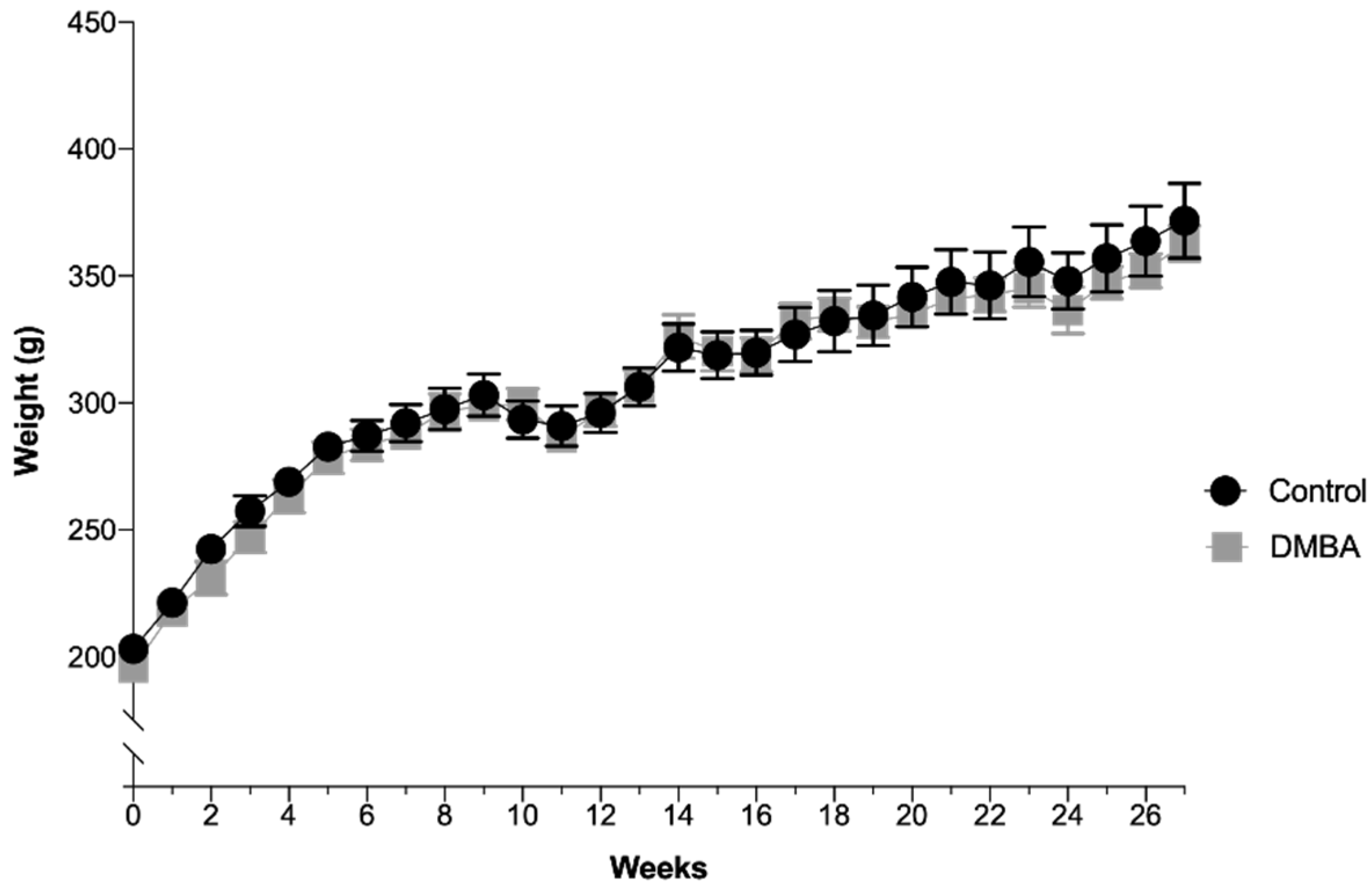
2.1.1. Animals’ Weight
2.1.2. Urinalysis
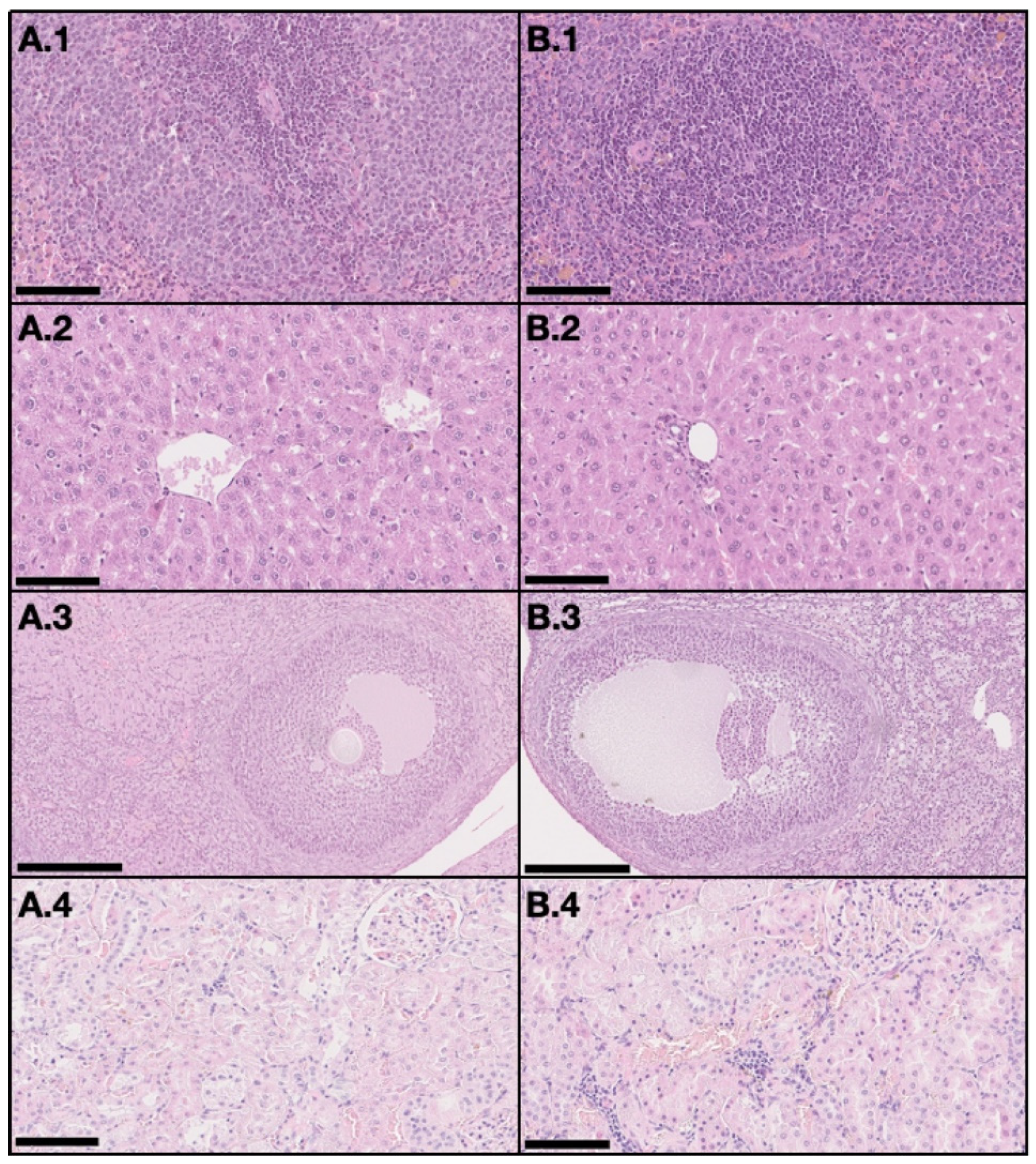
2.1.3. Blood Samples
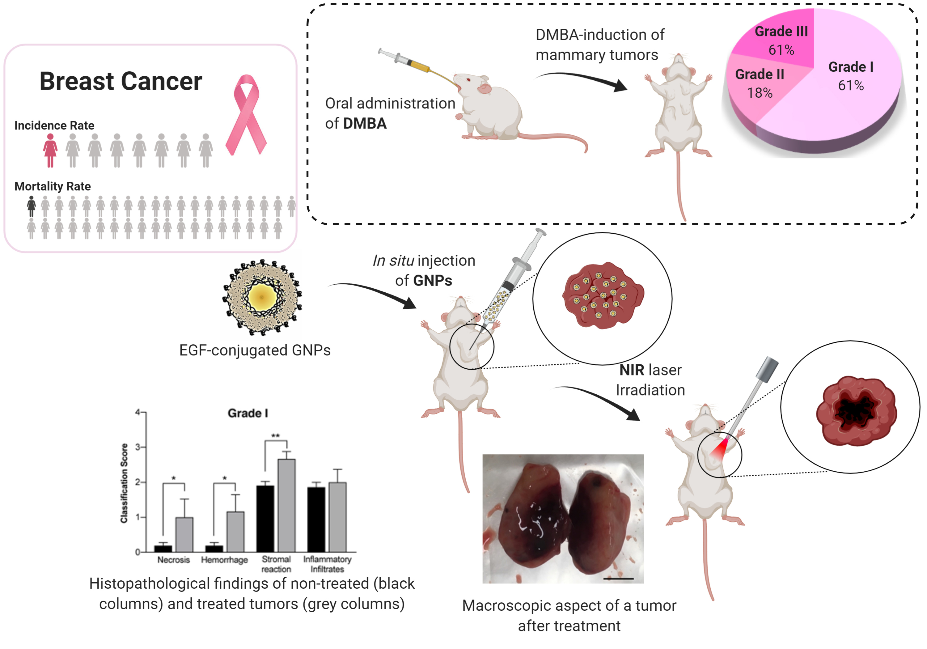
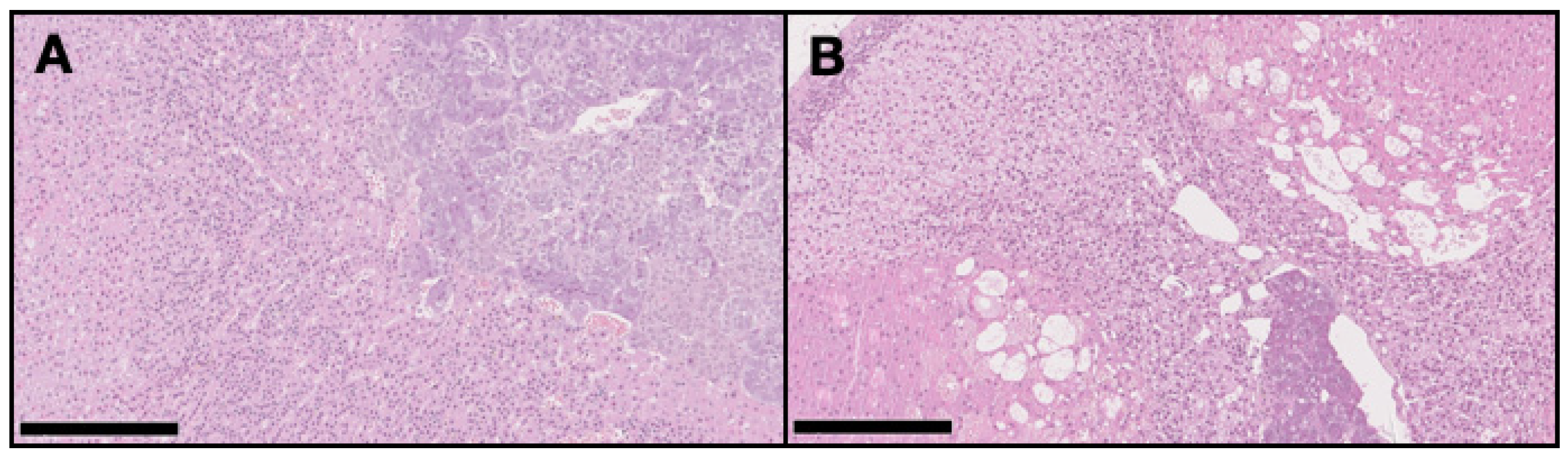
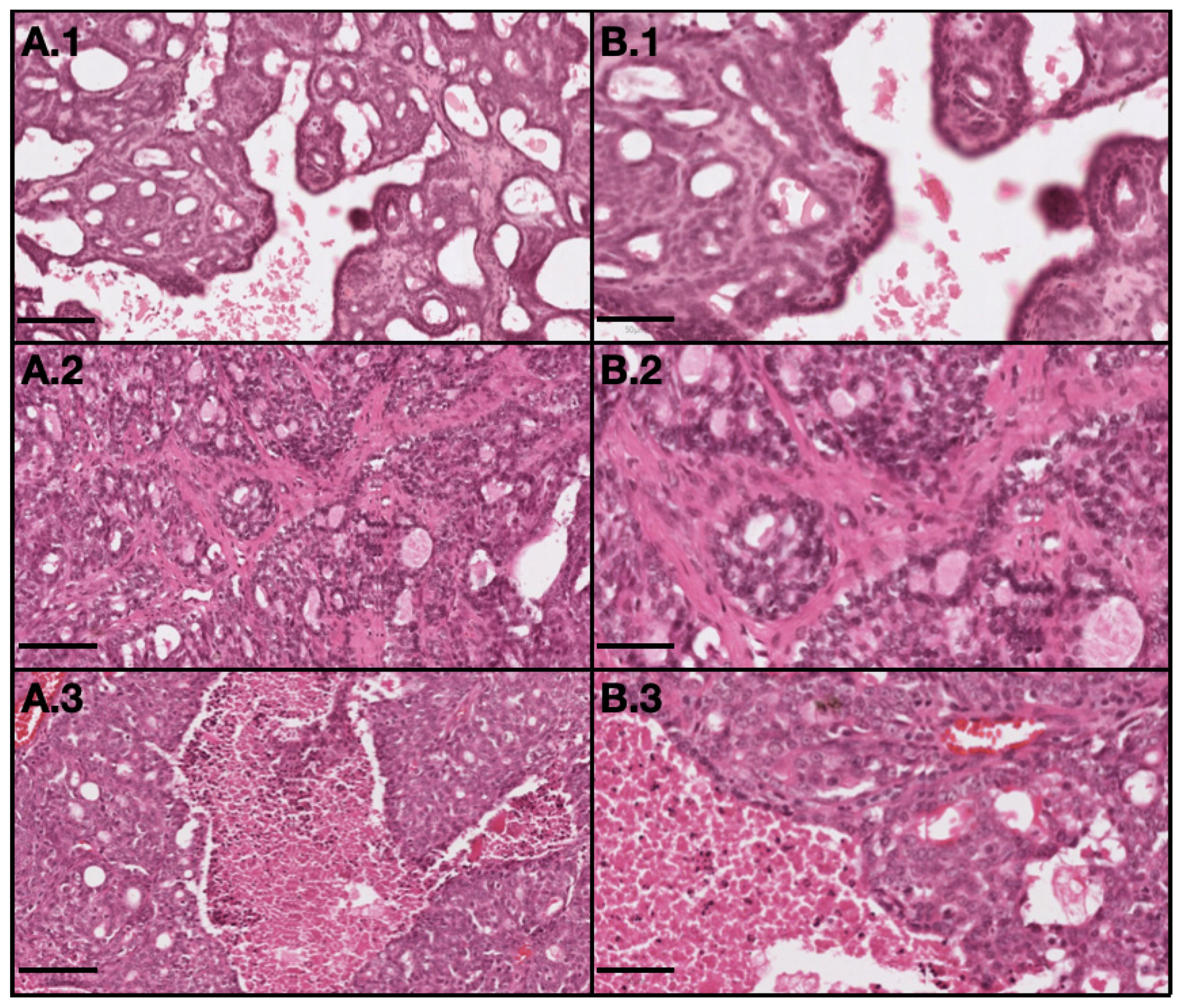
2.1.4. Histopathological Classification of the Breast Tumors
Breast Tumors Classification
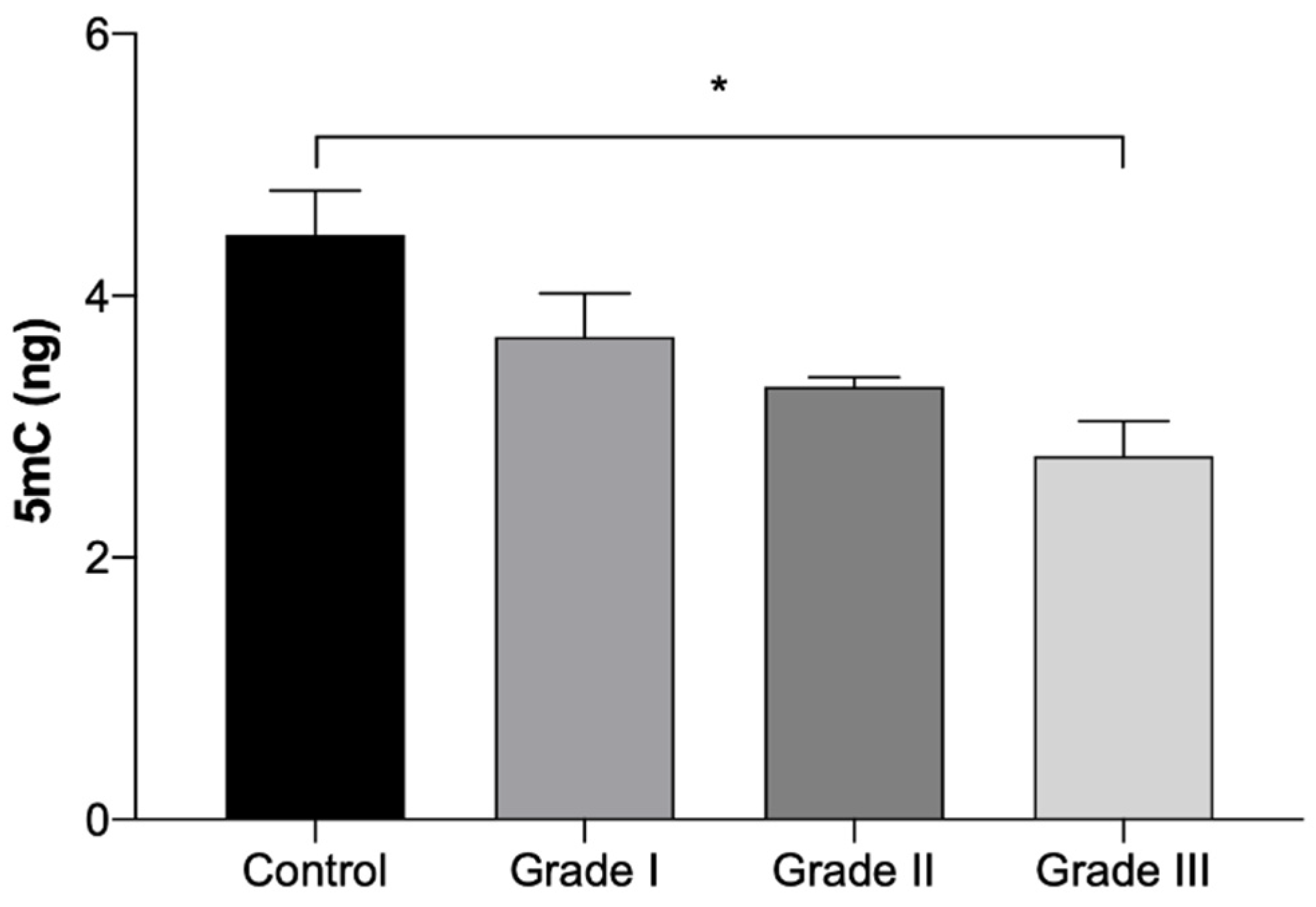
2.1.5. Epigenetic Alterations
DNA Methylation
2.2. EGF-Conjugated GNPs for Photothermal Treatment
2.2.1. Size, PdI, Maximum Absorbance Peak and Morphology of GNPs
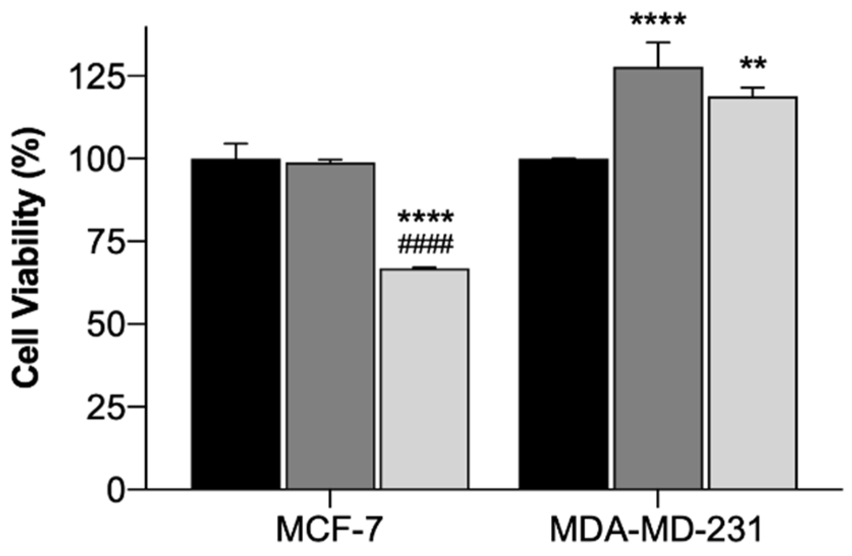
2.2.2. In Vitro Photothermal Therapy with Functionalized Gold-Nanoparticles
2.2.3. Preliminary Safety Assessment for Potential In Vivo Applications Using Hemolytic Activity Assay
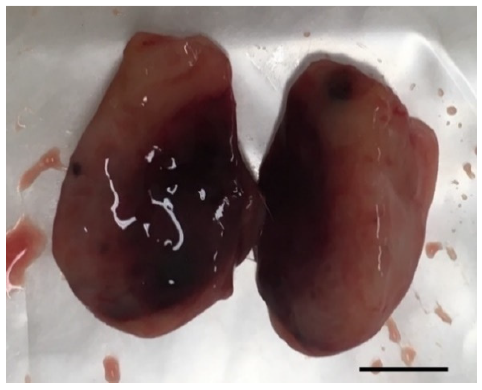
2.2.4. Preliminary In Vivo Photothermal Therapy with Functionalized Gold-Nanoparticles
3. Discussion
4. Materials and Methods
4.1. In Vivo Studies
Development and Characterization of an Experimental Model
- 1.
- Urinalysis
- 2.
- Blood Samples Analysis
- Hematological Parameters
- Biochemical Parameters
- 3.
- Histopathological Assessment
- 4.
- DNA Extraction
- 5.
- DNA Methylation
4.2. EGF-Conjugated GNPs Preparation and Characterization
4.2.1. EGF-Conjugated GNPs Preparation
4.2.2. Characterization of the GNPs
4.2.3. In Vitro Photothermal Therapy with Functionalized Gold-Nanoparticles
Cell Culture and Incubation with the EGF-Conjugated GNPs
Laser Irradiation Procedure
MTT Assay
4.2.4. Preliminary Safety Assessment for Future In Vivo Applications Using Hemolytic Activity Assay
4.2.5. In Vivo Photothermal Therapy with Functionalized Gold-Nanoparticles
4.3. Statistical Analysis
5. Conclusions
Supplementary Materials
Author Contributions
Funding
Acknowledgments
Conflicts of Interest
Abbreviations
| 5mC | 5-methylcytosine |
| Abs | Absorbance |
| ALP | Alkaline Phosphatase |
| ALT | Alanine Transaminase |
| AST | Aspartate Transaminase |
| ATCC | American Type Culture Collection |
| BRCA1 | Breast Cancer 1 gene |
| BRCA2 | Breast Cancer 2 gene |
| BUN | Blood Urea Nitrogen |
| CI | Confidence Interval |
| DGAV | Directorate-General for Food and Veterinary |
| DLS | Dynamic Light Scattering |
| DMBA | 7,12-dimethylbenzantracene |
| DMEM | Dulbecco’s Modified Eagle’s Medium |
| DMSO | Dimethyl Sulfoxide |
| DNA | Deoxyribonucleic acid |
| EDTA | Ethylenediamine tetraacetic acid |
| EGF | Epidermal Growth Factor |
| EGF-conjugated GNPs | Gold Nanoparticles coated with a combination of Hyaluronic and Oleic Acids to which EGF was added |
| EGFR | Epidermal Growth Factor Receptor |
| ER | Estrogen Receptor |
| GLOBOCAN | Global Cancer Observatory Reports |
| GNPs | Gold Nanoparticles |
| H&E | Hematoxylin and Eosin stain |
| HA | Hyaluronic acid |
| HAOA coating | Coating of Hyaluronic and Oleic Acids |
| HAOA-coated GNPs | Gold Nanoparticles coated with a combination of Hyaluronic and Oleic Acids |
| HCT | Hematocrit |
| HER2 | Human Epidermal Growth Factor Receptor 2 |
| LMC | Left Mammary Chain |
| MCF-7 cells | Michigan Cancer Foundation-7 Cells |
| MCH | Mean Corpuscular Hemoglobin |
| MCHC | Mean Corpuscular Hemoglobin Concentration |
| MCV | Mean Corpuscular Volume |
| MDA-MB-231 cells | M. D. Anderson Cancer Center- MB-231 Cells |
| microRNAs | Micro-Ribonucleic Acid |
| MTT | 3-(4,5-Dimethylthiazol-2-yl)-2,5-Diphenyltetrazolium Bromide |
| NGS | Nottingham grading System |
| NIR | Near-infrared |
| NPs | Nanoparticles |
| OA | Oleic Acid |
| OD | Optical Density |
| ORBEA | Animal Welfare and Ethics Body |
| PBS | Phosphate Buffered Saline |
| PdI | Polydispersity Index |
| PR | Progesterone Receptor |
| PTT | Photothermal Therapy |
| RMC | Right Mammary Chain |
| RT | Room Temperature |
| SD | Standard Deviation |
| SEM | Standard Error of the Mean |
| SPR | Surface Plasmon Resonance |
| TEB | Terminal End Buds |
| TEM | Transmission Electron Microscopy |
| TE Buffer | Tris + EDTA Buffer |
References
- Bray, F.; Ferlay, J.; Soerjomataram, I.; Siegel, R.L.; Torre, L.A.; Jemal, A. Global cancer statistics 2018: GLOBOCAN estimates of incidence and mortality worldwide for 36 cancers in 185 countries. CA. Cancer J. Clin. 2018, 68, 394–424. [Google Scholar] [CrossRef] [PubMed]
- Fitzmaurice, C.; Allen, C.; Barber, R.M.; Barregard, L.; Bhutta, Z.A.; Brenner, H.; Dicker, D.J.; Chimed-Orchir, O.; Dandona, R.; Dandona, L.; et al. Global, Regional, and National Cancer Incidence, Mortality, Years of Life Lost, Years Lived With Disability, and Disability-Adjusted Life-years for 32 Cancer Groups, 1990 to 2015. JAMA Oncol. 2017, 3, 524. [Google Scholar] [PubMed]
- DeSantis, C.E.; Ma, J.; Gaudet, M.M.; Newman, L.A.; Miller, K.D.; Goding Sauer, A.; Jemal, A.; Siegel, R.L. Breast cancer statistics, 2019. CA. Cancer J. Clin. 2019, 69, 438–451. [Google Scholar] [CrossRef] [PubMed]
- Nebbioso, A.; Tambaro, F.P.; Dell’Aversana, C.; Altucci, L. Cancer epigenetics: Moving forward. PLoS Genet. 2018, 14, 1–25. [Google Scholar] [CrossRef] [PubMed]
- Alvarado, A.; Faustino-Rocha, A.I.; Colaço, B.; Oliveira, P.A. Experimental mammary carcinogenesis—Rat models. Life Sci. 2017, 173, 116–134. [Google Scholar] [CrossRef] [PubMed]
- Alvarado, A.; Faustino-Rocha, A.I.; Ferreira, R.; Mendes, R.; Duarte, J.A.; Pires, M.J.; Colaço, B.; Oliveira, P.A. Prognostic factors in an exercised model of chemically-induced mammary cancer. Anticancer Res. 2016, 36, 2181–2188. [Google Scholar] [PubMed]
- Alvarado, A.; Lopes, A.C.; Faustino-Rocha, A.I.; Cabrita, A.M.S.; Ferreira, R.; Oliveira, P.A.; Colaço, B. Prognostic factors in MNU and DMBA-induced mammary tumors in female rats. Pathol. Res. Pract. 2017, 213, 441–446. [Google Scholar] [CrossRef]
- Sheikh, A.; Hussain, S.A.; Ghori, Q.; Naeem, N.; Fazil, A.; Giri, S.; Sathian, B.; Mainali, P.; Al Tamimi, D.M. The spectrum of genetic mutations in breast cancer. Asian Pac. J. Cancer Prev. 2015, 16, 2177–2185. [Google Scholar] [CrossRef]
- Ha, S.M.; Chae, E.Y.; Cha, J.H.; Kim, H.H.; Shin, H.J.; Choi, W.J. Association of BRCA Mutation Types, Imaging Features, and Pathologic Findings in Patients With Breast Cancer With BRCA1 and BRCA2 Mutations. AJR Am. J. Roentgenol. 2017, 209, 920–928. [Google Scholar] [CrossRef]
- Strumylaitė, L.; Mechonošina, K.; Tamašauskas, S. Environmental factors and breast cancer. Medicina 2010, 46, 867–873. [Google Scholar]
- Hiatt, R.A.; Brody, J.G. Environmental Determinants of Breast Cancer. Annu. Rev. Public Health 2018, 39, 113–133. [Google Scholar] [CrossRef] [PubMed]
- Baylin, S.B.; Jones, P.A. Epigenetic determinants of cancer. Cold Spring Harb. Perspect. Biol. 2016, 8, 1–36. [Google Scholar] [CrossRef] [PubMed]
- Drake, T.M.; Søreide, K. Cancer epigenetics in solid organ tumours: A primer for surgical oncologists. Eur. J. Surg. Oncol. 2019, 45, 736–746. [Google Scholar] [CrossRef]
- Hamidi, T.; Singh, A.K.; Chen, T. Genetic alterations of DNA methylation machinery in human diseases. Epigenomics 2015, 7, 247–265. [Google Scholar] [CrossRef] [PubMed]
- Jones, M.E.; Schoemaker, M.J.; Wright, L.B.; Ashworth, A.; Swerdlow, A.J. Smoking and risk of breast cancer in the Generations Study cohort. Breast Cancer Res. 2017, 19, 1–14. [Google Scholar] [CrossRef]
- Gaudet, M.M.; Carter, B.D.; Brinton, L.A.; Falk, R.T.; Gram, I.T.; Luo, J.; Milne, R.L.; Nyante, S.J.; Weiderpass, E.; Beane Freeman, L.E.; et al. Pooled analysis of active cigarette smoking and invasive breast cancer risk in 14 cohort studies. Int. J. Epidemiol. 2016, 46, 881–893. [Google Scholar] [CrossRef]
- Bjerkaas, E.; Parajuli, R.; Weiderpass, E.; Engeland, A.; Maskarinec, G.; Selmer, R.; Gram, I.T. Smoking duration before first childbirth: An emerging risk factor for breast cancer? Results from 302,865 Norwegian women. Cancer Causes Control 2013, 24, 1347–1356. [Google Scholar] [CrossRef]
- Catsburg, C.; Miller, A.B.; Rohan, T.E. Active cigarette smoking and risk of breast cancer. Int. J. Cancer 2015, 136, 2204–2209. [Google Scholar] [CrossRef]
- Dossus, L.; Boutron-Ruault, M.-C.; Kaaks, R.; Gram, I.T.; Vilier, A.; Fervers, B.; Manjer, J.; Tjonneland, A.; Olsen, A.; Overvad, K.; et al. Active and passive cigarette smoking and breast cancer risk: Results from the EPIC cohort. Int. J. Cancer 2014, 134, 1871–1888. [Google Scholar] [CrossRef]
- Xue, F.; Willett, W.C.; Rosner, B.; E Hankinson, S.; Michels, K.B. Cigarette Smoking and the Incidence of Breast Cancer. Arch. Intern. Med. 2011, 171, 125–133. [Google Scholar] [CrossRef]
- Gaudet, M.M.; Gapstur, S.M.; Sun, J.; Diver, W.R.; Hannan, L.M.; Thun, M.J. Active Smoking and Breast Cancer Risk: Original Cohort Data and Meta-Analysis. JNCI J. Natl. Cancer Inst. 2013, 105, 515–525. [Google Scholar] [CrossRef] [PubMed]
- Gram, I.T.; Little, M.A.; Lund, E.; Braaten, T. The fraction of breast cancer attributable to smoking: The Norwegian women and cancer study 1991–2012. Br. J. Cancer 2016, 115, 616–623. [Google Scholar] [CrossRef] [PubMed][Green Version]
- Reynolds, P.; Hurley, S.; Goldberg, D.E.; Anton-Culver, H.; Bernstein, L.; Deapen, D.; Horn-Ross, P.L.; Peel, D.; Pinder, R.; Ross, R.K.; et al. Active Smoking, Household Passive Smoking, and Breast Cancer: Evidence From the California Teachers Study. JNCI J. Natl. Cancer Inst. 2004, 96, 29–37. [Google Scholar] [CrossRef]
- Al-Delaimy, W.K.; Cho, E.; Chen, W.Y.; Colditz, G.; Willet, W.C. A prospective study of smoking and risk of breast cancer in young adult women. Cancer Epidemiol. Biomark. Prev. 2004, 13, 398–404. [Google Scholar]
- Gram, I.T.; Braaten, T.; Terry, P.D.; Sasco, A.J.; Adami, H.-O.; Lund, E.; Weiderpass, E. Breast Cancer Risk Among Women Who Start Smoking as Teenagers. Cancer Epidemiol. Biomark. Prev. 2005, 14, 61–66. [Google Scholar]
- Luo, J.; Horn, K.; Ockene, J.K.; Simon, M.S.; Stefanick, M.L.; Tong, E.; Margolis, K.L. Interaction Between Smoking and Obesity and the Risk of Developing Breast Cancer Among Postmenopausal Women: The Women’s Health Initiative Observational Study. Am. J. Epidemiol. 2011, 174, 919–928. [Google Scholar] [CrossRef]
- Gram, I.T.; Park, S.-Y.; Kolonel, L.N.; Maskarinec, G.; Wilkens, L.R.; Henderson, B.E.; Le Marchand, L. Smoking and Risk of Breast Cancer in a Racially/Ethnically Diverse Population of Mainly Women Who Do Not Drink Alcohol: The MEC Study. Am. J. Epidemiol. 2015, 182, 917–925. [Google Scholar] [CrossRef]
- Luo, J.; Margolis, K.L.; Wactawski-Wende, J.; Horn, K.; Messina, C.; Stefanick, M.L.; Tindle, H.A.; Tong, E.; Rohan, T.E. Association of active and passive smoking with risk of breast cancer among postmenopausal women: A prospective cohort study. BMJ 2011, 342, d1016. [Google Scholar] [CrossRef]
- Costa, E.; Ferreira-Gonçalves, T.; Chasqueira, G.; Cabrita, A.S.; Figueiredo, I.V.; Reis, C.P. Experimental Models as Refined Translational Tools for Breast Cancer Research. Sci. Pharm. 2020, 88, 32. [Google Scholar] [CrossRef]
- El-Abd, E.; Shalaby, E.; Matalkah, F. Animal Models of Breast Cancer. In Omics Approaches in Breast Cancer: Towards Next-Generation Diagnosis, Prognosis and Therapy; Springer: Heidelberg, Germany, 2014; pp. 297–314. ISBN1 978-81-322-0842-6. ISBN2 978-81-322-0843-3 (Online). [Google Scholar]
- Dai, X.; Cheng, H.; Bai, Z.; Li, J. Breast cancer cell line classification and Its relevance with breast tumor subtyping. J. Cancer 2017, 8, 3131–3141. [Google Scholar] [CrossRef]
- Lacroix, M.; Leclercq, G. Relevance of breast cancer cell lines as models for breast tumours: An update. Breast Cancer Res. Treat. 2004, 83, 249–289. [Google Scholar] [CrossRef] [PubMed]
- Akbari Bazm, M.; Naseri, L.; Khazaei, M. Methods of inducing breast cancer in animal models: A systemic review. World Cancer Res. J. 2018, 5, 1182. [Google Scholar]
- Clarke, R. Animal models of breast cancer: Their diversity and role in biomedical research. Breast Cancer Res. Treat. 1996, 39, 1–6. [Google Scholar] [CrossRef] [PubMed]
- European Commission. Seventh Report on the Statistics on the Number of Animals used for Experimental and other Scientific Purposes in the Member States of the European Union; European Commission: Brussels, Belgium, 2013. [Google Scholar]
- Hoenerhoff, M.J.; Shibata, M.A.; Bode, A.; Green, J.E. Pathologic progression of mammary carcinomas in a C3(1)/SV40 T/t-antigen transgenic rat model of human triple-negative and Her2-positive breast cancer. Transgenic Res. 2011, 20, 247–259. [Google Scholar] [CrossRef]
- Mullins, L.J.; Brooker, G.; Mullins, J.J. Transgenesis in the Rat BT—Transgenesis Techniques: Principles and Protocols; Clarke, A.R., Ed.; Springer: Totowa, NJ, USA, 2002; pp. 255–270. ISBN 978-1-59259-178-7. [Google Scholar]
- Cardiff, R.D. Epilog: Comparative medicine, one medicine and genomic pathology. Breast Dis. 2007, 28, 107–110. [Google Scholar] [CrossRef]
- Park, M.K.; Lee, C.H.; Lee, H. Mouse models of breast cancer in preclinical research. Lab. Anim. Res. 2018, 34, 160. [Google Scholar] [CrossRef]
- Liu, Y.; Yin, T.; Feng, Y.; Cona, M.M.; Huang, G.; Liu, J.; Song, S.; Jiang, Y.; Xia, Q.; Swinnen, J.V.; et al. Mammalian models of chemically induced primary malignancies exploitable for imaging-based preclinical theragnostic research. Quant. Imaging Med. Surg. 2015, 5, 708–70829. [Google Scholar]
- Medina, D. Chemical carcinogenesis of rat and mouse mammary glands. Breast Dis. 2007, 28, 63–68. [Google Scholar] [CrossRef]
- Pohanish, R.P. Sittig’s Handbook of Toxic and Hazardous Chemicals and Carcinogens; Elsevier Science: Amsterdam, The Netherlands, 2011; ISBN 9781437778694. [Google Scholar]
- Agrawal, A.; Verma, P.; Goyal, P.K. Chemomodulatory Effects of Aegle Marmelos Against DMBA- Induced Skin Tumorigenesis in Swiss Albino Mice. Asian Pac. J. Cancer Prev. J Cancer Prev. 2010, 11, 1311–1314. [Google Scholar]
- Dimitrova-Shumkovska, J.; Veenman, L.; Ristoski, T.; Leschiner, S.; Gavish, M. Decreases in Binding Capacity of the Mitochondrial 18 kDa Translocator Protein Accompany Oxidative Stress and Pathological Signs in Rat Liver After DMBA Exposure. Toxicol. Pathol. 2010, 38, 957–968. [Google Scholar] [CrossRef]
- Loomis, T.A.; Hayes, A.W. Loomis’s Essentials of Toxicology; Academic Press: Cambridge, MA, USA, 1996; ISBN 9780080535630. [Google Scholar]
- Gao, J.; Lauer, F.T.; Mitchell, L.A.; Burchiel, S.W. Microsomal Expoxide Hydrolase Is Required for 7,12-Dimethylbenz[a]anthracene (DMBA)-Induced Immunotoxicity in Mice. Toxicol. Sci. 2007, 98, 137–144. [Google Scholar] [CrossRef] [PubMed]
- Marusyk, A.; Almendro, V.; Polyak, K. Intra-tumour heterogeneity: A looking glass for cancer? Nat. Rev. Cancer 2012, 12, 323–334. [Google Scholar] [CrossRef] [PubMed]
- Perou, C.M.; Sørlie, T.; Eisen, M.B.; van de Rijn, M.; Jeffrey, S.S.; Rees, C.A.; Pollack, J.R.; Ross, D.T.; Johnsen, H.; Akslen, L.A.; et al. Molecular portraits of human breast tumours. Nature 2000, 406, 747–752. [Google Scholar] [CrossRef] [PubMed]
- Curtis, C.; Shah, S.P.; Chin, S.-F.; Turashvili, G.; Rueda, O.M.; Dunning, M.J.; Speed, D.; Lynch, A.G.; Samarajiwa, S.; Yuan, Y.; et al. The genomic and transcriptomic architecture of 2,000 breast tumours reveals novel subgroups. Nature 2012, 486, 346–352. [Google Scholar] [CrossRef] [PubMed]
- Koboldt, D.C.; Fulton, R.S.; McLellan, M.D.; Schmidt, H.; Kalicki-Veizer, J.; McMichael, J.F.; Fulton, L.L.; Dooling, D.J.; Ding, L.; Mardis, E.R.; et al. Comprehensive molecular portraits of human breast tumours. Nature 2012, 490, 61–70. [Google Scholar]
- Zou, L.; Wang, H.; He, B.; Zeng, L.; Tan, T.; Cao, H.; He, X.; Zhang, Z.; Guo, S.; Li, Y. Current Approaches of Photothermal Therapy in Treating Cancer Metastasis with Nanotherapeutics. Theranostics 2016, 6, 762–772. [Google Scholar] [CrossRef]
- Liu, T.-M.; Conde, J.; Lipiński, T.; Bednarkiewicz, A.; Huang, C.-C. Smart NIR linear and nonlinear optical nanomaterials for cancer theranostics: Prospects in photomedicine. Prog. Mater. Sci. 2017, 88, 89–135. [Google Scholar] [CrossRef]
- Jacques, S.L. Optical properties of biological tissues: A review. Phys. Med. Biol. 2013, 58, R37–R61. [Google Scholar] [CrossRef]
- Abadeer, N.S.; Murphy, C.J. Recent Progress in Cancer Thermal Therapy Using Gold Nanoparticles. J. Phys. Chem. C 2016, 120, 4691–4716. [Google Scholar] [CrossRef]
- Silva, C.O.; Rijo, P.; Molpeceres, J.; Ascensão, L.; Roberto, A.; Fernandes, A.S.; Gomes, R.; Pinto Coelho, J.M.; Gabriel, A.; Vieira, P.; et al. Bioproduction of gold nanoparticles for photothermal therapy. Ther. Deliv. 2016, 7, 287–304. [Google Scholar] [CrossRef]
- Amendola, V.; Pilot, R.; Frasconi, M.; Maragò, O.M.; Iatì, M.A. Surface plasmon resonance in gold nanoparticles: A review. J. Phys. Condens. Matter 2017, 29, 203002. [Google Scholar] [CrossRef] [PubMed]
- Kennedy, L.C.; Bickford, L.R.; Lewinski, N.A.; Coughlin, A.J.; Hu, Y.; Day, E.S.; West, J.L.; Drezek, R.A. A New Era for Cancer Treatment: Gold-Nanoparticle-Mediated Thermal Therapies. Small 2011, 7, 169–183. [Google Scholar] [CrossRef] [PubMed]
- Silva, C.O.; Petersen, S.B.; Reis, C.P.; Rijo, P.; Molpeceres, J.; Fernandes, A.S.; Gonçalves, O.; Gomes, A.C.; Correia, I.; Vorum, H.; et al. EGF functionalized polymer-coated gold nanoparticles promote EGF photostability and EGFR internalization for photothermal therapy. PLoS ONE 2016, 11, 1–29. [Google Scholar] [CrossRef] [PubMed]
- Herrera-Gayol, A.; Jothy, S. Effects of hyaluronan on the invasive properties of human breast cancer cells in vitro. Int. J. Exp. Pathol. 2001, 82, 193–200. [Google Scholar] [CrossRef] [PubMed]
- Kim, W.; Na, K.-Y.; Lee, K.-H.; Lee, H.W.; Lee, J.K.; Kim, K.-T. Selective uptake of epidermal growth factor-conjugated gold nanoparticle (EGF-GNP) facilitates non-thermal plasma (NTP)-mediated cell death. Sci. Rep. 2017, 7, 10971. [Google Scholar] [CrossRef]
- Davidson, N.E.; Gelmann, E.P.; Lippman, M.E.; Dickson, R.B. Epidermal Growth Factor Receptor Gene Expression in Estrogen Receptor-Positive and Negative Human Breast Cancer Cell Lines. Mol. Endocrinol. 1987, 1, 216–223. [Google Scholar] [CrossRef]
- Veeck, J.; Esteller, M. Breast Cancer Epigenetics: From DNA Methylation to microRNAs. J. Mammary Gland Biol. Neoplasia 2010, 15, 5–17. [Google Scholar] [CrossRef]
- Pasculli, B.; Barbano, R.; Parrella, P. Epigenetics of breast cancer: Biology and clinical implication in the era of precision medicine. Semin. Cancer Biol. 2018, 51, 22–35. [Google Scholar] [CrossRef]
- Zeng, Y.; Chen, T. DNA methylation reprogramming during mammalian development. Genes 2019, 10, 257. [Google Scholar] [CrossRef]
- Patil, V.; Ward, R.L.; Hesson, L.B. The evidence for functional non-CpG methylation in mammalian cells. Epigenetics 2014, 9, 823–828. [Google Scholar] [CrossRef]
- Lauridsen, M.; Hansen, S.H.; Jaroszewski, J.W.; Cornett, C. Human Urine as Test Material in 1H NMR-Based Metabonomics: Recommendations for Sample Preparation and Storage. Anal. Chem. 2007, 79, 1181–1186. [Google Scholar] [CrossRef] [PubMed]
- Schreier, C.; Kremer, W.; Huber, F.; Neumann, S.; Pagel, P.; Lienemann, K.; Pestel, S. Reproducibility of NMR Analysis of Urine Samples: Impact of Sample Preparation, Storage Conditions, and Animal Health Status. Biomed Res. Int. 2013, 2013, 878374. [Google Scholar] [CrossRef] [PubMed]
- Askenazi, D.J.; Moore, J.F.; Fineberg, N.; Koralkar, R.; Clevenger, S.; Sharer, J.D. Comparison of Methods, Storage Conditions, and Time to Analysis of Serum and Urine Creatinine Measured from Microsamples by Liquid Chromatography Mass Spectrometery (LC/MS) vs. Jaffe. J. Clin. Lab. Anal. 2014, 28, 405–408. [Google Scholar] [CrossRef] [PubMed]
- Kumar, P.; Nagarajan, A.; Uchil, P. Analysis of Cell Viability by the MTT Assay. Cold Spring Harb. Protoc. 2018, 2018, pdb.prot095505. [Google Scholar] [CrossRef]
- Gaspar, M.M.; Calado, S.; Pereira, J.; Ferronha, H.; Correia, I.; Castro, H.; Tomás, A.M.; Cruz, M.E.M. Targeted delivery of paromomycin in murine infectious diseases through association to nano lipid systems. Nanomed. Nanotechnol. Biol. Med. 2015, 11, 1851–1860. [Google Scholar] [CrossRef]
- Beynen, L.F.; van Zutphen, V.; Baumans, A.C. Principles of Laboratory Animal Science, Revised Edition, 1st Edition—A Contribution to the Humane Use and Care of Animals and to the Quality of Experimental Results, 1st ed.; Elsevier: Amsterdam, The Netherlands, 2001; ISBN 9780444506122. [Google Scholar]
- Liska, J.; Macejova, D.; Galbavy, S.; Baranova, M.; Zlatos, J.; Stvrtina, S.; Mostbock, S.; Weiss, R.; Scheiblhofer, S.; Thalhamer, J.; et al. Treatment of 1-methyl-1-nitrosourea-induced mammary tumours with immunostimulatory CpG motifs and 13-cis retinoic acid in female rats: Histopathological study. Exp. Toxicol. Pathol. 2003, 55, 173–179. [Google Scholar] [CrossRef]
- Iannaccone, P.M.; Jacob, H.J. Rats! DMM Dis. Model. Mech. 2009, 2, 206–210. [Google Scholar] [CrossRef]
- Faustino-Rocha, A.I.; Ferreira, R.; Oliveira, P.A.; Gama, A.; Ginja, M. N-Methyl-N-nitrosourea as a mammary carcinogenic agent. Tumor Biol. 2015, 36, 9095–9117. [Google Scholar] [CrossRef]
- Fagundes, D.J.; Taha, M.O. Animal disease model: Choice’s criteria and current animals specimens. Acta Cirúrgica Bras. 2004, 19, 59–65. [Google Scholar] [CrossRef]
- Gal, A.; Baba, A.; Miclaus, V.; Bouari, C.; Bolfă, P.; Borza, G.; Catoi, C. Comparative aspects regarding MNU-induced mammary carcinogenesis in immature Sprague-Dowley and Whistar rats. Bull. Univ. Agric. Sci. Vet. Med. Cluj Napoca Vet. Med. 2011, 68, 159–163. [Google Scholar]
- Lopes, A.C.; Cova, T.F.G.G.; Pais, A.A.; Pereira, J.L.; Colaço, B.; Cabrita, A. Improving discrimination in the grading of rat mammary tumors using two-dimensional mapping of histopathological observations. Exp. Toxicol. Pathol. 2014, 66, 73–80. [Google Scholar] [CrossRef] [PubMed]
- Russo, I.H.; Russo, J. Mammary gland neoplasia in long-term rodent studies. Environ. Health Perspect. 1996, 104, 938–967. [Google Scholar] [CrossRef] [PubMed]
- Masso-Welch, P.A.; Darcy, K.M.; Stangle-Castor, N.C.; Ip, M.M. A Developmental Atlas of Rat Mammary Gland Histology. J. Mammary Gland Biol. Neoplasia 2000, 5, 165–185. [Google Scholar] [CrossRef] [PubMed]
- Mann, P.C.; Boorman, G.A.; LoIIioi, L.O.; McMnrtin, D.N.G.D. Proliferative Lesions of the Mammary Gland in Rats. Guid. Toxicol. Pathol. Soc. Toxicol. Pathol. Regist. Pathol. Forces Inst. Pathol. 1996, IS-2. Available online: chrome-extension://ikhdkkncnoglghljlkmcimlnlhkeamad/pdf-viewer/web/viewer.html?file=https%3A%2F%2Fwww.toxpath.org%2Fdocs%2FSSNDC%2FMammaryProliferativeRat.pdf (accessed on 31 December 1996).
- Russo, J.; Russo, I.H. Experimentally induced mammary tumors in rats. Breast Cancer Res. Treat. 1996, 39, 7–20. [Google Scholar] [CrossRef]
- Fu, X.; Latendresse, J.R.; Muskhelishvili, L.; Blaydes, B.S.; Delclos, K.B. Dietary modulation of 7,12-dimethylbenz[a]anthracene (DMBA)-induced adrenal toxicity in female Sprague-Dawley rats. Food Chem. Toxicol. Int. J. Publ. Br. Ind. Biol. Res. Assoc. 2005, 43, 765–774. [Google Scholar] [CrossRef]
- Murad, T.M.; Leibach, J.; von Haam, E. Latent effect of DMBA on adrenal glands of Sprague-Dawley rats: An ultrastructural study. Exp. Mol. Pathol. 1973, 18, 305–315. [Google Scholar] [CrossRef]
- Huggins, C.; MORII, S. Selective adrenal necrosis and apoplexy induced by 7, 12-dimethylbenz(a)anthracene. J. Exp. Med. 1961, 114, 741–760. [Google Scholar] [CrossRef]
- Reis, A.H.O.; Vargas, F.R.; Lemos, B. Biomarkers of genome instability and cancer epigenetics. Tumor Biol. 2016, 37, 13029–13038. [Google Scholar] [CrossRef]
- Feinberg, A.P.; Vogelstein, B. Hypomethylation distinguishes genes of some human cancers from their normal counterparts. Nature 1983, 301, 89–92. [Google Scholar] [CrossRef]
- Gama-Sosa, M.A.; Slagel, V.A.; Trewyn, R.W.; Oxenhandler, R.; Kuo, K.C.; Gehrke, C.W.; Ehrlich, M. The 5-methylcytosine content of DNA from human tumors. Nucleic Acids Res. 1983, 11, 6883–6894. [Google Scholar] [CrossRef] [PubMed]
- Hanahan, D.; Weinberg, R.A.A. Hallmarks of cancer: The next generation. Cell 2011, 144, 646–674. [Google Scholar] [CrossRef] [PubMed]
- Li, Z.-H.; Hu, P.-H.; Tu, J.-H.; Yu, N.-S. Luminal B breast cancer: Patterns of recurrence and clinical outcome. Oncotarget 2016, 7, 65024–65033. [Google Scholar] [CrossRef]
- Maxwell, J.; Roberts, A.; Cil, T.; Somogyi, R.; Osman, F. Current Practices and Barriers to the Integration of Oncoplastic Breast Surgery: A Canadian Perspective. Ann. Surg. Oncol. 2016, 23, 3259–3265. [Google Scholar] [CrossRef]
- Baskar, R.; Lee, K.A.; Yeo, R.; Yeoh, K.-W. Cancer and Radiation Therapy: Current Advances and Future Directions. Int. J. Med. Sci. 2012, 9, 193–199. [Google Scholar] [CrossRef]
- Schleh, C.; Semmler-Behnke, M.; Lipka, J.; Wenk, A.; Hirn, S.; Schäffler, M.; Schmid, G.; Simon, U.; Kreyling, W.G. Size and surface charge of gold nanoparticles determine absorption across intestinal barriers and accumulation in secondary target organs after oral administration. Nanotoxicology 2012, 6, 36–46. [Google Scholar] [CrossRef]
- Hillyer, J.F.; Albrecht, R.M. Gastrointestinal persorption and tissue distribution of differently sized colloidal gold nanoparticles. J. Pharm. Sci. 2001, 90, 1927–1936. [Google Scholar] [CrossRef]
- Semmler-Behnke, M.; Lipka, J.; Wenk, A.; Hirn, S.; Schäffler, M.; Tian, F.; Schmid, G.; Oberdörster, G.; Kreyling, W.G. Size dependent translocation and fetal accumulation of gold nanoparticles from maternal blood in the rat. Part. Fibre Toxicol. 2014, 11, 33. [Google Scholar] [CrossRef]
- Sonavane, G.; Tomoda, K.; Sano, A.; Ohshima, H.; Terada, H.; Makino, K. In vitro permeation of gold nanoparticles through rat skin and rat intestine: Effect of particle size. Colloids Surf. B Biointerfaces 2008, 65, 1–10. [Google Scholar] [CrossRef]
- Hirn, S.; Semmler-Behnke, M.; Schleh, C.; Wenk, A.; Lipka, J.; Schäffler, M.; Takenaka, S.; Möller, W.; Schmid, G.; Simon, U.; et al. Particle size-dependent and surface charge-dependent biodistribution of gold nanoparticles after intravenous administration. Eur. J. Pharm. Biopharm. 2011, 77, 407–416. [Google Scholar] [CrossRef]
- Hirsch, L.R.; Stafford, R.J.; Bankson, J.A.; Sershen, S.R.; Rivera, B.; Price, R.E.; Hazle, J.D.; Halas, N.J.; West, J.L. Nanoshell-mediated near-infrared thermal therapy of tumors under magnetic resonance guidance. Proc. Natl. Acad. Sci. USA 2003, 100, 13549–13554. [Google Scholar] [CrossRef] [PubMed]
- Theodossiou, T.A.; Ali, M.; Grigalavicius, M.; Grallert, B.; Dillard, P.; Schink, K.O.; Olsen, C.E.; Wälchli, S.; Inderberg, E.M.; Kubin, A.; et al. Simultaneous defeat of MCF7 and MDA-MB-231 resistances by a hypericin PDT–tamoxifen hybrid therapy. NPJ Breast Cancer 2019, 5, 13. [Google Scholar] [CrossRef]
- Ciccone, V.; Terzuoli, E.; Donnini, S.; Giachetti, A.; Morbidelli, L.; Ziche, M. Stemness marker ALDH1A1 promotes tumor angiogenesis via retinoic acid/HIF-1α/VEGF signalling in MCF-7 breast cancer cells. J. Exp. Clin. Cancer Res. 2018, 37, 311. [Google Scholar] [CrossRef]
- Yuan, C.; Wang, C.; Wang, J.; Kumar, V.; Anwar, F.; Xiao, F.; Mushtaq, G.; Liu, Y.; Kamal, M.A.; Yuan, D. Inhibition on the growth of human MDA-MB-231 breast cancer cells in vitro and tumor growth in a mouse xenograft model by Se-containing polysaccharides from Pyracantha fortuneana. Nutr. Res. 2016, 36, 1243–1254. [Google Scholar] [CrossRef]
- Kumar, P.; Aggarwal, R. An overview of triple-negative breast cancer. Arch. Gynecol. Obstet. 2016, 293, 247–269. [Google Scholar] [CrossRef] [PubMed]
- Russo, J. Significance of Rat mammary tumors for human risk assessment. Toxicol. Pathol. 2015, 43, 145–170. [Google Scholar] [CrossRef] [PubMed]
- Tavassoli, F.A.; Devilee, P. World Health Organization Classification of Tumours. Pathology and Genetics of Tumours of the Breast and Female Genital Organs; Fattaneh, A., Tavassoli, Devilee, P., Eds.; IARC Press: Lyon, France, 2003; ISBN 9283224124. [Google Scholar]
- Rakha, E.A.; Reis-Filho, J.S.; Baehner, F.; Dabbs, D.J.; Decker, T.; Eusebi, V.; Fox, S.B.; Ichihara, S.; Jacquemier, J.; Lakhani, S.R.; et al. Breast cancer prognostic classification in the molecular era: The role of histological grade. Breast Cancer Res. 2010, 12, 207. [Google Scholar] [CrossRef]
- Ferreira-Gonçalves, T.; Ferreira, D.; Ferreira, H.C.P.R. Coated Gold Nanoparticles for Enhancement of Photothermal Therapy. In Proceedings of the Advanced Technologies for the Processing and Characterization of Nanostructures Materials—AERoGELS Cost Action 18125, Brussels, Belgium, 27 May 2019; p. 1. [Google Scholar]
- Faustino-Rocha, A.; Oliveira, P.A.; Pinho-Oliveira, J.; Teixeira-Guedes, C.; Soares-Maia, R.; da Costa, R.G.; Colaço, B.; Pires, M.J.; Colaço, J.; Ferreira, R.; et al. Estimation of rat mammary tumor volume using caliper and ultrasonography measurements. Lab Anim. 2013, 42, 217–224. [Google Scholar] [CrossRef]


| Parameter | Units | Control Group | DMBA Group | ||
|---|---|---|---|---|---|
| Absolute N. of Samples | % of Samples | Absolute N. of Samples | % of Samples | ||
| Bilirubin | Negative 1 mg/dL 2 mg/dL 4 mg/dL | 59 1 0 0 | 98.33 1.67 0.00 0.00 | 59 1 0 0 | 98.33 1.67 0.00 0.00 |
| Urobilinogen | Normal 2 mg/dL 4 mg/dL 8 mg/dL 12 mg/dL | 60 0 0 0 0 | 100.00 0.00 0.00 0.00 0.00 | 60 0 0 0 0 | 100.00 0.00 0.00 0.00 0.00 |
| Ketone | Negative 10 mg/dL 25 mg/dL 100 mg/dL 300 mg/dL | 60 0 0 0 0 | 100.00 0.00 0.00 0.00 0.00 | 60 0 0 0 0 | 100.00 0.00 0.00 0.00 0.00 |
| Ascorbic acid | Negative 20 mg/dL 40 mg/dL | 3 57 0 | 5.00 95.00 0.00 | 3 57 0 | 5.00 95.00 0.00 |
| Glucose | Normal 50 mg/dL 100 mg/dL 250 mg/dL 500 mg/dL 1000 mg/dL | 60 0 0 0 0 0 | 100.00 0.00 0.00 0.00 0.00 0.00 | 60 0 0 0 0 0 | 100.00 0.00 0.00 0.00 0.00 0.00 |
| Protein | Negative 30 mg/dL 100 mg/dL 500 mg/dL | 38 14 4 4 | 63.33 23.33 6.67 6.67 | 47 6 2 5 | 78.33 10.00 3.33 8.33 |
| Erythrocytes | Negative 5-10 Ery/µL 50 Ery/µL 300 Ery/µL | 47 11 2 0 | 78.33 18.33 3.33 0.00 | 44 16 0 0 | 73.33 26.67 0.00 0.00 |
| pH | 5 6 6.5 7 7.5 8 9 | 41 15 0 2 0 2 0 | 68.33 25.00 0.00 3.33 0.00 3.33 0.00 | 29 27 0 2 0 0 2 | 48.33 45.00 0.00 3.33 0.00 0.00 3.33 |
| Nitrite | Negative Positive | 59 1 | 98.33 1.67 | 59 1 | 98.33 1.67 |
| Leukocytes | Negative 25 Leu/µL 75 Leu/µL 500 Leu/µL | 60 0 0 0 | 100.00 0.00 0.00 0.00 | 60 0 0 0 | 100.00 0.00 0.00 0.00 |
| Control | DMBA | Unit | |||
|---|---|---|---|---|---|
| Mean ± SEM | CI 95% | Mean ± SEM | CI 95% | ||
| Erythrocytes | 7.4 ± 0.1 | [7.1;7.7] | 7.6 ± 0.2 | [7.2; 8.0] | 1012/L |
| Hemoglobin | 137.6 ± 2.5 | [131.7; 143.6] | 141.8 ± 3.6 | [133.3; 150.2] | g/L |
| HCT 1 | 0.41 ± 0.01 | [0.40; 0.43] | 0.43 ± 0.01 | [0.40; 0.46] | l/L |
| MCV 2 | 55.0 ± 0.5 | [53.8; 56.1] | 56.7 ± 0.9 | [54.7; 58.8] | fL |
| MCH 3 | 18.5 ± 0.2 | [18.1; 18.9] | 18.7 ± 0.3 | [17.9; 19.5] | pg |
| MCHC 4 | 336.9 ± 1.1 | [334.2; 339.5] | 329.1 ± 3.4 | [321.1; 337.1] | g/L |
| Leucocytes | 5.0 ± 0.7 | [3.3; 6.6] | 6.2 ± 1.4 | [2.9; 9.6] | 109/L |
| Neutrophils | 12.6 ± 2.4 | [6.8; 18.4] | 22.1 ± 6.7 | [6.4; 37.9] | % |
| Lymphocytes | 80.5 ± 2.4 | [74.8; 86.2] | 72.6 ± 7.1 | [55.9; 89.3] | % |
| Monocytes | 5.2 ± 0.6 | [3.7; 6.8] | 4.0 ± 0.9 | [1.9; 6.0] | % |
| Eosinophils | 1.6 ± 0.5 | [0.4; 2.9] | 1.2 ± 0.4 | [0.3; 2.2] | % |
| Platelets | 737.4 ± 27.7 | [671.9; 802.9] | 629.6 ± 60.1 | [487.6; 771.6] | 109/L |
| Glucose | 259.3 ± 29.4 | [189.8; 328.7] | 240.8 ± 22.8 | [186.7; 294.8] | mg/dL |
| Urea | 40.0 ± 1.8 | [35.8; 44.2] | 35.6 ± 4.6 | [24.6; 46.6] | mg/dL |
| Creatinine | 0.57 ± 0.03 | [0.50; 0.65] | 0.52 ± 0.02 | [0.47; 0.57] | mg/dL |
| BUN 5 | 18.7 ± 0.8 | [16.7; 20.6] | 16.7 ± 2.2 | [11.5; 21.8] | mg/dL |
| AST 6 | 192.9 ± 39.4 | [99.7; 286.1] | 164.1 ± 20.5 | [115.6; 212.7] | U/L |
| ALT 7 | 47.6 ± 4.9 | [36.0; 59.3] | 40.6 ± 7.5 | [22.9; 58.3] | U/L |
| ALP 8 | 72.2 ± 5.5 | [59.3; 85.2] | 71.2 ± 11.5 | [44.0; 98.4] | U/L |
| Calcium | 11.2 ± 0.4 | [10.3; 12.1] | 12.1 ± 0.8 | [10.1; 14.1] | mg/dL |
| N. Tumors | % Tumors | RMC 1 | LMC 2 | |
|---|---|---|---|---|
| Non-neoplastic | 13 | 22 | 4 | 9 |
| Benign neoplastic | 11 | 18 | 5 | 6 |
| In situ malignant neoplastic | 3 | 5 | 2 | 1 |
| Invasive malignant neoplastic | 33 | 55 | 19 | 14 |
| Total | 60 | 100 | 30 | 30 |
| Absolute N. of Tumors | % Tumors | Grade I (Absolute N.) | Grade II (Absolute N.) | Grade III (Absolute N.) | |
|---|---|---|---|---|---|
| RMC 1 | 19 | 58 | 11 | 5 | 3 |
| LMC 2 | 14 | 42 | 9 | 1 | 4 |
| Total | 33 | 100 | 20 | 6 | 7 |
| 1st pair | 7 | 21 | 4 | 0 | 3 |
| 2nd pair | 6 | 18 | 3 | 1 | 2 |
| 3rd pair | 8 | 24 | 6 | 1 | 1 |
| 4th pair | 9 | 27 | 6 | 2 | 1 |
| 5th pair | 1 | 3 | 0 | 1 | 0 |
| 6th pair | 2 | 6 | 1 | 1 | 0 |
| 5 mC | |||
|---|---|---|---|
| Mean ± SEM | CI 95% | ||
| Control (n = 12) | LMC 1 | 4.782 ± 0.610 | [3.088; 6.476] |
| RMC 2 | 4.236 ± 0.406 | [3.244; 5.228] | |
| Total | 4.463 ± 0.340 | [3.716; 5.211] | |
| Grade I (n = 16) | LMC 1 | 4.520 ± 0.330 | [3.714; 5.326] |
| RMC 2 | 3.038 ± 0.426 | [2.056; 4.021] | |
| Total | 3.687 ± 0.331 | [2.982; 4.393] | |
| Grade II (n = 6) | LMC 1 | 3.520 ± 0.001 | - |
| RMC 2 | 3.264 ± 0.068 | [3.076; 3.452] | |
| Total | 3.307 ± 0.070 | [3.126; 3.488] | |
| Grade III (n = 6) | LMC 1 | 2.973 ± 0.327 | [1.933; 4.012] |
| RMC 2 | 2.388 ± 0.426 | [-3.019; 7.794] | |
| Total | 2.778 ± 0.265 * | [2.098; 3.458] | |
| Main Peak (nm) | PdI | Maximum Absorbance Peak (nm) | |
|---|---|---|---|
| Core GNPs | 252.4 ± 9.3 | 0.734 ± 0.025 | 899 ± 1 |
| HAOA-coated GNPs | 334.4 ± 40.4 * | 0.637 ± 0.089 | A broad band |
| EGF-conjugated GNPs | 191.6 ± 17.3 ## | 0.384 ± 0.024 ***, ## | 823 ± 1 |
| GNPs Conc. (mg/mL) | Hemolysis (%) (Mean ± SD) | |
|---|---|---|
| Core GNPs | EGF-Conjugated GNPs | |
| 0.7 | 0.0 ± 0.1 | 2.0 ± 0.2 |
| 3.5 × 10⁻¹ | 0.0 ± 0.1 | 0.2 ± 0.2 |
| 17.5 × 10⁻² | 0.0 ± 0.1 | 0.0 ± 0.3 |
| 87.5 × 10⁻³ | 0.0 ± 0.2 | 0.0 ± 0.3 |
| 43.8 × 10⁻³ | 0.0 ± 0.1 | 0.0 ± 0.2 |
| 21.9 × 10⁻³ | 0.0 ± 0.1 | 0.0 ± 0.1 |
| 10.9 × 10⁻³ | 0.0 ± 0.1 | 0.0 ± 0.2 |
| 5.5 × 10⁻³ | 0.0 ± 0.2 | 0.0 ± 0.1 |
| 2.7 × 10⁻³ | 0.0 ± 0.1 | 0.0 ± 0.1 |
| 1.4 × 10⁻³ | 0.0 ± 0.1 | 0.0 ± 0.1 |
| 0.7 × 10⁻³ | 0.0 ± 0.2 | 0.0 ± 0.1 |
| 0.3 × 10⁻³ | 0.0 ± 0.1 | 0.0 ± 0.1 |
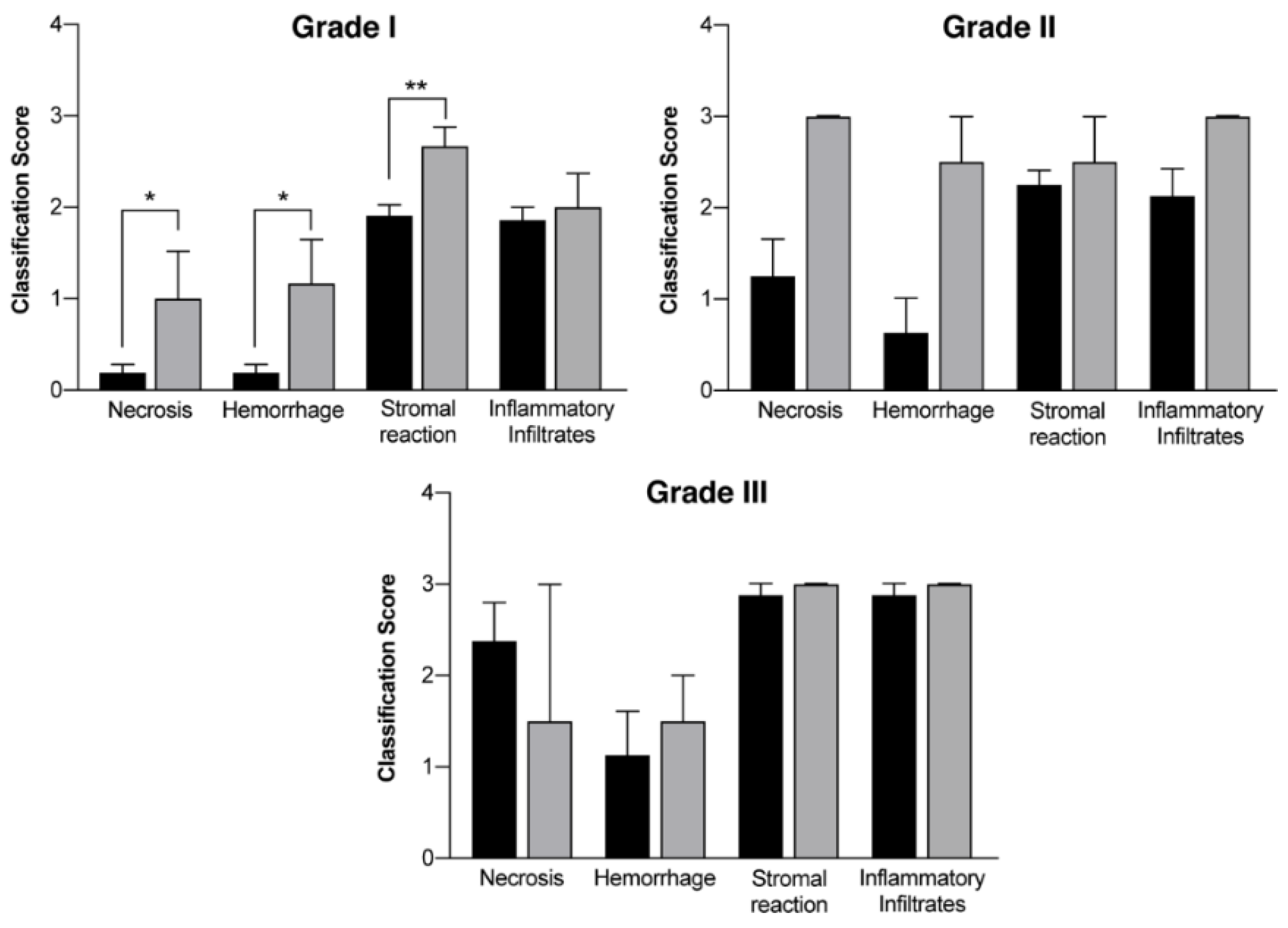
| Group | Tumor Grade | Necrosis | Hemorrhage | Stromal Reaction | Inflammatory Infiltrates |
|---|---|---|---|---|---|
| Control group (n > 10) | I | 0.19 ± 0.09 | 0.19 ± 0.09 | 1.91 ± 0.12 | 1.86 ± 0.14 |
| II | 1.25 ± 0.41 | 0.63 ± 0.38 | 2.25 ± 0.16 | 2.13 ± 0.30 | |
| III | 2.38 ± 0.42 | 1.13 ± 0.48 | 2.88 ± 0.13 | 2.88 ± 0.13 | |
| Treatment Group (n = 10) | I | 1.00 ± 0.52 * | 1.17 ± 0.48 * | 2.67 ± 0.21 ** | 2.00 ± 0.37 |
| II | 3.00 ± 0.01 | 2.50 ± 0.50 | 2.50 ± 0.50 | 3.00 ± 0.01 | |
| III | 1.50 ± 1.50 | 1.50 ± 0.50 | 3.00 ± 0.01 | 3.00 ± 0.01 |
| Necrosis | Hemorrhage | Stromal Reaction | Inflammatory Infiltrates |
|---|---|---|---|
| 0–not present | 0–not present | 0–absent | 0–absent |
| 1–focal (10%) | 1–focal (10%) | 1–mild | 1–mild |
| 2–moderate (20–70%) | 2–moderate (20–70%) | 2–moderate | 2–moderate |
| 3–extensive (>80%) | 3–extensive (>80%) | 3–high | 3–high |
Publisher’s Note: MDPI stays neutral with regard to jurisdictional claims in published maps and institutional affiliations. |
© 2020 by the authors. Licensee MDPI, Basel, Switzerland. This article is an open access article distributed under the terms and conditions of the Creative Commons Attribution (CC BY) license (http://creativecommons.org/licenses/by/4.0/).
Share and Cite
Costa, E.; Ferreira-Gonçalves, T.; Cardoso, M.; Coelho, J.M.P.; Gaspar, M.M.; Faísca, P.; Ascensão, L.; Cabrita, A.S.; Reis, C.P.; Figueiredo, I.V. A Step Forward in Breast Cancer Research: From a Natural-Like Experimental Model to a Preliminary Photothermal Approach. Int. J. Mol. Sci. 2020, 21, 9681. https://doi.org/10.3390/ijms21249681
Costa E, Ferreira-Gonçalves T, Cardoso M, Coelho JMP, Gaspar MM, Faísca P, Ascensão L, Cabrita AS, Reis CP, Figueiredo IV. A Step Forward in Breast Cancer Research: From a Natural-Like Experimental Model to a Preliminary Photothermal Approach. International Journal of Molecular Sciences. 2020; 21(24):9681. https://doi.org/10.3390/ijms21249681
Chicago/Turabian StyleCosta, Eduardo, Tânia Ferreira-Gonçalves, Miguel Cardoso, João M. P. Coelho, Maria Manuela Gaspar, Pedro Faísca, Lia Ascensão, António S. Cabrita, Catarina Pinto Reis, and Isabel V. Figueiredo. 2020. "A Step Forward in Breast Cancer Research: From a Natural-Like Experimental Model to a Preliminary Photothermal Approach" International Journal of Molecular Sciences 21, no. 24: 9681. https://doi.org/10.3390/ijms21249681
APA StyleCosta, E., Ferreira-Gonçalves, T., Cardoso, M., Coelho, J. M. P., Gaspar, M. M., Faísca, P., Ascensão, L., Cabrita, A. S., Reis, C. P., & Figueiredo, I. V. (2020). A Step Forward in Breast Cancer Research: From a Natural-Like Experimental Model to a Preliminary Photothermal Approach. International Journal of Molecular Sciences, 21(24), 9681. https://doi.org/10.3390/ijms21249681