Article

Nonanal Stimulates Growth Factors via Cyclic Adenosine Monophosphate (cAMP) Signaling in Human Hair Follicle Dermal Papilla Cells

Department of Food and Nutrition, Brain Korea 21 PLUS Project, Yonsei University, 50 Yonsei-ro, Seodaemun-gu, Seoul 120-749, Korea
*
Author to whom correspondence should be addressed.
These authors contributed equally to this work.
Int. J. Mol. Sci. 2020, 21(21), 8054; https://doi.org/10.3390/ijms21218054
Submission received: 29 September 2020 / Revised: 21 October 2020 / Accepted: 26 October 2020 / Published: 28 October 2020
(This article belongs to the Section Biochemistry)

Abstract

:
Human hair follicle dermal papilla cells (DPCs) are a specialized population of cells located in the hair follicles and regulate hair growth and development, particularly by releasing numerous growth factors in response to various physiological conditions. In the present study, we aimed to test whether nonanal, a scent compound from plants, stimulated growth factors in DPCs and to delineate the underlying mechanisms involved. We found that nonanal promoted DPC proliferation in a dose-dependent manner. Meanwhile, it also increased the intracellular cyclic adenosine monophosphate (cAMP) levels and the expression of various growth factor genes such as vascular endothelial growth factor, keratinocyte growth factor, and insulin-like growth factor 1. Furthermore, nonanal treatment stimulated DPC migration. Notably, the benefits of nonanal use were abrogated by cAMP inhibition. Our results reveal the potential of nonanal in preventing hair loss and suggest that its effects are cAMP-mediated in DPCs.

1. Introduction

Hair is an epidermal outgrowth composed of keratins originating from hair follicles deep down in the dermis. These hair follicles have been shown to produce increased amounts of keratin in response to growth factors; thus, growth factors are considered as key molecules that participate in hair growth initiation and suppression [1,2,3]. Dermal papilla, located at the base of the hair follicle, has its own blood supply and thus plays a crucial role in regulating numerous growth factors in response to various physiological conditions [4,5]. It has been reported that various causes of hair loss, including emotional stress, hormone imbalance, and nutritional deficiency, are characterized by the condensation of the dermal papilla along with impaired ability to produce growth factors [6,7,8].
At the cellular level, the production of growth factors is tightly regulated by several intracellular signaling pathways (e.g., Wnt/β-catenin and cyclic adenosine monophosphate (cAMP) that transmit a variety of extracellular stimuli. There are a few exogenous Wnt ligands that activate downstream β-catenin signaling, which leads to enhanced lymphoid enhancer-binding factor 1 (LEF1) transcription factor and contributes to growth factor gene enrichment [9,10,11]. Contrarily, cAMP, which can be activated by various agents targeting upstream G protein coupled receptor (GPCR; targeted by approximately 40% of all drugs), has been recognized as an important regulator of various growth factors, although the exact mechanism of growth factor regulation by cAMP has not been elucidated with consensus [12,13,14,15].
Cell proliferation might be a surrogate marker for increased growth factors in vitro [16,17,18], thus we attempted to explore the molecules that could enhance the proliferation of human hair follicle dermal papilla cells (DPCs). Since the beneficial effects of phytochemicals on hair growth have gained major interest, an in-house phytochemical library with about 500 entries, most of which have commercial usage such as food additives and cosmetic ingredients, was built as the first step. Then, through the screening process, we identified that nonanal induced a significant increase in the cell proliferation. Nonanal is a scent compound categorized as an aldehyde with the chemical formula C9H18O and has been widely reported in numerous natural essential oils, including cinnamon, rose, citrus, and pine [19,20]; however, its biological function remains unknown. This study aimed to investigate whether nonanal stimulated growth factors in DPCs and to delineate the mechanisms involved.

2. Results

2.1. Nonanal Increases Cell Proliferation

To evaluate whether nonanal affected cell proliferation in DPCs, we treated DPCs with either vehicle (dimethyl sulfoxide; DMSO) or various concentrations of nonanal ranging from 6.25 to 100 µM. After 24 h, the cells were observed using a microscope and a water-soluble tetrazolium salt (WST-1) assay was conducted (Figure 1A,B). Treatment of DPCs with nonanal significantly enhanced cell proliferation in a dose-dependent manner compared to that in vehicle controls. The proliferative effect seemed to be saturated at 50 µM nonanal in 24 h time point (Figure 1B,C); therefore, all further experiments were performed with 50 µM nonanal. On the other hand, nonanal treatment did not affect the proliferation of Hs68 dermal fibroblasts at 50 µM concentration (Figure S1).

2.2. Nonanal Enhances Gene Expression of Growth Factors

DPCs were treated with either vehicle or nonanal for 3, 6, 12, and 24 h. The mRNA expression levels of growth factors including vascular endothelial growth factor (VEGF), keratinocyte growth factor (KGF), and insulin-like growth factor 1 (IGF-1) in DPCs were then analyzed (Figure 2). Nonanal treatment significantly increased mRNA expressions of all growth factors studied, which peaked at 6 h, compared with vehicle controls.

2.3. Nonanal Does Not Affect the β-Catenin Signaling Pathway

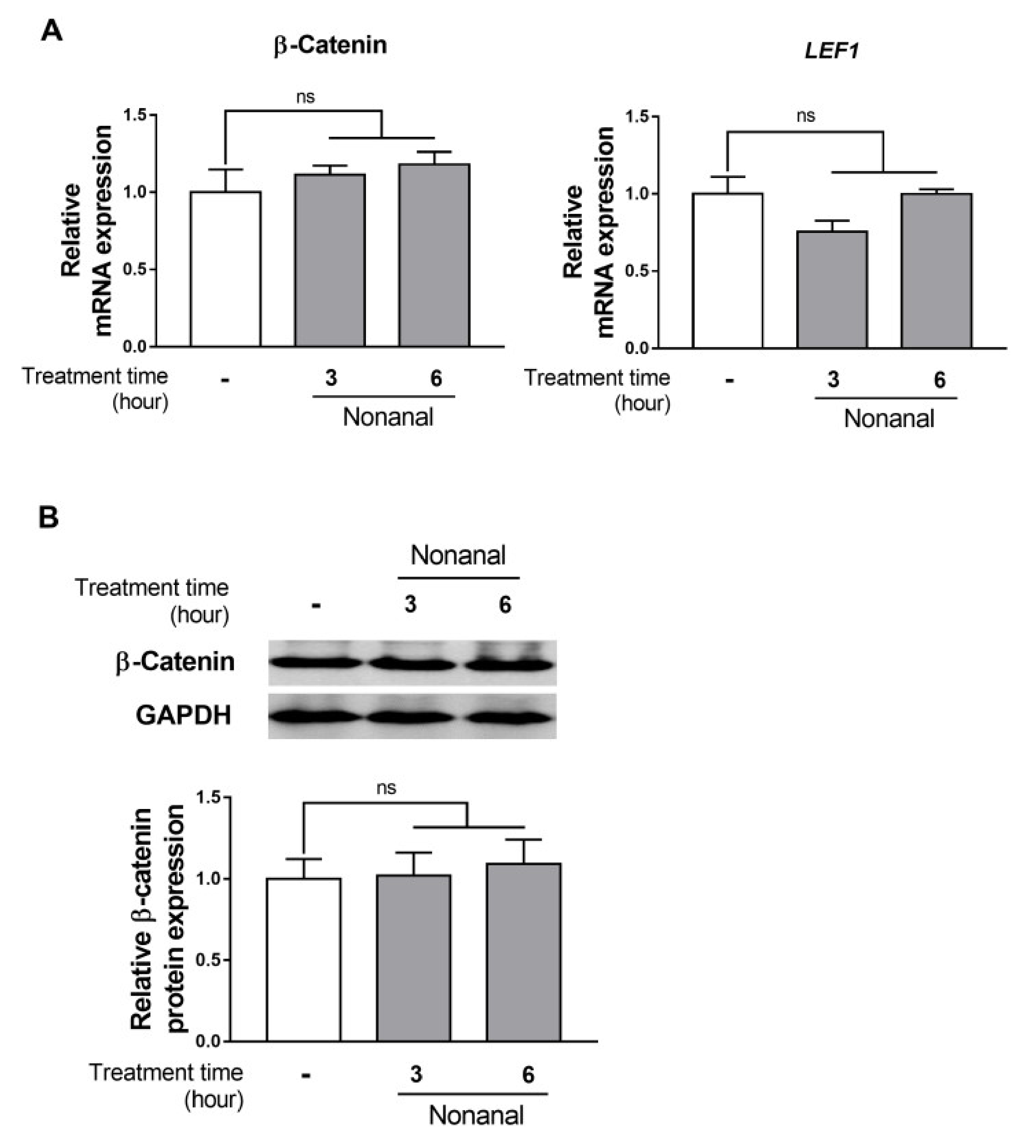
To explore the participation of β-catenin signaling in the nonanal-induced growth, β-catenin and its downstream effector, LEF1, expression levels were assessed in DPCs. DPCs were exposed to either vehicle or nonanal for 3 and 6 h. Nonanal treatment did not significantly affect β-catenin and LEF1 mRNA expression (Figure 3A). Consistent with the gene expression results, there was no change in β-catenin protein expression after treating DPCs with nonanal for 3 and 6 h (Figure 3B).

2.4. Nonanal Activates the cAMP/PKA Signaling Pathway

To investigate the role of the cAMP/ protein kinase A (PKA) pathway in the nonanal-induced growth, intracellular cAMP level and protein expression of PKA catalytic subunit α (PKA Cα), which is activated by cAMP, were evaluated in DPCs. DPCs were treated with nonanal for 5, 15, 30, and 60 min. The cAMP level significantly elevated at 5, 15, and 30 min and was maximum at 30 min, which then declined (Figure 4A). Furthermore, nonanal treatment significantly enhanced PKA Cα protein expression in DPCs compared with vehicle controls (Figure 4B).

2.5. cAMP Is Involved in Nonanal-Induced DPC Proliferation

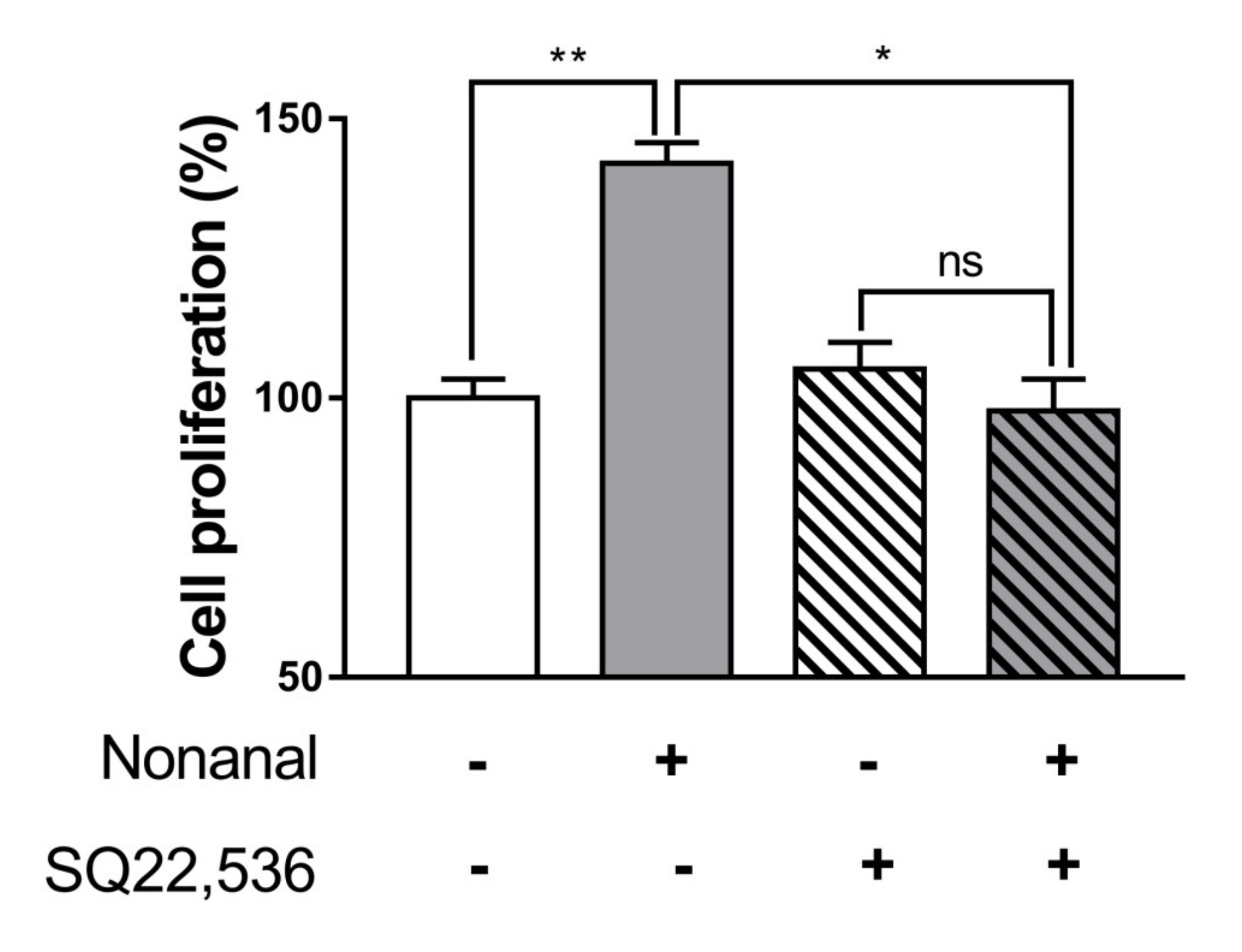
Since nonanal increases the intracellular cAMP levels, the role of cAMP in nonanal-induced DPC proliferation was evaluated. Nonanal treatment significantly increased cell proliferation, which was almost completely blocked by treating the cells with cAMP inhibitor SQ22,536 (50 µM; Figure 5).

2.6. cAMP Is Implicated in Nonanal-Induced Gene Expression of Growth Factors

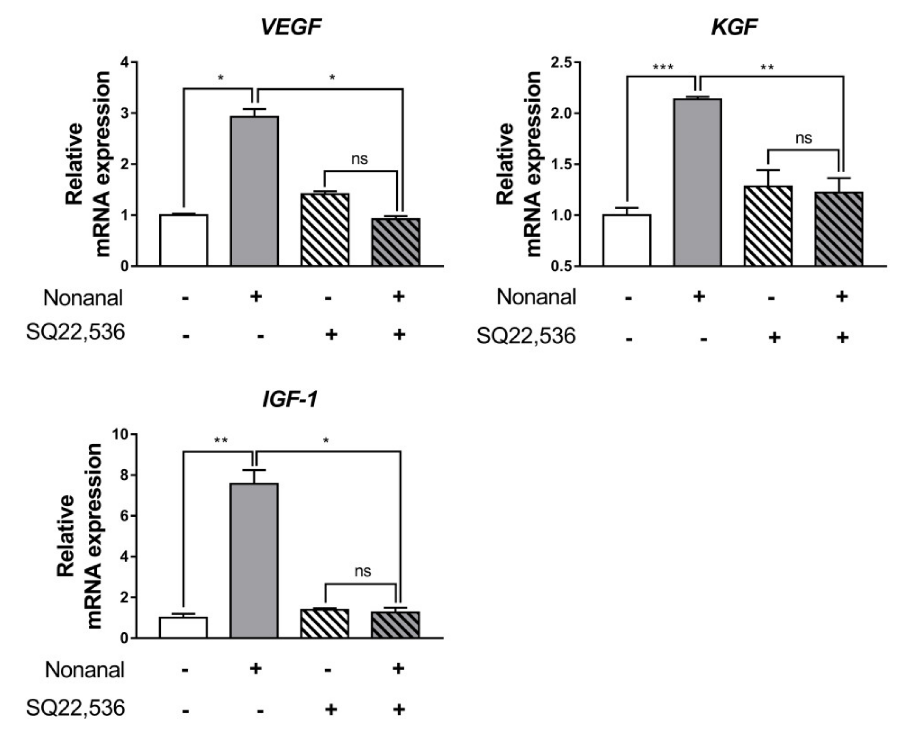
To investigate the role of cAMP in nonanal-induced gene expression of growth factors, we used 50 µM SQ22,536 to inhibit cAMP production in DPCs. The mRNA expression of VEGF, KGF, and IGF-1 was significantly enhanced by nonanal, but these effects are fully abolished by SQ22,536 (Figure 6).

2.7. Nonanal Promotes Cell Migration via cAMP

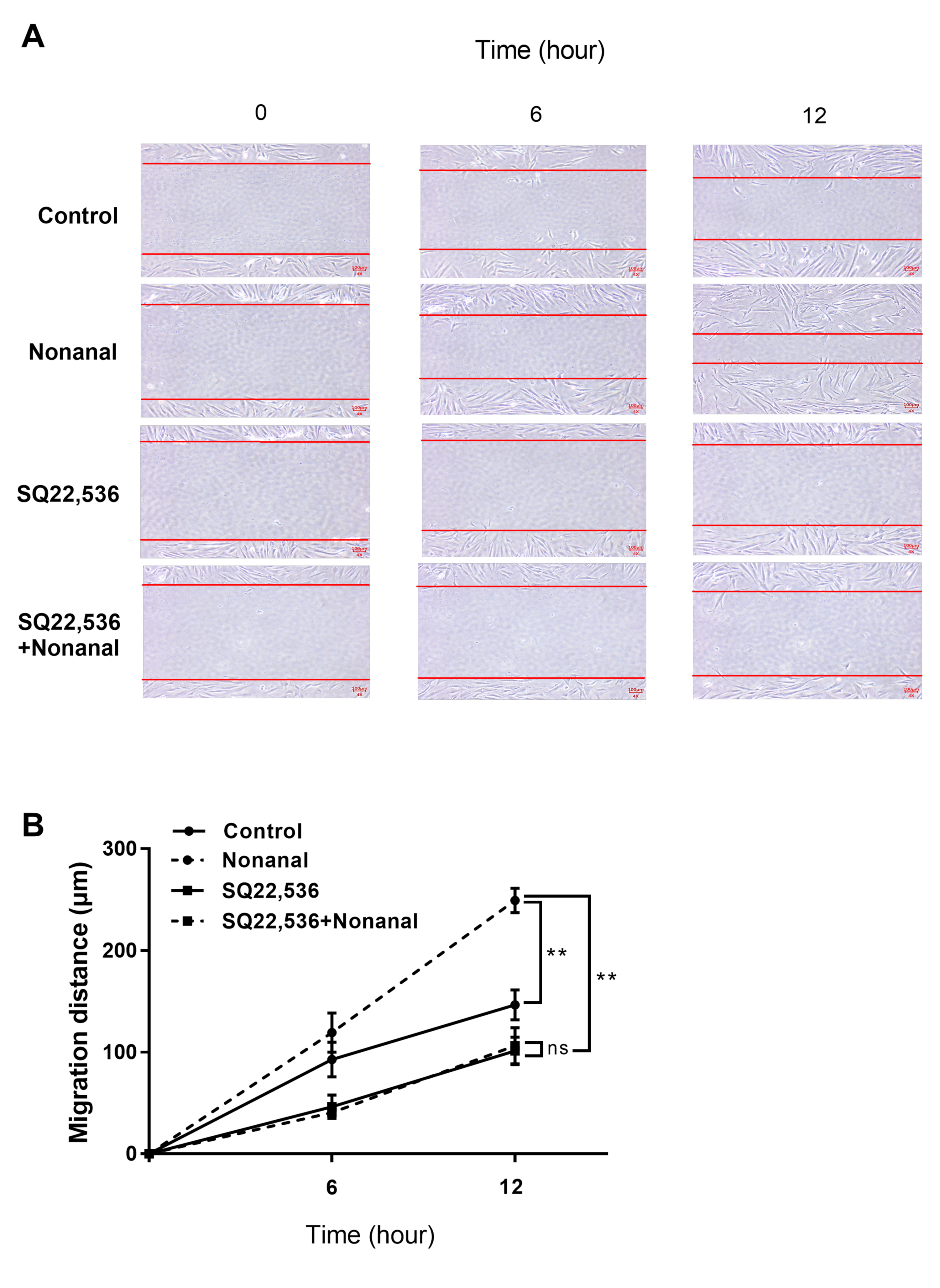
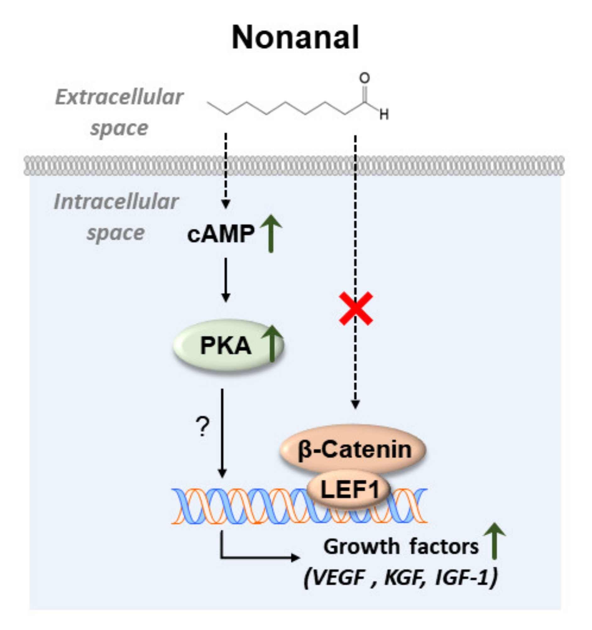
To determine whether nonanal affected DPC migration via cAMP, DPCs were treated with nonanal in the presence or absence of SQ22,536. Cell migration assay revealed that nonanal significantly increased migration in comparison with vehicle controls. However, nonanal-induced DPC migration was significantly inhibited by SQ22,536 (Figure 7A,B). Taken together, nonanal-induced cAMP pathway appears to contribute to the stimulation of growth factors in DPCs (Figure 8).

3. Discussion

Topical delivery of agents offers several advantages over oral delivery for treatment of extensive alopecia. There are fewer digestive enzymes, the pH is not extreme, and it shows considerable efficiency and bioavailability because of the proximity to the hair follicle. This may also lead to decreased systematic toxicity [21,22,23]. However, the skin epidermis can prevent the diffusion of many exogenous agents. It is commonly believed that only relatively low-molecular-weight (below 500 Da) lipophilic molecules can effectively permeate through the human skin [24]; nonanal is expected to be easily absorbed and reach the dermal papilla with topical application because of its lipophilicity and low molecular weight (142.2 Da). In addition, this compound has already been used in the cosmetic industry as a fragrance ingredient with no reports of skin irritation or sensitization [25,26,27].
It has been accepted that DPC has an essential role in the regulation of hair growth owing to its ability to secrete various growth factors. For example, KGF acts exclusively on the epidermis, where it stimulates the synthesis of hair keratin by inducing keratinocyte proliferation, migration, and differentiation [28,29]. However, several other growth factors act not only on the epidermis, but also on other locations in the skin for hair growth promotion. VEGF, as its name suggests, additionally increases vascularization around the hair follicles, thereby providing adequate nutrition and oxygen to the dermal papilla [30,31,32]. IGF-1, besides the epidermis and blood vessels, can act on the producer cell itself (autocrine signaling), which is known to strongly enhance DPC proliferation and migration [33,34,35]. In the present study, nonanal promotes proliferation and migration of DPCs. While the upregulation of growth factors such as IGF-1, KGF, and VEGF is a compelling potential mechanism, cAMP could also result in proliferation and migration through a number of mechanisms. Further study is needed to determine whether the increased levels of these factors are actually responsible for proliferation and migration induced by nonanal compound. If nonanal functions in the human scalp tissues in a way which is similar to that in DPCs, then the phenotypic improvement of hair loss may be accelerated through the simultaneous release of these growth factors.
In the present study, nonanal treatment stimulated DPC migration, which is known to be positively regulated by growth factors [18,36,37]. Cell migration, characterized by the orchestrated movements of cells in particular directions, is crucial in many developmental and morphogenic processes controlling organ size [38,39]. It has been shown that increased DPC migration can lead to the enhancement of dermal papilla volume, which determines hair follicle size and subsequently affects hair shaft thickness [40,41,42]. One clinical study has shown that the DPCs derived from bald scalps exhibit impaired migration activity [43].
We found that nonanal treatment did not alter β-catenin signaling, but activated the cAMP signaling pathway in DPCs (Figure 3 and Figure 4). Although mechanisms by which nonanal enhances the cAMP level are unknown, it is noteworthy that nonanal has been identified as a ligand of numerous olfactory receptors (ORs; e.g., OR1G1, OR52D1, and OR1A1) which constitute the largest GPCR subfamily, in each OR-transfected HEK293 and HeLa cells [44,45]. GPCR activation in the membrane can effectively stimulate adenylyl cyclase that catalyzes ATP to cAMP conversion [46,47]. The beneficial effects of nonanal seem to be specific to cell types as nonanal treatment did not affect the proliferation of Hs68 dermal fibroblasts, one of the major skin cell types. Further research is required to elucidate the exact mechanism by which nonanal activates the cAMP signaling in DPCs.

4. Materials and Methods

4.1. Cell Culture

Human follicle DPCs (55, female, Caucasian) were obtained from PromoCell (Heidelberg, Germany). These cells were isolated from human dermis of the scalp, originating from the occipital region. The cells were cultured in DPC growth medium with supplement mix (PromoCell) and 1% penicillin-streptomycin (10,000 U/mL; Gibco; Grand Island, NE, USA). The cells were incubated at 37 °C in 5% carbon dioxide humidified air and passaged every 3 days using 4-(2-hydroxyethyl)-1-piperazine ethanesulfonic acid (HEPES) buffer, trypsin/ethylene diamine tetra acetic acid (EDTA) solution, and trypsin neutralizing solution (PromoCell). Initial passage number of cells was 2, and the cells from passage numbers 3 to 5 were used for the study. Hs68 human dermal fibroblasts (American Type Culture Collection (ATCC), Manassas, VA, USA) were cultured in high-glucose Dulbecco’s modified Eagle’s medium (Hyclone, Logan, UT, USA) supplemented with 10% of fetal bovine serum (Hyclone) and 1% penicillin-streptomycin. Hs68 cells from passage numbers 5 to 7 were used for the study.

4.2. WST-1 Assay

WST-1 assay was performed to assess cell proliferation. DPCs were seeded in a 96-well plate at a density of 3000 cells per well and treated with vehicle (DMSO; Sigma-Aldrich; St. Louis, MO, USA) or various concentrations of nonanal (Sigma-Aldrich). When required, vehicle (DMSO) or SQ22,536 (Sigma-Aldrich), a cAMP inhibitor, was added 1 h before nonanal treatment. Images of the cells were captured using a Nikon Eclipse TS2 microscope equipped with the DMX1200 camera (Nikon, Tokyo, Japan). Subsequently, the treated cells were incubated for 2 h with the WST-1 reagent (Sigma-Aldrich) diluted 1:10 in culture medium. Absorbance was read at 440 nm using the Infinite M200 microplate reader (Tecan; Männedorf, Switzerland). In all the experiments, the final DMSO concentration was less than 0.1% (v/v). Furthermore, to determine whether the proliferation of Hs68 was also affected by nonanal treatment, the same method was employed as above.

4.3. cAMP Measurements

DPCs were seeded in a 12-well plate at a density of 40 000 cells per well and treated with nonanal at various time points. The cells were washed with HEPES buffer and lysed in 0.1 M HCl. Intracellular cAMP level in the lysates was measured using a cAMP enzyme-linked immunosorbent assay (ELISA) kit (Enzo Life Sciences; Farmingdale, NY, USA) following the manufacturer’s instructions. The cAMP level was normalized to the cellular protein content of the lysates, which was determined by the Bradford assay (BioRad; Hercules, CA, USA).

4.4. Real-Time PCR

DPCs were seeded in a 60 mm dish at a density of 200,000 cells and treated with vehicle or nonanal at various time points. When required, SQ22,536 was added 1 h before nonanal treatment. The RNA from the cells was isolated using the TRIzol reagent (Invitrogen; Carlsbad, CA, USA) and reverse-transcribed into cDNA with oligo(dT) primer (Invitrogen) and SuperScript IV reverse transcriptase (Invitrogen). The cDNA was amplified with gene-specific primers (Table 1) and SYBR green supermix (BioRad) on a CFX real-time system (BioRad). Data were analyzed using the comparative cycle threshold (Ct) method for relative quantitation and normalized to the levels of the internal control, glyceraldehyde 3-phosphate dehydrogenase (GAPDH).

4.5. Western Blotting

Western blotting was performed to determine the protein levels. DPCs were seeded in a 60-mm dish at a density of 200,000 cells, treated with either the vehicle or nonanal, and harvested at various time points. The cells were washed with HEPES buffer and lysed in a PRO-PREP protein extraction buffer (iNtRON; Seoul, Korea). Overall, 20 µg of protein was separated by sodium dodecyl sulfate-polyacrylamide gel electrophoresis (SDS-PAGE) and transferred onto a nitrocellulose membrane (Whatman; Kent, UK). Subsequently, the membrane was blocked with 5% bovine serum albumin (BSA; MP biomedicals; Irvine, CA, USA) in tris-buffered saline/Tween-20 (TBST) for 1 h and then incubated with a 1:1000-diluted anti-PKA Cα, anti-β-catenin, or anti-GAPDH antibodies (Cell Signaling; Herts, UK) overnight at 4 °C. The membrane was further incubated with a peroxidase-conjugated secondary antibody (Santa Cruz Biotechnology; Santa Cruz, CA, USA) for 1 h at 20 °C. Antibody binding was detected with electrochemiluminescence detection reagent (ECL; BioRad) and visualized using Ez-capture (ATTO; Tokyo, Japan). The protein levels were normalized to those of the loading control (GAPDH).

4.6. Migration Assay

DPCs were seeded in a 24-mm dish at a density of 20,000 cells per well and grown to confluence. On the day of the experiment, to inhibit cell proliferation, the cells were washed with HEPES buffer and preincubated with mitomycin C (15 μg/mL, Sigma-Aldrich) for 2 h. Subsequently, the cells were scratched in straight line with a sterile 200 μL tip. The cells were re-washed with HEPES buffer to remove cellular debris and incubated with vehicle or nonanal for 0, 6, and 12 h. When required, SQ22,536 was added 1 h before nonanal treatment. The same fields were photographed at each time point, and the area migrated by DPCs was measured using ImageJ (National Institutes of Health; NIH; Bethesda, MD, USA).

4.7. Statistical Analysis

All data are presented as mean ± standard error of mean (SEM). Statistical analysis was performed by the Student’s t-test using SPSS Statistics 24 (SPSS; Chicago, IL, USA). Differences were considered as significant at p < 0.05.

5. Conclusions

In summary, we have demonstrated that nonanal increases the growth factor levels through cAMP-mediated signaling in DPCs. Although further in vivo experimental models should be tested to confirm the in vivo relevance of these in vitro findings, the current study highlights the potential utility of nonanal for treating hair loss associated with various physiological states and provides insights into the molecular mechanisms of the effects of nonanal in DPCs.

Supplementary Materials

Supplementary materials can be found at https://www.mdpi.com/1422-0067/21/21/8054/s1.

Author Contributions

Conceptualization, S.P., W.K. and T.P.; methodology, S.P. and B.S.; formal analysis, S.P. and W.K.; data curation, S.P.; writing—original draft preparation, S.P., W.K., B.S. and D.C.; writing—review and editing, S.P., W.K. and T.P.; funding acquisition, T.P. All authors have read and agreed to the published version of the manuscript.

Funding

This work was supported by Innopolis Foundation through Technology Commercialization services(or Project), funded by Ministry of Science and ICT (2020-JB-RD-0080) and the National Research Foundation of Korea (NRF) grant funded by the Korean government (MSIT) (No. 2019R1A2C2003340).

Conflicts of Interest

The authors declare no conflict of interest. The funders had no role in the design of the study; in the collection, analyses, or interpretation of data; in the writing of the manuscript, or in the decision to publish the results.

References

  1. Danilenko, D.M.; Ring, B.D.; Pierce, G.F. Growth factors and cytokines in hair follicle development and cycling: Recent insights from animal models and the potentials for clinical therapy. Mol. Med. Today 1996, 2, 460–467. [Google Scholar] [CrossRef]
  2. Shirakata, Y. Regulation of epidermal keratinocytes by growth factors. J. Dermatol. Sci. 2010, 59, 73–80. [Google Scholar] [CrossRef]
  3. Zhang, X.; Wang, Y.; Gao, Y.; Liu, X.; Bai, T.; Li, M.; Li, L.; Chi, G.; Xu, H.; Liu, F. Maintenance of high proliferation and multipotent potential of human hair follicle-derived mesenchymal stem cells by growth factors. Int. J. Mol. Med. 2013, 31, 913–921. [Google Scholar] [CrossRef] [Green Version]
  4. Han, J.H.; Kwon, O.S.; Chung, J.H.; Cho, K.H.; Eun, H.C.; Kim, K.H. Effect of minoxidil on proliferation and apoptosis in dermal papilla cells of human hair follicle. J. Dermatol. Sci. 2004, 34, 91–98. [Google Scholar] [CrossRef]
  5. Rahmani, W.; Abbasi, S.; Hagner, A.; Raharjo, E.; Kumar, R.; Hotta, A.; Magness, S.; Metzger, D.; Biernaskie, J. Hair follicle dermal stem cells regenerate the dermal sheath, repopulate the dermal papilla, and modulate hair type. Dev. Cell 2014, 31, 543–558. [Google Scholar] [CrossRef] [PubMed] [Green Version]
  6. Phillips, T.G.; Slomiany, W.P.; Allison, R. Hair loss: Common causes and treatment. Am. Fam. Physician 2017, 96, 371–378. [Google Scholar] [PubMed]
  7. Navarro, M.; Asín, M.; Martínez, M.; Martínez, A.; Molina, C.; Moscoso, L.; Pino, A.; Orive, G.; Anitua, E. Management of androgenetic alopecia: A comparative clinical study between plasma rich in growth factors and topical minoxidil. Eur. J. Plast. Surg. 2016, 39, 173–180. [Google Scholar] [CrossRef]
  8. Trüeb, R.M. Molecular mechanisms of androgenetic alopecia. Exp. Gerontol. 2002, 37, 981–990. [Google Scholar] [CrossRef]
  9. Leirós, G.J.; Ceruti, J.M.; Castellanos, M.L.; Kusinsky, A.G.; Balañá, M.E. Androgens modify Wnt agonists/antagonists expression balance in dermal papilla cells preventing hair follicle stem cell differentiation in androgenetic alopecia. Mol. Cell. Endocrinol. 2017, 439, 26–34. [Google Scholar] [CrossRef]
  10. Enshell-Seijffers, D.; Lindon, C.; Kashiwagi, M.; Morgan, B.A. β-catenin activity in the dermal papilla regulates morphogenesis and regeneration of hair. Dev. Cell 2010, 18, 633–642. [Google Scholar] [CrossRef] [Green Version]
  11. Myung, P.S.; Takeo, M.; Ito, M.; Atit, R.P. Epithelial Wnt ligand secretion is required for adult hair follicle growth and regeneration. J. Investig. Dermatol. 2013, 133, 31–41. [Google Scholar] [CrossRef] [PubMed] [Green Version]
  12. Chéret, J.; Bertolini, M.; Ponce, L.; Lehmann, J.; Tsai, T.; Alam, M.; Hatt, H.; Paus, R. Olfactory receptor OR2AT4 regulates human hair growth. Nat. Commun. 2018, 9, 1–12. [Google Scholar] [CrossRef] [PubMed]
  13. Hwang, K.-A.; Hwang, Y.-L.; Lee, M.-H.; Kim, N.-R.; Roh, S.-S.; Lee, Y.; Kim, C.D.; Lee, J.-H.; Choi, K.-C. Adenosine stimulates growth of dermal papilla and lengthens the anagen phase by increasing the cysteine level via fibroblast growth factors 2 and 7 in an organ culture of mouse vibrissae hair follicles. Int. J. Mol. Med. 2012, 29, 195–201. [Google Scholar] [PubMed] [Green Version]
  14. Choi, M.; Choi, S.-J.; Jang, S.; Choi, H.-I.; Kang, B.-M.; Hwang, S.T.; Kwon, O. Shikimic acid, a mannose bioisostere, promotes hair growth with the induction of anagen hair cycle. Sci. Rep. 2019, 9, 1–8. [Google Scholar] [CrossRef] [Green Version]
  15. Stevens, R.C.; Cherezov, V.; Katritch, V.; Abagyan, R.; Kuhn, P.; Rosen, H.; Wüthrich, K. The GPCR Network: A large-scale collaboration to determine human GPCR structure and function. Nat. Rev. Drug Discov. 2013, 12, 25–34. [Google Scholar] [CrossRef] [Green Version]
  16. Sun, Y.N.; Cui, L.; Li, W.; Yan, X.T.; Yang, S.Y.; Kang, J.I.; Kang, H.K.; Kim, Y.H. Promotion effect of constituents from the root of Polygonum multiflorum on hair growth. Bioorganic Med. Chem. Lett. 2013, 23, 4801–4805. [Google Scholar] [CrossRef]
  17. Gasparri, F.; Mariani, M.; Sola, F.; Galvani, A. Quantification of the proliferation index of human dermal fibroblast cultures with the ArrayScan™ high-content screening reader. J. Biomol. Screen. 2004, 9, 232–243. [Google Scholar] [CrossRef] [Green Version]
  18. Madaan, A.; Verma, R.; Singh, A.T.; Jaggi, M. Review of hair follicle dermal papilla cells as in vitro screening model for hair growth. Int. J. Cosmet. Sci. 2018, 40, 429–450. [Google Scholar] [CrossRef] [Green Version]
  19. Guenther, E.; Althausen, D. The Essential Oils; Van Nostrand: New York, NY, USA, 1948; Volume 1. [Google Scholar]
  20. Omonov, T.S.; Kharraz, E.; Foley, P.; Curtis, J.M. The production of biobased nonanal by ozonolysis of fatty acids. RSC Adv. 2014, 4, 53617–53627. [Google Scholar] [CrossRef]
  21. Heyneman, C.A.; Lawless-Liday, C.; Wall, G.C. Oral versus topical NSAIDs in rheumatic diseases. Drugs 2000, 60, 555–574. [Google Scholar] [CrossRef]
  22. Aqil, F.; Munagala, R.; Jeyabalan, J.; Vadhanam, M.V. Bioavailability of phytochemicals and its enhancement by drug delivery systems. Cancer Lett. 2013, 334, 133–141. [Google Scholar] [CrossRef] [PubMed] [Green Version]
  23. Fang, J.-Y.; Liu, P.-F.; Huang, C.-M. Decreasing systemic toxicity via transdermal delivery of anticancer drugs. Curr. Drug Metab. 2008, 9, 592–597. [Google Scholar] [CrossRef] [PubMed]
  24. Bos, J.D.; Meinardi, M.M. The 500 Dalton rule for the skin penetration of chemical compounds and drugs. Exp. Dermatol. Viewp. 2000, 9, 165–169. [Google Scholar] [CrossRef] [PubMed]
  25. Lachenmeier, D.W.; Gary, M.; Monakhova, Y.B.; Kuballa, T.; Mildau, G. Rapid NMR screening of total aldehydes to detect oxidative rancidity in vegetable oils and decorative cosmetics. Spectrosc. Eur. 2010, 22, 11. [Google Scholar]
  26. Ruzsanyi, V.; Mochalski, P.; Schmid, A.; Wiesenhofer, H.; Klieber, M.; Hinterhuber, H.; Amann, A. Ion mobility spectrometry for detection of skin volatiles. J. Chromatogr. B 2012, 911, 84–92. [Google Scholar] [CrossRef] [Green Version]
  27. Panzella, L. Natural Phenolic Compounds for Health, Food and Cosmetic Applications. Antioxidants 2020, 9, 427. [Google Scholar] [CrossRef]
  28. Rubin, J.S.; Bottaro, D.P.; Chedid, M.; Miki, T.; Ron, D.; Cheon, H.-G.; Taylor, W.G.; Fortney, E.; Sakata, H.; Finch, P.W. Keratinocyte growth factor. Cell Biol. Int. 1995, 19, 399–412. [Google Scholar] [CrossRef]
  29. Gibbs, S.; Silva Pinto, A.N.; Murli, S.; Huber, M.; Hohl, D.; Ponec, M. Epidermal growth factor and keratinocyte growth factor differentially regulate epidermal migration, growth, and differentiation. Wound Repair Regen. 2000, 8, 192–203. [Google Scholar] [CrossRef]
  30. Messenger, A.; Rundegren, J. Minoxidil: Mechanisms of action on hair growth. Br. J. Dermatol. 2004, 150, 186–194. [Google Scholar] [CrossRef]
  31. Yano, K.; Brown, L.F.; Detmar, M. Control of hair growth and follicle size by VEGF-mediated angiogenesis. J. Clin. Investig. 2001, 107, 409–417. [Google Scholar] [CrossRef] [Green Version]
  32. Wise, L.M.; Inder, M.K.; Real, N.C.; Stuart, G.S.; Fleming, S.B.; Mercer, A.A. The vascular endothelial growth factor (VEGF)-E encoded by orf virus regulates keratinocyte proliferation and migration and promotes epidermal regeneration. Cell. Microbiol. 2012, 14, 1376–1390. [Google Scholar] [CrossRef]
  33. Shigematsu, S.; Yamauchi, K.; Nakajima, K.; Iijima, S.; Aizawa, T.; Hashizume, K. IGF-1 regulates migration and angiogenesis of human endothelial cells. Endocr. J. 1999, 46, S59–S62. [Google Scholar] [CrossRef] [PubMed] [Green Version]
  34. Chiu, H.; Chang, C.; Chen, J.; Jee, S. Human hair follicle dermal papilla cell, dermal sheath cell and interstitial dermal fibroblast characteristics. J. Formos. Med. Assoc. Taiwan Yi Zhi 1996, 95, 667–674. [Google Scholar] [PubMed]
  35. Itami, S.; Kurata, S.; Takayasu, S. Androgen induction of follicular epithelial cell growth is mediated via insulin-like growth factor-I from dermal papilla cells. Biochem. Biophys. Res. Commun. 1995, 212, 988–994. [Google Scholar] [CrossRef]
  36. Guvakova, M.A. Insulin-like growth factors control cell migration in health and disease. Int. J. Biochem. Cell Biol. 2007, 39, 890–909. [Google Scholar] [CrossRef]
  37. Seeger, M.A.; Paller, A.S. The roles of growth factors in keratinocyte migration. Adv. Wound Care 2015, 4, 213–224. [Google Scholar] [CrossRef] [PubMed] [Green Version]
  38. Ridley, A.J.; Schwartz, M.A.; Burridge, K.; Firtel, R.A.; Ginsberg, M.H.; Borisy, G.; Parsons, J.T.; Horwitz, A.R. Cell migration: Integrating signals from front to back. Science 2003, 302, 1704–1709. [Google Scholar] [CrossRef] [Green Version]
  39. Lauffenburger, D.A.; Horwitz, A.F. Cell migration: A physically integrated molecular process. Cell 1996, 84, 359–369. [Google Scholar] [CrossRef] [Green Version]
  40. Lei, M.; Guo, H.; Qiu, W.; Lai, X.; Yang, T.; Widelitz, R.B.; Chuong, C.M.; Lian, X.; Yang, L. Modulating hair follicle size with W nt10b/DKK 1 during hair regeneration. Exp. Dermatol. 2014, 23, 407–413. [Google Scholar] [CrossRef] [Green Version]
  41. Elliott, K.; Messenger, A.G.; Stephenson, T.J. Differences in hair follicle dermal papilla volume are due to extracellular matrix volume and cell number: Implications for the control of hair follicle size and androgen responses. J. Investig. Dermatol. 1999, 113, 873–877. [Google Scholar] [CrossRef] [Green Version]
  42. Zhou, L.; Wang, H.; Jing, J.; Yu, L.; Wu, X.; Lu, Z. Regulation of hair follicle development by exosomes derived from dermal papilla cells. Biochem. Biophys. Res. Commun. 2018, 500, 325–332. [Google Scholar] [CrossRef] [PubMed]
  43. Upton, J.H.; Hannen, R.F.; Bahta, A.W.; Farjo, N.; Farjo, B.; Philpott, M.P. Oxidative stress–associated senescence in dermal papilla cells of men with androgenetic alopecia. J. Investig. Dermatol. 2015, 135, 1244–1252. [Google Scholar] [CrossRef] [PubMed] [Green Version]
  44. Schmiedeberg, K.; Shirokova, E.; Weber, H.-P.; Schilling, B.; Meyerhof, W.; Krautwurst, D. Structural determinants of odorant recognition by the human olfactory receptors OR1A1 and OR1A2. J. Struct. Biol. 2007, 159, 400–412. [Google Scholar] [CrossRef]
  45. Sanz, G.; Schlegel, C.; Pernollet, J.-C.; Briand, L. Comparison of odorant specificity of two human olfactory receptors from different phylogenetic classes and evidence for antagonism. Chem. Senses 2005, 30, 69–80. [Google Scholar] [CrossRef] [PubMed]
  46. Thomsen, W.; Frazer, J.; Unett, D. Functional assays for screening GPCR targets. Curr. Opin. Biotechnol. 2005, 16, 655–665. [Google Scholar] [CrossRef]
  47. Selbie, L.A.; Hill, S.J. G protein-coupled-receptor cross-talk: The fine-tuning of multiple receptor-signalling pathways. Trends Pharmacol. Sci. 1998, 19, 87–93. [Google Scholar] [CrossRef]
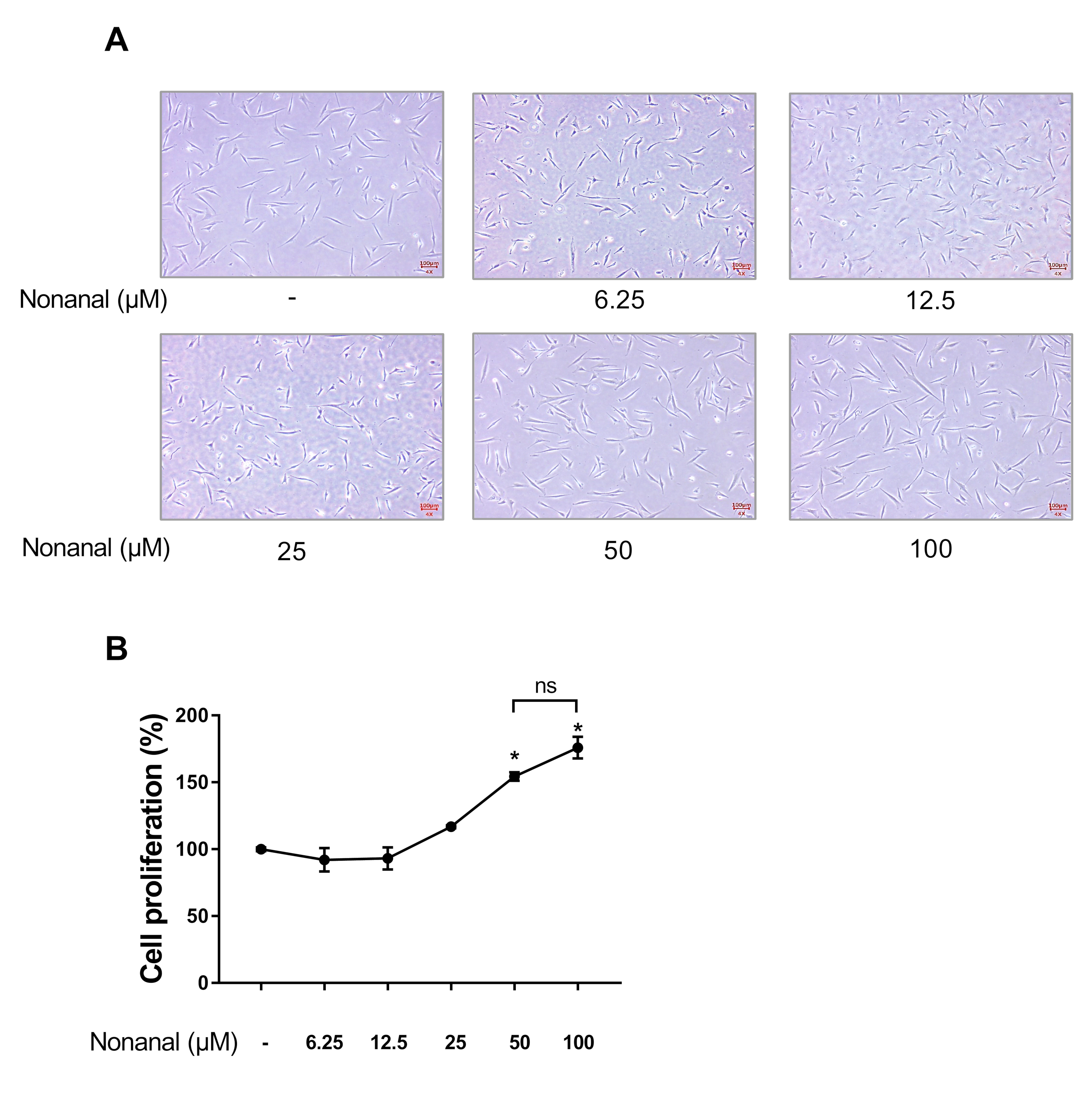
Figure 1. Effect of nonanal on dermal papilla cell (DPC) proliferation. DPCs were treated with vehicle (dimethyl sulfoxide; DMSO) or indicated concentrations of nonanal ranging from 6.25 to 100 µM. (A) Representative images of cells in response to nonanal treatment at indicated concentrations for 24 h were captured. (B) Cell proliferation was determined in response to nonanal treatment at indicated concentrations for 24 h by WST-1 assay. (C) DPCs were exposed to 50 µM of nonanal at different time points (12, 24, and 48 h) and cell proliferation was determined by WST-1 assay. Data were statistically analyzed using the Student’s t-test. Data are presented as mean ± standard error of mean (SEM) of the results of three independent experiments. * p < 0.05 vs. control.
Ijms 21 08054 g001
Figure 2. The time-course of mRNA expression of growth factors in response to nonanal treatment in DPCs. DPCs were treated with vehicle or 50 µM nonanal and the relative mRNA levels of growth factors were measured by real-time PCR over a 24 h time-course. Data were statistically analyzed using the Student’s t-test. Data are presented as mean ± SEM of the results of three independent experiments. * p < 0.05, ** p < 0.01, *** p < 0.001. ns, not significant.
Ijms 21 08054 g002
Figure 3. Effect of nonanal on β -catenin pathway in DPCs. DPCs were exposed to vehicle or 50 µM of nonanal at different time points (3 and 6 h). (A) Relative β-catenin and lymphoid enhancer-binding factor 1 (LEF1) mRNA levels were analyzed by real-time PCR. (B) β-catenin protein levels were detected by Western blot. Data were statistically analyzed using the Student’s t-test. Data are presented as mean ± SEM of the results of three independent experiments. ns, not significant.
Ijms 21 08054 g003
Figure 4. Effect of nonanal on the cyclic adenosine monophosphate (cAMP) pathway in DPCs. (A) DPCs were treated with vehicle or 50 µM nonanal for 5, 15, 30, and 60 min. Intracellular cAMP levels were determined using enzyme-linked immunosorbent assay (ELISA) kit. (B) The protein levels of protein kinase A catalytic subunit (PKA Cα) were detected by Western blotting after treatment with vehicle or 50 µM nonanal for 30 min. Data were statistically analyzed using the Student’s t-test. Data are presented as mean ± SEM of the results of three independent experiments. * p < 0.05, ** p < 0.01. ns, not significant.
Ijms 21 08054 g004
Figure 5. Effect of inhibiting cAMP production on nonanal-stimulated proliferation of DPCs. DPCs were treated with vehicle or 50 µM nonanal for 24 h. The vehicle or 50 µM SQ22,536, a cAMP inhibitor, was added 1 h before nonanal treatment. Cell proliferation was determined by WST-1 assay. Data were statistically analyzed using the Student’s t-test. Data are presented as mean ± SEM of the results of three independent experiments. * p < 0.05, ** p< 0.01. ns, not significant.
Ijms 21 08054 g005
Figure 6. Effect of cAMP inhibitors on nonanal-stimulated mRNA expression of growth factors. DPCs were treated with vehicle or 50 µM nonanal for 6 h. The vehicle or 50 µM SQ22,536 was added 1 h before nonanal treatment. Relative mRNA levels of the growth factors were measured by real-time PCR. Data were statistically analyzed using the Student’s t-test. Data are presented as mean ± SEM of the results of three independent experiments. * p < 0.05, ** p < 0.01, *** p < 0.001. ns, not significant.
Ijms 21 08054 g006
Figure 7. Effect of nonanal on DPC migration via the cAMP signaling pathway. Migration assay was conducted in confluent DPCs by drawing a straight line across the bottom of the dish, followed by incubation with either vehicle or 50 µM nonanal at different time points (0, 6, and 12 h). The vehicle or 50 µM SQ22,536 was added 1 h before nonanal treatment. (A) Phase-contrast microscopy images of the scratched area were captured. The lines delineate the migrating edges of cells. (B) Quantification of cell migration was performed by measuring the area occupied by the cells at different time points. Data were statistically analyzed using the Student’s t-test. Data are presented as mean ± standard error of mean (SEM) of the results of three independent experiments. ** p < 0.01. ns, not significant.
Ijms 21 08054 g007
Figure 8. A suggested mechanism by which nonanal induces the growth factors expression in DPCs via the cAMP signaling pathway. As shown in a schematic illustration, stimulation of the cAMP signaling pathway in response to nonanal increases levels of growth factors such as VEGF, KGF, and IGF-1. The detailed molecular mechanism by which cAMP-mediated growth factor secretion increases needs further investigation. Interestingly, β -catenin and LEF1 levels did not increase in response to nonanal treatment.
Ijms 21 08054 g008
Table 1. Primer sequences used for real-time PCR.
Gene NamePrimer Sequences (5′→ 3′)
Catenin beta 1 (CTNNB1)F: CCGACACCAAGAAGCAGAGATG
R: GTGGGATGGTGGGTGTAAGAG
Glyceraldehyde 3-phosphate dehydrogenase (GAPDH)F: AAATCAAGTGGGGCGATGC
R: AGGGGGCAGAGATGATGACC
Insulin-like growth factor-1 (IGF-1)F: TTGCTCTCAACATCTCCCATCT
R: TGCATCTTCACCTTCAAGAAAT
Keratinocyte growth factor (KGF)F: AGAGAAGGGATGCTGGAGGT
R: CGTAAGGGGCACTGTTTTAGAG
Lymphoid enhancer factor 1 (LEF1)F: CAACCCTACCCATCCTCACTG
R: GGCTCCTGCTCCTTTCTCTGT
Vascular endothelial growth factor (VEGF)F: CTTCTGAGTTGCCCAGGAGA
R: GGATGGAGGAAGGTCAACCA
Publisher’s Note: MDPI stays neutral with regard to jurisdictional claims in published maps and institutional affiliations.