Article

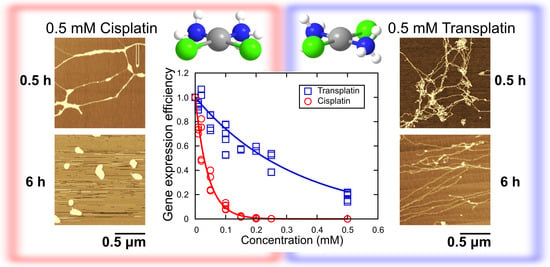
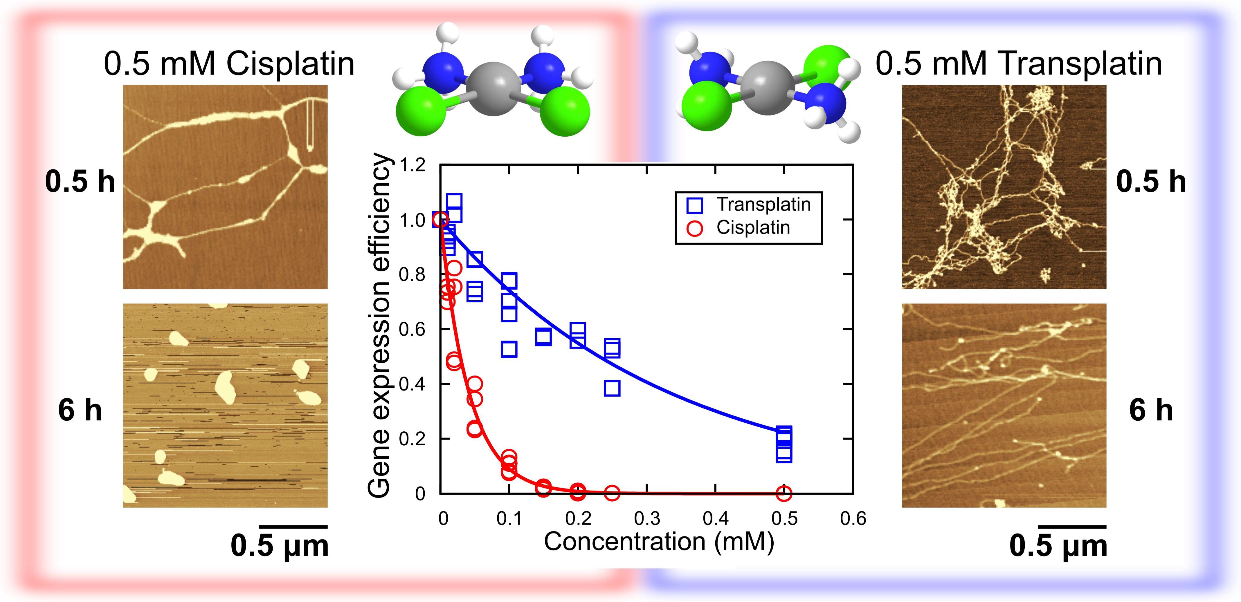
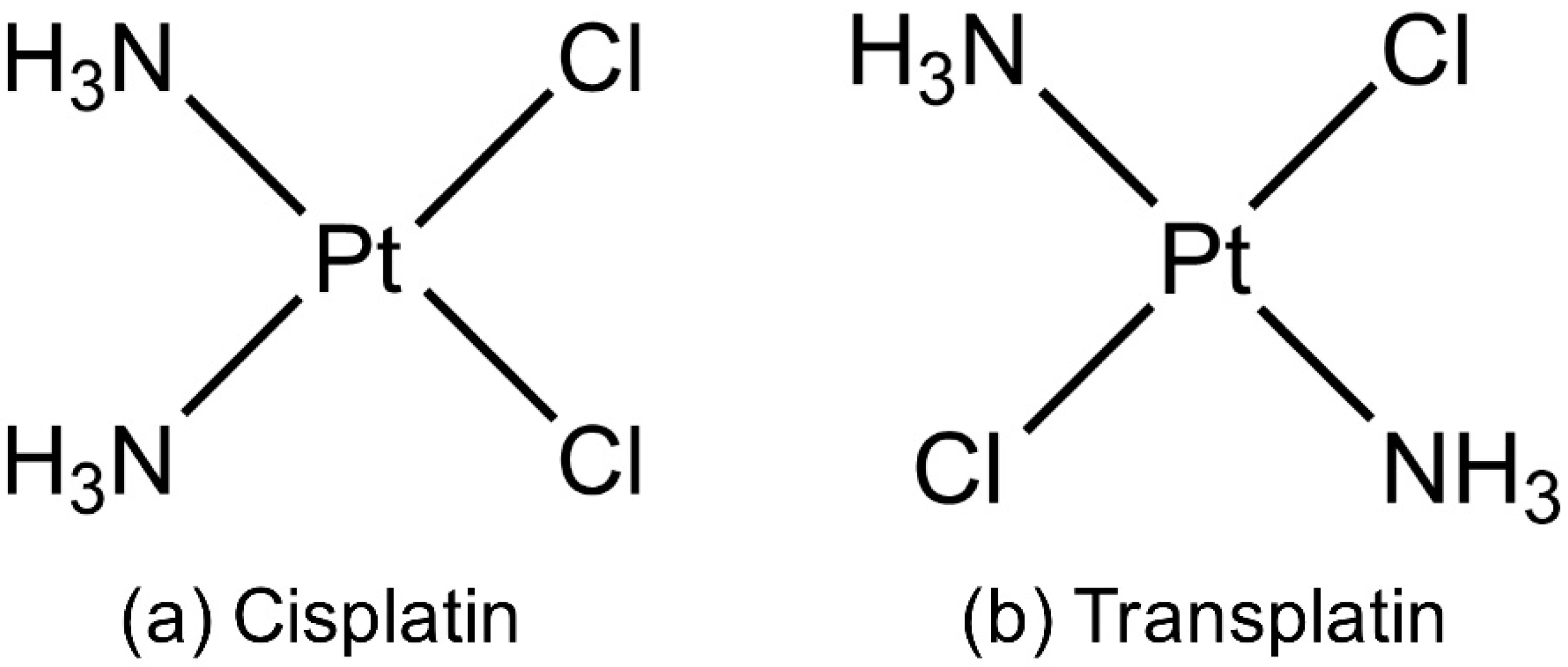
Different Effects of Cisplatin and Transplatin on the Higher-Order Structure of DNA and Gene Expression

1
Faculty of Life and Medical Sciences, Doshisha University, Kyotanabe, Kyoto 610-0394, Japan
2
Faculty of Pharmaceutical Sciences, Suzuka University of Medical Science, Suzuka, Mie 513-8670, Japan
*
Author to whom correspondence should be addressed.
Int. J. Mol. Sci. 2020, 21(1), 34; https://doi.org/10.3390/ijms21010034
Submission received: 31 October 2019 / Revised: 15 December 2019 / Accepted: 17 December 2019 / Published: 19 December 2019
(This article belongs to the Special Issue Cisplatin in Cancer Therapy: Molecular Mechanisms of Action)

Abstract

:
Despite the effectiveness of cisplatin as an anticancer agent, its trans-isomer, transplatin, is clinically ineffective. Although both isomers target nuclear DNA, there is a large difference in the magnitude of their biological effects. Here, we compared their effects on gene expression in an in vitro luciferase assay and quantified their effects on the higher-order structure of DNA using fluorescence microscopy (FM) and atomic force microscopy (AFM). The inhibitory effect of cisplatin on gene expression was about 7 times that of transplatin. Analysis of the fluctuation autocorrelation function of the intrachain Brownian motion of individual DNA molecules showed that cisplatin increases the spring and damping constants of DNA by one order of magnitude and these visco-elastic characteristics tend to increase gradually over several hours. Transplatin had a weaker effect, which tended to decrease with time. These results agree with a stronger inhibitory effect of cisplatin on gene expression. We discussed the characteristic effects of the two compounds on the higher-order DNA structure and gene expression in terms of the differences in their binding to DNA.

Graphical Abstract

1. Introduction

Clinical use of cisplatin (cis-diamminedichloridoplatinum(II); see Figure 1) [1,2] started in the United States more than 40 years ago [3]. Cisplatin and related compounds, such as carboplatin (cis-diammine(1,1-cyclobutanedicarboxylato)platinum(II)) [4,5] and oxaliplatin (1R,2R-diaminocyclohexane)oxalatoplatinum(II)) [6,7], are called “platinum-based drugs” and are widely used as anticancer agents. DNA is believed to be the critical target of the platinum-based drugs [8,9]. The structure of these drugs can be described by a general formula, cis-[PtA2 X2], where A = ammonia or amine, and X = leaving group such as halide or carboxylate. In contrast to cis-Pt(II) complexes, the trans-Pt(II) derivatives have not entered a clinical trial yet. In particular, transplatin (trans-diamminedichloridoplatinum(II); see Figure 1), was found to be much less effective than cisplatin in blocking cell division in bacteria [10] and in inhibiting the growth of cancer cells [11]. Therefore, transplatin is known as a therapeutically inactive stereoisomer of cisplatin.
One possible explanation for the therapeutic inactivity of transplatin is that it undergoes more effective detoxification by sulfur-containing scavengers such as glutathione [12] and metallothionein [13]. Higher doses of transplatin as compared to cisplatin are required for binding of an equal number of Pt atoms per DNA nucleotide in living cells [14]. It was reported that the introduction of a sterically hindered planar amine ligand into transplatin instead of one of the ammonia ligands greatly enhanced cytotoxicity [15,16,17,18,19], probably because the bulky amine ligand decreased the rate of reaction of transplatin derivatives with glutathione. Another reason could be a different mode of interaction with DNA; the platinum(II) atom preferentially binds to guanine-N7, followed by adenine-N7, adenine-N1, and cytosine-N3 [20]. Although both cisplatin and transplatin form covalent DNA adducts, their structure is distinctively different. For instance, cisplatin forms mainly 1,2-intrastrand crosslinks (~80%) and rarely interstrand crosslinks (~1.5%) between two nucleobases in double-stranded DNA [21,22], whereas transplatin can hardly form intrastrand crosslinks but forms monofunctional adducts [23] and interstrand crosslinks [24] at a higher frequency than cisplatin. Local and second-order conformational changes induced in DNA by the two stereoisomers have also been discussed as possible reasons for their different antitumor efficacies [25,26,27,28].
Using fluorescence microscopy (FM) and atomic force microscopy (AFM), we elucidated the higher-order conformational changes of single long DNA strings induced by some novel cationic dinuclear Pt(II) complexes [29,30,31], which are potent anticancer drug candidates [32]. We found that these complexes shrink DNA much more efficiently than cisplatin. Here, we addressed the mechanism underlying the markedly different antitumor activity of cisplatin and transplatin by measuring changes in the higher-order structure of genome-sized DNA by FM and AFM. We found that the DNA-shrinking effect of cisplatin and transplatin correlated well with their potency as inhibitors of gene expression. We revealed that transplatin induced weak DNA shrinking just after its addition to DNA, and this effect almost disappeared after several hours, whereas the DNA-shrinking effect of cisplatin monotonously increased with time. Such marked difference in the effects of the two compounds could be interpreted in terms of the different chemical mechanisms of their interaction with DNA.

2. Results

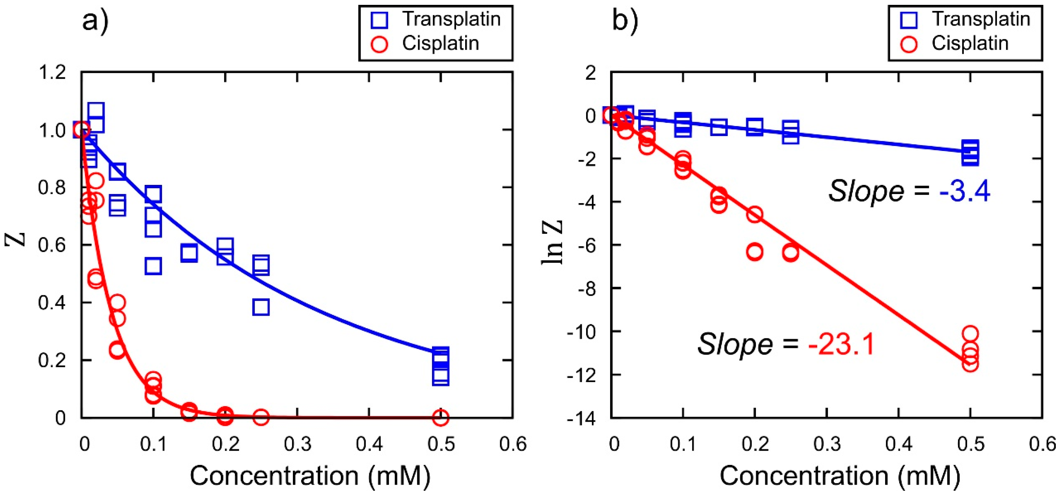
2.1. Luciferase Assay for Gene Expression and Higher-Order DNA Structure

Inhibitory Effect on Gene Expression

To investigate the effects of cisplatin and transplatin on gene expression, we performed a cell-free luciferase assay. Figure 2a shows the relative luminescence intensity ( Z ) as a marker for gene expression at various concentrations of cisplatin or transplatin. The concentration dependence of the inhibition of gene expression over a certain time period τ (i.e., 90 min) was analyzed as follows. Assuming first order reaction kinetics for the decrease of the concentration of the active site of DNA, x , caused by the binding of a Pt chemical with the concentration c , we expect d x / d t   ~ α c x ,   where α is the kinetic constant. Through integration, x / x o ~   e α c t   is deduced. Under the simple assumption, Z would be proportional to the total concentration of the product, luciferase. Thus, the dependence of Z on c for a reaction period of τ is given as Z   ~   e α c τ   . From this relationship, the relative kinetic constant was evaluated from the slope of the graph: ln Z   v s .   c . A plot of the logarithm of the Z   values vs. the concentrations of cisplatin or transplatin is shown in Figure 2b. The relative ratio of the slopes, −23.1 and −3.4, indicated that the inhibitory effect of cisplatin was about 7 times that of transplatin.

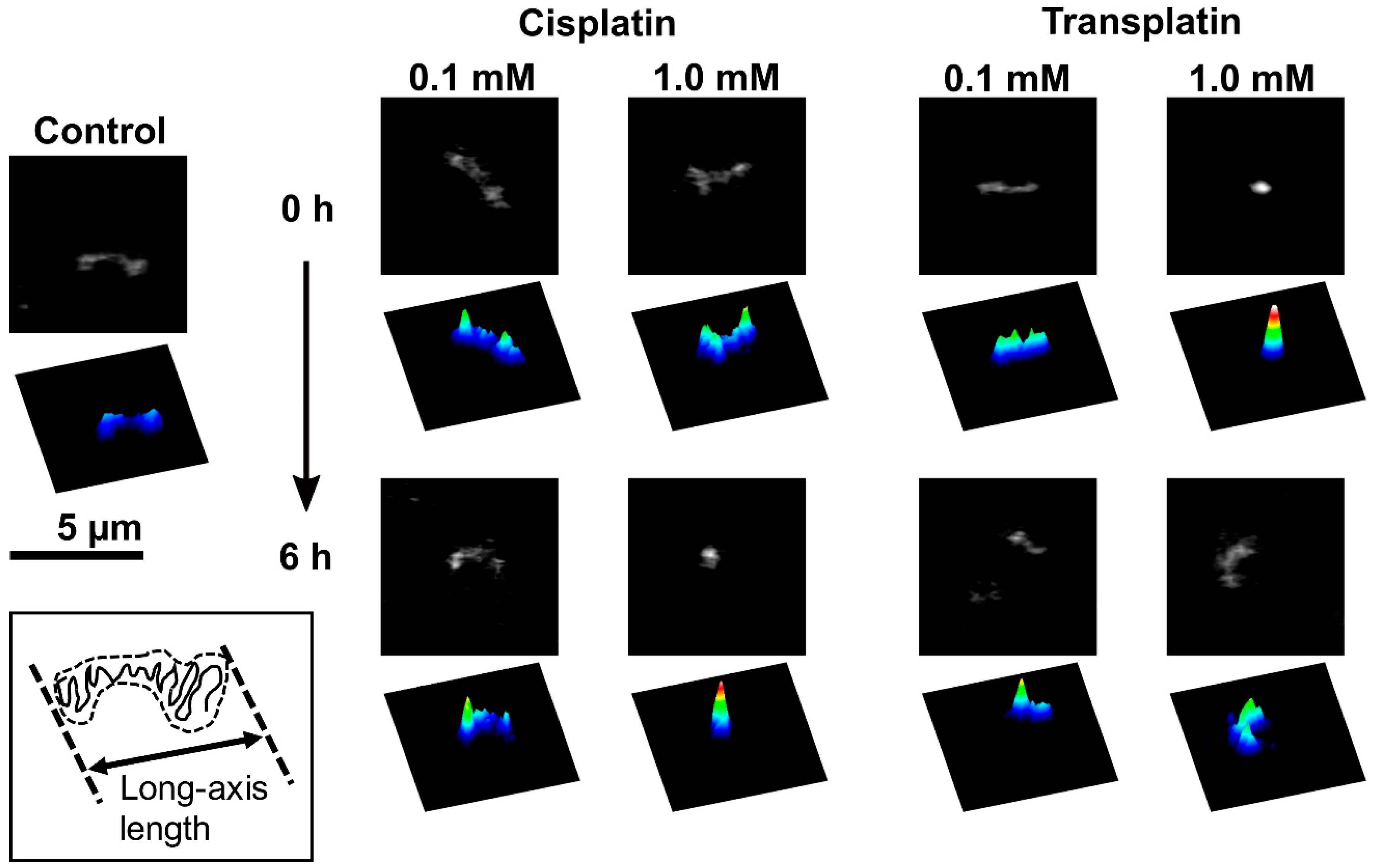
2.2. Higher-Order Conformational Change in T4 Phage DNA

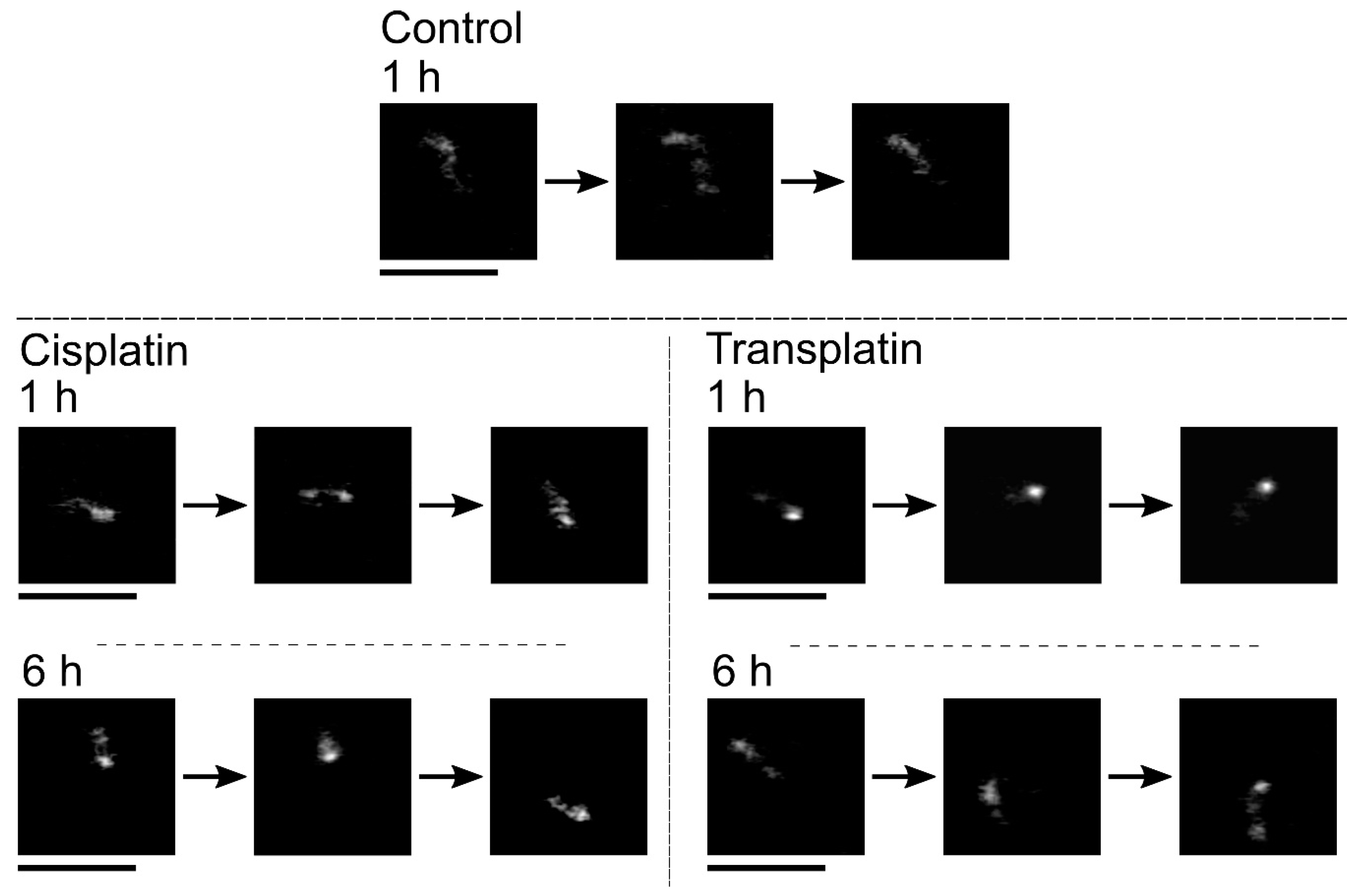
2.2.1. FM Observations

To investigate the effects of cisplatin and transplatin on the higher-order structure of DNA, we used FM to observe single DNA molecules in the presence or absence of cisplatin or transplatin. Typical FM images and corresponding quasi-3D profiles of fluorescence intensity are shown in Figure 3. FM allows the observation of individual DNA molecules exhibiting translational and intramolecular Brownian motion in bulk solution. In the absence of cisplatin or transplatin, DNA molecules existed in an elongated coil state (Figure 3, control). Upon the addition of cisplatin, a partial globule conformation was generated (Figure 3, 0.1 mM cisplatin). An increase in cisplatin concentration or longer incubation time generated a compact globule conformation (Figure 3, 1.0 mM cisplatin, 6 h). In contrast, at 1.0 mM transplatin, the compact DNA was unfolded into a coil conformation with time.
These observations clearly indicate that cisplatin and transplatin affect the higher-order structure of DNA by causing a shrunken state in a markedly different manner. Histograms of the long-axis length L of DNA as a function of the concentration of cisplatin and transplatin are shown in Figure 4. Both compounds shrank DNA molecules. In the presence of cisplatin, the degree of DNA shrinking increased with time. On the contrary, in the presence of transplatin, the DNA shrunken at 0 h gradually swelled, indicating a gradual decrease in the interaction between transplatin and DNA. To visualize the time-dependent effects of the two compounds, we plotted the average length L ¯ of DNA molecules at 0 h and 6 h against the compound concentration (Figure 5). Transplatin caused larger shrinking immediately after the addition compared to cisplatin. However, after several hours, DNA molecules swelled back in the presence of transplatin but not cisplatin.

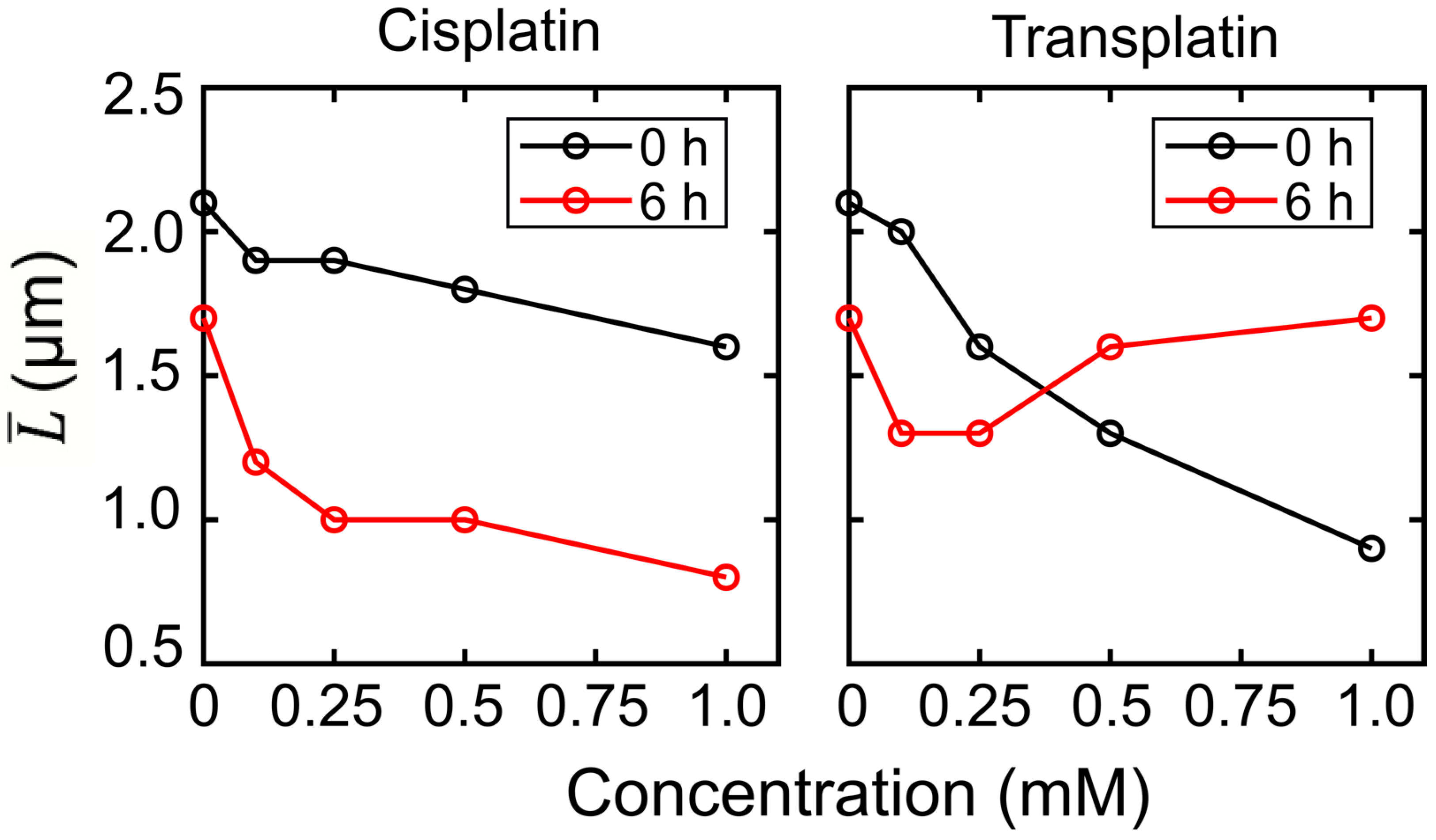
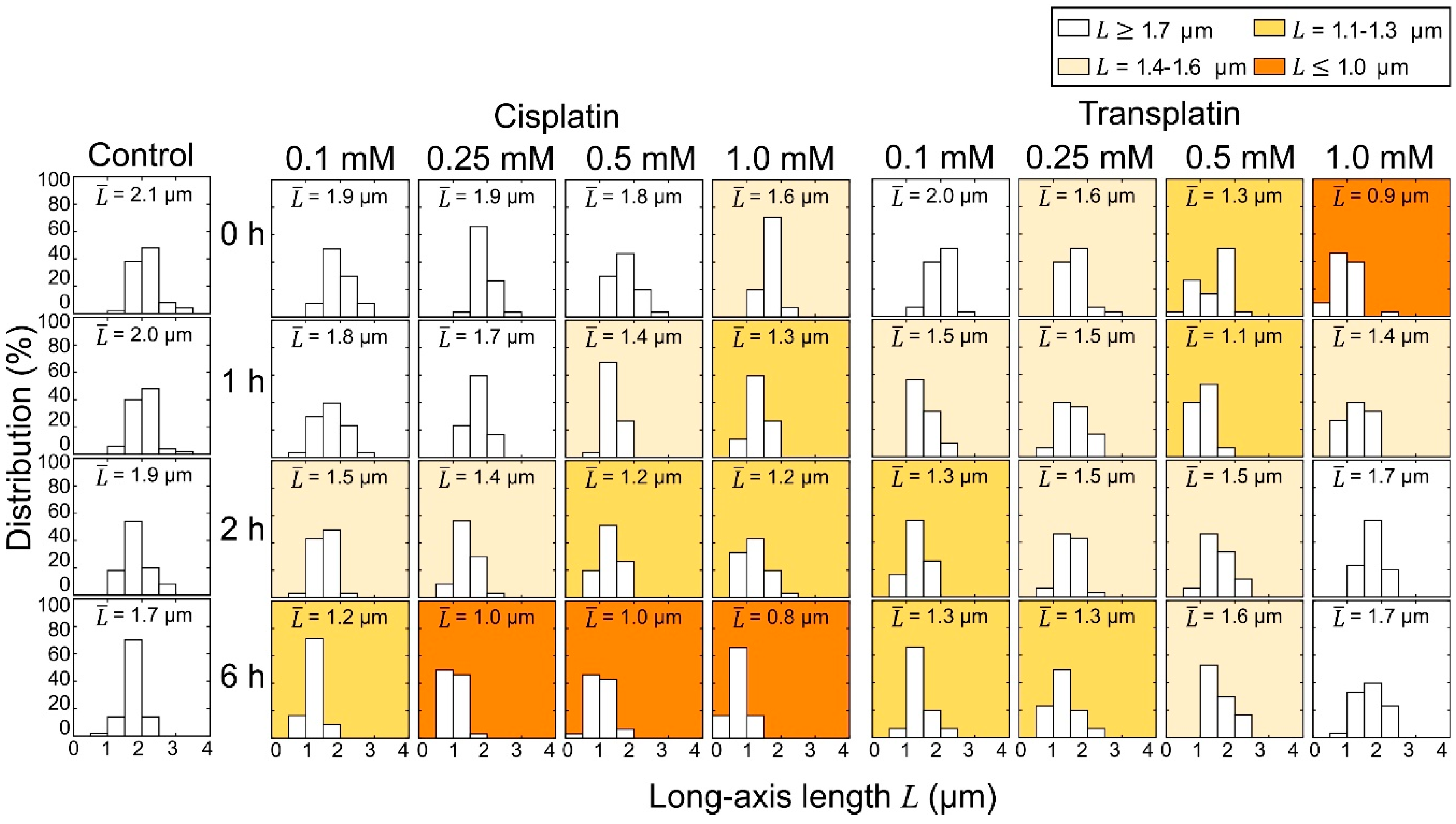
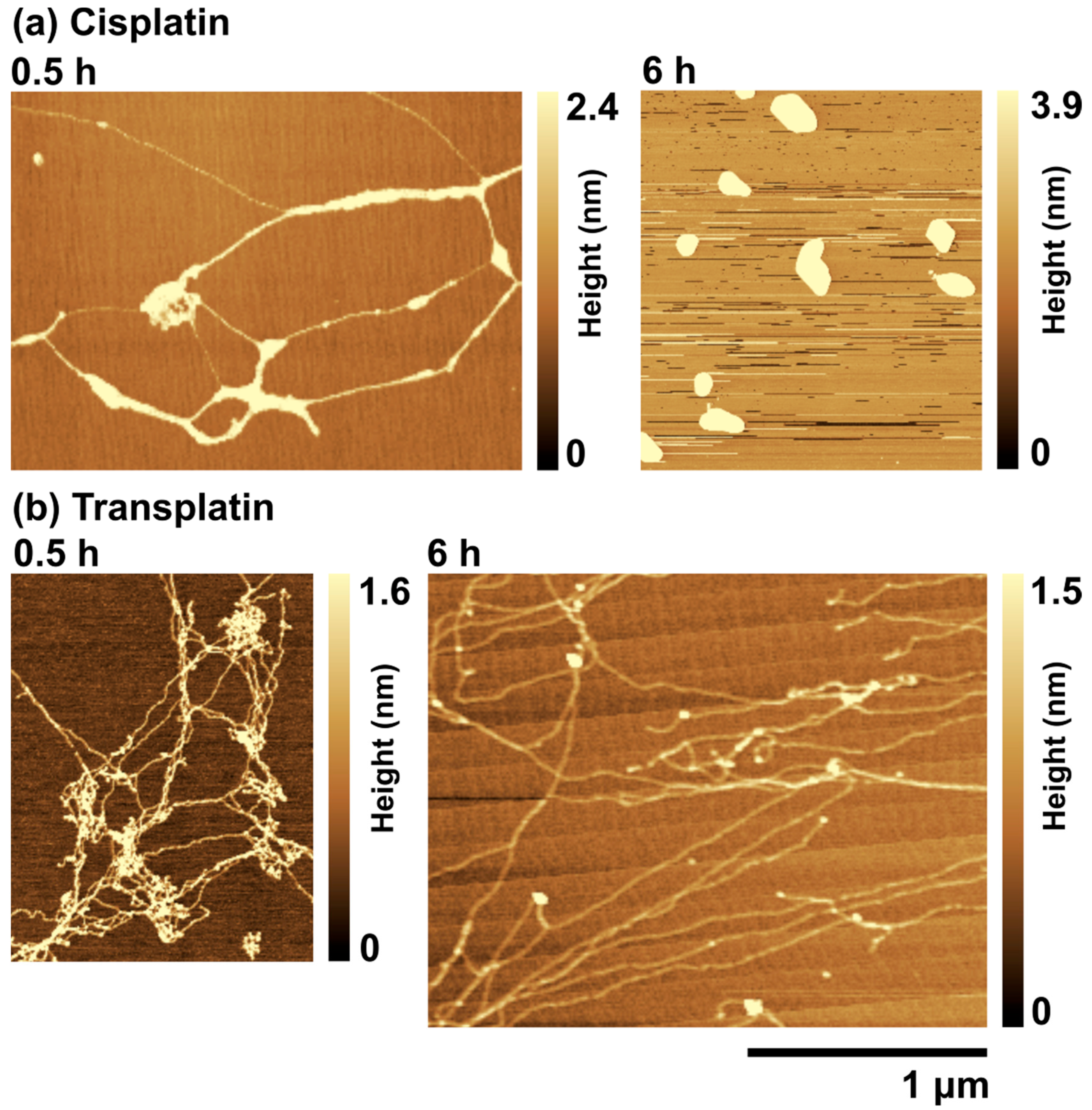
2.2.2. AFM Observations

To investigate the effects of cisplatin and transplatin on the higher-order structure of DNA in more detail, we used AFM. Typical AFM images of T4 DNA in the presence of cisplatin and transplatin are shown in Figure 6. At 0.5 h, cisplatin induced inter- and intramolecular crosslinks in DNA, which resulted in bundles of parallel aligned DNA segments (Figure 6a). At 6 h, DNA became folded and its structure became condensed (Figure 6a). This change was consistent with the results of FM observations, suggesting that the bond between DNA and cisplatin becomes stronger with time. On the other hand, in the presence of transplatin, a mesh-like structure was observed at 0.5 h, which loosened with time (Figure 6b).

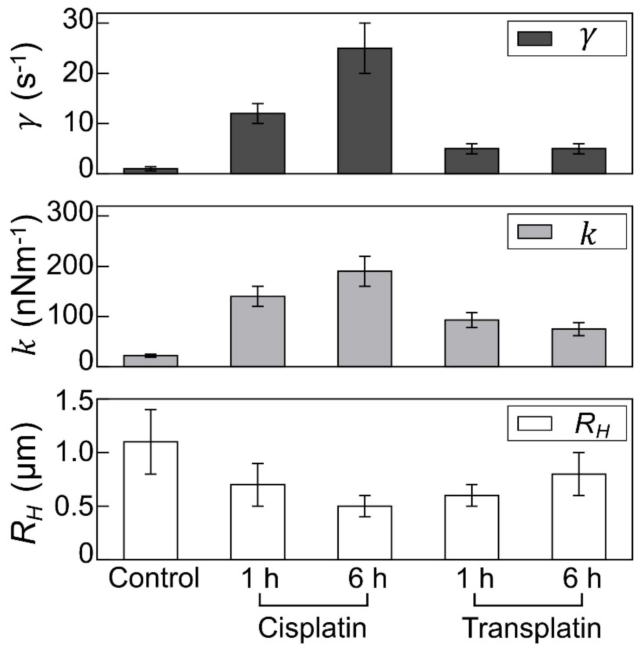
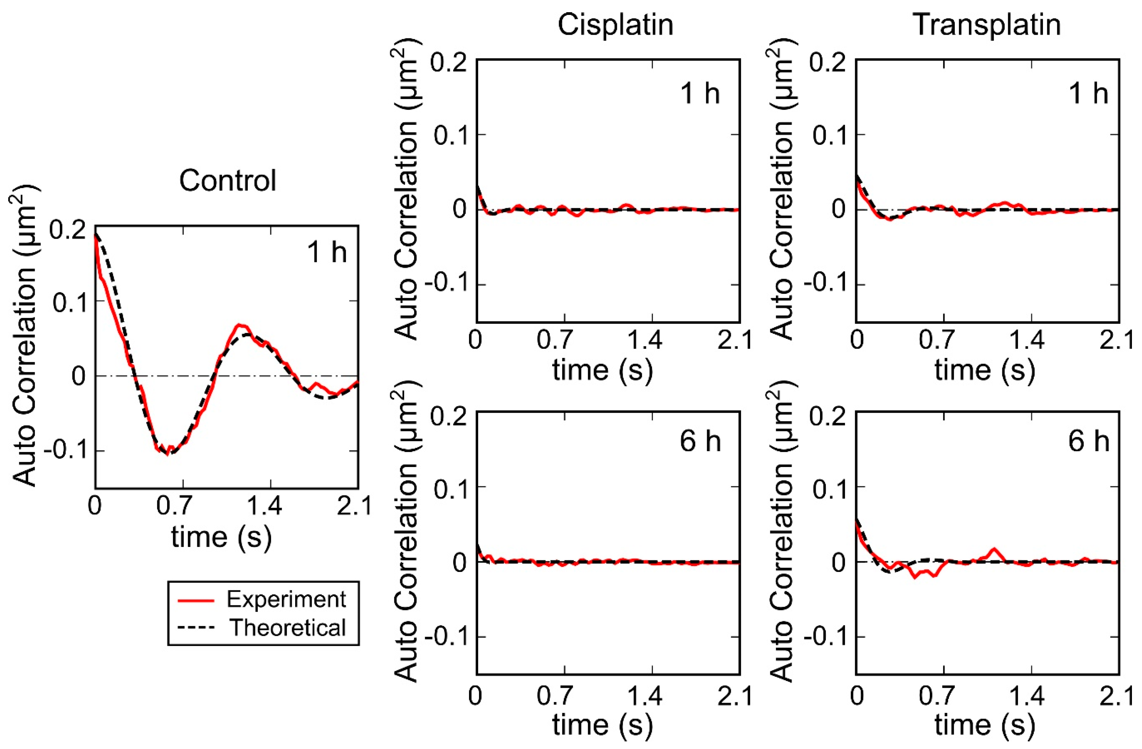
2.2.3. Visco-Elasticity and Hydrodynamic Radius RH of the DNA Molecule
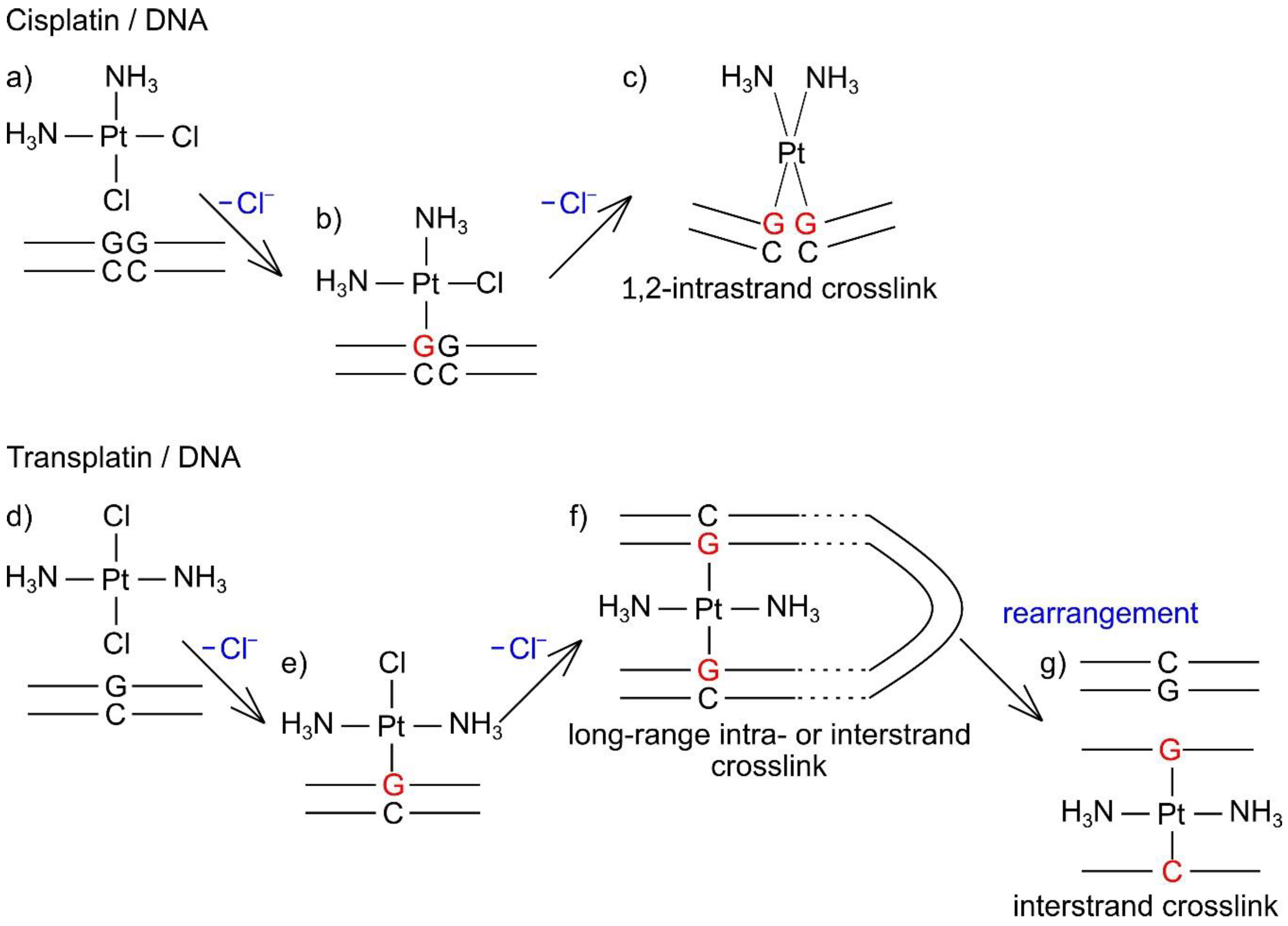
To evaluate the intramolecular Brownian motion of DNA (Figure 7), we quantified the fluctuation of single DNA molecules in the fluorescence images [32,33,34,35]. Autocorrelation of DNA obtained from the time-dependent fluctuation of the long-axis length is shown in Figure 8, and the deduced spring and damping constants are summarized in Figure 9. In the control state, the spring constant was 2.2 ± 0.3 × 10⁻⁸ Nm−1 and the damping constant was 1 ± 0.4 s−1. By the addition of the platinum compounds, the spring constant increased greatly. The increase of spring constant for the cisplatin–DNA complex was more than 10 times that of the control DNA at 6 h, whereas the transplatin–DNA complex was 5 times that of the control DNA. Hence, the increase in the spring constant was found to be more significant in DNA with cisplatin than with transplatin. It was also noted that the spring constant for the transplatin-DNA complex tended to decrease from 1 h to 6 h, in contrast to the increasing tendency of the cisplatin-DNA complex. As revealed in Figure 9, the change in the viscosity constant indicated a similar trend to the change in the spring constant. In addition to the aforementioned analysis of the intrachain fluctuation (Figure 7, Figure 8 and Figure 9), we analyzed the translational motion to evaluate the RH. Although the fluorescence images do not give precise information on the actual sizes of individual DNA molecules because of the presence of a blurring effect of 0.3–0.5 μm [33,34,35], the analysis of the Brownian motion, or translational fluctuation, can provide the RH as a reliable measure of the size of individual DNA molecules. It was apparent that the effect on the hydrodynamic radius RH was observed in a very similar manner, although the hydrodynamic radius RH did not change greatly and the visco-elastic properties of the platinum compound–DNA complex changed more clearly. Cisplatin decreased the RH (Figure 9), and the effect became stronger with time, whereas the effect of transplatin was weaker at 6 h than at 1 h.

3. Discussion

Inhibition of transcription is one of the mechanisms by which platinum-based drugs kill cancer cells [36]. The inhibition level appears to be related to the global DNA structure change induced by the formation of platinum–DNA adducts [37]. Therefore, the higher-order conformational change of DNA induced by each isomer might be related to its pharmaceutical activity or inactivity. There are no reports on the direct comparison of the higher-order structural changes induced in genome-sized DNA by cisplatin and transplatin. In this study, we used FM and AFM to monitor the structure of T4 phage DNA (166 kbp) at various concentrations of each platinum complex. When we previously monitored higher-order structural changes induced in DNA by cisplatin and a series of promising anticancer Pt(II) drug candidates [38], the inhibitory effects of these compounds on gene expression were correlated to the degree of DNA shrinkage: high DNA shrinkage resulted in a low expression level [19]. To shrink the DNA, it is necessary to neutralize the negative surface charge of the phosphate backbone, and the crosslinks between the segments along a DNA molecular chain, which also promotes DNA compaction [39,40]. In this study, both cisplatin and transplatin formed platinum(II)–DNA adducts, which neutralized the surface charge with positively charged Pt(II) ion and crosslinked DNA, and thus induced DNA shrinkage. The degree of shrinkage was higher in the presence of cisplatin than in the presence of transplatin after 6 h of exposure to the drug. The hydrolysis rate of cisplatin and transplatin affects their reactivity with biomolecules. Although trans effects between the two Cl ligands in transplatin appear to accelerate hydrolysis, by which a Cl ligand is displaced by a water molecule [41], it is difficult to determine the hydrolysis rate of which isomer is higher [42,43,44,45]. Indeed, reactions of each isomer with DNA are likely to provide comparable numbers of Pt atoms per DNA length in a cell-free medium [42,46,47]. Therefore, the different degrees of DNA shrinkage after 6 h are likely due to the different types of DNA crosslinks formed by cisplatin and transplatin, as discussed below. The inhibitory effect of cisplatin on gene expression was about 7 times that of transplatin. It has been reported that transplatin adducts are preferentially bypassed by RNA polymerase, and 4-fold more transplatin adducts than cisplatin adducts are required to similarly inhibit transcription [48]. It is also noted that similar effects of cisplatin on the shrinking of DNA has been reported for shorter DNA molecules [49,50].
Our most interesting finding came from FM and AFM observations over time. DNA molecules transiently became stiffer with a weak shrinkage immediately or 1 h after the addition of 0.5 or 1 mM transplatin and were softened back after several hours, whereas those in the presence of cisplatin continuously became stiffer and shrunken throughout the monitoring period. Compared to the control DNA molecules, a more significant shrinkage was observed in those with cisplatin than with transplatin. As mentioned in the Results (refer to Section 2.2.3), the spring and viscosity constants of DNA, which were evaluated from the time-dependent conformational fluctuation in solution observed through FM, reflect the effect of the platinum compounds on the physical property of their higher-order structure in a sensitive manner. As for the measurement of the elastic property of DNA, a number of publications have appeared, during the past couple of decades, that report the use of laser tweezers through the chemical modification with the binding of a micrometer-sized plastic sphere at the end of the DNA molecule (e.g., [51,52,53]). Unfortunately, most of these studies have reported the observed spring constant for DNA molecules shorter than several kbp, which is attributable to the technical difficulty of the measurement on individual giant DNA molecules. In the present study, we successfully applied our newly developed methodology for the analysis of intrachain Brownian motion that did not require the modification of DNA molecules longer than 100 kbp. It is expected that the application of the methodology on the measurement of the visco-elastic properties of giant DNA molecules adapted in the present study will open a new window for future studies on the relationship of the higher-order structure of genome-sized DNA with its biological functions.
Using simulated and experimental data, Dutta et al. [54] showed the relation between the local and higher-order DNA structure upon the formation of cisplatin 1,2-intrastrand crosslinks. The different types of DNA adducts made by these isomers likely result in different higher-order structural changes of DNA. A supposed interaction mechanism of cisplatin and transplatin with genome-sized DNA is shown in Figure 10. Cisplatin forms bifunctional DNA adducts, such as 1,2-intrastrand (major type) and interstrand crosslinks [8,55,56,57,58,59], and induces severe distortion of the double helix in the local structure, resulting in shrinking and kinking of the higher-order DNA structure. On the other hand, because of steric restrictions, transplatin cannot form 1,2-intrastrand crosslinks and provides interstrand crosslinks at a higher frequency than cisplatin [24], and a relatively large proportion of transplatin–DNA adducts remain monofunctional [60]. The local conformational changes in DNA around a 1,2-intrastrand [55,58] or interstrand [59] crosslink made by cisplatin seem to be more severe than those around an interstrand crosslink made by transplatin [61,62] and its monofunctional DNA adducts [63]. Given that the degree of local DNA conformational changes can affect the higher-order structure, the transient shrinkage of DNA upon addition of transplatin seems unexpected. One possible explanation is the formation of long-range intra- or interstrand crosslinks between two nucleobases that are distant in the DNA primary structure but are spatially close to each other. 1,3-GNG intrastrand crosslinks (where G = guanine, N = adenine, cytosine, or thymine) can be formed in the reaction between transplatin and single-stranded oligonucleotides [60], and such DNA adducts can be readily rearranged into interstrand crosslinks after hybridization of platinated single strands with complementary strands [24,64,65,66]. Although 1,3-GNG intrastrand crosslinks are unlikely to form in double-stranded DNA [67], the long-range intra- or interstrand crosslinks looking like interhelix crosslinks could be formed between two proximal nucleobases when the negative DNA charge is partially neutralized by the formation of monofunctional adducts of transplatin. Accordingly, a partially folded DNA string might allow the monofunctional adducts to form bifunctional adducts of the long-range crosslinks, which can be rearranged to form energetically more favorable interstrand crosslinks between complementary GC base pairs [24]. This hypothesis is supported by the fact that transplatin is more reactive than cisplatin; that a relatively large proportion (~50%) of all adducts on the double-stranded DNA are monofunctional [23]; and that transplatin, owing to its geometry, is a useful tool to prepare a DNA triplex with interstrand GG or GC crosslinking [68,69].

4. Materials and Methods

4.1. Materials

Plasmid DNA (luciferase T7 control DNA, 4331 bp) containing a gene encoding luciferase and a T7 RNA polymerase promoter sequence was purchased from Promega (Madison, WI, USA). T4 GT7 phage DNA (166 kbp; contour length, 57 µm) was purchased from Nippon Gene (Toyama, Japan). The fluorescent cyanine dye YOYO-1 (1,10-(4,4,7,7-tetramethyl-4,7-diazaundecamethylene)-bis-4-(3-methyl-2,3-dihydro-(benzo-1,3-oxazole)-2-methylidene)-quinolinium tetraiodide) was purchased from Molecular Probes Inc. (Eugene, OR, USA). The antioxidant 2-mercaptoethanol was purchased from Wako Pure Chemical Industries (Osaka, Japan). Cisplatin was synthesized according to a previous report [70]. Transplatin was purchased from Sigma Aldrich (Saint Louis, MO, USA). Other chemicals were of analytical grade.

4.2. Methods

4.2.1. Luciferase Assay for Gene Expression

The cell-free luciferase assay was carried out using a TnT T7 Quick Coupled Transcription/Translation System (Promega) similar to a previous report [71]. Plasmid DNA (4331 kbp) was used as a template. The final concentration of DNA was 1.5 μM in nucleotide units. The reaction mixture was incubated at 30 °C for 90 min in a Dry Thermo Unit (Taitec, Saitama, Japan). Luciferase expression was evaluated after the addition of luciferin (Luciferase Assay Reagent, Promega) by detecting the emission around 565 nm with a luminometer (Microtec Co., Chiba, Japan).

4.2.2. FM Observations

T4 phage DNA was dissolved in 10 mM Tris-HCl buffer (pH 7.5) containing 4% (v/v) 2-mercaptoethanol and 80 mM KCl in the presence of various concentrations of cisplatin or transplatin. DNA concentration was 1.5 µM in nucleotide units. To visualize individual DNA molecules, 0.05 µM YOYO-1 was added to the DNA solution after incubation with the Pt complexes. DNA was imaged using an Axiovert 135 TV microscope (Carl Zeiss, Oberkochen, Germany) equipped with a 100× oil-immersion objective lens with fluorescent illumination from a mercury lamp (100 W) via a filter set (Zeiss-10, excitation BP 450–490; beam splitter FT 510; emission BP 515–565). A high-sensitivity EBCCD camera (Hamamatsu Photonics, Shizuoka, Japan) was used to observe individual DNA molecules. All images were recorded on a DVD at 30 frames per second and were analyzed with Cosmos image-analysis software (Library, Tokyo, Japan).

4.2.3. AFM Measurements

For AFM imaging using an SPM-9700 microscope (Shimadzu, Kyoto, Japan), T4 GT7 phage DNA was dissolved (final concentration, 1.5 μM) in 10 mM Tris-HCl (pH 7.5) containing various concentrations of cisplatin or transplatin, incubated for 5 min, and then transferred onto a freshly cleaved mica surface (not pretreated with any cationic species) for 30 min at 25 °C. The mica was rinsed with pure water and dried under a gentle stream of nitrogen gas. All measurements were determined in air with tapping mode. A 200-μm long cantilever (OMCL-AC200TS-C2, Olympus, Tokyo, Japan) with a spring constant of 9 ± 20 N/m was used. The scanning rate was 0.4 Hz and images were captured on a 256 × 256 or 512 × 512 pixel format. The images were plane-fitted and flattened by the computer program equipped in the imaging module.

4.2.4. Evaluation of Visco-Elasticity and Hydrodynamic Radius RH from the Analysis of DNA Molecule Fluctuations Observed by FM

We evaluated the autocorrelation C ( t ) from the time-dependent fluctuation of the long-axis length, L ( t ) [33]:
C ( t ) =   L ( t ) L ¯   L ( 0 ) L ¯
where L ¯ is the time-average of L .
Based on a theoretical model of thermal fluctuation for a microscopic visco-elastic object with a harmonic potential, the autocorrelation function is represented as in Equation (2) to a reasonable approximation:
C ( t )   ~   k B T k e γ t cos ω t
k     k B T C ( 0 )
where k B is the Boltzmann constant, T is absolute temperature, k (N/m) is the spring constant, γ (1/s) is the damping constant, and ω (radian/s) is angular frequency. k is evaluated from the initial value of the autocorrelation function, C ( 0 ) , by using Equation (3), where C ( 0 ) is the value of C ( t ) at t = 0 . Then, the value of γ is determined from the fitting curve.
The hydrodynamic radius of individual DNA molecules was evaluated from the time-dependent fluctuation of their center of mass in fluorescence images as described previously [32,34,35]. From the Brownian motion trails of the center of mass of DNA, we obtained the time dependence of the mean-square displacement of each molecule, and from these values we obtained the two-dimensional diffusion constant D as follows:
( r ( 0 ) r ( t ) ) 2 = 4 D t
where r is a two-dimensional positional vector of the center of mass of DNA molecules and the symbol t   denotes a time average. In the actual analysis of the fluctuation, we used a modified equation [32,33,34,35] when a convectional flow was noticed. The hydrodynamic radius RH was calculated from D based on the Stokes–Einstein relationship:
R H =   k B T / 6 π η D
where η   is the viscosity of the solvent.

5. Conclusions

Using FM and AFM, we clearly delineated the difference between the effects of the geometric isomers cisplatin and transplatin on the higher-order structure of genome-sized DNA. The extent of DNA shrinkage was higher for cisplatin than for transplatin. This result agreed with a stronger inhibitory effect of cisplatin on gene expression in comparison with transplatin in a cell-free assay. Of note, transplatin induces DNA shrinkage transiently but more efficiently than cisplatin, probably because of the formation of long-range intra- or interstrand crosslinks between two nucleobases distant in the primary DNA structure but spatially close to each other, followed by a gradual rearrangement into an energetically preferable interstrand crosslink. This hypothesis was in line with a time-dependent rearrangement caused by transplatin on the order of several hours. Our finding of transient DNA shrinkage by transplatin was unprecedented and suggests that many undiscovered properties of genome-sized DNA molecules in the presence of biologically relevant compounds can be revealed by FM and AFM observations. We believe that our new experimental methodology to evaluate the visco-elastic properties of a single giant DNA molecule will be useful for future studies on the dynamic behavior of DNA molecules in solution. Additionally, AFM analysis of morphological changes of DNA at different time stages would be important and has been planned as our future project.

Author Contributions

S.K. and Y.Y. designed the study. T.K. performed all of the experiments and analyzed the data. S.K. and K.Y. supervised the experiments and theoretical interpretation. All authors have read and agreed to the published version of the manuscript.

Funding

This research was funded by KAKENHI grant numbers 15H02121 and 19K07018.

Conflicts of Interest

The authors declare no conflicts of interests.

Abbreviations

FMFluorescence microscopy
AFMAtomic force microscopy

References

  1. Rosenberg, B.; VanCamp, L.; Trosko, J.E.; Mansour, V.H. Platinum compounds: A new class of potent antitumour agents. Nature 1969, 222, 385–386. [Google Scholar] [CrossRef] [PubMed]
  2. Rosenberg, B.; VanCamp, L. The successful regression of large solid sarcoma 180 tumors by platinum compounds. Cancer Res. 1970, 30, 1799–1802. [Google Scholar] [PubMed]
  3. Prestayko, A.W.; D’aoust, J.; Issell, B.; Crooke, S. Cisplatin (cis-diamminedichloroplatinum II). Cancer Treat. Rev. 1979, 6, 17–39. [Google Scholar] [CrossRef]
  4. Calvert, A.H.; Harland, S.J.; Newell, D.R.; Siddik, Z.H.; Jones, A.C.; McElwain, T.J.; Raju, S.; Wiltshaw, E.; Smith, I.E.; Baker, J.M.; et al. Early clinical studies with cis-diammine-1,1-cyclobutane dicarboxylate platinum II. Cancer Chemother. Pharmacol. 1982, 9, 140–147. [Google Scholar] [CrossRef]
  5. Sharma, H.; Thatcher, N.; Baer, J.; Zaki, A.; Smith, A.; McAucliffe, C.A.; Crowther, D.; Owens, S.; Fox, B.W. Blood clearance of radioactively labelled cis-diammine 1,1-cyclobutane dicarboxylate platinum (II) (CBDCA) in cancer patients. Cancer Chemother. Pharmacol. 1983, 11, 5–7. [Google Scholar] [CrossRef]
  6. Tashiro, T.; Kawada, Y.; Sakurai, Y.; Kidani, Y. Antitumor activity of a new platinum complex, oxalato (trans-l-1,2-diaminocyclohexane)platinum (II): New experimental data. Biomed. Pharmacol. 1989, 43, 251–260. [Google Scholar] [CrossRef]
  7. Cvitkovic, E. Ongoing and unsaid on oxaliplatin: The hope. Br. J. Cancer 1998, 77, 8–11. [Google Scholar] [CrossRef] [Green Version]
  8. Jamieson, E.R.; Lippard, S.J. Structure, Recognition, and Processing of Cisplatin-DNA Adducts. Chem. Rev. 1999, 99, 2467–2498. [Google Scholar] [CrossRef]
  9. Reedijk, J. New clues for platinum antitumor chemistry: Kinetically controlled metal binding to DNA. Proc. Natl. Acad. Sci. USA 2003, 100, 3611–3616. [Google Scholar] [CrossRef] [Green Version]
  10. Rosenberg, B. Some biological effects of platinum compounds. Platin. Met. Rev. 1971, 15, 42–51. [Google Scholar]
  11. Johnson, N.P.; Hoeschele, J.D.; Rahn, R.O.; O’Neill, J.P.; Hsie, A.W. Mutagenicity, cytotoxicity, and DNA binding of platinum (II)-chloroammines in Chinese hamster ovary cells. Cancer Res. 1980, 40, 1463–1468. [Google Scholar] [PubMed]
  12. Dedon, P.C.; Borch, R.F. Characterization of the reactions of platinum antitumor agents with biologic and nonbiologic sulfur-containing nucleophiles. Biochem. Pharmacol. 1987, 36, 1955–1964. [Google Scholar] [CrossRef]
  13. Żelazowski, A.J.; Garvey, J.S.; Hoeschele, J.D. In vivo and in vitro binding of platinum to metallothionein. Arch. Biochem. Biophys. 1984, 229, 246–252. [Google Scholar] [CrossRef]
  14. Ciccarelli, R.B.; Solomon, M.J.; Varshavsky, A.; Lippard, S.J. In vivo effects of cis- and trans-diamminedichloroplatinum(II) on SV40 chromosomes: Differential repair, DNA-protein cross-linking, and inhibition of replication. Biochemistry 1985, 24, 7533–7540. [Google Scholar] [CrossRef] [PubMed]
  15. Farrell, N.; Ha, T.T.B.; Souchard, J.P.; Wimmer, F.L.; Cros, S.; Johnson, N.P. Cytostatic Trans-Platinum(Ii) Complexes. J. Med. Chem. 1989, 32, 2240–2241. [Google Scholar] [CrossRef] [PubMed]
  16. Farrell, N.; Kelland, L.R.; Roberts, J.D.; Van Beusichem, M. Activation of the trans geometry in platinum antitumor complexes: A survey of the cytotoxicity of trans complexes containing planar ligands in murine L1210 and human tumor panels and studies on their mechanism of action. Cancer Res. 1992, 52, 5065–5072. [Google Scholar] [PubMed]
  17. Aris, S.M.; Farrell, N.P. Towards antitumor active trans-platinum compounds. Eur. J. Inorg. Chem. 2009, 2009, 1293–1302. [Google Scholar] [CrossRef] [Green Version]
  18. Coluccia, M.; Nassi, A.; Loseto, F.; Boccarelli, A.; Mariggio, M.A.; Giordano, D.; Intini, F.P.; Caputo, P.; Natile, G. A Trans-Platinum Complex Showing Higher Antitumor-Activity Than the Cis Congeners. J. Med. Chem. 1993, 36, 510–512. [Google Scholar] [CrossRef]
  19. Coluccia, M.; Boccarelli, A.; Mariggio, M.A.; Cardellicchio, N.; Caputo, P.; Intini, F.P.; Natile, G. Platinum(II) complexes containing iminoethers: A trans platinum antitumour agent. Chem.-Biol. Interact. 1995, 98, 251–266. [Google Scholar] [CrossRef]
  20. Blommaert, F.A.; van Dijk-Knijnenburg, H.C.; Dijt, F.J.; den Engelse, L.; Baan, R.A.; Berends, F.; Fichtinger-Schepman, A.M. Formation of DNA adducts by the anticancer drug carboplatin: Different nucleotide sequence preferences in vitro and in cells. Biochemistry 1995, 34, 8474–8780. [Google Scholar] [CrossRef]
  21. Fichtinger-Schepman, A.M.; van der Veer, J.L.; den Hartog, J.H.; Lohman, P.H.; Reedijk, J. Adducts of the antitumor drug cis-diamminedichloroplatinum(II) with DNA: Formation, identification, and quantitation. Biochemistry 1985, 24, 707–713. [Google Scholar] [CrossRef] [PubMed]
  22. Fichtinger-Schepman, A.M.; Lohman, P.H.; Berends, F.; Reedijk, J.; van Oosterom, A.T. Interactions of the antitumour drug cisplatin with DNA in vitro and in vivo. IARC Sci. Publ. 1986, 78, 83–99. [Google Scholar]
  23. Eastman, A.; Barry, M.A. Interaction of trans-diamminedichloroplatinum(II) with DNA: Formation of monofunctional adducts and their reaction with glutathione. Biochemistry 1987, 26, 3303–3307. [Google Scholar] [CrossRef] [PubMed]
  24. Brabec, V.; Leng, M. DNA interstrand cross-links of trans-diamminedichloroplatinum(II) are preferentially formed between guanine and complementary cytosine residues. Proc. Natl. Acad. Sci. USA 1993, 90, 5345–5349. [Google Scholar] [CrossRef] [PubMed] [Green Version]
  25. Hofr, C.; Brabec, V. Thermal and thermodynamic properties of duplex DNA containing site-specific interstrand cross-link of antitumor cisplatin or its clinically ineffective trans isomer. J. Biol. Chem. 2001, 276, 9655–9661. [Google Scholar] [CrossRef] [Green Version]
  26. Kasparkova, J.; Brabec, V. Recognition of DNA interstrand cross-links of cis-diamminedichloroplatinum (II) and its trans isomer by DNA-binding proteins. Biochemistry 1995, 34, 12379–12387. [Google Scholar] [CrossRef]
  27. Kasparkova, J.; Pospisilova, S.; Brabec, V. Different recognition of DNA modified by antitumor cisplatin and its clinically ineffective trans isomer by tumor suppressor protein p53. J. Biol. Chem. 2001, 276, 16064–16069. [Google Scholar] [CrossRef] [Green Version]
  28. Kasparkova, J.; Thibault, T.; Kostrhunova, H.; Stepankova, J.; Vojtiskova, M.; Muchova, T.; Midoux, P.; Malinge, J.M.; Brabec, V. Different affinity of nuclear factor-kappa B proteins to DNA modified by antitumor cisplatin and its clinically ineffective trans isomer. FEBS J. 2014, 281, 1393–1408. [Google Scholar] [CrossRef]
  29. Yoshikawa, Y.; Komeda, S.; Uemura, M.; Kanbe, T.; Chikuma, M.; Yoshikawa, K.; Imanaka, T. Highly Efficient DNA Compaction Mediated by an In Vivo Antitumor-Active Tetrazolato-Bridged Dinuclear Platinum(II) Complex. Inorg. Chem. 2011, 50, 11729–11735. [Google Scholar] [CrossRef]
  30. Uemura, M.; Yoshikawa, Y.; Yoshikawa, K.; Sato, T.; Mino, Y.; Chikuma, M.; Komeda, S. Second- and higher-order structural changes of DNA induced by antitumor-active tetrazolato-bridged dinuclear platinum(II) complexes with different types of 5-substituent. J. Inorg. Biochem. 2013, 127, 169–174. [Google Scholar] [CrossRef]
  31. Komeda, S.; Yoneyama, H.; Uemura, M.; Muramatsu, A.; Okamoto, N.; Konishi, H.; Takahashi, H.; Takagi, A.; Fukuda, W.; Imanaka, T.; et al. Specific Conformational Change in Giant DNA Caused by Anticancer Tetrazolato-Bridged Dinuclear Platinum(II) Complexes: Middle-Length Alkyl Substituents Exhibit Minimum Effect. Inorg. Chem. 2017, 56, 802–811. [Google Scholar] [CrossRef] [PubMed]
  32. Kida, N.; Katsuda, Y.; Yoshikawa, Y.; Komeda, S.; Sato, T.; Saito, Y.; Chikuma, M.; Suzuki, M.; Imanaka, T.; Yoshikawa, K. Characteristic effect of an anticancer dinuclear platinum(II) complex on the higher-order structure of DNA. J. Biol. Inorg. Chem. 2010, 15, 701–707. [Google Scholar] [CrossRef] [PubMed]
  33. Carnerero, J.M.; Masuoka, S.; Baba, H.; Yoshikawa, Y.; Prado-Gotor, R.; Yoshikawa, K. Decorating a single giant DNA with gold nanoparticles. RSC Adv. 2018, 8, 26571–26579. [Google Scholar] [CrossRef] [Green Version]
  34. Araki, S.; Nakai, T.; Hizume, K.; Takeyasu, K.; Yoshikawa, K. Hydrodynamic radius of circular DNA is larger than that of linear DNA. Chem. Phys. Lett. 2006, 418, 255–259. [Google Scholar] [CrossRef] [Green Version]
  35. Matsumoto, M.; Sakaguchi, T.; Kimura, H.; Doi, M.; Minagawa, K.; Matsuzawa, Y.; Yoshikawa, K. Direct observation of brownian motion of macromolecules by fluorescence microscope. J. Polym. Sci. B 1992, 30, 779–783. [Google Scholar] [CrossRef]
  36. Jamieson, E.R.; Jacobson, M.P.; Barnes, C.M.; Chow, C.S.; Lippard, S.J. Structural and kinetic studies of a cisplatin-modified DNA icosamer binding to HMG1 domain B. J. Biol. Chem. 1999, 274, 12346–12354. [Google Scholar] [CrossRef] [Green Version]
  37. Sandman, K.E.; Marla, S.S.; Zlokarnik, G.; Lippard, S.J. Rapid fluorescence-based reporter-gene assays to evaluate the cytotoxicity and antitumor drug potential of platinum complexes. Chem. Biol. 1999, 6, 541–551. [Google Scholar] [CrossRef] [Green Version]
  38. Komeda, S.; Yoneyama, H.; Uemura, M.; Tsuchiya, T.; Hoshiyama, M.; Sakazaki, T.; Hiramoto, K.; Harusawa, S. Synthesis and structure-activity relationships of tetrazolato-bridged dinuclear platinum(II) complexes: A small modification at tetrazole C5 markedly influences the in vivo antitumor efficacy. J. Inorg. Biochem. 2019, 192, 82–86. [Google Scholar] [CrossRef]
  39. Onoa, G.B.; Cervantes, G.; Moreno, V.; Prieto, M.J. Study of the interaction of DNA with cisplatin and other Pd (II) and Pt (II) complexes by atomic force microscopy. Nucleic Acids Res. 1998, 26, 1473–1480. [Google Scholar] [CrossRef] [Green Version]
  40. Malina, J.; Farrell, N.P.; Brabec, V. DNA condensing effects and sequence selectivity of DNA binding of antitumor noncovalent polynuclear platinum complexes. Inorg. Chem. 2014, 53, 1662–1671. [Google Scholar] [CrossRef]
  41. Lippert, B. Trans-diammineplatinum (II): What makes it different from cis-DDP? Coordination chemistry of a neglected relative of cisplatin and its interaction with nucleic acids. Met. Ions Biol. Syst. 1996, 33, 105–142. [Google Scholar] [PubMed]
  42. Bancroft, D.P.; Lepre, C.A.; Lippard, S.J. Platinum-195 NMR kinetic and mechanistic studies of cis- and trans-diamminedichloroplatinum(II) binding to DNA. J. Am. Chem. Soc. 1990, 112, 6860–6871. [Google Scholar] [CrossRef]
  43. Miller, S.E.; Gerard, K.J.; House, D.A. The hydrolysis products of cis-diamminedichloroplatinum (II) 6. A kinetic comparison of the cis-and trans-isomers and other cis-di (amine) di (chloro) platinum (II) compounds. Inorg. Chim. Acta 1991, 190, 135–144. [Google Scholar] [CrossRef]
  44. Arpalahti, J.; Mikola, M.; Mauristo, S. Kinetics and mechanism of the complexation of cis-diamminedichloroplatinum (II) with the purine nucleoside inosine in aqueous solution. Inorg. Chem. 1993, 32, 3327–3332. [Google Scholar] [CrossRef]
  45. Mikola, M.; Arpalahti, J. Kinetics and mechanism of the complexation of trans-diamminedichloroplatinum (II) with the purine nucleoside inosine in aqueous solution. Inorg. Chem. 1994, 33, 4439–4445. [Google Scholar] [CrossRef]
  46. Bierbach, U.; Qu, Y.; Hambley, T.W.; Peroutka, J.; Nguyen, H.L.; Doedee, M.; Farrell, N. Synthesis, structure, biological activity, and DNA binding of platinum (II) complexes of the type trans-[PtCl2 (NH3) L](L= planar nitrogen base). Effect of L and cis/trans isomerism on sequence specificity and unwinding properties observed in globally platinated DNA. Inorg. Chem. 1999, 38, 3535–3542. [Google Scholar]
  47. Navarro, M.; Castro, W.; Higuera-Padilla, A.R.; Sierraalta, A.; Abad, M.J.; Taylor, P.; Sánchez-Delgado, R.A. Synthesis, characterization and biological activity of trans-platinum (II) complexes with chloroquine. J. Inorg. Biochem. 2011, 105, 1684–1691. [Google Scholar] [CrossRef] [Green Version]
  48. Mello, J.A.; Lippard, S.J.; Essigmann, J.M. DNA adducts of cis-diamminedichloroplatinum(II) and its trans isomer inhibit RNA polymerase II differentially in vivo. Biochemistry 1995, 34, 14783–14791. [Google Scholar] [CrossRef]
  49. Hou, X.-M.; Zhang, X.-H.; Wei, K.-J.; Ji, C.; Dou, S.-X.; Wang, W.-C.; Li, M.; Wang, P.-Y. Cisplatin induces loop structures and condensation of single DNA molecules. Nucleic Acids Res. 2009, 37, 1400–1410. [Google Scholar] [CrossRef]
  50. Liu, Z.; Tan, S.; Zu, Y.; Fu, Y.; Meng, R.; Xing, Z. The interactions of cisplatin and DNA studied by atomic force microscopy. Micron 2010, 41, 833–839. [Google Scholar] [CrossRef]
  51. Lima, C.; Caquito, J.; de Oliveira, R.; Rocha, M. Pixantrone anticancer drug as a DNA ligand: Depicting the mechanism of action at single molecule level. Eur. Phys. J. E 2019, 42, 130. [Google Scholar] [CrossRef] [PubMed]
  52. Bustamante, C.; Smith, S.B.; Liphardt, J.; Smith, D. Single-molecule studies of DNA mechanics. Curr. Opin. Struct. Biol. 2000, 10, 279–285. [Google Scholar] [CrossRef]
  53. Wang, M.D.; Yin, H.; Landick, R.; Gelles, J.; Block, S.M. Stretching DNA with optical tweezers. Biophys. J. 1997, 72, 1335–1346. [Google Scholar] [CrossRef] [Green Version]
  54. Dutta, S.; Rivetti, C.; Gassman, N.R.; Young, C.G.; Jones, B.T.; Scarpinato, K.; Guthold, M. Analysis of single, cisplatin-induced DNA bends by atomic force microscopy and simulations. J. Mol. Recognit. 2018, 31, e2731. [Google Scholar] [CrossRef] [PubMed]
  55. Yang, D.; van Boom, S.S.G.E.; Reedijk, J.; van Boom, J.H.; Wang, A.H.J. Structure and isomerization of an intrastrand cisplatin-cross-linked octamer DNA duplex by NMR analysis. Biochemistry 1995, 34, 12912–12920. [Google Scholar] [CrossRef] [PubMed]
  56. Van Boom, S.S.; Yang, D.; Reedijk, J.; van der Marel, G.A.; Wang, A.H. Structural effect of intra-strand cisplatin-crosslink on palindromic DNA sequences. J. Biomol. Struct. Dyn. 1996, 13, 989–998. [Google Scholar] [CrossRef] [PubMed]
  57. Shellard, S.A.; Fichtinger-Schepman, A.M.; Lazo, J.S.; Hill, B.T. Evidence of differential cisplatin-DNA adduct formation, removal and tolerance of DNA damage in three human lung carcinoma cell lines. Anti-Cancer Drugs 1993, 4, 491–500. [Google Scholar] [CrossRef]
  58. Takahara, P.M.; Rosenzweig, A.C.; Frederick, C.A.; Lippard, S.J. Crystal structure of double-stranded DNA containing the major adduct of the anticancer drug cisplatin. Nature 1995, 377, 649–652. [Google Scholar] [CrossRef]
  59. Coste, F.; Malinge, J.M.; Serre, L.; Shepard, W.; Roth, M.; Leng, M.; Zelwer, C. Crystal structure of a double-stranded DNA containing a cisplatin interstrand cross-link at 1.63 A resolution: Hydration at the platinated site. Nucleic Acids Res. 1999, 27, 1837–1846. [Google Scholar] [CrossRef] [Green Version]
  60. Eastman, A.; Jennerwein, M.M.; Nagel, D.L. Characterization of bifunctional adducts produced in DNA by trans-diamminedichloroplatinum(II). Chem.-Biol. Interact. 1988, 67, 71–80. [Google Scholar] [CrossRef]
  61. Paquet, F.; Boudvillain, M.; Lancelot, G.; Leng, M. NMR solution structure of a DNA dodecamer containing a transplatin interstrand GN7-CN3 cross-link. Nucleic Acids Res. 1999, 27, 4261–4268. [Google Scholar] [CrossRef] [PubMed] [Green Version]
  62. Brabec, V.; Sip, M.; Leng, M. DNA conformational change produced by the site-specific interstrand cross-link of trans-diamminedichloroplatinum (II). Biochemistry 1993, 32, 11676–11681. [Google Scholar] [CrossRef] [PubMed]
  63. Brabec, V.; Reedijk, J.; Leng, M. Sequence-dependent distortions induced in DNA by monofunctional platinum (II) binding. Biochemistry 1992, 31, 12397–12402. [Google Scholar] [CrossRef] [PubMed]
  64. Kasparkova, J.; Marini, V.; Bursova, V.; Brabec, V. Biophysical studies on the stability of DNA intrastrand cross-links of transplatin. Biophys. J. 2008, 95, 4361–4371. [Google Scholar] [CrossRef] [Green Version]
  65. Giraud-Panis, M.-J.; Leng, M. Transplatin-modified oligonucleotides as modulators of gene expression. Pharmacol. Ther. 2000, 85, 175–181. [Google Scholar] [CrossRef]
  66. Dalbiès, R.; Payet, D.; Leng, M. DNA double helix promotes a linkage isomerization reaction in trans-diamminedichloroplatinum (II)-modified DNA. Proc. Natl. Acad. Sci. USA 1994, 91, 8147–8151. [Google Scholar] [CrossRef] [Green Version]
  67. Boudvillain, M.; Dalbies, R.; Aussourd, C.; Leng, M. Intrastrand cross-links are not formed in the reaction between transplatin and native DNA: Relationm with the clinical inefficency of transplatin. Nucleic Acids Res. 1995, 23, 2381–2388. [Google Scholar] [CrossRef] [Green Version]
  68. Campbell, M.A.; Miller, P.S. Transplatin-conjugated triplex-forming oligonucleotides form adducts with both strands of DNA. Bioconjugate Chem. 2009, 20, 2222–2230. [Google Scholar] [CrossRef] [Green Version]
  69. Colombier, C.; Lippert, B.; Leng, M. Interstrand cross-linking reaction in triplexes containing a monofunctional transplatin-adduct. Nucleic Acids Res. 1996, 24, 4519–4524. [Google Scholar] [CrossRef] [Green Version]
  70. Alderden, R.A.; Hall, M.D.; Hambley, T.W. The discovery and development of cisplatin. J. Chem. Educ. 2006, 83, 728. [Google Scholar] [CrossRef]
  71. Shimizu, Y.; Yoshikawa, Y.; Kenmotsu, T.; Komeda, S.; Yoshikawa, K. Conformational transition of DNA by dinuclear Pt(II) complexes causes cooperative inhibition of gene expression. Chem. Phys. Lett. 2017, 678, 123–129. [Google Scholar] [CrossRef]
Figure 1. Chemical structures of the anticancer drug (a) cisplatin and its geometric isomer (b) transplatin.
Ijms 21 00034 g001
Figure 2. Effects of cisplatin and transplatin on gene expression efficiency. (a) The vertical axis is the relative emission intensity of the luciferin–luciferase reaction, Z , where the intensity in the absence of platinum complexes is taken as unity. (b) Quantification of the efficiency through the plots of ln Z vs. concentration using the same data as in (a).
Ijms 21 00034 g002
Figure 3. Fluorescence microscopy (FM) images of single T4 DNA molecules fluctuating in solution at 0.1 and 1.0 mM cisplatin or transplatin. Corresponding quasi-3D profiles are also shown. The DNA concentration was 1.5 µM in the nucleotide units.
Ijms 21 00034 g003
Figure 4. Distribution of the long-axis length L of T4 DNA observed by FM. At each condition, 50 DNA molecules were measured and the mean values, L ¯ , are given in the boxes. The DNA concentration was 1.5 µM in nucleotide units.
Ijms 21 00034 g004
Figure 5. Average long-axis length L ¯ of T4 DNA vs. concentration of cisplatin or transplatin. The DNA concentration was 1.5 µM in the nucleotide units.
Ijms 21 00034 g005
Figure 6. Atomic force microscopy (AFM) images of T4 DNA in the presence of (a) 0.5 mM cisplatin or (b) 0.5 mM transplatin. All specimens were gently adsorbed on the mica surface without shear stress and then dried. The DNA concentration was 1.5 µM in nucleotide units.
Ijms 21 00034 g006
Figure 7. Intra-chain and translational Brownian motion of single T4 DNA molecules in bulk solution as observed by fluorescence microscopy. The time interval between neighboring frames is 0.1 s. Control: no cisplatin or transplatin. The concentration of cisplatin and transplatin was 0.5 mM. Scale bars, 5 µm.
Ijms 21 00034 g007
Figure 8. Autocorrelation function for the time-dependent fluctuation of the long-axis length of the single DNA molecules shown in Figure 7. Control: no Pt compounds. The concentration of cisplatin and transplatin was 0.5 mM. Solid lines, autocorrelation of DNA obtained from the time-dependent fluctuation of the long-axis length; broken lines, fitted curves based on Equation (2).
Ijms 21 00034 g008
Figure 9. Visco-elastic properties and the hydrodynamic radius of DNA molecules. Upper and middle panels: Visco-elasticity of single T4 DNA molecules (166 kbp) in the presence of cisplatin or transplatin evaluated from the autocorrelation function of intrachain thermal fluctuation shown in Figure 8. k , spring constant; γ , damping constant. Lower panel: hydrodynamic radius RH. The RH values were calculated for at least 100 DNA molecules per sample. DNA concentration was 1.5 µM in nucleotide units. Concentration of cisplatin and transplatin was 0.5 mM.
Ijms 21 00034 g009
Figure 10. A supposed interaction mechanisms of cisplatin and transplatin with genome-sized DNA. (a) Unbound transplatin and DNA. (b) Cisplatin–DNA complex as a monofunctional adduct. (c) Formation of a cisplatin–DNA 1,2-intrastrand crosslink. (d) Unbound transplatin and DNA. (e) Transplatin–DNA complex as a monofunctional adduct. (f) Formation of a transplatin–DNA long-range intra- or interstrand crosslink. (g) Formation of a transplatin/DNA interstrand crosslink.
Ijms 21 00034 g010

