Review

Alternative Splicing in Angiogenesis

Institute of Biomedical and Clinical Sciences, Medical School, College of Medicine and Health, University of Exeter, Exeter EX4 4PY, UK
*
Author to whom correspondence should be addressed.
Int. J. Mol. Sci. 2019, 20(9), 2067; https://doi.org/10.3390/ijms20092067
Submission received: 26 March 2019 / Revised: 15 April 2019 / Accepted: 23 April 2019 / Published: 26 April 2019
(This article belongs to the Special Issue Pre-mRNA Splicing 2018)

Abstract

:
Alternative splicing of pre-mRNA allows the generation of multiple splice isoforms from a given gene, which can have distinct functions. In fact, splice isoforms can have opposing functions and there are many instances whereby a splice isoform acts as an inhibitor of canonical isoform function, thereby adding an additional layer of regulation to important processes. Angiogenesis is an important process that is governed by alternative splicing mechanisms. This review focuses on the alternative spliced isoforms of key genes that are involved in the angiogenesis process; VEGF-A, VEGFR1, VEGFR2, NRP-1, FGFRs, Vasohibin-1, Vasohibin-2, HIF-1α, Angiopoietin-1 and Angiopoietin-2.

1. Alternative Splicing

The human genome consists of an estimated 20,000 protein-coding genes [1], which can give rise to a much greater number of proteins. Alternative splicing is one of the main processes that accounts for the increased protein to gene ratio. In fact, alternative splicing is reported to be responsible for 40% of protein modifications [2]. It involves the formation of multiple mRNA products from a single pre-mRNA, and many of these mRNA products are then translated into proteins that may have distinct structures or functions [3]. This important process therefore increases the coding capacity of a given gene, and as it has been reported that more than 95% of pre-mRNAs are alternatively spliced in mammals [4], research into alternative splicing has gathered momentum over the past two decades. Alternative splicing is heavily regulated. However, like most biological processes, alternative splicing is open to faults, which can result in disease. It has been suggested that around 33% of mutations that cause disease affect splicing [5].

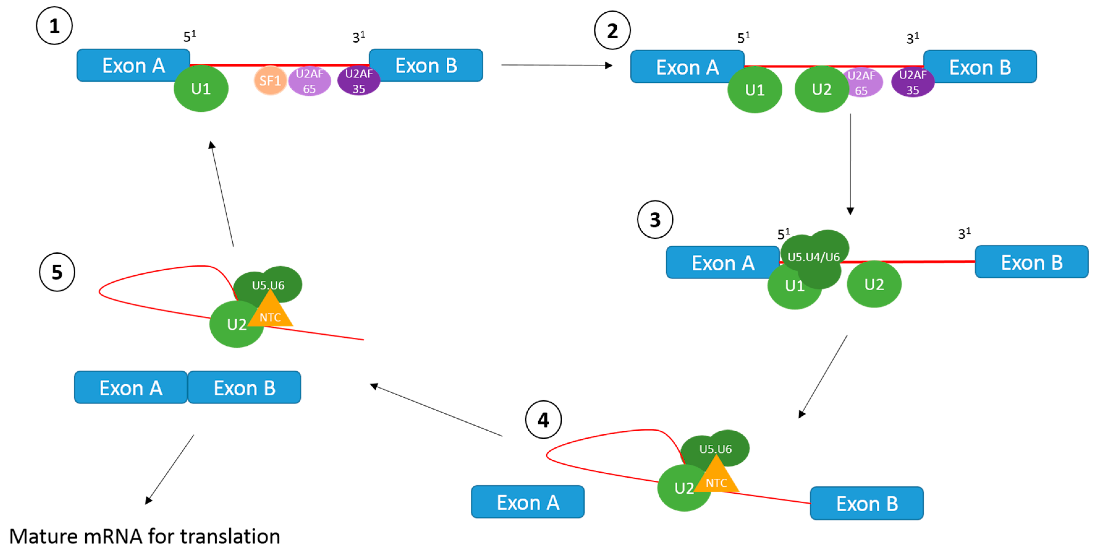
1.1. The Splicing Reaction

Splicing of pre-mRNAs involves the excision of introns and joining together of exons, which forms the mature mRNA transcript that is then translated into protein [6]. This is achieved by the spliceosome, which is a macromolecule structure comprised of five small nuclear ribonucleoprotein particles (snRNPs) and over 100 proteins [7].
The splicing reaction occurs in multiple stages, as outlined in Figure 1. Briefly, the CAG GURAGU consensus sequence at the 5′ of the intron is bound by the U1 snRNP, whilst SF1 binds to the branch point sequence YNYURAC, and the 35kDa U2AF subunit (U2AF35) binds to the 3′ splice site via an AG dinucleotide. In addition, the downstream polypyrimidine sequence of the branch point is bound by the U2AF65 subunit. The U2 snRNP displaces the SF1 protein at the branch point sequence. Following this, the U5.U4/U6 tri-snRNP is recruited to the U1 snRNP, and the NineTeen Complex (NTC) connects to the spliceosome. After conformational and compositional rearrangements, the catalytic function of the spliceosome is activated, and a transesterification reaction occurs. This releases the U1 and U4 snRNPs and forms an intron lariat at the 3′ part of the exon. A further transesterification reaction at the 3′ splice site releases the 3′ exon, which leads to exon ligation and excision of the lariat containing U4, U5 and U6 snRNPs. The spliced mature mRNA transcript can then be translated into protein [7,8].

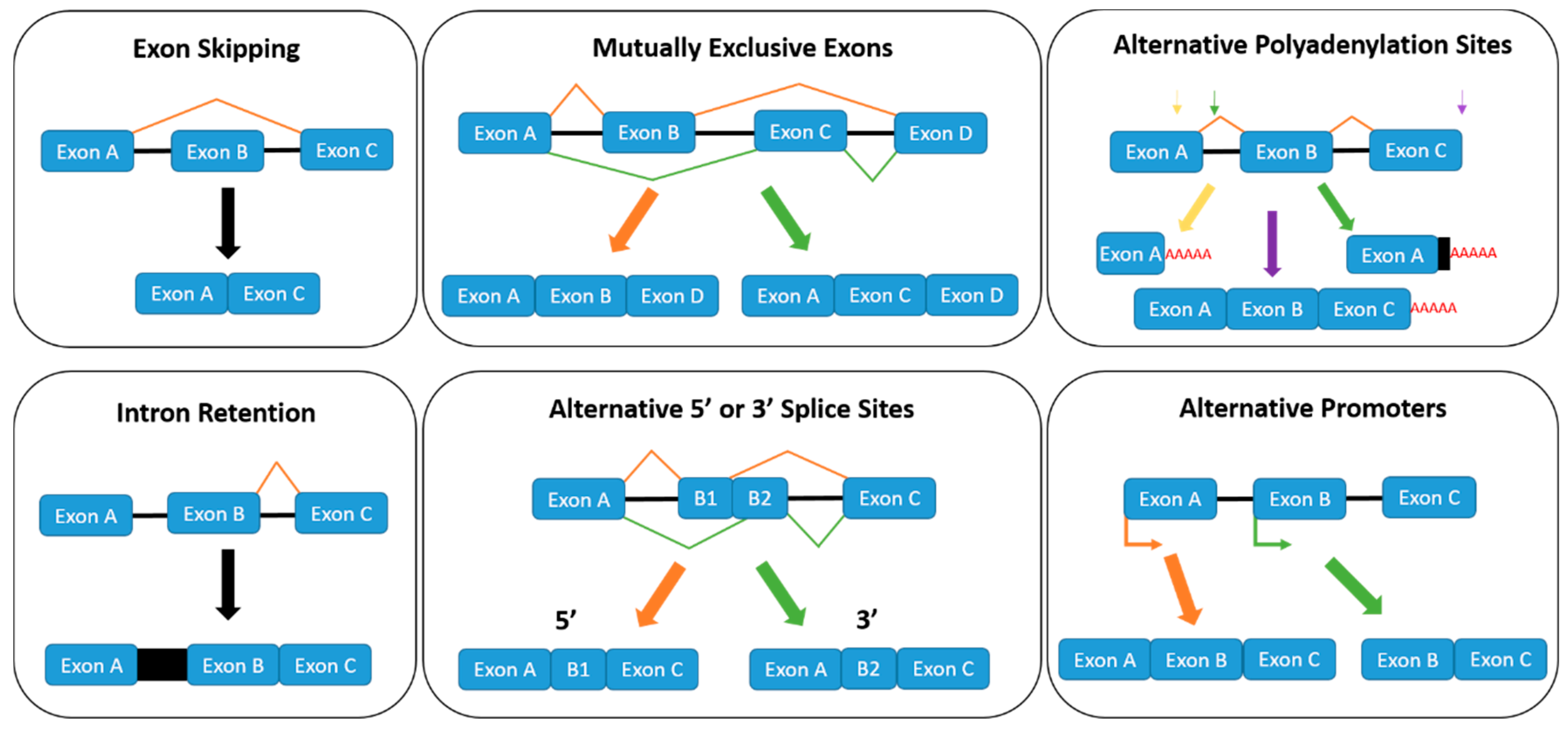
1.2. Regulation of Alternative Splicing
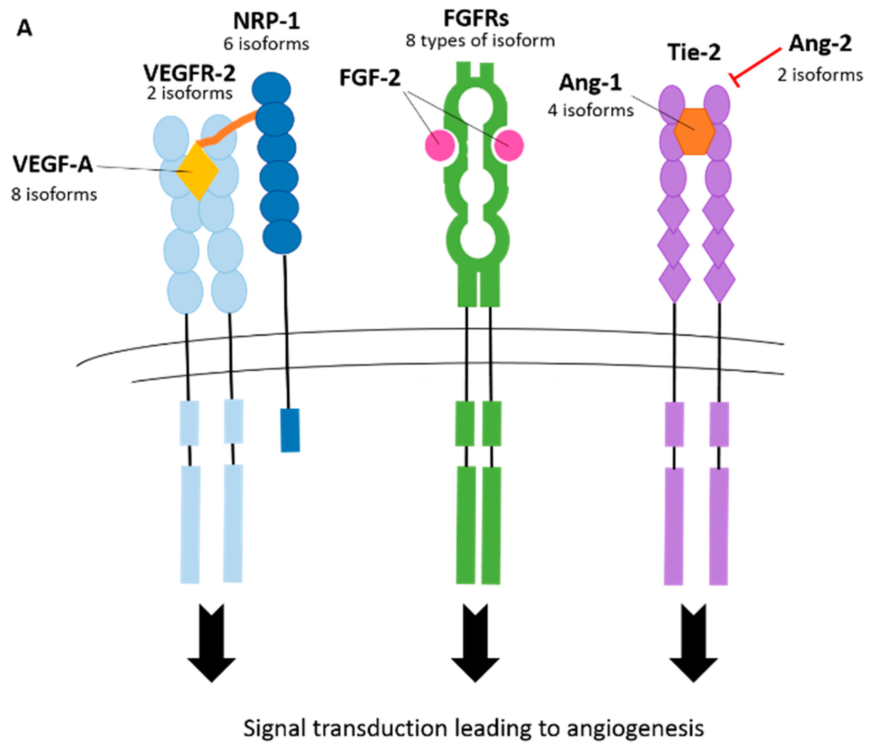
Alternative splicing arises when exons or introns are either included or excluded from a mature mRNA transcript. The major alternative splicing patterns (exon skipping, intron retention, mutually exclusive exons and alternative 3′ or 5′ splice sites) are illustrated in Figure 2, which forms through the recognition of short cis-acting splicing regulatory elements (SREs) by trans-acting splice factors or RNA binding proteins (RBPs). SREs are categorised depending on where in the pre-mRNA the RNA binding protein is binding (exon or intron), and whether they enhance or inhibit splicing. This forms four subgroups of SREs; exonic splicing enhancers (ESEs), intronic splicing enhancers (ISEs), exonic splicing silencers (ESSs) and intronic splicing silencers (ISSs).
RBPs recognise particular SREs and determine which sections of pre-mRNA are included in the mature transcript. Therefore, the regulation of alternative splicing can be influenced by the amount and activity of RBPs, the two main studied groups of which are serine/arginine rich (SR) proteins (12 discovered to date [9]) and heterogenous nuclear ribonucleoproteins (hnRNPs) (23 discovered to date [10]). Originally, it was thought that SR proteins bind to ESEs and promote exon inclusion, whereas spliceosome access to the polypyrimidine tract is blocked by hnRNPs, thereby promoting exon exclusion [11]. For example, hnRNP H/F has been shown to bind to exon 18b regulatory sequences of T-cell factor-3, which represses the inclusion of exon 18b and results in an increase in the E12 isoform that contains exon 18a instead of exon 18b [12]. However, instances whereby SR proteins promote exon exclusion and hnRNP proteins promote exon inclusion have since been reported [13,14,15,16]. In addition, it has been reported that SR proteins and hnRNPs are antagonists of each other when binding to SREs [17]. Therefore, changes in the ratio of SR protein to hnRNPs in the nucleus can alter splicing.
The SR proteins SRSF1, SRSF2 and SRSF6 are involved in the regulation of the spliceosome itself. They promote the binding of the U1 and U2 snRNPs to the 5′ and 3′ splice sites, respectively [18]. Therefore, the binding of SR proteins to SREs is highly regulated through phosphorylation activation by SR protein kinases, such as CDC-like-kinase 1 (CLK1) and SRSF-protein-kinase 1 (SRPK1). For instance, an established pathway for SRSF1-mediated splicing involves phosphorylation of SRSF1 by SRPK1, which facilitates the binding of SRSF1 to a transportin protein (Tr-SR) and results in the nuclear import of SRSF1 [19]. Once in the nucleus, SRSF1 is stored as nuclear speckles, and subsequent hyper-phosphorylation by CLK1 releases SRSF1 from nuclear speckle storage and allows SRSF1 to bind to SREs on pre-mRNA for the splicing reaction [20]. In addition to the major alternative splicing patterns usually documented, two further alternative splicing mechanisms are also depicted in Figure 2; alternative polyadenylation and alternative promoters. Multiple polyadenylation sites can exist throughout a pre-mRNA sequence, which can be cut prematurely to the canonical isoform, and a polyadenylation tail is added. A recent model of VEGFR-1 mRNA alternative polyadenylation regulation has been proposed whereby demethylated hnRNP D is unable to bind to ARE in intron 13 [21]. This results in the premature cutting of the pre-mRNA by cleavage factors and the addition of the poly-A tail to the truncated pre-mRNA, which produces the soluble VEGFR-1 (sVEGFR-1) isoform [21]. However, when hnRNP D is methylated, it binds to ARE in intron 13 and causes the splicing out of the intron, which produces the membrane-bound canonical isoform of VEGFR-1 [21].

2. Angiogenesis

Angiogenesis is an important process that is defined as the formation of new blood vessels from pre-existing vasculature. It is vital during embryonic development, wound healing, hair growth, and reproduction [22]. However, dysregulation of angiogenesis is associated with a range of pathologies in adults, such as cancer, diabetic retinopathy, rheumatoid arthritis and endometriosis [23,24]. This has led to the study of complex signalling pathways that regulate angiogenesis.

Angiogenic Vessel Formation

The angiogenesis process is activated by pro-angiogenic signals, such as vascular endothelial growth factor-A (VEGF-A), angiopoietin-2 (ANG-2) and fibroblast growth factor (FGF) [25]. Ang-2 and matrix metalloproteinases (MMPs) mediate the detachment of pericytes from the vessel wall. This causes the loosening of junctions that are between the endothelial cell monolayer and the nascent vessel dilates. VEGF-A, which is the most extensively investigated member of the VEGF family, increases endothelial cell layer permeability which forces plasma proteins out of the blood vessels and into the surrounding tissue, where they form an extracellular matrix scaffold for the attachment of endothelial cells. The formation of the vascular sprout is guided by the tip cell (stimulated by VEGF-A receptors, neuropilins, Delta-like 4 (DLL4) and JAGGED1 (JAG1), and the elongation of the stalk is facilitated by neighbouring stalk cells (stimulated by NOTCH, NOTCH regulated Ankyrin repeat protein (NRARP), wingless/integrateds (WNT)s, placental growth factor (PIGF) and fibroblast growth factors (FGFs)) [26]. Recently, the macrophage stimulating-1/Forkhead box protein O1 (MST1/FOXO1) cascade was identified to direct the migration of tip cells towards hypoxic regions [27]. The vascular lumen is then established and this is mediated by a variety of factors including: vascular endothelial cadherin (VE-cadherin), CD34, sialomucins, VEGF-A and hedgehog ligands. Following this, the cells become covered in pericytes, which is signalled by the expression of factors, such as platelet-derived growth factor-β (PDGF-B), angiopoietin-1 (ANG-1), transforming growth factor-β (TGF-β), ephrin-B2 and NOTCH. The basement membrane is reformed through the inhibition of MMPs by a tissue inhibitor of metalloproteinases (TIMPs) and plasminogen activator inhibitor-1 (PaI-1), which results in the maturation of the vessel [26]. The important process of angiogenesis is highly regulated at many levels, including transcriptional [28,29], post-transcriptional [30], microRNAs [30,31], allosteric enhancement [32], and as discussed in this review, alternative splicing. Many genes involved in angiogenesis have been shown to be alternatively spliced, and there are many instances whereby isoforms negatively regulate the canonical isoform (Figure 3). This review will discuss current known splicing events that occur in genes associated with angiogenesis and the functions of their splice isoforms (if known) as shown in Table 1. The major splicing events are shown in Figure 4.

3. Splicing in Angiogenesis

3.1. Vascular Endothelial Growth Factor-A (VEGF-A)

The binding of VEGF-A to VEGF receptor 2 (VEGFR2) is considered to be the main stimulatory signal of angiogenesis in vivo [60]. Studies have shown that VEGF-A is secreted by many different cell types, including: endothelial cells, fibroblasts, smooth muscle cells, platelets, neutrophils and macrophages [34]. VEGF-A is produced by 60% of tumours [61] and has therefore been extensively investigated for anti-cancer therapeutics.
The VEGF-A mRNA contains eight exons, the splicing of which gives rise to a variety of isoforms. The six major isoforms of VEGF-A are: VEGF-A111, VEGF-A121, VEGF145, VEGF-A165, VEGF-A189 and VEGF-A206 (where the subscripted numbers denoted the number of amino acids present in the isoform) [34]. Additionally, another less commonly expressed isoform has also been reported, known as VEGF-A183 [62]. The bioavailability of the isoforms is governed by a mRNA sequence that spans exons 6a and 7 determines the binding capacity of the isoforms to heparin and heparin sulphate proteoglycans that are located on the cell surface and extracellular matrix [34,63]. VEGF-A145, VEGF-A189 and VEGF-A206 strongly bind to both the cell surface and extracellular matrix, whereas VEGF-A111 and VEGF-A121, which both lack exons 6 and 7, are readily diffusible [35,36,37]. VEGF-A165 is moderately diffusible with an estimated 50–70% being bound to the cell surface or extracellular matrix following secretion [35]. VEGF-A165 is the most abundantly expressed isoform and most potent initiator of angiogenesis [63]. In fact, VEGF-A189 has less angiogenic activity when compared to the VEGF-A165 and VEGF-A121 isoforms [64,65]. In addition, VEGF-A165 has increased mitogenic effects in comparison to VEGF-A189, which is caused by its heparin binding ability that allows the attachment to the neuropilin co-receptor [66].
It is hypothesized that many splice factors and signaling pathways are involved in the regulation of VEGF-A isoform expression. Indeed, CAPER-α, a transcriptional coactivator for steroid receptors has been shown to alter the ratio of VEGF-A189 to VEGF-A165 in Ewing cells [67]. Furthermore, an earlier study also showed that siRNA knockdowns of CAPER-α increased the VEGF-A121/VEGF-A189 expression ratio in breast cancer cells [68]. Therefore, it can be concluded that CAPER-α is involved in the regulation of alternative splicing of these three isoforms of VEGF-A, and it is proposed that perhaps CAPER-α may also govern the selection of other VEGF-A isoforms. An additional layer of VEGF-A regulation has been uncovered, whereby microRNAs (particularly miRNA-297 and -299 in tumour-associated macrophages; and miR-574-3p in human myeloid cells) bind to the CA-rich element (CARE) in the 3′ UTR of VEGF-A, which results in the negative regulation of VEGF-A expression [69,70]. However, upon hypoxic insult, heterogenous nuclear ribonucleoprotein L (hnRNP L) is translocated from the nucleus to the cytoplasm, where it can interact with VEGF-A mRNA. More specifically, hnRNP L binds to CARE, thereby inhibiting the binding of the microRNAs to the sequence and subsequently induces the splicing of VEGF-A [69]. In addition, it has been shown that increased hnRNP L binding to the CARE element during hypoxia also reduces the binding of the GAIT complex to the GAIT element, which accordingly results in decreased translational repression of VEGF-A [71]. However, whether these mechanisms induce splicing for the expression of a certain isoform of VEGF-A is yet to be determined.
Almost 20 years ago, another family of VEGF-A isoforms was discovered, which arise from alternative splicing of exon 8 (Figure 4A). The isoforms have the same number of amino acids, but alternative sequences in the C terminus [72]. So far, four alternative VEGF-AXXXb isoforms have been identified: VEGF-A165b, VEGF-A121b, VEGF-A189b, and VEGF-A145b [72,73,74,75]. It is generally accepted that the VEGF-Axxxb family of isoforms are inhibitors of angiogenesis, and the VEGF-Axxxa family promotes angiogenesis (Figure 3B), which arguably makes them the most intriguing splicing event of VEGF-A. In fact, it has been reported that VEGF-Axxxb expression is downregulated in cancer, diabetic retinopathy, Denys Drash syndrome and retinal vein occlusion, and upregulated in systemic sclerosis and asthma [38].
The aberrant splicing of VEGF-A exon 8 has been extensively investigated, and a few different signalling pathways have been reported to be involved in the production VEGF-Axxxb. In the first pathway, stimulation by growth factors (e.g., insulin growth factor (IGF)) promotes the phosphorylation of SRSF1 by SR protein kinase 1, which allows transportation of SRSF1 into the nucleus. SRSF1 then binds with the proximal splice site of exon 8 and results in the production of the VEGF-Axxxa isoform [76]. However, knockdown of SRSF1 and knockdown and inhibition of SRPK1 altered the splicing of VEGF-A mRNA to produce more of the VEGF-Axxxb isoform, and so it is thought that VEGF-Axxxb production is linked to the inhibition of this pathway [77].
The other three pathways described so far involve the selection of the distal splice site of VEGF-A exon 8, which results in production of the VEGF-Axxxb isoform. One pathway involves the stimulation of CLK1/4 phosphorylation of SRFS6 by TGF-β, which leads to distal splice site selection of VEGF-A exon 8 [78,79]. The remaining two pathways that have been uncovered to date involve VEGF-Axxxb splice site selection by SRSF2. One such pathway includes the wingless integrated 5a/orphan receptor tyrosine kinase-like receptor-2/Jun N-terminal kinase (Wnt5a/Ror2/JNK) dependent mechanism [80], and the other pathway involves E2F1 [81].

3.2. Vascular Endothelial Growth Factor Receptors (VEGFRs)

In order to facilitate a cellular response, a VEGF-A dimer binds to paired tyrosine kinase receptors, named vascular endothelial growth factor receptors (VEGFRs), which have been found in a range of cell types, such as endothelial cells [82], haematopoietic [83] and smooth muscle cells [84]. There are three types of VEGFRs: VEGFR1, VEGFR2 and VEGFR3, which are encoded by different genes; however, VEGFR1 and VEGFR2 share similar structures [85,86]. The VEGF-A family of isoforms bind to VEGFR1 and VEGFR2 to facilitate angiogenesis, whereas VEGFC/D isoforms bind to VEGFR3 to drive lymphangiogenesis for the formation of lymph vessels [87]. VEGFR1 has decreased kinase activity when compared to VEGFR2, despite having a higher affinity for the binding of VEGF-A, and so is highly regarded as a decoy receptor [88,89]. Therefore, most research into the roles of VEGFRs in angiogenesis have focused on VEGFR2.
An additional step is required to activate VEGFR2 signalling after VEGF-A has bound to the receptor. The binding of allosteric sites of VEGFR2 are necessary to cause a conformational twist of the receptor, which leads to its activation [34]. Similar structural arrangements are caused by VEGF-A165, VEGF-A165b and VEGF-A121, which ultimately allows the transduction of the signal [90]. VEGF-A165 has been shown to activate phosphorylation of VEGFR2, AKT and ERK the most, whereas VEGF-A165b activation of VEGFR2 results in diminished stimulation of angiogenesis [91,92,93], and it is for this reason the ‘anti-angiogenic’ label has been applied to VEGF-A165b. Most evidence points to a partial agonistic function for VEGF-A121 [93,94]; however, there have also been some cases where VEGF-A121 has shown full agonistic qualities. For example, several independent groups have shown that VEGF-A121 activation induced phosphorylation of VEGFR2, but at a reduced rate to VEGF-A165a [95,96,97]. In addition, when compared to VEGF-A165, VEGF-A121 generated less motility and sprouting in human umbilical vein endothelial (HUVEC) cells [95]. Studies into the effect of VEGF-A121 on ex-vivo angiogenic sprouting and vascular permeability have demonstrated VEGF-A121 to have both equal and lower levels of generation to VEGF-A165 [34].
VEGFR2 signalling is complicated further by splicing of VEGFR2. Endogenous soluble VEGFR2 (esVEGFR2) [39] arises from partial retention of intron 13 and a consequential early termination stop codon. The resultant protein contains six out of the seven extracellular immunoglobulin-like domains and has a unique c-terminal sequence that does not exist in the canonical form of VEGFR2. It can be noted that there is another soluble variant of VEGFR2 which is formed through proteolytic shedding from the cell surface, which is a post-translational modification and is not formed from alternative splicing of VEGFR2 [98]. Both of these variants act on the lymphangiogenic system through trapping of VEGF-C, which obstructs the activation of VEGFR3, resulting in the decrease of lymphatic endothelial cell proliferation [98]. Lymphangiogenesis in tumours promotes metastasis of malignant cancer cells. In accordance with this, researchers have observed a correlation between sVEGFR2 downregulation and progression of malignant neuroblastoma, which suggests a protective function for these variants in malignancies [39]. Another truncated VEGFR2 isoform has been discovered in rat retina, which contains the entire extracellular domain and a part of the intracellular domain. Little is known about this isoform; however, it has confirmed VEGF-A activity and leads to increased cytoplasmic calcium and therefore is thought to be functional in the angiogenic system [99].
Although VEGFR1 is heavily regarded as a ‘decoy’ receptor for angiogenesis, it has been implemented in tumour progressive processes. In adults, VEGFR1 has been shown to activate inflammation, tumour growth, metastasis; increase chemotaxis of monocytes and has been linked to increased release of MMP9 from human vascular smooth muscle cells [99,100]. To date, four alternatively spliced variants of VEGFR1 have also been discovered: sVEGFR1-i13 (shown in Figure 4A), sVEGFR1-i14, sVEGFR1-e15a and sVEGFR1-e15b [101,102,103]. The sVEGFR1-i13 and sVEGFR1-i14 isoforms have extensions beyond exon 13 and exon 14, respectively, whereby sVEGFR1-i13 arises from an extended read through of a potential splice site in exon 13 (Figure 4A), and sVEGFR1-i14 arises from a skipped splice site. The sVEGFR1-e15a and sVEGFR1-i15b isoforms arise from the inclusion of an alternative exon in place of exon 15 (either exon 15a or 15b) [102,103]. Membrane-bound VEGFR1 and the soluble isoforms share the first 13 exons, which code for the first six N-terminal immunoglobulin-like domains [103]. The soluble isoforms lack all the protein domains that are encoded after exon 13 in membrane-bound VEGFR1: the seventh immunoglobulin-like extracellular domain, the membrane-anchoring region, the juxtamembrane domain and the intracellular signaling tyrosine kinase domains [103]. This enables the soluble isoforms to have retained affinity for VEGF-A ligands, neuropilin-1 (NRP-1) co-receptor and extracellular heparin sulphate proteoglycans, but prevents signal transduction. In essence, sVEGFR1 isoforms trap VEGF-A and form inert heterodimers with VEGF receptors [39], which has caused them to be regarded as potent anti-angiogenic factors. Investigations into the mechanisms that control the splicing of VEGFR1 have been conducted, and numerous studies have provided evidence that hnRNP D plays a role in the regulation of sVEGFR1-i13. One such study executed in human dermal microvascular endothelial cells (HMVEC) unearthed two sequences within intron 13 that are responsible for the premature poly(A) signal found in sVEGFR1, and proposed that hnRNP D binds to an A-and-U-rich-element (AURE) sequence (AUUUA) located in intron 13 downstream of the sVEGFR1 processing site [104]. Indeed, overexpression of hnRNP D was shown to significantly decrease the sVEGFR1/mVEGFR1 (membrane-bound VEGFR1) ratio [104]. However, a later study conducted in human macrophage-like U937 cells provided evidence that hnRNP D increases the expression of sVEGF1 mRNA with the use of peptides that were modelled on the regulatory domain of hnRNP D [105]. Furthermore, this finding was strengthened by the peptide-mediated suppressed expression of VEGF-A [105]. This discrepancy in the role of hnRNP D in VEGFR1 splicing regulation could be due to a cell type-specific effect and/or additional modifications, such as the altered expression of other factors yet to be uncovered as being involved in the alternative splicing of VEGFR1. In addition, regulation of splicing events, particularly of genes that are of particular importance, can be very complicated with many molecules involved, and so it could be that there are other molecular mechanisms taking place that override the effect of hnRNP D. Indeed, there has been evidence of other molecules and pathways that regulate the expression of sVEGFR1-i13. Studies conducted in endothelial cells have revealed that the U2A65 splicing factor is implicated in the positive regulation of sVEGFR1-i13 [106], whereas JuMonJi Domain containing protein-6 (JMJD6) and NOTCH1 have been shown to be negative modulators of the isoform [106,107]. Moreover, a recent study conducted in squamous lung carcinoma cells has shown evidence for a more accomplished mechanism, whereby VEGF-A165 promotes the expression of SOX2, which directly binds to the promoter of SRSF2 and induces transcription [101]. The SRSF2 splice factor promotes splicing of VEGFR1 towards the sVEGFR1-i13 isoform [101].

3.3. Neuropilins (NRPs)

NRPs are expressed in many distinct cell types, including neurons, blood vessels and immune cells [108]. NRPs are transmembrane co-receptors that function in both VEGF signalling (through dimerization with VEGFR2) and semaphorin/plexin signalling, which implicate NRPs in both angiogenesis and lymphangiogenesis, and neuronal tissue pattern development, respectively [109]. For the basis of this review, the role of NRPs in axonal development will be kept brief, as this subject matter is beyond the scope of this review. However, it can be noted that the semaphorin-3 (SEMA3) family of glycoproteins binds to NRP receptors to facilitate axonal guidance.
The structure of NRPs consists of various domains with different functions. The a1 and a2 domains are involved in binding to SEMA3, and the b1 and b2 domains are required for the binding of VEGF-A; in particular, VEGF-A165, VEGF-A121 and VEGF-A189 have been shown to bind to NRP1 [108]. In fact, VEGF-A165 has been shown to be paramount for the complex formation between VEGFR2 and NRP1 [110]. VEGF-A165 can also bind to NRP2, however, with a 50-fold less than NRP1 [111]. Interestingly, exon 8 of VEGF-A has been shown to be crucial for binding to NRPs, and consequently VEGF165b has been shown to be unable to bind to NRPs [112]. In addition to VEGF-A, the b1 and b2 domains of NRP1 have also been shown to bind to other angiogenic-associated factors, including: VEGF-B, VEGF-C, VEGF-D, placental growth factor-2 (PLGF2), hepatocyte growth factor (HGF), fibroblast growth factors (FGFs), and transforming growth factor beta 1 (TGFβ1) [108], which further corroborates that NRP1 plays a key role in angiogenesis.
NRP1 mRNA consists of 17 exons and to date, six alternatively spliced isoforms of NRP1 have been discovered. The first to be discovered were s11NRP1 and s12NRP1, which are composed of the first 11 and 12 exons of NRP1, respectively [40,113]. An additional two more isoforms were later discovered: sIIINRP1 and sIVNRP1. sIIINRP1 is formed from exons 1-9 and exon 12, with cassette exon skipping of exons 10 and 11 [114]. The structure of sivNRP1 is similar to that of sIIINRP1, however, it also retains exon 10 [114]. All four of these isoforms are soluble and do not contain the C-terminal transmembrane and cytoplasmic domains. The resulting proteins formed are able to sequester NRP1 binding factors, but are unable to transmit signals, and therefore act as antagonists of NRP1 signalling [40]. Hence, these soluble isoforms of NRP1 have been associated with anti-angiogenic and anti-tumourigenic functions. Another splice variant of NRP1, named NRP1-ΔE16, is formed by the skipping of exon 16 and the addition of an arginine (AAG) [41]. Not much is documented on this isoform, as it was not found to have any functional differences to full length NRP1.
More recently, a further splice isoform of NRP1, NRP-1Δ7, was discovered [42]. This isoform is formed by the deletion of just seven amino acids in exon 11 caused by the utilization of an alternative donor site located 21 bases upstream of the conventional splice site (Figure 4A). The seven amino acids that are deleted in NRP1-Δ7 omit a coding sequence that contains two aspartic residues that are located two residues downstream of the glycosylation site and are required for efficient glycosylation [115]. Glycosylation is a post translational modification that can alter the structure and function of a protein. NRP1 is reported to be glycosylated at Serine-612, whereby long chains of oligosaccharides are formed. Various different chains can assemble on NRP1, which are dependent on the cell type. For example, in smooth muscle cells, chondroitin sulfate is added, whilst in endothelial cells heparin sulfate is assembled on the glycosylation site of NRP1 [116]. The number and nature of the added oligosaccharide chain(s) can modulate the function of a protein. Indeed, it has been reported that mutated Serine-612 prevents glycosylation at the site, increased cell invasion in human glioblastoma cells [117] and reduced fibronectin fibrillation in a human hepatic cell line [118]. Further studies conducted in modified prostate (PC3) and breast (MDA-MB-231) cancer cells that expressed either recombinant full length NRP1 or NRP1-Δ7, showed that NRP1-Δ7 has anti-tumorigenic properties, including decreased proliferation, migration and anchorage-dependent growth whilst the overexpression of full length NRP1 had the opposite effect [42]. Therefore, NRP1-Δ7 is considered to function in the inhibition of angiogenesis and tumorigenesis and could have therapeutic potential.
NRP2 functions in lymphangiogenesis through dimerization with VEGFR3 and activation by VEGF-C [119]. Indeed, NRP2 knockout mice presented with impaired lymphangiogenesis [120]. In addition, NRP2 has been implicated in tumour metastasis. This was shown using a monoclonal antibody to inhibit VEGF-C binding to NRP2, which in turn decreased metastasis [121]. Recently, a new splice isoform of NRP2 named s9NRP2 has been discovered and is thought to antagonize VEGF-C/NRP2 signalling [122]. The isoform is formed from the retention of an intron between exons 9 and 10 that encodes a premature stop codon in the b2 domain. This results in an unpaired cysteine residue that forms a disulphide bridge with an unpaired cysteine from another s9NRP2-producing dimer. There has been evidence to show that s9NRP2 inhibits the binding of VEGF-C to NRP2, however, with no effect on the interaction of VEGF-C to VEGFR-3 [122]. Further research is required to establish whether the isoform also inhibits the formation of metastasis.

3.4. Fibroblast Growth Factor Receptors (FGFRs)

There are at least five types of FGFRs named FGFR 1–5 [123]; FGFR 1-3 have a similar structure, whereas FGFR4 lacks the transmembrane and intracytoplasmic domains. FGFR5 and an additional receptor have also been proposed, designated as FGFR6 [124]. FGFRs have the same protein structure that is generally found in receptor tyrosine kinases and are involved in the regulation of key biological cellular processes, such as proliferation, differentiation, migration and survival [125]. There are a number of different types of alternatively spliced isoforms that can arise in FGFRs, which will be discussed below.
The most described alternative splicing event in the literature is generated from the use of alternative exons in the c-terminus of IgIII of FGFRs1-3 [43]. The IgIII domain is comprised of three domains: IIIa, IIIb and IIIc, which are encoded by exons 7–9, respectively. The alternative use of exons 8 and 9 form the IIIb and IIIc isoforms for FGFR1-3 (Figure 4A), which have different ligand affinities and tissue specificities. i.e., the IIIb isoform is predominantly found in the epithelial tissues, whereas the IIIc isoform is expressed in the mesenchymal tissues. The regulation of this splicing switch is therefore strongly implicated in epithelial-mesenchymal-transition (EMT). A number of RBPs have been implicated in the regulation of splicing of the c-terminus of IgIII. ESRP1 and ESRP2 promote the expression of the IIIb isoform, and hnRNP F/H, K and forkhead box-2 (FOX-2) silence the IIIc isoform. On the other hand, hnRNP A1 and PTB are associated with silencing of IIIb [126]. Furthermore, hnRNP M has been shown to bind to ISE/ISS-3 of FGFR2 and promote exon IIIc skipping [127]. Another more detailed splicing mechanism for the regulation of IIIb and IIIc splicing involves RBM4 and nPTB [128]. In short, RBM4 promotes the expression of the IIIb isoform, whereas nPTB induces exon IIIc inclusion. Interestingly, the RBM4 splice factor is also implicated in promoting exon 10 skipping of PTB to produce the nPTB splice variant [128], and this therefore shows the extent that splicing mechanisms are tightly controlled. The splicing regulation of FGFR2 is a prime example of how RNA-binding proteins work in combination to bring about a splicing profile. A mesenchymal phenotype is linked to the promotion of tumorigenesis, and aberrant expression of the IIIb and IIIc isoforms has been documented in animal models of bladder cancer and epithelial cell tumours [129,130,131]. Furthermore, elevated expression of both the IIIc and IIIb isoforms has been shown in a variety of cancerous cells and tissues [43]. However, expression of FGFR-2 IIIb in oesophageal and colorectal cancers was associated with better differentiation and decreased expression of the isoform correlated with proliferation, invasion and poor prognosis [44]. Furthermore, the loss of the IIIb isoform and increase in IIIc expression has been associated with a more aggressive cancer phenotype [44]. In light of the above, it could be concluded that the IIIc isoform is a tumour promoter, and IIIb acts as a tumour suppressor; however, increased expression of FGFR-2 IIIb has also been linked to cell transformation and tumour progression in some cancers, and so the precise role of FGFR-2 IIIb in cancers remains controversial [44]. For example, in gastric cells, FGFR-2 IIIb expression was associated with cellular invasion of the gastric wall; however, decreased expression of FGFR-2 IIIb in gastric cells was also linked to proliferation, invasion and poor prognosis [44]. Furthermore, FGF-7 and FGFR-2 IIIb have been implicated in tumour angiogenesis through inducing an increase in VEGF-A expression in colorectal and pancreatic cancers [132,133]. This is a prime example of how there is a lot of cross-talk between different arms of angiogenesis signalling, and therefore associates FGF-7 and the FGFR-2 IIIb isoform in particular in the promotion of angiogenesis.
The next splicing event of FGFRs that will be discussed is the inclusion or exclusion of exons that encode the IgI and acid box domains, which are formed from exons three and four respectively in FGFRs 1–3, and exons two and three respectively in FGFR4 [134,135]. Both domains are involved in the auto-inhibition of FGFRs [43]. In fact, isoforms that do not contain these domains have heparin sulfate proteoglycan attachments, which strengthens the affinity for binding to ligands and enhances signalling transduction of FGFRs [136]. The excision of the IgI domain produces an isoform that has two loops in the extracellular domain (formed by IgII and IgIII), named FGFRβ, whereas the inclusion of the exon produces FGFRα, which has three loops in the extracellular domain [137]. Therefore, the FGFRβ isoforms, which do not contain the auto-inhibitory IgI domain, have a higher affinity for FGFs and enhanced signalling [45,46]. Indeed, FGFR-1β has been shown to increase proliferation through having a stronger affinity for FGF1 and displaying augmented signalling [43]. Furthermore, an elevation in the expression ratio of FGFR1β:FGFR1α has been linked to tumour progression [43]. Recently, the polypyrimidine tract-binding protein 1 (PTPBP1) splicing repressor has been implicated in the regulation of alternative splicing of FGFR1α and FGFR1β. The study conducted in breast cancer cells revealed that PTBP1 represses the splicing of FGFR1β [138]. However, PTBP1 has also been reported to induce α exon deletion through the binding of intronic splicing silencer sequences that flank the α exon resulting in the positive regulation of FGFR1β [139]. Therefore, further investigation is required to clarify the precise role of PTBP1 in the regulation of FGFR1α/FGFR1β splicing. In addition, the SRSF6 splicing factor has been implicated in the exclusion of the α exon, which has been attributed to the presence of two exonic splicing enhancers located in the α exon, which can be bound by SRSF6 to facilitate the exclusion of the α exon [140]. Indeed, knock downs of SRSF6 resulted in the increased expression of FGFR1α [140].
Another class of isoforms are formed from the skipping of exons 8, 9 and 10. This region encodes the transmembrane domain, which is responsible for anchoring FGFRs to the cellular membrane. Therefore, this alternative splicing event produces soluble FGFR receptors, which can be found in other locations in the cell other than the cell membrane. For example, a soluble isoform of FGFR3, which arises from the deletion of exon 8 as well as exon 7, has been shown to be predominantly found in the nucleus of breast epithelial cells [45]. Although the precise functions of these isoforms are yet to be determined and may be diverse depending on the splicing events that exist in other regions of the sequence, it can be proposed that many of the soluble isoforms act as an antagonist through the binding of ligands without the activation of signal transduction, which is true of other soluble tyrosine kinase receptors. Indeed, a soluble isoform of FGFR4 has been shown to diminish FGF1 signalling in human Michigan Cancer Foundation-7 (MCF-7) breast cancer cells [43].
A further group of FGFR splice isoforms arise from the inclusion of differing carboxy-terminal sequences, termed C1, C2 and C3. The distinct sequences produce proteins that have differential retention of tyrosine residues, which function as docking sites for cytoplasmic signalling proteins and are also involved in autophosphorylation of the receptor [47]. The C3 variant has been documented to be the most transforming of the isoforms when compared to C2, which has moderate transforming activity, and C1 which has weak activity. The increased transforming activity elicited by the C3 isoform strongly suggests a role for these variants in oncogenesis. In support of this, elevated expression of the C3 isoform has been detected in gastric cell lines and in human breast cancer cell lines [47].
Another type of splice isoform that arises in FGFRs is produced via the inclusion or deletion of six nucleotides (GTAACA), known as the VT motif which codes for two amino acids (valine and threonine) in the juxtamembrane region of FGFR1-3 [141]. The inclusion or exclusion of the VT motif affects the signalling capability of the receptor, as the exclusion of the VT motif prevents the binding of effector molecules. In fact, isoforms that contain the VT motif are able to activate the Ras/MAPK signalling pathway [48]. Therefore, as one of the major downstream pathways of FGFR is affected, it has been postulated that the absence of the VT motif could have major implications for the function of FGFR. However, other signalling pathways that rely on receptor kinase activity for activation remain unaffected by the exclusion of the VT motif, such as the downstream effectors of phospholipase C-γ (PLCγ) [141]. Therefore, the inclusion or exclusion of the VT motif may selectively activate different signalling mechanisms in varied cell types.

3.5. Vasohibins

Vasohibin-1 is a negative regulator of angiogenesis that is typically expressed in endothelial cells and is regulated by VEGF-A and FGF-2 [51]. Whilst vasohibin-1 inhibits VEGF-stimulated angiogenesis, it does not inhibit the phosphorylation and subsequent activation of VEGFRs; however, vasohibin-1 does inhibit FGF-2-activated angiogenesis [142]. In fact, it was also shown that vasohibin-1 is activated by VEGFR-2 and its downstream effector, protein kinase C-δ (PKC-δ), in endothelial cells resulting in the inhibition of angiogenesis [51]. PKC-δ is also involved in the induction of vasohibin-1 by FGF-2, which further implies that this factor is imperative to the regulation of vasohibin-1 expression [51]. Vasohibin-1 has also been shown to regulate the hypoxia inducible factor-1α (HIF-1α) pathway through inducing the degradation of HIF-1α by prolyl hydroxylase, which suggests that hypoxia and vasohibin-1 act in concert to regulate one another [143]. Therefore, the expression of vasohibin-1 is highly regulated in a number of ways.
Later, another gene which shared 52.5% homology with vasohibin-1 was discovered and designated vasohibin-2 [144]. Human vasohibin-2 was shown to also exert anti-angiogenic activity; however, unlike vasohibin-1, vasohibin-2 was not found to induce VEGF–A or FGF-2. Furthermore, vasohibin-2 is not regulated by cytokines or growth factors [144]. However, a microRNA (mir-200b) has been shown to target and therefore diminish the expression of vasohibin-2 [144,145].
Studies of multiple collections of microarray data has revealed that the two genes have entirely different profiles of co-expression with other genes, suggesting that vasohibin-1 and vasohibin-2 have distinct functions [144]. In accordance, vasohibin-1 is expressed in endothelial cells of newly formed blood vessels behind the sprouting front and acts to halt angiogenesis, whereas vasohibin-2 is expressed in mononuclear cells that help form the sprout and promotes angiogenesis [144]. Indeed, vasohibin-2 has been linked to tumour growth through the stimulation of angiogenesis [145,146], whereas vasohibin-1 has been shown to be an effective treatment for angiogenesis in various animal models, including diabetic nephropathy, pulmonary fibrosis, ocular angiogenesis and cancers [144].
The two main isoforms of vasohibin-1 that have been documented in the literature are the 365aa isoform, known as vasohibin-1A (VASH1A) formed by seven exons, and the 204aa isoform, known as vasohibin-1B (VASH1B), which consists of four exons [50] (Figure 4A). The proteins generated from these isoforms have the same N-terminus, which contains a nuclear localisation signal, but a different C-terminus, which is thought to be important for anti-angiogenic activity [49]. Interestingly, a study of in vivo blood vessel growth conducted in chicken chorioallantoic membrane showed that whilst VASH1A did not inhibit vessel growth, the vessels were shown to be irregular; however, VASH1B inhibited vessel growth strongly [49]. Furthermore, in vitro studies of angiogenesis showed that VASH1B inhibited endothelial cell growth, migration and tube formation [49]. VASH1A on the other hand, only had a significant effect on migration in vitro; in fact VASH1A significantly increased endothelial cell migration [49]. Moreover, the VASH1B isoform was shown to induce apoptosis in endothelial and fibroblast cells, whereas VASH1A was not shown to only induce apoptosis in fibroblasts [49]. A later study was able to explain the differences in functions exerted by the two vasohibin isoforms [50]. It was shown that VASH1A promoted normalisation of abnormal tumour blood vessels, whereas VASH1B induced autophagy, which promotes either cellular survival or cellular death in order to maintain homeostasis. Therefore, the same group investigated the effects of combination treatments that included both isoforms and found that the combination treatments had the highest anti-tumour effects with marked vascular normalisation when compared to treatment with either isoform alone [50]. Therefore, combination treatments with VASH1A and VASH1B may be useful in antiangiogenic treatments, as the existing vasculature can be normalised by VASH1A and pruned by VASH1B.
The alternative splicing of vasohibin-2 is less documented. Vasohibin-2 contains 11 exons which are alternatively spliced to produce transcripts that are: 355aa, 311aa, 290aa, 156aa, 117aa and 104aa. The largest isoform (355aa) has been shown to be predominantly expressed in HUVEC cells, and the 290aa splice variant has been shown to have antiangiogenic activity [51]. The function of the other isoforms have not yet been classified. As mentioned in the beginning of this section, vasohibin-2 was found to be involved in the stimulation of angiogenesis and is highly expressed at the sprouting front. The drive of angiogenesis by vasohibin-2 was linked to high expression of the protein in mononuclear cells at the sprouting front, whereas anti-angiogenic activity of vasohibin-2 was identified in endothelial cells. This suggests that perhaps vasohibin-2 has distinct functions in different cell types. Another reason for this discrepancy could be that splice isoforms of vasohibin-2 whose function has not yet been determined could have opposing functions.

3.6. Hypoxia Inducible Factor-α (HIF-α)

Hypoxia is a feature of many diseases, such as cancers whereby inadequate vasculature causes pockets of low oxygen. This exerts a pro-apoptotic response in some cancer cells [147], as they do not receive the oxygen they require for survival. Therefore, hypoxia elicits various responses including the promotion of angiogenesis in order to provide cells with the oxygen that they need. The most established signalling pathway that is induced by hypoxia is the hypoxia inducible factor-α (HIF-α) pathway. In short, HIF-α forms a dimer with HIF-β, also known as aryl hydrocarbon receptor nuclear translocator (ARNT), which then binds to HIF-response cis-elements to facilitate the transcription of target genes, including VEGF-A. However, in normoxic conditions, prolyl hydroxylases (PHD) interacts with HIF-α, allowing the von Hippel-Lindau (VHL) protein to bind and target the degradation of HIF-α by the proteasome thereby preventing any transcription of HIF-targeted genes [8]. There are three homologous of HIF-α and HIF-β that are designated HIF-1α and HIF-1β, HIF-2α and HIF-2β, and HIF-3α and HIF-3β [55]. The HIF-1α and HIF-2α subunits share similar domain assemblies and undergo proteolytic regulation [55]. However, HIF-3α has been generally considered as a negative regulator of HIF-1α, but not HIF-2α [148]. The most studied of these are the HIF-1α subunits.
Several splice isoforms of HIF-1α have been described in the literature, which arise from cassette exon skipping: HIF-1α Δ11, HIF-1α Δ12, HIF-1α Δ11&12, HIF-1α Δ14, and HIF-1α417 [53,54,149]. The splicing scheme for HIF-1α Δ14 is shown in Figure 4A. Most of these isoforms lack exons that encode parts of the oxygen-dependent degradation domain which is targeted by prolyl hydroxylases (PHDs), or C-terminus transactivation domains, which are required for transcription of HIF-targeted genes [149]. As most of these isoforms have reduced or completely abolished HIF function and have a dampening effect on the initiation of HIF-activated transcription of target genes, it is therefore predicted that these isoforms are stable in normoxia in order to maintain negative regulation of HIF signalling in the presence of oxygen. In accordance with this, a study found that the HIF-1α Δ14 isoform is expressed at higher levels than the canonical isoform during normoxia [54]. During hypoxia, however, both the canonical isoform of HIF-1α and HIF-1α Δ14 were able to dimerise with HIF-1β and activate the VEGF-A promoter [54]. However, HIF-1α Δ14 was shown to have a three-fold less potency than the canonical form of HIF-1α [54]. Furthermore, HIF-1α Δ12 and HIF-1α Δ11&12 both block the dimerization of HIF-1α and HIF-1β by isolating HIF-1β to the cytoplasm [53]. Therefore, these two isoforms act as dominant-negative regulators of HIF-1 transcription. The skipping of exon 10 produces the HIF-1α417 splice variant, which causes a frame-shift and results in a truncated protein of 417 amino acids in length [53]. The resultant protein lacks a transactivation domain and so was thought not to act as a principal transcription factor. However, further experiments showed that the splice variant amplified HIF-1β-mediated transcription of the erythropoietin (EPO) reporter gene [53], which shows another mechanism in which HIF target genes are transcribed. In contrast to the other isoforms, there is evidence to show that HIF-1α Δ11 promotes tumorigenesis through the enhancement of HIF-1 activity. Indeed, overexpression of HIF-1α Δ11 increased tumour growth in vivo [52]. An additional alternative splicing event involves the insertion of three bases (TAG) between exons 1 and 2 of HIF-1α, which substitutes an asparagine in place of a lysine and inserts an additional arginine [44]. This significance of this isoform remains to be elucidated. Another splice variant of HIF-1α arises from an alternative upstream translational start site, which produces a protein that has an N-terminus that is 24 amino acids longer than canonical HIF-1α named HIF-1α Alt1 [150].
HIF-3α, the dominant negative regulator of HIF-1α, has also been reported to undergo alternative splicing [47,151]. The most established alternative splice isoform of HIF-3α is inhibitory Per/Arnt/Sim (IPAS), which is formed through multiple alternative splicing events and is inducible by hypoxia (Figure 4B). IPAS shares exons 2, 4 and 5 with the canonical HIF-3α; however, IPAS also contains an alternative exon upstream of exon 1 (designated 1a), as well as an additional exon before exon 4 (designated exon 4a) and exon 16. The use of alternative splice sites in exons 3 and 6 are also present in IPAS [151]. The inclusion of exon 4a, together with the use of the alternative 3′ splice site of exon 3, results in a frame shift. The resultant protein lacks transactivation domains and so cannot initiate transcription [53]. IPAS dimerises with HIF-α subunits, thereby preventing the transcription of HIF target genes, such as VEGF-A [55]. Indeed, in cornea epithelium, IPAS was found to negatively regulate VEGF-A gene expression and therefore, was shown to indirectly dampen angiogenesis [56]. Another splice variant of HIF-3α that is generated by the inclusion of intron 7 and known as HIF-3α4 is reported to be similar to IPAS [55,152]. HIF-3α4 forms a complex with HIF-1α, which prevents the binding of HIF to hypoxia response elements, and subsequent transcription of target HIF genes. Indeed, inducible HIF-3α4 was shown to hamper angiogenesis and proliferation [55].

3.7. Angiopoietin

Angiopoietins and Tie receptors are involved in endothelial cell survival and vascular maturation. There are four members of the angiopoietin family, designated angiopoietin 1–4 (Ang-1-4) [153]. Ang-1 and Ang-2 are the most characterised in the literature. They share around 60% homology and both bind to the same site of the TEK receptor tyrosine kinase-2 (Tie-2) receptor with similar affinity [153]. Ang-1 is expressed by smooth muscle cells and perivascular cells, whereas, Ang-2 is primarily expressed by endothelial cells and is stored in Weibel-Palade bodies within the cells [153,154]. Upon cytokine activation of the endothelium, Ang-2 is released from the Weibel-Palade bodies through a neuroligin-2-dependent mechanism and binds to the Tie-2 receptor [155]. Originally, it was thought that Ang-1 acted as an agonist of the Tie-2 receptor and Ang-2 was an antagonist of the Tie-2 receptor, as it has been shown to bind to Tie-2 but did not induce autophosphorylation of the receptor [156]. However, further investigations have shown that Ang-2 can also act as an agonist of the Tie-2 receptor. A range of solutions have been proposed to explain the contrasting functions assigned to Ang-2, such as cell type-specific effects, Ang-2 stimulation duration, the presence of co-receptors (e.g., Tie-1), and the presence or absence of other molecules [153]. Indeed, it has been shown in chicken testis that Ang-2 induces angiogenesis in the presence of VEGF-A, whereas in the absence of VEGF-A, Ang-2 was associated with vascular regression [58]. Alternative spliced variants of Ang-2 with varied functions could also account for the discrepancies for functional assignment of Ang-2.
The first discovered isoform was the Ang-2B variant, which was detected in chicken testis, and arises from the inclusion of exon 1B instead of exon 1 [58]. The study found that the canonical isoform (assigned Ang-2A) had a higher expression than the alternatively spliced Ang-2B isoform in immature testis and fully regressed testis, whereas the Ang-2B isoform was expressed more highly in adult quiescent testis than the Ang-2A isoform. Therefore, the study theorised that although a precise function could not be assigned to the Ang-2B isoform, there was an indication that Ang-2A may be involved in vascular remodelling in immature testis and fully regressed testis, whereas the Ang-2B isoform may be involved in inactivating the vasculature in quiescent testis [58]. Soon after the discovery of the Ang-2B isoform, another splice variant was discovered in humans, which arises from the skipping of exon 2, named Ang-2443 (and also known as Ang-2C) [59,157,158] (Figure 4A). In likeness to Ang-2, the splice variant was also shown to bind to Tie-2 but did not stimulate Tie-2 phosphorylation, thereby acting as an antagonist of Tie-2 signalling activation [59]. Therefore, there is an indication that it may be involved in the regulation of angiogenesis. Furthermore, moderate Ang-2443 expression was also found in the C33A and CaSki cervical carcinoma cell lines and the isoform was also detected in primary tumour tissues, hemangioma and breast carcinoma, suggesting that the isoform may be involved in tumorigenesis of non-endothelial tumour cells [59]. Moreover, high expression of the isoform was detected during early macrophage differentiation, suggesting a role in the regulation of inflammatory processes [59]. Ang-1 has four reported splice variants, which are named according to their size; however, the type of splicing that generates each of them is not clear. Albeit, there has been some research into the function of the isoforms. Both 1.5 kb (full-length Ang-1) and 1.3 kb bind to Tie-2 and induce its autophosphorylation, whereas the 0.9 kb and 0.7 kb splice variants also bind to the Tie receptor but are unable to induce its autophosphorylation [57]. Therefore, the 0.9kb and 0.7kb splice variants act as inhibitors of Ang-1 signalling.
Several studies have implicated Ang-1 and Ang-2 in the promotion and inhibition of tumourigenesis [159,160,161,162]. There could be a few reasons for the discrepancies in tumorigenic function of Ang-1 and Ang-2, including: the effects of Ang-1 or Ang-2 may be cell-type specific; both angiopoietins may be increased and so determining the function of one may be construed as the function exerted by the other highly expressed angiopoietin [153]; or the variation could be due to differential expression of Ang-1 or Ang-2 splice variants.

4. Manipulation of Alternative Splicing as a Potential Therapy for Angiogenic Associated Diseases

Many genes are alternatively spliced to produce splice isoforms that act as negative regulators. Therefore, there is an interest in exploiting this system in order to switch splicing profiles in disease to favour certain isoforms for therapeutic benefit. In other words, the general idea is to downregulate the expression of disease-causing isoforms to favour the expression of isoforms that do not promote disease progression.
One of the most common methods for achieving this is through the use of splice switching oligonucleotides (SSOs), which function through binding to cis-elements, and inhibiting the binding of splice factors at a specific splice site. In 2016, the FDA approved the first exon skipping SSO to be used in humans known as eteplirsen for the treatment of Duchenne Muscular Dystrophy (DMD) [163,164].
Another method of altering splicing profiles is through the manipulation of molecular mechanisms that regulate the splicing of a given gene by using small molecule compounds. An example of this is SPHINX, which is a small molecular compound that inhibits the SRPK1 splice factor kinase. SRPK1 phosphorylates and activates the SRSF1 splice factor, which is involved in the induction of the VEGF-Axxx pro-angiogenic isoform. However, when SRPK1 is inhibited by SPHINX, SRSF1 is not activated and the splicing profile is switched to favour the anti-angiogenic VEGF-Axxxb splice isoform. SPHINX has been shown to successfully reduce angiogenesis in several mouse models, including: ocular neovascularization [165], melanoma xenografts [166] and orthotopic prostate cancer [167]. Recently, a more potent form of SPHINX has been developed, named SPHINX31, which has a reported IC50 of 6nM [168]. In a similar thread, recently, another group uncovered the SRPK1-inhibitory function of an existent FDA-approved drug originally described as an ALK1 inhibitor called Alectinib, which had a half maximal inhibitory concentration (IC50) for SRPK1 kinase activity of 11nM [169]. Therefore, the next few years should yield some exciting findings and possible options for splicing-targeted angiogenic therapy.

5. Conclusions

Angiogenesis is a complicated process that is regulated by a variety of factors, and alternative splicing of angiogenic genes adds another layer of regulation to the angiogenic process. There are many splice isoforms that have a negative effect compared to the canonical isoform, which allows for careful regulation of the complicated angiogenic process. The importance of careful regulation is shown in pathologies whereby angiogenesis is dys-regulated, such as cancer, diabetic nephropathy, rheumatoid arthritis and endometriosis. Indeed, there have been many studies that have identified aberrant expression of angiogenic splice isoforms in cancers as described in this review. It is hoped that future studies will uncover more of the mechanisms associated with the generation of the splice isoforms mentioned for therapeutic benefit. Furthermore, the identification of additional splice isoforms of angiogenic-associated genes will allow more scope for the development of therapeutic drugs.

Author Contributions

E.B. drafted the manuscript and edited subsequent versions. S.O. edited various versions of the manuscript, including the final one.

Funding

Funding for this study was supported by grants from British Heart Foundation to SO (PG/15/53/31371), Diabetes UK to SO (17/0005668).

Conflicts of Interest

The authors declare no conflicts of interest.

References

  1. Salzberg, S.L. Open questions: How many genes do we have? BMC Biol. 2018, 16, 94. [Google Scholar] [CrossRef] [PubMed]
  2. Ponomarenko, E.A.; Poverennaya, E.V.; Ilgisonis, E.V.; Pyatnitskiy, M.A.; Kopylov, A.T.; Zgoda, V.G.; Lisitsa, A.V.; Archakov, A.I. The Size of the Human Proteome: The Width and Depth. Int. J. Anal. Chem. 2016, 2016, 7436849. [Google Scholar] [CrossRef] [PubMed]
  3. Mthembu, N.N.; Mbita, Z.; Hull, R.; Dlamini, Z. Abnormalities in alternative splicing of angiogenesis-related genes and their role in HIV-related cancers. HIV/AIDS 2017, 9, 77–93. [Google Scholar] [CrossRef]
  4. Kornblihtt, A.R.; Schor, I.E.; Allo, M.; Dujardin, G.; Petrillo, E.; Munoz, M.J. Alternative splicing: A pivotal step between eukaryotic transcription and translation. Nature reviews. Mol. Cell Biol. 2013, 14, 153–165. [Google Scholar]
  5. Lim, K.H.; Ferraris, L.; Filloux, M.E.; Raphael, B.J.; Fairbrother, W.G. Using positional distribution to identify splicing elements and predict pre-mRNA processing defects in human genes. Proc. Natl. Acad. Sci. USA 2011, 108, 11093–11098. [Google Scholar] [CrossRef] [PubMed]
  6. Matera, A.G.; Wang, Z. A day in the life of the spliceosome. Nat. Rev. Mol. Cell Biol. 2014, 15, 108–121. [Google Scholar] [CrossRef]
  7. Coltri, P.P.; Dos Santos, M.G.P.; da Silva, G.H.G. Splicing and cancer: Challenges and opportunities. RNA 2019, e1527. [Google Scholar] [CrossRef]
  8. Bowler, E. The Effect of Hypoxia on Alternative Splicing in Prostate Cancer Cell Lines. Ph.D. Thesis, University of the West of England, Bristol, UK, 2017. [Google Scholar]
  9. Manley, J.L.; Krainer, A.R. A rational nomenclature for serine/arginine-rich protein splicing factors (SR proteins). Genes Dev. 2010, 24, 1073–1074. [Google Scholar] [CrossRef]
  10. Geuens, T.; Bouhy, D.; Timmerman, V. The hnRNP family: Insights into their role in health and disease. Hum. Genet. 2016, 135, 851–867. [Google Scholar] [CrossRef]
  11. Fu, X.D.; Ares, M., Jr. Context-dependent control of alternative splicing by RNA-binding proteins. Nature reviews. Genetics 2014, 15, 689–701. [Google Scholar]
  12. Yamazaki, T.; Liu, L.; Lazarev, D.; Al-Zain, A.; Fomin, V.; Yeung, P.L.; Chambers, S.M.; Lu, C.W.; Studer, L.; Manley, J.L. TCF3 alternative splicing controlled by hnRNP H/F regulates E-cadherin expression and hESC pluripotency. Genes Dev. 2018, 32, 1161–1174. [Google Scholar] [CrossRef] [PubMed]
  13. Comiskey, D.F., Jr.; Jacob, A.G.; Singh, R.K.; Tapia-Santos, A.S.; Chandler, D.S. Splicing factor SRSF1 negatively regulates alternative splicing of MDM2 under damage. Nucleic Acids Res. 2015, 43, 4202–4218. [Google Scholar] [CrossRef] [PubMed]
  14. Anczukow, O.; Akerman, M.; Clery, A.; Wu, J.; Shen, C.; Shirole, N.H.; Raimer, A.; Sun, S.; Jensen, M.A.; Hua, Y.; et al. SRSF1-Regulated Alternative Splicing in Breast Cancer. Mol. Cell 2015, 60, 105–117. [Google Scholar] [CrossRef] [PubMed]
  15. de Miguel, F.J.; Sharma, R.D.; Pajares, M.J.; Montuenga, L.M.; Rubio, A.; Pio, R. Identification of alternative splicing events regulated by the oncogenic factor SRSF1 in lung cancer. Cancer Res. 2014, 74, 1105–1115. [Google Scholar] [CrossRef] [PubMed]
  16. Wang, E.; Aslanzadeh, V.; Papa, F.; Zhu, H.; de la Grange, P.; Cambi, F. Global profiling of alternative splicing events and gene expression regulated by hnRNPH/F. PLoS ONE 2012, 7, e51266. [Google Scholar] [CrossRef]
  17. Kedzierska, H.; Piekielko-Witkowska, A. Splicing factors of SR and hnRNP families as regulators of apoptosis in cancer. Cancer Lett. 2017, 396, 53–65. [Google Scholar] [CrossRef]
  18. Howard, J.M.; Sanford, J.R. The RNAissance family: SR proteins as multifaceted regulators of gene expression. RNA 2015, 6, 93–110. [Google Scholar] [CrossRef]
  19. Serrano, P.; Aubol, B.E.; Keshwani, M.M.; Forli, S.; Ma, C.T.; Dutta, S.K.; Geralt, M.; Wuthrich, K.; Adams, J.A. Directional Phosphorylation and Nuclear Transport of the Splicing Factor SRSF1 Is Regulated by an RNA Recognition Motif. J. Mol. Biol. 2016, 428, 2430–2445. [Google Scholar] [CrossRef]
  20. Naro, C.; Sette, C. Phosphorylation-mediated regulation of alternative splicing in cancer. Int. J. Cell Biol. 2013, 2013, 151839. [Google Scholar] [CrossRef]
  21. Ikeda, T.; Yoshitomi, Y.; Saito, H.; Shimasaki, T.; Yamaya, H.; Kobata, T.; Ishigaki, Y.; Tomosugi, N.; Yoshitake, Y.; Yonekura, H. Regulation of soluble Flt-1 (VEGFR-1) production by hnRNP D and protein arginine methylation. Mol. Cell. Biochem. 2016, 413, 155–164. [Google Scholar] [CrossRef]
  22. Failla, C.M.; Carbo, M.; Morea, V. Positive and Negative Regulation of Angiogenesis by Soluble Vascular Endothelial Growth Factor Receptor-1. Int. J. Mol. Sci. 2018, 19, 1306. [Google Scholar] [CrossRef] [PubMed]
  23. Otrock, Z.K.; Mahfouz, R.A.; Makarem, J.A.; Shamseddine, A.I. Understanding the biology of angiogenesis: Review of the most important molecular mechanisms. Blood Cells Mol. Dis. 2007, 39, 212–220. [Google Scholar] [CrossRef] [PubMed]
  24. Djokovic, D.; Calhaz-Jorge, C. Angiogenesis as a therapeutic target in endometriosis. Acta Medica Portuguesa 2014, 27, 489–497. [Google Scholar] [CrossRef]
  25. Truby, L.K.; Topkara, V.K. Angiopoietin-2: Marker or mediator of angiogenesis in continuous-flow left ventricular assist device patients? J. Thor. Dis. 2016, 8, 3042–3045. [Google Scholar] [CrossRef]
  26. Carmeliet, P.; Jain, R.K. Molecular mechanisms and clinical applications of angiogenesis. Nature 2011, 473, 298–307. [Google Scholar] [CrossRef] [PubMed]
  27. Kim, Y.H.; Choi, J.; Yang, M.J.; Hong, S.P.; Lee, C.K.; Kubota, Y.; Lim, D.S.; Koh, G.Y. A MST1-FOXO1 cascade establishes endothelial tip cell polarity and facilitates sprouting angiogenesis. Nat. Commun. 2019, 10, 838. [Google Scholar] [CrossRef] [PubMed]
  28. Fish, J.E.; Cantu Gutierrez, M.; Dang, L.T.; Khyzha, N.; Chen, Z.; Veitch, S.; Cheng, H.S.; Khor, M.; Antounians, L.; Njock, M.S.; et al. Dynamic regulation of VEGF-inducible genes by an ERK/ERG/p300 transcriptional network. Development 2017, 144, 2428–2444. [Google Scholar] [CrossRef]
  29. Coma, S.; Allard-Ratick, M.; Akino, T.; van Meeteren, L.A.; Mammoto, A.; Klagsbrun, M. GATA2 and Lmo2 control angiogenesis and lymphangiogenesis via direct transcriptional regulation of neuropilin-2. Angiogenesis 2013, 16, 939–952. [Google Scholar] [CrossRef]
  30. Chang, S.H.; Hla, T. Post-transcriptional gene regulation by HuR and microRNAs in angiogenesis. Curr. Opin. Hematol. 2014, 21, 235–240. [Google Scholar] [CrossRef]
  31. Kir, D.; Schnettler, E.; Modi, S.; Ramakrishnan, S. Regulation of angiogenesis by microRNAs in cardiovascular diseases. Angiogenesis 2018, 21, 699–710. [Google Scholar] [CrossRef]
  32. Jo, D.H.; Kim, J.H.; Kim, K.W.; Suh, Y.G.; Kim, J.H. Allosteric regulation of pathologic angiogenesis: Potential application for angiogenesis-related blindness. Arch. Pharm. Res. 2014, 37, 285–298. [Google Scholar] [CrossRef] [PubMed]
  33. Nowak, D.G.; Woolard, J.; Amin, E.M.; Konopatskaya, O.; Saleem, M.A.; Churchill, A.J.; Ladomery, M.R.; Harper, S.J.; Bates, D.O. Expression of pro- and anti-angiogenic isoforms of VEGF is differentially regulated by splicing and growth factors. J. Cell Sci. 2008, 121, 3487–3495. [Google Scholar] [CrossRef] [PubMed]
  34. Peach, C.J.; Mignone, V.W.; Arruda, M.A.; Alcobia, D.C.; Hill, S.J.; Kilpatrick, L.E.; Woolard, J. Molecular Pharmacology of VEGF-A Isoforms: Binding and Signalling at VEGFR2. Int. J. Mol. Sci. 2018, 19, 1264. [Google Scholar] [CrossRef] [PubMed]
  35. Krilleke, D.; DeErkenez, A.; Schubert, W.; Giri, I.; Robinson, G.S.; Ng, Y.S.; Shima, D.T. Molecular mapping and functional characterization of the VEGF164 heparin-binding domain. J. Biol. Chem. 2007, 282, 28045–28056. [Google Scholar] [CrossRef] [PubMed]
  36. Houck, K.A.; Leung, D.W.; Rowland, A.M.; Winer, J.; Ferrara, N. Dual regulation of vascular endothelial growth factor bioavailability by genetic and proteolytic mechanisms. J. Biol. Chem. 1992, 267, 26031–26037. [Google Scholar] [PubMed]
  37. Houck, K.A.; Ferrara, N.; Winer, J.; Cachianes, G.; Li, B.; Leung, D.W. The vascular endothelial growth factor family: Identification of a fourth molecular species and characterization of alternative splicing of RNA. Mol. Endocrinol. 1991, 5, 1806–1814. [Google Scholar] [CrossRef] [PubMed]
  38. Bates, D.O.; Mavrou, A.; Qiu, Y.; Carter, J.G.; Hamdollah-Zadeh, M.; Barratt, S.; Gammons, M.V.; Millar, A.B.; Salmon, A.H.; Oltean, S.; et al. Detection of VEGF-A(xxx)b isoforms in human tissues. PLoS ONE 2013, 8, e68399. [Google Scholar] [CrossRef] [PubMed]
  39. Pavlakovic, H.; Becker, J.; Albuquerque, R.; Wilting, J.; Ambati, J. Soluble VEGFR-2: An antilymphangiogenic variant of VEGF receptors. Ann. N. Y. Acad. Sci. 2010, 1207, E7–E15. [Google Scholar] [CrossRef] [PubMed]
  40. Gagnon, M.L.; Bielenberg, D.R.; Gechtman, Z.; Miao, H.Q.; Takashima, S.; Soker, S.; Klagsbrun, M. Identification of a natural soluble neuropilin-1 that binds vascular endothelial growth factor: In vivo expression and antitumor activity. Proc. Natl. Acad. Sci. USA 2000, 97, 2573–2578. [Google Scholar] [CrossRef] [PubMed]
  41. Tao, Q.; Spring, S.C.; Terman, B.I. Characterization of a new alternatively spliced neuropilin-1 isoform. Angiogenesis 2003, 6, 39–45. [Google Scholar] [CrossRef]
  42. Hendricks, C.; Dubail, J.; Brohee, L.; Delforge, Y.; Colige, A.; Deroanne, C. A Novel Physiological Glycosaminoglycan-Deficient Splice Variant of Neuropilin-1 Is Anti-Tumorigenic In Vitro and In Vivo. PLoS ONE 2016, 11, e0165153. [Google Scholar] [CrossRef]
  43. Gong, S.G. Isoforms of receptors of fibroblast growth factors. J. Cell. Physiol. 2014, 229, 1887–1895. [Google Scholar] [CrossRef]
  44. Ishiwata, T. Role of fibroblast growth factor receptor-2 splicing in normal and cancer cells. Front. Biosci. 2018, 23, 626–639. [Google Scholar] [CrossRef]
  45. Johnston, C.L.; Cox, H.C.; Gomm, J.J.; Coombes, R.C. Fibroblast growth factor receptors (FGFRs) localize in different cellular compartments. A splice variant of FGFR-3 localizes to the nucleus. J. Boil. Chem. 1995, 270, 30643–30650. [Google Scholar] [CrossRef]
  46. Barnard, J.C.; Williams, A.J.; Rabier, B.; Chassande, O.; Samarut, J.; Cheng, S.Y.; Bassett, J.H.; Williams, G.R. Thyroid hormones regulate fibroblast growth factor receptor signaling during chondrogenesis. Endocrinology 2005, 146, 5568–5580. [Google Scholar] [CrossRef]
  47. Cha, J.Y.; Maddileti, S.; Mitin, N.; Harden, T.K.; Der, C.J. Aberrant receptor internalization and enhanced FRS2-dependent signaling contribute to the transforming activity of the fibroblast growth factor receptor 2 IIIb C3 isoform. J. Biol. Chem. 2009, 284, 6227–6240. [Google Scholar] [CrossRef]
  48. Twigg, S.R.; Burns, H.D.; Oldridge, M.; Heath, J.K.; Wilkie, A.O. Conserved use of a non-canonical 5′ splice site (/GA) in alternative splicing by fibroblast growth factor receptors 1, 2 and 3. Hum. Mol. Genet. 1998, 7, 685–691. [Google Scholar] [CrossRef]
  49. Kern, J.; Bauer, M.; Rychli, K.; Wojta, J.; Ritsch, A.; Gastl, G.; Gunsilius, E.; Untergasser, G. Alternative splicing of vasohibin-1 generates an inhibitor of endothelial cell proliferation, migration, and capillary tube formation. Arterioscl. Thromb. Vascular Biol. 2008, 28, 478–484. [Google Scholar] [CrossRef]
  50. Horie, S.; Suzuki, Y.; Kobayashi, M.; Kadonosono, T.; Kondoh, S.; Kodama, T.; Sato, Y. Distinctive role of vasohibin-1A and its splicing variant vasohibin-1B in tumor angiogenesis. Cancer Gene Ther. 2016, 23, 133–141. [Google Scholar] [CrossRef]
  51. Sato, Y.; Sonoda, H. The vasohibin family: A negative regulatory system of angiogenesis genetically programmed in endothelial cells. Arterioscl. Thromb. Vascular Biol. 2007, 27, 37–41. [Google Scholar] [CrossRef]
  52. Chun, Y.S.; Lee, K.H.; Choi, E.; Bae, S.Y.; Yeo, E.J.; Huang, L.E.; Kim, M.S.; Park, J.W. Phorbol ester stimulates the nonhypoxic induction of a novel hypoxia-inducible factor 1alpha isoform: Implications for tumor promotion. Cancer Res. 2003, 63, 8700–8707. [Google Scholar]
  53. Lee, K.H.; Park, J.W.; Chun, Y.S. Non-hypoxic transcriptional activation of the aryl hydrocarbon receptor nuclear translocator in concert with a novel hypoxia-inducible factor-1alpha isoform. Nucleic Acids Res. 2004, 32, 5499–5511. [Google Scholar] [CrossRef]
  54. Gothie, E.; Richard, D.E.; Berra, E.; Pages, G.; Pouyssegur, J. Identification of alternative spliced variants of human hypoxia-inducible factor-1α. J. Biol. Chem. 2000, 275, 6922–6927. [Google Scholar] [CrossRef]
  55. Ando, H.; Natsume, A.; Iwami, K.; Ohka, F.; Kuchimaru, T.; Kizaka-Kondoh, S.; Ito, K.; Saito, K.; Sugita, S.; Hoshino, T.; et al. A hypoxia-inducible factor (HIF)-3alpha splicing variant, HIF-3α4 impairs angiogenesis in hypervascular malignant meningiomas with epigenetically silenced HIF-3α4. Biochem. Biophys. Res. Commun. 2013, 433, 139–144. [Google Scholar] [CrossRef]
  56. Makino, Y.; Cao, R.; Svensson, K.; Bertilsson, G.; Asman, M.; Tanaka, H.; Cao, Y.; Berkenstam, A.; Poellinger, L. Inhibitory PAS domain protein is a negative regulator of hypoxia-inducible gene expression. Nature 2001, 414, 550–554. [Google Scholar] [CrossRef]
  57. Huang, Y.Q.; Li, J.J.; Karpatkin, S. Identification of a family of alternatively spliced mRNA species of angiopoietin-1. Blood 2000, 95, 1993–1999. [Google Scholar]
  58. Mezquita, J.; Mezquita, B.; Pau, M.; Mezquita, C. Characterization of a novel form of angiopoietin-2 (Ang-2B) and expression of VEGF and angiopoietin-2 during chicken testicular development and regression. Biochem. Biophys. Res. Commun. 1999, 260, 492–498. [Google Scholar] [CrossRef]
  59. Kim, I.; Kim, J.H.; Ryu, Y.S.; Jung, S.H.; Nah, J.J.; Koh, G.Y. Characterization and expression of a novel alternatively spliced human angiopoietin-2. J. Biol. Chem. 2000, 275, 18550–18556. [Google Scholar] [CrossRef]
  60. Gacche, R.N.; Meshram, R.J. Angiogenic factors as potential drug target: Efficacy and limitations of anti-angiogenic therapy. Biochimi. Biophys. Acta 2014, 1846, 161–179. [Google Scholar] [CrossRef]
  61. Berse, B.; Brown, L.F.; Van de Water, L.; Dvorak, H.F.; Senger, D.R. Vascular permeability factor (vascular endothelial growth factor) gene is expressed differentially in normal tissues, macrophages, and tumors. Mol. Biol. Cell 1992, 3, 211–220. [Google Scholar] [CrossRef]
  62. Neufeld, G.; Cohen, T.; Gengrinovitch, S.; Poltorak, Z. Vascular endothelial growth factor (VEGF) and its receptors. FASEB J. 1999, 13, 9–22. [Google Scholar] [CrossRef]
  63. Panoilia, E.; Schindler, E.; Samantas, E.; Aravantinos, G.; Kalofonos, H.P.; Christodoulou, C.; Patrinos, G.P.; Friberg, L.E.; Sivolapenko, G. A pharmacokinetic binding model for bevacizumab and VEGF165 in colorectal cancer patients. Cancer Chemother. Pharmacol. 2015, 75, 791–803. [Google Scholar] [CrossRef] [PubMed]
  64. Cohen, T.; Gitay-Goren, H.; Sharon, R.; Shibuya, M.; Halaban, R.; Levi, B.Z.; Neufeld, G. VEGF121, a vascular endothelial growth factor (VEGF) isoform lacking heparin binding ability, requires cell-surface heparan sulfates for efficient binding to the VEGF receptors of human melanoma cells. J. Biol. Chem. 1995, 270, 11322–11326. [Google Scholar] [CrossRef] [PubMed]
  65. Cheng, S.Y.; Nagane, M.; Huang, H.S.; Cavenee, W.K. Intracerebral tumor-associated hemorrhage caused by overexpression of the vascular endothelial growth factor isoforms VEGF121 and VEGF165 but not VEGF189. Proc. Natl. Acad. Sci. USA 1997, 94, 12081–12087. [Google Scholar] [CrossRef] [PubMed]
  66. Soker, S.; Takashima, S.; Miao, H.Q.; Neufeld, G.; Klagsbrun, M. Neuropilin-1 is expressed by endothelial and tumor cells as an isoform-specific receptor for vascular endothelial growth factor. Cell 1998, 92, 735–745. [Google Scholar] [CrossRef]
  67. Huang, G.; Zhou, Z.; Wang, H.; Kleinerman, E.S. CAPER-α alternative splicing regulates the expression of vascular endothelial growth factor165 in Ewing sarcoma cells. Cancer 2012, 118, 2106–2116. [Google Scholar] [CrossRef]
  68. Dowhan, D.H.; Hong, E.P.; Auboeuf, D.; Dennis, A.P.; Wilson, M.M.; Berget, S.M.; O’Malley, B.W. Steroid hormone receptor coactivation and alternative RNA splicing by U2AF65-related proteins CAPERα and CAPERβ. Mol. Cell 2005, 17, 429–439. [Google Scholar] [CrossRef]
  69. Jafarifar, F.; Yao, P.; Eswarappa, S.M.; Fox, P.L. Repression of VEGFA by CA-rich element-binding microRNAs is modulated by hnRNP L. EMBO J. 2011, 30, 1324–1334. [Google Scholar] [CrossRef]
  70. Yao, P.; Wu, J.; Lindner, D.; Fox, P.L. Interplay between miR-574-3p and hnRNP L regulates VEGFA mRNA translation and tumorigenesis. Nucleic Acids Res. 2017, 45, 7950–7964. [Google Scholar] [CrossRef] [PubMed]
  71. Ray, P.S.; Jia, J.; Yao, P.; Majumder, M.; Hatzoglou, M.; Fox, P.L. A stress-responsive RNA switch regulates VEGFA expression. Nature 2009, 457, 915. [Google Scholar] [CrossRef]
  72. Bates, D.O.; Cui, T.G.; Doughty, J.M.; Winkler, M.; Sugiono, M.; Shields, J.D.; Peat, D.; Gillatt, D.; Harper, S.J. VEGF165b, an inhibitory splice variant of vascular endothelial growth factor, is down-regulated in renal cell carcinoma. Cancer Res. 2002, 62, 4123–4131. [Google Scholar]
  73. Rennel, E.S.; Varey, A.H.; Churchill, A.J.; Wheatley, E.R.; Stewart, L.; Mather, S.; Bates, D.O.; Harper, S.J. VEGF(121)b, a new member of the VEGF(xxx)b family of VEGF-A splice isoforms, inhibits neovascularisation and tumour growth in vivo. Br. J. Cancer 2009, 101, 1183–1193. [Google Scholar] [CrossRef] [PubMed]
  74. Miller-Kasprzak, E.; Jagodzinski, P.P. 5-Aza-2′-deoxycytidine increases the expression of anti-angiogenic vascular endothelial growth factor 189b variant in human lung microvascular endothelial cells. Biomed. Pharmacother. 2008, 62, 158–163. [Google Scholar] [CrossRef]
  75. Perrin, R.M.; Konopatskaya, O.; Qiu, Y.; Harper, S.; Bates, D.O.; Churchill, A.J. Diabetic retinopathy is associated with a switch in splicing from anti- to pro-angiogenic isoforms of vascular endothelial growth factor. Diabetologia 2005, 48, 2422–2427. [Google Scholar] [CrossRef] [PubMed]
  76. Nowak, D.G.; Amin, E.M.; Rennel, E.S.; Hoareau-Aveilla, C.; Gammons, M.; Damodoran, G.; Hagiwara, M.; Harper, S.J.; Woolard, J.; Ladomery, M.R.; et al. Regulation of vascular endothelial growth factor (VEGF) splicing from pro-angiogenic to anti-angiogenic isoforms: A novel therapeutic strategy for angiogenesis. J. Biol. Chem. 2010, 285, 5532–5540. [Google Scholar] [CrossRef]
  77. Amin, E.M.; Oltean, S.; Hua, J.; Gammons, M.V.; Hamdollah-Zadeh, M.; Welsh, G.I.; Cheung, M.K.; Ni, L.; Kase, S.; Rennel, E.S.; et al. WT1 mutants reveal SRPK1 to be a downstream angiogenesis target by altering VEGF splicing. Cancer Cell 2011, 20, 768–780. [Google Scholar] [CrossRef] [PubMed]
  78. Biselli-Chicote, P.M.; Oliveira, A.R.; Pavarino, E.C.; Goloni-Bertollo, E.M. VEGF gene alternative splicing: Pro- and anti-angiogenic isoforms in cancer. J. Cancer Res. Clin. Oncol. 2012, 138, 363–370. [Google Scholar] [CrossRef]
  79. Oltean, S.; Gammons, M.; Hulse, R.; Hamdollah-Zadeh, M.; Mavrou, A.; Donaldson, L.; Salmon, A.H.; Harper, S.J.; Ladomery, M.R.; Bates, D.O. SRPK1 inhibition in vivo: Modulation of VEGF splicing and potential treatment for multiple diseases. Biochem. Soc. Trans. 2012, 40, 831–835. [Google Scholar] [CrossRef]
  80. Kikuchi, R.; Nakamura, K.; MacLauchlan, S.; Ngo, D.T.; Shimizu, I.; Fuster, J.J.; Katanasaka, Y.; Yoshida, S.; Qiu, Y.; Yamaguchi, T.P.; et al. An antiangiogenic isoform of VEGF-A contributes to impaired vascularization in peripheral artery disease. Nat. Med. 2014, 20, 1464–1471. [Google Scholar] [CrossRef]
  81. Merdzhanova, G.; Gout, S.; Keramidas, M.; Edmond, V.; Coll, J.L.; Brambilla, C.; Brambilla, E.; Gazzeri, S.; Eymin, B. The transcription factor E2F1 and the SR protein SC35 control the ratio of pro-angiogenic versus antiangiogenic isoforms of vascular endothelial growth factor-A to inhibit neovascularization in vivo. Oncogene 2010, 29, 5392–5403. [Google Scholar] [CrossRef]
  82. Witmer, A.N.; Dai, J.; Weich, H.A.; Vrensen, G.F.; Schlingemann, R.O. Expression of vascular endothelial growth factor receptors 1, 2, and 3 in quiescent endothelia. J. Histochem. Cytochem. 2002, 50, 767–777. [Google Scholar] [CrossRef]
  83. Kabrun, N.; Buhring, H.J.; Choi, K.; Ullrich, A.; Risau, W.; Keller, G. Flk-1 expression defines a population of early embryonic hematopoietic precursors. Development 1997, 124, 2039–2048. [Google Scholar] [PubMed]
  84. Ishida, A.; Murray, J.; Saito, Y.; Kanthou, C.; Benzakour, O.; Shibuya, M.; Wijelath, E.S. Expression of vascular endothelial growth factor receptors in smooth muscle cells. J. Cell. Physiol. 2001, 188, 359–368. [Google Scholar] [CrossRef]
  85. Koch, S.; Claesson-Welsh, L. Signal transduction by vascular endothelial growth factor receptors. Cold Spring Harb. Perspect. Med. 2012, 2, a006502. [Google Scholar] [CrossRef] [PubMed]
  86. Smith, G.A.; Fearnley, G.W.; Tomlinson, D.C.; Harrison, M.A.; Ponnambalam, S. The cellular response to vascular endothelial growth factors requires co-ordinated signal transduction, trafficking and proteolysis. Biosci. Rep. 2015, 35, 253. [Google Scholar] [CrossRef]
  87. Simons, M.; Gordon, E.; Claesson-Welsh, L. Mechanisms and regulation of endothelial VEGF receptor signalling. Nat. Rev. Mol. Cell Biol. 2016, 17, 611–625. [Google Scholar] [CrossRef] [PubMed]
  88. Meyer, R.D.; Mohammadi, M.; Rahimi, N. A single amino acid substitution in the activation loop defines the decoy characteristic of VEGFR-1/FLT-1. J. Biol. Chem. 2006, 281, 867–875. [Google Scholar] [CrossRef]
  89. Waltenberger, J.; Claesson-Welsh, L.; Siegbahn, A.; Shibuya, M.; Heldin, C.H. Different signal transduction properties of KDR and Flt1, two receptors for vascular endothelial growth factor. J. Biol. Chem. 1994, 269, 26988–26995. [Google Scholar]
  90. Sarabipour, S.; Ballmer-Hofer, K.; Hristova, K. VEGFR-2 conformational switch in response to ligand binding. eLife 2016, 5, e13876. [Google Scholar] [CrossRef]
  91. Fearnley, G.W.; Smith, G.A.; Abdul-Zani, I.; Yuldasheva, N.; Mughal, N.A.; Homer-Vanniasinkam, S.; Kearney, M.T.; Zachary, I.C.; Tomlinson, D.C.; Harrison, M.A.; et al. VEGF-A isoforms program differential VEGFR2 signal transduction, trafficking and proteolysis. Biol. Open 2016, 5, 571–583. [Google Scholar] [CrossRef]
  92. Woolard, J.; Wang, W.Y.; Bevan, H.S.; Qiu, Y.; Morbidelli, L.; Pritchard-Jones, R.O.; Cui, T.G.; Sugiono, M.; Waine, E.; Perrin, R.; et al. VEGF165b, an inhibitory vascular endothelial growth factor splice variant: Mechanism of action, in vivo effect on angiogenesis and endogenous protein expression. Cancer Res. 2004, 64, 7822–7835. [Google Scholar] [CrossRef]
  93. Cebe Suarez, S.; Pieren, M.; Cariolato, L.; Arn, S.; Hoffmann, U.; Bogucki, A.; Manlius, C.; Wood, J.; Ballmer-Hofer, K. A VEGF-A splice variant defective for heparan sulfate and neuropilin-1 binding shows attenuated signaling through VEGFR-2. Cell. Mol. Life Sci. 2006, 63, 2067–2077. [Google Scholar] [CrossRef]
  94. Kawamura, H.; Li, X.; Harper, S.J.; Bates, D.O.; Claesson-Welsh, L. Vascular endothelial growth factor (VEGF)-A165b is a weak in vitro agonist for VEGF receptor-2 due to lack of coreceptor binding and deficient regulation of kinase activity. Cancer Res. 2008, 68, 4683–4692. [Google Scholar] [CrossRef]
  95. Pan, Q.; Chathery, Y.; Wu, Y.; Rathore, N.; Tong, R.K.; Peale, F.; Bagri, A.; Tessier-Lavigne, M.; Koch, A.W.; Watts, R.J. Neuropilin-1 binds to VEGF121 and regulates endothelial cell migration and sprouting. J. Biol. Chem. 2007, 282, 24049–24056. [Google Scholar] [CrossRef]
  96. Fearnley, G.W.; Odell, A.F.; Latham, A.M.; Mughal, N.A.; Bruns, A.F.; Burgoyne, N.J.; Homer-Vanniasinkam, S.; Zachary, I.C.; Hollstein, M.C.; Wheatcroft, S.B.; et al. VEGF-A isoforms differentially regulate ATF-2-dependent VCAM-1 gene expression and endothelial-leukocyte interactions. Mol. Boil. Cell 2014, 25, 2509–2521. [Google Scholar] [CrossRef]
  97. Shiying, W.; Boyun, S.; Jianye, Y.; Wanjun, Z.; Ping, T.; Jiang, L.; Hongyi, H. The Different Effects of VEGFA121 and VEGFA165 on Regulating Angiogenesis Depend on Phosphorylation Sites of VEGFR2. Inflamm. Bowel Dis. 2017, 23, 603–616. [Google Scholar] [CrossRef]
  98. Albuquerque, R.J.; Hayashi, T.; Cho, W.G.; Kleinman, M.E.; Dridi, S.; Takeda, A.; Baffi, J.Z.; Yamada, K.; Kaneko, H.; Green, M.G. Alternatively spliced vascular endothelial growth factor receptor-2 is an essential endogenous inhibitor of lymphatic vessel growth. Nat. Med. 2009, 15, 1023. [Google Scholar] [CrossRef]
  99. Ferrara, N.; Davis-Smyth, T. The biology of vascular endothelial growth factor. Endocr. Rev. 1997, 18, 4–25. [Google Scholar] [CrossRef]
  100. Gille, H.; Kowalski, J.; Li, B.; LeCouter, J.; Moffat, B.; Zioncheck, T.F.; Pelletier, N.; Ferrara, N. Analysis of biological effects and signaling properties of Flt-1 (VEGFR-1) and KDR (VEGFR-2). A reassessment using novel receptor-specific vascular endothelial growth factor mutants. J. Biol. Chem. 2001, 276, 3222–3230. [Google Scholar] [CrossRef]
  101. Abou Faycal, C.; Gazzeri, S.; Eymin, B. A VEGF-A/SOX2/SRSF2 network controls VEGFR1 pre-mRNA alternative splicing in lung carcinoma cells. Sci. Rep. 2019, 9, 336. [Google Scholar] [CrossRef]
  102. Heydarian, M.; McCaffrey, T.; Florea, L.; Yang, Z.; Ross, M.; Zhou, W.; Maynard, S. Novel splice variants of sFlt1 are upregulated in preeclampsia. Placenta 2009, 30, 250–255. [Google Scholar] [CrossRef]
  103. Wu, F.T.; Stefanini, M.O.; Mac Gabhann, F.; Kontos, C.D.; Annex, B.H.; Popel, A.S. A systems biology perspective on sVEGFR1: Its biological function, pathogenic role and therapeutic use. J. Cell. Mol. Med. 2010, 14, 528–552. [Google Scholar] [CrossRef]
  104. Ikeda, T.; Sun, L.; Tsuruoka, N.; Ishigaki, Y.; Yoshitomi, Y.; Yoshitake, Y.; Yonekura, H. Hypoxia down-regulates sFlt-1 (sVEGFR-1) expression in human microvascular endothelial cells by a mechanism involving mRNA alternative processing. Biochem. J. 2011, 436, 399–407. [Google Scholar] [CrossRef]
  105. Fellows, A.; Mierke, D.F.; Nichols, R.C. AUF1-RGG peptides up-regulate the VEGF antagonist, soluble VEGF receptor-1 (sFlt-1). Cytokine 2013, 64, 337–342. [Google Scholar] [CrossRef]
  106. Boeckel, J.N.; Guarani, V.; Koyanagi, M.; Roexe, T.; Lengeling, A.; Schermuly, R.T.; Gellert, P.; Braun, T.; Zeiher, A.; Dimmeler, S. Jumonji domain-containing protein 6 (Jmjd6) is required for angiogenic sprouting and regulates splicing of VEGF-receptor 1. Proc. Natl. Acad. Sci. USA 2011, 108, 3276–3281. [Google Scholar] [CrossRef]
  107. Kangsamaksin, T.; Murtomaki, A.; Kofler, N.M.; Cuervo, H.; Chaudhri, R.A.; Tattersall, I.W.; Rosenstiel, P.E.; Shawber, C.J.; Kitajewski, J. NOTCH decoys that selectively block DLL/NOTCH or JAG/NOTCH disrupt angiogenesis by unique mechanisms to inhibit tumor growth. Cancer Discov. 2015, 5, 182–197. [Google Scholar] [CrossRef]
  108. Raimondi, C.; Brash, J.T.; Fantin, A.; Ruhrberg, C. NRP1 function and targeting in neurovascular development and eye disease. Prog. Retin. Eye Res. 2016, 52, 64–83. [Google Scholar] [CrossRef]
  109. Yao, X.; Bouyain, S. Splicing and proteolytic processing in VEGF signaling: Now it is the coreceptor’s turn. Structure 2015, 23, 610–611. [Google Scholar] [CrossRef]
  110. Herzog, B.; Pellet-Many, C.; Britton, G.; Hartzoulakis, B.; Zachary, I.C. VEGF binding to NRP1 is essential for VEGF stimulation of endothelial cell migration, complex formation between NRP1 and VEGFR2, and signaling via FAK Tyr407 phosphorylation. Mol. Boil. Cell 2011, 22, 2766–2776. [Google Scholar] [CrossRef]
  111. Parker, M.W.; Xu, P.; Li, X.; Vander Kooi, C.W. Structural basis for selective vascular endothelial growth factor-A (VEGF-A) binding to neuropilin-1. J. Biol. Chem. 2012, 287, 11082–11089. [Google Scholar] [CrossRef]
  112. Vander Kooi, C.W.; Jusino, M.A.; Perman, B.; Neau, D.B.; Bellamy, H.D.; Leahy, D.J. Structural basis for ligand and heparin binding to neuropilin B domains. Proc. Natl. Acad. Sci. USA 2007, 104, 6152–6157. [Google Scholar] [CrossRef]
  113. Rossignol, M.; Gagnon, M.L.; Klagsbrun, M. Genomic organization of human neuropilin-1 and neuropilin-2 genes: Identification and distribution of splice variants and soluble isoforms. Genomics 2000, 70, 211–222. [Google Scholar] [CrossRef]
  114. Cackowski, F.C.; Xu, L.; Hu, B.; Cheng, S.Y. Identification of two novel alternatively spliced Neuropilin-1 isoforms. Genomics 2004, 84, 82–94. [Google Scholar] [CrossRef] [PubMed]
  115. Zhang, L.; David, G.; Esko, J.D. Repetitive Ser-Gly sequences enhance heparan sulfate assembly in proteoglycans. J. Biol. Chem. 1995, 270, 27127–27135. [Google Scholar] [CrossRef] [PubMed]
  116. Shintani, Y.; Takashima, S.; Asano, Y.; Kato, H.; Liao, Y.; Yamazaki, S.; Tsukamoto, O.; Seguchi, O.; Yamamoto, H.; Fukushima, T.; et al. Glycosaminoglycan modification of neuropilin-1 modulates VEGFR2 signaling. EMBO J. 2006, 25, 3045–3055. [Google Scholar] [CrossRef]
  117. Frankel, P.; Pellet-Many, C.; Lehtolainen, P.; D’Abaco, G.M.; Tickner, M.L.; Cheng, L.; Zachary, I.C. Chondroitin sulphate-modified neuropilin 1 is expressed in human tumour cells and modulates 3D invasion in the U87MG human glioblastoma cell line through a p130Cas-mediated pathway. EMBO Rep. 2008, 9, 983–989. [Google Scholar] [CrossRef] [PubMed]
  118. Yaqoob, U.; Cao, S.; Shergill, U.; Jagavelu, K.; Geng, Z.; Yin, M.; de Assuncao, T.M.; Cao, Y.; Szabolcs, A.; Thorgeirsson, S.; et al. Neuropilin-1 stimulates tumor growth by increasing fibronectin fibril assembly in the tumor microenvironment. Cancer Res. 2012, 72, 4047–4059. [Google Scholar] [CrossRef]
  119. Xu, Y.; Yuan, L.; Mak, J.; Pardanaud, L.; Caunt, M.; Kasman, I.; Larrivee, B.; Del Toro, R.; Suchting, S.; Medvinsky, A.; et al. Neuropilin-2 mediates VEGF-C-induced lymphatic sprouting together with VEGFR3. J. Biol. Chem. 2010, 188, 115–130. [Google Scholar]
  120. Yuan, L.; Moyon, D.; Pardanaud, L.; Breant, C.; Karkkainen, M.J.; Alitalo, K.; Eichmann, A. Abnormal lymphatic vessel development in neuropilin 2 mutant mice. Development 2002, 129, 4797–4806. [Google Scholar]
  121. Caunt, M.; Mak, J.; Liang, W.C.; Stawicki, S.; Pan, Q.; Tong, R.K.; Kowalski, J.; Ho, C.; Reslan, H.B.; Ross, J.; et al. Blocking neuropilin-2 function inhibits tumor cell metastasis. Cancer Cell 2008, 13, 331–342. [Google Scholar] [CrossRef]
  122. Parker, M.W.; Linkugel, A.D.; Goel, H.L.; Wu, T.; Mercurio, A.M.; Vander Kooi, C.W. Structural basis for VEGF-C binding to neuropilin-2 and sequestration by a soluble splice form. Structure 2015, 23, 677–687. [Google Scholar] [CrossRef]
  123. Sleeman, M.; Fraser, J.; McDonald, M.; Yuan, S.; White, D.; Grandison, P.; Kumble, K.; Watson, J.D.; Murison, J.G. Identification of a new fibroblast growth factor receptor, FGFR5. Gene 2001, 271, 171–182. [Google Scholar] [CrossRef]
  124. Leelayuwat, C.; Abraham, L.J.; Pinelli, M.; Townend, D.C.; Wilks, A.F.; Dawkins, R.L. The primate MHC contains sequences related to the fibroblast growth factor receptor gene family. Tissue Antigens 1996, 48, 59–64. [Google Scholar] [CrossRef] [PubMed]
  125. Ranieri, D.; Rosato, B.; Nanni, M.; Belleudi, F.; Torrisi, M.R. Expression of the FGFR2c mesenchymal splicing variant in human keratinocytes inhibits differentiation and promotes invasion. Mol. Carcinogenesis 2018, 57, 272–283. [Google Scholar] [CrossRef] [PubMed]
  126. Michelle, L.; Barbier, J.; Chabot, B. hnRnP and hnRnP-Like Proteins in Splicing control. RNA Binding Proteins 2012, 1–25. [Google Scholar]
  127. Hovhannisyan, R.H.; Carstens, R.P. Heterogeneous ribonucleoprotein M is a splicing regulatory protein that can enhance or silence splicing of alternatively spliced exons. J. Biol. Chem. 2007, 282, 36265–36274. [Google Scholar] [CrossRef] [PubMed]
  128. Liang, Y.C.; Lin, W.C.; Lin, Y.J.; Lin, J.C. The impact of RNA binding motif protein 4-regulated splicing cascade on the progression and metabolism of colorectal cancer cells. Oncotarget 2015, 6, 38046–38060. [Google Scholar] [CrossRef]
  129. Yan, G.; Fukabori, Y.; McBride, G.; Nikolaropolous, S.; McKeehan, W. Exon switching and activation of stromal and embryonic fibroblast growth factor (FGF)-FGF receptor genes in prostate epithelial cells accompany stromal independence and malignancy. Mol. Cell. Biol. 1993, 13, 4513–4522. [Google Scholar] [CrossRef]
  130. Savagner, P.; Valles, A.; Jouanneau, J.; Yamada, K.; Thiery, J. Alternative splicing in fibroblast growth factor receptor 2 is associated with induced epithelial-mesenchymal transition in rat bladder carcinoma cells. Mol. Biol. Cell 1994, 5, 851–862. [Google Scholar] [CrossRef]
  131. Oltean, S.; Sorg, B.S.; Albrecht, T.; Bonano, V.I.; Brazas, R.M.; Dewhirst, M.W.; Garcia-Blanco, M.A. Alternative inclusion of fibroblast growth factor receptor 2 exon IIIc in Dunning prostate tumors reveals unexpected epithelial mesenchymal plasticity. Proc. Natl. Acad. Sci. USA 2006, 103, 14116–14121. [Google Scholar] [CrossRef]
  132. Narita, K.; Fujii, T.; Ishiwata, T.; Yamamoto, T.; Kawamoto, Y.; Kawahara, K.; Nakazawa, N.; Naito, Z. Keratinocyte growth factor induces vascular endothelial growth factor-A expression in colorectal cancer cells. Int. J. Oncol. 2009, 34, 355–360. [Google Scholar]
  133. Cho, K.; Ishiwata, T.; Uchida, E.; Nakazawa, N.; Korc, M.; Naito, Z.; Tajiri, T. Enhanced expression of keratinocyte growth factor and its receptor correlates with venous invasion in pancreatic cancer. Am. J. Pathol. 2007, 170, 1964–1974. [Google Scholar] [CrossRef]
  134. Lang, L.; Teng, Y. Fibroblast Growth Factor Receptor 4 Targeting in Cancer: New Insights into Mechanisms and Therapeutic Strategies. Cells 2019, 8, 31. [Google Scholar] [CrossRef]
  135. Kostrzewa, M.; Muller, U. Genomic structure and complete sequence of the human FGFR4 gene. Mamm. Genome 1998, 9, 131–135. [Google Scholar] [CrossRef]
  136. Roghani, M.; Moscatelli, D. Prostate cells express two isoforms of fibroblast growth factor receptor 1 with different affinities for fibroblast growth factor-2. Prostate 2007, 67, 115–124. [Google Scholar] [CrossRef]
  137. Groth, C.; Lardelli, M. The structure and function of vertebrate fibroblast growth factor receptor 1. Int. J. Dev. Boil. 2002, 46, 393–400. [Google Scholar]
  138. Zhao, M.; Zhuo, M.L.; Zheng, X.; Su, X.; Meric-Bernstam, F. FGFR1beta is a driver isoform of FGFR1 alternative splicing in breast cancer cells. Oncotarget 2019, 10, 30–44. [Google Scholar] [CrossRef]
  139. Bruno, I.G.; Jin, W.; Cote, G.J. Correction of aberrant FGFR1 alternative RNA splicing through targeting of intronic regulatory elements. Hum. Mol. Genet. 2004, 13, 2409–2420. [Google Scholar] [CrossRef]
  140. Jin, W.; Cote, G.J. Enhancer-dependent splicing of FGFR1 alpha-exon is repressed by RNA interference-mediated down-regulation of SRp55. Cancer Res. 2004, 64, 8901–8905. [Google Scholar] [CrossRef]
  141. Burgar, H.R.; Burns, H.D.; Elsden, J.L.; Lalioti, M.D.; Heath, J.K. Association of the signaling adaptor FRS2 with fibroblast growth factor receptor 1 (Fgfr1) is mediated by alternative splicing of the juxtamembrane domain. J. Biol. Chem. 2002, 277, 4018–4023. [Google Scholar] [CrossRef]
  142. Watanabe, K.; Hasegawa, Y.; Yamashita, H.; Shimizu, K.; Ding, Y.; Abe, M.; Ohta, H.; Imagawa, K.; Hojo, K.; Maki, H.; et al. Vasohibin as an endothelium-derived negative feedback regulator of angiogenesis. J. Clin. Investig. 2004, 114, 898–907. [Google Scholar] [CrossRef] [PubMed]
  143. Kozako, T.; Matsumoto, N.; Kuramoto, Y.; Sakata, A.; Motonagare, R.; Aikawa, A.; Imoto, M.; Toda, A.; Honda, S.; Shimeno, H.; et al. Vasohibin induces prolyl hydroxylase-mediated degradation of hypoxia-inducible factor-1alpha in human umbilical vein endothelial cells. FEBS Lett. 2012, 586, 1067–1072. [Google Scholar] [CrossRef] [PubMed]
  144. Sato, Y. The vasohibin family: A novel family for angiogenesis regulation. J. Biochem. 2013, 153, 5–11. [Google Scholar] [CrossRef]
  145. Takahashi, Y.; Koyanagi, T.; Suzuki, Y.; Saga, Y.; Kanomata, N.; Moriya, T.; Suzuki, M.; Sato, Y. Vasohibin-2 expressed in human serous ovarian adenocarcinoma accelerates tumor growth by promoting angiogenesis. Mol. Cancer Res. 2012, 10, 1135–1146. [Google Scholar] [CrossRef] [PubMed]
  146. Bai, X.; Margariti, A.; Hu, Y.; Sato, Y.; Zeng, L.; Ivetic, A.; Habi, O.; Mason, J.C.; Wang, X.; Xu, Q. Protein kinase C{delta} deficiency accelerates neointimal lesions of mouse injured artery involving delayed reendothelialization and vasohibin-1 accumulation. Arterioscl. Thromb. Vasc. Biol. 2010, 30, 2467–2474. [Google Scholar] [CrossRef]
  147. Wang, X.; Li, J.; Wu, D.; Bu, X.; Qiao, Y. Hypoxia promotes apoptosis of neuronal cells through hypoxia-inducible factor-1α-microRNA-204-B-cell lymphoma-2 pathway. Exp. Biol. Med. 2016, 241, 177–183. [Google Scholar] [CrossRef]
  148. Kim, S.H.; Hwang, D.; Park, H.; Yang, E.G.; Chung, H.S.; Kim, S.Y. The action of HIF-3alpha variants on HIF-2α-HIF-1β heterodimer formation is directly probed in live cells. Exp. Cell Res. 2015, 336, 329–337. [Google Scholar] [CrossRef]
  149. Green, L.; Cookson, A.; Bruce, I.N.; Donn, R.P.; Ray, D.W. Identification of multiple, oxygen-stable HIF1 alpha isoforms, and augmented expression of adrenomedullin in rheumatoid arthritis. Clin. Exp. Rheumatol. 2013, 31, 672–682. [Google Scholar]
  150. Lukashev, D.; Sitkovsky, M. Preferential expression of the novel alternative isoform I.3 of hypoxia-inducible factor 1alpha in activated human T lymphocytes. Hum. Immunol. 2008, 69, 421–425. [Google Scholar]
  151. Makino, Y.; Kanopka, A.; Wilson, W.J.; Tanaka, H.; Poellinger, L. Inhibitory PAS domain protein (IPAS) is a hypoxia-inducible splicing variant of the hypoxia-inducible factor-3α locus. J. Boil. Chem. 2002, 277, 32405–32408. [Google Scholar] [CrossRef]
  152. Maynard, M.A.; Evans, A.J.; Shi, W.; Kim, W.Y.; Liu, F.F.; Ohh, M. Dominant-negative HIF-3 alpha 4 suppresses VHL-null renal cell carcinoma progression. Cell Cycle 2007, 6, 2810–2816. [Google Scholar] [CrossRef]
  153. Thomas, M.; Augustin, H.G. The role of the Angiopoietins in vascular morphogenesis. Angiogenesis 2009, 12, 125–137. [Google Scholar] [CrossRef]
  154. Fiedler, U.; Scharpfenecker, M.; Koidl, S.; Hegen, A.; Grunow, V.; Schmidt, J.M.; Kriz, W.; Thurston, G.; Augustin, H.G. The Tie-2 ligand angiopoietin-2 is stored in and rapidly released upon stimulation from endothelial cell Weibel-Palade bodies. Blood 2004, 103, 4150–4156. [Google Scholar] [CrossRef]
  155. Pergolizzi, M.; Bizzozero, L.; Riccitelli, E.; Pascal, D.; Samarelli, A.V.; Bussolino, F.; Arese, M. Modulation of Angiopoietin 2 release from endothelial cells and angiogenesis by the synaptic protein Neuroligin 2. Biochem. Biophys. Res. Commun. 2018, 501, 165–171. [Google Scholar] [CrossRef]
  156. Maisonpierre, P.C.; Suri, C.; Jones, P.F.; Bartunkova, S.; Wiegand, S.J.; Radziejewski, C.; Compton, D.; McClain, J.; Aldrich, T.H.; Papadopoulos, N.; et al. Angiopoietin-2, a natural antagonist for Tie2 that disrupts in vivo angiogenesis. Science 1997, 277, 55–60. [Google Scholar] [CrossRef]
  157. Pichiule, P.; Chavez, J.C.; LaManna, J.C. Hypoxic regulation of angiopoietin-2 expression in endothelial cells. J. Biol. Chem. 2004, 279, 12171–12180. [Google Scholar] [CrossRef]
  158. Mezquita, J.; Mezquita, P.; Montserrat, P.; Mezquita, B.; Francone, V.; Vilagrasa, X.; Mezquita, C. Genomic structure and alternative splicing of chicken angiopoietin-2. Biochem. Biophys. Res. Commun. 2000, 275, 643–651. [Google Scholar] [CrossRef]
  159. Michael, I.P.; Orebrand, M.; Lima, M.; Pereira, B.; Volpert, O.; Quaggin, S.E.; Jeansson, M. Angiopoietin-1 deficiency increases tumor metastasis in mice. BMC Cancer 2017, 17, 539. [Google Scholar] [CrossRef]
  160. Yang, P.; Chen, N.; Yang, D.; Crane, J.; Huang, B.; Dong, R.; Yi, X.; Guo, J.; Cai, J.; Wang, Z. Cervical cancer cell-derived angiopoietins promote tumor progression. Tumour Boil. 2017, 39, 1010428317711658. [Google Scholar] [CrossRef]
  161. Dong, Z.; Chen, J.; Yang, X.; Zheng, W.; Wang, L.; Fang, M.; Wu, M.; Yao, M.; Yao, D. Ang-2 promotes lung cancer metastasis by increasing epithelial-mesenchymal transition. Oncotarget 2018, 9, 12705–12717. [Google Scholar] [CrossRef]
  162. Yu, Q.; Stamenkovic, I. Angiopoietin-2 is implicated in the regulation of tumor angiogenesis. Am. J. Pathol. 2001, 158, 563–570. [Google Scholar] [CrossRef]
  163. Aartsma-Rus, A.; Krieg, A.M. FDA Approves Eteplirsen for Duchenne Muscular Dystrophy: The Next Chapter in the Eteplirsen Saga. Nucleic Acid Therap. 2017, 27, 1–3. [Google Scholar] [CrossRef] [PubMed]
  164. Verma, A. Recent Advances in Antisense Oligonucleotide Therapy in Genetic Neuromuscular Diseases. Ann. Indian Acad. Neurol. 2018, 21, 3–8. [Google Scholar] [PubMed]
  165. Gammons, M.V.; Fedorov, O.; Ivison, D.; Du, C.; Clark, T.; Hopkins, C.; Hagiwara, M.; Dick, A.D.; Cox, R.; Harper, S.J.; et al. Topical antiangiogenic SRPK1 inhibitors reduce choroidal neovascularization in rodent models of exudative AMD. Investig. Ophthalmol. Visual Sci. 2013, 54, 6052–6062. [Google Scholar] [CrossRef]
  166. Gammons, M.V.; Lucas, R.; Dean, R.; Coupland, S.E.; Oltean, S.; Bates, D.O. Targeting SRPK1 to control VEGF-mediated tumour angiogenesis in metastatic melanoma. Br. J. Cancer 2014, 111, 477–485. [Google Scholar] [CrossRef] [PubMed]
  167. Mavrou, A.; Brakspear, K.; Hamdollah-Zadeh, M.; Damodaran, G.; Babaei-Jadidi, R.; Oxley, J.; Gillatt, D.A.; Ladomery, M.R.; Harper, S.J.; Bates, D.O.; et al. Serine-arginine protein kinase 1 (SRPK1) inhibition as a potential novel targeted therapeutic strategy in prostate cancer. Oncogene 2015, 34, 4311–4319. [Google Scholar] [CrossRef] [PubMed]
  168. Batson, J.; Toop, H.D.; Redondo, C.; Babaei-Jadidi, R.; Chaikuad, A.; Wearmouth, S.F.; Gibbons, B.; Allen, C.; Tallant, C.; Zhang, J.; et al. Development of Potent, Selective SRPK1 Inhibitors as Potential Topical Therapeutics for Neovascular Eye Disease. ACS Chem. Biol. 2017, 12, 825–832. [Google Scholar] [CrossRef] [PubMed]
  169. Hatcher, J.M.; Wu, G.; Zeng, C.; Zhu, J.; Meng, F.; Patel, S.; Wang, W.; Ficarro, S.B.; Leggett, A.L.; Powell, C.E.; et al. SRPKIN-1: A Covalent SRPK1/2 Inhibitor that Potently Converts VEGF from Pro-angiogenic to Anti-angiogenic Isoform. Cell Chem. Biol. 2018, 25, 460–470. [Google Scholar] [CrossRef] [PubMed]
Figure 1. The splicing reaction. U1 snRNP binds to the CAG GURAGU consensus sequence, SF1 binds to the branch point sequence and U2AF35 binds to the 3′ splice site. The downstream polypyrimidine sequence of the branch point is bound by the U2AF65 subunit. SF1 is displaced by U2 and the U5. U4/U6 tri-snRNP is recruited to U1. Conformational and compositional rearrangements occur, which results in the release of U1 and U4, the addition of the NineTeen Complex (NTC), and the first transesterification reaction. A second transesterification reaction occurs which releases the lariat. Exons are ligated together and continue to translation. Introns are degraded and snRNPs are reprocessed for other splicing reactions.
Ijms 20 02067 g001
Figure 2. Different alternative splicing mechanisms; exon skipping, mutually exclusive exons, alternative polyadenylation sites, intron retention, alternative 5′ or 3′ splice sites and alternative promoters. The coloured splicing patterns correspond to the coloured arrows.
Ijms 20 02067 g002
Figure 3. Alternative splicing of tyrosine kinase receptors and ligands that modulate angiogenesis. (A) Tyrosine kinase receptors: VEGFR-2 (with NRP-1 co-receptor), FGFRs and Tie-2 are shown with corresponding major ligands: VEGF-A, FGF-2, Ang-1, respectively. The number of currently known isoforms is indicated. (B) The effect of canonical VEGF-Axxx and alternatively spliced VEGF-Axxxb on the activation of the VEGFR-2 receptor and downstream signalling cascade leading to angiogenesis. Canonical VEGF is proangiogenic, whereas VEGF-Axxxb acts a partial agonist of the receptor and does not induce angiogenesis.
Ijms 20 02067 g003aIjms 20 02067 g003b
Figure 4. (A) Splicing schemes of major isoforms associated with angiogenesis. The coloured splicing patterns correspond to the coloured arrows. Premature stop codon shown by Ijms 20 02067 i001
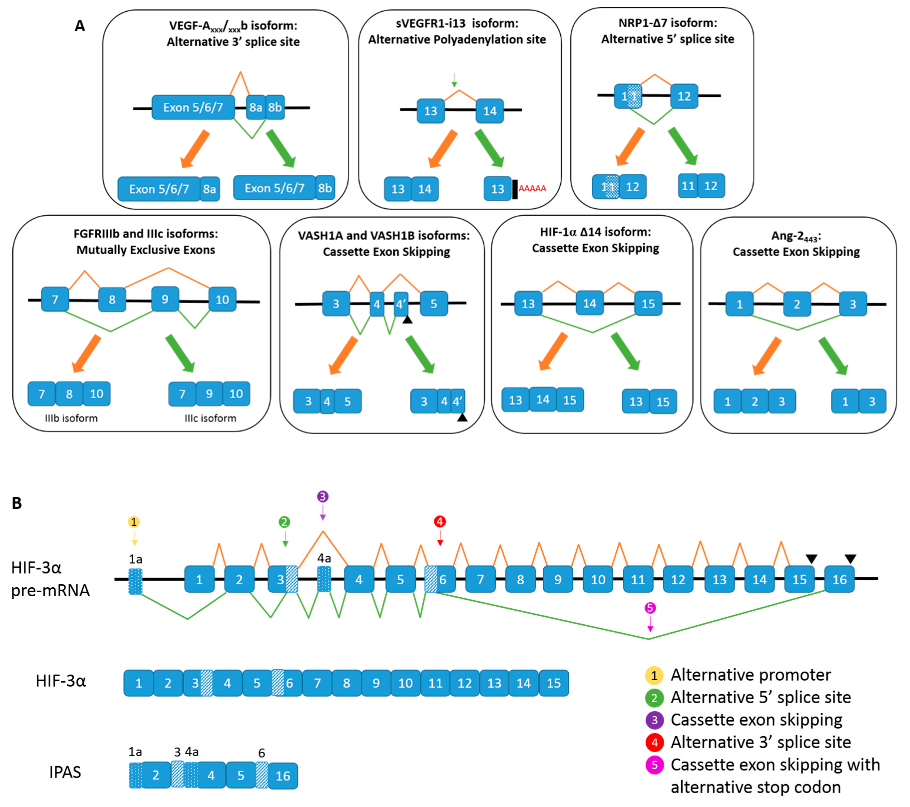
. (B) Alternative spicing of HIF-3α, and the generation of IPAS mRNA.
Ijms 20 02067 g004
Table 1. Splice isoforms of key angiogenic genes and their known functions.
GeneSplice VariantsFunction
VEGF-AVEGF-A111Proangiogenic. Diffusible [33,34]
VEGF-A121Proangiogenic. Diffusible. Implicated in tumorigenesis [33,35,36]
VEGF-A145Proangiogenic. Bind to cell surface and extracellular matrix [33,34]
VEGF-A165Proangiogenic. Moderately diffusible. Implicated in tumorigenesis [33,36]
VEGF-A189Proangiogenic. Implicated in tumorigenesis [33,36,37]
VEGF-A206Proangiogenic. Strongly bind to cell surface and ECM [33,37]
VEGF-A183Proangiogenic [33]
VEGF-AxxxbAnti-angiogenic. Downregulated in cancer, diabetic retinopathy, Denys Drash syndrome, retinal vein occlusion. Upregulated in systemic sclerosis and asthma [33,38]
VEGFR1sVEGFR1Potent anti-angiogenic [39]
VEGFR2sVEGFR2Decreases lymphangiogenesis. Downregulated in neuroblastoma [39]
esVEGFR2Decreases lymphangiogenesis [39]
NRP-1s11NRP1, s12NRP1, sIIINRP1, sIVNRP1Soluble isoform. Antagonists of NRP1 signalling. Anti-angiogenic and anti-tumorigenic [40]
NRP1-ΔE16No functional difference to full length NRP1 [41]
NRP1Δ7Affects glycosylation status of NRP1. Anti-tumorigenic in prostate cancer and breast cancer cells [42]
FGFRsIIIbEMT. Found in epithelial tissues. Evidence as a tumour suppressor and as a tumour promoter [43,44]
IIIcEMT. Found in mesenchymal tissues. Tumourigenic [43]
FGFRαContains autoinhibitory IgI domain which results in a lower affinity for FGFs and decreased signalling compared to FGFRβ [45,46]
FGFRβHigher affinity for FGFs and enhanced signalling. Increases proliferation and linked to tumourigenesis [43,45,46]
Soluble receptorsCan be found in locations in the cell other than the cell membrane. Precise function unknown [43,45]
C1, C2 and C3C3 has the most transforming activity, C2 has moderate transforming activity and C1 has the least transforming activity. C3 implicated in oncogenesis [47]
Deletion of the VT motifPrevention of binding of some effector molecules. Suggested to be unable to activate the downstream Ras/MAPK signalling pathway [48]
Vasohibin-1VASH1A and VASH-1BBoth are anti-angiogenic. VASH-1A promotes normalisation of abnormal tumour blood vessels. VASH-1B prunes vasculature [49,50]
Vasohibin-2355aaPredominantly expressed in HUVECs. Function unknown [51]
290aaAnti-angiogenic activity [52]
311aa, 156aa, 117aa, 104aaFunction unknown
HIF-1αHIF-1αΔ11Promotes tumorigenesis through enhancement of HIF activity [52]
HIF-1αΔ12 and HIF-1αΔ11&12Inhibits dimerisation of HIF-1α and HIF-1β. Act as dominant regulators of HIF-1 transcription [53]
HIF-1αΔ14Less potent activator of HIF-1 transcription than canonical form of HIF-1α [54]
HIF-1α417Amplifies HIF-1β-mediated transcription of EPO gene [53]
HIF-1TAGFunction unknown
HIF-1α Alt1Function unknown
HIF-3αIPASDimerises with HIF-α subunits but cannot initiate transcription of HIF target genes, such as VEGF. Dampens angiogenesis [53,55,56]
HIF-3α4Forms complex with HIF-1α and prevents HIF transcription. Hampers angiogenesis and proliferation [55]
Ang-11.5 kb, 1.3 kbBind to Tie-2 and induce its autophosphorylation [57]
0.9 kb, 0.7 kbBind to Tie-2 but do not induce its autophosphorylation [57]
Ang-2Ang-2BNot precise function but indication of inactivation of the vasculature and vascular remodelling [58]
Ang-2443Antagonist of Tie-2 signalling activation. Suggestive role in the regulation of inflammatory processes [59]

