Article

MicroRNA29a Reverts the Activated Hepatic Stellate Cells in the Regression of Hepatic Fibrosis through Regulation of ATPase H+ Transporting V1 Subunit C1

1
School of Pharmaceutical Science, Jiangnan University, Wuxi 214122, China
2
National Engineering Laboratory for Cereal Fermentation Technology, Jiangnan University, Wuxi 214122, China
3
The Key Laboratory of Industrial Biotechnology, Ministry of Education; School of Biotechnology, Jiangnan University, Wuxi 214122, China
*
Authors to whom correspondence should be addressed.
Int. J. Mol. Sci. 2019, 20(4), 796; https://doi.org/10.3390/ijms20040796
Submission received: 5 January 2019 / Revised: 7 February 2019 / Accepted: 11 February 2019 / Published: 13 February 2019
(This article belongs to the Section Molecular Biology)

Abstract

:
Activated hepatic stellate cells (aHSCs) play a key role in liver fibrosis. During the regression of fibrosis, aHSCs are transformed into inactivated cells (iHSCs), which are quiescent lipid-containing cells and express higher levels of lipid-related genes, such as peroxisome proliferators-activated receptors gamma (PPARγ). Here, we investigated the role of MicroRNA29a (Mir29a) in the resolution of liver fibrosis. Mir29a and lipid-related genes were up-regulated after the recovery of CCl4-induced liver fibrosis in mice. PPARγ agonist rosiglitazone (RSG) promoted de-differentiation of aHSCs to iHSCs and up-regulated MIR29a expression in a human HSC cell line LX-2. MIR29a mimics in vitro promoted the expression of lipid-related genes, while decreased the expression of fibrosis-related genes. MIR29a inhibitor showed the reverse effects. ATPase H+ transporting V1 subunit C1 (Atp6v1c1) was increased in liver fibrosis, while down-regulated after the recovery in mice, and negatively regulated by MIR29a in LX-2 cells. Knockdown of ATP6V1C1 by siRNA decreased alpha-smooth muscle actin (α-SMA) and increased lipid-related genes expression. Simultaneous addition of MIR29a mimics and ATP6V1C1 siRNA further increased RSG promoted expression of lipid-related proteins in vitro. Collectively, MIR29a plays an important role during the trans-differentiation of aHSCs in the resolution of liver fibrosis, in part, through regulation of ATP6V1C1.

1. Introduction

Fibrosis diseases cause serious harm to human health [1], such as pulmonary fibrosis, cirrhosis, glomerulosclerosis, myelofibrosis, and hypertrophic scars [2]. Liver fibrosis is the liver’s wound healing response to a variety of external injuries, which can cause cirrhosis and even hepatocellular carcinoma. However, there is no effective treatment currently [3]. More and more evidence showed that the process of liver fibrosis is reversible after the removal of pathogenic factors [4,5,6]. So, the study of the molecular mechanisms in the resolution of liver fibrosis may be essential for prevention and treatment.
Quiescent hepatic stellate cells (qHSCs) are present in normal liver and store retinoids in large amounts of lipid droplets [7]. Under pathological conditions, qHSCs are activated by cytokines released by immune cells and hepatocytes, and gradually lose their retinoid-filled lipid droplets [8]. At the same time, the synthesis and deposition of extracellular matrix (ECM), including collagen type I (Col1), are increased [9,10]. Genetic tracing studies revealed that HSCs are the main ECM producer during hepatic fibrogenesis [11,12].
Activated hepatic stellate cells (aHSCs) may revert to inactivated HSC (iHSCs) in vivo and in vitro [13,14,15]. iHSCs and qHSCs are both rich in lipid droplets and have lipofibroblast properties. iHSCs highly expressed lipid-related genes, such as peroxisome proliferators-activated receptors gamma (PPARγ), and lowly expressed fibrotic-related genes, such as alpha-smooth muscle actin (α-SMA) and COL1. However, iHSCs can differentiate into myofibroblasts faster than qHSCs in response to fibrogenic stimuli [5,6]. It is well known that PPARγ signaling plays an important role in the trans-differentiation of myofibroblasts to lipofibroblast. Overexpression of PPARγ promotes the de-differentiation of aHSCs into iHSCs in vitro [16]. PPARγ agonist rosiglitazone (RSG) can inhibit ECM production and may serve as potential therapeutics for lung fibrosis and intestinal fibrosis [17,18]. The above studies indicate that the trans-differentiation of myofibroblast to lipofibroblast may play a role in reversing fibrosis.
MicroRNAs (miRNAs) are non-encoding single-stranded RNA molecules of about 22 nucleotides in length encoded by endogenous genes [19]. They are involved in post-transcriptional gene expression regulation in plants and animals [20]. The MIR29 family has been reported to participate in the development of cardiac fibrosis, liver fibrosis, renal fibrosis, and pulmonary fibrosis [21,22,23,24]. MIR29 has three mature members, MIR29a, MIR29b, and MIR29c. MiR29 is significantly under-expressed in human and murine fibrotic liver tissues [25,26]. Overexpression of Mir29 in mouse HSCs leads to a decrease in collagen deposition through direct target ECM production [27]. Patients with advanced cirrhosis have a significantly lower serum level of MIR29a compared with healthy volunteers or patients with early hepatic fibrosis [28]. Mir29a also influences the expression of genes associated with lipid metabolism in mouse C2C12 myoblasts [29,30]. These studies suggest that enhancing MIR29 may be a promising anti-fibrotic therapy. However, the mechanism of action of MIR29a in liver fibrosis remains largely unclear.
Vacuolar adenosine triphosphatase (V-ATPase) has been shown to play an important role in the maintenance of the intracellular pH. V-ATPase is composed of a cytosolic V1 domain and a transmembrane V0 domain. The V1 domain consists of three A subunits, three B subunits, two G subunits plus the C, D, E, F, and H subunits. V-ATPase inhibitor affects the proliferation, activation, and metabolic activity of HSC [31]. ATP6V1C1, one of the main genes regulating V-ATPase activity, has been implicated in the regulation of energy metabolism [32,33]. ATP6V1C1 expression is up-regulated in the activated primary human HSC and co-localized with HSC activation marker α-SMA in human liver tissues of patients with cirrhosis [34].
In this study, we focused on investigating the role of MIR29a in the resolution of liver fibrosis. Mouse liver fibrosis model induced by CCl4 and human HSC cell line LX-2 were used. We hypothesized that MIR29a promotes the resolution of liver fibrosis, and ATP6V1C1 may be a potential target of MIR29a.

2. Results

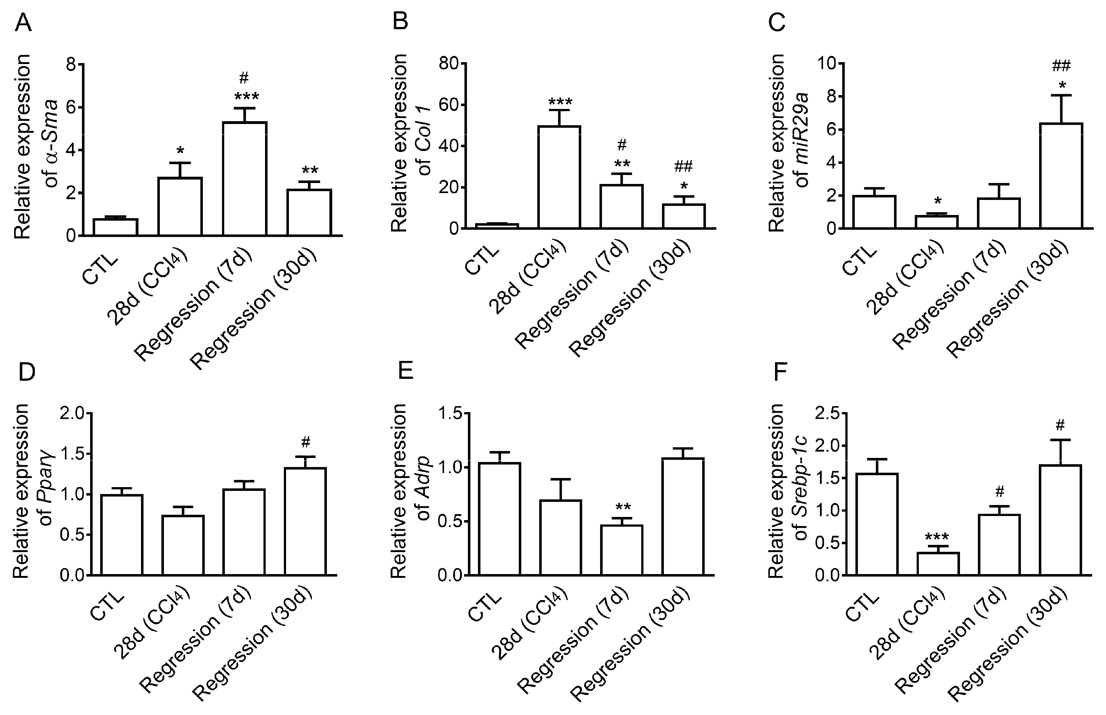
2.1. Expression of Mir29a, Fibrogenic- and Adipogenic-Related Genes in the Progression and Resolution of Liver Fibrosis in Mice

First of all, we established the liver fibrosis model by repetitive CCl4 injection in mice. By quantitative real-time PCR (qRT-PCR), we found that α-Sma and Col1 were significantly up-regulated (Figure 1A,B), while the expression of Mir29a was decreased in liver tissues after 28 days with CCl4 treatment, as compared to controls (CTL) (Figure 1C). Meanwhile, the adipogenic transcription factors Pparγ, adipose differentiation-related protein (Adrp), and sterol regulatory element binding transcription factor 1 (Srebp-1c) were down-regulated in liver tissues after 28 days of CCl4 treatment, as compared to CTL (Figure 1D–F). After 7 days of recovery from CCl4, the expression of Col1 was down-regulated (Figure 1B), while mRNA levels of Mir29a, Pparγ, and Srebp-1c were increased in liver tissues compared with those of the model group (Figure 1C,D,F). The mRNA levels of α-Sma and Col1 were significantly reduced 30 days after recovery (Figure 1A,B), indicating that the degree of liver fibrosis was greatly reduced. The expression of Mir29a was increased (Figure 1C), and mRNA levels of Pparγ, Adrp, and Srebp-1c were similar in the liver after 30 days of recovery compared to the CTL group (Figure 1D–F). These data indicated that Mir29a is negatively correlated with the fibrosis progress and may play a role in the resolution of liver fibrosis.

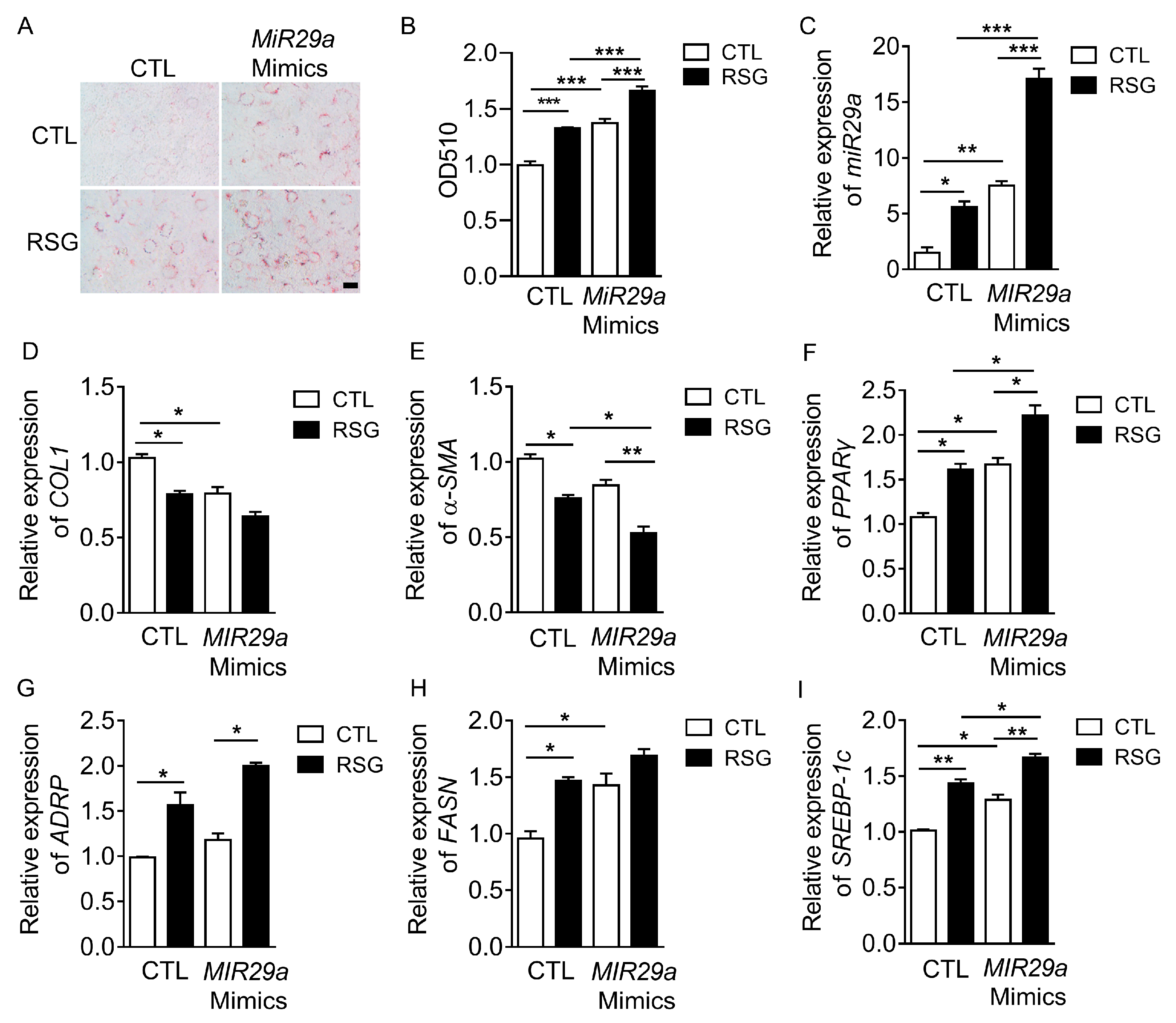
2.2. The Effect of MIR29a on aHSCs Trans-Differentiation to iHSCs

PPARγ plays a key role in the reversal of aHSC to iHSC. We found that PPARγ agonist RSG (1.25–80 μM) showed no significant effect on cell viability (Figure S1A). Treatment with RSG (5 μM) resulted in a dramatic up-regulation of PPARγ expression in LX-2 cells (Figure S1B). RSG up-regulated MIR29a expression in LX-2 cells (Figure S1C). Instead, mRNA levels of both COL1 and α-SMA in LX-2 cells were down-regulated by RSG (Figure S1D–E). We also confirmed that RSG induced the adipogenic related genes (PPARγ, ADRP, FASN, C/EBPα) expression and down-regulated α-SMA expression in protein levels (Figure S1F). Then, to determine whether MIR29a plays a role in the aHSC trans-differentiation, we used MIR29a mimics or MIR29a inhibitor in LX-2 cells treated with RSG. The results of Oil red O staining showed that MIR29a mimics or RSG induced lipid droplets formation in LX-2 cells (Figure 2A,B). The quantitative data revealed that MIR29a mimics further promoted RSG-induced lipid accumulation (Figure 2B). MIR29a expression was higher in LX-2 cells treated with RSG (Figure 2C). MIR29a mimics alone decreased the expression of the fibrosis-related genes COL1 and α-SMA compared to the CTL group (Figure 2D,E). Meanwhile, overexpression of MIR29 with its mimics resulted in 1.7, 1.3, 1.5, and 1.4 fold up-regulation of PPARγ, ADRP, FAS, and SREBP-1c expression in LX-2 cells, respectively (Figure 2F–I). Furthermore, PPARγ, ADRP, FASN, and SREBP-1c were increased all above 1.5 fold upon treatment with both RSG and MIR29a mimics (Figure 2F–I). Conversely, MIR29a inhibitor increased COL1 and αSMA (Figure S2B,C) but decreased PPARγ, ADRP, FASN, and SREBP-1c expression (Figure S2D–G). These results suggested that MIR29a may promote aHSC trans-differentiation.

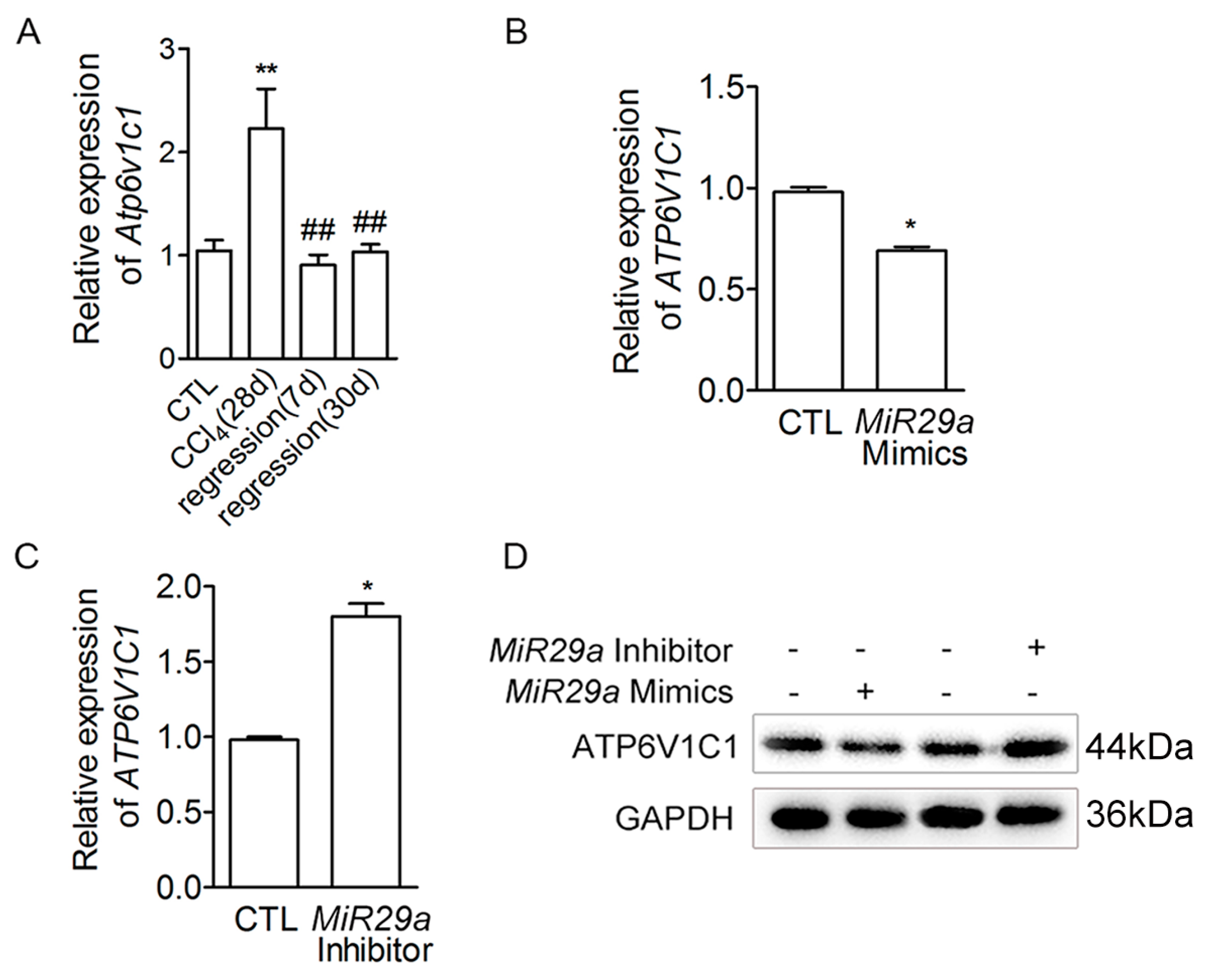
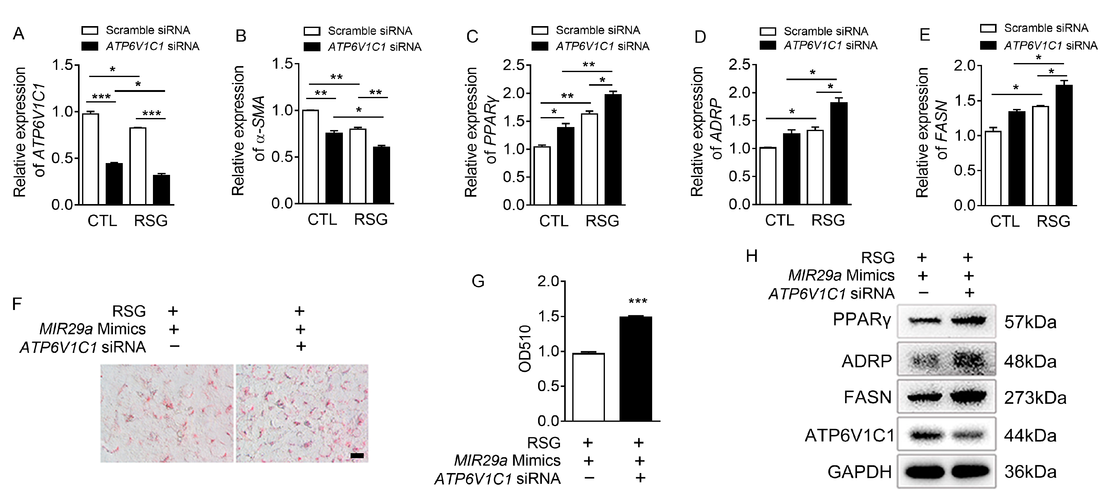
2.3. The Effect of ATP6V1C1 on aHSCs Trans-Differentiation to iHSCs

To investigate the potential new target of MIR29a, we used the bioinformatics analysis and found that ATP6V1C1 might be the potential target of MIR29a [35]. We first examined the expression of Atp6v1c1 in murine liver tissues. The mRNA level of Atp6v1c1 in liver tissues was increased more than two-fold after 28 days with CCl4 treatment and was again similar to that of CTL after fibrosis recovery (Figure 3A). We also found that MIR29a negatively regulated the expression of ATP6V1C1 by qRT-PCR and western blotting analyses (Figure 3B–D).
We then examined whether the reduction of ATP6V1C1 by siRNA can play a role in the HSC trans-differentiation. As expected, ATP6V1C1 siRNA effectively decreased ATP6V1C1 expression relative to a scramble siRNA control in LX-2 cells (Figure 4A). Reduction of ATP6V1C1 by siRNA alone led to the inhibition of α-SMA (Figure 4B) and up-regulation of PPARγ, ADRP, and FASN (Figure 4C–E). In addition, RSG slightly decreased ATP6V1C1 expression (Figure 4A). ATP6V1C1 siRNA further promoted the expression of PPARγ, ADRP, FASN induced by RSG (Figure 4C–E). The results of Oil red O staining showed that knockdown of ATP6V1C1 results in a 1.5 fold up-regulation of MIR29a mimics and RSG-induced lipid differentiation (Figure 4F,G). At the protein level, knockdown of ATP6V1C1 increased the expression of PPARγ, ADRP, and FASN in LX-2 cells treated with MIR29a mimics and RSG (Figure 4H). These data suggested that ATP6V1C1 inhibits the HSC lipogenesis and may be the target of MIR29a in the progress of HSC trans-differentiation.

3. Discussion

This work highlights the importance of MIR29a during the regression of liver fibrosis. MiR29a has been known as a direct target of TGFβ signaling and is down-regulated when liver fibrosis occurs. Overexpressing Mir29a/b markedly reduced the degree of liver fibrosis induced by CCl4 in mice and decreased Collagen expression in LX-2 cells [28,36]. Ectopic expression of Mir29b also blunted the increased expression of α-SMA, caused cell cycle arrest, and induced apoptosis in activated HSCs [37]. Liver-specific Mir29ab1 knockout mice enhanced mortality and fibrosis in response to CCl4 treatment compared with wild-type CTL mice [38]. Consistent with these studies, our study found that Mir29a was down-regulated in CCl4-induced liver fibrosis in mice. We further showed that the expression level of Mir29a in the liver was also recovered and comparable to that of normal mice after 30 days of recovery from CCl4 treatment. In vitro, MIR29a mimics significantly decreased the expression of COL1 and α-SMA in human LX-2 cells.
A treatment of aHSC with ectopic expression of PPARγ induced the expression of a panel of adipogenic transcription factors and caused the phenotypic reversal to qHSC [16]. In vitro study also found that Mir29a promotes lipid production in mouse skeletal muscle cells [29,30]. As expected, we found that RSG increased the expression of PPARγ and induced lipid droplet formation in LX-2 cells. Moreover, our work showed that RSG up-regulated MIR29a expression. Meanwhile, MIR29a promoted lipid droplet formation and up-regulated the expression of PPARγ and several adipogenic transcription factors. These data suggest that MIR29a plays an important role in the process of aHSCs to iHSCs trans-differentiation during liver fibrosis resolution.
The previous study reported that V-ATPase, including ATP6V1C1, was co-localized with HSC activation marker α-SMA in human cirrhotic liver tissues determined by immunohistochemistry analysis, and pharmaceutical inhibition of V-ATPase led to the down-regulation of gene expression of HSC activation markers [34]. In this study, by bioinformatics analysis, we revealed that ATP6V1C1 might be the target of MIR29a [35]. The expression of Atp6v1c1 was negatively correlated with Mir29a in CCl4-induced liver fibrosis in mice as well as in human HSC in vitro. Next, we confirmed that ATP6V1C1 was negatively regulated by Mir29a in LX-2 cells. ATP6V1C1 is a potent negative regulator of HSC trans-differentiation. Knockdown of ATP6V1C1 led to the inhibition of α-SMA, whereas increased the expression of the adipogenic transcription factors, including PPARγ, ADRP, and FASN. ATP6V1C1 siRNA further increased lipid deposition and the expression of lipid-related protein stimulated by MIR29a mimics and RSG. Thus, ATP6V1C1 may be regulated by MIR29a and promote the fibrogenesis while inhibiting the lipogenesis progress during liver fibrosis.

4. Materials and Methods

4.1. Chemicals and Reagents

Carbon tetrachloride (CCl4), olive oil, oil red O, and RSG were from Sigma-Aldrich (St Louis, MO, USA). The mimics and inhibitor of MIR29a were purchased from the Ruibo Company (Guangzhou, China). ATP6V1C1 siRNA and scramble siRNA control were purchased from Santa Cruz Biotechnology (Santa Cruz, CA, USA). The antibodies ATP6V1C1, PPARγ, ADRP, FASN, and GAPDH were purchased from Santa Cruz Biotechnology (Santa Cruz, CA, USA).

4.2. Animal Model of Liver Fibrosis and Treatment

All animal experiments were approved by the Animal Research Committee of Jiangnan University (JN.No 20171115c0400424[72]; 2017.12.24–2018.4.24). Male C57LB/6 at 8 weeks were purchased from Shanghai SLAC Laboratory Animal CO. LTD and were allowed to freely drink tap water and diet (Shanghai SLAC Laboratory Animal Co., Shanghai, China). Liver fibrosis was induced by intraperitoneal (i.p.) injection of 0.5 mL/kg CCl4 (25% solution in olive oil) twice weekly for 28 days [39]. The mouse livers were harvested at 7 and 30 days after CCl4 injury (n = 5 per group).

4.3. Cell Culture, Cell Viability, and Drug Treatment

Human HSCs cell line LX-2 cells were obtained from the cell bank of Xiangya Central Experiment Laboratory of Central South University (Changsha, China). Cells were cultured in DMEM supplemented with 10% FBS (Gibco, Grand Island, NY, USA), 100 U mL−1 penicillin, and 100 mg mL−1 streptomycin in a humidified atmosphere of 5% CO2 at 37 °C. MIR29a mimics (100 nM), MIR29a inhibitor (200 nM), ATP6V1C1 siRNA (60 nM) or scramble siRNA control (60 nM) were transiently transfected into LX-2 cells for 24 h using Lipofectamine RNAi max (Invitrogen, Carlsbad, CA, USA). The cells were then treated with 5 µM RSG for 24 h.

4.4. RNA Isolation and qRT-PCR Analysis

Trizol reagent (Invitrogen, Carlsbad, CA, USA) was used for extracting RNA from mouse liver tissues or cells according to the manufacturer’s protocol. Total RNA (1 mg) was transcribed into cDNA using M-MLV and random hexamer primers (Thermo Fisher Scientific, Waltham, MA, USA). Real-time PCR was performed as previously described [40]. Glycerol phosphate dehydrogenase (GAPDH) was used as an invariant control. Gene expressions were measured relative to the endogenous reference gene GAPDH using the comparative CT method. Sequences of the specific primer sets are as follows: Gapdh (NM-008085), forward, 5’-TTG TCA TGG GAG TGA ACG AGA-3’; reverse, 5’-CAG GCA GTT GGT GGT ACA GG-3’; GAPDH (NM-014364), forward, 5’-TGT GGG CAT CAA TGG ATT TGG-3’; reverse, 5’-ACA CCA TGT ATT CCG GGT CAA T-3’; Col1 (NM-007742), forward, 5’-GCT CCT CTT AGG GGC CAC T-3’; reverse, 5’-ATT GGG GAC CCT TAG GCC AT-3’; COL1 (NM-000088), forward, 5’-GAG GGC CAA GAC GAA GAC ATC-3’; reverse, 5’-CAG ATC ACG TCA TCG CAC AAC-3’; α-Sma (NM-007392), forward, 5’-CCC AGA CAT CAG GGA GTA ATG G-3’; reverse, 5’-TCT ATC GGA TAC TTC AGC GTC A-3’; α-SMA (NM-001613), forward, 5’-GTG TTG CCC CTG AAG AGC AT-3’; reverse, 5’-GCT GGG ACA TTG AAA GTC TCA-3’; Pparγ (NM-001308352), forward, 5’-CTT GGC TGC GCT TAC GAA GA-3’; reverse, 5’-GAA AGC TCG TCC ACG TCA GAC-3’; PPARγ (NM-001172698), forward, 5’-GAT GCC AGC GAC TTT GAC TC-3’; reverse, 5’-ACC CAC GTC ATC TTC AGG GA-3’; Adrp (NM-007408), forward, 5’-CTT GTG TCC TCC GCT TAT GTC-3’; reverse, 5’-GCA GAG GTC ACG GTC TTC AC-3’; ADRP (NM-001122), forward, 5’-ATG GCA TCC GTT GCA GTT GAT-3’; reverse, 5’-GGA CAT GAG GTC ATA CGT GGA G-3’; Srebp-1c (NM-011480), forward, 5’-TGA CCC GGC TAT TCC GTG A-3’; reverse, 5’-CTG GGC TGA GCA ATA CAG TTC-3’; SREBP-1C (NM-001005291), forward, 5’-GCC CCT GTA ACG ACC ACT G-3’; reverse, 5’-CAG CGA GTC TGC CTT GAT G-3’; FASN (NM-012393), forward, 5’-CCC AGT CCT TCA CTT CTA TGT TC-3’; reverse, 5’-GTA GCA CAG TTC AGT CTC GAC-3’. The expression level of mature MIR29a was quantified by TaqMan microRNA assays (Mm04238191_s1, Applied Biosystems, Foster city, CA, USA).

4.5. Western Blot Analysis

Cells were washed with ice-cold Dulbecco’s Phosphate Buffered Saline (Gibco, Grand Island, NY, USA) and resuspended in RIPA buffer with protease inhibitor (Sigma-Aldrich, St.Louis, MO, USA). Protein was resolved by SDS-polyacrylamide gel electrophoresis and transferred to PVDF membranes. After blocking, they were probed with primary antibodies overnight at 4 °C, then incubated with horseradish peroxidase-conjugated secondary antibody for 1 h at room temperature. The bands were visualized using ECL reagents (Thermofisher Scientific, Waltham, MA, USA). The ratio of the relevant protein was subjected to internal control (GAPDH).

4.6. Oil Red O Staining

Oil Red O solution 0.5% in isopropanol was used to stain fat droplets in cells. After removing the staining solution, the dye retained in the cells was eluted into isopropanol, and the optical density (OD) at 510 nm was measured with a microplate reader (Thermo Labsystems, Waltham, MA, USA).

4.7. Statistical Analysis

Data are expressed as means ± SEM. Differences in measured variables between experimental and control groups were assessed by using Student’s test. Differences in multiple groups were assessed by using one-way analysis of variance (ANOVA), and the Tukey test was used for determining the significance. Results were considered statistically significant at p < 0.05. All analyses were conducted in GraphPad Prism (La Jolla, CA, USA).

5. Conclusions

Our study found that Mir29a and the adipogenic transcription factors were up-regulated during the process of fibrosis regression in mice. MIR29a down-regulated the expression of ATP6V1C1, inhibited the activation of HSCs, and positively regulated the adipogenic transcription factors. ATP6V1C1 negatively regulated adipogenic transcription factors. We propose a new mechanism of action of MIR29a in the resolution of liver fibrosis. MIR29a plays an important role during the resolution of liver fibrosis, which may be through the regulation of ATP6V1C1.

Supplementary Materials

Supplementary materials can be found at https://www.mdpi.com/1422-0067/20/4/796/s1.

Author Contributions

Experimental design and interpretation of results, F.J., Y.G., J.-S.S., Z.-H.X., H.-Y.X.; conductance of experiments, F.J. and X.-Y.X.; writing, F.J. and Y.G.

Funding

This work was supported by the grant from the National Natural Science Foundation of China (Grant No. 31771514), China Postdoctoral Science Foundation (Grant No. 2018M640455), National first-class discipline program of Light Industry Technology and Engineering (Grant No. LITE2018-11), the Six talent peaks project in Jiangsu Province (Grant No. 2015-SWYY-006), and Fundamental Research Funds for the Central Universities (Grant No. 1102050205181880, JUSRP21824).

Conflicts of Interest

The authors declare no conflict of interest.

Abbreviations

aHSCsActivated hepatic stellate cells
iHSCsInactivated hepatic stellate cells
qHSCsQuiescent hepatic stellate cells
HSCsHepatic stellate cells
MiRNAsMicroRNAs
PPARγPeroxisome proliferators-activated receptors gamma
ADRPAdipose differentiation-related protein
SREBP-1cSterol regulatory element binding transcription factor 1
FASNFatty acid synthase
ATP6V1C1ATPase H+ transporting V1 subunit C1
CCl4Carbon tetrachloride
RSGRosiglitazone
COL1Collagen type I
α-SMAAlpha-smooth muscle actin
ECMExtracellular matrix
TGF-βTransforming growth factor-β
GAPDHGlycerol phosphate dehydrogenase

References

  1. Kim, Y.; Ejaz, A.; Tayal, A.; Spolverato, G.; Bridges, J.F.; Anders, R.A.; Pawlik, T.M. Temporal trends in population-based death rates associated with chronic liver disease and liver cancer in the United States over the last 30 years. Cancer 2015, 120, 3058–3065. [Google Scholar] [CrossRef] [PubMed]
  2. Mehal, W.Z.; Iredale, J.; Friedman, S.L. Scraping fibrosis: Expressway to the core of fibrosis. Nat. Med. 2011, 17, 552–553. [Google Scholar] [CrossRef] [PubMed] [Green Version]
  3. Bataller, R.; Brenner, D.A. Liver fibrosis. J. Clin. Investig. 2005, 115, 209–218. [Google Scholar] [CrossRef]
  4. Eyraud, D. Clinics and Research in Hepatology and Gastroenterology. Editorial. Clin. Res. Hepatol. Gastroenterol. 2015, 39, 151. [Google Scholar] [CrossRef] [PubMed]
  5. Kisseleva, T.; Cong, M.; Paik, Y.H.; Scholten, D.; Jiang, C.; Benner, C.; Iwaisako, K.; Mooremorris, T.; Scott, B.; Tsukamoto, H.; et al. Myofibroblasts revert to an inactive phenotype during regression of liver fibrosis. Proc. Natl. Acad. Sci. USA 2012, 109, 9448–9453. [Google Scholar] [CrossRef] [PubMed] [Green Version]
  6. Xiao, L.; Xu, J.; Brenner, D.A.; Kisseleva, T. Reversibility of Liver Fibrosis and Inactivation of Fibrogenic Myofibroblasts. Curr. Pathobiol. Rep. 2013, 1, 209–214. [Google Scholar] [Green Version]
  7. Geerts, A. History, heterogeneity, developmental biology, and functions of quiescent hepatic stellate cells. Semin. Liver Dis. 2001, 21, 311–336. [Google Scholar] [CrossRef]
  8. Friedman, S.L. Hepatic Stellate Cells: Protean, Multifunctional, and Enigmatic Cells of the Liver. Physiol. Rev. 2008, 88, 125–172. [Google Scholar] [CrossRef] [Green Version]
  9. Canbay, A.; Higuchi, H.; Bronk, S.F.; Taniai, M.; Sebo, T.J.; Gores, G.J. Fas enhances fibrogenesis in the bile duct ligated mouse: A link between apoptosis and fibrosis. Gastroenterology 2002, 123, 1323–1330. [Google Scholar] [CrossRef]
  10. Magdaleno, F.; Schierwagen, R.; Uschner, F.E.; Trebicka, J. “Tipping” extracellular matrix remodeling towards regression of liver fibrosis: Novel concepts. Minerva. Gastroenterol. Dietol. 2018, 64, 51–61. [Google Scholar]
  11. Mederacke, I.; Hsu, C.C.; Troeger, J.S.; Huebener, P.; Mu, X.; Dapito, D.H.; Pradere, J.P.; Schwabe, R.F. Fate-tracing reveals hepatic stellate cells as dominant contributors to liver fibrosis independent of its etiology. Nat. Commun. 2013, 4, 2823. [Google Scholar] [CrossRef] [PubMed]
  12. Iwaisako, K.; Jiang, C.; Zhang, M.; Cong, M.; Mooremorris, T.J.; Park, T.J.; Liu, X.; Xu, J.; Wang, P.; Paik, Y.H.; et al. Origin of myofibroblasts in the fibrotic liver in mice. Proc. Natl. Acad. Sci. USA 2014, 111, 3297–3305. [Google Scholar] [CrossRef] [PubMed]
  13. Xie, G.; Wang, X.; Wang, L.; Wang, L.; Atkinson, R.D.; Kanel, G.C.; Gaarde, W.A.; Deleve, L.D. Role of differentiation of liver sinusoidal endothelial cells in progression and regression of hepatic fibrosis in rats. Gastroenterology 2012, 142, 918–927. [Google Scholar] [CrossRef] [PubMed]
  14. Iredale, J.P.; Benyon, R.C.; Pickering, J.; Mccullen, M.; Northrop, M.; Pawley, S.; Hovell, C.; Arthur, M.J. Mechanisms of spontaneous resolution of rat liver fibrosis. Hepatic stellate cell apoptosis and reduced hepatic expression of metalloproteinase inhibitors. J. Clin. Investig. 1998, 102, 538–549. [Google Scholar] [CrossRef] [PubMed]
  15. Troeger, J.S.; Mederacke, I.; Gwak, G.Y.; Dapito, D.H.; Mu, X.; Hsu, C.C.; Pradere, J.P.; Friedman, R.A.; Schwabe, R.F. Deactivation of Hepatic Stellate Cells During Liver Fibrosis Resolution in Mice. Gastroenterology 2012, 143, 1073.e22–1083.e22. [Google Scholar] [CrossRef]
  16. She, H.; Xiong, S.; Hazra, S.; Tsukamoto, H. Adipogenic transcriptional regulation of hepatic stellate cells. J. Biol. Chem. 2005, 280, 4959–4967. [Google Scholar] [CrossRef]
  17. El, A.E.; Moiseenko, A.; Kheirollahi, V.; De, L.S.; Crnkovic, S.; Kwapiszewska, G.; Kosanovic, D.; Schwind, F.; Schermuly, R.T.; Henneke, I. Two-Way Conversion between Lipogenic and Myogenic Fibroblastic Phenotypes Marks the Progression and Resolution of Lung Fibrosis. Cell Stem Cell 2016, 20, 261–273. [Google Scholar]
  18. Koo, J.B.; Nam, M.; Jung, Y.; Yoo, J.; Kim, D.H.; Kim, G.; Shin, S.J.; Lee, K.M.; Hahm, K.B.; Kim, J.W.; et al. Anti-fibrogenic effect of PPAR-γ agonists in human intestinal myofibroblasts. BMC Gastroenterol. 2017, 17, 73. [Google Scholar] [CrossRef] [Green Version]
  19. Kim, V.N. MicroRNA biogenesis: Coordinated cropping and dicing. Nat. Rev. Mol. Cell Biol. 2005, 6, 376–385. [Google Scholar] [CrossRef]
  20. Bartel, D.P. MicroRNAs: Target Recognition and Regulatory Functions. Cell 2009, 136, 215–233. [Google Scholar] [CrossRef] [Green Version]
  21. Rooij, E.V.; Sutherland, L.B.; Thatcher, J.E.; Dimaio, J.M.; Naseem, R.H.; Marshall, W.S.; Hill, J.A.; Olson, E.N. Dysregulation of microRNAs after Myocardial Infarction Reveals a Role of miR-29 in Cardiac Fibrosis. Proc. Natl. Acad. Sci. USA 2008, 105, 13027–13032. [Google Scholar] [CrossRef] [PubMed]
  22. Xie, T.; Liang, J.; Geng, Y.; Liu, N.; Kurkciyan, A.; Kulur, V.; Leng, D.; Deng, N.; Liu, Z.; Song, J.; et al. MicroRNA-29c Prevents Pulmonary Fibrosis by Regulating Epithelial Cell Renewal and Apoptosis. Am. J. Respir. Cell Mol. Biol. 2017, 57, 721–732. [Google Scholar] [CrossRef] [PubMed]
  23. Kriegel, A.J.; Liu, Y.; Fang, Y.; Ding, X.; Liang, M. The miR-29 family: Genomics, cell biology, and relevance to renal and cardiovascular injury. Physiol. Genom. 2012, 44, 237–244. [Google Scholar] [CrossRef]
  24. He, Y.; Huang, C.; Lin, X.; Li, J. MicroRNA-29 family, a crucial therapeutic target for fbrosis diseases. Biochimie 2013, 95, 1355–1359. [Google Scholar] [CrossRef] [PubMed]
  25. Zhang, Y.; Wu, L.; Wang, Y.; Zhang, M.; Li, L.; Zhu, D.; Li, X.; Gu, H.; Zhang, C.Y.; Zen, K. Protective role of estrogen-induced miRNA-29 expression in carbon tetrachloride-induced mouse liver injury. J. Biol. Chem. 2012, 287, 14851–14862. [Google Scholar] [CrossRef] [PubMed]
  26. Knabel, M.K.; Ramachandran, K.; Karhadkar, S.; Hwang, H.W.; Creamer, T.J.; Chivukula, R.R.; Sheikh, F.; Clark, K.R.; Torbenson, M.; Montgomery, R.A.; et al. Systemic Delivery of scAAV8-Encoded MiR-29a Ameliorates Hepatic Fibrosis in Carbon Tetrachloride-Treated Mice. PLoS ONE 2015, 10, e0124411. [Google Scholar] [CrossRef]
  27. Bandyopadhyay, S.; Friedman, R.C.; Marquez, R.T.; Keck, K.; Kong, B.; Icardi, M.S.; Brown, K.E.; Burge, C.B.; Schmidt, W.N.; Wang, Y.; et al. Hepatitis C Virus Infection and Hepatic Stellate Cell Activation Downregulate miR-29: MiR-29 Overexpression Reduces Hepatitis C Viral Abundance in Culture. J. Infect. Dis. 2011, 203, 1753–1762. [Google Scholar] [CrossRef]
  28. Roderburg, C.; Urban, G.W.; Bettermann, K.; Vucur, M.; Zimmermann, H.; Schmidt, S.; Janssen, J.; Koppe, C.; Knolle, P.; Castoldi, M.; et al. Micro-RNA profiling reveals a role for miR-29 in human and murine liver fibrosis. Hepatology 2011, 53, 209–218. [Google Scholar] [CrossRef]
  29. Zhou, Y.; Gu, P.; Shi, W.; Li, J.; Hao, Q.; Cao, X.; Lu, Q.; Zeng, Y. MicroRNA-29a induces insulin resistance by targeting PPARδ in skeletal muscle cells. Int. J. Mol. Med. 2016, 37, 931–938. [Google Scholar] [CrossRef] [Green Version]
  30. Wu, P.; Wang, Q.; Jiang, C.; Chen, C.; Liu, Y.; Chen, Y.; Zeng, Y. MicroRNA-29a is involved lipid metabolism dysfunction and insulin resistance in C2C12 myotubes by targeting PPARδ. Mol. Med. Rep. 2018, 17, 8493–8501. [Google Scholar] [CrossRef]
  31. Marrone, G.; De Chiara, F.; Bottcher, K.; Levi, A.; Dhar, D.; Longato, L.; Mazza, G.; Zhang, Z.; Marrali, M.; Fernándeziglesias, A.; et al. The adenosine monophosphate–activated protein kinase—Vacuolar adenosine triphosphatase–pH axis: A key regulator of the profibrogenic phenotype of human hepatic stellate cells. Hepatology 2018, 68, 1140–1153. [Google Scholar] [CrossRef] [PubMed]
  32. Zhao, J.; Benlekbir, S.; Rubinstein, J.L. Electron cryomicroscopy observation of rotational states in a eukaryotic V-ATPase. Nature 2015, 521, 241–245. [Google Scholar] [CrossRef] [PubMed]
  33. Forgac, M. Vacuolar ATPases: Rotary proton pumps in physiology and pathophysiology. Nat. Rev. Mol. Cell Biol. 2007, 8, 917–929. [Google Scholar] [CrossRef] [PubMed]
  34. Sagnelli, E.; Potenza, N.; Onorato, L.; Sagnelli, C.; Coppola, N.; Russo, A. Micro-RNAs in hepatitis B virus-related chronic liver diseases and hepatocellular carcinoma. World J. Hepatol. 2018, 10, 558–570. [Google Scholar] [CrossRef] [PubMed]
  35. Agarwal, V.; Bell, G.W.; Nam, J.W.; Bartel, D.P. Predicting effective microRNA target sites in mammalian mRNAs. eLife 2015, 4, e05005. [Google Scholar] [CrossRef] [PubMed]
  36. Bian, E.B.; Li, J.; Zhao, B. miR-29, a potential therapeutic target for liver fibrosis. Gene 2014, 544, 259–260. [Google Scholar] [CrossRef] [PubMed]
  37. Wang, J.; Chu, E.S.; Chen, H.Y.; Man, K.; Go, M.Y.; Huang, X.R.; Lan, H.Y.; Sung, J.J.; Yu, J. microRNA-29b prevents liver fibrosis by attenuating hepatic stellate cell activation and inducing apoptosis through targeting PI3K/AKT pathway. Oncotarget 2014, 6, 7325–7338. [Google Scholar] [CrossRef] [PubMed] [Green Version]
  38. Kogure, T.; Costinean, S.; Yan, I.; Braconi, C.; Croce, C.; Patel, T. Hepatic miR-29ab1 expression modulates chronic hepatic injury. J. Cell. Mol. Med. 2012, 16, 2647–2654. [Google Scholar] [CrossRef] [PubMed]
  39. Iredale, J.P. Models of liver fibrosis: Exploring the dynamic nature of inflammation and repair in a solid organ. J. Clin. Investig. 2007, 117, 539–548. [Google Scholar] [CrossRef]
  40. Geng, Y.; Dong, Y.Y.; Yu, M.Y.; Zhang, L.; Yan, X.H.; Sun, J.X.; Qiao, L.; Geng, H.X.; Nakajima, M.; Furuichi, T.; et al. Follistatin-like 1 (Fstl1) is a bone morphogenetic protein (BMP) 4 signaling antagonist in controlling mouse lung development. Proc. Natl. Acad. Sci. USA 2011, 108, 7058–7063. [Google Scholar] [CrossRef] [Green Version]
Figure 1. Expression of Mir29a and adipogenic-related genes expression increased, fibrogenic-related genes expression decreased in the resolution of liver fibrosis in mice. (AF) Mice and littermate control mice were treated with oil or CCl4 for 28 days, and liver tissues were harvested for the following analyses at the indicated time points. qRT-PCR analysis of α-Sma, Col1, Mir29a, Pparγ, Adrp, and Srebp-1c mRNA expression (n = 5 per group). Error bar represents SEM. * p < 0.05, ** p < 0.01, and *** p < 0.001 vs. oil group. # p < 0.05, ## p < 0.01 vs. CCl4 group.
Ijms 20 00796 g001
Figure 2. MIR29a mimics decreased the expression of fibrosis-related genes and increased the expression of adipogenic transcription factors. (A,B) MIR29a mimics (100 nM) were transiently transfected into LX-2 cells for 48 h, and the cells were treated with RSG (5 µM) for 24 h. Conventional Oil red O staining was conducted. After removing the staining solution, the dye retained in the cells was eluted into isopropanol, and OD510 was determined. Scale bar: 10 µm. (C–I) The expression of MIR29a, COL1, α-SMA, PPARγ, ADRP, FASN, and SREBP-1c mRNA were assessed by qRT-PCR (n = 3 per group). Error bar represents SEM. * p < 0.05, ** p < 0.01, and *** p < 0.001.
Ijms 20 00796 g002
Figure 3. The expression of ATP6V1C1 in cells and CCl4-induced mice. (A) Mice and littermate control mice were treated with oil or CCl4 for 28 days, and liver tissues were harvested for the following analyses at the indicated time points. qRT-PCR analysis of Atp6v1c1 mRNA expression (n = 5 per group). Error bar represents SEM. ** p < 0.01 vs. CTL group. ## p < 0.01 vs. CCl4 (28d) group. (B,C) MIR29a mimics (100 nM) or MIR29a inhibitor (200 nM) were transiently transfected into LX-2 cells for 24 h, and the cells were treated with RSG (5 µM) for 24 h. The expression of ATP6V1C1 mRNA was assessed by qRT-PCR (n = 3 per group). Error bar represents SEM. * p < 0.05. (D) Protein expression levels of ATP6V1C1 in cell extracts were assessed by Western blot. GAPDH was used as a loading control.
Ijms 20 00796 g003
Figure 4. Knockdown of ATP6V1C1 attenuated the activation of LX-2 cells and up-regulated the expression of the adipogenic transcription factors. (AE) ATP6V1C1 siRNA (60 nM) transfection was performed on LX-2 cells using Lipofectamine RNAimax. The gene expressions of ATP6V1C1, α-SMA, PPARγ, ADRP, and FASN mRNA were assessed by qRT-PCR (n = 3 per group). (F,G) MIR29a mimics (100 nM) and ATP6V1C1 siRNA (60 nM) were transiently transfected into LX-2 cells for 24 h, and the cells were treated with RSG (5 µM) for 24 h. Conventional Oil red O staining was conducted. After removing the staining solution, the dye retained in the cells was eluted into isopropanol, and OD510 was determined. Scale bar: 10 μm. (H) Protein expression levels of PPARγ, ADRP, FASN, and ATP6V1C1 in cell extracts were assessed by Western blot. GAPDH was used as a loading control. Throughout, the error bar represents SEM. * p < 0.05, ** p < 0.01, and *** p < 0.001.
Ijms 20 00796 g004

