Decreased Activity of Blood Acid Sphingomyelinase in the Course of Multiple Myeloma

 ,
, 
 , and
, and
Abstract
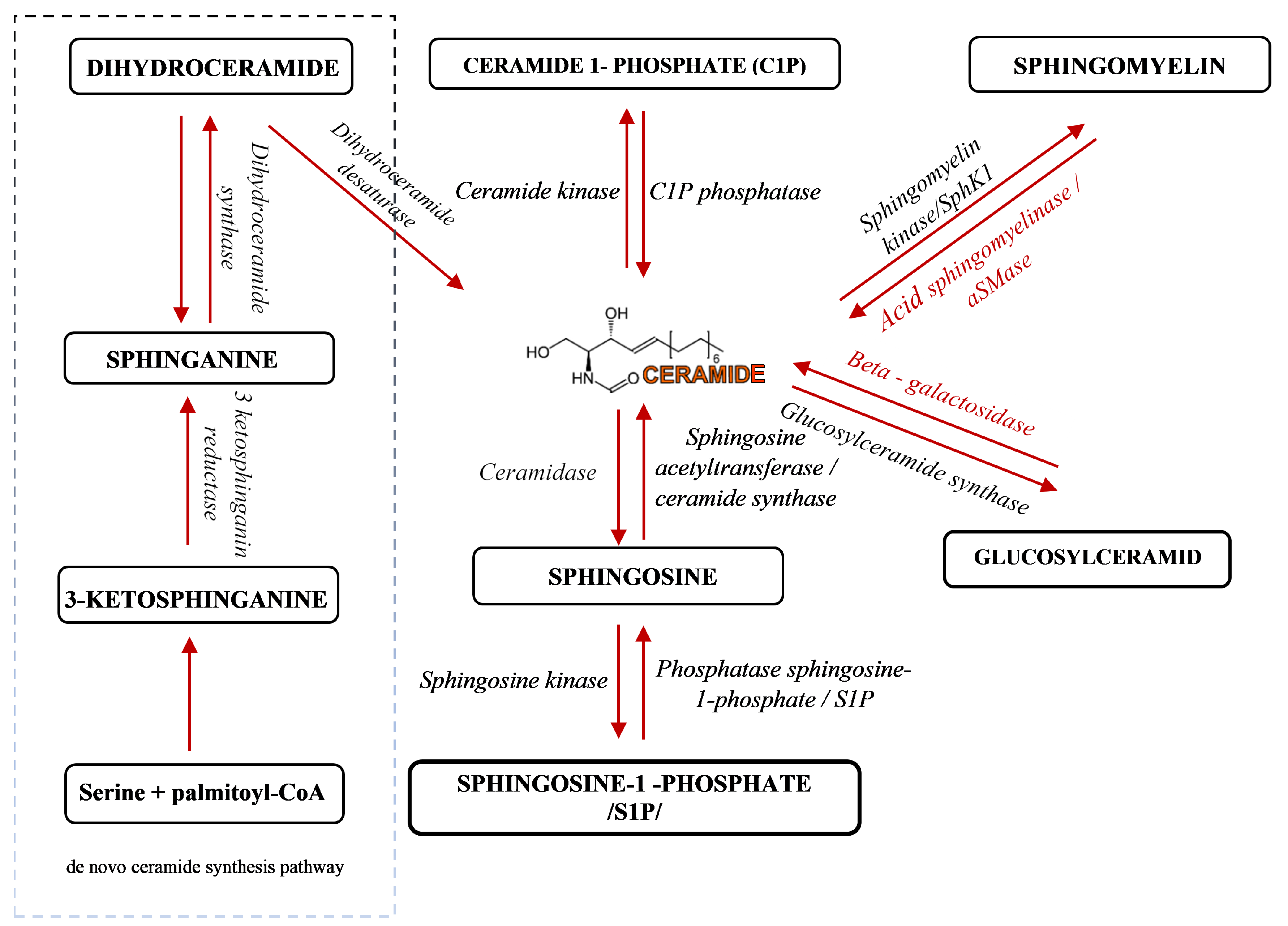
1. Introduction
2. Results
2.1. Clinical Characteristics of the Patient Group
2.2. Outline of Beta Glucosidase and Beta Galactosidase Activity
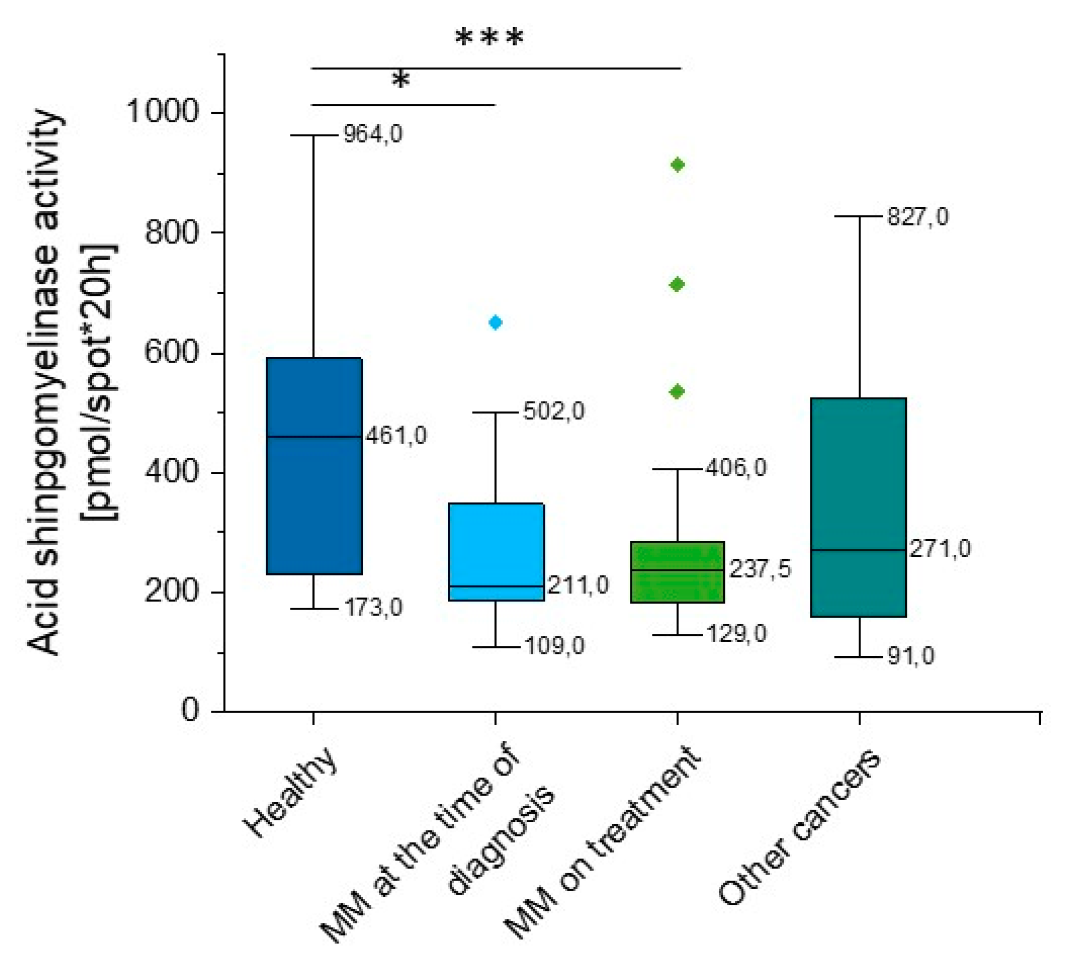
2.3. Outline of Acid Sphingomyelinase (aSMase) Activity
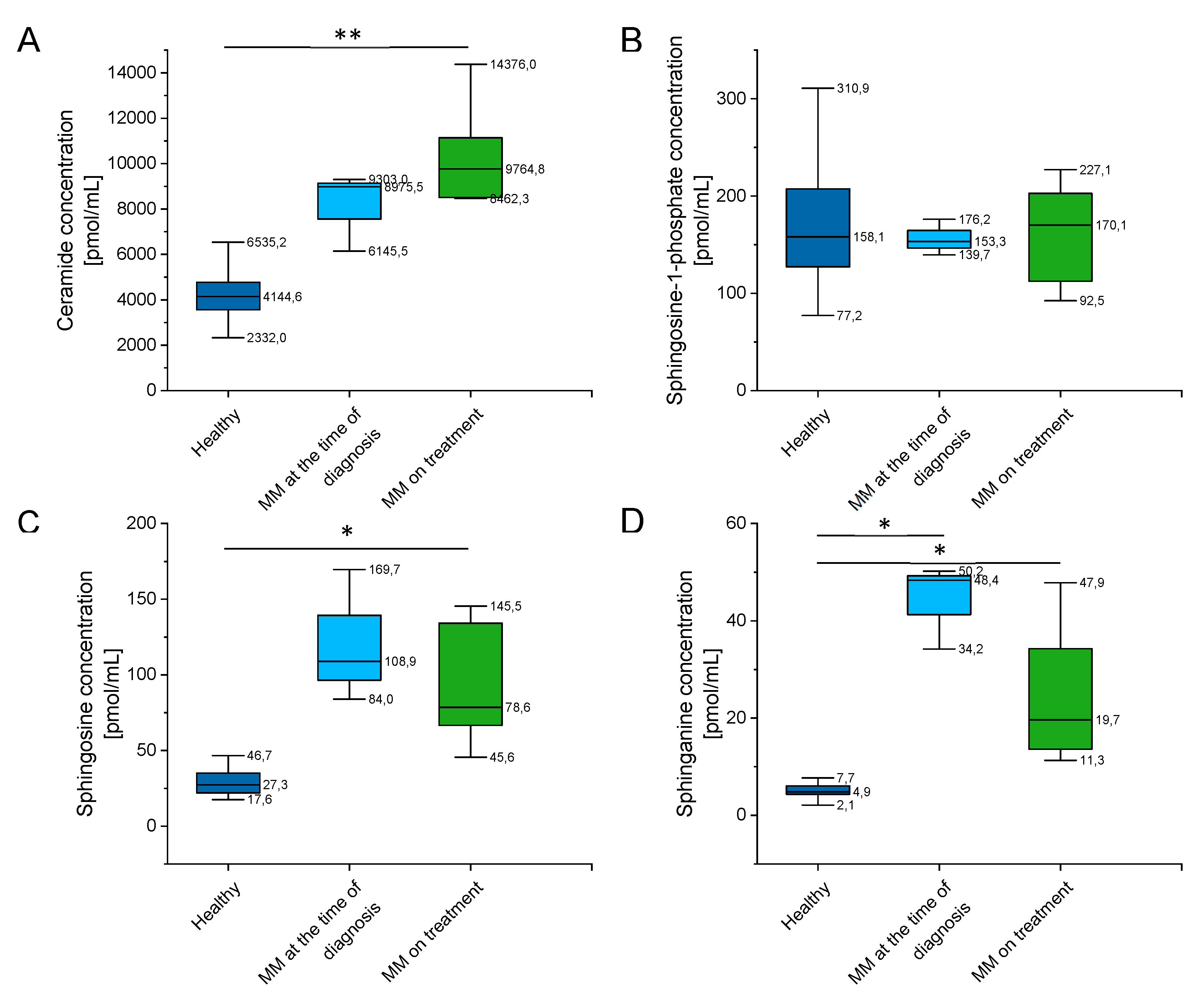
2.4. Outline of Ceramide, Sphingosine-1-Phosphate (S1P), Sphingosine (SFO), and Sphinganine (SFA) Concentration
2.5. Outline Enzyme Activity Depending on the International Staging System (ISS) for Multiple Myeloma (MM)
3. Discussion
4. Materials and Methods
4.1. Acquiring Blood Samples
4.2. Blood Drop Test
4.3. Colorimetric Evaluation of aSMase Activity
4.4. Assessment of Phospholipids Serum Levels
4.5. Statistical Analysis
5. Conclusions
Supplementary Materials
Author Contributions
Funding
Conflicts of Interest
Abbreviations
| SM | Sphingomyeline |
| aSMase | Acid sphingomyelinase |
| Cer | Ceramide |
| Sph | Sphingosine |
| CerS | Ceramide synthase |
References
- Walker, B.A.; Mavrommatis, K.; Wardell, C.P.; Ashby, T.C.; Bauer, M.; Davies, F.E.; Rosenthal, A.; Wang, H.; Qu, P.; Hoering, A.; et al. Identification of novel mutational drivers reveals oncogene dependencies in multiple myeloma. Blood 2018, 132, 587–597. [Google Scholar] [CrossRef] [PubMed]
- Rasche, L.; Weinhold, N. Pathogenesis of multiple myeloma. Der Internist 2019, 60, 3–9. [Google Scholar] [CrossRef] [PubMed]
- Tsukamoto, S.; Huang, Y.; Kumazoe, M.; Lesnick, C.; Yamada, S.; Ueda, N.; Suzuki, T.; Yamashita, S.; Kim, Y.H.; Fujimura, Y.; et al. Sphingosine Kinase-1 Protects Multiple Myeloma from Apoptosis Driven by Cancer-Specific Inhibition of RTKs. Mol. Cancer Ther. 2015, 14, 2303–2312. [Google Scholar] [CrossRef] [PubMed]
- Watek, M.; Piktel, E.; Wollny, T.; Durnas, B.; Fiedoruk, K.; Lech-Maranda, E.; Bucki, R. Defective Sphingolipids Metabolism and Tumor Associated Macrophages as the Possible Links Between Gaucher Disease and Blood Cancer Development. Int. J. Mol. Sci. 2019, 20, 843. [Google Scholar] [CrossRef] [PubMed]
- Wollny, T.; Watek, M.; Durnas, B.; Niemirowicz, K.; Piktel, E.; Zendzian-Piotrowska, M.; Gozdz, S.; Bucki, R. Sphingosine-1-Phosphate Metabolism and Its Role in the Development of Inflammatory Bowel Disease. Int. J. Mol. Sci. 2017, 18, 741. [Google Scholar] [CrossRef] [PubMed]
- Wątek, M.; Durnaś, B.; Wollny, T.; Żendzian-Piotrowska, M.; Pasiarski, M.; Góźdź, S. Rola i potencjał terapeutyczny sfingolipidowego szlaku sygnalizacyjnego w nowotworach hematologicznych Role and therapeutic potential of sphingolipids signaling in hematological malignances. Hematologia 2018, 9, 318–329. [Google Scholar] [CrossRef]
- Durnaś, B.; Fiedoruk, K.; Cieśluk, M.; Deptuła, P.; Król, G.; Piktel, E.; Savage, P.B.; Bucki, R. Lizozym nasila bakteriobójczą aktywność cerageniny CSA-13 w stosunku do Bacillus subtilis. Med. Stud. Studia Med. 2019, 35, 1–9. [Google Scholar] [CrossRef]
- Kurek, K.; Piotrowska, D.M.; Wiesiolek-Kurek, P.; Chabowska, A.; Lukaszuk, B.; Zendzian-Piotrowska, M. The role of sphingolipids in selected cardiovascular diseases. Postepy Hig. Med. Dosw. 2013, 67, 1018–1026. [Google Scholar] [CrossRef]
- Furuya, H.; Shimizu, Y.; Kawamori, T. Sphingolipids in cancer. Cancer Metastasis Rev. 2011, 30, 567–576. [Google Scholar] [CrossRef]
- Clarke, C.J.; Wu, B.X.; Hannun, Y.A. The neutral sphingomyelinase family: Identifying biochemical connections. Adv. Enzym. Regul. 2011, 51, 51–58. [Google Scholar] [CrossRef]
- Hammad, S.M. Blood sphingolipids in homeostasis and pathobiology. Adv. Exp. Med. Biol. 2011, 721, 57–66. [Google Scholar] [CrossRef] [PubMed]
- Wątek, M.; Durnaś, B.; Wollny, T.; Pasiarski, M.; Góźdź, S.; Marzec, M.; Chabowska, A.; Wolak, P.; Żendzian-Piotrowska, M.; Bucki, R. Unexpected profile of sphingolipid contents in blood and bone marrow plasma collected from patients diagnosed with acute myeloid leukemia. Lipids Health Dis. 2017, 16, 235. [Google Scholar] [CrossRef] [PubMed]
- Astudillo, L.; Therville, N.; Colacios, C.; Segui, B.; Andrieu-Abadie, N.; Levade, T. Glucosylceramidases and malignancies in mammals. Biochimie 2016, 125, 267–280. [Google Scholar] [CrossRef] [PubMed]
- Mistry, P.K.; Taddei, T.; vom Dahl, S.; Rosenbloom, B.E. Gaucher disease and malignancy: A model for cancer pathogenesis in an inborn error of metabolism. Crit. Rev. Oncog. 2013, 18, 235–246. [Google Scholar] [CrossRef] [PubMed]
- De Fost, M.; Vom Dahl, S.; Weverling, G.J.; Brill, N.; Brett, S.; Haussinger, D.; Hollak, C.E. Increased incidence of cancer in adult Gaucher disease in Western Europe. Blood Cells Mol. Dis. 2006, 36, 53–58. [Google Scholar] [CrossRef] [PubMed]
- Nair, S.; Branagan, A.R.; Liu, J.; Boddupalli, C.S.; Mistry, P.K.; Dhodapkar, M.V. Clonal Immunoglobulin against Lysolipids in the Origin of Myeloma. N. Engl. J. Med. 2016, 374, 555–561. [Google Scholar] [CrossRef] [PubMed]
- Lidove, O.; Belmatoug, N.; Froissart, R.; Lavigne, C.; Durieu, I.; Mazodier, K.; Serratrice, C.; Douillard, C.; Goizet, C.; Cathebras, P.; et al. Déficit en sphingomyélinase acide (maladie de Niemann-Pick B): Une étude rétrospective multicentrique de 28 patients adultes. La Rev. Med. Interne 2017, 38, 291–299. [Google Scholar] [CrossRef]
- Mühle, C.; Weinland, C.; Gulbins, E.; Lenz, B.; Kornhuber, J. Peripheral Acid Sphingomyelinase Activity Is Associated with Biomarkers and Phenotypes of Alcohol Use and Dependence in Patients and Healthy Controls. Int. J. Mol. Sci. 2018, 19, 4028. [Google Scholar] [CrossRef]
- Savic, R.; Schuchman, E.H. Use of acid sphingomyelinase for cancer therapy. Adv. Cancer Res. 2013, 117, 91–115. [Google Scholar] [CrossRef]
- Lee, R.E. The pathology of Gaucher disease. Prog. Clin. Biol. Res. 1982, 95, 177–217. [Google Scholar]
- Xiong, X.; Lee, C.F.; Li, W.; Yu, J.; Zhu, L.; Kim, Y.; Zhang, H.; Sun, H. Acid Sphingomyelinase regulates the localization and trafficking of palmitoylated proteins. Biol. Open 2019. [Google Scholar] [CrossRef] [PubMed]
- Zhu, L.; Xiong, X.; Kim, Y.; Okada, N.; Lu, F.; Zhang, H.; Sun, H. Acid sphingomyelinase is required for cell surface presentation of Met receptor tyrosine kinase in cancer cells. J. Cell Sci. 2016, 129, 4238–4251. [Google Scholar] [CrossRef] [PubMed]
- Tirodkar, T.S.; Voelkel-Johnson, C. Sphingolipids in apoptosis. Exp. Oncol. 2012, 34, 231–242. [Google Scholar] [PubMed]
- Cieśluk, M.; Piktel, E.; Wątek, M.; Durnaś, B.; Wollny, T.; Król, G.; Bucki, R. Zewnątrzkomórkowe pułapki neutrofilowe jako główne źródło eDNA. Med. Stud. Studia Med. 2017, 33, 137–145. [Google Scholar] [CrossRef]
- Smith, E.L.; Schuchman, E.H. The unexpected role of acid sphingomyelinase in cell death and the pathophysiology of common diseases. FASEB J. 2008, 22, 3419–3431. [Google Scholar] [CrossRef] [PubMed]
- Lin, T.; Genestier, L.; Pinkoski, M.J.; Castro, A.; Nicholas, S.; Mogil, R.; Paris, F.; Fuks, Z.; Schuchman, E.H.; Kolesnick, R.N.; et al. Role of acidic sphingomyelinase in Fas/CD95-mediated cell death. J. Biol. Chem. 2000, 275, 8657–8663. [Google Scholar] [CrossRef]
- Ogretmen, B. Sphingolipid metabolism in cancer signalling and therapy. Nat. Rev. Cancer 2018, 18, 33–50. [Google Scholar] [CrossRef]
- Hannun, Y.A.; Obeid, L.M. Principles of bioactive lipid signalling: Lessons from sphingolipids. Nat. Rev. Mol. Cell Biol. 2008, 9, 139–150. [Google Scholar] [CrossRef]
- Rajkumar, S.V. Multiple myeloma: 2018 update on diagnosis, risk-stratification, and management. Am. J. Hematol. 2018, 93, 981–1114. [Google Scholar] [CrossRef]
- Kyle, R.A.; Rajkumar, S.V. Treatment of multiple myeloma: A comprehensive review. Clin. Lymphoma Myeloma 2009, 9, 278–288. [Google Scholar] [CrossRef]
- Wnorowska, U.; Wątek, M.; Durnaś, B.; Głuszek, K.; Piktel, E.; Niemirowicz, K.; Bucki, R. Zewnątrzkomórkowy DNA jako istotny składnik oraz cel terapeutyczny biofilmu bakteryjnego. Med. Stud. Studia Med. 2015, 31, 132–138. [Google Scholar] [CrossRef]
- Fu, D.; Li, Y.; Li, J.; Shi, X.; Yang, R.; Zhong, Y.; Wang, H.; Liao, A. The effect of S1P receptor signaling pathway on the survival and drug resistance in multiple myeloma cells. Mol. Cell. Biochem. 2017, 424, 185–193. [Google Scholar] [CrossRef] [PubMed]
- Ricci, C.; Onida, F.; Ghidoni, R. Sphingolipid players in the leukemia arena. Biochim. Biophys. Acta 2006, 1758, 2121–2132. [Google Scholar] [CrossRef] [PubMed]
- Tsukamoto, S.; Hirotsu, K.; Kumazoe, M.; Goto, Y.; Sugihara, K.; Suda, T.; Tsurudome, Y.; Suzuki, T.; Yamashita, S.; Kim, Y.; et al. Green tea polyphenol EGCG induces lipid-raft clustering and apoptotic cell death by activating protein kinase Cdelta and acid sphingomyelinase through a 67 kDa laminin receptor in multiple myeloma cells. Biochem. J. 2012, 443, 525–534. [Google Scholar] [CrossRef] [PubMed]
- Venkata, J.K.; Stuart, R.K.; Costa, L.J.; An, N.; Cai, H.; Coker, W.J.; Song, J.; Gibbs, K.; Matson, T.; Smith, C.D.; et al. Sphingolipids as a Novel Target For The Treatment Of Multiple Myeloma. Blood 2013, 122, 3163. [Google Scholar] [CrossRef]
- Torti, L.; Pulini, S.; Morelli, A.M.; Bacci, F.; Di Bartolomeo, P. New Myeloma Diagnostic Criteria: To Treat or Not to Treat? Monocentric Experience of 220 Newly Multiple Myeloma Diagnosed Patients Retrospectively Analyzed. Blood 2016, 128, 5629. [Google Scholar] [CrossRef]
- Zhang, X.K.; Elbin, C.S.; Turecek, F.; Scott, R.; Chuang, W.L.; Keutzer, J.M.; Gelb, M. Multiplex lysosomal enzyme activity assay on dried blood spots using tandem mass spectrometry. Methods Mol. Biol. 2010, 603, 339–350. [Google Scholar] [CrossRef]
- Metz, T.F.; Mechtler, T.P.; Orsini, J.J.; Martin, M.; Shushan, B.; Herman, J.L.; Ratschmann, R.; Item, C.B.; Streubel, B.; Herkner, K.R.; et al. Simplified newborn screening protocol for lysosomal storage disorders. Clin. Chem. 2011, 57, 1286–1294. [Google Scholar] [CrossRef]
- Baranowski, M.; Charmas, M.; Długołęcka, B.; Górski, J. Exercise increases plasma levels of sphingoid base-1 phosphates in humans. Acta Physiol. 2011, 203, 373–380. [Google Scholar] [CrossRef]


| Group | Patients | Median Age | M:F |
|---|---|---|---|
| Healthy | 17 | 52.58 (29–79) | 12:5 |
| MM at the time of diagnosis | 14 | 65.5 (42–86) | 8:6 |
| MM on treatment | 55 | 65.13 (42–88) | 24:31 |
| Other cancers * | 14 | 54.28 (28–76) | 9:5 |
© 2019 by the authors. Licensee MDPI, Basel, Switzerland. This article is an open access article distributed under the terms and conditions of the Creative Commons Attribution (CC BY) license (http://creativecommons.org/licenses/by/4.0/).
Share and Cite
Wątek, M.; Piktel, E.; Barankiewicz, J.; Sierlecka, E.; Kościołek-Zgódka, S.; Chabowska, A.; Suprewicz, Ł.; Wolak, P.; Durnaś, B.; Bucki, R.; et al. Decreased Activity of Blood Acid Sphingomyelinase in the Course of Multiple Myeloma. Int. J. Mol. Sci. 2019, 20, 6048. https://doi.org/10.3390/ijms20236048
Wątek M, Piktel E, Barankiewicz J, Sierlecka E, Kościołek-Zgódka S, Chabowska A, Suprewicz Ł, Wolak P, Durnaś B, Bucki R, et al. Decreased Activity of Blood Acid Sphingomyelinase in the Course of Multiple Myeloma. International Journal of Molecular Sciences. 2019; 20(23):6048. https://doi.org/10.3390/ijms20236048
Chicago/Turabian StyleWątek, Marzena, Ewelina Piktel, Joanna Barankiewicz, Ewa Sierlecka, Sylwia Kościołek-Zgódka, Anna Chabowska, Łukasz Suprewicz, Przemysław Wolak, Bonita Durnaś, Robert Bucki, and et al. 2019. "Decreased Activity of Blood Acid Sphingomyelinase in the Course of Multiple Myeloma" International Journal of Molecular Sciences 20, no. 23: 6048. https://doi.org/10.3390/ijms20236048
APA StyleWątek, M., Piktel, E., Barankiewicz, J., Sierlecka, E., Kościołek-Zgódka, S., Chabowska, A., Suprewicz, Ł., Wolak, P., Durnaś, B., Bucki, R., & Lech-Marańda, E. (2019). Decreased Activity of Blood Acid Sphingomyelinase in the Course of Multiple Myeloma. International Journal of Molecular Sciences, 20(23), 6048. https://doi.org/10.3390/ijms20236048