Article

Proteomic Investigation of S-Nitrosylated Proteins During NO-Induced Adventitious Rooting of Cucumber

College of Horticulture, Gansu Agricultural University, Lanzhou 730070, China
*
Author to whom correspondence should be addressed.
Int. J. Mol. Sci. 2019, 20(21), 5363; https://doi.org/10.3390/ijms20215363
Submission received: 10 September 2019 / Revised: 19 October 2019 / Accepted: 23 October 2019 / Published: 28 October 2019

Abstract

:
Nitric oxide (NO) acts an essential signaling molecule that is involved in regulating various physiological and biochemical processes in plants. However, whether S-nitrosylation is a crucial molecular mechanism of NO is still largely unknown. In this study, 50 μM S-nitrosoglutathione (GSNO) treatment was found to have a maximum biological effect on promoting adventitious rooting in cucumber. Meanwhile, removal of endogenous NO significantly inhibited the development of adventitious roots implying that NO is responsible for promoting the process of adventitious rooting. Moreover, application of GSNO resulted in an increase of intracellular S-nitrosothiol (SNO) levels and endogenous NO production, while decreasing the S-nitrosoglutathione reductase (GSNOR) activity during adventitious rooting, implicating that S-nitrosylation might be involved in NO-induced adventitious rooting in cucumber. Furthermore, the identification of S-nitrosylated proteins was performed utilizing the liquid chromatography/mass spectrometry/mass spectrometry (LC-MS/MS) and biotin-switch technique during the development of adventitious rooting. Among these proteins, the activities and S-nitrosylated level of glyceraldehyde-3-phosphate dehydrogenase (GAPDH), tubulin alpha chain (TUA), and glutathione reductase (GR) were further analyzed as NO direct targets. Our results indicated that NO might enhance the S-nitrosylation level of GAPDH and GR, and was found to subsequently reduce these activities and transcriptional levels. Conversely, S-nitrosylation of TUA increased the expression level of TUA. The results implied that S-nitrosylation of key proteins seems to regulate various pathways through differential S-nitrosylation during adventitious rooting. Collectively, these results suggest that S-nitrosylation could be involved in NO-induced adventitious rooting, and they also provide fundamental evidence for the molecular mechanism of NO signaling during adventitious rooting in cucumber explants.

1. Introduction

Free radical nitric oxide (NO) is generated via non-enzymatic [1] and enzymatic pathways [2,3] in plants. As a multifunctional physiological regulator, NO has been shown to be involved in every aspect of plant growth and every developmental process in plants [4]. Furthermore, an increasing body of evidence has indicated that NO could play an essential role in response to various abiotic stresses [5,6,7].
Previous studies suggested that NO could exert its effects depending on the cyclic guanosine monophosphate (cGMP) signaling pathway [8,9]. For example, NO could promote the adventitious rooting of marigold through the cGMP-dependent pathway [10].
Additionally, the emerging picture is that NO also could operate biological functions through protein S-nitrosylation which is a NO-dependent posttranslational modification (PTM) [11,12]. It has been shown that NO groups could be covalently bound to cysteine (Cys) residues of target proteins, resulting in the formation of S-nitrosothiols during S-nitrosylation [11,13]. At present, increasing evidence demonstrates that S-nitrosylation might be involved in processes for regulating the growth, development, and stress responses in plants [14,15,16].
In general, proteomics deals with the large-scale determination of gene and cellular function directly at the protein level [17]. Recently, global protein S-nitrosylation has been identified using the proteomic approach. According to Hu et al. [18], more than 2200 S-nitrosylated proteins have been identified in mammals and plants. In the present work, several S-nitrosylated proteins have been identified using proteomic analyses in different plants. For example, Lindermayr et al. [19] identified proteins, which, when treated with NO, were involved in various pathways such as cytoskeleton organization, metabolic processes, redox homeostasis, as well as cellular signaling transduction. Moreover, Morisse et al. [20] identified 492 S-nitrosylated proteins and 392 sites in chlamydomonas reinhardtii cells, which were treated with S-nitrosoglutathione (GSNO). Moreover, 926 proteins that undergo nitrosylation have been identified in Arabidopsis [18]. Certain NO target proteins have been pointed out as important for regulating physiological and pathological cellular processes through proteomic and transcriptomic analyses [21,22,23]. Although the identification research on S-nitrosylation is increasing, the mechanism of S-nitrosylation during root development remains unclear. The aim of this study was to identify possible candidates for S-nitrosylation during adventitious rooting to reveal the biological function of NO at the protein level in plants. Therefore, we conducted this experiment to detect and identify the S-nitrosylated proteins during NO-induced adventitious rooting in cucumber explants. The objective of this study was to decipher the novel role of protein S-nitrosylation in the process of adventitious root development in order to further improve our understanding of NO signaling transduction in molecular mechanisms.

2. Results

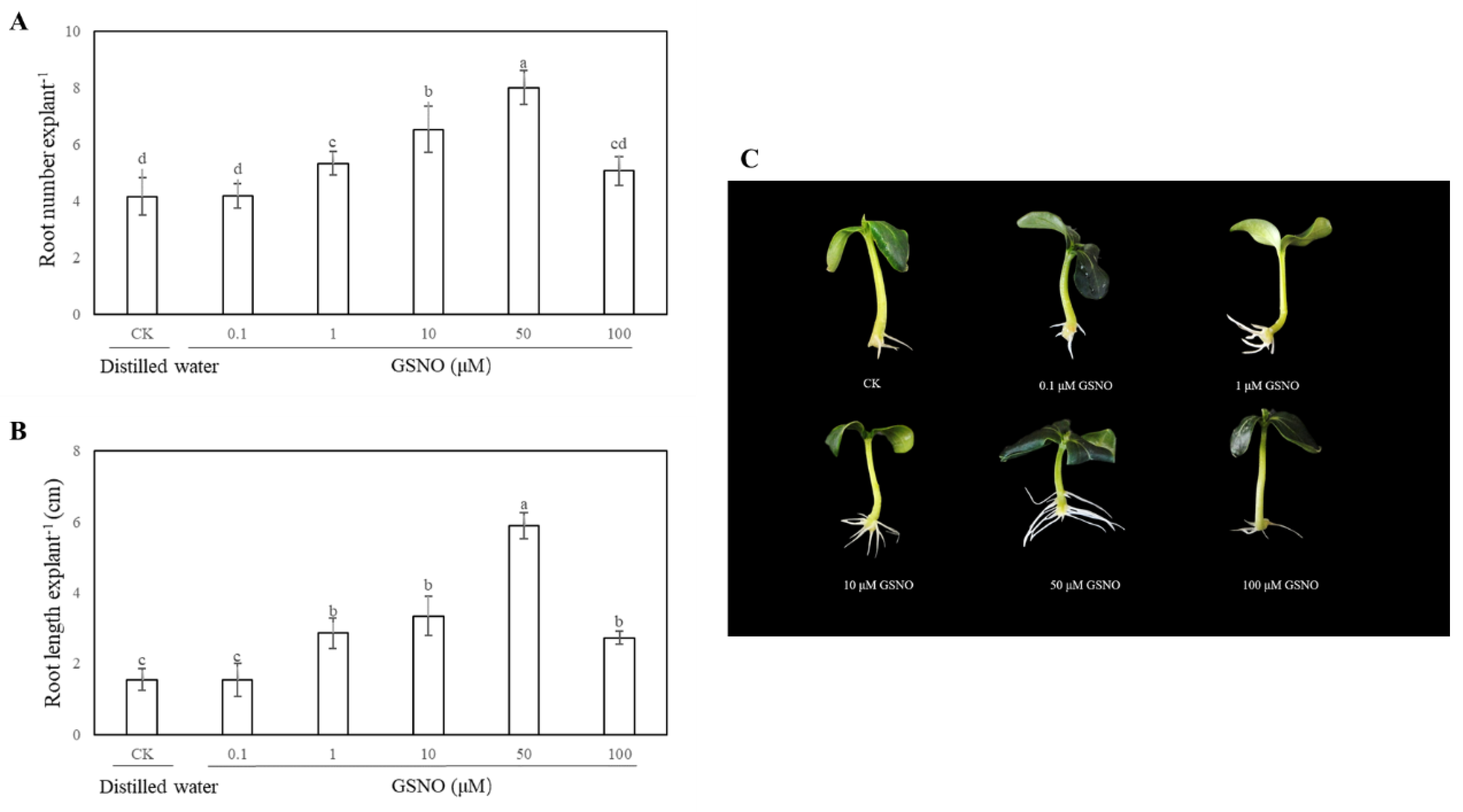
2.1. Effect of Exogenous S-Nitrosoglutathione (GSNO) on Adventitious Rooting in Cucumber

In order to access the effects of exogenous GSNO on adventitious root development of cucumber, explants were cultivated with different concentrations of GSNO (0, 0.1, 1, 10, 50 and 100 μM). As shown in Figure 1, there is no significant difference between the control and 0.1 μM GSNO. Meanwhile, lower concentrations of GSNO (1, 10, and 50 μM) significantly promoted the development of adventitious root. However, a higher dose of GSNO (100 μM) obviously decreased the number and length of adventitious roots, indicating exogenous GSNO could have a concentration-dependent effect on adventitious rooting. Moreover, the root number and length of 50 μM GSNO-treated explants increased by 92% and 280.60%, respectively, when compared with the control (Figure 1). These results revealed that 50 μM GSNO had the maximum biological effect on adventitious rooting. Therefore, 50 μM GSNO was used for the following experiments.

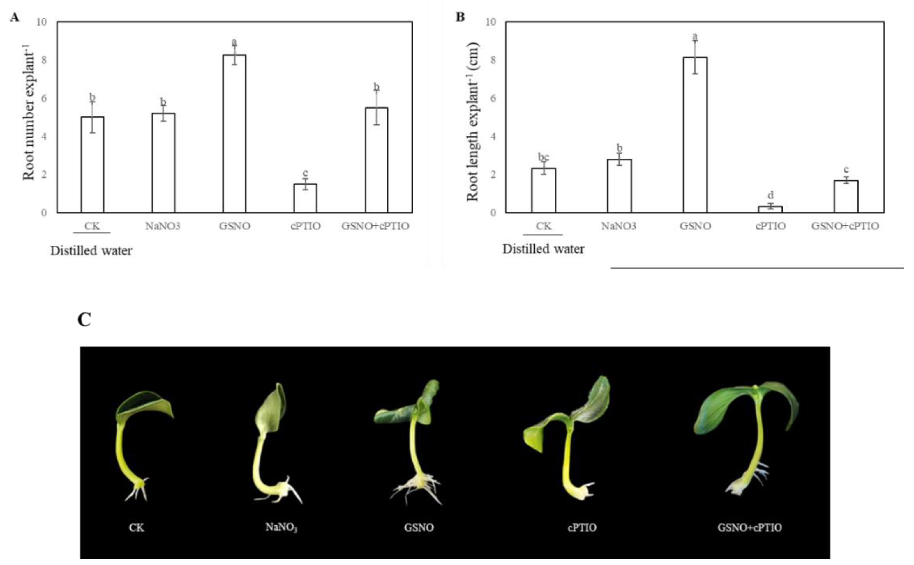
2.2. Effect of Nitric Oxide (NO) Scavenger on Adventitious Rooting in Cucumber

In order to further confirm the effect of NO on adventitious rooting, NO scavenger 2-(4-carboxy-2-phenyl)-4, 4, 5, 5-tetramethylimidazoline-1-oxyl-3-oxide (cPTIO), and a normal product of NO decomposition, NaNO3 were applied in our research. Figure 2 showed that application of cPTIO alone clearly inhibited the adventitious root development. NaNO3 treatment as a control for NO decomposition had no effect on adventitious root development. However, GSNO + cPTIO treatment significantly reversed the inhibitive effect of NO scavengers (Figure 2). These results indicate that NO is responsible for the development of adventitious root in cucumber explants.

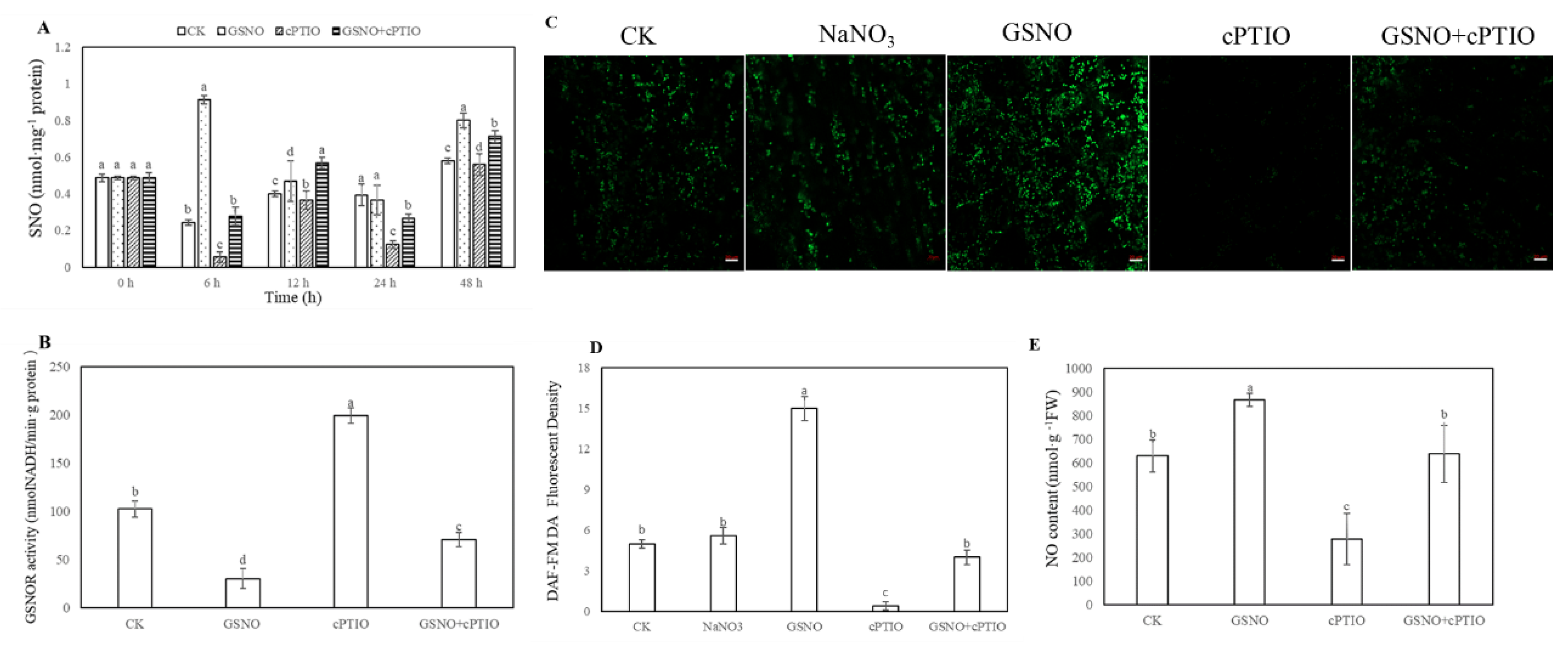
2.3. Effect of GSNO on the Levels of Total S-Nitrosothiol (SNO), and S-Nitrosoglutathione Reductase (GSNOR) Activity and Endogenous NO Level During the Development of Adventitious Roots in Cucumber

To further elucidate whether S-nitrosylation was involved in the process of adventitious rooting, the level of endogenous S-nitrosothiol (SNO) was tested during adventitious rooting (Figure 3A). As shown in Figure 3A, during adventitious rooting, treatment with GSNO strikingly elevated the endogenous SNO level. At 6 h, nitroso groups with GSNO treatment reached the maximum value and were significantly higher than that of cPTIO treatment. On the contrary, lower S-nitrosoglutathione reductase (GSNOR) activity was found in GSNO treatment relative to that of control or cPTIO treatment at 6 h (Figure 3B). Additionally, application of GSNO treatment significantly enhanced the fluorescent intensity of NO production in cucumber hypocotyl. Meanwhile, there was no significant difference between distilled water (CK) treatment and sodium nitrate (NaNO3) treatment (Figure 3C,D). However, the production of endogenous NO was remarkably reduced in hypocotyl after NO scavenger treatment (Figure 3).

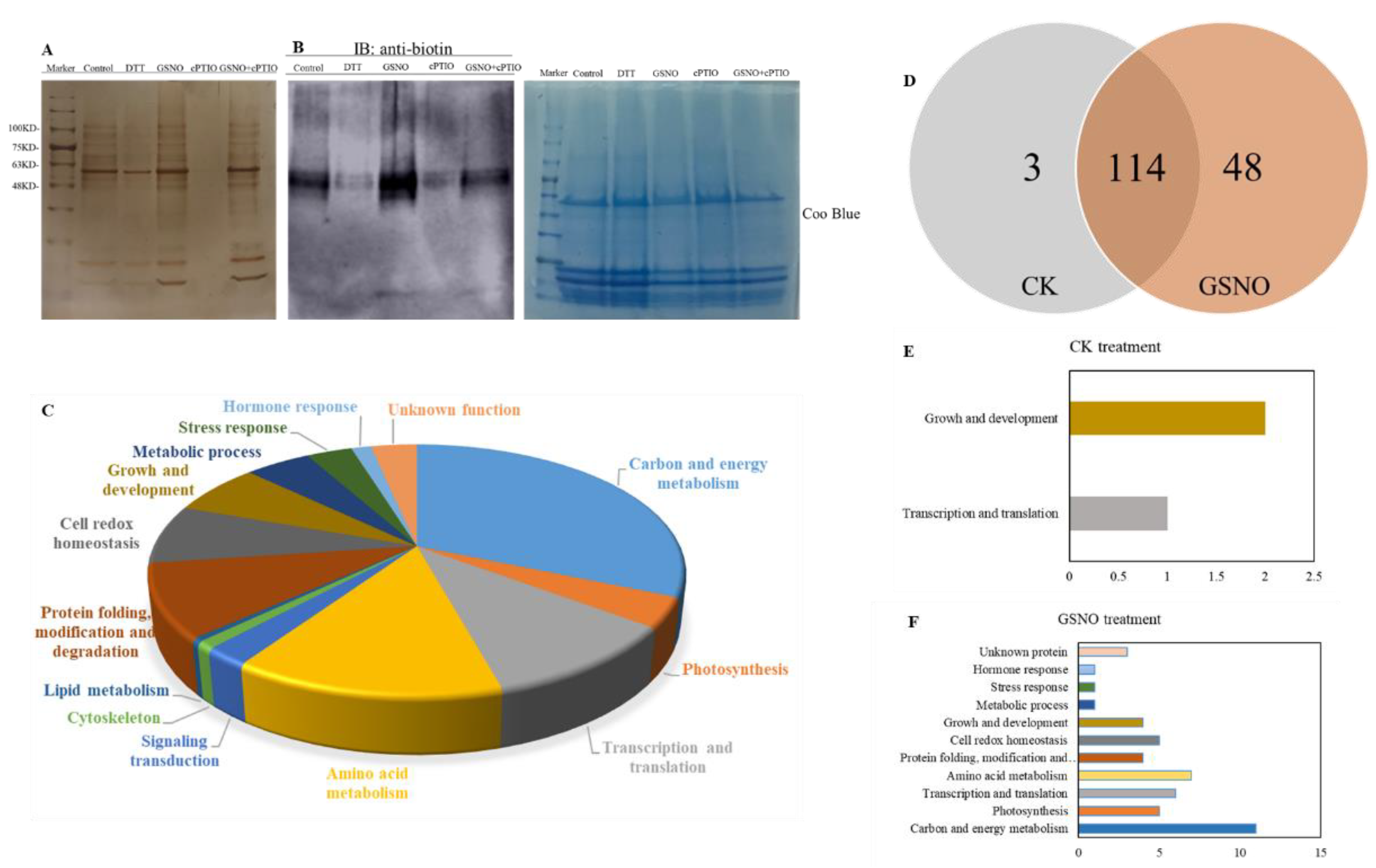
2.4. Identification of S-Nitrosylated Proteins During NO-Induced Adventitious Rooting in Cucumber

In order to further identify whether there exist possible candidates for S-nitrosylation during NO-induced adventitious rooting in cucumber explants, biotin switch detection and liquid chromatography/mass spectrometry/mass spectrometry (LC-MS/MS) were performed (Figure 4). As shown in Figure 4A,B, GSNO treatment obviously increased nitrosylation of proteins during adventitious rooting of cucumber, when compared to those of the control treatment. However, cPTIO treatment remarkably inhibited potential candidates for S-nitrosylation. Moreover, our results indicated that 167 proteins were identified from control, GSNO treatment, and GSNO + cPTIO treatment (Table 1). These identified proteins might be involved in various processes during adventitious rooting such as carbon and energy metabolism, photosynthesis, transcription and translation, and so on (Figure 4C). During adventitious rooting, approximately 40% were found to function in carbon and energy metabolism, 25.5% in the process of genetic information, and 8.5% in the growth and development process (Figure 4C). Additionally, identified proteins were found to function related to redox homeostasis, signaling transduction, and hormone response, about 9.7%, 3.0%, and 1.8%, respectively (Figure 4C). Among these proteins, three and 48 proteins were identified from the control and GSNO treatment, respectively (Figure 4D–F). As shown in Figure 4D, 114 proteins are common to both the control and GSNO treatment. These results implied that S-nitrosylation might be involved in NO-induced adventitious rooting in cucumber.

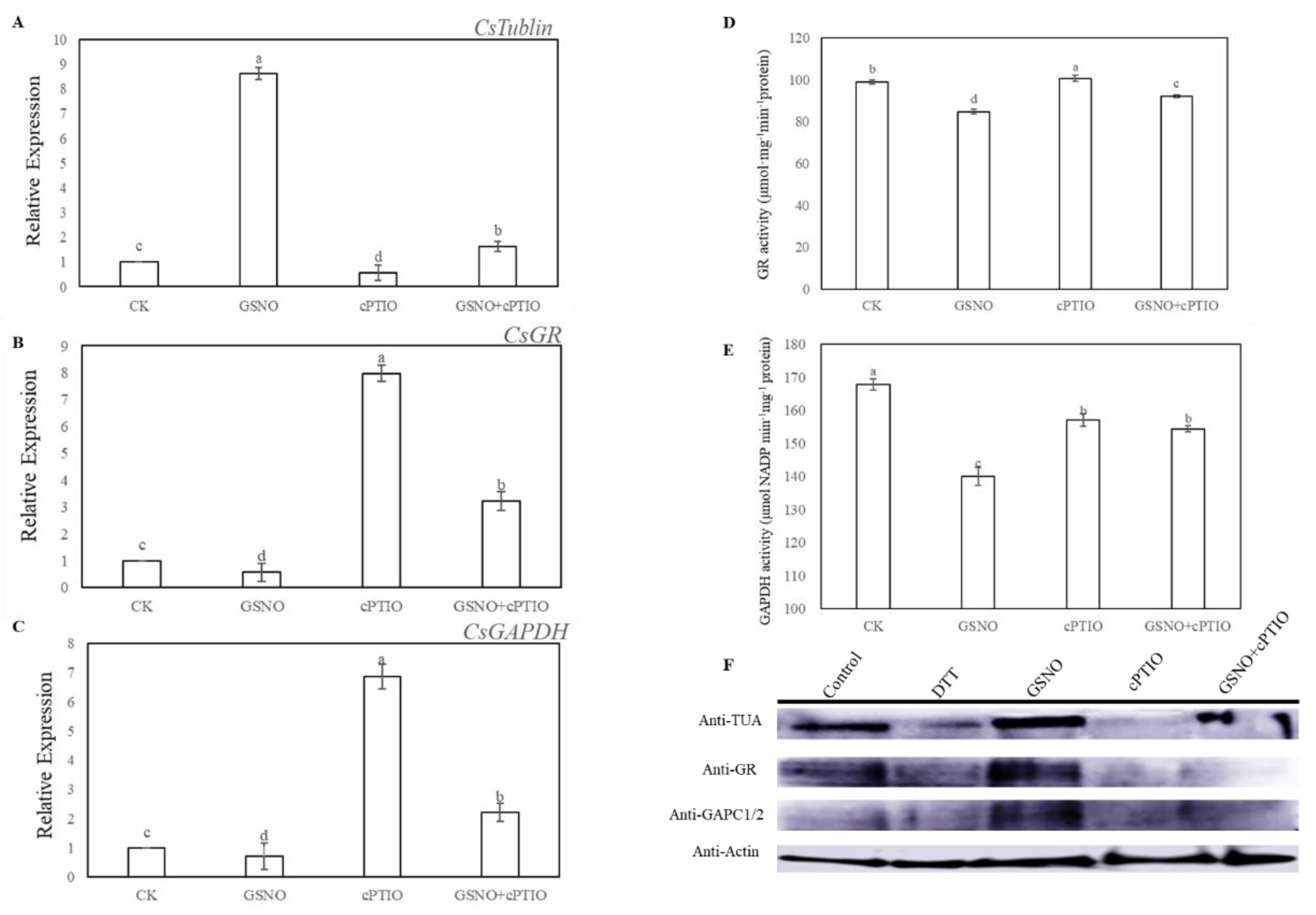
2.5. Effect of GSNO on the Activities and S-Nitrosylation Level of Tubulin Alpha Chain (TUA), Glutathione Reductase (GR), and Glyceraldehyde-3-Phosphate Dehydrogenase (GAPDH) During Adventitious Rooting

Here, tubulin alpha chain (TUA), glutathione reductase (GR), and glyceraldehyde-3-phosphate dehydrogenase (GAPDH) were selected as candidate proteins to further assess the level of nitrosylation during NO-induced adventitious rooting in cucumber. At 6 h, GSNO treatment significantly increased the expression level of TUA, and remarkably decreased the expression level and activities of GR and GAPDH (Figure 5A–E). However, exogenous application of GSNO significantly enhanced the nitrosylation level of these proteins, which was detected by the biotin switch technique (Figure 5D). On the contrary, the S-nitrosylation level of these proteins was largely blocked by the treatment of cPTIO (Figure 5D). Interestingly, removal of endogenous NO significantly inhibited the expression level of TUA but improved the expression level and activities of GR and GAPDH during the development of adventitious roots in cucumber (Figure 5A–E).

3. Discussion

In our study, the data presented herein demonstrated the evidence that there is a molecular mechanism of NO function to induce the development of adventitious rooting in cucumber. As previously reported in other researches, NO might play a critical role in affecting the root development [24,25,26]. For example, Yuan et al. [27] found the level of endogenous NO might be enhanced under cadmium (Cd) stress to inhibit the growth of root meristem in Arabidopsis through regulating auxin accumulation and transport. Alternatively, NO might act as a necessary factor affecting adventitious rooting [28,29,30]. According to our results, NO was indispensable for promoting adventitious rooting in cucumber (Figure 1 and Figure 2). Interestingly, research suggested that NO could partly exert its influence on the process of root growth and development through S-nitrosylation [31,32,33]. To investigate potential NO regulation of physiological processes through modifying cysteine residues of proteins [19], the changes of S-nitrosylation level and endogenous NO content during adventitious rooting of cucumber explants were analyzed (Figure 3). Application of exogenous NO significantly increased the level of endogenous SNO and endogenous NO production. However, SNO and NO level with cPTIO treatment significantly were lower than those of the control and GSNO treatment, implying that NO might enhance the endogenous nitrosylation level during adventitious rooting (Figure 3A,C–E). Previously, Wang et al. [32] found that SNP could enhance the level of SNO. Our results indicated that NO might affect process of adventitious rooting through enhancing the endogenous nitrosylation. Moreover, it is known that GSNOR can regulate global levels of S-nitrosylation [34,35] Additionally, Lin et al. [36] found that S-nitrosoglutathione reductase (OsGSNOR) overexpression transgenic plants were detected with a lower SNO content indicating that GSNOR might play a vital role in SNO homeostasis. As mentioned above, NO might inhibit the activity of GSNOR1 preventing S-nitrosoglutathione scavenging [35]. As depicted in Figure 3B, a lower GSNOR activity was detected in GSNO treatment, which also suggested that GSNOR regulates the total level of SNOs during NO-induced adventitious rooting in cucumber.
For a deeper insight, S-nitrosylated proteins were identified during adventitious rooting of cucumber (Figure 4; Table 1). Among these proteins, a large amount of the S-nitrosylated proteins identified were closely related to carbon and energy metabolism, implying this process could be regulated by S-nitrosylation, during adventitious rooting of cucumber. Previous research suggested that carbohydrates and nitrogen compounds might provide nutrition and energy during adventitious root formation and development [37]. In our study, for example, pyruvate kinase, malate dehydrogenase, and malate synthase were involved in the tricarboxylic acid (TCA) cycle, which acts as an iconic process for carbohydrate metabolism [38]. However, the molecular mechanisms of these protein functions during the development of adventitious roots are still not established. Here, our results imply that these proteins may be S-nitrosylated during NO-induced adventitious rooting of cucumber. Moreover, cytoskeleton change might affect cell shape and translocate organelles which could enhance cell response to intracellular and extracellular signaling [39]. Potential candidates of S-nitrosylation during adventitious rooting in cucumber are also related to cytoskeleton structure including tubulin α and tubulin β [40]. Tubulin α and tubulin β have been demonstrated to be S-nitrosylated in mammals and plants [40,41]. These results indicate that the S-nitrosylation of tubulin variants could act as an important mediator in NO-promoted development of adventitious roots in cucumber. Additionally, another cluster of potential candidates for S-nitrosylation includes metabolic enzymes such as GAPDH, glucose-6-phosphate isomerase, fructose-bisphosphate aldolase, phosphoglycerate kinase, and so on (Table 1). Previous studies have reported that H2O2 treatment might affect fructose-1,6-biphosphate aldolase and 2-phosphoglycerate hydrolase undergoing S-glutathionylation [42]. Meanwhile, Lindermayr et al. [19] suggested that the glycolysis-related enzymes are sensitive to S-nitrosylation. Thus, these metabolism enzymes, which are identified as targets for S-nitrosylation, imply that S-nitrosylation of metabolic proteins could mediate adventitious root development.
In our study, there were 116 S-nitrosylated proteins from both control and GSNO treatments (Figure 4C). These proteins participated in different processes of cellular metabolism, such as lipid metabolism, transcription and translation, hormone response, and signaling transduction (Figure 4D,E). As a consequence, these S-nitrosylated proteins with different functions might play a vital role in affecting the process of adventitious rooting. As previously reported in Wang et al. [32], NO could inhibit the growth of primary roots through S-nitrosylation of plastidial GAPDH. Our results indicated that NO could enhance the S-nitrosylation level of GAPDH, however, it was shown to decrease the expression level and activity of GAPDH during adventitious rooting (Figure 5C,E). In animals, some research has demonstrated that NO could inhibit GAPDH activity through S-nitrosylation [43,44]. Additionally, GAPDH activity was clearly inhibited by exogenous NO during NO-repression of the process of primary root growth [24]. These results might indicate that GAPDH is a key target for NO-specific PTM. Furthermore, we demonstrated evidence for the first time that GR and TUA could be over-nitrosylated under NO treatment during adventitious rooting (Figure 5). According to a previous study, GR had been shown to play an essential role for cell redox homeostasis [45]. Moreover, TUA has been found to play an essential role in cytoskeleton development [46]. The development of adventitious roots may be closely related with cell division and cell growth [47]. As depicted in Figure 5A, NO significantly increased the expression level of TUA, suggesting that the cell cycle process plays a vital role during adventitious root growth [48]. In addition, Begara-Morales [49] found that chloroplastic and cytosolic GR in peas are S-nitrosylated by GSNO, however, NO-based modification did not significantly affect this protein activity. In a previous study on mammal cells, an inhibitory effect on GR activity was shown after exposure to GSNO for a longer time [50]. According to our results, GSNO significantly decreased the expression level and activity of GR during adventitious rooting, implying that S-nitrosylation of GR induced by GSNO might inhibit protein activity and this change could be related to the development of adventitious roots in cucumber. Although some S-nitrosylation of proteins during adventitious rooting have been identified, whether the activities and functions of these identified proteins have been changed due to S-nitrosylation directly, needs to be further investigated. In the future, these results could provide valuable information for future investigations.

4. Materials and Methods

4.1. Plant Materials

Cucumber (Cucumis sativus ‘BaiLv 1′) seeds were supplied by the Gansu Academy of Agricultural Sciences, Lanzhou, China. The seeds were germinated in petri dishes on filter papers soaked with distilled water and maintained at 25 ± 1 °C for 6 days with a 14 h photoperiod (photosynthetically active radiation = 200 μmol s–1 m–2). After removing the primary roots of 6-day-old seedlings, the cucumber explants were then maintained under the same conditions of temperature and photoperiod for another 5 days under different treatments as indicated below. These media were changed every day in order to keep the solution fresh. The number and length of adventitious roots per explant were counted and measured.

4.2. Treatments of Explants

Explants were placed in petri dishes containing distilled water (control) and different concentrations of S-nitrosoglutathione (GSNO, a donor of NO, Sigma, St Louis, MO, USA) as indicated in Figure 1 and kept at 25 ± 1 °C 200 μM 2-(4-carboxy-2-phenyl)-4, 4, 5, 5-tetramethylimidazoline-1-oxyl-3-oxide (c-PTIO; Sigma, St Louis, MO, USA), 100 μM sodium nitrate (NaNO3, degradation product of NO, Solarbio, Beijing, China) was added alone and with a suitable concentration of GSNO. The concentrations of NO scavenger and NaNO3 were based on the results of a preliminary experiment.

4.3. Determination of Endogenous SNO Content, NO Production, and GSNOR Activity

SNO content was determined as described by Feechan et al. [34] with minor modifications. Fresh cucumber explants were homogenized with extraction buffer (50 mM Tris-HCl, pH 8.0), 150 mM NaCl, and 1 mM protease inhibitor phenylmethanesulfonyl fluoride (PMSF) in an ice bath for 20 min. The centrifugation was performed at 10,000 rpm for 15 min at 4 °C. The absorbance of the mixture reaction, which includes 1.5 mL of the supernatant, 1.5 mL of 0.1% N-(1-naphthyl)-ethylenediamine, and 1% sulfanilamide, with and without adding HgCl2, was taken for 20 min in the dark. SNO content was recorded photometrically at 540 nm [34].
The level of endogenous NO in cucumber hypocotyls at 6 h was detected by NO fluorescent probe 4-amino-5-methylamino-2′,7′- diaminofluoresceindiacetate (DAF-FM DA) [51]. The hypocotyls were loaded with 5 μM DAF-FM DA for 30 min at 37 °C in the dark. The samples were then washed three times with fresh buffer. DAF-FM DA fluorescence was visualized using a laser scanning confocal microscope (Leica TCS SL; Leica Microsystems, Wetzlar, Hessen, Germany). The excitation wavelength was 488 nm and the emission wavelength was 515 nm.
NO content was measured according to the Greiss reagent method with some modifications [52]. A quantity of 0.2 g of explants was finely frozen in liquid nitrogen with the extract mixture (4 mL of 50 mM ice cold acetic acid buffer, containing 4% zinc diacetate). After that, centrifugation was performed at 10,000× g for 15 min at 4 °C, and then the supernatants were collected. For each sample, 0.1 g of charcoal was added. After that, the supernatants were filtered and collected again, and then 1 mL of the mixture was pipetted into 1 mL of Greiss reagent. They were allowed to react for 30 min at room temperature. Then the absorbance was assayed at 540 nm.
S-nitrosoglutathione reductase (GSNOR) activity was measured using the method of Durner et al. [53]. Samples were homogenized with 20 mM Tris-HCl (pH 8.0, 0.2 mM NADH, and 0.5 mM EDTA) and centrifuged at 10,000 rpm for 20 min at 4 °C. The reaction was started by adding GSNO and the absorbance of the sample was measured at 340 nm.

4.4. Biotin-Switch Assay and Identification of Biotinylated Proteins

Cucumber explants were ground in liquid nitrogen, extracted by HEN-2 Buffer (250 mM Hepes-NaOH, EDTA, neocuproine and proteinase inhibitor), followed by centrifugation at 13,000 g for 10 min at 4 °C. Then, extracted protein was incubated in blocking buffer (250 mM Hepes, EDTA, SDS, methylmethane thiosulphonate (MMTS)) for 30 min at 50 °C under dark conditions. Subsequently, the MMTS was removed by cold acetone. The protein was resuspended with HEN-1 buffer (250 mM Hepes, EDTA, SDS) and 1 mM sodium ascorbate and biotin-HPDP (Sigma, St Louis, MO, USA) were added for labeling. The S-nitrosylated proteins were identified by LC-MS/MS and measured by immunoblot analysis [54].

4.5. Western Blotting

For western blot analysis, proteins from different treatments were resolved using SDS-PAGE on 12% polyacrylamide gels, and transferred to polyvinylidene difluoride membranes (PVDF, Novex, San Diego, CA, USA) utilizing a wet transfer device (BioRad, Barcelona, Spain) at 105 V for 70 min at 4 °C. The immunoreaction was performed with rabbit polyclonal antibodies against Biotin (1:2500) (Agrisera, Vännäs, Sweden), TUA (1:5000) (Agrisera, Vännäs, Sweden), GR (1:5000) (Agrisera, Vännäs, Sweden), and GAPDH (1:2000) (Agrisera, Vännäs, Sweden), Actin (1:2500) (Agrisera, Vännäs, Sweden). The blot was incubated in secondary antibody (goat anti-rabbit IgG), diluted to 1:10,000, for 1 h at 25 °C.

4.6. GR, GAPDH Activity

GR activity was determined according to Foyer et al. [55]. A 0.2 g quantity of explant was ground in liquid nitrogen with the extract mixture, followed by centrifugation at 12,000 g for 20 min at 4 °C. Then, a total of 100 μL of enzyme extract was transferred into 2 mL of reaction mixture (25 mM sodium phosphate buffer, pH 7.0, 0.1 mM EDTA, 0.5 mM oxidized glutathione (GSSG), 0.12 mM NADPH). GR activity was evaluated by measuring the decrease in absorbance at 340 nm due to NADPH oxidation.
The measurement of GAPDH activity was according to the method of Piattoni et al. [56]. Crude protein extraction was performed with 200 μL reaction buffer (50 mM Tris-HCl, pH 8.5, 10 mM sodium arsenate, 2 mM NAD+, 1 U/mL aldolase, 1.2 mM fructose-1,6-diphosphate) at 30 °C. Then, the reaction was monitored at 340 nm.

4.7. Gene Expression Analyses by RT-qPCR

The method of real time RT-PCT (RT-qPCR) analyses and statistical data analyses reference the procedure of Zhao et al. [57]. The cDNA was amplified using the following primers: for Actin (accession No. AB010922.1), F: TTGAATCCCAAGGCGAATAG and R: TGCGACCACTGGCATAAAG; for CsTUA (accession No. AJ715498.1), F: 5′-TTGTTCCTGGAGGCGATCTT-3′ and R: 5′- ACAAATGCGCGCTTAGCATA-3′. For CsGR (accession No. NM_001308836.1): F: 5′- GATATGAGAGCCGTGGTTGC-3′ and 5′- AGTCGCAAACAACAC AGCAT-3′; for CsGAPDH (accession No. NM_001305758.1), 5′- TGACGA GTCCATCATCAGCAATGC-3′ and 5′- CAATGTTGAGTGCAGCAGCTCTTG-3′. The expression analyses were conducted three times independently.

4.8. Statistical Analysis
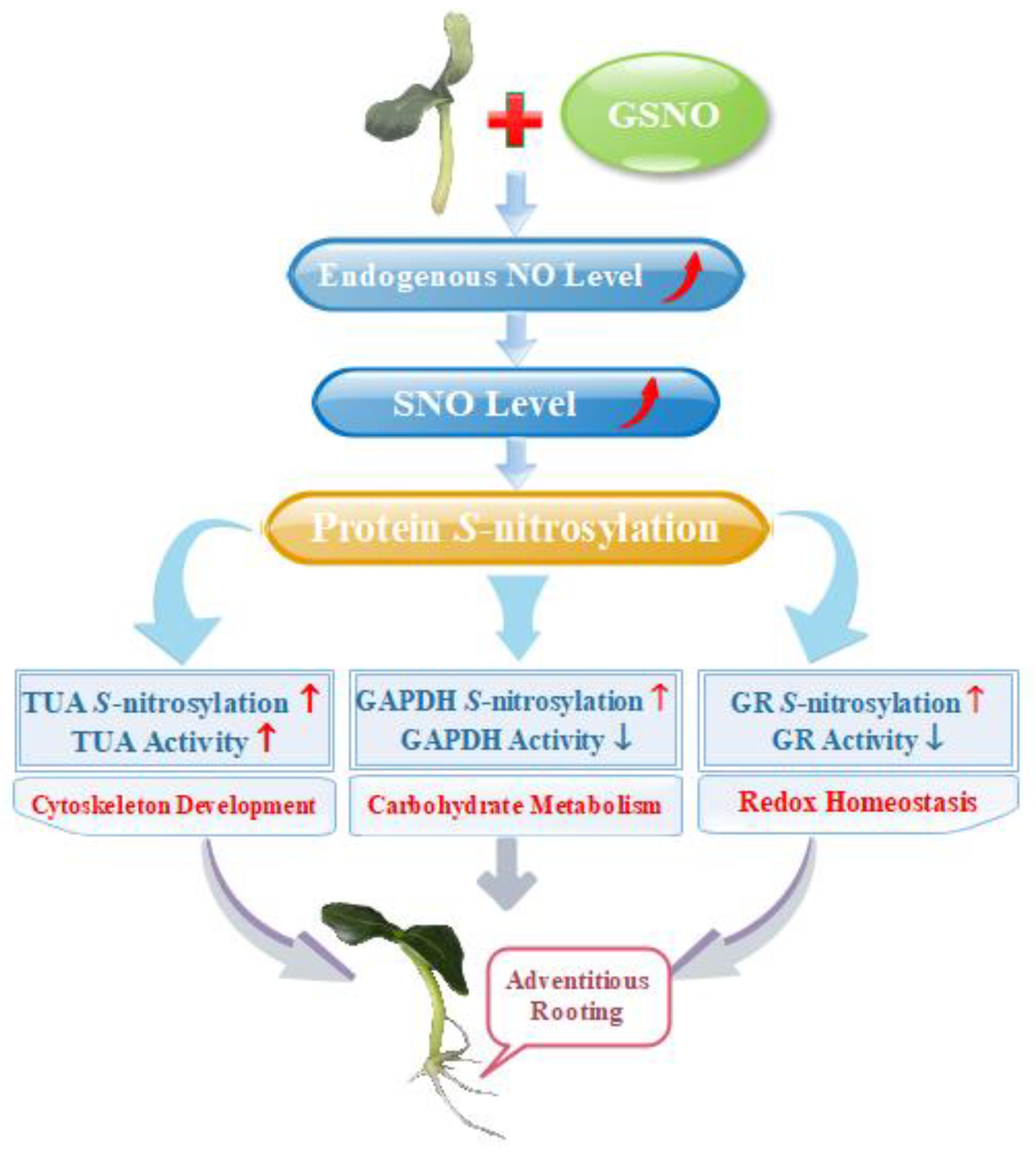
The statistical analyses was analyzed using the Statistical Package for Social Sciences for Windows (version 13.00; SPSS, Inc., Chicago, IC, United States) and statistical differences were analyzed through Duncan’s multiple range test (p < 0.05). In the analysis of variance (ANOVA), results were expressed as the mean values ± SE from three independent replicates.

5. Conclusion

Taken together, the evidence presented in this study showed that there are a series of S-nitrosylated proteins during NO-induction of the development of adventitious roots, which highlights the effect of NO-based posttranslational modification on regulating the development of adventitious roots in cucumber. Moreover, differential S-nitrosylation of key proteins regulated various pathways during adventitious rooting (Figure 6). Thus, our work demonstrated that S-nitrosylation process is an essential modulator during adventitious rooting of cucumber. Further work should focus on deciphering the function of such S-nitrosylated proteins on affecting adventitious root development. Therefore, corresponding genetic and proteomic evidences should be provided to further investigate mechanisms.

Author Contributions

Conceptualization, J.Y.(Jihua Yu) and W.L.; Formal analysis, L.N., J.Y.(Jian Yu), J.L. and L.H.; Funding acquisition, J.Y.(Jihua Yu); Investigation, L.N., J.Y.(Jian Yu) and Y.W.; Methodology, L.N., W.L. and X.X.; Project administration, J.Y.(Jihua Yu) and J.X.; Resources, J.Y.(Jihua Yu); Supervision, J.Y.(Jihua Yu), W.L. and J.X.; Writing – original draft, L.N.; Writing – review & editing, J.Y.(Jihua Yu) and W.L.

Funding

This work was supported by the National key research and development projects (2018YFD0201205), the National Natural Science Foundation of China (No. 31660584), China Agriculture Research System (CARS-23-C-07), Gansu Provience Science and Technology Key Project Fund (No.17ZD2NA015) and Natural Science Foundation of Gansu References Province, China (1610RJZA098).

Conflicts of Interest

The authors declare no conflict of interest. Declare conflicts of interest or state “The authors declare no conflict of interest.” Authors must identify and declare any personal circumstances or interest that may be perceived as inappropriately influencing the representation or interpretation of reported research results. Any role of the funders in the design of the study; in the collection, analyses or interpretation of data; in the writing of the manuscript, or in the decision to publish the results must be declared in this section. If there is no role, please state “The funders had no role in the design of the study; in the collection, analyses, or interpretation of data; in the writing of the manuscript, or in the decision to publish the results”.

References

  1. Skiba, U.; Smith, K. Nitrification and denitrification as sources of nitric oxide and nitrous oxide in a sandy loam soil. Soil Biol. Biochem. 1993, 25, 1527–1536. [Google Scholar] [CrossRef]
  2. Rockel, P.; Strube, F.; Rockel, A.; Wildt, J.; Kaiser, W.M. Regulation of nitric oxide (NO) production by plant nitrate reductase in vivo and in vitro. J. Exp. Bot. 2002, 53, 103–110. [Google Scholar] [CrossRef] [PubMed]
  3. Corpas, F.; Barroso, J. Nitric oxide synthase-like activity in higher plants. Nitric Oxide-Biol. Chem. 2017, 68, 5–6. [Google Scholar] [CrossRef] [PubMed]
  4. Niu, L.; Liao, W. Hydrogen Peroxide Signaling in Plant Development and Abiotic Responses: Crosstalk with Nitric Oxide and Calcium. Front. Plant Sci. 2016, 7. [Google Scholar] [CrossRef]
  5. He, H.; Huang, W.; Oo, T.L.; Gu, M.; He, L.-F. Nitric oxide inhibits aluminum-induced programmed cell death in peanut (Arachis hypoganea L.) root tips. J. Hazard. Mater. 2017, 333, 285–292. [Google Scholar] [CrossRef]
  6. Akram, N.A.; Iqbal, M.; Muhammad, A.; Ashraf, M.; Al-Qurainy, F.; Shafiq, S. Aminolevulinic acid and nitric oxide regulate oxidative defense and secondary metabolisms in canola (Brassica napus L.) under drought stress. Protoplasma 2018, 255, 163–174. [Google Scholar] [CrossRef]
  7. Alamri, S.A.; Siddiqui, M.H.; Al-Khaishany, M.Y.; Khan, M.N.; Ali, H.M.; Alakeel, K.A. Nitric oxide-mediated cross-talk of proline and heat shock proteins induce thermotolerance in Vicia faba L. Environ. Exp. Bot. 2019, 161, 290–302. [Google Scholar] [CrossRef]
  8. Neill, S.J.; Desikan, R.; Hancock, J.T. Nitric oxide signalling in plants. New Phytol. 2003, 159, 11–35. [Google Scholar] [CrossRef] [Green Version]
  9. Pagnussat, G.C.; Lanteri, M.L.; Lamattina, L. Nitric oxide and cyclic GMP are messengers in the indole acetic acid-induced adventitious rooting process. Plant. Physiol. 2003, 132, 1241–1248. [Google Scholar] [CrossRef]
  10. Liao, W.; Xiao, H.; Zhang, M. Role and relationship of nitric oxide and hydrogen peroxide in adventitious root development of marigold. Acta Physiol. Plant. 2009, 31, 1279. [Google Scholar] [CrossRef]
  11. Astier, J.; Rasul, S.; Koen, E.; Manzoor, H.; Besson-Bard, A.; Lamotte, O.; Jeandroz, S.; Durner, J.; Lindermayr, C.; Wendehenne, D. S-nitrosylation: An emerging post-translational protein modification in plants. Plant Sci. 2011, 181, 527–533. [Google Scholar] [CrossRef] [PubMed]
  12. Gaston, B.M.; Carver, J.; Doctor, A.; Palmer, L.A. S-nitrosylation signaling in cell biology. Mol. Interv. 2003, 3, 253–263. [Google Scholar] [CrossRef] [PubMed]
  13. Hess, D.T.; Stamler, J.S. Regulation by S-nitrosylation of protein post-translational modification. J. Biol. Chem. 2012, 287, 4411–4418. [Google Scholar] [CrossRef]
  14. Astier, J.; Kulik, A.; Koen, E.; Besson-Bard, A.; Bourque, S.; Jeandroz, S.; Lamotte, O.; Wendehenne, D. Protein S-nitrosylation: what’s going on in plants? Free Radic. Biol. Med. 2012, 53, 1101–1110. [Google Scholar] [CrossRef] [PubMed]
  15. Lamotte, O.; Bertoldo, J.B.; Besson-Bard, A.; Rosnoblet, C.; Aimé, S.; Hichami, S.; Terenzi, H.; Wendehenne, D. Protein S-nitrosylation: Specificity and identification strategies in plants. Front. Chem. 2015, 2, 114. [Google Scholar] [CrossRef]
  16. Feng, J.; Chen, L.; Zuo, J. Protein S-nitrosylation in plants: Current progresses and challenges. J. Integr. Plant Biol. 2019. [Google Scholar] [CrossRef]
  17. Aebersold, R.; Mann, M. Mass spectrometry-based proteomics. Nature 2003, 422, 198. [Google Scholar] [CrossRef]
  18. Hu, J.; Huang, X.; Chen, L.; Sun, X.; Lu, C.; Zhang, L.; Wang, Y.; Zuo, J. Site-specific nitrosoproteomic identification of endogenously S-nitrosylated proteins in Arabidopsis. Plant Physiol. 2015, 167, 1731–1746. [Google Scholar] [CrossRef]
  19. Lindermayr, C.; Saalbach, G.; Durner, J. Proteomic identification of S-nitrosylated proteins in Arabidopsis. Plant Physiol. 2005, 137, 921–930. [Google Scholar] [CrossRef]
  20. Morisse, S.; Zaffagnini, M.; Gao, X.-H.; Lemaire, S.D.; Marchand, C.H. Insight into protein S-nitrosylation in Chlamydomonas reinhardtii. Redox Signal. 2014, 21, 1271–1284. [Google Scholar] [CrossRef]
  21. de Pinto, M.C.; Locato, V.; Sgobba, A.; del Carmen Romero-Puertas, M.; Gadaleta, C.; Delledonne, M.; De Gara, L. S-nitrosylation of ascorbate peroxidase is part of programmed cell death signaling in tobacco Bright Yellow-2 cells. Plant Physiol. 2013, 163, 1766–1775. [Google Scholar] [CrossRef] [PubMed]
  22. Correa-Aragunde, N.; Foresi, N.; Lamattina, L. Nitric oxide is a ubiquitous signal for maintaining redox balance in plant cells: Regulation of ascorbate peroxidase as a case study. J. Exp. Bot. 2015, 66, 2913–2921. [Google Scholar] [CrossRef] [PubMed]
  23. Yun, B.-W.; Feechan, A.; Yin, M.; Saidi, N.B.; Le Bihan, T.; Yu, M.; Moore, J.W.; Kang, J.-G.; Kwon, E.; Spoel, S.H. S-nitrosylation of NADPH oxidase regulates cell death in plant immunity. Nature 2011, 478, 264. [Google Scholar] [CrossRef] [PubMed]
  24. Lombardo, M.C.; Lamattina, L. Abscisic acid and nitric oxide modulate cytoskeleton organization, root hair growth and ectopic hair formation in Arabidopsis. Nitric Oxide 2018, 80, 89–97. [Google Scholar] [CrossRef] [PubMed]
  25. Yu, Q.-X.; Ahammed, G.J.; Zhou, Y.-H.; Shi, K.; Zhou, J.; Yu, Y.; Yu, J.-Q.; Xia, X.-J. Nitric oxide is involved in the oxytetracycline-induced suppression of root growth through inhibiting hydrogen peroxide accumulation in the root meristem. Sci. Rep. 2017, 7, 43096. [Google Scholar] [CrossRef] [PubMed]
  26. Liu, M.; Liu, X.X.; He, X.L.; Liu, L.J.; Wu, H.; Tang, C.X.; Zhang, Y.S.; Jin, C.W. Ethylene and nitric oxide interact to regulate the magnesium deficiency-induced root hair development in Arabidopsis. New Phytol. 2017, 213, 1242–1256. [Google Scholar] [CrossRef]
  27. Yuan, H.M.; Huang, X. Inhibition of root meristem growth by cadmium involves nitric oxide-mediated repression of auxin accumulation and signalling in Arabidopsis. Plant Cell Environ. 2016, 39, 120–135. [Google Scholar] [CrossRef]
  28. Lanteri, M.L.; Laxalt, A.M.; Lamattina, L. Nitric oxide triggers phosphatidic acid accumulation via phospholipase D during auxin-induced adventitious root formation in cucumber. Plant Physiol. 2008, 147, 188–198. [Google Scholar] [CrossRef]
  29. Jin, X.; Liao, W.B.; Yu, J.H.; Ren, P.J.; Dawuda, M.; Wang, M.; Niu, L.J.; Li, X.P.; Xu, X.T. Nitric oxide is involved in ethylene-induced adventitious rooting in marigold (Tagetes erecta L.). Sci. Hortic. 2017, 97, 620–631. [Google Scholar] [CrossRef]
  30. Niu, L.; Yu, J.; Liao, W.; Yu, J.; Zhang, M.; Dawuda, M.M. Calcium and calmodulin are involved in Nitric Oxide-induced adventitious rooting of cucumber under simulated osmotic stress. Front. Plant Sci. 2017, 8, 1684. [Google Scholar] [CrossRef]
  31. Feng, J.; Wang, C.; Chen, Q.; Chen, H.; Ren, B.; Li, X.; Zuo, J. S-nitrosylation of phosphotransfer proteins represses cytokinin signaling. Nat. Commun. 2013, 4, 1529. [Google Scholar] [CrossRef] [PubMed]
  32. Wang, J.; Wang, Y.; Lv, Q.; Wang, L.; Du, J.; Bao, F.; He, Y.-K. Nitric oxide modifies root growth by S-nitrosylation of plastidial glyceraldehyde-3-phosphate dehydrogenase. Biochem. Biophys. Res. Commun. 2017, 488, 88–94. [Google Scholar] [CrossRef] [PubMed]
  33. Terrile, M.C.; París, R.; Calderón-Villalobos, L.I.; Iglesias, M.J.; Lamattina, L.; Estelle, M.; Casalongué, C.A. Nitric oxide influences auxin signaling through S-nitrosylation of the Arabidopsis TRANSPORT INHIBITOR RESPONSE 1 auxin receptor. Plant J. 2012, 70, 492–500. [Google Scholar] [CrossRef] [PubMed]
  34. Feechan, A.; Kwon, E.; Yun, B.-W.; Wang, Y.; Pallas, J.A.; Loake, G.J. A central role for S-nitrosothiols in plant disease resistance. Proc. Natl. Acad. Sci. USA 2005, 102, 8054–8059. [Google Scholar] [CrossRef]
  35. Frungillo, L.; Skelly, M.J.; Loake, G.J.; Spoel, S.H.; Salgado, I. S-nitrosothiols regulate nitric oxide production and storage in plants through the nitrogen assimilation pathway. Nat. Commun. 2014, 5, 5401. [Google Scholar] [CrossRef]
  36. Lin, A.; Wang, Y.; Tang, J.; Xue, P.; Li, C.; Liu, L.; Hu, B.; Yang, F.; Loake, G.J.; Chu, C. Nitric oxide and protein S-nitrosylation are integral to hydrogen peroxide-induced leaf cell death in rice. Plant Physiol. 2012, 158, 451–464. [Google Scholar] [CrossRef]
  37. Rapaka, V.K.; Bessler, B.; Schreiner, M.; Druege, U. Interplay between initial carbohydrate availability, current photosynthesis, and adventitious root formation in Pelargonium cuttings. Plant Sci. 2005, 168, 1547–1560. [Google Scholar] [CrossRef]
  38. Akram, M. Citric acid cycle and role of its intermediates in metabolism. Cell Biochem. Biophys. 2014, 68, 475–478. [Google Scholar] [CrossRef]
  39. Nick, P. Signals, motors, morphogenesis-the cytoskeleton in plant development. Plant Biol. 1999, 1, 169–179. [Google Scholar] [CrossRef]
  40. Jaffrey, S.R.; Erdjument-Bromage, H.; Ferris, C.D.; Tempst, P.; Snyder, S.H. Protein S-nitrosylation: A physiological signal for neuronal nitric oxide. Nat. Cell Biol. 2001, 3, 193. [Google Scholar] [CrossRef]
  41. Yemets, A.I.; Krasylenko, Y.A.; Lytvyn, D.I.; Sheremet, Y.A.; Blume, Y.B. Nitric oxide signalling via cytoskeleton in plants. Plant Sci. 2011, 181, 545–554. [Google Scholar] [CrossRef] [PubMed]
  42. Shenton, D.; Grant, C.M. Protein S-thiolation targets glycolysis and protein synthesis in response to oxidative stress in the yeast Saccharomyces cerevisiae. Biochem. J. 2003, 374, 513–519. [Google Scholar] [CrossRef] [PubMed]
  43. Sen, N.; Hara, M.R.; Kornberg, M.D.; Cascio, M.B.; Bae, B.-I.; Shahani, N.; Thomas, B.; Dawson, T.M.; Dawson, V.L.; Snyder, S.H. Nitric oxide-induced nuclear GAPDH activates p300/CBP and mediates apoptosis. Nat. Cell Biol. 2008, 10, 866. [Google Scholar] [CrossRef] [PubMed]
  44. Kornberg, M.D.; Sen, N.; Hara, M.R.; Juluri, K.R.; Nguyen, J.V.K.; Snowman, A.M.; Law, L.; Hester, L.D.; Snyder, S.H. GAPDH mediates nitrosylation of nuclear proteins. Nat. Cell Biol. 2010, 12, 1094. [Google Scholar] [CrossRef]
  45. Meyer, A.J. The integration of glutathione homeostasis and redox signaling. J. Plant Physiol. 2008, 165, 1390–1403. [Google Scholar] [CrossRef]
  46. Bao, Y.; Kost, B.; Chua, N.H. Reduced expression of α-tubulin genes in Arabidopsis thaliana specifically affects root growth and morphology, root hair development and root gravitropism. Plant J. 2001, 28, 145–157. [Google Scholar] [CrossRef]
  47. Zhu, Y.; Liao, W.; Niu, L.; Wang, M.; Ma, Z. Nitric oxide is involved in hydrogen gas-induced cell cycle activation during adventitious root formation in cucumber. BMC Plant Biol. 2016, 16, 146. [Google Scholar] [CrossRef]
  48. Begara-Morales, J.C.; Sánchez-Calvo, B.; Chaki, M.; Mata-Pérez, C.; Valderrama, R.; Padilla, M.N.; López-Jaramillo, J.; Luque, F.; Francisco, J.C.; Barroso, J.B. Differential molecular response of monodehydroascorbate reductase and glutathione reductase by nitration and S-nitrosylation. J. Exp. Bot. 2015, 66, 5983–5996. [Google Scholar] [CrossRef]
  49. Lorbiecke, R.; Sauter, M. Adventitious root growth and cell-cycle induction in deepwater rice. Plant Physiol. 1999, 119, 21–30. [Google Scholar] [CrossRef]
  50. Beltrán, B.; Orsi, A.; Clementi, E.; Moncada, S. Oxidative stress and S-nitrosylation of proteins in cells. Br. J. Pharmacol. 2000, 129, 953–960. [Google Scholar] [CrossRef]
  51. Graziano, M.; Lamattina, L. Nitric oxide accumulation is required for molecular and physiological responses to iron deficiency in tomato roots. Plant J. 2007, 52, 949–960. [Google Scholar] [CrossRef]
  52. Xuan, W.; Xu, S.; Li, M.; Han, B.; Zhang, B.; Zhang, J.; Lin, Y.; Huang, J.; Shen, W.; Cui, J. Nitric oxide is involved in hemin-induced cucumber adventitious rooting process. J. Plant Physiol. 2012, 169, 1032–1039. [Google Scholar] [CrossRef]
  53. Durner, J.; Wendehenne, D.; Klessig, D.F. Defense gene induction in tobacco by nitric oxide, cyclic GMP, and cyclic ADP-ribose. Proc. Natl. Acad. Sci. USA 1998, 95, 10328–10333. [Google Scholar] [CrossRef] [Green Version]
  54. Jaffrey, S.R.; Snyder, S.H. The biotin switch method for the detection of S-nitrosylated proteins. Sci. STKE 2001, 86, pl1. [Google Scholar] [CrossRef]
  55. Foyer, C.H.; Halliwell, B. The presence of glutathione and glutathione reductase in chloroplasts: A proposed role in ascorbic acid metabolism. Planta 1976, 133, 21–25. [Google Scholar] [CrossRef]
  56. Piattoni, C.; Guerrero, S.; Iglesias, A. A differential redox regulation of the pathways metabolizing glyceraldehyde-3-phosphate tunes the production of reducing power in the cytosol of plant cells. Int. J. Mol. Sci. 2013, 14, 8073–8092. [Google Scholar] [CrossRef]
  57. Zhao, Y.; Liu, W.; Xu, Y.-P.; Cao, J.-Y.; Braam, J.; Cai, X.-Z. Genome-wide identification and functional analyses of calmodulin genes in Solanaceous species. BMC Plant Biol. 2013, 13, 70. [Google Scholar] [CrossRef]
Figure 1. Effect of different concentrations of S-nitrosoglutathione (GSNO) on adventitious root development in cucumber explants. The primary roots were removed from hypocotyl of 5-day-old seedlings. Explants were incubated for 5 days with different concentrations of GSNO. The numbers (A) and root length (B) of adventitious roots were expressed as mean ± SE (n = 3). Ten explants were used per replicate. Photographs (C) were taken after five days of the treatments indicated. Bars with different lowercase letters were significantly different by Duncan’s multiple range test (p < 0.05). Bars with different lowercase letters were significantly different by Duncan’s multiple range test.
Ijms 20 05363 g001
Figure 2. Effect of NO scavenger 2-(4-carboxy-2-phenyl)-4, 4, 5, 5-tetramethylimidazoline-1-oxyl-3-oxide (cPTIO) on adventitious root development in cucumber explants. The primary roots were removed from 5-day-old seedlings. Explants were then incubated for 5 days with distilled water (CK) or 100 μM sodium nitrate (NaNO3), 50 μM GSNO, 200 μM cPTIO, or 50 μM GSNO + 200 μM cPTIO. The numbers (A) and root length (B) of adventitious roots were expressed as mean ± SE (n = 3). Ten explants were used per replicate. Photographs (C) were taken after five days of the treatments indicated. Bars with different lowercase letters were significantly different by Duncan’s multiple range test (p < 0.05). Bars with different lowercase letters were significantly different by Duncan’s multiple range test.
Ijms 20 05363 g002
Figure 3. Effect of GSNO on the levels of total S-nitrosothiol (SNO) (A), S-nitrosoglutathione reductase (GSNOR) activity (B) and endogenous NO level (C, D, E) during the development of adventitious roots in cucumber. Explants were incubated with distilled water (CK) or 100 μM NaNO3, 200 μM cPTIO, 50 μM GSNO, or 50 μM GSNO + 200 μM cPTIO. The levels of total SNO (A) were determined during adventitious rooting. GSNOR activity (B) and endogenous NO levels (C, E) in cucumbers were detected after 6 h of treatment. 4-amino-5-methylamino-2′,7′- di aminofluoresceindiacetate (DAF-FM DA) was utilized to detect endogenous NO of a longitudinal section from the tip of the hypocotyls. Changes in fluorescence intensity of NO (C) were monitored by fluorescence microscopy after 6 h. The DAF-FM DA fluorescence density of endogenous NO (D) was analyzed by ImageJ software. Bars with different lowercase letters were significantly different by Duncan’s multiple range test (p < 0.05). Bars with different lowercase letters were significantly different by Duncan’s multiple range test.
Ijms 20 05363 g003
Figure 4. Identification of S-nitrosylated proteins during the development of adventitious rooting. Total S-nitrosylated proteins were detected through liquid chromatography/mass spectrometry/mass spectrometry (LC-MS/MS) and the western blotting method after explants were incubated with distilled water (CK) or 50 μM GSNO, 200 μM cPTIO, or 50 μM GSNO + 200 μM cPTIO for 6 h (A, B). Functional categorization of S-nitrosylated proteins from CK, GSNO and GSNO + cPTIO treatment (C). The number of S-nitrosylated proteins in CK and GSNO treated explants (D). Functional categorization of S-nitrosylated proteins from CK treatment alone (E). Functional categorization of S-nitrosylated proteins from GSNO treatment alone (F).
Ijms 20 05363 g004
Figure 5. Effect of GSNO on the expression levels, enzymatic activities and S-nitrosylation level of tubulin alpha chain (TUA), glutathione reductase (GR), and glyceraldehyde-3-phosphate dehydrogenase (GAPDH) during adventitious rooting. Explants were incubated with distilled water (CK) or 200 μM cPTIO, 50 μM GSNO, or 50 μM GSNO + 200 μM cPTIO. TUA, GR and GAPDH expression level (A, B, C), and GR and GADPH activity (D, E) in cucumber explants was determined at 6 h of treatment. Immunoblot analysis of S-nitrosylated proteins in vivo (F). After biotinylation, proteins were purified with neutravidin-agarose, separated by sodium dodecyl sulfate-polyacrylamide gel (SDS-PAGE), and immunoblotted with anti-TUA, anti-GR, and anti-GAPDH antibodies. Bars with different lowercase letters were significantly different by Duncan’s multiple range test (p < 0.05). Bars with different lowercase letters were significantly different by Duncan’s multiple range test.
Ijms 20 05363 g005
Figure 6. Schematic model of NO-induced S-nitrosylation during adventitious rooting in cucumber. NO-enhanced endogenous NO concentration and SNO levels, which triggers S-nitrosylation of proteins to induce adventitious root development. Differential S-nitrosylation of TUA, GAPDH and GR might regulate various pathways during NO-promoted the development of adventitious roots. The increase is indicated by the red arrow. The decrease is indicated by the blue arrow.
Ijms 20 05363 g006
Table 1. S-nitrosylated proteins identified from the control, S-nitrosoglutathione (GSNO), and GSNO + 2-(4-carboxy-2-phenyl)-4, 4, 5, 5-tetramethylimidazoline-1-oxyl-3-oxide (cPTIO) treatment during adventitious rooting of cucumber seedlings.
Accession NumberProtein NameMol MassPeptide Sequence
A0A0A0K9P511S globulin subunit beta-like54 kDaSSLLAFLC11LAVFING NGFEETVC299TLRLKHN
A0A0A0K67426S protease regulatory subunit 747 kDaAKKVNDLC56GIKESDT QPLQVARC91TKIINPN MARSKKAC263IVFFDEV
A0A0A0K3C426S proteasome non-ATPase regulatory subunit 2 homolog98 kDaGLIYLGSC539NEEVAQA
A0A0A0LQ324-alpha-glucanotransferase64 kDaYSGQDANC140GNTLLIS
A0A0A0KAJ960S ribosomal protein L344 kDaKDDATKPC41RLTAFLG
A0A0A0KXM86-phosphogluconate dehydrogenase, decarboxylating53 kDaAYLEKGDC103IIDGGNE
Q08375Acetyl-CoA acyltransferase (3-ketoacyl-coa thiolase)48 kDaSIENAQNC191LLPMGVT FASQFVYC370RNKLGLD LGATGARC401VATLLHE AVFERGDC440VDELCNA
A0A0A0LFR2Acetyltransferase component of pyruvate dehydrogenase complex58 kDaNRSQFLQC75QRGVSMM YYLTVDTC341VDKLMDL FMSVTLSC509DHRVIDG
A0A0A0KHD6Aconitate hydratase95 kDaPAVVDLAC103MRDAMNR ALVAKKAC442ELGLEVK
A0A0A0KJ21Actin41 kDaEDIQPLVC12DNGTGMV TYNSIMKC287DVDIRKD
A0A0A0KRC5Acyl-coenzyme A oxidase73 kDaQHLMESTC457KVQKAED SARMSVEC486AKRLSQF KDQLQKLC544SIYALFT
A0A0A0LNE3Adenosylhomocysteinase53 kDaEMPGLMAC42RTEFGPS
A0A0A0KSC6Adenylosuccinate lyase60 kDaMEIGANC7RVLDQPR LEFFHFSC186TSEDINN
A0A0A0K9F9Aldehyde dehydrogenase family 7 member B454 kDaQYMRRSTC490TINYGNE
H6WX41Alkaline alpha galactosidase 386 kDaHHTDAVYC441AKQTAVV SSAKPRQC744IVDSSVV
A0A0A0KMH9Alpha-mannosidase114 kDaMEKQANSC8LPFSFLV NNSIQGAC76VQNVLDS QPKILSQC470PLLNISF
A0A0A0L5C9Aminopeptidase99 kDaQPSSIQAC82EVSQILV AFALSMAC587QQSVTSL
A0A0A0KF04Aminotransferase 244 kDaDHTIKAVC142IVHNETA
A0A0A0LEK8Aspartate aminotransferase50 kDaNRVTTVQC163LSGTGSL
G3EIZ8ATP synthase subunit alpha54 kDaAESETLYC202VYVAIGQ
A0A2D0UXD2Betaine aldehyde dehydrogenase54 kDaAKLEAIDC100GKPLEEA
A0A0A0K2H5Beta-xylosidase/alpha-L-arabinofuranosidase 2-like84 kDaLAGLDLDC344GDFLGKH PGCANVAC485TSAQLDE
A0A0A0KYI1Biotin carboxylase58 kDaMDAAMPLC8KSARAPS KLADESVC117IGEAPSS SAAVSRGC142TMLHPGY
A0A0A0LD02Carbonic anhydrase35 kDaSTASINTC9LFSLNKS ACSDSRVC167PSHVLDF
G8EX76Chloroplast transketolase80 kDaEGIANEAC246SLAGHWG
A0A0A0LCU8Coatomer subunit beta106 kDaMEKSC5TLLVHFD STAVIYEC262AGTLVSL RAAANTYC284QLLLSQS MKSTNMKC879LTPISAL
A0A0A0LBW6D-3-phosphoglycerate dehydrogenase63 kDaAAATEHGC144LVVNAPT
A0A0A0KG56Dihydrolipoyl dehydrogenase 2, chloroplastic-like59 kDaKLVPHVYC393IGDANGK
A0A0A0LTJ3Elongation factor Ts, mitochondrial121 kDaTGAGMMDC693KKALAES TGAGMMDC936KKALSET
A0A0A0K581Eukaryotic translation initiation factor 3 subunit B60 kDaTTKTLGYC112FIEYGTP
A0A0A0LC36Eukaryotic translation initiation factor 3 subunit C106 kDaTKARAMLC519DIYHHAL SWDQPSGC785IIFHDVT
A0A0A0L3P3Ferredoxin--NADP reductase, chloroplastic46 kDaDSKTVSLC213VKRLVYT
A0A0A0K8H3Fructose-1,6-bisphosphatase, cytosolic36 kDaLVSSGRTC95ILVSEED
A0A0A0KKE4Fructose-bisphosphate aldolase38 kDaMSC3YRGKYAD
A0A0A0KEY8Glucose-1-phosphate adenylyltransferase57 kDaPNLKRKLC58ISSLIAD
A0A0A0LRW2Glucose-6-phosphate isomerase67 kDaMASISGIC8SSSPSLK AVLNEASC559KEPVEPL
A0A0A0KPY1Glutamate decarboxylase56 kDaMVDENTIC205VAAILGS KKKTNGVC499
A0A0A0K488Glutamate-1-semialdehyde 2,1-aminomutase 2, chloroplastic-like54 kDaSVGIGLPC47STKLSHT
A0A0A0K8Q7Glutathione reductase59 kDaAGGVGGTC122VIRGCVP
A0A0A0K8C1Glyceraldehyde-3-phosphate dehydrogenase36 kDaNIVSNASC154TTNCLAP NASCTTNC158LAPLAKV
A0A0A0LN17Glycine cleavage system P protein113 kDaTFVISNNC252HPQTIDI NPASAAMC688GMKIVSV
A0A0A0LAN5Glycosyltransferase55 kDaQLTPRPNC123IISDMCI
A0A0A0KHX5Glyoxysomal fatty acid beta-oxidation multifunctional protein MFP-a79 kDaMC2HALLVTI NLKHTIAC303IDAVETG
A0A0A0LNA7Guanosine nucleotide diphosphate dissociation inhibitor49 kDaSEGETAKC278KKVVCDP
A0A0A0K921Heat shock 70 kDaa protein 15-like92 kDaVIDQLVYC704INSYREA
A0A0A0KXG3Heat shock protein 7070 kDaNMDLFRKC319MEPVEKC CMEPVEKC326LRDAKMD MKELESIC609NPIIAKM
A0A0A0K5T7Ketol-acid reductoisomerase63 kDaNISVIAVC242PKGMGPS CMDILYEC394YEDVASG
A0A0A0LXB9L-ascorbate oxidase65 kDaYMFWSPDC54VENIVMG GTASISQC116AINPGET ELSGKEKC236APFILHV IPPKALAC574GSTALVK
A0A0A0L5B9Lon protease homolog 2, peroxisomal98 kDaDLKLASAC757ESNLLEG
A0A0A0LR30Lsocitrate lyase64 kDaQLKTFSEC320VTDAIMN
A0A0A0L0E4Malate dehydrogenase36 kDaCTAIAKYC142PNALVNM
A0A0A0LUC5Malate synthase65 kDaKGMYKEAC533KMFTRQC
A0A0A0L5H2Methionine S-methyltransferase120 kDaVDSFLALC15QQSGDAA QLERIVGC210IPQILNP HALSVYSC364QLLQPNQ HLPAQREC664DKSASSR CGWDVIEC997HAGVSVV ADFKRIAC1082SS
A0A0A0LEZ3Methionine synthase84 kDaIPSNTFSC64YDQVLDT HLVVSTSC328SLLHTAV
A0A0A0LIC6Methylenetetrahydrofolate reductase72 kDaETMMHLTC128TNMPVEK YEKFMKYC446LGKLRSS
A0A0A0KI79Mg-protoporphyrin IX chelatase45 kDaKGRPQVQC60NVATEIN KVKISRVC350AELNVDG
A0A0A0LN97Multicopper oxidase60 kDaDGVYGTTC99PIPPGKN
A0A0A0KIJ0Ncharacterized protein55 kDaIEPVPESC99VSTLEER
A0A0A0L679Phospho-2-dehydro-3-deoxyheptonate aldolase57 kDaFLLQGGDC124AESFKEF NSRYHTHC479DPRLNAS
A0A0A0KEF3Phosphoglycerate kinase50 kDaQVVKADDC177IGPEVEK
A0A0A0KTJ4Phospholipase D92 kDaYFSQRRGC178KVTLYQD KFYEPHRC209WEDVFDA LFPESIEC736VKSVNQL
A0A0A0L987Phosphoribulokinase46 kDa****MAVC4TVYTTQS
A0A0A0L989Polyadenylate-binding protein71 kDaAFGSILSC146KVALDSS
A0A0A0K809Presequence protease 1, chloroplastic/mitochondrial-like122 kDaVFLRSLTC12SSLVCNR RGKAMSGC743AEDLFNL SLLSRKNC847LVNITAD
A0A0A0K8X9Protease Do-like 2, chloroplastic68 kDaAAAMASSC9FSPFDST VLARGVDC204DIALLSV LKFGNLPC230LQDAVTV AAIAASSC571ILRDYGI
A0A0A0LRK5Purple acid phosphatase54 kDaVLCDLGVC26NGGITSG
A0A0A0L0U0Pyrophosphate--fructose 6-phosphate 1-phosphotransferase subunit alpha67 kDaETFAEAKC208PTKVVGV ASHVALEC276TLQSHPN RTIVKPGC584SQEVLKA
A0A0A0KH95Pyrophosphate--fructose 6-phosphate 1-phosphotransferase subunit beta61 kDaLKTRVIGC224PKTIDGD SFGFDTAC247RIYAEMI
A0A218KBQ1Pyruvate kinase55 kDaKPGNNILC143SDGTITL QKMMIYKC287NLAGKPV AVLDGTDC328VMLSGES
A0A0A0KAU8RuBisCO large subunit-binding protein subunit alpha64 kDaLSSASILC14SSHKSLR
A0A0A0KFZ8RuvB-like helicase51 kDaPQTKFVQC224PDGELQK
A0A0A0KBZ1S-(hydroxymethyl)glutathione dehydrogenase40 kDaTQGQVITC10KAAVAWE GVDYSFEC271IGNVNVM
C4PAW8Sedoheptulose-1,7-bisphosphatase42 kDaGLIRLLTC93MGEALRT SHFCKYAC148SEEVPEL
A0A0A0K8A3Selenium-binding protein 2-like53 kDaKDTGYVGC277ALTSNMV
A8CM21Stachyose synthase96 kDaSSAINKGC383TSCSCKA GLTNMFNC792SGTIQHL
A0A0A0KGA1Succinate-semialdehyde dehydrogenase58 kDaGPALASGC230TVVIKPS NSGQTC346VCANRILVQ
A0A0A0LVU2T-complex protein 1 subunit delta57 kDaRSLHDALC404VVRCLVN AITLATEC519VRMILKI
A0A0A0LZU0T-complex protein 1 subunit eta60 kDaFADRDIFC313AGRVAEE NAATEAAC517LILSVDE
A0A0A0LLK5Tocopherol cyclase57 kDaPLCGIHHC16SFKLVEA
A0A0A0KBL8Transketolase, chloroplastic80 kDaNRSSRSRC65GVVRASV EGIANEAC249SLAGHWG
A0A0A0K6A8Tubulin alpha chain49 kDaGIQVGNAC20WELYCLE TIQFVDWC347PTGFKCG AKVQRAVC376MISNSTS
A0A0A0LCY8Tubulin beta chain50 kDaLHIQGGQC12GNQIGAK ATMSGVTC238CLRFPGQ NNVKSTVC354DIPPTGL
A0A0A0K9N4Ubiquitin carboxyl-terminal hydrolase 654 kDaYMNSTLQC121LHSVPEL MQQDAEEC200WTQLLYT ESVYSLKC256HISQEVN
A0A0A0KZ30UDP-glucose 6-dehydrogenase52 kDaMVKIC5CIGAGYV TKEAHAVC417ILTEWDE
A0A0A0KZU3Gamma aminobutyrate transaminase 256 kDaTNPKLGSC18AKDVAAL
A0A0A0LHR0PALP domain-containing protein58 kDaSSPFTLVC36SSATSDS
A0A0A0LQL1Uncharacterized protein110 kDaLARGQLRC391IGATTLE
A0A0A0LTW3UVR domain-containing protein96 kDaRRRKASRC26VPRAMFE LARGELQC343IGATTLD
A0A0A0KSQ4Probable nucleoredoxin 163 kDaWICEGGVC559RKA
A0A0A0L5E7Uncharacterized protein43 kDaQQFTGLRC13APLSSSR
A0A0A0LNR8Peptidase_S9 domain-containing protein85 kDaILSGEVSC428ISPANSN PVKDVSNC514LTKGASE AAARNPVC653NLALMVG
A0A0A0KN12Oxalate--CoA ligase-like55 kDaKLRFIRS291CSASLAPS
A0A0A0K983Uncharacterized protein69 kDaTTDGKTNC422LNAAVGT AMVTQAYC569DVPFSYT
A0A0A0KI31Glyoxysomal fatty acid beta-oxidation Multifunctional protein MFP-a79 kDaGLEVAMAC124HARLSTK NLKHPLVC251IDVVETG
A0A0A0KIK3enolase isoform47 kDaQIKTGAPC408RSERLAK
A0A0A0KL58AA_TRNA_LIGASE_II domain-containing protein51 kDaTATERTLC402CILENYQ ATERTLC403CILENYQK
A0A0A0KPT0Protein kinase domain-containing protein127 kDaRGAAKGLC974FLHHNCI YLEITLRC1124VEEFPSK
A0A0A0KQJ3Alpha-amylase 3, chloroplastic isoform101 kDaLDPLLYHC13AKGKHRF RPCSFTYC37PNKLLCH NWELTVGC112NLAGKWI ISVSVRKC292SETTKYL
A0A0A0KTH8Malate dehydrogenase, chloroplastic48 kDaSRTSRVTC49SINQVEA CNTNALIC230LKNAPKI ELLAEKRC413VAHLTGE
A0A0A0KW042-hydroxyacyl-CoA lyase60 kDaDISEIPNC154VARVLNS RSLAIGKC274DVALVVG
A0A0A0KWS02,3-bisphosphoglycerate-independent Phosphoglycerate mutase61 kDaNGVRTFAC356SETVKFG
A0A0A0L7Y511S globulin seed storage protein 2-like57 kDaSSGLIVKC260DEEMSFL NGIEETVC297TARVQHN
A0A0A0LFS9Cell division control protein 48 homolog E89 kDaCTEAALQC426IREKMDV KARQSAPC576VLFFDEL
A0A0A0LHX3Uncharacterized protein71 kDaNTPQQLAC176IDVIEDG KVPLCIPC201EDKVFRE
A0A0A0LJ13Triosephosphate isomerase, chloroplastic32 kDaEGLGVIAC177IGELLEE
A0A0A0LTR4Beta-glucosidase 44-like57 kDaLPVVCMLC14AATAMHL
A0A0A0LV53Lysosomal beta glucosidase-like68 kDaNVCSNVNC542VVVVVSG
A0A0A0KD01Uncharacterized protein91 kDaHLNAAASC154QIQFVCK KELDEAIC328WAKVSET NLEDRLAC546KDNSSPL
A0A0A0KTK6Aminotran_1_2 domain-containing protein52 kDaKVPDVLYC417LKLLEAT
A0A0A0KW15Uncharacterized protein51 kDaEIKEGCGC460KG
A0A0A0L0K6Uncharacterized protein50 kDaDGVYGTTC103PIPPGKN
A0A0A0L0Q4Ribos_L4_asso_C domain-containing protein44 kDaQGAFGNMC100RGGRMFA
A0A0A0L5U9Acyl-CoA dehydrogenase family member 1091 kDaSTVGNQMC262DVAYFCL NLEYGHLC511EIMGRSI SDATNIEC579SITREGD SGAMDPRC605KILIVMG
A0A0A0LI90Aldedh domain-containing protein53 kDaHKAPIAEC98LVKEIAK
A0A0A0LRM411-beta-hydroxysteroid dehydrogenase 1B-like38 kDaPVETADEC267AKGVVRG
A0A0A0LUA8Aldedh domain-containing protein59 kDaKVGPALAC232GNTVVLK GKSPFIVC325EDADVDK
A0A0A0K8W3Uncharacterized protein109 kDaMKNC4SNALSAN KLLRNYRC701HPDILHL
A0A0A0KNB1OMPdecase domain-containing protein52 kDaSTSYDLVC73GVPYTAL EKIGPEIC274LLKTHVD
A0A0A0L1I8DNA mismatch repair protein MLH3 isoform136 kDaAYVLNLEC311PVSFYDL KKSRMQSC394QASLIDS RVLNSKAC1128RGAIMFG
A0A0A0LAP3Uncharacterized protein60 kDaMVTHC5INLHLHR
A0A0A0LL68E2F_TDP domain-containing protein47 kDaALALPPQC47CLQYHRP ACFSERQC318RMIIKST
A0A0A0LPD2B5 domain-containing protein66 kDaANRYDLLC76LEGLAQA TKNVFIEC256TATDLTK
A0A0A0LRD9Programmed cell death protein 478 kDaDTFEACRC309IRQLGVT VVSEACQC606IRDLGMP
A0A0A0LSH7DEAD-box ATP-dependent RNA helicase 5648 kDaKDLLKNEC166PHIVVGT
A0A0A0LYN5Asparagine--tRNA ligase, cytoplasmic 164 kDaLQVETYAC324ALSSVYT DLQDDMNC368AEAYVRF
A0A0A0LYR4Arginine--tRNA ligase, cytoplasmic isoform66 kDaAEVVEEAC526TNLLPNV
A0A0A0KMJ3Uncharacterized protein111 kDaMARLVLPC8KSVGLAR QASRKLIC80SVATEPL DIMAKYTC241RIEADKS
A0A0A0KSN9T-complex protein 1 subunit zeta 159 kDaMERLVLAC331GGEAVNS NVKNPHSC375TILIKGP
A0A0A0L246Uncharacterized protein57 kDaLEDTLVAC63LDRIFKT RSRAMVIC278GRLLSKE FSLVDESC295LRNLISA LLSSFPTC345VKHVIYA
A0A0A0L3I1Peptidase_S9 domain-containing protein81 kDaMSPC4ALLRLFR VKEGDEPC132DITPKEF NFVDKFSC651PIILFQG
A0A0A0L6P6HATPase_c domain-containing protein80 kDaKKSFENLC548KTIKDIL DRIVDSPC573CLVTGEY RIVDSPC574CLVTGEYG
A0A0A0LFM9T-complex protein 1 subunit theta58 kDaKYAADAVC516TVLRVDQ
A0A0A0LIF5Chaperonin CPN60-2, mitochondrial61 kDaVAGDGTTC122ATILTRA TNQKNQKC244ELEDPLI
A0A0A0LXZ3UVR domain-containing protein102 kDaLARGELQC404IGATTLD
A0A0A0M2C3RuBisCO large subunit-binding protein subunit beta, chloroplastic64 kDaMAVEYENC280KLLLVDK KTFLMSDC584VVVEIKE
A0A0A0KSV2Bifunctional aspartokinase/homoserine dehydrogenase 1, chloroplastic101 kDaQVAVIPNC490SILAAVG
A0A0A0KWR4Probable serine protease EDA2 isoform55 kDaMDLWLSEC480QSTTGRN
A0A0A0LCI75-methyltetrahydropteroyltriglutamate--Homocysteine methyltransferase-like84 kDaHLVVSTSC328SLLHTAV
A0A0A0LK02SET domain-containing protein57 kDaRANEELIC413QVVRNAC
A0A0A0LZR25-methyltetrahydropteroyltriglutamate--homocysteine methyltransferase91 kDaKIVVSTSC390SLLHTAV
A0A0A0M063Glyco_transf_20 domain-containing protein97 kDaLVKELSEC861SVSNLS
A0A0A0KK36Probable polygalacturonase48 kDaWNIHPVYC208RNVVVRY
A0A0A0LHX3Peroxisomal fatty acid beta-oxidation multifunctional protein AIM1 isoform71 kDaNTPQQLAC176IDVIEDG KVPLCIPC201EDKVFRE
A0A0A0LL68legumin J47 kDaALALPPQC47CLQYHRP ACFSERQC318RMIIKST
A0A0A0LNN6Uncharacterized protein55 kDaNGFEETVC313TLRLKHS
A0A0A0L6K0Uncharacterized protein37 kDaGFVFPKKC75NEVVIKL PEYVQKSC147SLNQEET AGEEGLEC293ISMIVAT
A0A0A0L7C4Acetyl-coenzyme A synthetase, chloroplastic/glyoxysomal isoform89 kDaNLIVTSSC10NAVRPFP SSTTTSSC75LLRPPFA LAQRIIDC329KPKIVIT LVSHPQC699AEAAVVG
A0A0A0LBK43-ketoacyl-CoA thiolase 2, peroxisomal47 kDaLGTTGARC401VATLLSE
A0A0A0LT72NAB domain-containing protein40 kDaRTSSSPSC20DTFSSNR KAGEMARC248MLKLRDD
A0A0A0LU46Probable aspartyl aminopeptidase56 kDaAATNDAKC36KNNAVVT VVRNDMSC449GSTIGPI
A0A0A0LXJ84-hydroxy-3-methylbut-2-en-1-yl Diphosphate synthase (ferredoxin), chloroplastic82 kDaVALRVAEC181FDKIRVN
A0A0A0KGD1Elongation factor 2-like84 kDaETVEDVPC355GNTVAMV
A0A0A0L9F9WD_REPEATS_REGION domain-containing protein120 kDaaMAC3IKGVNRS
A0A0A0KTQ0PKS_ER domain-containing protein40 kDaPSQLNSYC16HFISSKL
A0A0A0KN12Oxalate--CoA ligase-like55 kDaKLRFIRSC291SASLAPS
A0A0A0LXU24-coumarate--CoA ligase-like 759 kDaIHSPKILC165FNDLVNM GRELMEEC326ANNIPSA
A0A0A0KEW1Agglutinin domain-containing protein53 kDaENESSWPC93TLFNFIP LLATKAKC419DIPFSYT
A0A0A0KHT0F-box domain-containing protein45 kDaRLLLLRRC66YSTATKK
A0A0A0KLY1ANK_REP_REGION domain-containing protein56 kDaMC2SGSKNKV KVDVNRAC109GSDLTTA
A0A0A0KT59Uncharacterized protein89 kDaPCGLSLSC66SLSLSLS DKAVESLC320RIGSQMR AGKVTKFC517RILSPEL AIQHILPC532VKELSSD
A0A0A0KZ23PCI domain-containing protein37 kDaTRNYSEKC105INNIMDF
A0A0A0LQN5Minotran_1_2 domain-containing protein52 kDaPGNPTGQC226LSEANLR
A0A0A0LBA6Starch branching enzyme I99 kDaFPAVPPLC17KRSDSTF
A0A0A0KM90Uroporphyrinogen decarboxylase43 kDaMSC3IHNSPLP IHNSPLPC11FSASSSS
A0A0A0K6R4V-type proton ATPase catalytic subunit A68 kDaAIPGAFGC256GKTVISQ

