Novel Insights into Regulation of Human Teeth Biomineralization: Deciphering the Role of Post-Translational Modifications in a Tooth Protein Extract

 ,
,  ,
,
Abstract
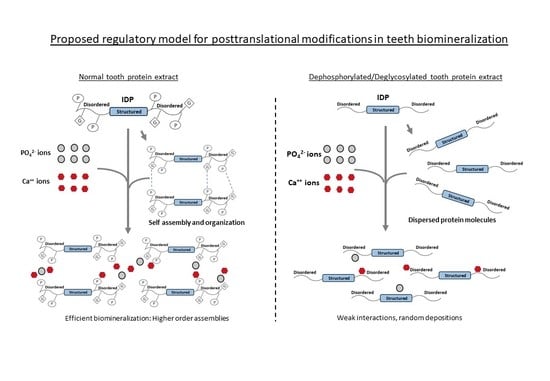
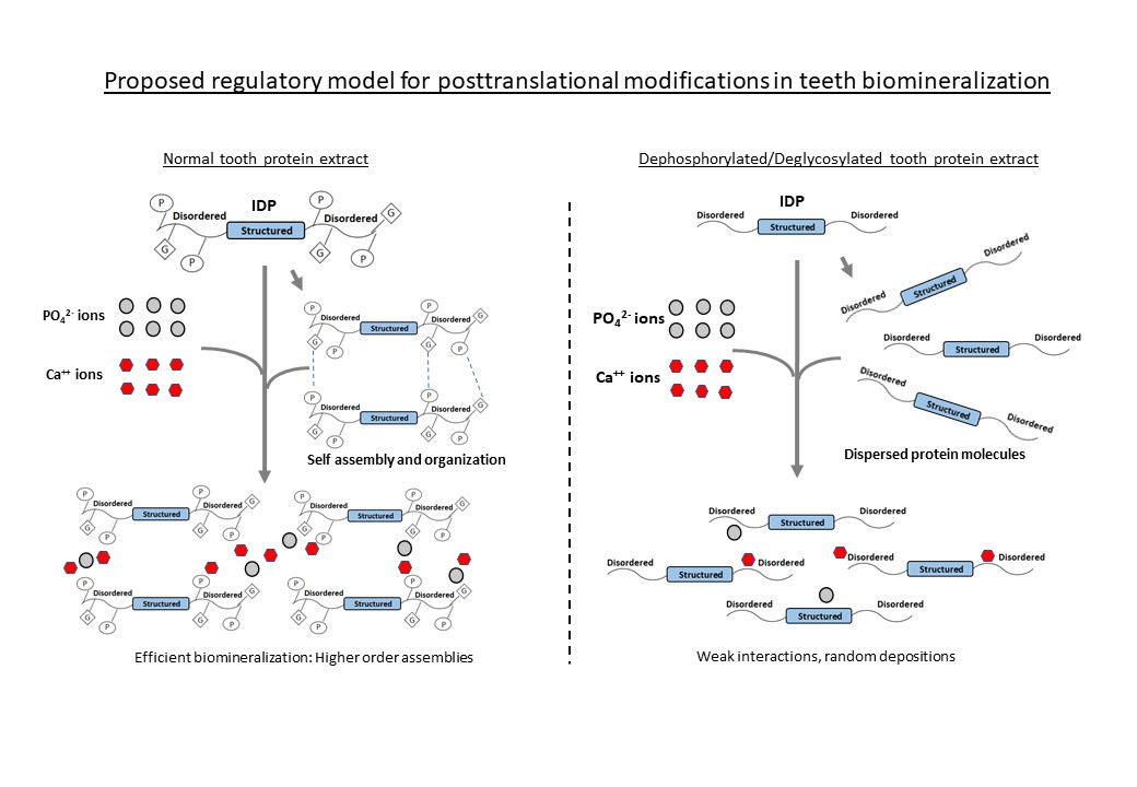
1. Introduction
2. Results and Discussion
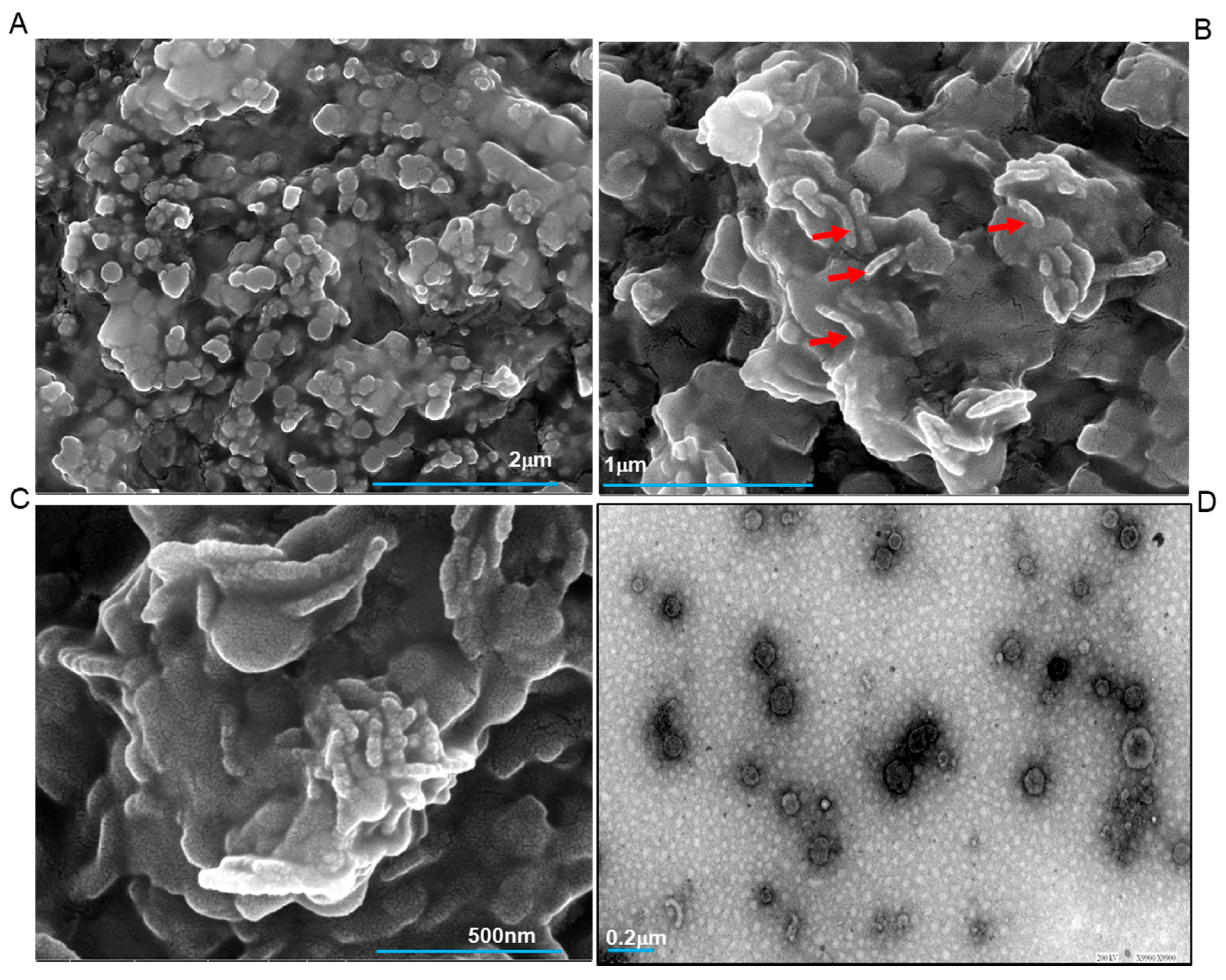
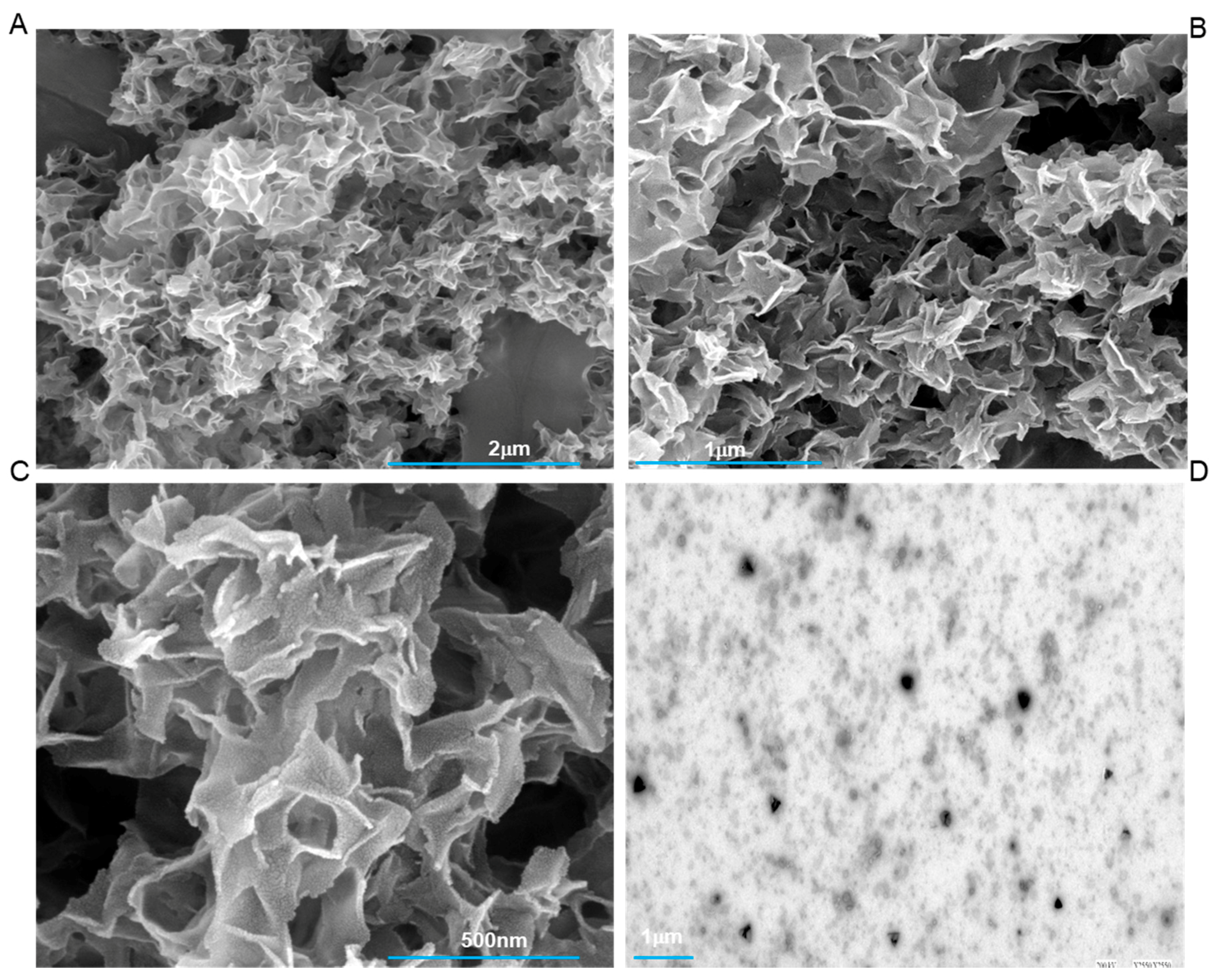
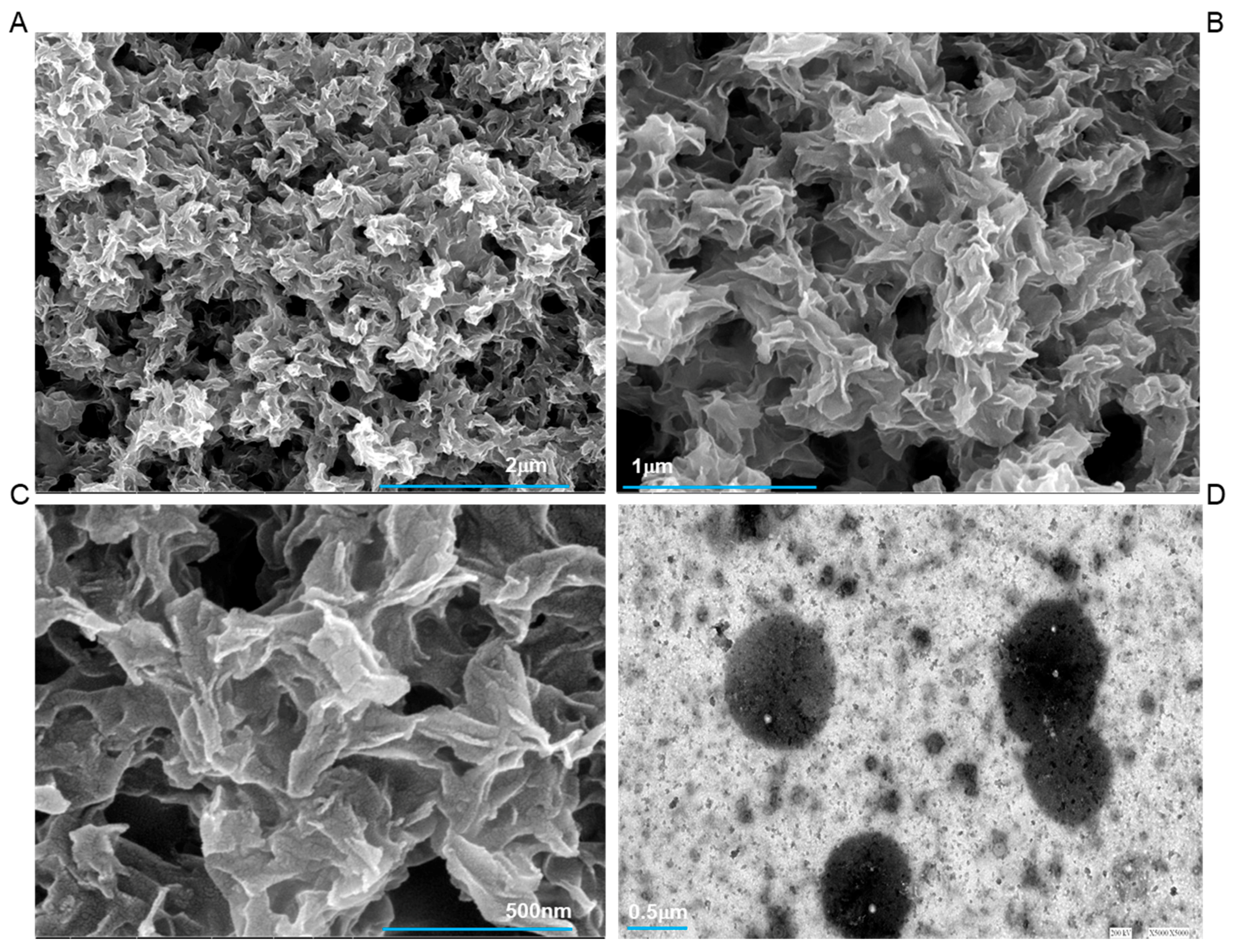
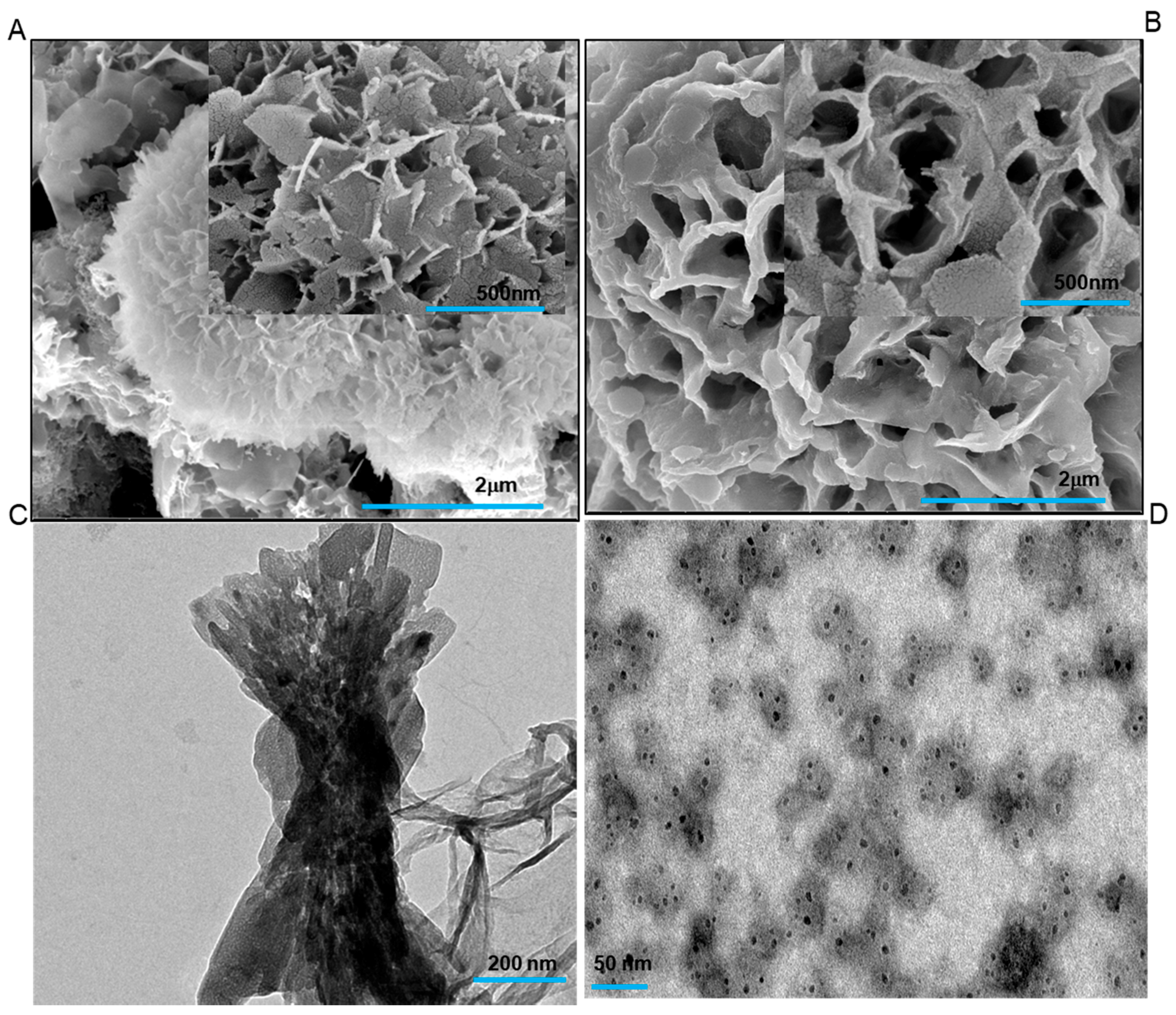
2.1. Scanning and Transmission Electron Microscopy (SEM and TEM) Analysis
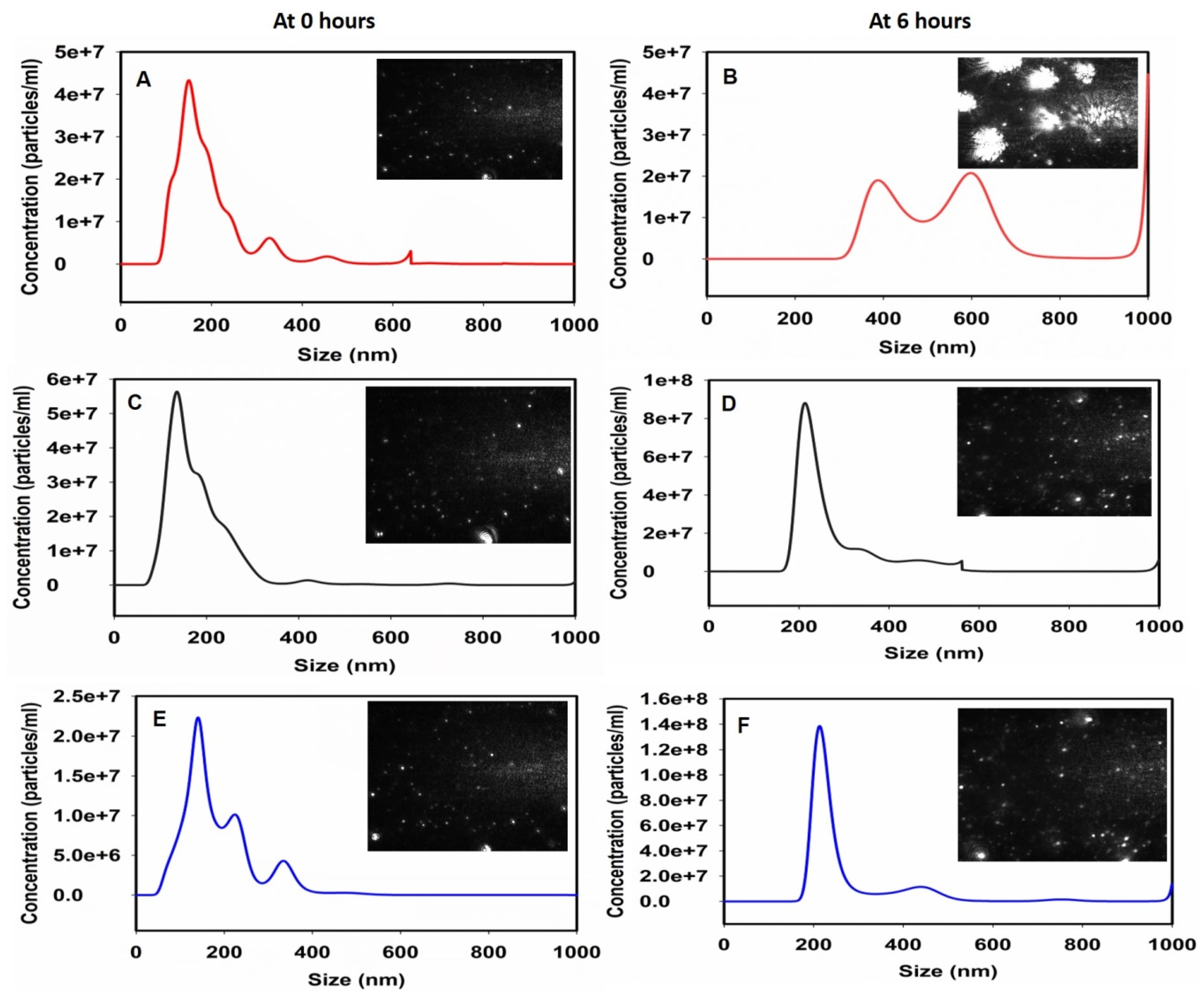
2.2. Nanoparticle Tracking Analysis (NTA)
3. Materials and Methods
3.1. Isolation of Proteins from Human Teeth
3.2. Dephosphorylation of the Tooth Protein Extract
3.3. Deglycosylation of the Tooth Protein Extract
3.4. Zeta Potential Measurements
3.5. In Vitro Mineralization Assay
3.6. High-Resolution Field Emission Scanning Electron Microscopy (HR FESEM)
3.7. High-Resolution Transmission Electron Microscopy (HRTEM)
3.8. Nanoparticle Tracking Analysis (NTA)
4. Conclusions
Supplementary Materials
Author Contributions
Funding
Acknowledgments
Conflicts of Interest
References
- Boskey, A.L.; Villarreal-Ramirez, E. Intrinsically disordered proteins and biomineralization. Matrix Biol. 2016, 52–54, 43–59. [Google Scholar] [CrossRef] [PubMed]
- Kalmar, L.; Homola, D.; Varga, G.; Tompa, P. Structural disorder in proteins brings order to crystal growth in biomineralization. Bone 2012, 51, 528–534. [Google Scholar] [CrossRef] [PubMed]
- Habchi, J.; Tompa, P.; Longhi, S.; Uversky, V.N. Introducing protein intrinsic disorder. Chem. Rev. 2014, 114, 6561–6588. [Google Scholar] [CrossRef] [PubMed]
- He, G.; Gajjeraman, S.; Schultz, D.; Cookson, D.; Qin, C.; Butler, W.T.; Hao, J.; George, A. Spatially and temporally controlled biomineralization is facilitated by interaction between self-assembled dentin matrix protein 1 and calcium phosphate nuclei in solution. Biochemistry 2005, 44, 16140–16148. [Google Scholar] [CrossRef] [PubMed]
- Fan, D.; Lakshminarayanan, R.; Moradian-Oldak, J. The 32kDa enamelin undergoes conformational transitions upon calcium binding. J. Struct. Biol. 2008, 163, 109–115. [Google Scholar] [CrossRef] [PubMed]
- Delak, K.; Harcup, C.; Lakshminarayanan, R.; Sun, Z.; Fan, Y.; Moradian-Oldak, J.; Evans, J.S. The tooth enamel protein, porcine amelogenin, is an intrinsically disordered protein with an extended molecular configuration in the monomeric form. Biochemistry 2009, 48, 2272–2281. [Google Scholar] [CrossRef] [PubMed]
- Amos, F.F.; Ndao, M.; Ponce, C.B.; Evans, J.S. A C-RING-like domain participates in protein self-assembly and mineral nucleation. Biochemistry 2011, 50, 8880–8887. [Google Scholar] [CrossRef] [PubMed]
- Tartaix, P.H.; Doulaverakis, M.; George, A.; Fisher, L.W.; Butler, W.T.; Qin, C.; Salih, E.; Tan, M.; Fujimoto, Y.; Spevak, L.; et al. In vitro effects of dentin matrix protein-1 on hydroxyapatite formation provide insights into in vivo functions. J. Biol. Chem. 2004, 279, 18115–18120. [Google Scholar] [CrossRef] [PubMed]
- Chang, E.P.; Russ, J.A.; Verch, A.; Kröger, R.; Estroff, L.A.; Evans, J.S. The intrinsically disordered C-RING biomineralization protein, AP7, creates protein phases that introduce nanopatterning and nanoporosities into mineral crystals. Biochemistry 2014, 53, 4317–4319. [Google Scholar] [CrossRef] [PubMed]
- Gao, J.; Xu, D. Correlation between posttranslational modification and intrinsic disorder in protein. Pac. Symp. Biocomput. 2012, 94–103. [Google Scholar]
- George, A.; Veis, A. Phosphorylated proteins and control over apatite nucleation, crystal growth, and inhibition. Chem. Rev. 2008, 108, 4670–4693. [Google Scholar] [CrossRef] [PubMed]
- Iijima, M.; Fan, D.; Bromley, K.M.; Sun, Z.; Moradian-Oldak, J. Tooth enamel proteins enamelin and amelogenin cooperate to regulate the growth morphology of octacalcium phosphate crystals. Cryst. Growth Des. 2010, 10, 4815–4822. [Google Scholar] [CrossRef] [PubMed]
- Wiedemann-Bidlack, F.B.; Kwak, S.Y.; Beniash, E.; Yamakoshi, Y.; Simmer, J.P.; Margolis, H.C. Effects of phosphorylation on the self-assembly of native full-length porcine amelogenin and its regulation of calcium phosphate formation in vitro. J. Struct. Biol. 2011, 173, 250–260. [Google Scholar] [CrossRef] [PubMed]
- Kwak, S.Y.; Green, S.; Wiedemann-Bidlack, F.B.; Beniash, E.; Yamakoshi, Y.; Simmer, J.P.; Margolis, H.C. Regulation of calcium phosphate formation by amelogenins under physiological conditions. Eur. J. Oral Sci. 2011, 119, 103–111. [Google Scholar] [CrossRef] [PubMed]
- Kwak, S.Y.; Kim, S.; Yamakoshi, Y.; Simmer, J.P.; Beniash, E.; Margolis, H.C. Regulation of calcium phosphate formation by native amelogenins in vitro. Connect. Tissue Res. 2014, 55, 21–24. [Google Scholar] [CrossRef] [PubMed]
- Tarasevich, B.J.; Howard, C.J.; Larson, J.L.; Snead, M.L.; Simmer, J.P.; Paine, M.; Shaw, W.J. The nucleation and growth of calcium phosphate by amelogenin. J. Cryst. Growth 2007, 304, 407–415. [Google Scholar] [CrossRef] [PubMed]
- Sharma, V.; Srinivasan, A.; Roychoudhury, A.; Rani, K.; Tyagi, M.; Dev, K.; Nikolajeff, F.; Kumar, S. Characterization of protein extracts from different types of human teeth and insight in biomineralization. Sci. Rep. 2019, 9, 9314. [Google Scholar] [CrossRef] [PubMed]
- Kwak, S.Y.; Wiedemann-Bidlack, F.B.; Beniash, E.; Yamakoshi, Y.; Simmer, J.P.; Litman, A.; Margolis, H.C. Role of 20-kDa amelogenin (P148) phosphorylation in calcium phosphate formation in vitro. J. Biol. Chem. 2009, 284, 18972–18979. [Google Scholar] [CrossRef] [PubMed]
- Akita, H.; Fukae, M.; Shimoda, S.; Aoba, T. Localization of glycosylated matrix proteins in secretory porcine enamel and their possible functional roles in enamel mineralization. Arch. Oral Biol. 1992, 37, 953–962. [Google Scholar] [CrossRef]


© 2019 by the authors. Licensee MDPI, Basel, Switzerland. This article is an open access article distributed under the terms and conditions of the Creative Commons Attribution (CC BY) license (http://creativecommons.org/licenses/by/4.0/).
Share and Cite
Sharma, V.; Rani, K.; Roychoudhury, A.; Chawla, A.; Nikolajeff, F.; Kumar, S. Novel Insights into Regulation of Human Teeth Biomineralization: Deciphering the Role of Post-Translational Modifications in a Tooth Protein Extract. Int. J. Mol. Sci. 2019, 20, 4035. https://doi.org/10.3390/ijms20164035
Sharma V, Rani K, Roychoudhury A, Chawla A, Nikolajeff F, Kumar S. Novel Insights into Regulation of Human Teeth Biomineralization: Deciphering the Role of Post-Translational Modifications in a Tooth Protein Extract. International Journal of Molecular Sciences. 2019; 20(16):4035. https://doi.org/10.3390/ijms20164035
Chicago/Turabian StyleSharma, Vaibhav, Komal Rani, Ajoy Roychoudhury, Amita Chawla, Fredrik Nikolajeff, and Saroj Kumar. 2019. "Novel Insights into Regulation of Human Teeth Biomineralization: Deciphering the Role of Post-Translational Modifications in a Tooth Protein Extract" International Journal of Molecular Sciences 20, no. 16: 4035. https://doi.org/10.3390/ijms20164035
APA StyleSharma, V., Rani, K., Roychoudhury, A., Chawla, A., Nikolajeff, F., & Kumar, S. (2019). Novel Insights into Regulation of Human Teeth Biomineralization: Deciphering the Role of Post-Translational Modifications in a Tooth Protein Extract. International Journal of Molecular Sciences, 20(16), 4035. https://doi.org/10.3390/ijms20164035