Review

bHLH–PAS Proteins: Their Structure and Intrinsic Disorder

by
Marta Kolonko
and
Beata Greb-Markiewicz
*
Department of Biochemistry, Faculty of Chemistry, Wroclaw University of Science and Technology, Wybrzeże Wyspiańskiego 27, 50-370 Wroclaw, Poland
*
Author to whom correspondence should be addressed.
Int. J. Mol. Sci. 2019, 20(15), 3653; https://doi.org/10.3390/ijms20153653
Submission received: 24 June 2019 / Revised: 15 July 2019 / Accepted: 16 July 2019 / Published: 26 July 2019

Abstract

:
The basic helix–loop–helix/Per-ARNT-SIM (bHLH–PAS) proteins are a class of transcriptional regulators, commonly occurring in living organisms and highly conserved among vertebrates and invertebrates. These proteins exhibit a relatively well-conserved domain structure: the bHLH domain located at the N-terminus, followed by PAS-A and PAS-B domains. In contrast, their C-terminal fragments present significant variability in their primary structure and are unique for individual proteins. C-termini were shown to be responsible for the specific modulation of protein action. In this review, we present the current state of knowledge, based on NMR and X-ray analysis, concerning the structural properties of bHLH–PAS proteins. It is worth noting that all determined structures comprise only selected domains (bHLH and/or PAS). At the same time, substantial parts of proteins, comprising their long C-termini, have not been structurally characterized to date. Interestingly, these regions appear to be intrinsically disordered (IDRs) and are still a challenge to research. We aim to emphasize the significance of IDRs for the flexibility and function of bHLH–PAS proteins. Finally, we propose modern NMR methods for the structural characterization of the IDRs of bHLH–PAS proteins.

1. Introduction to bHLH–PAS Proteins

The basic helix–loop–helix/Per-ARNT-SIM (bHLH–PAS) proteins are a class of transcriptional regulators that commonly occur in living organisms. They play an important role in the regulation of a variety of developmental and physiological events [1]. The maintenance of cellular and systemic oxygen homeostasis is performed by hypoxia-inducible factor 1α (HIF1-α) [2]. In the hypoxia condition, HIF1-α is translocated to the nucleus [3] where it regulates transcription activity related to angiogenesis, cell proliferation/survival, glucose metabolism, and iron metabolism. The incorrect control of the listed processes is fundamental in many diseases, including cancer, strokes, and heart disease [2]. Some bHLH–PAS family members act as receptors for different high and low molecular ligands [1]. The only known small ligand-activated bHLH–PAS protein, aryl hydrocarbon receptor (AHR), is involved in toxin metabolism and binds highly toxic ligands, such as TCDD [4]. The ligated AHR migrates to the nucleus and mediates a wide range of biological responses to poisons. This mediation comprises a wasting syndrome, hepatotoxicity, teratogenesis, and tumor promotion [4]. Overexpression and constitutive activation of the AHR have been observed in various types of tumors [5]. Importantly, the AHR has been described as a critical modulator of host–environment interactions, especially for immune and inflammatory responses [6].
Another interesting example of a bHLH–PAS family member is the single-minded protein (SIM), which plays a significant role during central nerve cord [7] and genital imaginal disc development [8]. As shown, SIM gene mutations contribute to certain dysmorphic features of brain development and also the mental retardation in Down syndrome [9]. Interestingly, SIM overexpression is also associated with breast and prostate cancer [10], which indicates connections between their apparently unrelated signaling pathways.
Members of the bHLH–PAS family were shown to be targets for disease therapy. AHR, highly expressed in multiple organs and tissues, may influence tumorigenesis both by direct effect on the cancer cells and by modulation of the immune system. For this reason, the development of selective AHR modulators active against multiple tumors is a desirable direction of research [11]. Also, targeting of the HIF1-α pathway as a novel cancer therapy is a current project [12]. As AHR was shown to modulate the immune response in the respiratory tract, this protein can be potentially used also as a therapeutic object for the treatment of various inflammatory lung diseases [13,14]. Another member of the family, expressed mainly in the brain, neuronal PAS domain-containing protein 4 (NPAS4) has been proposed as a novel therapeutic target for depression and neurodegenerative diseases [15] and as a component of new stroke therapies [16]. Additionally, NPAS4, whose expression was also detected in the pancreas, was proposed to be a therapeutic target for diabetes [17] and as a treatment during pancreas transplantation [18].
In spite of performing a high diversity of functions, the bHLH–PAS proteins family exhibits a relatively well-conserved domain structure in the N-terminal part of their sequence (Figure 1). The bHLH region contains approximately 60 amino acid (aa) residues and can be divided into two functionally distinctive parts: the basic region responsible for DNA binding (approximately 15 aa), and the neighboring C-terminal HLH region, which takes part in protein dimerization [19]. The PAS domain is located in the central part of the protein and usually comprises about 300 aa residues [1]. It is divided into two structurally conserved regions named PAS-A and PAS-B, which are often connected to a single PAS-associated C-terminal (PAC) motif [20]. The PAS-A and PAS-B regions are separated by a poorly conserved link [1]. The PAS-A region is critical for selecting a dimerization partner and ensuring the specificity of target gene activation [21]. The PAS-B region is usually responsible for sensing diverse exogenous and endogenous signals, and is accompanied by energetic and conformational changes that regulate protein activity [21]. Contrary to conserved domains, the C-termini of bHLH–PAS proteins present significant variability [21] and contain variable transcription activation/repression domains (TAD/RPD) (Figure 1) [22,23]. An example is the mammalian SIM existing in two isoforms: SIM1 and SIM2. Both isoforms present a high amino acid identity in their N-termini (90% identity in the bHLH and PAS regions) and extreme diversity in their C-termini [24]. While SIM1 activates the expression of target genes, SIM2 acts as an inhibitor. Interestingly, the opposite transcriptional effect disappears after the deletion of both SIM1 and SIM2 C-termini, resulting in proteins with a similar activity [25,26]. Moffet and Pelletier [26] demonstrated that a distinct SIM2 C-terminal sequence comprises two repression domains with a high proline/serine and proline/alanine content, respectively. It is a feature of “repressor motifs”, which can also be found in a large number of other transcriptional repressors [25,26]. Due to the highly variable amino acid sequence and the lack of predefined domains, C-termini are believed to be responsible for the specific modulation of the functioning of bHLH–PAS proteins and the recognition of partner proteins necessary for their unique action [21].
Generally, bHLH–PAS proteins can be divided into two classes. While the expression of class I proteins is specifically regulated by diverse physiological states and/or environmental signals [30], class II proteins are expressed continuously and serve as heterodimerization partners for class I members. Only the dimer of the two bHLH–PAS proteins acts as a functional transcription factor complex, regulating the expression of genes under its control [22]. Mammalian bHLH–PAS transcription factors are listed in Table 1.

2. bHLH–PAS Protein Conservation between Organisms

bHLH–PAS proteins are highly conserved among different organisms, including vertebrates and invertebrates [33]. Most mammalian representatives possess orthologs in insect species. An example is the Drosophila melanogaster TANGO (TGO) protein, which is a homologue of the mammalian class II protein, ARNT [34]. TGO is known as the general dimerization partner for Similar (SIMA), Trachealess (TRH), Single-minded (SIM) protein, Spineless (SS), and Dysfusion (DYS), performing functions equivalent to mammalian ones.
In 2017, the Nobel Prize in Physiology or Medicine was awarded to J. C. Hall, M. Rosbash, and M. W. Young for their discoveries of molecular mechanisms controlling the circadian rhythm in D. melanogaster. As shown, the two bHLH–PAS transcription factors CLOCK and CYCLE play a key role as transcriptional activators for period (per) and timeless (tim) genes [35,36]. Thanks to the conservation of circadian bHLH–PAS proteins between D. melanogaster and mammals [35], the explanation of the fly daily rhythm enabled the understanding of a similar, though much more complicated, process in mammals, controlled by two orthologous to CLOCK/CYCLE heterodimers: CLOCK/BMAL1 and NPAS-2/BMAL1 [22].
In spite of significant similarities, some exceptions between vertebrates and invertebrates can be noticed. The bHLH–PAS transcription factor, Methoprene-tolerant protein (MET), occurs exclusively in insects and to date has no known ortholog in nonarthropod organisms. MET has been recently confirmed as the juvenile hormone (JH) receptor playing a significant role during insect development and maturation [37]. Interestingly, in a few species of insects, like D. melanogaster and Bombyx mori, there exist the MET paralogs named germ-cell expressed (GCE) and MET2, respectively [38]. MET and GCE participate in modulating JH signaling during D. melanogaster development, but their functions are not fully redundant and the proteins exhibit tissue-specific distribution [39]. In turn, the MET2 protein function in B. mori is not yet defined [40].

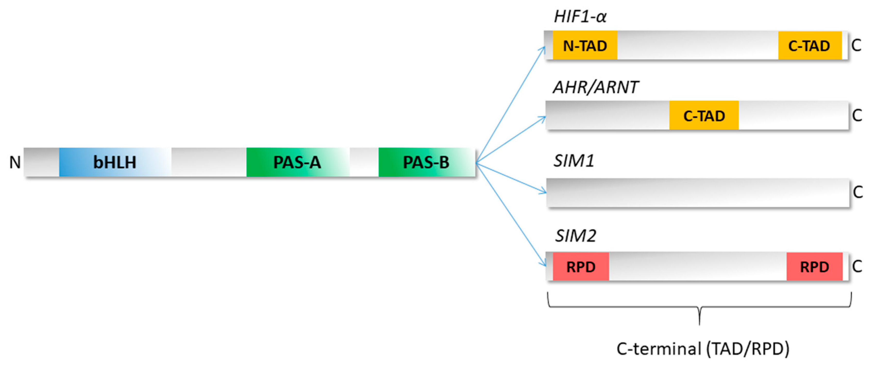
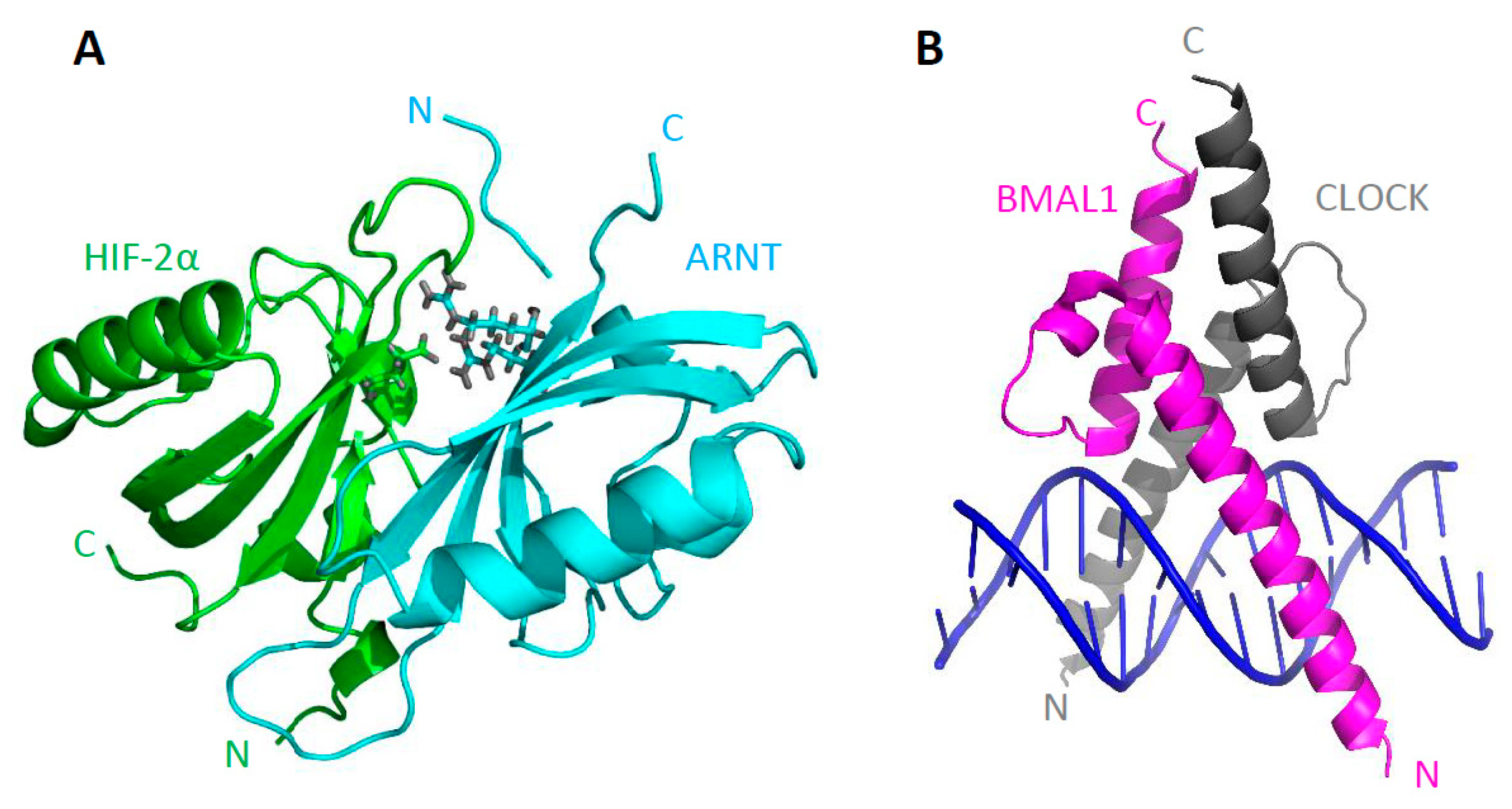
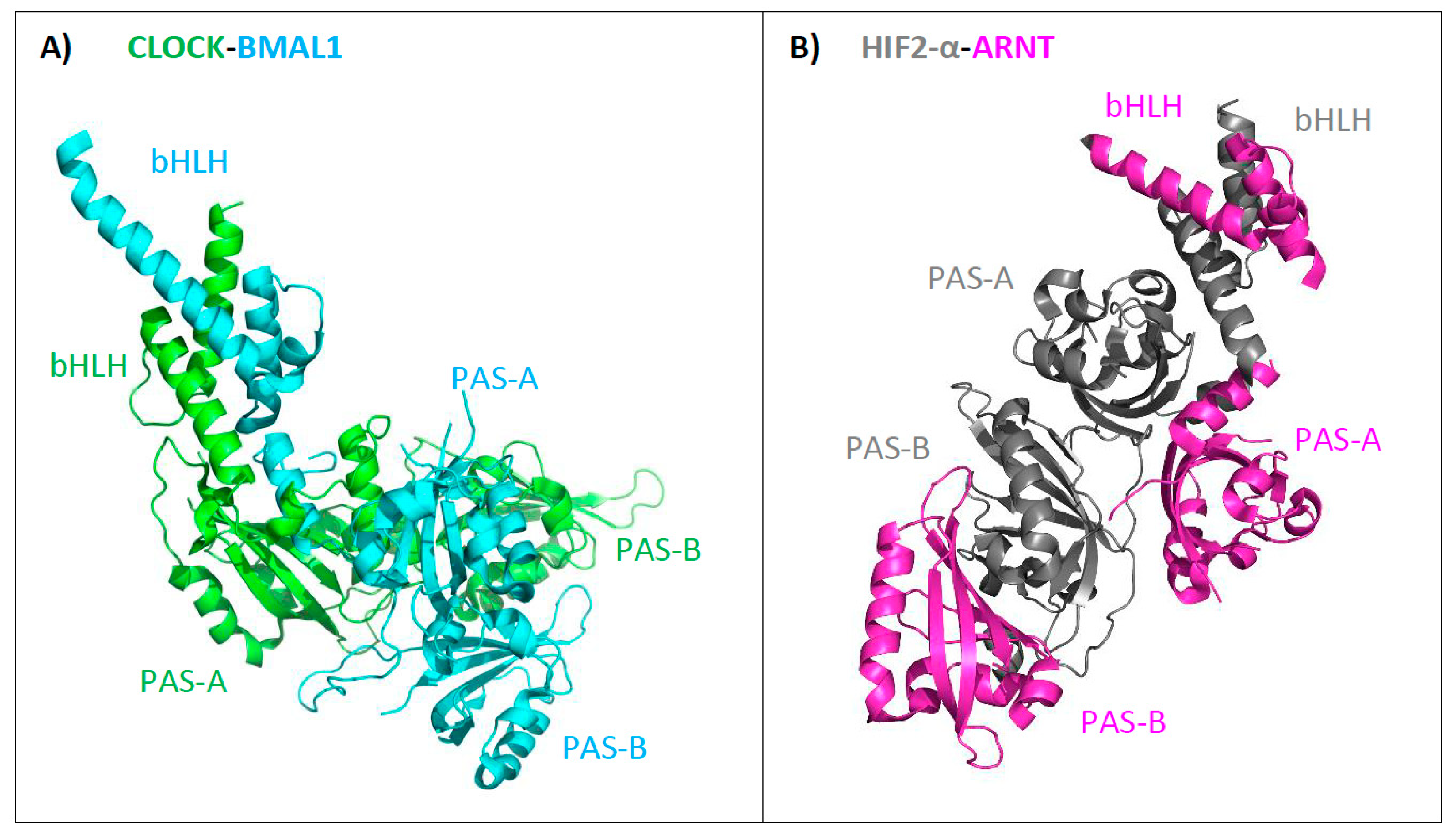
3. Structure of bHLH–PAS Proteins

To date, our knowledge regarding the tertiary structure of bHLH–PAS proteins is limited. All determined structures comprise single isolated domains (PAS-A or PAS-B) or adjacent domains connected with flexible aa chains. C-termini, however, comprising an extensive part of proteins, have not yet been structurally characterized. These regions are not homologous to any described domains and seem to be very disordered. Consequently, it can be seen to be a huge challenge for scientists to determine their structure and combine it with specific protein functions. All bHLH–PAS structures deposited in the Protein Data Bank (PDB) are listed in Table 2 (Nuclear Resonance Magnetism (NMR) structures) and Table 3 (X-ray structures). Most of the listed assemblies correspond to heterodimers.
The first step in determining the structure of bHLH–PAS proteins was the isolation and characterization of PAS-B domains from HIF2-α (Figure 2A) [41] and ARNT (Figure 2B) [42]. Both structures were obtained using the NMR technique and presented a fold characteristic for the PAS domain: a five-stranded antiparallel β-sheet flanked by several α-helices [42]. The next step was the crystallization of the isolated PAS-A domain of AHR (Figure 2C) and the PAS-B domains of ARNT (not shown) and HIF1-α (not shown). Interestingly, the tertiary architecture of all structurally characterized PAS domains is very conserved (Figure 2), despite the fact that their primary sequence is highly divergent (sequence identity lower than 20%) [43].
Further experiments led to the cocrystallization of PAS-B domains from the HIF2-α/ARNT heterodimer, which revealed that these two domains form an interaction interface via their β-sheets in an antiparallel form (Figure 3A) [42]. Another measurement covering bHLH domains of BMAL1/CLOCK bound to the DNA defined domain structure and binding properties specifying interactions taking place (Figure 3B) [44]. A typical bHLH domain comprises two long α helices connected by a short loop. The first helix includes the basic domain and interacts with the major groove of the DNA [45]. All presented structures allowed an insight into the organization of bHLH–PAS proteins; however, the structure of the multidomain bHLH–PAS protein was still missing.
A turning point was the year 2012, when the first heterodimer comprising the bHLH–PAS-A/PAS-B domains (CLOCK-BMAL1) was crystallized [47] and its structure was resolved (Figure 4A). In 2015, the architecture of two other heterodimers, HIF1-α-ARNT (not shown) and HIF2-α-ARNT (Figure 4B), were obtained [48]. All determined structures present the position of the defined domains in relation to each other in the functional heterodimers. In general, the individual PAS domains are not involved in equal interactions, and the obtained structures are highly asymmetric. Importantly, two groups of heterodimers (based on BMAL-1 or ARNT proteins as a dimerization partner) present separate types of quaternary architecture. All domains in the BMAL-1 group are close spatially to each other (Figure 4A), while ARNT domains do not create intramolecular interactions and can wrap up around a partner protein (Figure 4B) [22,48].
To date, all available structural information concerns mammalian bHLH–PAS proteins. There is almost no information about the structure of proteins derived from other organisms, including invertebrate D. melanogaster. It would be interesting to verify evolutionary conservation of the entire bHLH–PAS fold in different organisms. The majority of reported protein structures include defined N-terminal domains, while the structural information about C-terminal regions is still missing and limited to short peptides bound to interacting proteins [22]. An example is a short motif featuring a conserved sequence LIXXL found in D. melanogaster MET and GCE, which represents a novel nuclear receptor (NR) box. The Docking models of the MET/GCE NR box associating peptides to the orphan nuclear receptor (FTZ-F1) ligand-binding domain (LBD) revealed their α-helical structure, necessary for hydrophobic interaction [49].

4. Unique Properties of the C-Terminal Domains of bHLH–PAS Proteins as IDRs

While the N-terminal part of bHLH–PAS proteins is responsible for interactions with DNA, ligands/cofactors binding, and heterodimerization, their C-termini are usually responsible for the regulation of the protein and the activity of created complexes [50]. The variability of the amino acid sequence of C-terminal fragments, their transactivation role, and the lack of homology to any described domains prompted us to ask the question about the structural character of these regions and the relationship of their character with the performed function. For a long time, it was believed that spontaneous folding into a well-defined and stable tertiary structure is required for the protein action [51]. However, it is actually known that more than 20–30% of eukaryotic proteins do not have a stable tertiary structure in physiological conditions, but at the same time still perform important biological functions. Such proteins are referred to as intrinsically disordered proteins (IDPs). Simultaneously, over 70% of proteins involved in signal transduction cascades have long intrinsically disordered regions (IDRs). Importantly, the lack of a defined structure is critical for the functionality of IDPs and IDRs [52]. Additionally, the conformational plasticity and elongated shape make them a frequent target of different kinds of post-translational modifications (phosphorylation, acetylation, methylation, and others) that regulate protein activity [53]. IDPs were identified as elements of cellular signaling which control mechanisms and protein interaction networks [54]. IDPs were also shown to take part in disease-related signaling transduction; for example, intrinsically disordered amyloid β-peptides are involved in Alzheimer’s disease [55]. Therefore, IDPs can be seen to be targets for drug design strategies.

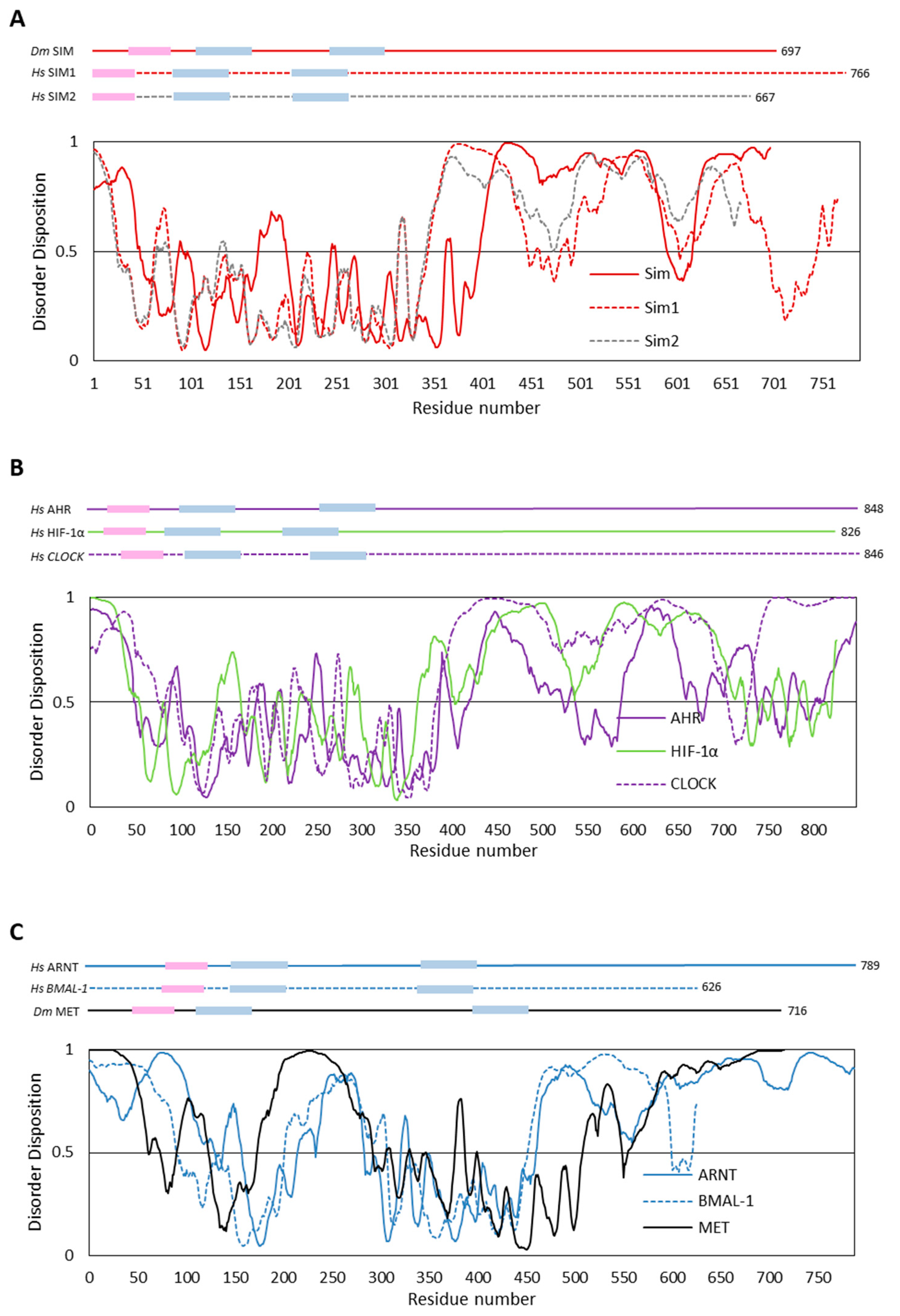
4.1. In Silico Analyses of Selected bHLH–PAS Proteins

To estimate the occurrence of putative IDRs in bHLH–PAS proteins, we performed in silico analyses of the composition, hydropathy, and sequence complexity of amino acid sequences corresponding to selected proteins. We used the previously described human SIM1 and SIM2, as well as their D. melanogaster ortholog, SIM (Figure 5A), representing the class I of the family. To obtain a wider spectrum, we studied other human class I members, AHR, HIF1-α, and CLOCK (Figure 5B), which are engaged in different signal transduction pathways. As mentioned previously, class I proteins dimerize with class II proteins to form a functional complex and are crucial for heterodimer specificity. As each bHLH–PAS class II transcription factor is able to interact with different class I members, we found it to be extremely interesting to perform in silico analysis of the structure of class II members. We chose human ARNT, human BMAL1 (Figure 5C), and, additionally, D. melanogaster MET (Figure 5C) as a unique protein with an unknown mammalian homolog. MET can be classified as a class II bHLH–PAS family member based on its ability to not only create heterodimers with its paralog GCE, but also homodimers [56].
We performed in silico analysis using the predictors of intrinsically disordered regions: PONDR-VSL2 [57], PONDR-FIT [58], IUPred [59], and IsUnstruct [60]. Since the results of all the employed predictors were compatible, for the purpose of simplicity, we decided to show only one representative result (PONDR-VSL2) for each protein (Figure 5). All the results (Figure 5) substantiate our hypothesis and indicate the intrinsic character of the long C-termini. It is worth noting that for proteins representing class I (see Figure 5A,B), short ordered fragments in their C-termini are visible. Such fragments are able to act as TAD/RPD or so-called molecular recognition elements (MoREs) [61,62]. The presence of MoREs makes the interactions between partner proteins highly specific and reversible [52]. The presented results revealed some subtle differences in the regions that comprise preserved domains. The structure predicted for class I proteins (Figure 5A,B) is undeniably more ordered, while class II proteins show a marked structure relaxation in their middle part (Figure 5C), which is C-terminally linked to the PAS-A domain responsible for specificity of gene activation by bHLH–PAS proteins [63]. Such a difference explains the ability of class II proteins to serve as an interaction partner for different proteins [22]. The ability of IDRs (and IDPs) to interact with several partners is an undeniable advantage in molecular recognition processes [64]. Importantly, the resulting induced folding may differ depending on the binding partner. For example, a disordered region of p53 protein, a known cell cycle regulator and a tumor suppressor [65], folds into alpha helix or beta strand, depending on the partner protein [66].

4.2. The Impact of Disordered Regions on Protein Function

The flexibility and disorder detected in individual C-termini can be related to the ability of individual bHLH–PAS proteins to perform diverse functions. The differences between SIM1 and SIM2 C-termini, regarding their opposite functions (gene activation/repression), have previously been described. C-terminal regions of two other studied proteins, AHR (class I member) and ARNT (class II member), are characterized by the presence of TADs [67], in which functions are mediated by CBP/p300 and RIP140 coactivators. The C-terminal region of ARNT was additionally proposed to be a crucial activator of the estrogen receptor (ER) [68]. Interestingly, the suppression of AHR activity is also connected with the C-terminus and is mediated by the binding of the small peptide inhibitor [69]. Another repressor of the AHR signaling pathway, AHRR, is distinguished from AHR by the presence of three SUMOylation sites in its C-terminus. As shown, SUMOylation is crucial for full suppressive activity of AHRR [70].
Moreover, the C-terminus of another studied protein, HIF1-α, is characterized by the presence of TADs, and it also interacts with the CBP/p300 coactivator. The C-terminus is additionally responsible for protein stability/degradation and contains sequence motifs influencing subcellular localization: nuclear localization signal (NLS) and nuclear export signal (NES) [71,72].
Another remarkable class I bHLH–PAS protein is CLOCK, comprising a domain with histone transacetylase (HAT) activity in the C-terminus. This domain is responsible for histones acetylation, which affects the transcriptional stimulation of clock-controlled genes. Additional acetylation is performed on the R537 residue of the partner protein, BMAL1. R537 residue is located in the C-terminal part of BMAL1 and its modification facilitates the cryptochrome (CRY1)-mediated repression of specific gene transcription [73]. Importantly, CRY1 competes with the CBP/p300 coactivator for BMAL1 TAD binding, and is not able to bind the C-terminus in the paralog protein, BMAL2. Therefore, C-termini distinguish the circadian functions of these two BMAL paralogs [74].

4.3. Structural Analysis of bHLH–PAS C-Terminal Fragments

To date, the only structurally characterized C-terminal fragment of the bHLH–PAS protein is the D. melanogaster MET C-terminus (MET/C) [75]. It was shown by a series of in vitro analyses that MET/C exhibits a highly disordered character and exists in a solution in extended flexible form with predispositions for conformational changes. It is interesting to note that some short secondary motifs in the structure of MET/C have been predicted. Such short ordered fragments can be important during partner recognition and interactions. It was hypothesized that the intrinsic disorder of the C-terminal fragment was indispensable for the functionality of MET due to it modulating the protein’s action in a context-specific way. It enables cross-talk between JH signaling and other signaling pathways during D. melanogaster development. Previously, it was shown that Met interacts with FTZ-F1 by its C-terminus [49], thereby modulating stage-specific responses to the hormones during D. melanogaster metamorphosis [76].
As all the in vitro analyses results obtained for the MET/C [75] were consistent with the in silico studies presented above (Figure 5C), we hypothesize that the disorder character of the bHLH–PAS proteins subfamily C-terminal fragments can be a more common characteristic and also be very important for their functionality. Previously, the importance of the disordered character of regions flanking the bHLH domain of bHLH transcription factors was shown [77,78,79].

4.4. Structural Analysis of IDPs

While C-terminal regions of the bHLH–PAS family are considered as IDRs, it can be challenging to detect and characterize them. The reason is that IDPs and IDRs do not adopt a single stable structure and the energetically most favorable conformations can be very distinguished [80,81]. The tiny conformational changes can promote IDPs/IDRs aggregation [82]. Additionally, it was shown that IDPs/IDRs can be highly sensitive to proteolysis [83]. Currently, studies focused on the characterization of IDRs and IDPs are rapidly developing, and techniques enabling the study of proteins in solution are still improving.
There are a number of bioinformatics tools allowing primary recognition of disordered proteins. Since IDPs are characterized by the specific aa composition (a low content of hydrophobic and a high content of charged residues [84]), the Composition Profiler [85] is commonly used to compare aa distribution between the studied protein and IDPs (DisProt3.4 database)/globular proteins (PDB S25 database). Additionally, for IDPs and globular proteins distinguishing, the Uversky diagram plotting mean net charge versus mean hydrophobicity is useful [86]. Disorder predictors (like PONDR-VSL2 [57], PONDR-FIT [58], IUPred [59], and IsUnstruct [60] used in this work) allow determining the probability of IDR occurrence utilizing the neural networks, trained on selected sets of ordered and disordered sequences. Another predictor, DynaMine, provides information about protein backbone flexibility [87,88]. IDPs, once purified, can be identified by various experimental methods. First, the underestimated mobility during SDS-PAGE electrophoresis can indicate the extended and elongated shape of the protein [75]. Hydrodynamic analysis comprising Size Exclusion Chromatography (SEC) [89] and Analytical Ultracentrifugation (AUC) are commonly used to determine hydrodynamic properties, like the Stokes radius (RS), the sedimentation coefficients (s), and the frictional ratios (f/f0) [90]. The Circular Dichroism (CD) is useful for secondary structures content calculation [91]. All listed techniques allow obtaining preliminary insight into protein structure properties.
One technique commonly used to study the overall shape and structural transitions of biological macromolecules in solution is small-angle X-ray scattering (SAXS) [92]. However, SAXS only provides limited information about the low-resolution overall shape of the molecule, so it is important to combine it with complementary high-resolution methods like NMR that present the local structure [93]. NMR offers unique opportunities that are based on analyzing the deviations from an idealized random coil devoid of any structural propensity [94]. The random coil exhibits characteristic chemical shifts, which are averages of all the possible conformations that amino acids can adopt in a solution. Therefore, NMR chemical shift deviations from random coil values can be used to evaluate the local transient secondary structure of IDPs [80]. The main problem during spectra assignments of IDPs is spectra overlapping (low chemical shifts dispersion) and a significant proton exchange with bulk water that reduces 1HN signal intensities, which in turn leads to low signal-to-noise ratios [94]. The exchange with water can be reduced by conducting measurements in low temperature or low pH [95]. Low-resolution spectra require the development of a novel NMR technique. Recently, IDP-dedicated methods such as 13C-direct detected experiments, paramagnetic relaxation enhancements (PREs), or residual dipolar couplings (RDCs) have been described [96].

5. Conclusions

The available structure characterization of bHLH–PAS proteins is limited to the relatively well-conserved domains bHLH, PAS-A, and PAS-B. Importantly, all structures deposited in the Protein Data Bank are obtained for mammalian family members, the majority of them being heterodimers. On the other hand, the important parts of bHLH–PAS factors, which comprise their long C-termini, have not yet been structurally characterized. These fragments perform important functions in the specific modulation of protein action and for the recognition of interacting partners.
Performed in silico analysis revealed that the C-termini of representatives of the class I bHLH–PAS protein family members (SIM, SIM1, SIM2, AHR, HIF1-α, and Clock), and also class II (ARNT, BMAL1, and MET), are predicted as intrinsically disordered regions (IDRs) and are not homologous to any described domains. We discussed the known functions of the presented C-termini proteins according to their disorder character. Moreover, we proposed NMR techniques for intrinsically disordered C-termini characterization [94]. We believe that the structural properties of subsequent IDRs predicted in the sequences of bHLH–PAS transcription factors (mainly C-termini) need to be resolved for a full understanding of the way of bHLH–PAS family transcription factors function.

Funding

The work was supported by The National Science Centre (NCN): PRELUDIUM predoctoral grant UMO-2017/27/N/NZ1/01783 and ETIUDA doctoral scholarship UMO-2018/28/T/NZ1/00337, and partially supported by a statutory activity subsidy 0401/0143/18 from the Polish Ministry of Science and Higher Education for the Faculty of Chemistry of Wroclaw University of Science and Technology.

Acknowledgments

We would like to thank Andrzej Ożyhar (Department of Biochemistry, Faculty of Chemistry, Wroclaw University of Science and Technology) for all valuable comments and suggestions.

Conflicts of Interest

The authors declare no conflict of interest.

Abbreviations

Aaamino acid
AHRaryl hydrocarbon receptor
AHRRaryl hydrocarbon receptor repressor
ARNTaryl hydrocarbon receptor nuclear translocator
bHLH–PAShelix–loop–helix/Per-ARNT-SIM
CLOCKcircadian locomotor output cycles protein kaput
CRY1cryptochrome
DYSdysfusion protein
ERestrogen receptor
GCEgerm cell-expressed protein
HAThistone transacetylase
HIFhypoxia-inducible factors
IDPsintrinsically disordered proteins
IDRsintrinsically disordered regions
JHjuvenile hormone
LBDligand-binding domain
METmethoprene-tolerant protein
MoREsmolecular recognition elements
MET/CC-terminus of the MET protein
NESnuclear export signal
NLSnuclear localization signal
NMRNuclear Resonance Magnetism
NPASneuronal PAS domain-containing proteins
NRboxes nuclear receptor boxes
PACPAS-associated C-terminal motif
PDBProtein Data Bank
PREsparamagnetic relaxation enhancements
RDCsresidual dipolar couplings
SAXSSmall-angle X-ray Scattering
SIMsingle-minded proteins
SIMAsimilar protein
SSspineless protein
TAD or TRDtransactivation or repression domain
TGOTANGO protein
TRHtrachealess protein

References

  1. Crews, S.T. Control of cell lineage-specific development and transcription by bHLH-PAS proteins. Genes Dev. 1998, 12, 607–620. [Google Scholar] [CrossRef] [PubMed] [Green Version]
  2. Lee, J.-W.; Bae, S.-H.; Jeong, J.-W.; Kim, S.-H.; Kim, K.-W. Hypoxia-inducible factor (HIF-1)α: Its protein stability and biological functions. Exp. Mol. Med. 2004, 36, 1–12. [Google Scholar] [CrossRef] [PubMed]
  3. Ema, M.; Hirota, K.; Mimura, J.; Abe, H.; Yodoi, J.; Sogawa, K.; Poellinger, L.; Fujii-Kuriyama, Y. Molecular mechanisms of transcription activation by HLF and HIF1alpha in response to hypoxia: Their stabilization and redox signal-induced interaction with CBP/p300. EMBO J. 1999, 18, 1905–1914. [Google Scholar] [CrossRef] [PubMed]
  4. Petrulis, J.R.; Kusnadi, A.; Ramadoss, P.; Hollingshead, B.; Perdew, G.H. The hsp90 Co-chaperone XAP2 Alters Importin β Recognition of the Bipartite Nuclear Localization Signal of the Ah Receptor and Represses Transcriptional Activity. J. Biol. Chem. 2003, 278, 2677–2685. [Google Scholar] [CrossRef] [PubMed]
  5. Xue, P.; Fu, J.; Zhou, Y. The Aryl Hydrocarbon Receptor and Tumor Immunity. Front. Immunol. 2018, 9, 286. [Google Scholar] [CrossRef] [PubMed] [Green Version]
  6. Neavin, D.; Liu, D.; Ray, B.; Weinshilboum, R. The Role of the Aryl Hydrocarbon Receptor (AHR) in Immune and Inflammatory Diseases. Int. J. Mol. Sci. 2018, 19, 3851. [Google Scholar] [CrossRef]
  7. Freer, S.M.; Lau, D.C.; Pearson, J.C.; Talsky, K.B.; Crews, S.T. Molecular and functional analysis of Drosophila single-minded larval central brain expression. Gene Expr. Patterns 2011, 11, 533–546. [Google Scholar] [CrossRef] [PubMed] [Green Version]
  8. Pielage, J.; Steffes, G.; Lau, D.C.; Parente, B.A.; Crews, S.T.; Strauss, R.; Klämbt, C. Novel behavioral and developmental defects associated with Drosophila single-minded. Dev. Biol. 2002, 249, 283–299. [Google Scholar] [CrossRef]
  9. Chen, H.; Chrast, R.; Rossier, C.; Gos, A.; Antonarakis, S.E.; Kudoh, J.; Yamaki, A.; Shindoh, N.; Maeda, H.; Minoshima, S.; et al. Single–minded and Down syndrome? Nat. Genet. 1995, 10, 9–10. [Google Scholar] [CrossRef]
  10. Bersten, D.C.; Sullivan, A.E.; Peet, D.J.; Whitelaw, M.L. bHLH–PAS proteins in cancer. Nat. Rev. Cancer 2013, 13, 827–841. [Google Scholar] [CrossRef]
  11. Safe, S.; Lee, S.O.; Jin, U.H. Role of the aryl hydrocarbon receptor in carcinogenesis and potential as a drug target. Toxicol. Sci. 2013, 1–16. [Google Scholar] [CrossRef] [PubMed]
  12. Masoud, G.N.; Li, W. HIF-1α pathway: Role, regulation and intervention for cancer therapy. Acta Pharm. Sin. B 2015, 5, 378–389. [Google Scholar] [CrossRef] [PubMed]
  13. Beamer, C.A.; Shepherd, D.M. Role of the aryl hydrocarbon receptor (AhR) in lung inflammation. Semin. Immunopathol. 2013, 35, 693–704. [Google Scholar] [CrossRef] [PubMed] [Green Version]
  14. Puccetti, M.; Paolicelli, G.; Oikonomou, V.; De Luca, A.; Renga, G.; Borghi, M.; Pariano, M.; Stincardini, C.; Scaringi, L.; Giovagnoli, S.; et al. Towards Targeting the Aryl Hydrocarbon Receptor in Cystic Fibrosis. Mediat. Inflamm. 2018, 2018, 1601486. [Google Scholar] [CrossRef] [PubMed]
  15. Zhang, Z.; Fei, P.; Mu, J.; Li, W.; Song, J. Hippocampal expression of aryl hydrocarbon receptor nuclear translocator 2 and neuronal PAS domain protein 4 in a rat model of depression. Neurol. Sci. 2014, 35, 277–282. [Google Scholar] [CrossRef] [PubMed]
  16. Choy, F.C.; Klarić, T.S.; Koblar, S.A.; Lewis, M.D. The Role of the Neuroprotective Factor Npas4 in Cerebral Ischemia. Int. J. Mol. Sci. 2015, 16, 29011–29028. [Google Scholar] [CrossRef] [PubMed] [Green Version]
  17. Sabatini, P.V.; Lynn, F.C. All-encomPASsing regulation of β-cells: PAS domain proteins in β-cell dysfunction and diabetes. Trends Endocrinol. Metab. 2015, 26, 49–57. [Google Scholar] [CrossRef]
  18. Speckmann, T.; Sabatini, P.V.; Nian, C.; Smith, R.G.; Lynn, F.C. Npas4 Transcription Factor Expression Is Regulated by Calcium Signaling Pathways and Prevents Tacrolimus-induced Cytotoxicity in Pancreatic Beta Cells. J. Biol. Chem. 2016, 291, 2682–2695. [Google Scholar] [CrossRef] [Green Version]
  19. Li, X.; Duan, X.; Jiang, H.; Sun, Y.; Tang, Y.; Yuan, Z.; Guo, J.; Liang, W.; Chen, L.; Yin, J.; et al. Genome-Wide Analysis of Basic/Helix-Loop-Helix Transcription Factor Family in Rice and Arabidopsis. Plant Physiol. 2006, 141, 1167–1184. [Google Scholar] [CrossRef] [Green Version]
  20. Ponting, C.P.; Aravind, L. PAS: A multifunctional domain family comes to light. Curr. Biol. 1997, 7, R674–R677. [Google Scholar] [CrossRef]
  21. Kewley, R.J.; Whitelaw, M.L.; Chapman-Smith, A. The mammalian basic helix–loop–helix/PAS family of transcriptional regulators. Int. J. Biochem. Cell Biol. 2004, 36, 189–204. [Google Scholar] [CrossRef]
  22. Wu, D.; Rastinejad, F. Structural characterization of mammalian bHLH-PAS transcription factors. Curr. Opin. Struct. Biol. 2017, 43, 1–9. [Google Scholar] [CrossRef] [PubMed]
  23. Partch, C.L.; Gardner, K.H. Coactivator recruitment, a new role for PAS domains in transcriptional regulation by the bHLH-PAS family. J. Cell. Physiol. 2010, 223, 553–557. [Google Scholar] [CrossRef] [PubMed]
  24. Woods, S.L.; Whitelaw, M.L. Differential Activities of Murine Single Minded 1 (SIM1) and SIM2 on a Hypoxic Response Element. J. Biol. Chem. 2002, 277, 10236–10243. [Google Scholar] [CrossRef] [PubMed] [Green Version]
  25. Moffett, P.; Reece, M.; Pelletier, J. The murine Sim-2 gene product inhibits transcription by active repression and functional interference. Mol. Cell. Biol. 1997, 17, 4933–4947. [Google Scholar] [CrossRef] [PubMed] [Green Version]
  26. Moffett, P.; Pelletier, J. Different transcriptional properties of mSim-1 and mSim-2. FEBS Lett. 2000, 466, 80–86. [Google Scholar] [CrossRef] [Green Version]
  27. Vorrink, S.U.; Domann, F.E. Regulatory crosstalk and interference between the xenobiotic and hypoxia sensing pathways at the AhR-ARNT-HIF1α signaling node. Chem. Biol. Interact. 2014, 218, 82–88. [Google Scholar] [CrossRef] [PubMed]
  28. Tal, R.; Shaish, A.; Bangio, L.; Peled, M.; Breitbart, E.; Harats, D. Activation of C-transactivation domain is essential for optimal HIF-1α-mediated transcriptional and angiogenic effects. Microvasc. Res. 2008, 76, 1–6. [Google Scholar] [CrossRef] [PubMed]
  29. Fukunaga, B.N.; Probst, M.R.; Reisz-Porszasz, S.; Hankinson, O. Identification of functional domains of the aryl hydrocarbon receptor. J. Biol Chem. 1995, 270, 29270–29278. [Google Scholar] [CrossRef]
  30. Fribourgh, J.L.; Partch, C.L. Assembly and function of bHLH-PAS complexes. Proc. Natl. Acad. Sci. USA 2017, 114, 5330–5332. [Google Scholar] [CrossRef]
  31. Hoffman, E.C.; Reyes, H.; Chu, F.F.; Sander, F.; Conley, L.H.; Brooks, B.A.; Hankinson, O. Cloning of a factor required for activity of the Ah (dioxin) receptor. Science 1991, 252, 954–958. [Google Scholar] [CrossRef] [PubMed]
  32. Andreasen, E.A.; Spitsbergen, J.M.; Tanguay, R.L.; Stegeman, J.J.; Heideman, W.; Peterson, R.E. Tissue-Specific Expression of AHR2, ARNT2, and CYP1A in Zebrafish Embryos and Larvae: Effects of Developmental Stage and 2,3,7,8-Tetrachlorodibenzo-p-dioxin Exposure. Toxicol. Sci. 2002, 68, 403–419. [Google Scholar] [CrossRef] [PubMed] [Green Version]
  33. Crews, S.T.; Fan, C.-M. Remembrance of things PAS: Regulation of development by bHLH–PAS proteins. Curr. Opin. Genet. Dev. 1999, 9, 580–587. [Google Scholar] [CrossRef]
  34. Sonnenfeld, M.; Ward, M.; Nystrom, G.; Mosher, J.; Stahl, S.; Crews, S. The Drosophila tango gene encodes a bHLH-PAS protein that is orthologous to mammalian Arnt and controls CNS midline and tracheal development. Development 1997, 124, 4571–4582. [Google Scholar] [PubMed]
  35. Panda, S.; Hogenesch, J.B.; Kay, S.A. Circadian rhythms from flies to human. Nature 2002, 417, 329–335. [Google Scholar] [CrossRef] [PubMed]
  36. Paranjpe, D.A.; Sharma, V.K. Evolution of temporal order in living organisms. J. Circadian Rhythm. 2005, 3, 7. [Google Scholar] [CrossRef]
  37. Li, K.-L.; Lu, T.-M.; Yu, J.-K. Genome-wide survey and expression analysis of the bHLH-PAS genes in the amphioxus Branchiostoma floridae reveal both conserved and diverged expression patterns between cephalochordates and vertebrates. Evodevo 2014, 5, 20. [Google Scholar] [CrossRef] [PubMed]
  38. Cheng, D.; Meng, M.; Peng, J.; Qian, W.; Kang, L.; Xia, Q. Genome-wide comparison of genes involved in the biosynthesis, metabolism, and signaling of juvenile hormone between silkworm and other insects. Genet. Mol. Biol. 2014, 37, 444–459. [Google Scholar] [CrossRef] [PubMed] [Green Version]
  39. Abdou, M.; Peng, C.; Huang, J.; Zyaan, O.; Wang, S.; Li, S.; Wang, J. Wnt Signaling Cross-Talks with JH Signaling by Suppressing Met and gce Expression. PLoS ONE 2011, 6, e26772. [Google Scholar] [CrossRef]
  40. Jindra, M.; Bellés, X.; Shinoda, T. Molecular basis of juvenile hormone signaling. Curr. Opin. Insect. Sci. 2015, 11, 39–46. [Google Scholar] [CrossRef]
  41. Erbel, P.J.A.; Card, P.B.; Karakuzu, O.; Bruick, R.K.; Gardner, K.H. Structural basis for PAS domain heterodimerization in the basic helix-loop-helix-PAS transcription factor hypoxia-inducible factor. Proc. Natl. Acad. Sci. USA 2003, 100, 15504–15509. [Google Scholar] [CrossRef] [PubMed]
  42. Card, P.B.; Erbel, P.J.A.; Gardner, K.H. Structural Basis of ARNT PAS-B Dimerization: Use of a Common Beta-sheet Interface for Hetero- and Homodimerization. J. Mol. Biol. 2005, 353, 664–677. [Google Scholar] [CrossRef] [PubMed]
  43. Henry, J.T.; Crosson, S. Ligand-binding PAS domains in a genomic, cellular, and structural context. Annu. Rev. Microbiol. 2011, 65, 261–286. [Google Scholar] [CrossRef] [PubMed]
  44. Wang, Z.; Wu, Y.; Li, L.; Su, X.-D. Intermolecular recognition revealed by the complex structure of human CLOCK-BMAL1 basic helix-loop-helix domains with E-box DNA. Cell Res. 2013, 23, 213. [Google Scholar] [CrossRef] [PubMed]
  45. Jones, S. An overview of the basic helix-loop-helix proteins. Genome Biol. 2004, 5, 226. [Google Scholar] [CrossRef] [PubMed]
  46. Scheuermann, T.H.; Tomchick, D.R.; Machius, M.; Guo, Y.; Bruick, R.K.; Gardner, K.H. Artificial ligand binding within the HIF2 PAS-B domain of the HIF2 transcription factor. Proc. Natl. Acad. Sci. USA 2009, 106, 450–455. [Google Scholar] [CrossRef] [PubMed]
  47. Huang, N.; Chelliah, Y.; Shan, Y.; Taylor, C.A.; Yoo, S.-H.; Partch, C.; Green, C.B.; Zhang, H.; Takahashi, J.S. Crystal structure of the heterodimeric CLOCK:BMAL1 transcriptional activator complex. Science 2012, 337, 189–194. [Google Scholar] [CrossRef] [PubMed]
  48. Wu, D.; Potluri, N.; Lu, J.; Kim, Y.; Rastinejad, F. Structural integration in hypoxia-inducible factors. Nature 2015, 524, 303–308. [Google Scholar] [CrossRef]
  49. Bernardo, T.J.; Dubrovsky, E.B. The Drosophila juvenile hormone receptor candidates methoprene-tolerant (MET) and germ cell-expressed (GCE) utilize a conserved LIXXL motif to bind the FTZ-F1 nuclear receptor. J. Biol. Chem. 2012, 287, 7821–7833. [Google Scholar] [CrossRef]
  50. Furness, S.G.B.; Lees, M.J.; Whitelaw, M.L. The dioxin (aryl hydrocarbon) receptor as a model for adaptive responses of bHLH/PAS transcription factors. FEBS Lett. 2007, 581, 3616–3625. [Google Scholar] [CrossRef]
  51. Mirsky, A.E.; Pauling, L. On the Structure of Native, Denatured, and Coagulated Proteins. Proc. Natl. Acad. Sci. USA 1936, 22, 439–447. [Google Scholar] [CrossRef] [PubMed] [Green Version]
  52. Tompa, P. Intrinsically unstructured proteins. Trends Biochem. Sci. 2002, 27, 527–533. [Google Scholar] [CrossRef]
  53. Wright, P.E.; Dyson, H.J. Intrinsically disordered proteins in cellular signalling and regulation. Nat. Rev. Mol. Cell Biol. 2014, 16, 18–29. [Google Scholar] [CrossRef] [PubMed]
  54. Uversky, V.N. Flexible Nets of Malleable Guardians, Intrinsically Disordered Chaperones in Neurodegenerative Diseases. Chem. Rev. 2011, 111, 1134–1166. [Google Scholar] [CrossRef] [PubMed]
  55. Ball, K.A.; Wemmer, D.E.; Head-Gordon, T. Comparison of Structure Determination Methods for Intrinsically Disordered Amyloid-β Peptides. J. Phys. Chem. B 2014, 118, 6405–6416. [Google Scholar] [CrossRef] [PubMed]
  56. Godlewski, J.; Wang, S.; Wilson, T.G. Interaction of bHLH-PAS proteins involved in juvenile hormone reception in Drosophila. Biochem. Biophys. Res. Commun. 2006, 342, 1305–1311. [Google Scholar] [CrossRef] [PubMed]
  57. Li, X.; Romero, P.; Rani, M.; Dunker, A.K.; Obradovic, Z. Predicting Protein Disorder for N-, C-, and Internal Regions. Genome Inform. 1999, 10, 30–40. [Google Scholar]
  58. Xue, B.; Dunbrack, R.L.; Williams, R.W.; Dunker, A.K.; Uversky, V.N. PONDR-FIT: A meta-predictor of intrinsically disordered amino acids. Biochim. Biophys. Acta 2010, 1804, 996–1010. [Google Scholar] [CrossRef] [Green Version]
  59. Dosztanyi, Z.; Csizmok, V.; Tompa, P.; Simon, I. IUPred: Web server for the prediction of intrinsically unstructured regions of proteins based on estimated energy content. Bioinformatics 2005, 21, 3433–3434. [Google Scholar] [CrossRef]
  60. Lobanov, M.Y.; Sokolovskiy, I.V.; Galzitskaya, O.V. IsUnstruct: Prediction of the residue status to be ordered or disordered in the protein chain by a method based on the Ising model. J. Biomol. Struct. Dyn. 2013, 31, 1034–1043. [Google Scholar] [CrossRef]
  61. Dziedzic-Letka, A.; Ozyhar, A. Intrinsically disordered proteins. Postepy Biochem. 2012, 58, 100–109. [Google Scholar] [PubMed]
  62. Vacic, V.; Oldfield, C.J.; Mohan, A.; Radivojac, P.; Cortese, M.S.; Uversky, V.N.; Dunker, A.K. Characterization of Molecular Recognition Features, MoRFs, and Their Binding Partners. J. Proteome Res. 2007, 6, 2351–2366. [Google Scholar] [CrossRef] [PubMed] [Green Version]
  63. Zelzer, E.; Wappner, P.; Shilo, B.-Z. The PAS domain confers target gene specificity of Drosophila bHLH/PAS proteins. Genes Dev. 1997, 11, 2079–2089. [Google Scholar] [CrossRef] [PubMed]
  64. Uversky, V.N. Natively unfolded proteins: A point where biology waits for physics. Protein Sci. 2002, 11, 739–756. [Google Scholar] [CrossRef] [PubMed] [Green Version]
  65. Bell, S.; Klein, C.; Müller, L.; Hansen, S.; Buchner, J. p53 contains large unstructured regions in its native state. J. Mol. Biol. 2002, 322, 917–927. [Google Scholar] [CrossRef]
  66. Riley, T.; Sontag, E.; Chen, P.; Levine, A. Transcriptional control of human p53-regulated genes. Nat. Rev. Mol. Cell Biol. 2008, 9, 402–412. [Google Scholar] [CrossRef] [PubMed]
  67. Jain, S.; Dolwick, K.M.; Schmidt, J.V.; Bradfield, C.A. Potent transactivation domains of the Ah receptor and the Ah receptor nuclear translocator map to their carboxyl termini. J. Biol. Chem. 1994, 269, 31518–31524. [Google Scholar] [PubMed]
  68. Brunnberg, S.; Pettersson, K.; Rydin, E.; Matthews, J.; Hanberg, A.; Pongratz, I. The basic helix-loop-helix-PAS protein ARNT functions as a potent coactivator of estrogen receptor-dependent transcription. Proc. Natl. Acad. Sci. USA 2003, 100, 6517–6522. [Google Scholar] [CrossRef]
  69. Ren, L.; Thompson, J.D.; Cheung, M.; Ngo, K.; Sung, S.; Leong, S.; Chan, W.K. Selective suppression of the human aryl hydrocarbon receptor function can be mediated through binding interference at the C-terminal half of the receptor. Biochem. Pharmacol. 2016, 107, 91–100. [Google Scholar] [CrossRef] [Green Version]
  70. Kawajiri, K.; Fujii-Kuriyama, Y. The aryl hydrocarbon receptor: A multifunctional chemical sensor for host defense and homeostatic maintenance. Exp. Anim. 2017, 66, 75–89. [Google Scholar] [CrossRef]
  71. Kallio, P.J.; Okamoto, K.; Brien, S.O.; Carrero, P.; Makino, Y.; Tanaka, H.; Poellinger, L. Signal transduction in hypoxic cells: Inducible nuclear translocation and recruitment of the CBP/p300 coactivator by the hypoxia-inducible factor-1 α. EMBO J. 1998, 17, 6573–6586. [Google Scholar] [CrossRef] [PubMed]
  72. Mylonis, I.; Chachami, G.; Paraskeva, E.; Simos, G. Atypical CRM1-dependent nuclear export signal mediates regulation of hypoxia-inducible factor-1alpha by MAPK. J. Biol. Chem. 2008, 283, 27620–27627. [Google Scholar] [CrossRef] [PubMed]
  73. Doi, M.; Hirayama, J.; Sassone-Corsi, P. Circadian regulator CLOCK is a histone acetyltransferase. Cell 2006, 125, 497–508. [Google Scholar] [CrossRef] [PubMed]
  74. Xu, H.; Gustafson, C.L.; Sammons, P.J.; Khan, S.K.; Parsley, N.C.; Ramanathan, C.; Lee, H.-W.; Liu, A.C.; Partch, C.L. Cryptochrome 1 regulates the circadian clock through dynamic interactions with the BMAL1 C terminus. Nat. Struct. Mol. Biol. 2015, 22, 476–484. [Google Scholar] [CrossRef] [PubMed] [Green Version]
  75. Kolonko, M.; Ożga, K.; Hołubowicz, R.; Taube, M.; Kozak, M.; Ożyhar, A.; Greb-Markiewicz, B. Intrinsic Disorder of the C-Terminal Domain of Drosophila Methoprene-Tolerant Protein. PLoS ONE 2016, 11, e0162950. [Google Scholar] [CrossRef] [PubMed]
  76. Broadus, J.; McCabe, J.R.; Endrizzi, B.; Thummel, C.S.; Woodard, C.T. The Drosophila beta FTZ-F1 orphan nuclear receptor provides competence for stage-specific responses to the steroid hormone ecdysone. Mol. Cell 1999, 3, 143–149. [Google Scholar] [CrossRef]
  77. Fuxreiter, M.; Simon, I.; Bondos, S. Dynamic protein–DNA recognition: Beyond what can be seen. Trends Biochem. Sci. 2011, 36, 415–423. [Google Scholar] [CrossRef]
  78. Guo, X.; Bulyk, M.L.; Hartemink, A.J. Intrinsic disorder within and flanking the DNA-binding domains of human transcription factors. Biocomputing 2012, 104–115. [Google Scholar] [CrossRef]
  79. Roschger, C.; Schubert, M.; Regl, C.; Andosch, A.; Marquez, A.; Berger, T.; Huber, C.G.; Lütz-Meindl, U.; Cabrele, C. The Recombinant Inhibitor of DNA Binding Id2 Forms Multimeric Structures via the Helix-Loop-Helix Domain and the Nuclear Export Signal. Int. J. Mol. Sci. 2018, 19, 1105. [Google Scholar] [CrossRef]
  80. Eliezer, D. Biophysical characterization of intrinsically disordered proteins. Curr. Opin. Struct. Biol. 2009, 19, 23–30. [Google Scholar] [CrossRef] [Green Version]
  81. Jensen, M.R.; Zweckstetter, M.; Huang, J.; Blackledge, M. Exploring Free-Energy Landscapes of Intrinsically Disordered Proteins at Atomic Resolution Using NMR Spectroscopy. Chem. Rev. 2014, 114, 6632–6660. [Google Scholar] [CrossRef] [PubMed]
  82. Levine, Z.A.; Larini, L.; LaPointe, N.E.; Feinstein, S.C.; Shea, J.-E. Regulation and aggregation of intrinsically disordered peptides. Proc. Natl. Acad. Sci. USA 2015, 112, 2758–2763. [Google Scholar] [CrossRef] [PubMed] [Green Version]
  83. Tolkatchev, D.; Plamondon, J.; Gingras, R.; Su, Z.; Ni, F. Recombinant Production of Intrinsically Disordered Proteins for Biophysical and Structural Characterization. In Instrumental Analysis of Intrinsically Disordered Proteins; John Wiley & Sons, Inc.: Hoboken, NJ, USA, 2010; pp. 653–670. [Google Scholar]
  84. Dunker, A.K.; Lawson, J.D.; Brown, C.J.; Williams, R.M.; Romero, P.; Oh, J.S.; Oldfield, C.J.; Campen, A.M.; Ratliff, C.M.; Hipps, K.W.; et al. Intrinsically disordered protein. J. Mol. Graph. Model. 2001, 19, 26–59. [Google Scholar] [CrossRef] [Green Version]
  85. Vacic, V.; Uversky, V.N.; Dunker, A.K.; Lonardi, S. Composition Profiler: A tool for discovery and visualization of amino acid composition differences. BMC Bioinform. 2007, 8, 211. [Google Scholar] [CrossRef] [PubMed]
  86. Uversky, V.N.; Gillespie, J.R.; Fink, A.L. Why are “natively unfolded” proteins unstructured under physiologic conditions? Proteins 2000, 41, 415–427. [Google Scholar]
  87. Cilia, E.; Pancsa, R.; Tompa, P.; Lenaerts, T.; Vranken, W.F. From protein sequence to dynamics and disorder with DynaMine. Nat. Commun. 2013, 4. [Google Scholar] [CrossRef] [PubMed]
  88. Cilia, E.; Pancsa, R.; Tompa, P.; Lenaerts, T.; Vranken, W.F. The DynaMine webserver: Predicting protein dynamics from sequence. Nucleic Acids Res. 2014, 42, W264–W270. [Google Scholar] [CrossRef]
  89. Uversky, V.N. Size-exclusion chromatography in structural analysis of intrinsically disordered proteins. Methods Mol. Biol. 2012, 896, 179–194. [Google Scholar] [CrossRef]
  90. Schuck, P. Size-distribution analysis of macromolecules by sedimentation velocity ultracentrifugation and lamm equation modeling. Biophys. J. 2000, 78, 1606–1619. [Google Scholar] [CrossRef]
  91. Greenfield, N.; Fasman, G.D. Computed circular dichroism spectra for the evaluation of protein conformation. Biochemistry 1969, 8, 4108–4116. [Google Scholar] [CrossRef]
  92. Kikhney, A.G.; Svergun, D.I. A practical guide to small angle X-ray scattering (SAXS) of flexible and intrinsically disordered proteins. FEBS Lett. 2015, 589, 2570–2577. [Google Scholar] [CrossRef] [PubMed] [Green Version]
  93. Kachala, M.; Valentini, E.; Svergun, D.I. Application of SAXS for the Structural Characterization of IDPs. In Intrinsically Disordered Proteins Studied by NMR Spectroscopy; Springer: Cham, Switzerland, 2015; pp. 261–289. [Google Scholar]
  94. Konrat, R. NMR contributions to structural dynamics studies of intrinsically disordered proteins. J. Magn. Reson. 2014, 241, 74–85. [Google Scholar] [CrossRef] [PubMed] [Green Version]
  95. Solyom, Z.; Schwarten, M.; Geist, L.; Konrat, R.; Willbold, D.; Brutscher, B. BEST-TROSY experiments for time-efficient sequential resonance assignment of large disordered proteins. J. Biomol. NMR 2013, 55, 311–321. [Google Scholar] [CrossRef] [PubMed]
  96. Kosol, S.; Contreras-Martos, S.; Cedeño, C.; Tompa, P. Structural Characterization of Intrinsically Disordered Proteins by NMR Spectroscopy. Molecules 2013, 18, 10802–10828. [Google Scholar] [CrossRef] [PubMed] [Green Version]
Figure 1. Schematic representation of the bHLH–PAS protein domain structure. The N-terminal part of bHLH–PAS proteins is characterized by the presence of defined domains: bHLH (blue), PAS-A and PAS-B (green). The C-terminal part presents significant diversity and contains variable transactivation/repression domains (TAD/RPD). The C-termini of selected proteins (HIF1-α, AHR/ARNT, SIM1, and SIM2) are presented. Yellow boxes indicate TADs while the red box indicates RPD. Based on [26,27,28,29].
Ijms 20 03653 g001
Figure 2. Representation of the PAS fold: a five-stranded antiparallel β-sheet is flanked by several α-helices. (A) HIF2-α PAS-B obtained with NMR (PDB 1P97), (B) ARNT PAS-B obtained with NMR (PDB 1X0O), (C) AHR PAS-A domain obtained with X-ray (PDB 4M4X).
Ijms 20 03653 g002
Figure 3. (A) HIF2-α PAS-B (green) and ARNT PAS-B (blue) heterodimer (3F1P, [46]). Amino acids creating a salt bridge are marked (HIF2-α E247, ARNT R362, ARNT R379). (B) BMAL1 bHLH (magenta) and CLOCK bHLH (grey) domains with E-box DNA (blue) (1H10, [44]).
Ijms 20 03653 g003
Figure 4. Representatives of the two groups of the bHLH–PAS heterodimers. (A) Overall structure of the CLOCK-BMAL1 heterodimer (4f3l, [47]), (B) overall structure of the HIF2-α–ARNT heterodimer (4zp4, [48]).
Ijms 20 03653 g004
Figure 5. Prediction of intrinsically disordered regions. The top panel presents the domain structure of the analyzed bHLH–PAS proteins. Pink indicates the bHLH domain, whereas blue represents PAS domains. The length of the proteins is marked. The bottom panel presents a prediction of intrinsically disordered regions based on the amino acid sequence of proteins. All calculations were performed using PONDR-VLS2 software [57]. A score over 0.5 indicates disorder. (A) The class I proteins: D. melanogaster SIM (red line) and its H. sapiens orthologs SIM1 (dashed red line) and SIM2 (dashed grey line). (B) The class I proteins: H. sapiens AHR (violet line), HIF1-α (green line), and CLOCK (violet dashed line). (C) The class II proteins: H. sapiens ortholog ARNT (blue line), BMAL-1 (blue dashed line), and D. melanogaster Met (black line).
Ijms 20 03653 g005
Table 1. Mammalian class I and class II bHLH–PAS proteins [1,21,30,31,32].
Class IClass IIType of Signal
hypoxia-inducible factors (HIF; HIF1-α, HIF2-α, and HIF3-α)aryl hydrocarbon receptor nuclear translocator (ARNT), also known as HIF1-β and ARNT2regulated by hypoxia
aryl hydrocarbon receptor (AHR); aryl hydrocarbon receptor repressor (AHRR)regulated by xenobiotics
single-minded proteins (SIM1 and SIM2)developmentally regulated
neuronal PAS domain proteins (NPAS)developmentally regulated
circadian locomotor output cycles protein kaput (CLOCK)Circadian rhythm proteins (BMAL1 and BMAL2, also known as ARNTL and ARNTL2)circadian rhythms
Table 2. bHLH–PAS protein structures deposited in the PDB obtained with NMR.
FormProteinSegmentOrganismPDB ID
monomersHIF2-αPAS-B domainHomo sapiens1P97
ARNT PAS-B domainHomo sapiens1X0O
dimerHIF-2a:ARNTPAS-B domainsHomo sapiens2A24
Table 3. bHLH–PAS protein structures deposited in the PDB obtained with X-ray diffraction.
FormProteinSegmentOrganismPDB ID
monomersAHR PAS-A Mus musculus4M4X
ARNT PAS-B Homo sapiens2B02
HIF1-αPAS-B Homo sapiens4H6J
dimersARNT HomodimerPAS-B Homo sapiens4EQ1
HIF2-α:ARNT PAS-B Homo sapiens3F1P
HIF2-α:ARNT with artificial ligandPAS-B Homo sapiens3F1O
HIF2-α:ARNT PAS-B Homo sapiens6D0C
ARNT/HIF transcription factor/coactivator complexPAS-B Homo sapiens, Mus musculus4PKY
ARNT transcription factor/coactivator complexPAS-B domainHomo sapiens, Mus musculus4LPZ
HIF2-α:ARNT bHLH; PAS-A; PAS-BMus musculus4ZP4
HIF2-α:ARNT with HRE DNAbHLH; PAS-A; PAS-BMus musculus4ZPK
HIF1-α:ARNT with HRE DNAbHLH; PAS-A; PAS-BMus musculus4ZPR
AHR:ARNT bHLH; PAS-A Homo sapiens5NJ8
AHR:ARNT bound to the dioxin response element (DRE)bHLH; PAS-AHomo sapiens, Mus musculus5V0L
AHRR:ARNT bHLH; PAS-A; PAS-BHomo sapiens, Bos taurus5Y7Y
NPAS1:ARNT bHLH; PAS-A; PAS-BMus musculus5SY5
NPAS3:ARNT in complex with HRE DNAbHLH; PAS-A; PAS-BMus musculus5SY7
CLOCK-BMAL1 bHLH Homo sapiens4H10
CLOCK:BMAL1 bHLH; PAS-A; PAS-BMus musculus4F3L

