Article

New Materials Based on Cationic Porphyrins Conjugated to Chitosan or Titanium Dioxide: Synthesis, Characterization and Antimicrobial Efficacy

by
Kelly A. D. F. Castro
1,*,
Nuno M. M. Moura
1,*,
Flávio Figueira
1,2,
Rosalina I. Ferreira
3,
Mário M. Q. Simões
1,
José A. S. Cavaleiro
1,
M. Amparo F. Faustino
1,*,
Armando J. D. Silvestre
2,
Carmen S. R. Freire
2,
João P. C. Tomé
4,
Shirley Nakagaki
5,
A. Almeida
3 and
M. Graça P. M. S. Neves
1,*
1
QOPNA & LAQV-REQUIMTE, Department of Chemistry, University of Aveiro, 3810-193 Aveiro, Portugal
2
CICECO, Departamento de Química, Universidade de Aveiro, 3810-193 Aveiro, Portugal
3
CESAM, Department of Biology, University of Aveiro, 3810-193 Aveiro, Portugal
4
CQE, Departamento de Engenharia Química, Instituto Superior Técnico, Universidade de Lisboa, Av. Rovisco Pais, n1, 1049-001 Lisboa, Portugal
5
Laboratório de Bioinorgânica e Catálise, Departamento de Química, Universidade Federal do Paraná, Curitiba, Paraná 81531-990, Brasil
*
Authors to whom correspondence should be addressed.
Int. J. Mol. Sci. 2019, 20(10), 2522; https://doi.org/10.3390/ijms20102522
Submission received: 29 March 2019 / Revised: 15 May 2019 / Accepted: 18 May 2019 / Published: 22 May 2019
(This article belongs to the Special Issue Insights into Photodynamic Therapy)

Abstract

:
The post-functionalization of 5,10,15-tris(1-methylpyridinium-4-yl)-20-(pentafluorophenyl)porphyrin tri-iodide, known as a highly efficient photosensitizer (PS) for antimicrobial photodynamic therapy (aPDT), in the presence of 3- or 4-mercaptobenzoic acid, afforded two new tricationic porphyrins with adequate carboxylic pending groups to be immobilized on chitosan or titanium oxide. The structural characterization of the newly obtained materials confirmed the success of the porphyrin immobilization on the solid supports. The photophysical properties and the antimicrobial photodynamic efficacy of the non-immobilized porphyrins and of the new conjugates were evaluated. The results showed that the position of the carboxyl group in the mercapto units or the absence of these substituents in the porphyrin core could modulate the action of the photosensitizer towards the bioluminescent Gram-negative Escherichia coli bacterium. The antimicrobial activity was also influenced by the interaction between the photosensitizer and the type of support (chitosan or titanium dioxide). The new cationic porphyrins and some of the materials were shown to be very stable in PBS and effective in the photoinactivation of E. coli bacterium. The physicochemical properties of TiO2 allowed the interaction of the PS with its surface, increasing the absorption profile of TiO2, which enables the use of visible light, inactivating the bacteria more efficiently than the corresponding PS immobilized on chitosan.

1. Introduction

Nowadays, antimicrobial photodynamic therapy (aPDT) is considered to be a promising approach to inactivate a broad spectrum of pathogens, namely, microorganism strains with high resistance to conventional antibiotics [1,2,3]. The objectives and procedures behind aPDT are similar to those of photodynamic therapy (PDT), where the production of reactive oxygen species (ROS) provided by the activation of a photosensitizer (PS), at an adequate radiation wavelength, in the presence of molecular oxygen, is responsible for the oxidation of cellular components in target tissues, leading to cell death. The process can involve electron-transfer reactions from the PS triplet state to surrounding substrates, thereby facilitating cytotoxic species, such as superoxide anion, hydroxyl and lipid-derived radicals (mechanism Type I) and/or energy transfer from the PS triplet state to ground state molecular oxygen (3O2) to produce excited-state singlet oxygen (1O2) (mechanism Type II). Most of the success behind this approach is associated with the availability of photosensitizers with adequate structural features. In this regard, porphyrins are recognized as an essential class of derivatives to be associated with PDT and aPDT applications, since their action can be triggered under visible light [3,4,5,6,7,8,9,10,11,12,13].
Several reports found in literature show that Gram-positive and Gram-negative bacterial strains are susceptible to ROS produced by porphyrins; however, their efficacy is highly dependent on the used porphyrin structure and its charge. In particular, neutral, cationic or anionic porphyrin derivatives can inactivate Gram-positive bacteria [14], but an effective photo-inactivation of Gram-negative bacteria (without membrane disrupting agents) requires the presence of positive charges in the porphyrin structure [14,15,16,17,18,19,20]. The relationship between the porphyrin surrounding charges and the capability to inactivate Gram-negative bacteria can be explained by taking the bacteria physiology into consideration. While the Gram-positive bacteria cell wall is composed by a thick porous layer of peptidoglycan with attached teichoic acids, the Gram-negative bacteria cell wall includes a relatively thin layer of peptidoglycan surrounded by an asymmetric highly impermeable bilayer with an inner leaflet of phospholipids and outer leaflets of lipopolysaccharide. Each lipopolysaccharide imparts a strongly negative charge on the surface of Gram-negative bacterial cells, limiting the permeation of neutral and negatively charged PS [21]. However, PS containing positive charges are able to promote a strong electrostatic interaction with the negatively charged sites at the outer surface of the bacterial cells, thus facilitating their accumulation in key subcellular compartments.
The possibility of extending the aPDT protocol to clinical and environmental contexts, usually requires the PS encapsulation into delivery agents or its immobilization on solid supports [22,23]. Regarding the latter perspective, porphyrins have already proved to be an effective PS in the control of insect pests, food pathogenic microorganisms, and disinfection/sterilization of surfaces and residual waters [3,24,25,26,27]. For instance, porphyrins incorporated into chitosan films and nanomagnetic materials provided a high degree of reusability without affecting their aPDT capabilities [22,23,27,28,29,30]. The development of photoactive materials is also particularly relevant in the inactivation of bacteria able to form biofilms [28,31,32,33]. For instance, Kishen and co-workers [32] studied the efficacy of Rose Bengal conjugated with chitosan against dental biofilms and recently our group reported that porphyrin derivatives attached to chitosan are efficient materials for preventing Listeria innocua biofilms proliferation [28].
Herein, the development of different photoactive materials was achieved by pairing the adequate porphyrin structure to the materials where it will be supported, aiming to modulate and to fine-tune the type of application where porphyrins can be used. Materials such as chitosan and titanium oxide (TiO2) exhibit properties of technological interest, namely those related to antimicrobial activity. Chitosan shows low toxicity, good adsorption to mucous membranes, antibacterial properties and the capacity to promote covalent or non-covalent interaction with other molecules, making it a promising material for drug encapsulation [34,35]. On the other hand, TiO2 is a relatively low-cost material that is also non-toxic and highly photoactive while keeping its stability when irradiated with a suitable wavelength [22,36,37]. In fact, TiO2 produces ROS that can be used to kill microorganisms, facilitating the disinfection/inactivation of some pathogens under ultraviolet light irradiation [37,38]. Both materials show antimicrobial capabilities and, combined with porphyrins, it is possible to fine-tune and enhance those features [27,39,40].
In recent years, our studies have focused on the design of porphyrin derivatives with adequate structural features to be used as PS in aPDT, namely of Gram-negative and Gram-positive bacteria [7,14,20,22,28,41,42,43,44,45]. In this context, the development of photoactive functional materials, as well as their reusability, represents an important challenge for a sustainable development of aPDT protocols [22].
During our studies concerning aPDT, we found that the tricationic porphyrin 5,10,15-tris(1-methylpyridinium-4-yl)-20-(pentafluorophenyl)porphyrin tri-iodide (P2 in Scheme 1) bearing a pentafluorophenyl group is a highly efficient PS due to its ability to photoinactivate a wide range of microorganisms under a lower irradiation protocol than the one needed for other PS [46,47,48,49,50,51]. In an attempt to develop new materials based on that porphyrinic core and solid supports with antimicrobial capabilities such as chitosan and titanium dioxide, we decided to decorate the meso-pentafluorophenyl group of P2 with other substituents containing carboxylic groups which will improve the interaction of the macrocycle with chitosan and titanium dioxide. It is expected that the synergistic combination of the P2 derivatives with the antimicrobial capability of the materials could lead to an effective inactivation of both the Gram-positive and the Gram-negative bacteria upon irradiation with visible light.
In the present study, the further functionalization of the efficient photosensitizer P2 with carboxylic groups was achieved by involving nucleophilic aromatic substitution of the p-fluorine atom of the C6F5 unit with 3- or 4-mercaptobenzoic acid (Scheme 1); it was considered that the position of the carboxylic group could influence the photoinactivation efficiency of the PS and/or of the PS hybrid materials. The new porphyrins obtained, P3 and P4, both bearing carboxylic groups, and also (for comparison) the template P2 were subsequently immobilized on chitosan films (CF) or on TiO2 giving rise to materials Px-CF or Px-TiO2, respectively (where x stands for the porphyrin derivative number). The efficacy of the photodynamic action of P2, P3 and P4 and of the new materials was assessed in the presence of recombinant bioluminescent E. coli. This Gram-negative bacterium is an excellent model to monitor the effectiveness of a photoinactivation process, since its light output is a sensitive sign of its metabolic activity [16,19,20,52,53,54,55]. Additionally, E. coli is one of the most common Gram-negative pathogens in humans, causing several serious illnesses, and it is known to develop multidrug resistance [38,56].

2. Results and Discussion

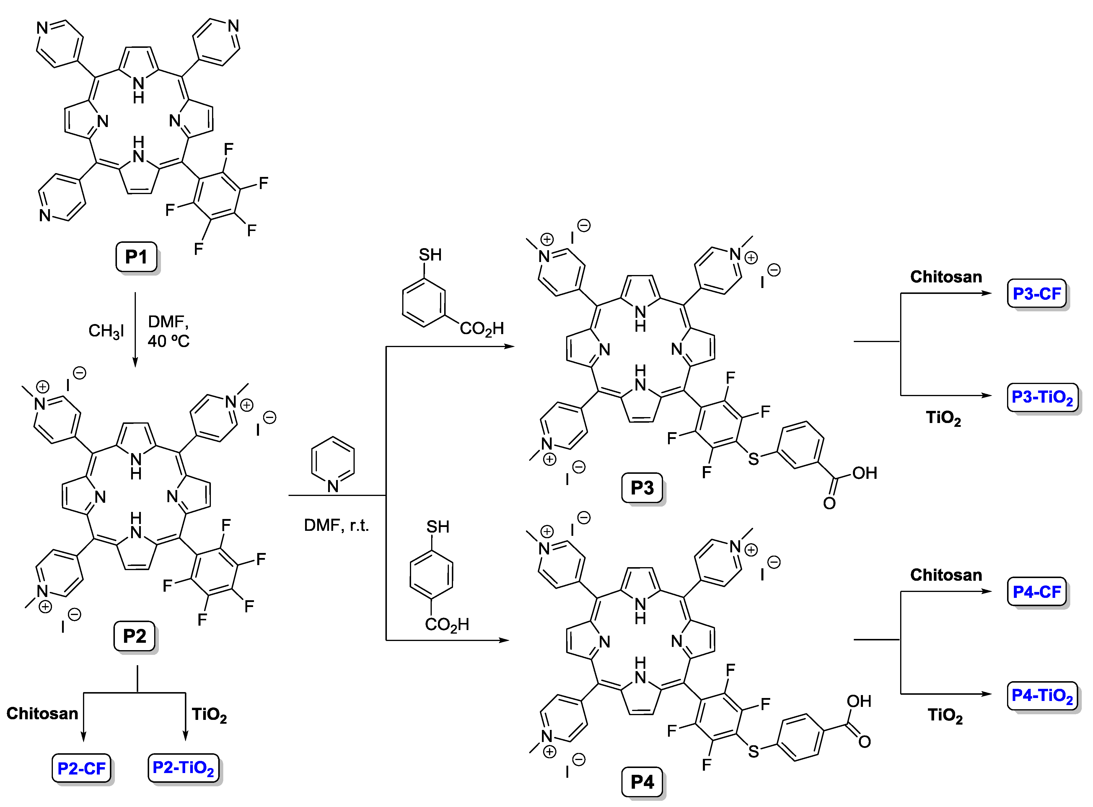
2.1. Synthesis of Porphyrin Derivatives

The general synthetic procedure used to obtain the new cationic porphyrin derivatives P3 and P4 bearing carboxyl groups is provided in Scheme 1. Both porphyrins were obtained using the tricationic porphyrin P2. The neutral precursor P1 was prepared by reacting pyrrole with the adequate stoichiometric proportion of pentafluorobenzaldehyde and pyridine-4-carbaldehyde in a mixture of acetic acid/nitrobenzene according to a literature procedure [46]. The quaternization of the pyridyl groups was carried out with methyl iodide and the required cationic porphyrin P2 [5,10,15-tris(1-methylpyridinium-4-yl)-20-(pentafluorophenyl)porphyrin tri-iodide] was quantitatively obtained [46]. The following aromatic nucleophilic substitution took place with 3- or 4-mercaptobenzoic acid in the presence of pyridine and DMF at room temperature. The required porphyrins P3 and P4 have been obtained with 90% and 93% yield, respectively [28,57,58]. The structures of the obtained products were confirmed by spectroscopic techniques (Figures S1–S15). The analytical data obtained for the intermediates is in accordance with the data previously published [59].
The 1H NMR spectra of compounds P3 and P4 showed the expected signals assigned to their structures. The insertion of 3-mercaptobenzoic acid residue rendered a non-symmetric pending group giving rise to three signals depicted at δ 7.69, δ 8.05–7.97 and δ 8.23 ppm, assigned to the resonances of H5, H4 and H6, and H2 of the 3-mercaptobenzoic unit. On the other hand, the insertion of the 4-mercaptobenzoic group gave rise to two doublets depicted at δ 8.03 and δ 7.78 ppm. All these assignments were confirmed through correlations found in 2D NMR spectra (COSY). The 19F NMR spectra of both derivatives also confirmed the success of the nucleophilic substitution by the disappearance of the resonance signal of the p-fluorine nucleus.
The absorption, emission and excitation spectra of P3 and P4 were recorded in DMF solutions (1 μM) at 298 K and were compared with those from the precursors P1 and P2 (see Table 1 and Figures S13 and S14, Supporting Information). The absorption spectra of P3 and P4 show the typical profile expected for free base meso-tetraarylporphyrins with the Soret band at 420 nm and four weak Q bands in the 500–650 nm range. The phyllo type Q-bands (IV > II > III > I) evidence a significant reduction in the intensity of bands III and I, which is a typical feature of tetraarylporphyrins containing electronegative substituents at the ortho positions of the phenyl rings [60].
The steady-state fluorescence emission spectra of all compounds, after their excitation at ca. 420 nm, show essentially two bands: one centered at ca. 648 nm and the other at 711 nm (Table 1 and Figure S16). These emission bands can be assigned to Qx(0–0) and Qx(0–1) transitions, typical of the free-base porphyrins with a D2h symmetry due to a nearly unchanged vibronic state upon excitation [61]. The perfect match between the absorption and the excitation spectra observed for all the compounds rules out the presence of any emissive impurities (Figures S14 and S17, Supporting Information).
The Stokes shift for P3 and P4 suggest a change in the electronic nature of the excited state compared to the ground state [62]. The fluorescence yields (ΦF) were determined using Equation (1), and 5,10,15,20-tetraphenylporphyrin (H2TPP) as reference (0.11 in DMF) [63,64]. In this equation, ΦF is the emission quantum yield of the sample, Φst the emission quantum yield of the standard, Ast represents the absorbance of the standard and As the absorbance of the sample at the excitation wavelength. Furthermore, Sst and Ss represent the integrated emission band areas of the standard H2TPP and of the sample, respectively. The results summarized in Table 1 show that P1 and P3 presented a slightly higher fluorescence quantum yield than P2 and P4.
Φ F = Φ s t ( S s S s t   × A s t A s )

2.2. Immobilization of Porphyrins on Chitosan

The immobilization of the cationic porphyrins P2, P3 and P4 on chitosan was performed by adding the adequate amount of each porphyrin dissolved in THF to a solution of chitosan in an aqueous acetic acid solution (1% v/v), according to a procedure previously reported by our group [28]. The amount of porphyrin immobilized was calculated indirectly by UV-Vis, considering the amount of unreacted porphyrin in the supernatants resulting from the washing process. The chitosan-porphyrin films, designated as P2-CF, P3-CF and P4-CF, were prepared by dissolving the porphyrins and chitosan in a 1% acetic acid solution followed by casting in a ventilated oven at 30 °C. The percentage of each porphyrin immobilized on the chitosan support is summarized in Table 2.
In Table 2 it is possible to observe that P3 and P4 lead to higher immobilization percentages than P2, most certainly due to the strong interactions (electrostatic and/or hydrogen bond) between the porphyrinic carboxylic unit and the chitosan cationic ammonium groups. On the other hand, although P2 interacts with chitosan in a lesser extent, this interaction seems also to be facilitated by hydrogen bonding involving the fluorine atoms and the chitosan OH and NH2 groups; additionally, one cannot exclude the interactions between the π-conjugated electron cloud of the macrocycle rings and the hydroxyl groups of chitosan [65].
All the chitosan-porphyrin films were characterized using a wide array of solid-state spectroscopic techniques such as UV-Vis and fluorescence, ATR-FTIR and powder X-ray diffraction (PXRD).
The presence of the porphyrin on the chitosan films P2-CF, P3-CF and P4-CF was promptly confirmed through UV-Vis and fluorescence spectra in the solid state. All the absorption spectra (Figure 1) displayed the typical Soret band accompanied by four Q-bands. All these bands are red-shifted and broader when compared with the corresponding absorption bands in solution (Table 1) [66]. A similar situation was observed with the UV-Vis of P2, P3 and P4 in the solid state (Figure S15, Supporting Information). The Soret bands suffered a red-shift of ca. 10 nm and in the Q-band region, depending on the porphyrin, red-shifts of ca. 5–16 nm were also detected. These shifts are attributed to small alterations of porphyrin molecular environment due to its interaction with chitosan [28,67].
The solid-state fluorescence spectra of P2, P3, P4 (Figure 2a–c) and of the immobilized porphyrins on chitosan (P2-CF, P3-CF and P4-CF, Figure 2e,f) are illustrated in Figure 2. All the hybrids show two emission bands in the red region upon excitation at 420 nm, which correspond to the S1→S0 transitions of the porphyrins. The two bands were observed at 663 and 711 nm for P2-CF, 661 and 714 nm for P3-CF and 657 and 712 nm for P4-CF. When compared with the corresponding emission of porphyrins P2-P4 in solution (Table 1) a red-shift of ca. 9-15 nm was observed in the first emission band. The low emission observed in the solid state for P2-P4 can probably be attributed to their tendency to form aggregates, leading to fluorescence quenching. Figure 3 shows the visual appearance of chitosan and of the P2-CF, P3-CF and P4-CF materials under ultraviolet light. The fluorescence intensities have not a linear dependence with the concentration of the porphyrin present in chitosan (P3 and P4 lead to higher immobilization percentages than P2, Table 2) but are dependent on the substituents present on the porphyrin core. This behavior is probably due to self-absorption and self-quenching of the PS supported in the films as described previously by Tian and co-workers [61].
The ATR-FTIR spectra of the Px-CF conjugates displayed the characteristic bands of chitosan (Figure S18, Supporting Information). The band at 3450 cm−1 is attributed to overlapped OH and N–H vibrations, while the bands at 2916 cm−1, 2867 cm−1 are associated with C–H and –C=O of the amide group CONHR vibrations of chitosan.
The bands at 1589, 1412 and 1332 cm−1 are associated with the bending vibrations of the N–H, C–H, O–H bonds; the vibration at 1152 cm−1 is due to νas(C=O) oxygen bridges and the band at 893 cm−1 to ω(C–H) from the polysaccharide’s structure. Most of the characteristic peaks of the porphyrin core are significantly reduced, when compared with chitosan, due to the low porphyrin immobilization on CF films (Table 2).
The PXRD of P3-CF and P4-CF showed some differences in the crystallinity when compared with CF. The peak observed at 42.9° for CF due to the crystalline phase of chitosan was not observed in P3-CF and P4-CF, indicating that the porphyrin immobilization led to a loss of crystallinity [68]. More specifically, it is possible to note for P3-CF a new set of peaks at 8.5, 11.8 and 15.2°, attributed to the chitosan chain rearrangements during the porphyrin immobilization process. Most of the peaks in the PXRD of these materials are characteristic peaks of chitosan while the presence of peaks related to the supported porphyrins are not visible [28,69]. Interestingly, the PXRD of P2-CF shows a predominant amorphous phase of chitosan (Figure S20, Supporting Information).

2.3. Immobilization of Porphyrins on Titanium Dioxide

The immobilization of the cationic porphyrins P2, P3 and P4 on TiO2 was performed by adding an adequate amount of each porphyrin dissolved in methanol to solid TiO2 (see details in the experimental section).
The amount of each porphyrin immobilized on TiO2 was also calculated by UV-Vis, considering the amount of porphyrin in the supernatants resulting from the washing process. The immobilization percentages of each porphyrin on the TiO2 support are summarized in Table 3. It is possible to note that the presence of the carboxylic groups in the porphyrin core is a fundamental requisite for an efficient electrostatic interaction with the TiO2 solid. Porphyrin P2, without the acidic groups, was completely removed after the washing process, while P3 and P4 showed higher than 90% immobilizations.
The UV-Vis spectra of the solids P3-TiO2 and P4-TiO2 show the typical porphyrin absorption bands confirming the success of P3 and P4 immobilization on TiO2 (Figure 4). The wavelengths of the Soret band at 420 nm and the Q-bands at 512, 547, 588, 649 nm are similar to those of the non-immobilized porphyrins in solution. However, when the spectra are compared with the absorption spectra of the porphyrins in the solid state, a blue shift can be observed (Figure S15, Supporting Information) [70,71]. This is probably due to interactions between the porphyrin plane and the support in an attempt of the macrocycle to acquire a more planar conformation in order to maximize the electrostatic interaction [72].
Figure 5 shows the solids TiO2, P3-TiO2 and P4-TiO2 under ultraviolet light and Figure 6 shows the corresponding emission spectra in the solid state. Contrarily to the low emission observed with P3 and P4 in the solid state, both hybrids showed a strong fluorescence. Hybrid P3-TiO2 shows two emission bands at 658 and 715 nm, with the first one more pronounced than the second one (Figure 6e), while P4-TiO2 exhibits the same profile but with the two bands at 667 and 717 nm (Figure 6d). The fluorescence excitation spectra (λem at 715 or 717 nm) show the typical features of porphyrins. These emission bands are slightly shifted when compared with those obtained when the same porphyrins were immobilized on chitosan. This behavior can be attributed to the distinct immobilization modes on the support and agrees with the distortions in the structure of the porphyrins indicated by the UV-Vis results. García-Sánchez and co-workers [73] observed a shift in the emission spectra for porphyrins immobilized on silica. The authors attributed this behavior to the different polarity existing inside the silica pores and the interactions with the surface groups.
The ATR-FTIR of P3-TiO2 and P4-TiO2 exhibit the typical vibration bands of titanium dioxide in the region of 3400 cm−1 (ν OH), at 1630 cm−1 attributed to the bending vibration of the hydroxyl group on the surface of TiO2 and at 481 cm−1 corresponding to the vibration of the Ti-O-Ti bonds [74]. In these solids, some characteristic bands due to the presence of the porphyrins were also observed at approximately 1593 and 1408 cm−1 arising from the symmetric and asymmetric vibrations of the C=O group and at 1472 cm−1 due to C=C vibrations (Figure S19, Supporting Information).
P3-TiO2 and P4-TiO2 hybrids displayed a PXRD pattern similar to those obtained from the pure support (Figure S21, Supporting Information). The two intense peaks at 25.3° and 27.4° (2θ) attributed to the anatase (101) and rutile (110) phases of TiO2, can be clearly observed in the hybrids, showing that the porphyrins do not interfere with the TiO2 crystallinity structure.

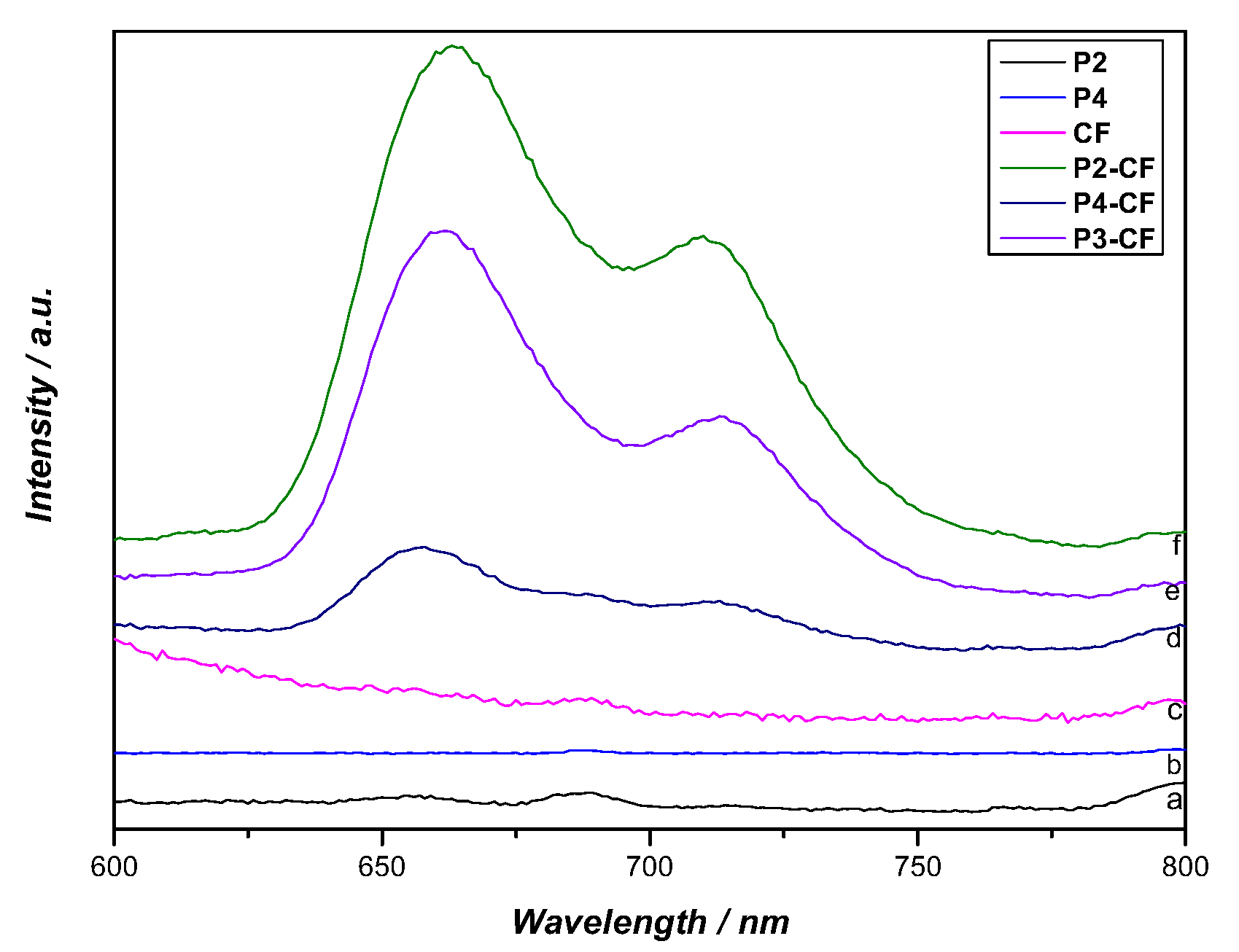
2.4. Singlet Oxygen Generation and Photostability

Knowing that the ability of a porphyrin or a material to act as a photosensitizer is related with its ability to generate ROS, namely 1O2, and also with its stability, these essential features were also considered in the characterization of the new compounds and materials.
The efficacy of compounds P3 and P4 to generate singlet oxygen was verified using 1,3-diphenylisobenzofuran (DPiBF) as a scavenger of singlet oxygen (1O2), and further compared with the ability of the precursors P1 and P2 (Figure 7). The degradation of DPiBF was monitored by the decrease of its band at 415 nm, after its irradiation in the presence of each porphyrin and atmospheric oxygen. The reaction between 1O2 and DPiBF in a 4+2 process affords 1,2-dibenzoylbenzene giving an indirect evidence of 1O2 generation.
The results summarized in Figure 7 show that all the porphyrins are good generators of 1O2, but the best efficacy was found for the new porphyrins P3 and P4. These two new porphyrins can generate 1O2 more efficiently than H2TPP, also considered an excellent 1O2 generator [75]. The best 1O2 generator was P3, followed by P4 and H2TPP, and finally by P1 and P2, both with a similar efficacy. These results suggest that the presence of the acid group and its position are important features for the efficiency in 1O2 generation and the lower efficacy of P1 and P2 is probably associated with aggregation phenomena.
The DPiBF decomposition provided by the materials PS-CF and PS-TiO2 was also investigated and the results are summarized in Figure 8. The results show that P3-TiO2 is a more efficient 1O2 producer than P4-TiO2. This result is in line with the better performance of P3 to generate 1O2 when compared with P4. Considering the chitosan materials, the higher DPiBF decay was caused by P2-CF and none of the other materials (P3-CF and P4-CF) were able to generate 1O2 under the tested conditions.
Interestingly, P2-CF showed almost the same 1O2 production as P2 in solution. This behavior might be related with an aggregation attenuation or an increased photostability of the porphyrin after its immobilization on the chitosan support.
The photostability studies were performed with P2-P4 in PBS, monitoring the decrease in absorbance of the corresponding Soret bands after different times of irradiation with a LED array at an irradiance of 10 mW·cm−2. P4 suffered a decay of 64%, followed by P2 (41%) and P3 (22%) during the irradiation period (Figure S22). Based on the observed degradation profiles, the studies were repeated in the absence of light, to verify if the decrease was not associated with an aggregation phenomenon. In fact, P4 showed a decrease of 41%, P2 (28%) and P3 (22%) in dark conditions, showing that somewhere up to a 23% decrease, the decrease is related to the photodecomposition of the macrocycle. In sum, P2 and P4 are more susceptible to photodegradation upon illumination, while P3 was quite photostable over the irradiation period investigated (30 min).

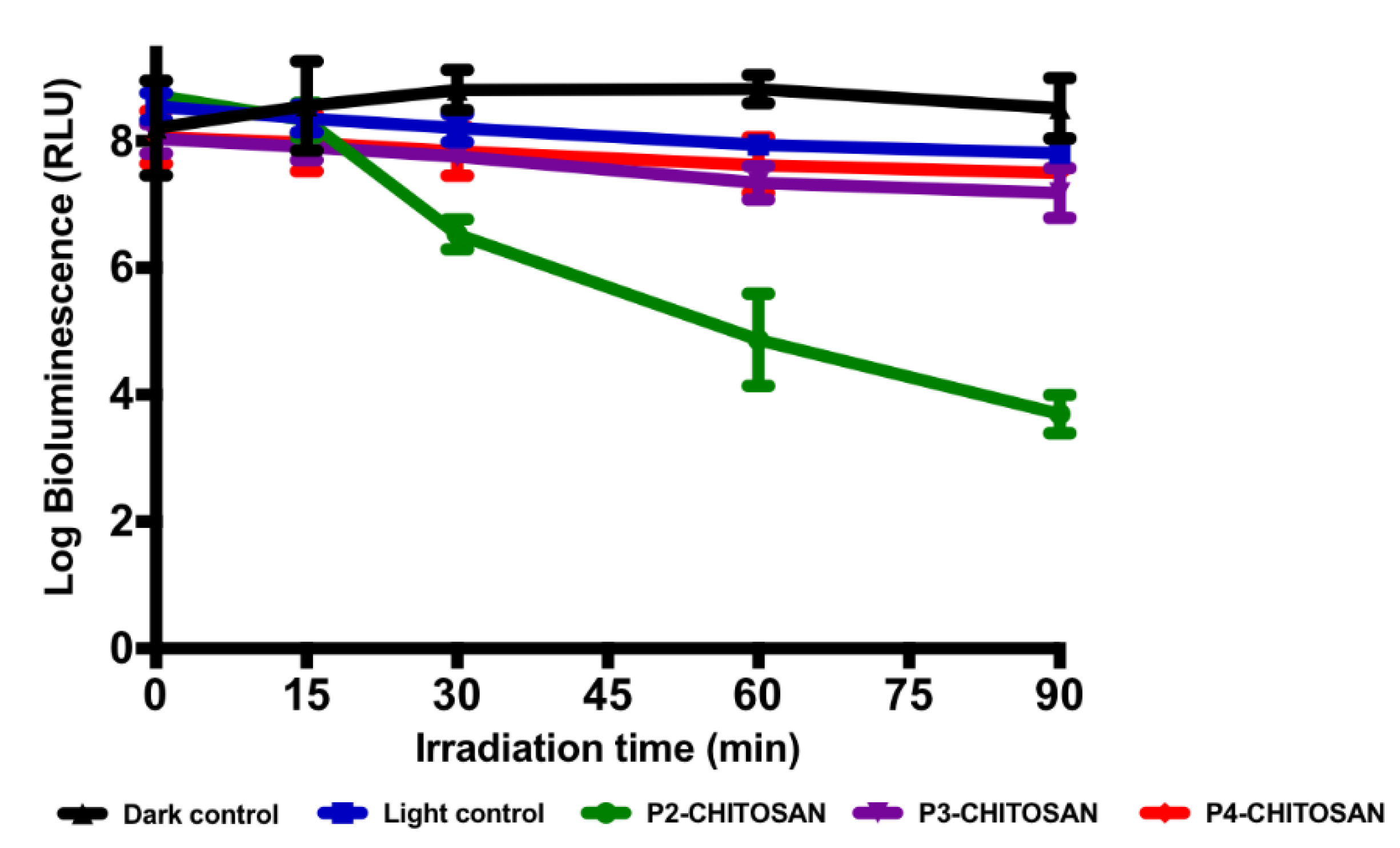
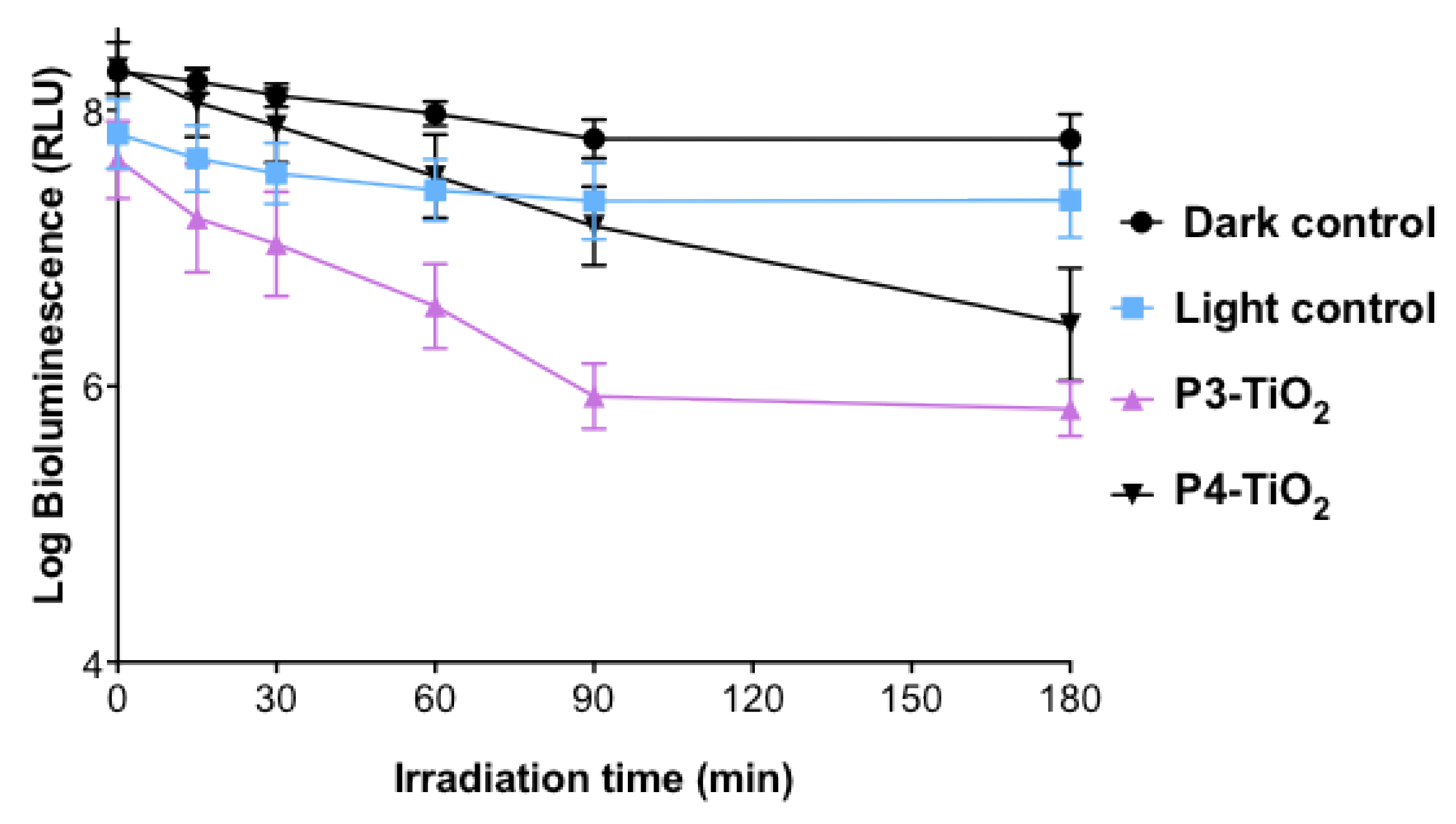
2.5. Photoinactivation Studies

It is commonly accepted that the success of photoinactivation closely depends on the structure of the photosensitizing agent [14]. As previously stated, the charge of the PS is extremely important for the inactivation of the microbial agents and, generally, positively charged PS are more efficient for Gram-negative strains, even if they are at low concentrations when compared to neutral or negatively charged PS [52]. Studies with cationic PS demonstrate that these derivatives are more likely to inactivate both Gram-positive and Gram-negative bacteria without the help of membrane disrupting agents [47,76].
The biological assays started by evaluating the photodynamic action of the new cationic porphyrins P3 and P4 at the concentration of 5.0 µM, under white light towards the recombinant bioluminescent E. coli (Figure 9). As mentioned above, this Gram-negative bacterium is an excellent bacterial model to monitor the effectiveness of a photoinactivation process, since its light output is a highly sensitive reporter of its metabolic activity. These set of experiments were not performed in the presence of the non-immobilized P2 since its efficacy has already been studied by some of us [46,47].
The results summarized in Figure 9 show that the bioluminescence of E. coli was not affected by light in the absence of the PS (light control) neither by the direct effect of any of the tested PS in the absence of light (dark control). These results indicate that any variation in the bioluminescence was due to the photodynamic effect of the PS. Photosensitizers P3 and P4 showed significant differences in terms of inactivation efficiency (ANOVA–p < 0.05) (Figure 9). P3 at a concentration of 5.0 µM was more effective than P4, causing a reduction of approximately 3.6 log in the bioluminescence, after only 30 min of irradiation. At this concentration, P3 inactivated completely E. coli (decrease of the bioluminescent signal below the quantification limit, 2 log) after 120 min of irradiation (with a total light dose of 21.6 J·cm−2) whilst P4 caused an inactivation of 3 log after 120 min of irradiation. Compound P3 can be considered a good PS against E. coli, since it showed a similar efficiency as P2 towards E. coli at the same concentration (5.0 μM) and under the same light irradiance [47]. The difference observed for each PS can be justified by a balance between the efficiency to produce 1O2 (P2 ~ P3 > P4) and their affinity to the bacteria which is more dependent on the PS structure.
Using different approaches (e.g., scanning electron microscopy (SEM), transmission electron microscopy (TEM), spectrophotometry, 1D electrophoresis, atomic force microscopy, proteomic, lipidomic, infrared spectroscopy) to evaluate the ultrastructural, morphological and functional changes in bacterial cells, at initial stages and during the course of the PDI process, it seems that the most affected cellular constituents in E.coli are external targets, namely proteins and lipids, which can affect normal cell membrane functionality or lead to significant cell membrane disintegration [77,78,79,80].
The results obtained for the photoinactivation efficacy of the new conjugates towards the bioluminescent E. coli were evaluated and are compiled in Figure 10 (Px-CF) and Figure 11 (Px-TiO2). It is known that chitosan can act as an antimicrobial agent, although Gram-negative bacteria are less susceptible to its effects [81].
Figure 10 shows for P2-CF and after 90 min of irradiation, a noticeable decrease of the bioluminescent signal to approximately 4.1 log. However, no significant differences were detected in the bioluminescent signal when the bacteria were exposed to P3 and P4 supported on chitosan (decrease of 0.63 and 0.31 log, respectively).
These results are most certainly related to the inability of these porphyrins to produce oxygen singlet, after being immobilized in chitosan. The porphyrins photodynamic performance in solution follows the order P2 ~ P3 > P4; however, when supported on chitosan films, only P2-CF, with the capacity to generate oxygen singlet, has a significant effect on the bacterial cells. Studies show that chitosan might have either bactericidal or bacteriostatic properties, depending on the type of bacterium, which is related to the thickness of the cell wall. Recent data shows that chitosan tends to act as bacteriostatic rather than bactericidal, which can also explain the visible changes in the dark controls [82].
The positive charges of chitosan and P2 can interfere with the negatively charged E. coli cell envelope, resulting in alterations of membrane permeability, which in combination with the 1O2 justifies the efficiency observed for P2-CF.
The combination of the 5,10,15,20-tetrakis(4-sulphonatophenyl)porphyrin (TPPS4−) or 5,10-bis(4-methylphenyl)-15,20-bis(4-N,N,N-trimethylammoniumphenyl)porphyrin (MPAP2+) and chitosan produced a potentiated photodynamic inactivation (PDI) effect. Both porphyrins TPPS4− and MPAP2+ at 5.0 µM in the presence of 0.25 mg/mL of chitosan caused a reduction of 3.5 log in the concentration of viable cells after a total light dose of 5.4 J·cm2 [81].
As shown in Figure 10, neither P3 nor P4 immobilized on chitosan are good photosensitizers. P3 and P4, when combined with chitosan, show almost no effect on the cells. Durantini and co-workers showed that chitosan was toxic to E. coli by itself at high concentrations (0.75 or 1.00 mg/mL) [81]. The concentration used in this work was 0.34 mg/mL and, as expected, no cytotoxic effect was observed for E. coli in the dark. Similar results were observed for P2-CF at a concentration of 3.0 μM after a total light dose of 10.8 J·cm−2. The porphyrins 5,10,15,20-tetrakis(4-aminophenyl)porphyrin (p-TAPP) and TMPyP incorporated into chitosan showed a considerable bactericidal effect under irradiation at 590 nm (2 log reduction after 120 min for p-TAPP and 4 log reduction after 140 min for TMPyP) [40]. The best results were obtained when P2 was incorporated into chitosan. The similarities seen in the UV-Vis spectra before and after the PDI biological assays show that PS-CF materials were photostable (Figure S23).
The positive effect of chitosan was also indicated when Luksiene and co-workers studied the photodynamic inactivation of the Gram (−) food pathogen Salmonella enterica using the chlorophyllin–chitosan hybrid as PS [30]. The authors verified that the inactivation caused by chlorophyllin (1.8 log, at 15 μM, light dose 38 J·cm−2) and chitosan (ca 1.8 log) separately gave rise in the presence of photoactivated Chl–CHS complex to a remarkable and swift decrease of Salmonella enterica viability (7 log) [30]. Considering that the inactivation of E. coli was achieved at lower PS concentration (3.0 μM vs 15 μM) and light dose (16.2 vs 38 J·cm−2), we can conclude that the new hybrid P2-CF is also a very promising material. No cytotoxic effect observed for chitosan alone can be justified to the low concentrations used since high concentrations (0.75 or 1.00 mg/mL) are required for E. coli inactivation, as showed by Durantini [81].
As stated before, TiO2 can be used as PS for the inactivation of some pathogens under ultraviolet light irradiation. The electronic properties of TiO2 can be easily modulated by linking a PS to its surface and the immobilization of the PS induces an extension of the absorption profile of TiO2 enabling the use of the visible light [83]. Macyk and co-workers showed that a TiO2 suspension did not result in any significant E. coli photoinactivation under visible light [84]. When P3 and P4 are supported on TiO2, it is noticeable that P3 and P4 at a concentration of 10 μM cause a reduction of approximately 2 log in the E. Coli bioluminescence after 180 min (32.4 J/cm2) (Figure 11). P3-TiO2 and P4-TiO2 have a higher effect on the bacterial cells compared to P3-CF and P4-CF. Compared to PS-TiO2, the 1O2 production from these PS-CF was much less efficient and could be disregarded.
TiO2-graphene functionalized with 5,10,15,20-tetrakis(4-carboxyphenyl)porphyrin showed a decrease of 64% in the concentration of viable cells of E. coli after 440 min under light irradiation (450 W xenon lamp) [27]. In this study, both porphyrins P3 and P4 supported on TiO2 showed a decrease of 99% in the concentration of viable cells of E. coli after 180 min.
Conversely, higher amounts of PS-TiO2 did not increase the photodynamic effect. In fact, the increase of material as a powder in these tests can obstruct light from reaching the PS, hampering the photosensitization process.

3. Materials and Methods

All analytical grade chemicals used in this study were purchased from Sigma-Aldrich or Merck. Chitosan was purified following the procedure described in the literature [28]. H2TPP was synthesized as described in the literature [85]. TiO2 was Evonik-Degussa P25.
1H, 19F and 13C solution NMR spectra were recorded on a Bruker Avance 300 (300.13 and 282.38 MHz, respectively), 500 (125.76 MHz for 13C) spectrometer. DMSO-d6 was used as solvent and tetramethylsilane (TMS) as the internal reference; the chemical shifts are expressed in ppm and the coupling constants (J) in Hertz (Hz). Unequivocal 1H assignments were made using 2D COSY (1H/1H). Electronic spectra (UV-Vis) were obtained on a Shimadzu UV-2501PC spectrophotometer, in the 350–800 nm range. Spectra of the solid samples were recorded on a Jasco V-560 spectrophotometer with reflectance accessory (JASCO ISV-469) in absorbance mode in the 250–850 range. Attenuated Total Reflectance Transmission Fourier Transform Infrared (ATR-FTIR) spectra were registered on a FTIR Bruker Tensor 27 spectrophotometer with ATR accessory ATR (ATR Golden Gate Diamond, Specac) in the 350–4000 cm−1 range; the spectra were collected with a resolution of 4 cm−1 and accumulation of 256 scans. For the X-ray diffraction (PXRD) measurements, self-oriented solids were placed on neutral glass sample holders. The measurements were performed in the reflection mode using a Panalytical Empyrean diffractometer (Cu Kα1,2 X-radiation, λ1 =1.540598 Å and λ2 =1.544426 Å), equipped with an X’Celerator detector and a flat-plate sample holder in a Bragg- Brentano para-focusing optics configuration (40 kV, 50 mA). Intensity data were collected by the step counting method (step 0.02) in continuous mode. The fluorescence emission spectra were recorded in DMF in 1 × 1 cm quartz optical cells under normal atmospheric conditions on a computer-controlled Horiba Jobin Yvon FluoroMax-3 spectrofluorimeter. The widths of both excitation and emission slits were set at 2.0 nm. The fluorescence quantum yield (φF) of porphyrins was calculated in DMF by comparison of the area below the corrected emission spectra using H2TPP as standard (λexc at 420 nm, φF = 0.11 in DMF) [64]. Fluorescence spectra of the solid samples were recorded using a fiber optic system connected to a Horiba-Jobin Yvon FluoroMax-3 spectrofluorimeter for appropriately λ (nm) of the solid compounds.

3.1. Synthesis of Porphyrin Derivatives

Porphyrin P1 was obtained by the reaction of the appropriate aldehydes in acetic acid and nitrobenzene, according to a literature procedure (see Scheme 1) [59]. The purity was confirmed by TLC and by 1H and 19F NMR spectroscopy.
Porphyrin P2. To a solution of P1 (0.106 mmol, 0.075 g) in N,N-dimethylformamide (DMF; 1 mL), an excess of methyl iodide (0.2 mL) was added in a sealed tube. The mixture was stirred overnight at 40 C. After cooling, diethyl ether was added to precipitate the porphyrin. The solid was isolated by filtration and washed with diethyl ether. The desired porphyrin was crystallized from CH2Cl2:CH3OH/hexane to yield P2. After that, the solid was dried under vacuum and then characterized.
P2: Yield: 98%; 1H NMR (300 MHz, DMSO-d6): δ 9.63–9.46 (8H, m, H-β), 8.92 (6H, d, J = 9.1 Hz, Ar-Py-m-H), 8.42 (6H, d, J = 9.1 Hz, Ar-Py-o-H), 4.32 (9H, s, -CH3), −3.15 (2H, s, N-H) ppm. 19F NMR (280 MHz, DMSO-d6): δ −135.45 (2F, dd, J = 24.8 and 6.1 Hz, Ar-o-F), −150.17 (1F, t, J = 22.6 Hz, Ar-p-F), between -160.17 and −160.37 (2F, m, Ar-m-F) ppm. UV-Vis (DMF) λmax, nm (log ε): 420 (4.90), 510 (3.83), 544 (3.19), 584 (3.36), 640 (2.70).
Porphyrin P3. To 3-mercaptobenzoic acid (0.015 mmol, 0.002 g) dissolved in DMF (5.0 mL) and containing 0.5 mL of pyridine, porphyrin P2 (0.014 mmol, 0.015 g) was added after 30 min in a single portion. The reaction mixture was stirred for 1 h at ambient temperature; afterwards, diethyl ether was added to precipitate the porphyrin, the solid was filtered and washed with diethyl ether. Porphyrin P3 was obtained by crystallization from methanol/chloroform.
P3: Yield: 90%; 1H NMR (300 MHz, DMSO-d6): δ 9.50–9.47 (8H, m, Ar-Py-m-H and H-β), 9.17 (6H, brs, H-β), 9.02 (6H, dd, J = 11.1, 6.7 Hz, Ar-Py-o-H), 8.23 (1H, s, S-Ar-COOH), 8.05–7.97 (2H, m, S-Ar-COOH), 7.69 (1H, t, J = 7.7 Hz, S-Ar-COOH), 4.72 (9H, s, -CH3), −3.11 (s, 2H, N-H) ppm. 19F NMR (280 MHz, DMSO-d6) δ −129.69 (2F, dd, J = 26.9 and 11.5 Hz, Ar-o-F), −134.52 (2F, dd, J = 26.9 and 11.5 Hz, Ar-m-F) ppm. 13C NMR (125 MHz, DMSO-d6): δ 166.6 (S-Ar-COOH), 162.4 (ArPy-Por, CH), 156.3 (ArPy-Por, CH), 144.3 (Ar-Py-m-H, -N-CH), 133.7 (Ar-Py-o-H, -N-CH), 133.3 (S-Ar, CH), 132.3 (S-Ar, CH), 132.2 (S-Ar, CH), 130.4 (S-Ar, CH), 129.9 (S-Ar, CH), 128.8 (S-Ar, CH), 116.2 (Por-ArF4, C-Por), 115.9, 103.9 (C-ArF4, C-S), 48.0 (CH3) ppm. UV-Vis (DMF) λmax, nm (log ε): 420 (5.03), 512 (3.92), 546 (3.41), 584 (3.48), 640 (2.90). HRMS-ESI(+): calcd. for C51H36F4N7O2S [M] 886.2571 found 886.2555.
Porphyrin P4. 4-Mercaptobenzoic acid (0.03 mmol, 0.004 g) was dissolved in DMF containing 0.5 mL of pyridine. The reaction mixture was kept under stirring at ambient temperature for 30 min. Afterwards, porphyrin P2 (0.03 mmol, 0.031 g) was added, and the reaction mixture was stirred for 1 h at ambient temperature. Then, diethyl ether was added to precipitate the porphyrin, the solid was filtered and washed with diethyl ether. The solid was dissolved in methanol and, after concentration, it was re-precipitated by the addition of acetone.
P4: Yield: 93%; 1H NMR (300 MHz, DMSO-d6): δ 9.57–9.50 (8H, m, Ar-Py-m-H and H-β), 9.18 (6H, brs, H-β), 9.06–9.00 (6H, m, Ar-Py-o-H), 8.03 (2H, d, J = 8.2 Hz, S-Ar-COOH), 7.78 (2H, d, J = 8.2 Hz, S-Ar-COOH), 4.73 (9H, s, –CH3), −3.10 (2H, s, N-H) ppm. 19F NMR (280 MHz, DMSO-d6): δ −129.31 (2F, dd, J = 26.7, 11.3 Hz, Ar-o-F), −134.42 (2F, dd, J = 26.7, 11.3 Hz, Ar-m-F) ppm. 13C NMR (125 MHz, DMSO-d6): δ 167.2 (S-Ar-COOH), 156.3 (ArPy-Por, CH), 144.3 (Ar-Py-m-H, -N-CH), 132.3, 132.2 (Ar-Py-o-H, -N-CH), 130.5 (S-Ar, CH), 127.7 (S-Ar, CH), 116.3 (Por-ArF4, C-Por), 103.9 (C-ArF4, C-S), 48.0 (CH3) ppm. UV-Vis (DMF) λmax, nm (log ε): 420 (5.29), 510 (4.15), 544 (3.61), 584 (3.73), 640 (3.01). HRMS-ESI(+): calcd. for C51H36F4N7O2S [M] 886.2571 found 886.2578.

3.2. Immobilization of Porphyrins on Chitosan

The porphyrin-chitosan (Px-chitosan where x = 2, 3 or 4) solids was prepared in two steps:
(i) 250 mg of chitosan were dissolved in 7.0 mL of 1% (v:v) aqueous acetic acid. Afterwards, porphyrins P2, P3 or P4 (5.0 mg) were added and the mixtures were stirred for 24 h at 30 °C. The solvent was evaporated under reduced pressure and the resulting solids were filtered and exhaustively washed with water and DMF. The solutions resulting from the washing process were analyzed by UV-Vis in order to quantify the Px that leaked from the chitosan and the loading of the porphyrin in each solid was calculated. The solids were dried in the oven at 55 °C for 72 h.
(ii) Each sample obtained (about 100 mg) was dissolved in 10 mL of 1% (v:v) aqueous acetic acid. The suspension was maintained at 40 °C under stirring until complete solubilization of the solid (3 h). The solution was poured into molds (5.0 × 5.0 cm) and allowed to dry at room temperature for 72 h. The films were denominated as P2-CF, P3-CF and P4-CF and characterized by ATR-FTIR, UV-Vis, PXRD and fluorescence of solid samples.

3.3. Immobilization of Porphyrins on Titanium Dioxide

To a solution of P2, P3, or P4 in methanol (0.789 µM, 10 mL) 0.5 g of TiO2 were added. The resulting suspension was stirred at ambient temperature for 6 h. The solids were isolated by centrifugation, washed with methanol and dried in the oven at 55 °C for 24 h. UV-Vis analysis of the solutions resulting from the washing process was performed and the loading of the porphyrin in each solid was calculated. The solids were named as P3-TiO2 and P4-TiO2 and characterized by the techniques described above. P2 was completely removed after the washing process of P2-TiO2, so this hybrid was not obtained.

3.4. Singlet Oxygen Generation and Photostability

In a 1 cm x 1 cm quartz cell, and to a solution of DPiBF at 50 μM, 0.5 µM of each PS in DMF were added and irradiated with a LED array at an irradiance of 10 mW cm−2. The LED array is composed of a matrix of 5 × 5 LEDs making a total of 25 light sources with an emission peak centered at 640 nm and a bandwidth at half maximum of ± 20 nm. Irradiation was conducted at ambient temperature under stirring. The decreasing of DPiBF was monitored by UV-Vis at 415 nm at intervals of 1 min during 15 min. The absorption decay is proportional to the production of 1O2. For the Px-CF and Px-TiO2 materials, a similar protocol was adopted, but the PS concentration used was 2.0 µM and the monitorization by UV-Vis was performed after centrifugation of the suspension irradiated and only the times 0, 5, 10 and 15 min were registered. Controls were also included. The experiments were performed in duplicate. H2TPP, an efficient singlet oxygen generator, was included as a reference.
The photostability studies of the porphyrin derivatives were performed by irradiation of a solution of each porphyrin in DMF (5.0 µM) with a LED array. The solutions were stirred and kept at ambient temperature. The stability of each porphyrin derivative was verified by UV-Vis at regular intervals up to 60 min.

3.5. Bacterial Strain and Culture Growth Conditions

The efficiency of the porphyrins in solution and after being incorporated on the materials to photoinactivate a bioluminescent E. coli was evaluated using a reliable and quick method that allows the monitorization of the activity of this bacterial strain in real time. Bioluminescent E. coli was transformed as described in a previous work [46] and stored at −80 °C in 10% glycerol. For the transformation, the plasmids pHK724 and pHK555 were inserted into competent cells of E. coli Top 10 (Invitrogen, USA), resulting in a bioluminescent strain. These plasmids contain the luxoperon from the bioluminescent marine bacterium Vibrio fischeri, required to produce light without the addition of exogenous substrates [46]. Before each assay, an aliquot of bioluminescent bacteria stored at −80 °C in 10% glycerol was aseptically plated on tryptic soy agar (TSA, Merck) supplemented with 50 mg·mL−1 of ampicillin (Amp) and with 34 mg·mL−1 of chloramphenicol (Cm), and stored at 4 °C. Before each assay, one isolated colony was aseptically transferred to 10 mL of tryptic soy broth (TSB, Merck) medium previously supplemented with Amp and Cm and was grown overnight at 25 °C under stirring (120 rpm). Afterwards, an aliquot was transferred into 10 mL TSB under the same growth conditions to reach stationary growth phase. An optical density at 600 nm (OD600) of 1.6 ± 0.1 corresponded to ≈108 colony forming units (CFU) mL−1 [46].

3.6. Irradiation Conditions for Photosensitization

All samples were exposed, in parallel, to white light (PAR radiation, 13 OSRAM 21 lamps of 18 W each, 380 700 nm) with a fluence rate of 3.0 mW cm−2 (measured with a light meter LI-COR model LI-250, Li-Cor Inc., Lincoln, NE, USA), at ambient temperature of less than 25 °C under 100 rpm mechanical stirring.

3.7. Photosensitization Procedure

Bacterial cultures mentioned above were grown overnight and then tenfold diluted in phosphate buffered saline (PBS), at pH 7.4, so that ~107 relative light units (RLUs) were reached. Five milliliters of the bacterial suspension were evenly distributed in glass beakers (previously sterilized) and the appropriate volume of the PS was added in order to attain final concentrations of 5.0 µM. For the immobilized PS, the corresponding mass of PS-CF or PS-TiO2 was added in order to achieve the final concentration of 3.0 or 10 µM, respectively. For the non-immobilized PS, the suspensions were incubated for 15 min in the dark to allow the binding of the PS to the bacterial cells, and subsequently exposed to irradiation conditions, during a maximum period of 120 min. For the materials prepared, the dark incubation time was 60 min and after which they were exposed to the same irradiation conditions, during a maximum period of 90 min and 180 min for Px-CF and Px-TiO2, respectively.
Dark and light controls were included in the experiments. In the light control, the bacterial suspensions without PS were exposed to the same irradiation protocol as the test suspensions. In the dark control, the PS was added and protected from light with aluminum foil. At the initial time, and after established times of irradiation, the bioluminescence was measured in the luminometer (GloMax® 20/20 Luminometer, Promega, Madinson, WI, USA). The percentage of decay of the bioluminescence is proportional to the concentration of viable cells (CFU). Three independent experiments with three replicates were performed and the results were averaged.

3.8. Statistical Analysis

Statistical analysis was performed in GraphPadPrism 7.0. The significance of the PDI effect of each PS and of the irradiation time on bacterial cell viability was assessed by an unvaried analysis of variance (ANOVA) model with the Bonferroni post hoc test. Normal distributions were assessed by the Bartlett’s test and homogeneity of variances was assessed by the Brown-Forsythe test. A value of p < 0.05 was considered significant.

4. Conclusions

We successfully accomplished the chemical modification of the 5,10,15-tris(1-methylpyridinium-4-yl)-20-(pentafluorophenyl)porphyrin. Moreover, two cationic porphyrins were obtained as a result of the substitution of the p-fluorine atoms on the meso-phenyl with thio-carboxylate acids. These cationic porphyrins were successfully immobilized on two different supports: chitosan and titanium oxide. The efficiency of the aPDT process against E. coli seems dependent on the structure of the porphyrin and on its ability to produce singlet oxygen. The results showed that the choice of the support and of the adequate porphyrin can be modulated to improve the efficiency of aPDT. The electronic properties of TiO2 allow linking the PS to its surface, increasing the absorption profile of TiO2, which enabled the use of the visible light, inactivating bacteria more efficiently than the PS-CF.

Supplementary Materials

Supplementary materials can be found at https://www.mdpi.com/1422-0067/20/10/2522/s1.

Author Contributions

Investigation—K.A.D.F.C. (porphyrin synthesis, materials preparation and biological studies), N.M.M.M. (porphyrin synthesis and characterization and materials preparation), F.F. (porphyrin synthesis), R.I.F. (biological studies); Supervision—A.A., A.J.D.S. and M.G.P.M.S.N.; Validation—J.A.S.C., M.A.F.F., A.A., and C.S.R.F.; Writing—original draft, K.A.D.F.C., N.M.M.M., M.M.Q.S., F.F.; Writing - review and editing, J.A.S.C., A.J.D.S., J.P.C.T., S.N. and M.G.P.M.S.N.

Funding

The authors are grateful to Conselho Nacional de Desenvolvimento Científico e Tecnológico, Coordenação de Aperfeiçoamento de Pessoal de Nível Superior, University of Aveiro and FCT/MCT for the financial support for QOPNA research Unit (FCT UID/QUI/00062/2019), FCOMP-01-0124-FEDER-037296), CESAM (UID/hyrinAMB/50017/2013), CICECO-Aveiro Institute of Materials, POCI-01-0145-FEDER-007679 (FCT Ref. UID/CTM/50011/2013), and the FCT project PREVINE (FCT-PTDC/ASP-PES/29576/2017), through national funds and, where applicable, co-financed by the FEDER, within the PT2020 Partnership Agreement, and to the Portuguese NMR Network. K.A.D.F. Castro also thanks CNPq for her post-doctoral scholarship (Process: 201107/2014-7). The research contracts of N.M.M. Moura (REF.-048-88-ARH/2018) and F. Figueira (REF. -168-89-ARH/2018) are funded by national funds (OE), through FCT—Fundação para a Ciência e a Tecnologia, I.P., in the scope of the framework contract foreseen in the numbers 4, 5 and 6 of the article 23, of the Decree-Law 57/2016, of August 29, changed by Law 57/2017, of July 19. FCT is also acknowledge for the research contract under Stimulus of Scientific Employment 2017 to C.S.R. Freire (CEECIND/00464/2017).

Conflicts of Interest

The authors declare no conflict of interest.

References

  1. Sperandio, F.F.; Huang, Y.Y.; Hamblin, M.R. Antimicrobial photodynamic therapy to kill Gram-negative bacteria. Recent Pat. Antiinfect Drug Discov. 2013, 8, 108–120. [Google Scholar] [CrossRef]
  2. Huang, L.; El-Hussein, A.; Xuan, W.; Hamblin, M.R. Potentiation by potassium iodide reveals that the anionic porphyrin TPPS4 is a surprisingly effective photosensitizer for antimicrobial photodynamic inactivation. J. Photochem. Photobiol. B 2018, 178, 277–286. [Google Scholar] [CrossRef]
  3. Mesquita, M.Q.; Dias, C.J.; Neves, M.G.P.M.S.; Almeida, A.; Faustino, M.A.F. Revisiting Current Photoactive Materials for Antimicrobial Photodynamic Therapy. Molecules 2018, 23, 2424. [Google Scholar] [CrossRef] [PubMed]
  4. Kou, J.; Dou, D.; Yang, L. Porphyrin photosensitizers in photodynamic therapy and its applications. Oncotarget 2017, 8, 81591–81603. [Google Scholar] [CrossRef] [Green Version]
  5. Gois, M.M.; Kurachi, C.; Santana, E.J.; Mima, E.G.; Spolidorio, D.M.; Pelino, J.E.; Salvador Bagnato, V. Susceptibility of Staphylococcus aureus to porphyrin-mediated photodynamic antimicrobial chemotherapy: an in vitro study. Lasers Med. Sci. 2010, 25, 391–395. [Google Scholar] [CrossRef] [PubMed]
  6. Mamone, L.; Ferreyra, D.D.; Gandara, L.; Di Venosa, G.; Vallecorsa, P.; Saenz, D.; Calvo, G.; Batlle, A.; Buzzola, F.; Durantini, E.N.; Casas, A. Photodynamic inactivation of planktonic and biofilm growing bacteria mediated by a meso-substituted porphyrin bearing four basic amino groups. J. Photochem. Photobiol. B 2016, 161, 222–229. [Google Scholar] [CrossRef] [PubMed] [Green Version]
  7. Oliveira, A.; Almeida, A.; Carvalho, C.M.; Tomé, J.P.C.; Faustino, M.A.F.; Neves, M.G.P.M.S.; Tomé, A.C.; Cavaleiro, J.A.S.; Cunha, Â. Porphyrin derivatives as photosensitizers for the inactivation of Bacillus cereus endospores. J. Appl. Microbiol. 2009, 106, 1986–1995. [Google Scholar] [CrossRef]
  8. Rossi, G.; Goi, D.; Comuzzi, C. The photodynamic inactivation of Staphylococcus aureus in water using visible light with a new expanded porphyrin. J. Water Health 2012, 10, 390–399. [Google Scholar] [CrossRef] [PubMed] [Green Version]
  9. Xing, C.; Xu, Q.; Tang, H.; Liu, L.; Wang, S. Conjugated polymer/porphyrin complexes for efficient energy transfer and improving light-activated antibacterial activity. J. Am. Chem. Soc. 2009, 131, 13117–13124. [Google Scholar] [CrossRef]
  10. Zhang, J.; Wu, X.; Cao, X.; Yang, F.; Wang, J.; Zhou, X.; Zhang, X.L. Synthesis and antibacterial study of 10, 15, 20-triphenyl-5-(4-hydroxy-3-(trimethylammonium)methyl)phenylporphyrin as models for combination of porphyrin and alkylating agent. Bioorg. Med. Chem. Lett. 2003, 13, 1097–1100. [Google Scholar] [CrossRef]
  11. Beirão, S.; Fernandes, S.; Coelho, J.; Faustino, M.A.F.; Tomé, J.P.C.; Neves, M.G.P.M.S.; Tomé, A.C.; Almeida, A.; Cunha, Â. Photodynamic inactivation of bacterial and yeast biofilms with a cationic porphyrin. Photochem. Photobiol. 2014, 90, 1387–1396. [Google Scholar] [CrossRef] [PubMed]
  12. Mesquita, M.Q.; Dias, C.J.; Gamelas, S.; Fardilha, M.; Neves, M.G.P.M.S.; Faustino, M.A.F. An insight on the role of photosensitizer nanocarriers for Photodynamic Therapy. An. Acad. Bras. Cienc. 2018, 90, 1101–1130. [Google Scholar] [CrossRef] [Green Version]
  13. Cardote, T.A.F.; Barata, J.F.B.; Amador, C.; Alves, E.; Neves, M.G.P.M.S.; Cavaleiro, J.A.S.; Cunha, Â.; Almeida, A.; Faustino, M.A.F. Evaluation of meso-substituted cationic corroles as potential antibacterial agents. An. Acad. Bras. Cienc. 2018, 90, 1175–1185. [Google Scholar] [CrossRef] [Green Version]
  14. Gomes, M.C.; Silva, S.; Faustino, M.A.F.; Neves, M.G.P.M.S.; Almeida, A.; Cavaleiro, J.A.S.; Tomé, J.P.C.; Cunha, Â. Cationic galactoporphyrin photosensitisers against UV-B resistant bacteria: oxidation of lipids and proteins by 1O2. Photochem. Photobiol. Sci. 2013, 12, 262–271. [Google Scholar] [CrossRef]
  15. Costa, D.C.; Gomes, M.C.; Faustino, M.A.F.; Neves, M.G.P.M.S.; Cunha, Â.; Cavaleiro, J.A.; Almeida, A.; Tomé, J.P.C. Comparative photodynamic inactivation of antibiotic resistant bacteria by first and second generation cationic photosensitizers. Photochem. Photobiol. Sci. 2012, 11, 1905–1913. [Google Scholar] [CrossRef]
  16. Spesia, M.B.; Lazzeri, D.; Pascual, L.; Rovera, M.; Durantini, E.N. Photoinactivation of Escherichia coli using porphyrin derivatives with different number of cationic charges. FEMS Immunol. Med. Microbiol. 2005, 44, 289–295. [Google Scholar] [CrossRef]
  17. Carvalho, C.M.B.; Gomes, A.T.P.C.; Fernandes, S.C.; Prata, A.C.C.; Almeida, M.A.; Cunha, M.A.; Tomé, J.P.C.; Faustino, M.A.F.; Neves, M.G.P.M.S.; Tomé, A.C.; Cavaleiro, J.A.S.; Lin, Z.; Rainho, J.P.; Rocha, J. Photoinactivation of bacteria in wastewater by porphyrins: Bacterial β-galactosidase activity and leucine-uptake as methods to monitor the process. J. Photochem. Photobiol. B 2007, 88, 112–118. [Google Scholar] [CrossRef]
  18. Merchat, M.; Bertolini, G.; Giacomini, P.; Villanueva, A.; Jori, G. Meso-substituted cationic porphyrins as efficient photosensitizers of gram-positive and gram-negative bacteria. J. Photochem. Photobiol. B 1996, 32, 153–157. [Google Scholar] [CrossRef]
  19. Pereira, M.A.; Faustino, M.A.F.; Tomé, J.P.C.; Neves, M.G.P.M.S.; Tomé, A.C.; Cavaleiro, J.A.S.; Cunha, Â.; Almeida, A. Influence of external bacterial structures on the efficiency of photodynamic inactivation by a cationic porphyrin. Photochem. Photobiol. Sci. 2014, 13, 680–690. [Google Scholar] [CrossRef]
  20. Mesquita, M.Q.; Menezes, J.C.J.M.D.S.; Neves, M.G.P.M.S.; Tomé, A.C.; Cavaleiro, J.A.S.; Cunha, Â.; Almeida, A.; Hackbarth, S.; Roder, B.; Faustino, M.A.F. Photodynamic inactivation of bioluminescent Escherichia coli by neutral and cationic pyrrolidine-fused chlorins and isobacteriochlorins. Bioorg. Med. Chem. Lett. 2014, 24, 808–812. [Google Scholar] [CrossRef] [PubMed]
  21. Delcour, A.H. Outer membrane permeability and antibiotic resistance. Biochim. Biophys. Acta 2009, 1794, 808–816. [Google Scholar] [CrossRef] [PubMed] [Green Version]
  22. Carvalho, C.M.B.; Alves, E.; Costa, L.; Tomé, J.P.C.; Faustino, M.A.F.; Neves, M.G.P.M.S.; Tomé, A.C.; Cavaleiro, J.A.S.; Almeida, A.; Cunha, Â.; Lin, Z.; Rocha, J. Functional cationic nanomagnet-porphyrin hybrids for the photoinactivation of microorganisms. ACS Nano 2010, 4, 7133–7140. [Google Scholar] [CrossRef] [PubMed]
  23. Alves, E.; Rodrigues, J.M.M.; Faustino, M.A.F.; Neves, M.G.P.M.S.; Cavaleiro, J.A.S.; Lin, Z.; Cunha, Â.; Nadais, M.H.; Tomé, J.P.C.; Almeida, A. A new insight on nanomagnet-porphyrin hybrids for photodynamic inactivation of microorganisms. Dyes Pigments 2014, 110, 80–88. [Google Scholar] [CrossRef]
  24. Almeida, J.; Tomé, J.P.C.; Neves, M.G.P.M.S.; Tomé, A.C.; Cavaleiro, J.A.S.; Cunha, Â.; Costa, L.; Faustino, M.A.F.; Almeida, A. Photodynamic inactivation of multidrug-resistant bacteria in hospital wastewaters: influence of residual antibiotics. Photochem. Photobiol. Sci. 2014, 13, 626–633. [Google Scholar] [CrossRef]
  25. Coppellotti, O.; Fabris, C.; Soncin, M.; Magaraggia, M.; Camerin, M.; Jori, G.; Guidolin, L. Porphyrin photosensitised processes in the prevention and treatment of water- and vector-borne diseases. Curr. Med. Chem. 2012, 19, 808–819. [Google Scholar] [CrossRef] [PubMed]
  26. Carvalho, C.M.B.; Tomé, J.P.C.; Faustino, M.A.F.; Neves, M.G.P.M.S.; Tomé, A.C.; Cavaleiro, J.A.S.; Costa, L.; Alves, E.; Oliveira, A.; Cunha, A.; Almeida, A. Antimicrobial photodynamic activity of porphyrin derivatives: potential application on medical and water disinfection. J. Porphyrins Phthalocyanines 2009, 13, 574–577. [Google Scholar] [CrossRef]
  27. Rahimi, R.; Zargari, S.; Yousefi, A.; Berijani, M.Y.; Ghaffarinejad, A.; Morsali, A. Visible light photocatalytic disinfection of E. coli with TiO2-graphene nanocomposite sensitized with tetrakis(4-carboxyphenyl)porphyrin. Appl. Surf. Sci. 2015, 355, 1098–1106. [Google Scholar] [CrossRef]
  28. Castro, K.A.D.F.; Moura, N.M.M.; Fernandes, A.; Faustino, M.A.F.; Simões, M.M.Q.; Cavaleiro, J.A.S.; Nakagaki, S.; Almeida, A.; Cunha, A.; Silvestre, A.J.D.; Freire, C.S.R.; Pinto, R.J.B.; Neves, M.G.P.M.S. Control of Listeria innocua biofilms by biocompatible photodynamic antifouling chitosan based materials. Dyes Pigments 2017, 137, 265–276. [Google Scholar] [CrossRef]
  29. Jiang, L.; Gan, C.R.; Gao, J.; Loh, X.J. A Perspective on the Trends and Challenges Facing Porphyrin-Based Anti-Microbial Materials. Small 2016, 12, 3609–3644. [Google Scholar] [CrossRef] [PubMed]
  30. Buchovec, I.; Lukseviciute, V.; Marsalka, A.; Reklaitis, I.; Luksiene, Z. Effective photosensitization-based inactivation of Gram (-) food pathogens and molds using the chlorophyllin-chitosan complex: towards photoactive edible coatings to preserve strawberries. Photochem. Photobiol. Sci. 2016, 15, 506–516. [Google Scholar] [CrossRef]
  31. Perni, S.; Prokopovich, P.; Parkin, I.P.; Wilson, M.; Pratten, J. Prevention of biofilm accumulation on a light-activated antimicrobial catheter material. J. Mat. Chem. 2010, 20, 8668–8673. [Google Scholar] [CrossRef] [Green Version]
  32. Shrestha, A.; Hamblin, M.R.; Kishen, A. Characterization of a Conjugate between Rose Bengal and Chitosan for Targeted Antibiofilm and Tissue Stabilization Effects as a Potential Treatment of Infected Dentin. Antimicrob. Agents Ch. 2012, 56, 4876–4884. [Google Scholar] [CrossRef] [PubMed] [Green Version]
  33. Vieira, C.; Santos, A.; Mesquita, M.Q.; Gomes, A.T.P.C.; Neves, M.G.P.M.S.; Faustino, M.A.F.; Almeida, A. Advances in aPDT based on the combination of a porphyrinic formulation with potassium iodide: Effectiveness on bacteria and fungi planktonic/biofilm forms and viruses. J. Porphyrins Phthalocyanines 2019, 23, 1–12. [Google Scholar] [CrossRef]
  34. Cooper, A.; Oldinski, R.; Ma, H.Y.; Bryers, J.D.; Zhang, M.Q. Chitosan-based nanofibrous membranes for antibacterial filter applications. Carbohydr. Polym. 2013, 92, 254–259. [Google Scholar] [CrossRef]
  35. Li, Q.L.; Mahendra, S.; Lyon, D.Y.; Brunet, L.; Liga, M.V.; Li, D.; Alvarez, P.J.J. Antimicrobial nanomaterials for water disinfection and microbial control: Potential applications and implications. Water Res. 2008, 42, 4591–4602. [Google Scholar] [CrossRef]
  36. Visai, L.; De Nardo, L.; Punta, C.; Melone, L.; Cigada, A.; Imbriani, M.; Arciola, C.R. Titanium oxide antibacterial surfaces in biomedical devices. Int. J. Artif. Organs 2011, 34, 929–946. [Google Scholar] [CrossRef]
  37. Reddy, P.V.L.; Kavitha, B.; Reddy, P.A.K.; Kim, K.H. TiO2-based photocatalytic disinfection of microbes in aqueous media: A review. Environ. Res. 2017, 154, 296–303. [Google Scholar] [CrossRef]
  38. Abrahamse, H.; Hamblin, M.R. New photosensitizers for photodynamic therapy. Biochem. J. 2016, 473, 347–364. [Google Scholar] [CrossRef] [Green Version]
  39. Yin, R.; Hamblin, M.R. Antimicrobial Photosensitizers: Drug Discovery Under the Spotlight. Curr. Med. Chem. 2015, 22, 2159–2185. [Google Scholar] [CrossRef] [PubMed]
  40. Camargo, C.R.; Martins, V.d.C.a.o.A.; Plepis, A.M.d.G.; Perussi, J.R. Development of Chitosan Membranes Containing Photosensitizer for Water Disinfection. GJSFR-C: Biological Science 2017, 17, 5–12. [Google Scholar]
  41. Lourenço, L.M.O.; Sousa, A.; Gomes, M.C.; Faustino, M.A.F.; Almeida, A.; Silva, A.M.S.; Neves, M.G.P.M.S.; Cavaleiro, J.A.S.; Cunha, Â.; Tomé, J.P.C. Inverted methoxypyridinium phthalocyanines for PDI of pathogenic bacteria. Photochem. Photobiol. Sci. 2015, 14, 1853–1863. [Google Scholar] [CrossRef]
  42. Pereira, J.B.; Carvalho, E.F.; Faustino, M.A.F.; Fernandes, R.; Neves, M.G.P.M.S.; Cavaleiro, J.A.S.; Gomes, N.C.M.; Cunha, Â.; Almeida, A.; Tomé, J.P.C. Phthalocyanine thio-pyridinium derivatives as antibacterial photosensitizers. Photochem. Photobiol. 2012, 88, 537–547. [Google Scholar] [CrossRef]
  43. Silva, E.M.P.; Giuntini, F.; Faustino, M.A.F.; Tomé, J.P.C.; Neves, M.G.P.M.S.; Tomé, A.C.; Silva, A.M.; Santana-Marques, M.G.; Ferrer-Correia, A.J.; Cavaleiro, J.A.S.; et al. Synthesis of cationic β-vinyl substituted meso-tetraphenylporphyrins and their in vitro activity against herpes simplex virus type 1. Bioorg. Med. Chem. Lett. 2005, 15, 3333–3337. [Google Scholar] [CrossRef]
  44. Gomes, M.C.; Woranovicz-Barreira, S.M.; Faustino, M.A.F.; Fernandes, R.; Neves, M.G.P.M.S.; Tomé, A.C.; Gomes, N.C.M.; Almeida, A.; Cavaleiro, J.A.; Cunha, Â.; Tomé, J.P.C. Photodynamic inactivation of Penicillium chrysogenum conidia by cationic porphyrins. Photochem. Photobiol. Sci. 2011, 10, 1735–1743. [Google Scholar] [CrossRef]
  45. Barata, J.F.; Pinto, R.J.; Vaz Serra, V.I.; Silvestre, A.J.; Trindade, T.; Neves, M.G.P.M.S.; Cavaleiro, J.A.S.; Daina, S.; Sadocco, P.; Freire, C.S. Fluorescent Bioactive Corrole Grafted-Chitosan Films. Biomacromolecules 2016, 17, 1395–1403. [Google Scholar] [CrossRef]
  46. Alves, E.; Carvalho, C.M.B.; Tomé, J.P.C.; Faustino, M.A.F.; Neves, M.G.P.M.S.; Tomé, A.C.; Cavaleiro, J.A.S.; Cunha, Â.; Mendo, S.; Almeida, A. Photodynamic inactivation of recombinant bioluminescent Escherichia coli by cationic porphyrins under artificial and solar irradiation. J. Ind. Microbiol. Biotechnol. 2008, 35, 1447–1454. [Google Scholar] [CrossRef]
  47. Alves, E.; Costa, L.; Carvalho, C.M.B.; Tomé, J.P.C.; Faustino, M.A.F.; Neves, M.G.P.M.S.; Tomé, A.C.; Cavaleiro, J.A.S.; Cunha, Â.; Almeida, A. Charge effect on the photoinactivation of Gram-negative and Gram-positive bacteria by cationic meso-substituted porphyrins. BMC Microbiol. 2009, 9, 70. [Google Scholar] [CrossRef]
  48. Alves, E.; Faustino, M.A.F.; Tomé, J.P.C.; Neves, M.G.P.M.S.; Tomé, A.C.; Cavaleiro, J.A.S.; Cunha, Â.; Gomes, N.C.M.; Almeida, A. Photodynamic antimicrobial chemotherapy in aquaculture: photoinactivation studies of Vibrio fischeri. PLoS ONE 2011, 6, e20970. [Google Scholar] [CrossRef]
  49. Alves, E.; Faustino, M.A.F.; Tomé, J.P.C.; Neves, M.G.P.M.S.; Tomé, A.C.; Cavaleiro, J.A.S.; Cunha, Â.; Gomes, N.C.; Almeida, A. Nucleic acid changes during photodynamic inactivation of bacteria by cationic porphyrins. Bioorg. Med. Chem. 2013, 21, 4311–4318. [Google Scholar] [CrossRef]
  50. Alves, E.; Santos, N.; Melo, T.; Maciel, E.; Dória, M.L.; Faustino, M.A.F.; Tomé, J.P.; Neves, M.G.P.M.S.; Cavaleiro, J.A.S.; Cunha, A.; Helguero, L.A.; Domingues, P.; Almeida, A.; Domingues, M.R. Photodynamic oxidation of Escherichia coli membrane phospholipids: new insights based on lipidomics. Rapid Commun. Mass Sp. 2013, 27, 2717–2728. [Google Scholar] [CrossRef]
  51. Alves, E.; Esteves, A.C.; Correia, A.; Cunha, Â.; Faustino, M.A.F.; Neves, M.G.P.M.S.; Almeida, A. Protein profiles of Escherichia coli and Staphylococcus warneri are altered by photosensitization with cationic porphyrins. Photochem. Photobiol. Sci. 2015, 14, 1169–1178. [Google Scholar] [CrossRef] [PubMed]
  52. Tavares, A.; Dias, S.R.; Carvalho, C.M.; Faustino, M.A.F.; Tomé, J.P.C.; Neves, M.G.P.M.S.; Tomé, A.C.; Cavaleiro, J.A.S.; Cunha, Â.; Gomes, N.C.; Alves, E.; Almeida, A. Mechanisms of photodynamic inactivation of a Gram-negative recombinant bioluminescent bacterium by cationic porphyrins. Photochem. Photobiol. Sci. 2011, 10, 1659–1669. [Google Scholar] [CrossRef] [PubMed]
  53. Moura, N.M.M.; Ramos, C.I.V.; Linhares, I.; Santos, S.M.; Faustino, M.A.F.; Almeida, A.; Cavaleiro, J.A.S.; Amado, F.M.L.; Lodeiro, C.; Neves, M.G.P.M.S. Synthesis, characterization and biological evaluation of cationic porphyrin-terpyridine derivatives. RSC Adv. 2016, 6, 110674–110685. [Google Scholar] [CrossRef]
  54. Caminos, D.A.; Spesia, M.B.; Durantini, E.N. Photodynamic inactivation of Escherichia coli by novel meso-substituted porphyrins by 4-(3-N,N,N-trimethylammoniumpropoxy)phenyl and 4-(trifluoromethyl)phenyl groups. Photochem. Photobiol. Sci. 2006, 5, 56–65. [Google Scholar] [CrossRef] [PubMed]
  55. Marciel, L.; Mesquita, M.Q.; Ferreira, R.; Moreira, B.; Neves, M.G.P.M.S.; Faustino, M.A.F.; Almeida, A. An efficient formulation based on cationic porphyrins to photoinactivate Staphylococcus aureus and Escherichia coli. Future Med. Chem. 2018, 10, 1821–1833. [Google Scholar] [CrossRef]
  56. Fair, R.J.; Tor, Y. Antibiotics and bacterial resistance in the 21st century. Perspect. Med. Chem. 2014, 6, 25–64. [Google Scholar] [CrossRef]
  57. Castro, K.A.D.F.; Simões, M.M.Q.; Neves, M.G.P.M.S.; Cavaleiro, J.A.S.; Ribeiro, R.R.; Wypych, F.; Nakagaki, S. Synthesis of new metalloporphyrin derivatives from [5,10,15,20-tetrakis (pentafluorophenyl)porphyrin] and 4-mercaptobenzoic acid for homogeneous and heterogeneous catalysis. Appl. Catal., A 2015, 503, 9–19. [Google Scholar] [CrossRef]
  58. Costa, J.I.T.; Tomé, A.C.; Neves, M.G.P.M.S.; Cavaleiro, J.A.S. 5,10,15,20-tetrakis(pentafluorophenyl)porphyrin: a versatile platform to novel porphyrinic materials. J. Porphyrins Phthalocyanines 2011, 15, 1116–1133. [Google Scholar] [CrossRef]
  59. Simões, C.; Gomes, M.C.; Neves, M.G.P.M.S.; Cunha, Â.; Tomé, J.P.C.; Tomé, A.C.; Cavaleiro, J.A.S.; Almeida, A.; Faustino, M.A.F. Photodynamic inactivation of Escherichia coli with cationic meso-tetraarylporphyrins—The charge number and charge distribution effects. Catal. Today 2016, 266, 197–204. [Google Scholar] [CrossRef]
  60. Gouterman, M. Spectra of Porphyrins. J. Mol. Spectrosc. 1961, 6, 138–163. [Google Scholar] [CrossRef]
  61. Tian, Y.Q.; Shumway, B.R.; Meldrum, D.R. A New Cross-Linkable Oxygen Sensor Covalently Bonded into Poly(2-hydroxyethyl methacrylate)-co-Polyacrylamide Thin Film for Dissolved Oxygen Sensing. Chem. Mater. 2010, 22, 2069–2078. [Google Scholar] [CrossRef] [Green Version]
  62. Moura, N.M.M.; Nunez, C.; Santos, S.M.; Faustino, M.A.F.; Cavaleiro, J.A.S.; Paz, F.A.A.; Neves, M.G.P.M.S.; Capelo, J.L.; Lodeiro, C. A New 3,5-Bisporphyrinylpyridine Derivative as a Fluorescent Ratiometric Probe for Zinc Ions. Chem. Eur. J. 2014, 20, 6684–6692. [Google Scholar] [CrossRef]
  63. Sharma, R.; Gautam, P.; Mobin, S.M.; Misra, R. β-Substituted ferrocenyl porphyrins: synthesis, structure, and properties. Dalton Trans. 2013, 42, 5539–5545. [Google Scholar] [CrossRef]
  64. Seybold, P.G.; Gouterman, M. Porphyrins: XIII: Fluorescence spectra and quantum yields. J. Mol. Spectrosc. 1969, 31, 1–13. [Google Scholar] [CrossRef]
  65. Castro, K.A.D.F.; Simões, M.M.Q.; Neves, M.G.P.M.S.; Cavaleiro, J.A.S.; Wypych, F.; Nakagaki, S. Glycol metalloporphyrin derivatives in solution or immobilized on LDH and silica: synthesis, characterization and catalytic features in oxidation reactions. Catal. Sci. Technol. 2014, 4, 129–141. [Google Scholar] [CrossRef]
  66. Musselman, R.L.; Larsen, R.W.; Hoffman, B.M. Electronic spectra of porphyrins in the solid state: Newly observed transitions, collective and structural effects, and protein-mimicking environments. Coord. Chem. Rev. 2013, 257, 369–380. [Google Scholar] [CrossRef]
  67. Fontana, C.R.; dos Santos, D.S., Jr.; Bosco, J.M.; Spolidorio, D.M.; Chierici Marcantonio, R.A. Evaluation of chitosan gel as antibiotic and photosensitizer delivery. Drug Deliv. 2008, 15, 417–422. [Google Scholar] [CrossRef]
  68. Ogawa, K. Effect of Heating an Aqueous Suspension of Chitosan on the Crystallinity and Polymorphs. Agric. Biol. Chem. 1991, 55, 2375–2379. [Google Scholar] [CrossRef]
  69. Ogawa, K.; Yui, T.; Okuyama, K. Three D structures of chitosan. Int. J. Biol. Macromol. 2004, 34, 1–8. [Google Scholar] [CrossRef] [PubMed]
  70. Yang, X.J.; Dai, Z.F.; Miura, A.; Tamai, N. Different back electron transfer from titanium dioxide nanoparticles to tetra (4-sulfonatophenyl) porphyrin monomer and its J-aggregate. Chem. Phys. Lett. 2001, 334, 257–264. [Google Scholar] [CrossRef]
  71. Nikolaou, V.; Angaridis, P.A.; Charalambidis, G.; Sharma, G.D.; Coutsolelos, A.G. A “click-chemistry” approach for the synthesis of porphyrin dyads as sensitizers for dye-sensitized solar cells. Dalton Trans. 2015, 44, 1734–1747. [Google Scholar] [CrossRef] [PubMed]
  72. Zervaki, G.E.; Tsaka, V.; Vatikioti, A.; Georgakaki, I.; Nikolaou, V.; Sharma, G.D.; Coutsolelos, A.G. A triazine di(carboxy)porphyrin dyad versus a triazine di(carboxy)porphyrin triad for sensitizers in DSSCs. Dalton Trans. 2015, 44, 13550–13564. [Google Scholar] [CrossRef] [PubMed]
  73. Quiroz-Segoviano, R.I.; Serratos, I.N.; Rojas-Gonzalez, F.; Tello-Solis, S.R.; Sosa-Fonseca, R.; Medina-Juarez, O.; Menchaca-Campos, C.; Garcia-Sanchez, M.A. On tuning the fluorescence emission of porphyrin free bases bonded to the pore walls of organo-modified silica. Molecules 2014, 19, 2261–2285. [Google Scholar] [CrossRef]
  74. Zhou, X.T.; Ji, H.B.; Huang, X.J. Photocatalytic degradation of methyl orange over metalloporphyrins supported on TiO2 Degussa P25. Molecules 2012, 17, 1149–1158. [Google Scholar] [CrossRef] [PubMed]
  75. Moura, N.M.M.; Faustino, M.A.F.; Neves, M.G.P.M.S.; Tomé, A.C.; Rakib, E.M.; Hannioui, A.; Mojahidi, S.; Hackbarth, S.; Roder, B.; Paz, F.A.A.; Silva, A.M.S.; Cavaleiro, J.A.S. Novel pyrazoline and pyrazole porphyrin derivatives: synthesis and photophysical properties. Tetrahedron 2012, 68, 8181–8193. [Google Scholar] [CrossRef]
  76. Mundt, J.M.; Rouse, L.; Van den Bossche, J.; Goodrich, R.P. Chemical and biological mechanisms of pathogen reduction technologies. Photochem. Photobiol. 2014, 90, 957–964. [Google Scholar] [CrossRef]
  77. Alves, E.; Faustino, M.A.F.; Neves, M.G.P.M.S.; Cunha, A.; Tomé, J.; Almeida, A. An insight on bacterial cellular targets of photodynamic inactivation. Future Med. Chem. 2014, 6, 141–164. [Google Scholar] [CrossRef]
  78. Caminos, D.A.; Spesia, M.B.; Pons, P.; Durantini, E.N. Mechanisms of Escherichia coli photodynamic inactivation by an amphiphilic tricationic porphyrin and 5,10,15,20-tetra(4-N,N,N-trimethylammoniumphenyl) porphyrin. Photochem. Photobiol. Sci. 2008, 7, 1071–1078. [Google Scholar] [CrossRef]
  79. Salmon-Divon, M.; Nitzan, Y.; Malik, Z. Mechanistic aspects of Escherichia coli photodynamic inactivation by cationic tetra-meso(N-methylpyridyl)porphine. Photochem. Photobiol. Sci. 2004, 3, 423–429. [Google Scholar] [CrossRef]
  80. Alves, E.; Moreirinha, C.; Faustino, M.A.F.; Cunha, Â.; Delgadillo, I.; Neves, M.G.P.M.S.; Almeida, A. Overall biochemical changes in bacteria photosensitized with cationic porphyrins monitored by infrared spectroscopy. Future Med. Chem. 2016, 8, 613–628. [Google Scholar] [CrossRef]
  81. Gsponer, N.S.; Spesia, M.B.; Durantini, E.N. Effects of divalent cations, EDTA and chitosan on the uptake and photoinactivation of Escherichia coli mediated by cationic and anionic porphyrins. Photodiagnosis Photodyn. 2015, 12, 67–75. [Google Scholar] [CrossRef]
  82. Goy, R.C.; Morais, S.T.B.; Assis, O.B.G. Evaluation of the antimicrobial activity of chitosan and its quaternized derivative on E-coli and S. aureus growth. Rev. Bras. Farmacogn. 2016, 26, 122–127. [Google Scholar] [CrossRef]
  83. Youssef, Z.; Vanderesse, R.; Colombeau, L.; Baros, F.; Roques-Carmes, T.; Frochot, C.; Wahab, H.; Toufaily, J.; Hamieh, T.; Acherar, S.; Gazzali, A.M. The application of titanium dioxide, zinc oxide, fullerene, and graphene nanoparticles in photodynamic therapy. Cancer Nanotechnol. 2017, 8, 6. [Google Scholar] [CrossRef] [PubMed]
  84. Mitoraj, D.; Janczyk, A.; Strus, M.; Kisch, H.; Stochel, G.; Heczko, P.B.; Macyk, W. Visible light inactivation of bacteria and fungi by modified titanium dioxide. Photochem. Photobiol. Sci. 2007, 6, 642–648. [Google Scholar] [CrossRef] [PubMed]
  85. Moura, N.M.M.; Cuerva, C.; Cavaleiro, J.A.S.; Mendes, R.F.; Paz, F.A.A.; Cano, M.; Neves, M.G.P.M.S.; Lodeiro, C. Metallomesogens with Luminescent Behaviour: Palladium Complexes Derived from Alkylamide Tetraarylporphyrins. ChemPlusChem 2016, 81, 262–273. [Google Scholar] [CrossRef]
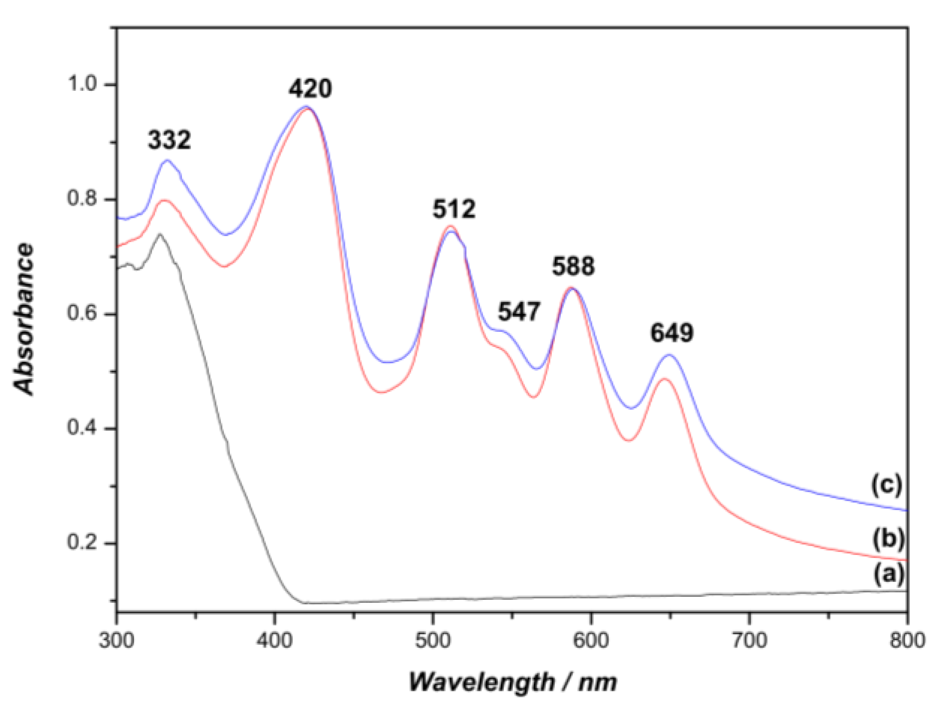
Scheme 1. Synthesis of porphyrinic derivatives and their immobilization on chitosan and on TiO2.
Ijms 20 02522 sch001
Figure 1. Solid state UV-Vis spectra of (a) P2-CF, (b) P3-CF and (c) P4-CF.
Ijms 20 02522 g001
Figure 2. Emission spectra in the solid state for (a) P2, (b) P3, (c) CF, (d) P4-CF, (e) P3-CF and (f) P2-CF.
Ijms 20 02522 g002
Figure 3. Digital photographs of PS-CF. From left to right: CF, P2-CF, P3-CF and P4-CF under ultraviolet light.
Ijms 20 02522 g003
Figure 4. UV-Vis spectra in the solid state of (a) TiO2, (b) P3-TiO2 and (c) P4-TiO2.
Ijms 20 02522 g004
Figure 5. Digital photographs of PS-TiO2. From left to right: TiO2, P3-TiO2 and P4-TiO2 under UV light.
Ijms 20 02522 g005
Figure 6. Emission spectra in the solid state of (a) P3, (b) P4, (c) TiO2, (d) P4-TiO2, and (e) P3-TiO2.
Ijms 20 02522 g006
Figure 7. Time-dependent photodecomposition of DPiBF (50 µM) photosensitized by P1, P2, P3 and P4 in DMF upon irradiation with red light (640 ± 20 nm) with or without PS (0.5 µM). H2TPP was used as the reference.
Ijms 20 02522 g007
Figure 8. Time-dependent photodecomposition of DPiBF (50 µM) photosensitized by P2-CF, P3-CF, P4-CF, P3-TiO2 and P4-TiO2 in DMF upon irradiation with red light (640 ± 20 nm) using 2.0 µM of PS.
Ijms 20 02522 g008
Figure 9. Photoinactivation of bioluminescent E. coli in the presence of P3-P4 at the concentration of 5.0 μM under 3.0 mW cm−2 of white light irradiation. The values represent the mean of three independent experiments with three replicates and the error bars indicate the standard deviation.
Ijms 20 02522 g009
Figure 10. Photoinactivation of the bioluminescent E. coli in the presence of porphyrins P2, P3 and P4 supported on chitosan at the concentration of 3.0 µM under 3.0 mW cm−2 of white light irradiation. The values represent the mean of three independent experiments with three replicates and the error bars indicate the standard deviation.
Ijms 20 02522 g010
Figure 11. Photoinactivation of bioluminescent E. coli with 10 μM of the porphyrins P3 and P4 supported on TiO2 under 3.0 mW·cm−2 of white light irradiation. The values represent the mean of three independent experiments with three replicates and the error bars indicate the standard deviation.
Ijms 20 02522 g011
Table 1. Selected photophysical properties of porphyrins P1–P4 in DMF.
PorphyrinSoret Band
λmax (nm)
Q Bands
λmax (nm)
λemission (nm) a)Stokes Shift
(nm)
ΦF b)
P1420512545585641648/71170.03
P2420510544584640649/71190.01
P3420512546584640649/71190.03
P4420510544584640648/71180.01
a) The optical density of all the samples was 0.05 at 420 nm; b) using H2TPP as the reference in DMF (ΦF = 0.11) [64].
Table 2. Porphyrin immobilization in chitosan.
SolidLoading (mol·g−1) a)w/w (%) b)Immobilization (%) c)
P2-CF7.87 × 10−60.8944%
P3-CF1.03 × 10−61.3065%
P4-CF1.09 × 10−61.3869%
a) Porphyrin (mol) / mass (g) of solid prepared; b) mass (g) of porphyrin per gram of chitosan; c) immobilization percentage based on the relative mass of porphyrin used.
Table 3. Porphyrin immobilization on titanium dioxide solid results.
SolidLoading
(Porphyrin Per Mass of Solid Prepared) (mol g1) a)
w/wb)
(%)
Immobilization c)
(%)
P2-TiO20 d)0c-
P3-TiO21.44 × 10−51.8291%
P4-TiO21.55 × 10−51.9696%
a) Porphyrin (mol) / mass (g) of titanium dioxide; b) mass (g) of porphyrin per mass (gram) of titanium dioxide; c) immobilization percentage based on the mass of porphyrin used; d) there was no interaction.

