Review

Genomic Features and Insights into the Taxonomy, Virulence, and Benevolence of Plant-Associated Burkholderia Species

1
Department of Integrated Biological Science, Pusan National University, Busan 46241, Korea
2
Department of Oriental Food and Culinary Arts, Youngsan University, Busan 48015, Korea
*
Author to whom correspondence should be addressed.
Int. J. Mol. Sci. 2019, 20(1), 121; https://doi.org/10.3390/ijms20010121
Submission received: 13 December 2018 / Revised: 24 December 2018 / Accepted: 24 December 2018 / Published: 29 December 2018
(This article belongs to the Section Molecular Plant Sciences)

Abstract

:
The members of the Burkholderia genus are characterized by high versatility and adaptability to various ecological niches. With the availability of the genome sequences of numerous species of Burkholderia, many studies have been conducted to elucidate the unique features of this exceptional group of bacteria. Genomic and metabolic plasticity are common among Burkholderia species, as evidenced by their relatively large multi-replicon genomes that are rich in insertion sequences and genomic islands and contain a high proportion of coding regions. Such unique features could explain their adaptability to various habitats and their versatile lifestyles, which are reflected in a multiplicity of species including free-living rhizospheric bacteria, plant endosymbionts, legume nodulators, and plant pathogens. The phytopathogenic Burkholderia group encompasses several pathogens representing threats to important agriculture crops such as rice. Contrarily, plant-beneficial Burkholderia have also been reported, which have symbiotic and growth-promoting roles. In this review, the taxonomy of Burkholderia is discussed emphasizing the recent updates and the contributions of genomic studies to precise taxonomic positioning. Moreover, genomic and functional studies on Burkholderia are reviewed and insights are provided into the mechanisms underlying the virulence and benevolence of phytopathogenic and plant-beneficial Burkholderia, respectively, on the basis of cutting-edge knowledge.

1. Introduction

The genus Burkholderia belongs to the subphylum of β-proteobacteria and encompasses Gram-negative bacterial species with high genetic versatility and adaptability to various ecological niches [1]. More specifically, they are able to occupy ecosystems as diverse as soils, plants, and even animal and human bodies as serious disease-causing agents [2,3,4]. Recently, Burkholderia organisms have drawn increasing attention as abundant and fundamental components of various ecosystems. The first member of the genus was isolated in 1942 from carnations displaying wilt and root-rot symptoms. It was initially named Phytomonas caryophylli and later renamed Pseudomonas caryophylli [5]. In 1950, another species was isolated from onions showing sour skin rot and named Pseudomonas cepacia [6,7]. Some members, originally included in the genus Pseudomonas, were later reclassified in the new Burkholderia genus due to improved taxonomic tools, the availability of techniques for DNA–DNA hybridization, as well as for 16S rRNA analysis [8]. Since then, many other species have been, and are still being, described and added to the genus.
The adaptability of Burkholderia spp. to diverse environments and their ecological versatility rely on the high genetic plasticity of their multi-chromosome genomes, rich in insertion sequences [9] and exhibiting a relatively high proportion of coding regions, allowing these bacteria to produce various metabolites with degradative potential [10]. Such metabolic capacity is a fundamental feature for their survival and fitness in different habitats [11].
The Burkholderia cepacia complex (BCC), one of the major components of the Burkholderia genus, comprises a group of related opportunistic human pathogen species responsible for pulmonary infections, particularly in immunocompromised and cystic fibrosis patients [9]. Due to the importance of BCC members as human pathogens, intensive research efforts and discussion have been devoted to the epidemiology of human diseases caused by Burkholderia spp. Other related species include animal pathogens such as Burkholderia mallei and plant pathogens such as Burkholderia glumae, Burkholderia gladioli, and Burkholderia plantarii. Notably, another group of plant-associated Burkholderia spp. comprises potentially beneficial species of plant growth-promoting or plant-symbiotic bacteria, favoring the healthy growth of plants by nitrogen fixation and protection against plant diseases. From the taxonomic standpoint, an effort is being made to better discriminate beneficial from pathogenic Burkholderia spp. [12].
In this review, the taxonomy of the Burkholderia genus is discussed, highlighting the major updates and the proposals of new delineated genera. Moreover, studies on the unique features of Burkholderia genomes, related to bacterial adaptability to a wide-range of ecological niches, are reviewed. The main focus of this review is on plant-associated species of the Burkholderia genus, including both phytopathogenic and beneficial symbiotic species, with a special attention to factors affecting virulence and benevolence, respectively. However, the insect-symbiotic Burkholderia is not covered in this review.

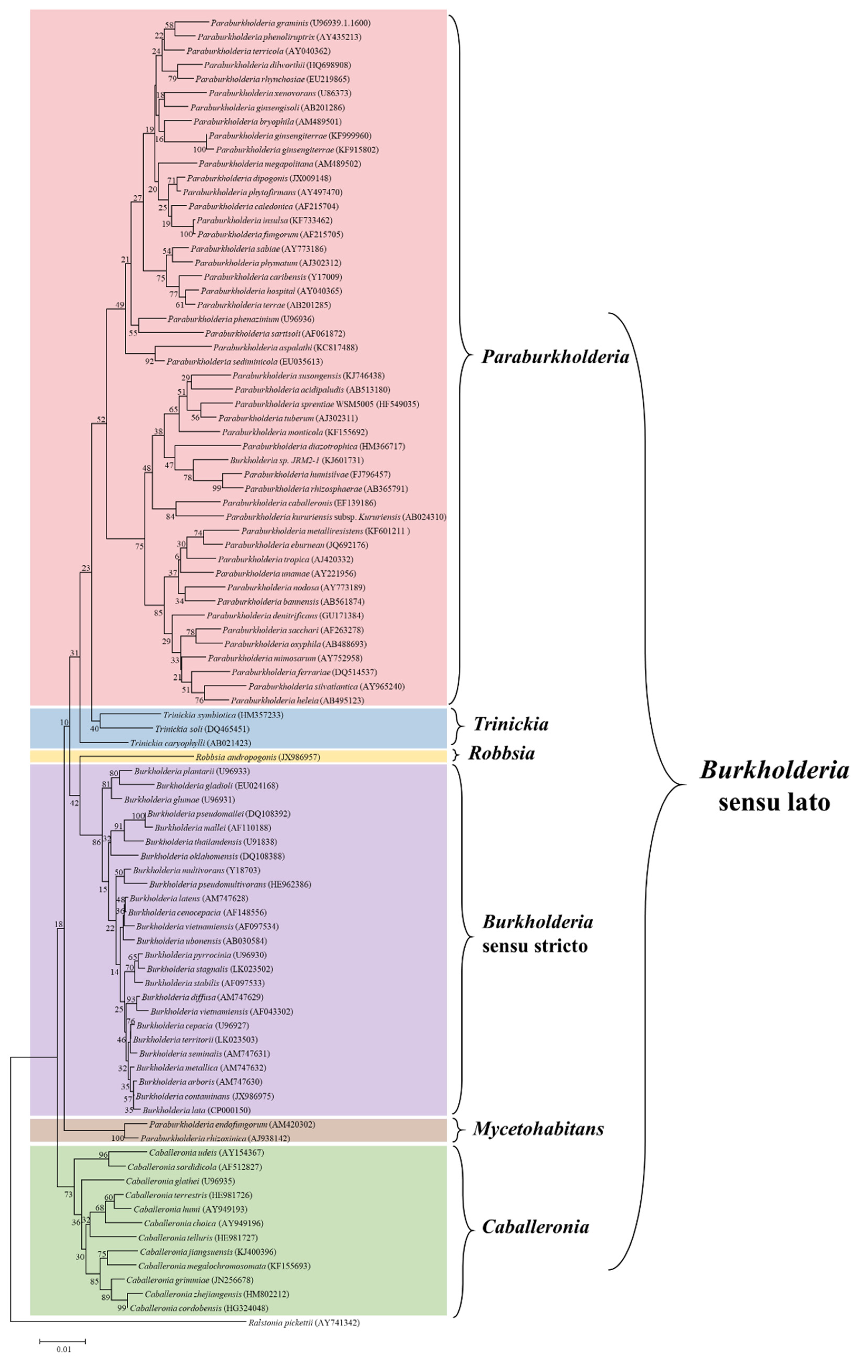
2. Taxonomic Updates of the Burkholderia Sensu Lato

Following the proposal of Burkholderia as a new genus in 1992 [8], and the continuous increase in the number of species within it, several taxonomic rearrangements have been suggested. Pieces of evidence from 16S rRNA analysis and the construction of phylogenetic trees based on other genes, such as recA, have increasingly supported the division of the Burkholderia genus into two distinct lineages [13]. Based on the suggested division, one group comprises human, animal, and plant pathogens or opportunistic pathogens, whereas the other group is represented by environmental xenobiotic-biodegrading, plant symbiotic, and plant growth-promoting species.
The distinction between the two lineages has been further supported by the identification of genomic within-group similarities and between-group differences. Ussery et al. [14] explored the genomic diversity among members of the Burkholderia genus and defined the pan- and core-genomes using the genome sequences available at the time of that study (56 full and partial genome sequences). A phylogenetic analysis conducted on 612 genes from the core Burkholderia genome resulted in a clear separation of the pathogenic Burkholderia spp. from members of the other clade. By combining comparative genomics with phylogenetic analyses based on 16s rRNA, recA, gyrB, and acdS genes, Gyaneshwar et al. [15] suggested the distinction of the Burkholderia genus into two subgenera and initially proposed the new genus, Caballeronia.
Consistently, multilocus sequence analysis using four housekeeping genes, namely atpD, gltB, lepA, and recA, in combination with 16S rRNA, confirmed the presence of different Burkholderia lineages by separating the genus into two major clusters [16]. The distinction was even more obvious after bioinformatics analysis of the virulence loci of Burkholderia genomes and functional pathogenicity tests [17]. In light of compelling evidence on the existence of systematic differences within the Burkholderia group, a novel genus, Paraburkholderia, was proposed based on the analysis of conserved sequence insertions/deletions [18]. Since then, several other species have been ascribed to the Paraburkholderia genus and the previously proposed Caballeronia genus [19]. Following reassessment of Burkholderia andropogonis, this species was separated into a new genus, Robbsia, as confirmed by phylogenetic and comparative genomic analysis [20]. Using whole-genome sequence data, Beukes et al. [21] addressed these complex phylogenetic relationships and confirmed the existence of generic boundaries among Burkholderia sensu lato bacteria. Although the analyzed genomes were similar in size and number of encoded genes, the genomes of Burkholderia sensu stricto showed higher G+C contents. The latter study includes a large-scale phylogenetic analysis on 106 common genes of Burkholderia sensu lato, specifically selected to limit phylogenetically irrelevant data. The results support the subdivision of Burkholderia sensu lato into five distinct lineages: Burkholderia sensu stricto, Paraburkholderia, Caballeronia, Robbsia, and Paraburkholderia rhizoxinica [21]. More recently, whole-genome analysis has been employed in the context of an international joint effort to investigate the taxonomic status of selected species of Burkholderia sensu lato [22]. Based on the phylogenetic analysis of conserved genes and average nucleotide and amino acid identities, two novel genera were proposed within the Burkholderia sensu lato assemblage. The first, Mycetohabitans, encompasses fungal symbiotic species with relatively small genomes. The second, Trinickia, includes soil and plant-associated species. Furthermore, the comparison of core-genomes revealed the presence of functional genus-specific genes. A historical overview of the major rearrangements and updates in Burkholderia taxonomy is summarized in Table 1.
It is clear that the Burkholderia sensu lato group has undergone several taxonomic changes and could be subjected to further adjustments in the future. The increased availability of genomic information, as well as the advancements in bioinformatics analysis, have enhanced our understanding of the evolutionary and taxonomic relationships between clinically, environmentally, industrially, and agriculturally important Burkholderia species. All this considered, the large group of Burkholderia sensu lato is currently composed of six genera, i.e., Burkholderia sensu stricto, Caballeronia, Paraburkholderia, Robbsia, Mycetohabitans, and Trinickia. The six genera were established after phylogenetic analysis of the main representative Burkholderia sensu lato strains using the neighbor-joining method, based on 16S rRNA sequences retrieved from the SILVA database (https://www.arb-silva.de/), as shown in Figure 1.
With regard to the scope of the current review, phytopathogenic Burkholderia species are classified in the Burkholderia sensu stricto group, closely related to the Burkholderia cepacia complex; the former Burkholderia andropogonis is currently a separate genus as Robbsia andropogonis; and Burkholderia caryophylli is now classified as Trinickia caryophylli. On the other hand, most of the plant-beneficial Burkholderia species were transferred to the Paraburkholderia and Caballeronia genera.

3. Genomic Features of the Plant-Associated Pathogenic and Beneficial Burkholderia

Because of the clinical importance of the B. cepacia complex, special consideration is devoted to these bacteria by genomic studies and, therefore, their genomes are well-characterized [9]. Besides common genomic features shared by most organisms of the large Burkholderia sensu lato group, functional features specific to the plant-associated groups are present and allow them to occupy various ecological niches with different lifestyles, as pathogens, legume nodulators, or biocontrol agents. Common features of Burkholderia species include relatively large multi-replicon genomes, the presence of genomic islands, and multiple insertion sequences conferring high genome plasticity [9,10].

3.1. Genome Size

Generally, Burkholderia organisms have larger genomes (7–8 Mbp on average) than other bacteria; however, the genome size greatly varies among different Burkholderia groups. The smallest genome among the free living Burkholderia (3.75 Mbp) is found in the fungal endosymbiont, Mycetohabitans rhizoxinica (basonym Burkholderia rhizoxinica), whereas the largest genome (11.5 Mbp) is found in the soil bacterium Paraburkholderia terrae (basonym Burkholderia terrae). It is suggested that genome size can provide insights into the evolutionary history and the relationships between Burkholderia species. It is also noted that the particular lifestyle of Burkholderia species might reflect selective pressure for a specific genome size [23].
Two main models for genome evolution in Burkholderia species were proposed: (i) plasmids might undergo rearrangements following parallel transfer, becoming established as additional chromosomes or megaplasmids and allowing for bacterial acquisition of essential genes; and (ii) loss of genomic information following rearrangements [24]. An example of the latter mechanism could be M. rhizoxinica, the obligate endosymbiont of the zygomycete Rhizopus microsporus, which is a causal agent of rice seedling blight development by production of rhizoxin [25]. The genome of M. rhizoxinica, containing one chromosome, a megaplasmid, and a plasmid, is remarkably small (relative to other Burkholderia), most likely because of rearrangements and deletion of genomic information, especially in genes coding for mobile genetic elements. The endosymbiotic lifestyle reduced the bacterial capacity to adapt to varying habitats and made efficient sensing superfluous [26,27]. On the other hand, a large part of the chromosomal coding region is devoted to the production of the secondary metabolite, rhizoxin [26,27].
Regarding the plant symbiotic Burkholderia, which can also be free-living in the bulk soil or water, a characteristic large genome size could explain their versatile lifestyle and the capacity to perform many plant-beneficial functions [28].
In another comparative genomic study of phytopathogenic Burkholderia species, Seo et al. [29] analyzed the genome sequences of different strains of B. glumae, B. gladioli, and B. plantarii and found genome size variations not only between species but also between different strains of the same species. For B. glumae, the smallest genome size, ~4.9 Mbp, was exhibited by the AU6208 strain, whereas the largest, ~7.2 Mbp, was found in the BGR1 strain. The relatively small genome size of B. glumae AU6208 could be attributed to genome rearrangements or deletions, as a result of adaptation to different hosts. Notably, although pathogenic to rice, AU6208 was originally isolated from infant patients, unlike other strains that were all isolated from rice as the original host. The genome size of B. gladioli BSR3 is ~9 Mbp and that of B. plantarii is ~8 Mbp.

3.2. Multi-Replicon Nature

A multi-replicon genome structure is found in most Burkholderia species with various numbers of chromosomes and plasmids. Martínez-Aguilar et al. [28] studied the multi-replicon genomes of plant-associated diazotrophic Burkholderia species and found that three strains of Paraburkholderia unamae and Paraburkholderia silvatlantica (formerly Burkholderia spp.) contain genomes of four replicons. The largest and smallest replicon sizes are ~3.31 and ~1.11 Mbp, respectively. In addition, the genomes of three strains of Paraburkholderia tropica (formerly Burkholderia tropica) contain five replicons ranging from ~3.24 to ~0.53 Mbp. The authors suggested that such multi-replicon large genomes could explain the capacity of these diazotrophic Burkholderia species to efficiently colonize the rhizosphere as well as endophytic environments.
A multi-replicon genome structure is also reported among the plant-pathogenic Burkholderia species. The genomes of the representative strains B. glumae BGR1 and B. gladioli BSR3 contain two chromosomes and four plasmids, while the B. plantarii ATCC43733 genome contain two chromosomes and one plasmid [29]. Agnoli et al. [30] found that deletion of the third chromosome from the genomes of several BCC species results in the attenuation of virulence and the loss of many functions such as the ability to utilize several substrates, antifungal activity, and biosynthesis of extracellular polymeric substances. Intriguingly, bacterial viability is not significantly affected, despite the massive loss of genomic information (~1 Mbp). This finding indicates that multi-replicon genomes are essential for adaptation but not necessarily for bacterial growth and survival.
While studying the pan- and core-genomes of Burkholderia species, Ussery et al. [14] found that only few hundred genes are conserved among all genomes to form the Burkholderia core genome, whereas more than 40,000 gene families are present in the Burkholderia pan genome. A more recent study of the available genome sequences, focusing on 110 genome sequences, shows that around 35,680 genes belong to pan-gene and 717 to core-gene families [23]. In the latter study, the number of shared genes has significantly dropped upon adding genes from different replicons to the analysis, which indicates the low percentage of genes shared by different genomic replicons of Burkholderia. In the same context, Seo et al. [29] analyzed the genomes of 106 Burkholderia species, representing the different groups, and found that 78,782 genes correspond to pan-genomes, while 587 are core genes conserved in all tested species. By considering the genomes of several strains of three plant pathogenic Burkholderia (B. glumae, B. gladioli and B. plantarii), 12,758 pan-genome genes are present in the tested strains, while 1908 genes are found conserved as the core-genome [29].

3.3. Genomic Islands and Multiple Insertion Sequences

Along with the above-mentioned features of Burkholderia genomes, other unique features are related to the genetic plasticity of these bacteria, such as the presence of genomic islands and multiple insertion sequences. Genomic islands refer to foreign DNA regions that have been horizontally acquired and became integrated within the genomes of several Burkholderia species. They have important evolutionary roles and can specify several accessory functions [31]. Multiple approaches have been used to identify these foreign DNAs, such as the analysis of G+C content, which has been found to differ in genomic islands with respect to the rest of the genome [32,33]. Genomic islands vary in their structure and content between Burkholderia species and account for more than 10% of the genomes in most species [24]. Genomic islands linked to pathogenicity and transmission have been extensively characterized. However, the coding capacity of genomic islands is not only associated with pathogenicity and virulence but also with adaptive traits such as symbiosis and metabolism [31]. An example of this is the environmental species Paraburkholderia xenovorans (formerly Burkholderia xenovorans), in which more than 20% of total genes are a recent acquisition and mainly involved in niche adaptation [34].
The genomes of Burkholderia species also contain many diverse insertion sequences that are mainly attributed to genomic rearrangements and replicon fusion and are related to transcriptional regulation [10,24,35,36]. This finding indicates that the presence of multiple insertion sequences in Burkholderia genomes has a role in phenotypic, as well as genetic diversity, by activating and/or inactivating adjacent genetic loci. Target genes may be inactivated or, in the case of adjacent promoter-less genes, activated by the promoters provided by insertion sequences [37]. Furthermore, the presence of multiple copies of the same insertion sequence may drive genetic rearrangements by fusion of two replicons, inversion, deletion, and duplication of the intervening region [38,39]. Accordingly, the transposition of insertion sequences is a very important evolutionary mechanism for the emergence of adaptation capacity and variability in Burkholderia species. A clear example of insertion sequence-mediated genomic alterations is provided by the genome of B. mallei, which is suggested to arise from several deletions and inversions of the Burkholderia pseudomallei genome. Evidence supporting such an assumption includes the presence of insertion sequences bordering most of the breakdown points and the presence of regions from chromosome 1 of B. pseudomallei on chromosome 2 of B. mallei [40]. In the phytopathogen B. glumae, the insertion sequences IS1417, IS1418, and IS1419 are characterized and found to be prevalent among different field strains. Apparently, these sequences may have been horizontally acquired by B. glumae from other related species, as they are also found in species such as B. gladioli and B. plantarii, with IS1417 being the most prevalent [41].

4. Plant Pathogenic Burkholderia

The Burkholderia group comprises several etiological agents of plant diseases causing significant economic losses by affecting the production of various crops. The major phytopathogenic species of Burkholderia group are B. glumae, B. gladioli, B. plantarii, R. andropogonis, and Trinickia caryophylli (formerly Burkholderia caryophylli) [42]. Although the mechanisms involved in pathogenicity and adaptation have not been fully elucidated, genomic studies and functional analyses, accompanied by phenotypic characterization, have enhanced our understanding of the virulence factors involved [29].
B. glumae, causing bacterial panicle blight and seedling rot of rice, was first reported in Japan, and is currently considered a major threat to rice cultivation worldwide [43,44]. In rice, the seed borne nature of B. glumae facilitates disease spread, which is estimated to cause the loss of up to 75% of heavily infested fields [45]. B. glumae is also found to be responsible for wilting symptoms in other hosts including pepper, sunflower, and tomato [46].
B. gladioli was first reported to cause corm rot in Gladiolus spp. [47]. Later, three pathovars were found: B. gladioli pv. gladioli, causing gladiolus rot; B. gladioli pv. alliicola, causing onion bulb rot; and B. gladioli pv. agaricicola, causing rapid soft rot of cultivated mushrooms [48,49,50].
B. plantarii was first reported to cause rice seedling blight in Japan and has been isolated from several other host plants [42,51]. Both B. plantarii and B. gladioli have also been reported to cause, in rice, a similar disease to that caused by the related species B. glumae [51,52].
R. andropogonis (formerly known as B. andropogonis) was first reported to be the causative agent of strip disease in sorghum and leaf spot in velvet bean, and since then it has been described to cause disease in a wide range of host plants [42,53,54,55].
T. caryophylli (formerly B. caryophylli) is responsible for a damaging disease of carnation in Japan, causing significant losses and was also found to cause onion rot [56,57].
The well-characterized fungal endosymbiont M. rhizoxinica (formerly B. rhizoxinica) could also be considered as a phytopathogen due to its cooperation with the phytopathogenic fungus Rhizopus microsporus in the development of rice seedling blight [42]. M. rhizoxinica controls the vegetative growth of the fungus [58]. However, although the main virulence factor of rice seedling blight was originally thought to be produced by the fungus, it was later revealed that M. rhizoxinica is responsible for rhizoxin production [59].

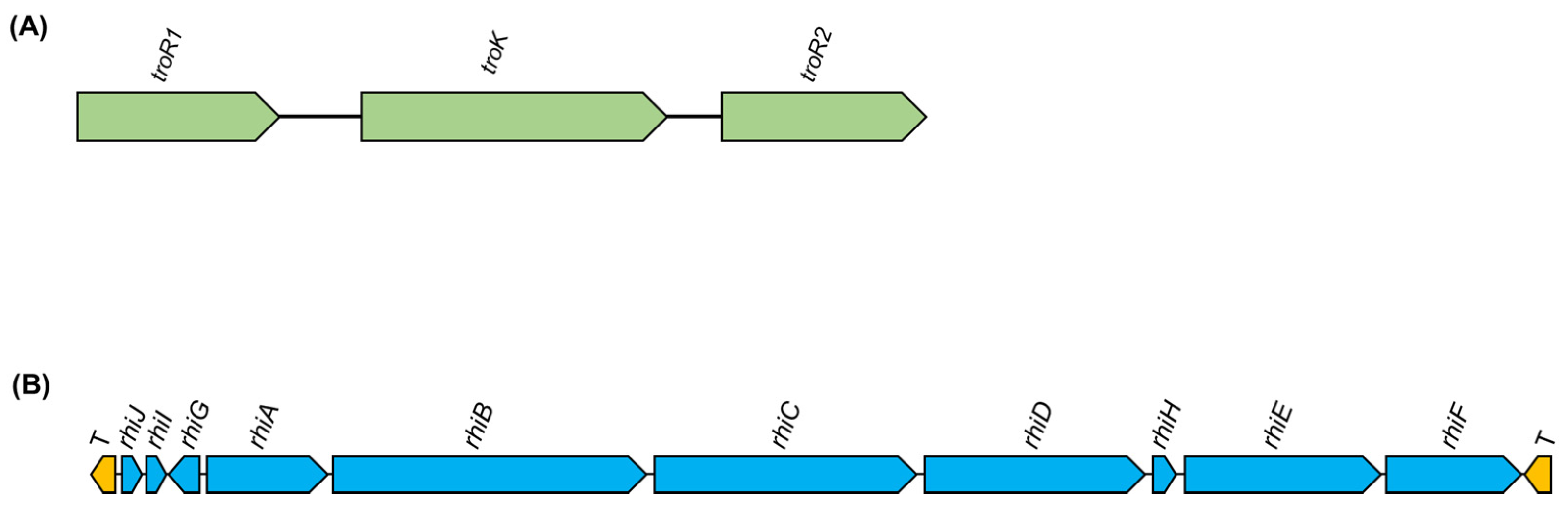
4.1. Virulence Factors in Phytopathogenic Burkholderia
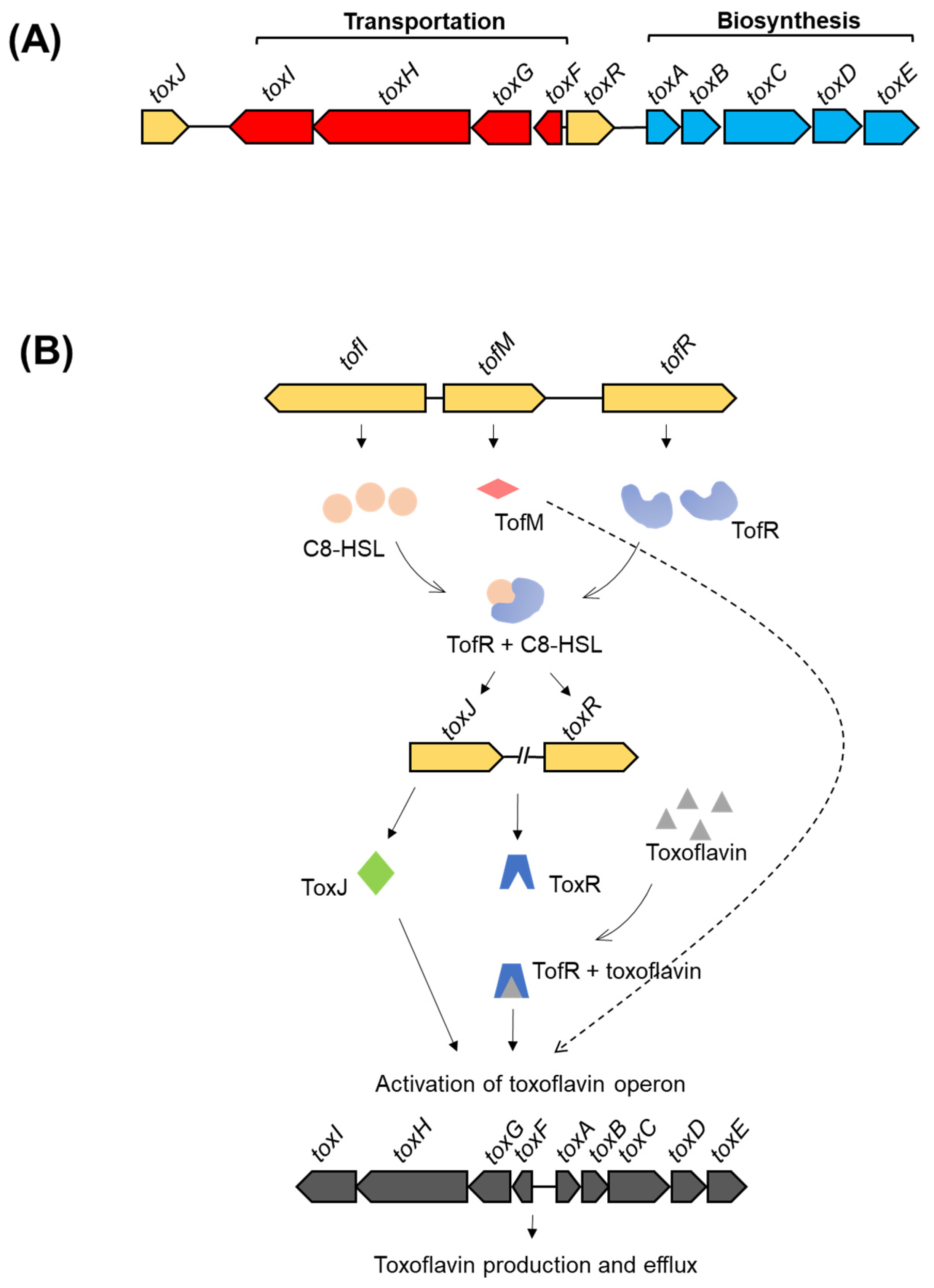
The first isolated members of Burkholderia group were phytopathogens with unique genomic features, as described above, enabling them to develop an arsenal of virulence factors, thereby causing diseases in various host plants [29,42]. The pathogenicity of phytopathogenic members of the Burkholderia group is complex and several virulence factors have been found to contribute to disease development. The virulence factors and their genetic regulation in B. glumae have been extensively studied due to the significant economic impact of bacterial panicle blight and bacterial grain rot on rice cultivations worldwide [44,60]. The major virulence factors include phytotoxins and other virulence-related enzymes, bacterial motility, secretion systems, and exopolysaccharides (EPSs). Some of these virulence factors are common among phytopathogenic Burkholderia, whereas others are species-specific.

4.1.1. Phytotoxins

Phytotoxins are plant-deleterious compounds produced by pathogens and one of the major responsible factors for the development of disease symptoms [61]. Toxoflavin, the most important and studied phytotoxin of phytopathogenic Burkholderia species, was first described as an azapteridine antibiotic in Burkholderia cocovenenans [62], later found in B. glumae [63], and more recently in the related species B. gladioli [64]. Toxoflavin from B. glumae is associated with the inhibition of sprout and root elongation in rice seedlings, as well as severe damage in toxin-treated panicles [65]. Moreover, Jeong et al. [46] showed that toxoflavin is also involved in development of severe wilting symptoms in several other crops, indicating that a broad range of host plants are affected. Toxoflavin is also toxic to mice and exhibits antibacterial and antifungal activities [66]. The wide spectrum of toxoflavin activities might be explained by its role as an active electron carrier between NADH and oxygen, involved in the production of reactive oxygen species such as hydrogen peroxides [67].
The genetic regulation of toxoflavin biosynthesis and transport is well characterized in B. glumae. A tox operon composed of five genes (toxA, toxB, toxC, toxD, and toxE) is responsible for toxin biosynthesis and is regulated by a LysR-type regulator, ToxR [68,69]. In a later study, another operon composed of four genes (toxF, toxG, toxH, and toxI) is responsible for toxoflavin transportation [70]. In the same study, the regulation of both tox operons is further elucidated. Upon activation by toxoflavin binding (as a co-inducer), ToxR regulates the expression of both operons. Moreover, the other regulator, ToxJ, is also involved in the activation of both tox operons. The activation of ToxJ is mediated by a quorum sensing (QS) system via the luxI homolog, tofI, and the luxR homolog, tofR, via AHL (N-acyl homoserine lactone) QS signals. That is, bacterial cell density is required for active toxoflavin production, so that bacterial cells can start toxoflavin-mediated attack of host cells only once a sufficiently high bacterial population has been reached [70]. An illustration of both tox operons and a proposed model of regulation is shown in Figure 2.
The complete genome sequence of B. gladioli BSR3 reveals the presence of the toxoflavin operons, as well as relevant regulatory genes [72]. To investigate the genetics of toxoflavin production and regulation in B. gladioli, Lee et al. [73] conducted a comparative genomic analysis of several strains of B. gladioli, including the non-toxoflavin-producing strain KACC11889 and B. glumae BGR1. The results indicate that the toxoflavin-producing strains of B. gladioli shared with B. glumae the genes for toxoflavin production and regulation, except that toxJ is located on another chromosome in B. gladioli. In addition, the genome of the non-toxoflavin-producing strain, B. gladioli KACC11889, contains the operons for biosynthesis and transport of toxoflavin, whereas the toxoflavin regulatory QS genes, tofI and tofR, are not found. Thus, the loss of these regulatory genes results in the inhibition of toxoflavin production, confirming the suggestion of a similar mechanism of toxoflavin regulation in B. gladioli and B. glumae [73].
Although toxJ regulatory genes are identified in the genome of the related species B. plantarii, toxoflavin biosynthesis and transport genes are not identified [29]. B. plantarii produces another phytotoxin, a non-benzenoid aromatic tropolone, which is responsible for seedling blight symptoms in rice, as confirmed by the replication of the characteristic bacterial-induced blight symptoms upon treatment of rice seedlings with exogenous tropolone [51,74]. Tropolone is also a potential iron chelator and has a wide range of biological roles such as antibacterial, antifungal, and insecticidal activities [75,76].
B. plantarii produces AHL-QS signal molecules resembling those of B. glumae, as the luxI family member, plaI, and the luxR family member, plaR, are highly similar to the B. glumae AHL-QS system genes, tofI and tofR. Through the generation of plaR knockout mutants, the role of the AHL-QS regulatory system in the virulence of B. plantarii to rice is established [77]. It was later revealed that treatment of B. plantarii with AHL-QS inhibitors resulted in the suppression of tropolone production by blocking the expression of plaI [78]. Therefore, a resemblance between the regulation of tropolone biosynthesis and that of toxoflavin, in B. glumae, could be inferred from the major role played by QS-related genes. This possibility is further supported by the presence of QS-AHL signal synthase-regulator loci within the tropolone biosynthesis operon (Seo et al., 2015 [29]). More recently, Miwa et al. [79] reported that a two-component signal transduction system regulates both tropolone biosynthesis and virulence in B. plantarii. Defects in any of the three genes encoding the two-component system (sensor histidine kinase and response regulators) resulted in the inhibition of toxin production and attenuation of bacterial virulence in rice. Three genes (i.e., troR1, troK, and troR2) are suggested to form an operon for tropolone synthesis, due to their adjacent chromosome position and their simultaneous requirement for toxin production [79]. A schematic organization of the two-component system genes controlling tropolone production is shown in Figure 3A. Further investigations are required to unravel the gene cluster organization and the regulation of tropolone biosynthesis, the main virulence factor in B. plantarii, and the possible regulatory roles of tropolone.
The other phytotoxin relevant for plant disease is rhizoxin, produced by M. rhizoxinica, the fungal endosymbiont of R. microspores. This bacterial endosymbiont alters the vegetative growth and sporulation of the host fungus by preventing the production of endosymbiont-free fungal spores [25,26]. The endosymbiont-produced rhizoxin is an effective inhibitor of eukaryotic cell growth by acting on β-tubulin and consequently blocking cell mitosis [80]. Besides its role as a virulence factor responsible for seedling blight symptoms in rice, various additional physiological functions have been attributed to rhizoxin, including reproduction in the host fungus and antibiotic activity against other microbes [25,81].
The availability and analysis of the complete genome sequence of M. rhizoxinica have allowed for the clarification of rhizoxin biosynthesis and the identification of the responsible gene cluster (rhi) [26,59]. The rhi locus is found on the large chromosome, flanked by transposase genes, indicating that it may represent a potentially mobile region of the genome [26,27]. It is also suggested that the rhizoxin gene cluster might have been transferred from the megaplasmid to the chromosome after horizontal acquisition, as concluded from the chromosomal location of the gene cluster, near the replication terminus, and the comparison of its G+C content with that of the rest of the chromosome [26]. Notably, in the comparative genomic analysis conducted by Seo et al. [29], genes involved in rhizoxin biosynthesis are also detected in other phytopathogenic Burkholderia spp., such as B. plantarii ATCC43733 and B. glumae PG1, for which the ability to produce rhizoxin has not been demonstrated. The organization on the rhi gene cluster is shown in Figure 3B.
The phytopathogen R. andropogonis produces the enol-ether amino acid phytotoxin, rhizobitoxine, that is responsible for leaf chlorosis in host plants [82]. Notably, rhizobitoxine has also been associated with a positive role in the symbiosis of legume nodulators. The phytotoxic activity of rhizobitoxine is related to inhibition of the methionine and ethylene biosynthetic pathways. The genes for rhizobitoxin biosynthesis (rtx operon) are identified in the legume nodulator, Bradyrhizobium elkanii [83,84]. However, further work is required to describe the biosynthetic genes and their regulation in R. andropogonis.
Another example of the versatility of Burkholderia species and their contribution to vital ecological aspects is the phytotoxin produced by another Burkholderia endo-symbiont. In this case, the endo-symbiont is associated with the pine wood nematode, Bursaphelenchus xylophilus, causing pine wilt disease, a devastating threat to pine trees worldwide [85]. The pine wood nematode endo-symbiont Burkholderia arboris produces pyochelin, which is a bacterial siderophore with several bioactivities due to its capacity to generate reactive oxygen species [86]. However, B. arboris-produced pyochelin is responsible for damaging plant tissues and is linked to the development of pine wilt disease, as the treatment of pine callus with purified pyochelin confirms its strong phytotoxicity [87]. Further studies are required to unveil the phytotoxic mechanism of pyochelin.
The different phytotoxins produced by the phytopathogenic species of Burkholderia group, their effect, and mode of action are summarized in Table 2.

4.1.2. Secretion Systems

The ability of Gram-negative bacteria, including Burkholderia spp., to control the translocation of potent effectors from their sites of synthesis to the exterior of the cell, or even to other surrounding organisms, is a very important tool for communication and survival [88]. Six types of bacterial secretion systems have been discovered and differentiated based on their structure, function, or type of secreted effectors [89]. Species possessing more than one type of secretion system are also common [90].
Phytopathogenic Burkholderia spp. exhibit a wide spectrum of secretion systems, playing essential roles in adaptation and virulence. Kang et al. [91] confirmed the association of Type 2 secretion system (T2SS) and T3SS with functions related to host plant colonization and bacterial growth, as indicated by the reduced virulence and retarded growth of T2SS- and Hrp T3SS-deficient mutants in host plants. Comparative genomic analysis of phytopathogenic Burkholderia reveals that the genes for T3SS are highly conserved in several strains of B. glumae, B. gladioli, and B. plantarii. However, within species-variation is observed in T1SS and T4SS [29].
Phytopathogenic Burkholderia organisms have different groups of T6SSs. Group 1 is conserved in all species [92]. T6SS is involved in the delivery of effectors to both eukaryotic and prokaryotic cells. Thus, it is suggested to be important for the competition with other surrounding microbes and for virulence to host plants [93].
Although several functions have been reported for different bacterial secretion systems, for many reasons, including the remarkable diversity and limited expression of effectors as well as the multifarious nature of the secretion systems, not all secreted effectors have been identified and their exact mechanisms of action are still elusive [91,94,95].

4.1.3. Other Virulence Factors

Several other factors are associated to the virulence of phytopathogenic Burkholderia species. Virulence-related enzymes such as lipases, proteases, and cellulases, as well as exopolysaccharides (EPS), contribute to the virulence of several species. For full virulence, the degradation of plant cell wall polymers by hydrolytic enzymes is crucial. Lipase is an important virulence factor for many bacterial pathogens, as it is responsible for the hydrolysis of the carboxyl ester bonds present in triacylglycerols, which liberate fatty acids and glycerol. Lipase could also play a role in the degradation of the epicuticle wax layer in plant tissues [96,97,98]. In B. glumae, lipase is also regulated by the AHL-QS system and is essential for panicle blight symptoms in rice [99]. The gene encoding lipase (LipA) is found in the three phytopathogenic species B. glumae, B. gladioli, and B. plantarii [29].
Pectin, mainly composed of polygalacturonic acid, is one of the major constituents of the plant cell wall. Polygalacturonases produced by phytopathogens can hydrolyze the pectic glycosidic bonds, depolymerizing pectin and causing rotting of plant tissues. B. glumae produces two different polygalacturonases (encoded by pehA and pehB) that are possibly regulated by QS and secreted by a T2SS. The pehA locus is present in B. glumae but not detected in B. gladioli and B. plantarii [29].
The EPSs have been shown to contribute to the pathogenesis of several plant pathogens by causing occlusion in the host vascular systems, leading to plant wilting [100]. Although the role of EPSs in the epidemiology of rice seedling blight has not yet been clearly established, they could explain the wide range of crops showing wilting symptoms in response to B. glumae infection [46]. In T. caryophylli, EPSs are suggested as important virulence factors, responsible for bacteria–plant interactions [101]. Further investigations are required to elucidate the contribution of EPSs to the virulence of phytopathogenic Burkholderia.

5. Plant-Beneficial and Symbiotic Burkholderia Species

If, on the one hand, pathogenic Burkholderia species are the subject of intensive research, mainly because of their economic implications, on the other hand, plant-beneficial and symbiotic species have attracted the attention of scientists due to their impressive ability to promote plant physiology and growth. The main intriguing findings about this group of bacteria are the discovery of nitrogen fixation ability [102] and the identification of plant endosymbionts and legume nodulators [103,104]. As a result of multiple taxonomic rearrangements and updates (see Section 2), the majority of plant-beneficial and symbiotic strains increasingly clustered together in separate clades, mainly within the Paraburkholderia group, based on the presence of several common features such as nitrogen fixation, metabolic versatility, and plant growth promoting ability [105]. Comparative genomic analyses between beneficial plant-associated Burkholderia species revealed a high degree of genetic synteny in their large multireplicon genomes [28]. Previous studies focusing on genomics and metabolism have reported the capacity of this Burkholderia subgroup to support healthy plant growth.

5.1. Benevolence Factors in Plant-Beneficial and Symbiotic Burkholderia

Beneficial plant-associated species of the Burkholderia sensu lato group have been reported to carry out several functions that are beneficial to plants, such as colonization of plant surfaces and rhizosphere, nitrogen fixation, production of siderophores, solubilization of phosphates, metabolism of volatile compounds, degradation of pollutants, production of plant growth-promoting phytohormones, reduction of plant ethylene levels, and protection from biotic and abiotic stress [106,107,108].

5.1.1. Colonization of Plant Tissues and the Role of EPSs

The ability of plant-beneficial bacteria to colonize the surface of vegetal tissues and the rhizosphere is important for the promotion of healthy plant growth. In this respect, the production of EPSs is essential not only to enhance bacterial tolerance to environmental stress but also for efficient colonization, by adhesion to the root surface, and for the aggregation of root-adhering soil particles [100,109]. Bacterial EPSs also play a key role in the interaction between the plant and the endophytic or associated bacteria. It has been suggested that bacteria–plant symbiosis in the β-rhizobia is initiated by the bacterial production of an infection thread allowing bacteria to invade and colonize plant roots. Jones et al. [110] reported that, unlike wild-type bacteria, mutants deficient in EPS production are unable to initiate a symbiotic relationship with a host plant. EPS-producing bacteria regulate the expression of plant defense-related genes to prepare plants for colonization. These findings suggest that bacterial EPSs could serve as signals regulating gene expression in plants [110]. Several members of plant-beneficial Burkholderia, such as Paraburkholderia tropica, Paraburkholderia caribensis, Paraburkholderia phytofirmans, and Paraburkholderia kururiensis, have been reported to synthesize EPSs, the structure of which has been elucidated [111,112,113,114].

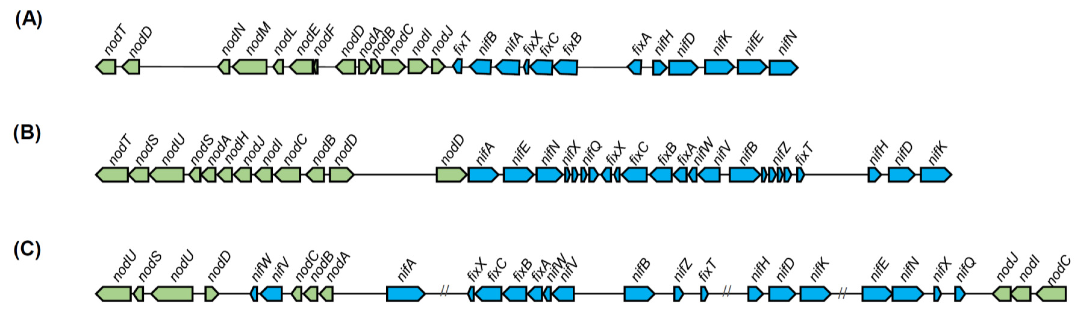
5.1.2. Nitrogen Fixation in Diazotrophic and Legume Nodulator Burkholderia

In Burkholderia bacteria, biological nitrogen fixation has been found in both plant endosymbiotic diazotrophic non-legume nodulator species, such as P. phytofirmans and Paraburkholderia unamae, and in legume nodulator species such as Paraburkholderia tuberum and P. phymatum [102,115].
The nitrogenase enzyme responsible for nitrogen fixation is composed of two structurally conserved components. Component I is encoded by nifD and nifK and component II is encoded mainly by nifH, forming the basic regions of the nif operon [116,117,118]. The nifHDK operon was first reported in the Burkholderia group in an endosymbiotic species of the arbuscular mycorrhizal fungus, Gigaspora margarita [118]. The presence of nifHDK coupled with nitrogen fixation has been confirmed in several other species in the Burkholderia group [102]. Moreover, the fix genes are also essential for nitrogen fixation, especially in microaerobic conditions. The nifHDK is controlled by the transcriptional regulator NifA. NifA and nifHDK, along with the fix genes, are regulated through the two-component signal transduction system through FixL and FixJ [119,120].
Legume nodulation is defined as the induction of nodule formation in plants by certain bacteria species, which fix atmospheric nitrogen for plants and receive carbons in exchange [121]. Previously, legume nodulation was thought to be an exclusive feature of the Rhizobiaceae members of α-Proteobacteria [122]. However, with the identification of Burkholderia legume nodulators, this hypothesis was disproved, and the term β-rhizobia was proposed to describe the legume nodulators in the β-proteobacteria [103,123,124,125,126,127]. Since then, several other Burkholderia species have been reported as legume nodulators [15].
The nodulation genes (nod), encoding proteins for production, transport, and regulation of the nod factor, along with the nif genes for nitrogen fixation, are considered as genes essential for the β-rhizobia symbiosis and are reported to be directly involved in Burkholderia nodulation ability [11]. The phylogenetic analyses conducted on the symbiosis-related genes, nodC and nifH, have evidenced that symbiosis is an ancient function in Burkholderia and these genes are vertically inherited and not the result of recent horizontal acquisition [104]. In contrast, in a recent study by Estrada-de los Santos et al. [22], phylogenies constructed by using nifD and nifH for nitrogen fixation and nodABCD for nodulation provided evidence that the nif genes have been acquired via horizontal gene transfer [22]. In the same study, the nif genes are widely present among Burkholderia sensu lato bacteria, especially in the Paraburkholderia group of plant-associated legume nodulators. However, nif genes are not found in Caballeronia, Robbsia or the newly assigned genus of fungal endosymbionts, Mycetohabitans.
With regard to the nod genes, which are only found in the group of nodulators, phylogenetic analysis has divided the nodulators into two clades based on the respective host plants: the mimosoid nodulator and the papilionoids nodulator clades. The divergence in the nod genes among the Paraburkholderia nodulators seems to be related to host plant specificity [120]. A more in-depth evaluation of nod gene organization supports the distinction between mimosoid and papilionoids nodulators. In the mimosoid-nodulators, the nod operon was organized as a single copy of nodBCIJHASU, differing from the related α-rhizobia and the papilionoid-nodulators [120]. The genetic organization of the nod and nif/fix gene clusters of representative species of α-rhizobia, mimosoid and papilionoid Paraburkholderia nodulators is shown in Figure 4. The papilionoid nodulators’ nod genes could have been acquired from the related α-rhizobia, whereas a different evolutionary origin is suggested for the nod genes of mimosoid nodulators [22].

5.1.3. Plant Growth Promotion

Plant growth promotion is common among members of the beneficial plant-associated Burkholderia, which possess several growth promoting factors involved not only in nitrogen fixation but also in ACC (1-aminocyclopropane-1-carboxylate) deaminase activity, phosphate solubilization, as well as siderophore and phytohormone production [105].
In plant tissues, ACC is an intermediate precursor in the production of the plant growth inhibitor, ethylene, in developing or stressed plants. The bacterially produced ACC deaminase enzyme catalyzes the conversion of ACC to ammonia and α-ketobutyrate, thereby reducing the ethylene levels in plant tissues, which in turn results in plant growth promotion, mainly manifesting as root elongation [128]. Production of ACC deaminase is a common feature in beneficial plant-associated Burkholderia species and the relevant encoding gene (acdS) is highly conserved and widely distributed among the rhizospheric, endophytic, and nodulator groups [129]. Furthermore, ACC deaminase activity is confirmed by comparing tomato plant responses to inoculation with an ACC deaminase-producing wild-type P. unamae strain and an acdS knockout mutant, which clearly demonstrates the plant growth-promoting properties of ACC deaminase-producing P. unamae [129]. In another study, an asdS deletion mutant of P. phytofirmans, devoid of ACC deaminase activity, lacked the ability to promote the root elongation of canola seedlings [130].
Phosphorus is an essential macronutrient for plant growth. Soil mineral and organic phosphates can be solubilized and become available for plants by the action of beneficial soil microbes [131]. A phosphate solubilization ability is reported in several species of plant-associated Paraburkholderia [132,133,134]. Among the species tested by Estrada-de los Santos et al. [22], only the soil bacterium Trinickia dabaoshanensis (formerly Paraburkholderia dabaoshanensis) can solubilize phosphate. The genetic background and mechanisms of phosphate solubilization have not been clearly characterized yet.
Plant growth regulators such as the auxin class of phytohormones are known to promote plant growth, particularly by controlling growth and development of plant roots [135]. Indole-3-acetic acid (IAA) is the most common and naturally occurring auxin and its presence has been extensively documented in several soil and rhizosphere bacteria. A clear example of IAA-producing species, among beneficial plant-associated Burkholderia, is the well-known plant growth-promoting P. phytofirmans, in which whole-genome sequence analysis reveals the presence of two independent putative IAA synthesis pathways, the indole-3-acetamide and the tryptophan side-chain oxidase pathway [136,137]. More recently, Zúñiga et al. [138], reported that IAA also plays a role in plant–microbe interactions (cell–cell communication) and that P. phytofirmans is not only a producer of IAA but can also degrade excess exogenous IAA that could suppress plant growth. The production of IAA by P. phytofirmans and its association with plant growth promotion and colonization were further confirmed by Naveed et al. [139] on maize plant, who found it to be dependent on L-Tryptophan.

5.1.4. Other Benevolence Factors in Plant-Associated Burkholderia

In addition to the above described beneficial effects of plant-associated Burkholderia, other plant-beneficial roles have been described. Several species exert antagonistic activities against phytopathogens including fungi, bacteria, and even nematodes. Broad-spectrum antibiotic activity is reported in a strain of Burkholderia ambifaria, exhibiting an effective control over several soil fungal pathogens [140]. Antifungal activity is reported in several other species, e.g., Paraburkholderia bryophila, Paraburkholderia megapolitana, Paraburkholderia ginsengiterrae, and Paraburkholderia panaciterrae [141,142]. Along with several other beneficial plant-associated Burkholderia, the latter species have also been found to produce siderophores, allowing them to compete for the available iron and dominate over pathogenic microbes [106]. In addition, the induction of resistance against biotic and abiotic stress was also reported, mainly by P. phytofirmans, as bacterial treatment triggers resistance against gray mold in grapevine and to improve plant tolerance to cold stress [143,144].
Another type of Burkholderia-beneficial interactions with plants is the intimate association of the plant leaf-nodulating Candidatus Burkholderia. Although the attempts to cultivate this group of bacteria remain unsuccessful, culture-independent molecular analyses are helpful to reveal the taxonomic position of these bacteria within the Burkholderia assemblage and the importance of this association to host plant [145]. The association of Ca. Burkholderia with leaves of Ardisia, Pavetta, Psychotria, and Serricanthe genera is vertically transmitted (i.e., transmitted from one generation to the next through seeds by incorporation to the reproductive stages), very specific and critical for the host plant development as the heat-treated seeds that germinate into aposymbiotic seedlings cannot reach maturity [146,147]. The genomic and functional studies on Candidatus Burkholderia kirkii and Candidatus Burkholderia crenata provide insights into secondary metabolism and possible beneficial roles to host plant such as the biosynthesis of antibiotic and insecticidal compounds for protection [145,148,149]. Future work is required to further characterize the mechanisms and roles of the unique symbiotic leaf–nodulating Ca. Burkholderia, which differs from the known symbiosis.
The main plant-associated phytopathogenic and beneficial species of the Burkholderia group, as well as their major virulence or benevolence factors, are summarized in Table 3.

6. Conclusions and Future Perspectives

Burkholderia organisms are ubiquitous in diverse environmental niches and display a high adaptability to changing environments. Unique genomic features and a large encoding capacity provide this group of bacteria with the flexibility required for adaptation to various lifestyles. Recent advancements in sequencing techniques and bioinformatics tools have led to a remarkable increase in the availability of genome sequences, improving our comprehension of the relationships between different Burkholderia species, their taxonomy, the mechanisms underlying their versatility and the potential factors involved in bacterial virulence or benevolence. The phytopathogenic Burkholderia group encompasses several important etiological agents of plant disease, mainly causing significant loss of rice crops. Comparative genomic analyses combined with functional and phenotypic studies have generated new knowledge of disease-causing mechanisms and involved virulence factors. However, further research efforts are needed to better characterize the virulence factors of phytopathogenic Burkholderia, their genetic background, and regulation.
Regarding the beneficial plant-associated Burkholderia, although several studies have reported efficient plant growth-promoting and potential biocontrol activities of several members of Burkholderia spp., their use in agriculture has been limited due to concerns related to their similarity with opportunistic human pathogens. However, based on current knowledge, including refined taxonomy, as well as the absence of pathogenic hallmarks in their genomes, a clear distinction between these bacteria and human pathogens has been established, suggesting that, in the coming years, members of the beneficial Burkholderia spp. could be utilized as suitable alternatives to agrochemicals to promote plant growth, manage plant diseases, and for biotechnological applications

Author Contributions

All authors contributed in writing the manuscript and figures preparation.

Funding

This work was supported by Youngsan University Research Fund of 2018 and by grants from the Strategic Initiative for Microbiomes in Agriculture and Food, Ministry of Agriculture, Food and Rural Affairs, Republic of Korea (No. 918019-04-1-HD030).

Conflicts of Interest

The authors declare no conflict of interest.

Abbreviations

BCCBurkholderia cepacia complex
MbpMillion base pair
QSQuorum sensing
AHLAcyl homoserine lactone
TSSType secretion systems
EPSExopolysaccharide
ACCAminocyclopropane-1-carboxylate
IAAIndole-3-acetic acid

References

  1. O’Sullivan, L.A.; Mahenthiralingam, E. Biotechnological potential within the genus Burkholderia. Lett. Appl. Microbiol. 2005, 41, 8–11. [Google Scholar] [CrossRef] [PubMed]
  2. Coenye, T.; LiPuma, J.J.; Henry, D.; Hoste, B.; Vandemeulebroecke, K.; Gillis, M.; Speert, D.P.; Vandamme, P. Burkholderia cepacia g enomovar VI, a new member of the Burkholderia cepacia complex isolated from cystic fibrosis patients. Int. J. Syst. Evol. Microbiol. 2001, 51, 271–279. [Google Scholar] [CrossRef] [PubMed]
  3. Salles, J.F.; De Souza, F.A.; van Elsas, J.D. Molecular method to assess the diversity of Burkholderia species in environmental samples. Appl. Environ. Microbiol. 2002, 68, 1595–1603. [Google Scholar] [CrossRef] [PubMed]
  4. Coenye, T.; Vandamme, P. Diversity and significance of Burkholderia species occupying diverse ecological niches. Environ. Microbiol. 2003, 5, 719–729. [Google Scholar] [CrossRef] [PubMed]
  5. Burkholder, W.H. Three bacterial plant pathogens: Phytomonas earyophylli sp. n., Phytomonas alliicola sp. n., and Phytomonas manihotis (Arthaud-Berthet et Sondar) Viégas. Phytopathology 1942, 32, 141–149. [Google Scholar]
  6. Burkholder, W.H. Sour skin, a bacterial rot of onion bulbs. Phytopathology 1950, 40, 115–117. [Google Scholar]
  7. Palleroni, N.J.; Holmes, B. Pseudomonas cepacia sp. nov., nom. rev. Int. J. Syst. Evol. Microbiol. 1981, 31, 479–481. [Google Scholar] [CrossRef]
  8. Yabuuchi, E.; Kosako, Y.; Oyaizu, H.; Yano, I.; Hotta, H.; Hashimoto, Y.; Ezaki, T.; Arakawa, M. Proposal of Burkholderia gen. nov. and transfer of seven species of the genus Pseudomonas homology group II to the new genus, with the type species Burkholderia cepacia (Palleroni and Holmes 1981) comb. nov. Microbiol. Immunol. 1992, 36, 1251–1275. [Google Scholar] [CrossRef] [PubMed]
  9. Mahenthiralingam, E.; Urban, T.A.; Goldberg, J.B. The multifarious, multi-replicon Burkholderia cepacia complex. Nat. Rev. Microbiol. 2005, 3, 144–156. [Google Scholar] [CrossRef]
  10. Lessie, T.G.; Hendrickson, W.; Manning, B.D.; Devereux, R. Genomic complexity and plasticity of Burkholderia cepacia. FEMS Microbiol. Lett. 1996, 144, 117–128. [Google Scholar] [CrossRef]
  11. Compant, S.; Nowak, J.; Coenye, T.; Clément, C.; Ait Barka, E. Diversity and occurrence of Burkholderia spp. in the natural environment. FEMS Microbiol. Rev. 2008, 32, 607–626. [Google Scholar] [CrossRef]
  12. Eberl, L.; Vandamme, P. Members of the genus Burkholderia: Good and bad guys. F1000Research 2016, 5. [Google Scholar] [CrossRef] [PubMed]
  13. Payne, G.W.; Vandamme, P.; Morgan, S.H.; LiPuma, J.J.; Coenye, T.; Weightman, A.J.; Jones, T.H.; Mahenthiralingam, E. Development of a recA gene-based identification approach for the entire Burkholderia genus. Appl. Environ. Microbiol. 2005, 71, 3917–3927. [Google Scholar] [CrossRef]
  14. Ussery, D.W.; Kiil, K.; Lagesen, K.; Sicheritz-Ponten, T.; Bohlin, J.; Wassenaar, T.M. The genus Burkholderia: Analysis of 56 genomic sequences. In Microbial Pathogenomics; de Reuse, H., Bereswill, S., Eds.; Krager: Basel, Switzerland, 2009; Volume 6, pp. 140–157. [Google Scholar]
  15. Gyaneshwar, P.; Hirsch, A.M.; Moulin, L.; Chen, W.M.; Elliott, G.N.; Bontemps, C.; Estrada-de los Santos, P.; Gross, E.; dos Reis, F.B., Jr.; Sprent, J.I.; et al. Legume-nodulating betaproteobacteria: Diversity, host range, and future prospects. Mol. Plant-Microbe Interact. 2011, 24, 1276–1288. [Google Scholar] [CrossRef] [PubMed]
  16. Estrada-de los Santos, P.; Vinuesa, P.; Martínez-Aguilar, L.; Hirsch, A.M.; Caballero-Mellado, J. Phylogenetic analysis of Burkholderia species by multilocus sequence analysis. Curr. Microbiol. 2013, 67, 51–60. [Google Scholar] [CrossRef] [PubMed]
  17. Angus, A.A.; Agapakis, C.M.; Fong, S.; Yerrapragada, S.; Estrada-De Los Santos, P.; Yang, P.; Song, N.; Kano, S.; Caballero-Mellado, J.; De Faria, S.M.; et al. Plant-associated symbiotic Burkholderia species lack hallmark strategies required in mammalian pathogenesis. PLoS ONE 2014, 9, e83779. [Google Scholar] [CrossRef] [PubMed]
  18. Sawana, A.; Adeolu, M.; Gupta, R.S. Molecular signatures and phylogenomic analysis of the genus Burkholderia: Proposal for division of this genus into the emended genus Burkholderia containing pathogenic organisms and a new genus Paraburkholderia gen. nov. harboring environmental species. Front. Genet. 2014, 5, 429. [Google Scholar] [CrossRef] [PubMed]
  19. Dobritsa, A.P.; Samadpour, M. Transfer of eleven species of the genus Burkholderia to the genus Paraburkholderia and proposal of Caballeronia gen. nov. to accommodate twelve species of the genera Burkholderia and Paraburkholderia. Int. J. Syst. Evol. Microbiol. 2016, 66, 2836–2846. [Google Scholar] [CrossRef]
  20. Lopes-Santos, L.; Castro, D.B.A.; Ferreira-Tonin, M.; Corrêa, D.B.A.; Weir, B.S.; Park, D.; Ottoboni, L.M.M.; Neto, J.R.; Destéfano, S.A.L. Reassessment of the taxonomic position of Burkholderia andropogonis and description of Robbsia andropogonis gen. nov., comb. nov. Antonie van Leeuwenhoek 2017, 110, 727–736. [Google Scholar] [CrossRef]
  21. Beukes, C.W.; Palmer, M.; Manyaka, P.; Chan, W.Y.; Avontuur, J.R.; van Zyl, E.; Huntemann, M.; Clum, A.; Pillay, M.; Palaniappan, K.; et al. Genome data provides high support for generic boundaries in Burkholderia sensu lato. Front. Microbiol. 2017, 8, 1154. [Google Scholar] [CrossRef]
  22. Estrada-de los Santos, P.; Palmer, M.; Chávez-Ramírez, B.; Beukes, C.; Steenkamp, E.; Briscoe, L.; Khan, N.; Maluk, M.; Lafos, M.; Humm, E.; et al. Whole genome analyses suggests that Burkholderia sensu lato contains two additional novel genera (Mycetohabitans gen. nov., and Trinickia gen. nov.): Implications for the evolution of diazotrophy and nodulation in the Burkholderiaceae. Genes 2018, 9, 389. [Google Scholar] [CrossRef]
  23. Leekitcharoenphon, P.; Buzard, G.S.; Ussery, D.W. Comparative Genomics in the Genus Burkholderia. In Burkholderia From Genomes to Function; Coenye, T., Mahenthiralingam, E., Eds.; Caister Academic Press: Norfolk, UK, 2014; pp. 31–50. ISBN 978-1-90823-97-3. [Google Scholar]
  24. Mahenthiralingam, E.; Drevinek, P. Comparative genomics of Burkholderia species. In Burkholderia: Molecular Microbiology and Genomics; Coenye, T., Vandamme, P., Eds.; Horizon Bioscience: Norfolk, UK, 2006; pp. 53–79. ISBN 978-1-904933-28-1. [Google Scholar]
  25. Partida-Martinez, L.P.; Monajembashi, S.; Greulich, K.O.; Hertweck, C. Endosymbiont-dependent host reproduction maintains bacterial-fungal mutualism. Curr. Biol. 2007, 17, 773–777. [Google Scholar] [CrossRef] [PubMed]
  26. Lackner, G.; Moebius, N.; Partida-Martinez, L.P.; Boland, S.; Hertweck, C. Evolution of an endofungal lifestyle: Deductions from the Burkholderia rhizoxinica genome. BMC Genom. 2011, 12, 210. [Google Scholar] [CrossRef] [PubMed]
  27. Lackner, G.; Moebius, N.; Partida-Martinez, L.; Hertweck, C. Complete genome sequence of Burkholderia rhizoxinica, an endosymbiont of Rhizopus microsporus. J. Bacteriol. 2011, 193, 783–784. [Google Scholar] [CrossRef] [PubMed]
  28. Martínez-Aguilar, L.; Díaz, R.; Peña-Cabriales, J.J.; Estrada-de los Santos, P.; Dunn, M.F.; Caballero-Mellado, J. Multichromosomal genome structure and confirmation of diazotrophy in novel plant-associated Burkholderia species. Appl. Environ. Microbiol. 2008, 74, 4574–4579. [Google Scholar] [CrossRef] [PubMed]
  29. Seo, Y.S.; Lim, J.Y.; Park, J.; Kim, S.; Lee, H.H.; Cheong, H.; Kim, S.M.; Moon, J.S.; Hwang, I. Comparative genome analysis of rice-pathogenic Burkholderia provides insight into capacity to adapt to different environments and hosts. BMC Genom. 2015, 16, 349. [Google Scholar] [CrossRef] [PubMed]
  30. Agnoli, K.; Schwager, S.; Uehlinger, S.; Vergunst, A.; Viteri, D.F.; Nguyen, D.T.; Sokol, P.A.; Carlier, A.; Eberl, L. Exposing the third chromosome of Burkholderia cepacia complex strains as a virulence plasmid. Mol. Microbiol. 2012, 83, 362–378. [Google Scholar] [CrossRef]
  31. Juhas, M.; Van Der Meer, J.R.; Gaillard, M.; Harding, R.M.; Hood, D.W.; Crook, D.W. Genomic islands: Tools of bacterial horizontal gene transfer and evolution. FEMS Microbiol. Rev. 2009, 33, 376–393. [Google Scholar] [CrossRef]
  32. Lawrence, J.G.; Ochman, H. Amelioration of bacterial genomes: Rates of change and exchange. J. Mol. Evol. 1997, 44, 383–397. [Google Scholar] [CrossRef]
  33. Bentley, S.D.; Parkhill, J. Comparative genomic structure of prokaryotes. Annu. Rev. Genet. 2004, 38, 771–791. [Google Scholar] [CrossRef]
  34. Chain, P.S.; Denef, V.J.; Konstantinidis, K.T.; Vergez, L.M.; Agulló, L.; Reyes, V.L.; Hauser, L.; Córdova, M.; Gómez, L.; González, M.; et al. Burkholderia xenovorans LB400 harbors a multi-replicon, 9.73-Mbp genome shaped for versatility. Proc. Natl. Acad. Sci. USA 2006, 103, 15280–15287. [Google Scholar] [CrossRef] [PubMed]
  35. Wood, M.S.; Byrne, A.; Lessie, T.G. IS406 and IS407, two gene-activating insertion sequences from Pseudomonas cepacia. Gene 1991, 105, 101–105. [Google Scholar] [CrossRef]
  36. Mahillon, J.; Chandler, M. Insertion sequences. Microbiol. Mol. Biol. Rev. 1998, 62, 725–774. [Google Scholar] [PubMed]
  37. Hübner, A.; Hendrickson, W. A fusion promoter created by a new insertion sequence, IS1490, activates transcription of 2, 4, 5-trichlorophenoxyacetic acid catabolic genes in Burkholderia cepacia AC1100. J. Bacteriol. 1997, 179, 2717–2723. [Google Scholar] [CrossRef] [PubMed]
  38. Barsomian, G.; Lessie, T.G. Replicon fusions promoted by insertion sequences on Pseudomonas cepacia plasmid pTGL6. Mol. Gen. Genet. 1986, 204, 273–280. [Google Scholar] [CrossRef] [PubMed]
  39. Gaffney, T.D.; Lessie, T.G. Insertion-sequence-dependent rearrangements of Pseudomonas cepacia plasmid pTGL1. J. Bacteriol. 1987, 169, 224–230. [Google Scholar] [CrossRef]
  40. Nierman, W.C.; DeShazer, D.; Kim, H.S.; Tettelin, H.; Nelson, K.E.; Feldblyum, T.; Ulrich, R.L.; Ronning, C.M.; Brinkac, L.M.; Daugherty, S.C.; et al. Structural flexibility in the Burkholderia mallei genome. Proc. Natl. Acad. Sci. USA 2004, 101, 14246–14251. [Google Scholar] [CrossRef]
  41. Hasebe, A.; Iida, S. The novel insertion sequences IS1417, IS1418, and IS1419 from Burkholderia glumae and their strain distribution. Plasmid 2000, 44, 44–53. [Google Scholar] [CrossRef]
  42. Gonzalez, C.F.; Venturi, V.; Engledow, A.S. The phytopathogenic Burkholderia. Burkholderia Mol. Microbiol. Genom. 2007, 1, 153–176. [Google Scholar]
  43. Goto, K.; Ohata, K. New bacterial diseases of rice (brown stripe and grain rot). Ann. Phytopathol. Soc. Jpn. 1956, 21, 46–47. [Google Scholar]
  44. Ham, J.H.; Melanson, R.A.; Rush, M.C. Burkholderia glumae: Next major pathogen of rice? Mol. Plant Pathol. 2011, 12, 329–339. [Google Scholar] [CrossRef] [PubMed]
  45. Trung, H.M.; Van, N.V.; Vien, N.V.; Lam, D.T.; Lien, M. Occurrence of rice grain rot disease in Vietnam. Int. Rice Res. Notes 1993, 18, 30. [Google Scholar]
  46. Jeong, Y.; Kim, J.; Kim, S.; Kang, Y.; Nagamatsu, T.; Hwang, I. Toxoflavin produced by Burkholderia glumae causing rice grain rot is responsible for inducing bacterial wilt in many field crops. Plant Dis. 2003, 87, 890–895. [Google Scholar] [CrossRef]
  47. Severini, G. Una bacteriosi dell’lxia maculate e del Gladiolus. Colvilli. Ann. Bot. (Rome) 1913, 11, 413–424. [Google Scholar]
  48. Hildebrand, D.C.; Palleroni, N.J.; Doudoroff, M. Synonymy of Pseudomonas gladioli Severini 1913 and Pseudomonas marginata (McCulloch 1921) Stapp 1928. Int. J. Syst. Bacteriol. 1973, 23, 433–437. [Google Scholar] [CrossRef]
  49. Young, J.M.; Dye, D.W.; Bradbury, J.F.; Panagopoulos, C.G.; Robbs, C.F. A proposed nomenclature and classification for plant pathogenic bacteria. N. Z. J. Agric. Res. 1978, 21, 153–177. [Google Scholar] [CrossRef] [Green Version]
  50. Lincoln, S.P.; Fermor, T.R.; Stead, D.E.; Sellwood, J.E. Bacterial soft rot of Agaricus bitorquis. Plant Pathol. 1991, 40, 136–144. [Google Scholar] [CrossRef]
  51. Azegami, K.; Nishiyama, K.; Watanabe, Y.; Kadota, I.; Ohuchi, A.; Fukazawa, C. Pseudomonas plantarii sp. nov., the causal agent of rice seedling blight. Int. J. Syst. Evol. Microbiol. 1987, 37, 144–152. [Google Scholar]
  52. Kato, T.; Tanaka, T.; Fujita, Y. Studies on bacterial seedling blight of rice. Classification of bacteria, obtained from disease seedling of rice in Yamagata perfecture. Bull. Yamagata Agric. Exp. Stn. 1992, 26, 103–109. [Google Scholar]
  53. Smith, E.F. Bacteria in Relation to Plant Diseases; Carnegie Institution of Washington Publication; Carnegie Institution of Washington: Washington, DC, USA, 1911; Volume 2, pp. 1–368. [Google Scholar]
  54. Moffett, M.L.; Hayward, A.C.; Fahy, P.C. Five new hosts of Pseudomonas andropogonis occurring in eastern Australia: Host range and characterisation of isolates. Plant Pathol. 1986, 35, 34–43. [Google Scholar] [CrossRef]
  55. Cother, E.J.; Noble, D.; Peters, B.J.; Albiston, A.; Ash, G.J. A new bacterial disease of jojoba (Simmondsia chinensis) caused by Burkholderia andropogonis. Plant Pathol. 2004, 53, 129–135. [Google Scholar] [CrossRef]
  56. Ballard, R.W.; Palleroni, N.J.; Doudoroff, M.; Stanier, R.Y. Taxonomy of the aerobic pseudomonads: Pseudomonas cepacia, P. marginata, P. alliicola and P. caryophylli. J. Gen. Microbiol. 1970, 60, 199–214. [Google Scholar] [CrossRef] [PubMed]
  57. Yamaguchi, T. Carnation. In Horticulture in Japan; Konishi, K., Iwahori, S., Kitagawa, H., Yakuwa, T., Eds.; Asakura Publishing: Tokyo, Japan, 1994; pp. 139–144. [Google Scholar]
  58. Lackner, G.; Moebius, N.; Hertweck, C. Endofungal bacterium controls its host by an hrp type III secretion system. ISME J. 2011, 5, 252–261. [Google Scholar] [CrossRef] [PubMed]
  59. Partida-Martinez, L.P.; Hertweck, C. A gene cluster encoding rhizoxin biosynthesis in “Burkholderia rhizoxina”, the bacterial endosymbiont of the fungus Rhizopus microsporus. ChemBioChem 2007, 8, 41–45. [Google Scholar] [CrossRef] [PubMed]
  60. Zhou-qi, C.; Bo, Z.; Guan-lin, X.; Bin, L.; Shi-wen, H. Research status and prospect of Burkholderia glumae, the pathogen causing bacterial panicle blight. Rice Sci. 2016, 23, 111–118. [Google Scholar] [CrossRef]
  61. Durbin, R.D. Bacterial phytotoxins: Mechanisms of action. Experientia 1991, 47, 776–783. [Google Scholar] [CrossRef]
  62. Levenberg, B.; Linton, S.N. On the biosynthesis of toxoflavin, an azapteridine antibiotic produced by Pseudomonas cocovenenans. J. Biol. Chem. 1966, 241, 846–852. [Google Scholar]
  63. Sato, Z.; Koiso, Y.; Iwasaki, S.; Matsuda, I.; Shirata, A. Toxins produced by Pseudomonas glumae. Jpn. J. Phytopathol. 1989, 55, 353–356. [Google Scholar] [CrossRef]
  64. Furuya, N.; Iiyama, K.; Shiozaki, N.; Matsuyama, N. Phytotoxin produced by Burkholderia gladioli. J. Fac. Agric. Kyushu Univ. 1997, 42, 33–37. [Google Scholar]
  65. Iiyama, K.; Futuya, N.; Takanami, Y.; Matsuyama, N. A role of phytotoxin in virulence of Pseudomonas glumae Kurita et Tabei. Jpn. J. Phytopathol. 1995, 61, 470–476. [Google Scholar] [CrossRef]
  66. Nagamatsu, T. Syntheses, Transformation, and Biological Activities of 7-Azapteridine Antibiotics: Toxoflavin, Fervenulin, Reumycin and Their Analogues. ChemInform 2001, 33, 261. [Google Scholar] [CrossRef]
  67. Latuasan, H.E.; Berends, W. On the origin of the toxicity of toxoflavin. Biochim. Biophys. Acta 1961, 52, 502–508. [Google Scholar] [CrossRef]
  68. Shingu, Y.; Yoneyama, K. Essential regulator gene toxR for toxoflavin biosynthesis of Burkholderia glumae. J. Gen. Plant Pathol. 2004, 70, 108–114. [Google Scholar] [CrossRef]
  69. Suzuki, F.; Sawada, H.; Azegami, K.; Tsuchiya, K. Molecular characterization of the tox operon involved in toxoflavin biosynthesis of Burkholderia glumae. J. Gen. Plant Pathol. 2004, 70, 97–107. [Google Scholar] [CrossRef]
  70. Kim, J.; Kim, J.G.; Kang, Y.; Jang, J.Y.; Jog, G.J.; Lim, J.Y.; Kim, S.; Suga, H.; Nagamatsu, T.; Hwang, I. Quorum sensing and the LysR-type transcriptional activator ToxR regulate toxoflavin biosynthesis and transport in Burkholderia glumae. Mol. Microbiol. 2004, 54, 921–934. [Google Scholar] [CrossRef] [PubMed]
  71. Chen, R.; Barphagha, I.K.; Karki, H.S.; Ham, J.H. Dissection of quorum-sensing genes in Burkholderia glumae reveals non-canonical regulation and the new regulatory gene tofM for toxoflavin production. PLoS ONE 2012, 7, e52150. [Google Scholar] [CrossRef]
  72. Seo, Y.S.; Lim, J.; Choi, B.S.; Kim, H.; Goo, E.; Lee, B.; Lim, J.S.; Choi, I.Y.; Moon, J.S.; Kim, J.; et al. Complete genome sequence of Burkholderia gladioli BSR3. J. Bacteriol. 2011, 193, 3149. [Google Scholar] [CrossRef]
  73. Lee, J.; Park, J.; Kim, S.; Park, I.; Seo, Y.S. Differential regulation of toxoflavin production and its role in the enhanced virulence of Burkholderia gladioli. Mol. Plant Pathol. 2016, 17, 65–76. [Google Scholar] [CrossRef]
  74. Azegami, K.; Nishiyama, K.; Watanabe, Y.; Suzuki, T.; Yoshida, M.; Nose, K.; Toda, S. Tropolone as a root growth-inhibitor produced by a plant pathogenic Pseudomonas sp. causing seedling blight of rice. Jpn. J. Phytopathol. 1985, 51, 315–317. [Google Scholar] [CrossRef]
  75. Wakimoto, S.; Hirayae, K.; Tsuchiya, K.; Kushima, Y.; Furuya, N.; Matsuyama, N. Production of antibiotics by plant pathogenic pseudomonads. Jpn. J. Phytopathol. 1986, 52, 835–842. [Google Scholar] [CrossRef]
  76. Bentley, R. A fresh look at natural tropolonoids. Nat. Prod. Rep. 2008, 25, 118–138. [Google Scholar] [CrossRef]
  77. Solis, R.; Bertani, I.; Degrassi, G.; Devescovi, G.; Venturi, V. Involvement of quorum sensing and RpoS in rice seedling blight caused by Burkholderia plantarii. FEMS Microbiol. Lett. 2006, 259, 106–112. [Google Scholar] [CrossRef] [PubMed]
  78. Wang, M.; Hashimoto, M.; Hashidoko, Y. Repression of tropolone production and induction of a Burkholderia plantarii pseudo-biofilm by carot-4-en-9, 10-diol, a cell-to-cell signaling disrupter produced by Trichoderma virens. PLoS ONE 2013, 8, e78024. [Google Scholar]
  79. Miwa, S.; Kihira, E.; Yoshioka, A.; Nakasone, K.; Okamoto, S.; Hatano, M.; Igarashi, M.; Eguchi, Y.; Kato, A.; Ichikawa, N.; et al. Identification of the three genes involved in controlling production of a phytotoxin tropolone in Burkholderia plantarii. J. Bacteriol. 2016, 198, 1604–1609. [Google Scholar] [CrossRef] [PubMed]
  80. Takahashi, M.; Iwasaki, S.; Kobayashi, H.; Okuda, S.; Murai, T.; Sato, Y. Rhizoxin binding to tubulin at the maytansine-binding site. Biochim. Biophys. Acta Gen. Subj. 1987, 926, 215–223. [Google Scholar] [CrossRef]
  81. Scherlach, K.; Busch, B.; Lackner, G.; Paszkowski, U.; Hertweck, C. Symbiotic cooperation in the biosynthesis of a phytotoxin. Angew. Chem. 2012, 124, 9753–9756. [Google Scholar] [CrossRef]
  82. Mitchell, R.E.; Frey, E.J. Rhizobitoxine and hydroxythreonine production by Pseudomonas andropogonis strains, and the implications to plant disease. Physiol. Mol. Plant Pathol. 1988, 32, 335–341. [Google Scholar] [CrossRef]
  83. Yasuta, T.; Okazaki, S.; Mitsui, H.; Yuhashi, K.I.; Ezura, H.; Minamisawa, K. DNA sequence and mutational analysis of rhizobitoxine biosynthesis genes in Bradyrhizobium elkanii. Appl. Environ. Microbiol. 2001, 67, 4999–5009. [Google Scholar] [CrossRef]
  84. Sugawara, M.; Okazaki, S.; Nukui, N.; Ezura, H.; Mitsui, H.; Minamisawa, K. Rhizobitoxine modulates plant–microbe interactions by ethylene inhibition. Biotechnol. Adv. 2006, 24, 382–388. [Google Scholar] [CrossRef]
  85. Mota, M.M.; Vieira, P. Pine Wilt Disease: A Worldwide Threat to Forest Ecosystems; Springer: Heidelberg, Germany, 2008. [Google Scholar]
  86. Adler, C.; Corbalán, N.S.; Seyedsayamdost, M.R.; Pomares, M.F.; de Cristóbal, R.E.; Clardy, J.; Kolter, R.; Vincent, P.A. Catecholate siderophores protect bacteria from pyochelin toxicity. PLoS ONE 2012, 7, e46754. [Google Scholar] [CrossRef]
  87. Le Dang, Q.; Son, S.W.; Cheon, H.M.; Choi, G.J.; Choi, Y.H.; Jang, K.S.; Lim, C.H.; Kim, J.C. Pyochelin isolated from Burkholderia arboris KRICT1 carried by pine wood nematodes exhibits phytotoxicity in pine callus. Nematology 2011, 13, 521–528. [Google Scholar] [CrossRef]
  88. Holland, I.B. The extraordinary diversity of bacterial protein secretion mechanisms. In Protein Secretion; Humana Press: New York, NY, USA, 2010; pp. 1–20. [Google Scholar]
  89. Green, E.R.; Mecsas, J. Bacterial secretion systems–an overview. Microbiol. Spectr. 2016, 4. [Google Scholar] [CrossRef]
  90. Gerlach, R.G.; Hensel, M. Protein secretion systems and adhesins: The molecular armory of Gram-negative pathogens. Int. J. Med. Microbiol. 2007, 297, 401–415. [Google Scholar] [CrossRef] [PubMed]
  91. Kang, Y.; Kim, J.; Kim, S.; Kim, H.; Lim, J.Y.; Kim, M.; Kwak, J.; Moon, J.S.; Hwang, I. Proteomic analysis of the proteins regulated by HrpB from the plant pathogenic bacterium Burkholderia glumae. Proteomics 2008, 8, 106–121. [Google Scholar] [CrossRef] [PubMed]
  92. Nguyen, T.T.; Lee, H.H.; Park, I.; Seo, Y.S. Genome-Wide Analysis of Type VI System Clusters and Effectors in Burkholderia Species. Plant Pathol. J. 2018, 34, 11–22. [Google Scholar] [PubMed]
  93. Cianfanelli, F.R.; Monlezun, L.; Coulthurst, S.J. Aim, load, fire: The type VI secretion system, a bacterial nanoweapon. Trends Microbiol. 2016, 24, 51–62. [Google Scholar] [CrossRef] [PubMed]
  94. Cornelis, G.R.; Van Gijsegem, F. Assembly and function of type III secretory systems. Annu. Rev. Microbiol. 2000, 54, 735–774. [Google Scholar] [CrossRef]
  95. Alteri, C.J.; Mobley, H.L. The versatile type VI secretion system. Microbiol. Spectr. 2016, 4. [Google Scholar] [CrossRef]
  96. Bornscheuer, U.T.; Bessler, C.; Srinivas, R.; Krishna, S.H. Optimizing lipases and related enzymes for efficient application. Trends Biotechnol. 2002, 20, 433–437. [Google Scholar] [CrossRef]
  97. Gupta, R.; Gupta, N.; Rathi, P. Bacterial lipases: An overview of production, purification and biochemical properties. Appl. Microbiol. Biotechnol. 2004, 64, 763–781. [Google Scholar] [CrossRef]
  98. Subramoni, S.; Suárez-Moreno, Z.R.; Venturi, V. Lipases as pathogenicity factors of plant pathogens. In Handbook of Hydrocarbon and Lipid Microbiology; Timmis, K.N., Ed.; Springer: Berlin, Germany, 2010; pp. 3269–3277. [Google Scholar]
  99. Devescovi, G.; Bigirimana, J.; Degrassi, G.; Cabrio, L.; LiPuma, J.J.; Kim, J.; Hwang, I.; Venturi, V. Involvement of a quorum-sensing-regulated lipase secreted by a clinical isolate of Burkholderia glumae in severe disease symptoms in rice. Appl. Environ. Microbiol. 2007, 73, 4950–4958. [Google Scholar] [CrossRef]
  100. Leigh, J.A.; Coplin, D.L. Exopolysaccharides in plant-bacterial interactions. Ann. Rev. Microbiol. 1992, 46, 307–346. [Google Scholar] [CrossRef] [PubMed]
  101. Molinaro, A.; De Castro, C.; Petersen, B.O.; Duus, J.Ø.; Parrilli, M.; Holst, O. Acetyl Substitution of the O-Specific Caryan from the Lipopolysaccharide of Pseudomonas (Burkholderia) caryophylli Leads to a Block Pattern. Angew. Chem. Int. Ed. 2000, 39, 156–160. [Google Scholar] [CrossRef]
  102. Estrada-de los Santos, P.; Bustillos-Cristales, R.; Caballero-Mellado, J. Burkholderia, a genus rich in plant-associated nitrogen fixers with wide environmental and geographic distribution. Appl. Environ. Microbiol. 2001, 67, 2790–2798. [Google Scholar] [CrossRef] [PubMed]
  103. Moulin, L.; Munive, A.; Dreyfus, B.; Boivin-Masson, C. Nodulation of legumes by members of the β-subclass of Proteobacteria. Nature 2001, 411, 948–950. [Google Scholar] [CrossRef] [PubMed]
  104. Bontemps, C.; Elliott, G.N.; Simon, M.F.; Dos Reis Junior, F.B.; Gross, E.; Lawton, R.C.; Neto, N.E.; de Fatima, L.M.; De Faria, S.M.; Sprent, J.I.; et al. Burkholderia species are ancient symbionts of legumes. Mol. Ecol. 2010, 19, 44–52. [Google Scholar] [CrossRef] [PubMed]
  105. Suárez-Moreno, Z.R.; Caballero-Mellado, J.; Coutinho, B.G.; Mendonça-Previato, L.; James, E.K.; Venturi, V. Common features of environmental and potentially beneficial plant-associated Burkholderia. Microb. Ecol. 2012, 63, 249–266. [Google Scholar] [CrossRef]
  106. Caballero-Mellado, J.; Onofre-Lemus, J.; Estrada-de los Santos, P.; Martínez-Aguilar, L. The tomato rhizosphere, an environment rich in nitrogen-fixing Burkholderia species with capabilities of interest for agriculture and bioremediation. Appl. Environ. Microbiol. 2007, 73, 5308–5319. [Google Scholar] [CrossRef]
  107. Perin, L.; Martinez-Aguilar, L.; Paredes-Valdez, G.; Baldani, J.I.; Estrada-de los Santos, P.; Reis, V.M.; Caballero-Mellado, J. Burkholderia silvatlantica sp. nov., a diazotrophic bacterium associated with sugar cane and maize. Int. J. Syst. Evol. Microbiol. 2006, 56, 1931–1937. [Google Scholar] [CrossRef]
  108. Zhang, H.; Hanada, S.; Shigematsu, T.; Shibuya, K.; Kamagata, Y.; Kanagawa, T.; Kurane, R. Burkholderia kururiensis sp. nov., a trichloroethylene (TCE)-degrading bacterium isolated from an aquifer polluted with TCE. Int. J. Syst. Evol. Microbiol. 2000, 50, 743–749. [Google Scholar] [CrossRef]
  109. Lynch, J.M.; Bragg, E. Microorganisms and soil aggregate stability. In Advances in Soil Science; Stewart, B.A., Ed.; Springer: New York, NY, USA, 1985; pp. 133–171. [Google Scholar]
  110. Jones, K.M.; Sharopova, N.; Lohar, D.P.; Zhang, J.Q.; VandenBosch, K.A.; Walker, G.C. Differential response of the plant Medicago truncatula to its symbiont Sinorhizobium meliloti or an exopolysaccharide-deficient mutant. Proc. Natl. Acad. Sci. USA 2008, 105, 704–709. [Google Scholar] [CrossRef] [PubMed]
  111. Vanhaverbeke, C.; Heyraud, A.; Achouak, W.; Heulin, T. Structural analysis of the exopolysaccharide from Burkholderia caribensis strain MWAP71. Carbohydr. Res. 2001, 334, 127–133. [Google Scholar] [CrossRef]
  112. Serrato, R.V.; Sassaki, G.L.; Cruz, L.M.; Pedrosa, F.O.; Gorin, P.A.; Iacomini, M. Culture conditions for the production of an acidic exopolysaccharide by the nitrogen-fixing bacterium Burkholderia tropica. Can. J. Microbiol. 2006, 52, 489–493. [Google Scholar] [CrossRef] [PubMed]
  113. Silipo, A.; Ierano, T.; Lanzetta, R.; Molinaro, A.; Parrilli, M. The Structure of the O-Chain Polysaccharide from the Gram-Negative Endophytic Bacterium Burkholderia phytofirmans Strain PsJN. Eur. J. Org. Chem. 2008, 2008, 2303–2308. [Google Scholar] [CrossRef]
  114. Hallack, L.F.; Passos, D.S.; Mattos, K.A.; Agrellos, O.A.; Jones, C.; Mendonça-Previato, L.; Previato, J.O.; Todeschini, A.R. Structural elucidation of the repeat unit in highly branched acidic exopolysaccharides produced by nitrogen fixing Burkholderia. Glycobiology 2009, 20, 338–347. [Google Scholar] [CrossRef]
  115. Caballero-Mellado, J.; Martínez-Aguilar, L.; Paredes-Valdez, G.; Estrada-de los Santos, P. Burkholderia unamae sp. nov., an N2-fixing rhizospheric and endophytic species. Int. J. Syst. Evol. Microbiol. 2004, 54, 1165–1172. [Google Scholar] [CrossRef]
  116. De Zamaroczy, M.; Delorme, F.; Elmerich, C. Regulation of transcription and promoter mapping of the structural genes for nitrogenase (nifHDK) of Azospirillum brasilense Sp7. Mol. Gen. Genet. 1989, 220, 88–94. [Google Scholar] [CrossRef]
  117. Ruvkun, G.B.; Ausubel, F.M. Interspecies homology of nitrogenase genes. Proc. Natl. Acad. Sci. USA 1980, 77, 191–195. [Google Scholar] [CrossRef]
  118. Minerdi, D.; Fani, R.; Gallo, R.; Boarino, A.; Bonfante, P. Nitrogen fixation genes in an endosymbiotic Burkholderia strain. Appl. Environ. Microbiol. 2001, 67, 725–732. [Google Scholar] [CrossRef]
  119. Foussard, M.; Garnerone, A.M.; Ni, F.; Soupène, E.; Boistard, P.; Batut, J. Negative autoregulation of the Rhizobium meliloti fixK gene is indirect and requires a newly identified regulator, FixT. Mol. Microbiol. 1997, 25, 27–37. [Google Scholar] [CrossRef]
  120. De Meyer, S.E.; Briscoe, L.; Martínez-Hidalgo, P.; Agapakis, C.M.; Estrada-de los Santos, P.E.; Seshadri, R.; Reeve, W.; Weinstock, G.; O’Hara, G.; Howieson, J.G.; et al. Symbiotic Burkholderia species show diverse arrangements of nif/fix and nod genes and lack typical high-affinity cytochrome cbb3 oxidase genes. Mol. Plant-Microbe Interact. 2016, 29, 609–619. [Google Scholar] [CrossRef] [PubMed]
  121. Long, S.R. Rhizobium-legume nodulation: Life together in the underground. Cell 1989, 56, 203–214. [Google Scholar] [CrossRef]
  122. Hirsch, A.M.; Lum, M.R.; Downie, J.A. What makes the rhizobia-legume symbiosis so special? Plant Physiol. 2001, 127, 1484–1492. [Google Scholar] [CrossRef] [PubMed]
  123. Vandamme, P.; Goris, J.; Chen, W.M.; De Vos, P.; Willems, A. Burkholderia tuberum sp. nov. and Burkholderia phymatum sp. nov., nodulate the roots of tropical legumes. Syst. Appl. Microbiol. 2002, 25, 507–512. [Google Scholar] [CrossRef] [PubMed]
  124. Chen, W.M.; Moulin, L.; Bontemps, C.; Vandamme, P.; Béna, G.; Boivin-Masson, C. Legume symbiotic nitrogen fixation by β-proteobacteria is widespread in nature. J. Bacteriol. 2003, 185, 7266–7272. [Google Scholar] [CrossRef] [PubMed]
  125. Chen, W.M.; de Faria, S.M.; Straliotto, R.; Pitard, R.M.; Simoes-Araujo, J.L.; Chou, J.H.; Chou, Y.J.; Barrios, E.; Prescott, A.R.; Elliott, G.N.; et al. Proof that Burkholderia strains form effective symbioses with legumes: A study of novel Mimosa-nodulating strains from South America. Appl. Environ. Microbiol. 2005, 71, 7461–7471. [Google Scholar] [CrossRef]
  126. Chen, W.M.; James, E.K.; Coenye, T.; Chou, J.H.; Barrios, E.; De Faria, S.M.; Elliott, G.N.; Sheu, S.Y.; Sprent, J.I.; Vandamme, P. Burkholderia mimosarum sp. nov., isolated from root nodules of Mimosa spp. from Taiwan and South America. Int. J. Syst. Evol. Microbiol. 2006, 56, 1847–1851. [Google Scholar] [CrossRef]
  127. Talbi, C.; Delgado, M.J.; Girard, L.; Ramirez-Trujillo, A.; Caballero-Mellado, J.; Bedmar, E.J. Burkholderia phymatum strains capable of nodulating Phaseolus vulgaris are present in Moroccan soils. Appl. Environ. Microbiol. 2010, 76, 4587–4591. [Google Scholar] [CrossRef]
  128. Glick, B.R.; Penrose, D.M.; Li, J. A model for the lowering of plant ethylene concentrations by plant growth-promoting rhizobacteria. J. Theor. Biol. 1998, 190, 63–68. [Google Scholar] [CrossRef]
  129. Onofre-Lemus, J.; Hernández-Lucas, I.; Girard, L.; Caballero-Mellado, J. ACC (1-aminocyclopropane-1-carboxylate) deaminase activity, a widespread trait in Burkholderia species, and its growth-promoting effect on tomato plants. Appl. Environ. Microbiol. 2009, 75, 6581–6590. [Google Scholar] [CrossRef]
  130. Sun, Y.; Cheng, Z.; Glick, B.R. The presence of a 1-aminocyclopropane-1-carboxylate (ACC) deaminase deletion mutation alters the physiology of the endophytic plant growth-promoting bacterium Burkholderia phytofirmans PsJN. FEMS Microbiol. Lett. 2009, 296, 131–136. [Google Scholar] [CrossRef]
  131. Rodríguez, H.; Fraga, R. Phosphate solubilizing bacteria and their role in plant growth promotion. Biotechnol. Adv. 1999, 17, 319–339. [Google Scholar] [CrossRef]
  132. de Oliveira-Longatti, S.M.; Marra, L.M.; Soares, B.L.; Bomfeti, C.A.; Da Silva, K.; Ferreira, P.A.A.; de Souza Moreira, F.M. Bacteria isolated from soils of the western Amazon and from rehabilitated bauxite-mining areas have potential as plant growth promoters. World J. Microbiol. Biotechnol. 2014, 30, 1239–1250. [Google Scholar] [CrossRef] [PubMed]
  133. Kaur, C.; Selvakumar, G.; Ganeshamurthy, A.N. Draft genome sequence of phosphate-solubilizing bacterium Paraburkholderia tropica strain P-31 isolated from pomegranate (Punica granatum) rhizosphere. Genome Announc. 2016, 4, e00844-16. [Google Scholar] [CrossRef]
  134. Hsu, P.C.L.; O’Callaghan, M.; Condron, L.; Hurst, M.R. Use of a gnotobiotic plant assay for assessing root colonization and mineral phosphate solubilization by Paraburkholderia bryophila Ha185 in association with perennial ryegrass (Lolium perenne L.). Plant Soil 2018, 425, 43–55. [Google Scholar] [CrossRef]
  135. Fukaki, H.; Tasaka, M. Hormone interactions during lateral root formation. Plant Mol. Biol. 2009, 69, 437–449. [Google Scholar] [CrossRef]
  136. Sessitsch, A.; Coenye, T.; Sturz, A.; Vandamme, P.; Barka, E.A.; Salles, J.; Van Elsas, J.; Faure, D.; Reiter, B.; Glick, B. Burkholderia phytofirmans sp. nov., a novel plant-associated bacterium with plant-beneficial properties. Int. J. Syst. Evol. Microbiol. 2005, 55, 1187–1192. [Google Scholar] [CrossRef]
  137. Weilharter, A.; Mitter, B.; Shin, M.V.; Chain, P.S.; Nowak, J.; Sessitsch, A. Complete genome sequence of the plant growth-promoting endophyte Burkholderia phytofirmans strain PsJN. J. Bacteriol. 2011, 193, 3383–3384. [Google Scholar] [CrossRef]
  138. Zúñiga, A.; Poupin, M.J.; Donoso, R.; Ledger, T.; Guiliani, N.; Gutiérrez, R.A.; González, B. Quorum sensing and indole-3-acetic acid degradation play a role in colonization and plant growth promotion of Arabidopsis thaliana by Burkholderia phytofirmans PsJN. Mol. Plant-Microbe Interact. 2013, 26, 546–553. [Google Scholar] [CrossRef]
  139. Naveed, M.; Qureshi, M.A.; Zahir, Z.A.; Hussain, M.B.; Sessitsch, A.; Mitter, B. L-Tryptophan-dependent biosynthesis of indole-3-acetic acid (IAA) improves plant growth and colonization of maize by Burkholderia phytofirmans PsJN. Ann. Microbiol. 2015, 65, 1381–1389. [Google Scholar] [CrossRef]
  140. Li, W.; Roberts, D.P.; Dery, P.D.; Meyer, S.L.F.; Lohrke, S.; Lumsden, R.D.; Hebbar, K.P. Broad spectrum anti-biotic activity and disease suppression by the potential biocontrol agent Burkholderia ambifaria BC-F. Crop Prot. 2002, 21, 129–135. [Google Scholar] [CrossRef]
  141. Vandamme, P.; Opelt, K.; Knöchel, N.; Berg, C.; Schönmann, S.; De Brandt, E.; Eberl, L.; Falsen, E.; Berg, G. Burkholderia bryophila sp. nov. and Burkholderia megapolitana sp. nov., moss-associated species with antifungal and plant-growth-promoting properties. Int. J. Syst. Evol. Microbiol. 2007, 57, 2228–2235. [Google Scholar] [CrossRef] [PubMed]
  142. Farh, M.E.A.; Kim, Y.J.; Van An, H.; Sukweenadhi, J.; Singh, P.; Huq, M.A.; Yang, D.C. Burkholderia ginsengiterrae sp. nov. and Burkholderia panaciterrae sp. nov., antagonistic bacteria against root rot pathogen Cylindrocarpon destructans, isolated from ginseng soil. Arch. Microbiol. 2015, 197, 439–447. [Google Scholar] [CrossRef] [PubMed]
  143. Barka, E.A.; Gognies, S.; Nowak, J.; Audran, J.C.; Belarbi, A. Inhibitory effect of endophyte bacteria on Botrytis cinerea and its influence to promote the grapevine growth. Biol. Control 2002, 24, 135–142. [Google Scholar] [CrossRef]
  144. Barka, E.A.; Nowak, J.; Clément, C. Enhancement of chilling resistance of inoculated grapevine plantlets with a plant growth-promoting rhizobacterium, Burkholderia phytofirmans strain PsJN. Appl. Environ. Microbiol. 2006, 72, 7246–7252. [Google Scholar] [CrossRef] [PubMed]
  145. Pinto-Carbó, M.; Gademann, K.; Eberl, L.; Carlier, A. Leaf nodule symbiosis: Function and transmission of obligate bacterial endophytes. Curr. Opin. Plant Biol. 2018, 44, 23–31. [Google Scholar] [CrossRef] [PubMed]
  146. Miller, I.M. Bacterial leaf nodule symbiosis. Adv. Bot. Res. 1990, 17, 163–234. [Google Scholar]
  147. Sinnesael, A.; Eeckhout, S.; Janssens, S.B.; Smets, E.; Panis, B.; Leroux, O.; Verstraete, B. Detection of Burkholderia in the seeds of Psychotria punctata (Rubiaceae)–Microscopic evidence for vertical transmission in the leaf nodule symbiosis. PLoS ONE 2018, 13, e0209091. [Google Scholar] [CrossRef]
  148. Carlier, A.; Fehr, L.; Pinto-Carbó, M.; Schäberle, T.; Reher, R.; Dessein, S.; König, G.; Eberl, L. The genome analysis of Candidatus Burkholderia crenata reveals that secondary metabolism may be a key function of the Ardisia crenata leaf nodule symbiosis. Environ. Microbiol. 2016, 18, 2507–2522. [Google Scholar] [CrossRef]
  149. Sieber, S.; Carlier, A.; Neuburger, M.; Grabenweger, G.; Eberl, L.; Gademann, K. Isolation and total synthesis of kirkamide, an aminocyclitol from an obligate leaf nodule symbiont. Angew. Chem. Int. Ed. 2015, 54, 7968–7970. [Google Scholar] [CrossRef] [PubMed]
  150. Zolg, W.; Ottow, J.C.G. Pseudomonas glathei sp. nov., a new nitrogen-scavenging rod isolated from acid lateritic relicts in Germany. Z. Allg. Mikrobiol. 1975, 15, 287–299. [Google Scholar]
  151. Aizawa, T.; Ve, N.B.; Nakajima, M.; Sunairi, M. Burkholderia heleia sp. nov., a nitrogen-fixing bacterium isolated from an aquatic plant, Eleocharis dulcis, that grows in highly acidic swamps in actual acid sulfate soil areas of Vietnam. Int. J. Syst. Evol. Microbiol. 2010, 60, 1152–1157. [Google Scholar] [CrossRef] [PubMed]
  152. Yang, H.C.; Im, W.T.; Kim, K.K.; An, D.S.; Lee, S.T. Burkholderia terrae sp. nov., isolated from a forest soil. Int. J. Syst. Evol. Microbiol. 2006, 56, 453–457. [Google Scholar] [CrossRef] [PubMed]
  153. Reis, V.M.; Estrada-de los Santos, P.; Tenorio-Salgado, S.; Vogel, J.; Stoffels, M.; Guyon, S.; Mavingui, P.; Baldani, V.L.D.; Schmid, M.; Baldani, J.I.; et al. Burkholderia tropica sp. nov., a novel nitrogen-fixing, plant-associated bacterium. Int. J. Syst. Evol. Microbiol. 2004, 54, 2155–2162. [Google Scholar] [CrossRef] [PubMed]
  154. de Oliveira Cunha, C.; Zuleta, L.F.G.; de Almeida, L.G.P.; Ciapina, L.P.; Borges, W.L.; Pitard, R.M.; Baldani, J.I.; Straliotto, R.; de Faria, S.M.; Hungria, M.; et al. Complete genome sequence of Burkholderia phenoliruptrix BR3459a (CLA1), a heat-tolerant, nitrogen-fixing symbiont of Mimosa flocculosa. J. Bacteriol. 2012, 194, 6675–6676. [Google Scholar] [CrossRef] [PubMed]
  155. Elliott, G.N.; Chen, W.M.; Chou, J.H.; Wang, H.C.; Sheu, S.Y.; Perin, L.; Reis, V.M.; Moulin, L.; Simon, M.F.; Bontemps, C.; et al. Burkholderia phymatum is a highly effective nitrogen-fixing symbiont of Mimosa spp. and fixes nitrogen ex planta. New Phytol. 2007, 173, 168–180. [Google Scholar] [CrossRef]
  156. Chen, W.M.; De Faria, S.M.; James, E.K.; Elliott, G.N.; Lin, K.Y.; Chou, J.H.; Sheu, S.Y.; Cnockaert, M.; Sprent, J.I.; Vandamme, P. Burkholderia nodosa sp. nov., isolated from root nodules of the woody Brazilian legumes Mimosa bimucronata and Mimosa scabrella. Int. J. Syst. Evol. Microbiol. 2007, 57, 1055–1059. [Google Scholar] [CrossRef]
  157. Nion, Y.A.; Toyota, K. Suppression of bacterial wilt and Fusarium wilt by a Burkholderia nodosa strain isolated from Kalimantan soils, Indonesia. Microbes Environ. 2008, 23, 134–141. [Google Scholar] [CrossRef]
  158. Martínez-Aguilar, L.; Salazar-Salazar, C.; Méndez, R.D.; Caballero-Mellado, J.; Hirsch, A.M.; Vásquez-Murrieta, M.S.; Estrada-de los Santos, P. Burkholderia caballeronis sp. nov., a nitrogen fixing species isolated from tomato (Lycopersicon esculentum) with the ability to effectively nodulate Phaseolus vulgaris. Antonie van Leeuwenhoek 2013, 104, 1063–1071. [Google Scholar] [CrossRef]
  159. Achouak, W.; Christen, R.; Barakat, M.; Martel, M.H.; Heulin, T. Burkholderia caribensis sp. nov., an exopolysaccharide-producing bacterium isolated from vertisol microaggregates in Martinique. Int. J. Syst. Evol. Microbiol. 1999, 49, 787–794. [Google Scholar] [CrossRef]
  160. Sheu, S.Y.; Chou, J.H.; Bontemps, C.; Elliott, G.N.; Gross, E.; dos Reis Junior, F.B.; Melkonian, R.; Moulin, L.; James, E.K.; Sprent, J.I.; et al. Burkholderia diazotrophica sp. nov., isolated from root nodules of Mimosa spp. Int. J. Syst. Evol. Microbiol. 2013, 63, 435–441. [Google Scholar] [CrossRef] [PubMed]
  161. De Meyer, S.E.; Cnockaert, M.; Ardley, J.K.; Van Wyk, B.E.; Vandamme, P.A.; Howieson, J.G. Burkholderia dilworthii sp. nov., isolated from Lebeckia ambigua root nodules. Int. J. Syst. Evol. Microbiol. 2014, 64, 1090–1095. [Google Scholar] [CrossRef] [PubMed]
  162. Sheu, S.Y.; Chen, M.H.; Liu, W.Y.; Andrews, M.; James, E.K.; Ardley, J.K.; De Meyer, S.E.; James, T.K.; Howieson, J.G.; Coutinho, B.G.; et al. Burkholderia dipogonis sp. nov., isolated from root nodules of Dipogon lignosus in New Zealand and Western Australia. Int. J. Syst. Evol. Microbiol. 2015, 65, 4716–4723. [Google Scholar] [CrossRef] [PubMed]
  163. Steenkamp, E.T.; van Zyl, E.; Beukes, C.W.; Avontuur, J.R.; Chan, W.Y.; Palmer, M.; Mthombeni, L.S.; Phalane, F.L.; Sereme, T.K.; Venter, S.N. Burkholderia kirstenboschensis sp. nov. nodulates papilionoid legumes indigenous to South Africa. Syst. Appl. Microbiol. 2015, 38, 545–554. [Google Scholar] [CrossRef] [PubMed]
  164. De Meyer, S.E.; Cnockaert, M.; Ardley, J.K.; Trengove, R.D.; Garau, G.; Howieson, J.G.; Vandamme, P. Burkholderia rhynchosiae sp. nov., isolated from Rhynchosia ferulifolia root nodules. Int. J. Syst. Evol. Microbiol. 2013, 63, 3944–3949. [Google Scholar] [CrossRef] [PubMed]
  165. Chen, W.M.; de Faria, S.M.; Chou, J.H.; James, E.K.; Elliott, G.N.; Sprent, J.I.; Bontemps, C.; Young, J.P.W.; Vandamme, P. Burkholderia sabiae sp. nov., isolated from root nodules of Mimosa caesalpiniifolia. Int. J. Syst. Evol. Microbiol. 2008, 58, 2174–2179. [Google Scholar] [CrossRef] [PubMed]
  166. Sheu, S.Y.; Chou, J.H.; Bontemps, C.; Elliott, G.N.; Gross, E.; James, E.K.; Sprent, J.I.; Young, J.P.W.; Chen, W.M. Burkholderia symbiotica sp. nov., isolated from root nodules of Mimosa spp. native to north-east Brazil. Int. J. Syst. Evol. Microbiol. 2012, 62, 2272–2278. [Google Scholar] [CrossRef] [PubMed]
Figure 1. Phylogenetic tree constructed by the neighbor-joining method using 16S rRNA sequences of representative strains of Burkholderia sensu lato assemblage. The 16S rRNA sequences were retrieved from the SILVA database (https://www.arb-silva.de/). Species names were corrected to match the current taxonomic positions and the accession numbers for each sequence are given in parenthesis following the species name. The updated genera and their phylogenetic relationships are also shown. Ralstonia pickettii (AY741342) was used as an outgroup.
Ijms 20 00121 g001
Figure 2. (A) A schematic representation of the tox operon for toxoflavin production and transport. Biosynthetic genes are shown in blue, transportation genes in red, and regulatory genes in yellow [69,70]. (B) An illustration of the proposed regulation of toxoflavin operons [70,71].
Ijms 20 00121 g002
Figure 3. (A) A schematic representation of the two-component system genes (sensor histidine kinase, troK and the two-response regulator genes, troR1 and troR2), which control the production of tropolone in Burkholderia plantarii (adapted from Miwa et al. [79]). (B) Schematic representation of the rhizoxin biosynthesis gene cluster (rhi). Rhizoxin biosynthetic genes are in blue and transposase genes in yellow. The annotation is adapted from Partida-Martinez et al. [25].
Ijms 20 00121 g003
Figure 4. Schematic illustration of nod and nif/fix gene cluster organization in representative species: (A) α-rhizobia Rhizobium leguminosarum; (B) the mimosoid-nodulator, Paraburkholderia phymatum; and (C) the papilionoid-nodulator, Paraburkholderia tuberum. Nod genes are shown in green and nif/fix genes are in blue. The “//” symbol means an interruption by other genes, some of which are symbiosis-related. The annotation is adapted from De Meyer et al. [120].
Ijms 20 00121 g004
Table 1. An overview of the major rearrangements and updates in the taxonomy of Burkholderia.
YearFindingDetailsReference
1942First isolation of BurkholderiaOriginally named Phytomonas caryophylli; then Pseudomonas caryophylli[5]
1992A new Burkholderia genus was proposedThe new genus comprised seven species from the genus Pseudomonas[8]
2011A second genus (Caballeronia) was suggestedBased on phylogenetic analysis of multiple genes and comparative genomics; however, the evidence was not sufficient to confirm the new grouping[15]
2014The genus Paraburkholderia was proposedBased on the analysis of conserved sequence in/dels[18]
2016Inclusion of several species in the Paraburkholderia genus and establishment of the Caballeronia genusEleven species were reclassified as Paraburkholderia and 14 species were transferred to the newly established Caballeronia genus[19]
2017Burkholderia andropogonis was separated in a newly proposed genus as Robbsia andropogonisBased on multilocus sequence, 16S rRNA gene phylogeny, and average nucleotide identity analyses, as well as tetranucleotide signature frequency and percentage of conserved proteins [20]
2017Confirmation of the genetic boundaries among the 4 established groups and suggestion of a fifth division: Paraburkholderia rhizoxinicaFive groups (Burkholderia sensu stricto, Paraburkholderia, Caballeronia, Robbsia, Paraburkholderia rhizoxinica) were separated based on maximum likelihood phylogenies using the amino acid and nucleotide sequence of 106 conserved proteins[21]
2018Two novel genera (Mycetohabitans and Trinickia) were proposedBased on whole-genome comparative study and phylogenetic analysis of conserved genes[22]
Table 2. Phytotoxins produced by phytopathogenic Burkholderia species, their major phytotoxic effect, and mode of action.
PhytotoxinProducing SpeciesMajor Phytotoxic EffectMode of ActionReferences
ToxoflavinB. glumae; B. gladioliSevere damage to rice panicles and inhibition of sprout and root elongation in seedlings.An active electron carrier between NADH and oxygen, producing reactive oxygen species[65,67]
TropoloneB. plantariiBlight symptomsA potential iron chelator with multiple biological roles[51,76]
RhizoxinMycetohabitans rhizoxinia *Seedling blight symptoms in rice; signaling element for bacterial-fungal symbiosis Acts on β-tubulin and blocks mitosis, inhibiting eukaryotic cell growth.[25,80,81]
RhizobitoxineRobbsia andropogonisLeaf chlorosis in host plantsInhibition of methionine and ethylene biosynthetic pathways[82,84]
PyochelinBurkholderia arborisNecrosis in Pinus densifloraND[87]
* M. rhizoxinica is considered as a phytopathogenic species, as it is responsible for the production of rhizoxin, the major virulence factor for rice seedling blight.
Table 3. A list of the main Phytopathogenic and plant-associated beneficial Burkholderia species, their major virulence or benevolence factor, and host or isolation sources.
CategorySpeciesHost/IsolationMajor Virulence/Benevolence FactorsReference
Phytopathogenic species
Burkholderia gladioliGladiolus, Onion, RiceToxoflavin, Lipase, T3SS[47,48,49,50]
Burkholderia glumaeRice and several other cropsToxoflavin, Lipase, T3SS, T6SS, EPSs, polygalacturonases[43,44]
Burkholderia plantariiRice and several other cropsTropolone, Lipase, T3SS[42,51]
Trinickia caryophylliCarnation and onionEPSs[22,56,57,101]
Mycetohabitans rhizoxiniaIn combination with host fungus causing rice seedling rotRhizoxin, T3SS[22,42,59]
Robbsia andropogonisSorghum, velvet beans, orchids, carnationRhizobitoxine[42,53,54,55,82]
Plant beneficial species
Free-living and endophytic
Paraburkholderia phytofirmansCereal and other crop soilsnif, ACC deaminase, EPSs, IAA[113,129,136,137]
Paraburkholderia xenovoransRhizospherenif, ACC deaminase[106,129]
Paraburkholderia unamaeCorn, sugarcane, coffee plants, and rhizospherenif, ACC deaminase[106,129]
Paraburkholderia silvatlanticaCorn rhizospherenif, ACC deaminase[107,129]
Paraburkholderia graminisRhizosphereACC deaminase[129]
Paraburkholderia bryophilaMoss gametophytesSiderophore, antifungal activity, phosphate solubilization[106,134,141]
Paraburkholderia kururiensisAquifer samplenif, ACC deaminase, EPSs[102,114,129]
Paraburkholderia ginsengiterraeGinseng rhizosphereAntifungal activity[142]
Paraburkholderia panaciterraeGinseng rhizosphereAntifungal activity[142]
Caballeronia glatheiFossil lateritic soilnif[150]
Paraburkholderia heleiaaquatic plant from highly acidic swampsnif[151]
Paraburkholderia megapolitanaMoss gametophytesSiderophore, antifungal activity[106,141]
Paraburkholderia terraeForest soilnif[152]
Paraburkholderia tropicaSugarcanenif, EPSs, phosphate solubilization[106,112,133,153]
Legume nodulators
Paraburkholderia phenoliruptrixMimosa root nodulesnod, nif, ACC deaminase[129,154]
Paraburkholderia phymatumRoot nodules of mimosa and other tropical legumesnod, nif, ACC deaminase[123,127,129,155]
Paraburkholderia tuberumRoot nodules of Papilionoid and tropical legumesnod, nif, ACC deaminase[123,129]
Paraburkholderia mimosarumMimosa root nodulesnod, nif[126]
Paraburkholderia nodosaMimosa root nodulesnod, nif, biocontrol activity[156,157]
Paraburkholderia caballeronisRhizosphere of tomatonod, nif[158]
Paraburkholderia caribensisvertisolnod, ACC deaminase, EPSs[111,124,129,159]
Paraburkholderia diazotrophicaMimosa nodulesnod, nif[160]
Paraburkholderia dilworthiiLebeckia ambigua root nodulesnod, nif[161]
Paraburkholderia dipogonisPapilionoid legume nodulesnod, nif[162]
Paraburkholderia kirstenboschensisPapilionoid legume nodulesnod, nif[163]
Paraburkholderia rhynchosiaeRhynchosia ferulifolia legumenod, nif[164]
Paraburkholderia sabiaeMimosa nodulesnod, nif[165]
Trinickia symbioticaMimosa nodulesnod, nif, siderophore[22,166]
Member that have been isolated from human clinical samples are excluded from the list of plant-associated beneficial species. Nod, can form nodules in legumes; nif, nitrogen fixation; EPSs, exopolysaccharides; T3SS, Type 3 secretion system; T6SS, Type 6 secretion system; IAA, indole acetic acid.

