Article

In Planta Preliminary Screening of ER Glycoprotein Folding Quality Control (ERQC) Modulators

1
Institute of Sciences of Food Production, C.N.R. Unit of Lecce, via Monteroni, I-73100 Lecce, Italy
2
Oxford Glycobiology Institute, Department of Biochemistry, University of Oxford, South Parks Road, Oxford OX1 3QU, UK
3
Leicester Institute of Structural and Chemical Biology, Department of Molecular and Cell Biology, University of Leicester, Henry Wellcome Building, Lancaster Road, Leicester LE1 7RH, UK
*
Authors to whom correspondence should be addressed.
Int. J. Mol. Sci. 2018, 19(7), 2135; https://doi.org/10.3390/ijms19072135
Submission received: 6 June 2018 / Revised: 10 July 2018 / Accepted: 19 July 2018 / Published: 23 July 2018
(This article belongs to the Special Issue Glycosylation and Glycoproteins 2017)

Abstract

:
Small molecule modulators of the Endoplasmic Reticulum glycoprotein folding quality control (ERQC) machinery have broad-spectrum antiviral activity against a number of enveloped viruses and have the potential to rescue secretion of misfolded but active glycoproteins in rare diseases. In vivo assays of candidate inhibitors in mammals are expensive and cannot be afforded at the preliminary stages of drug development programs. The strong conservation of the ERQC machinery across eukaryotes makes transgenic plants an attractive system for low-cost, easy and fast proof-of-concept screening of candidate ERQC inhibitors. The Arabidopsis thaliana immune response is mediated by glycoproteins, the folding of which is controlled by ERQC. We have used the plant response to bacterial peptides as a means of assaying an ERQC inhibitor in vivo. We show that the treatment of the plant with the iminosugar NB-DNJ, which is a known ER α-glucosidase inhibitor in mammals, influences the immune response of the plant to the bacterial peptide elf18 but not to the flagellin-derived flg22 peptide. In the NB-DNJ-treated plant, the responses to elf18 and flg22 treatments closely follow the ones observed for the ER α-glucosidase II impaired plant, At psl5-1. We propose Arabidopsis thaliana as a promising platform for the development of low-cost proof-of-concept in vivo ERQC modulation.

1. Introduction

Eukaryotic glycoproteins in the cellular secretory pathway require the endoplasmic reticulum (ER) protein-folding quality control (ERQC) machinery in order to fold correctly [1]. Small molecule modulators that target the ERQC α-glucosidases hamper glycoprotein entry into and/or exit from the ER glycoprotein folding cycle [2,3]. Such inhibitors have broad-spectrum antiviral activity against a number of enveloped viruses and have the potential to rescue the secretion of misfolded but active glycoproteins in rare diseases [4,5]. The strong conservation of the ERQC machinery across eukaryotes makes plants an attractive system for low-cost, easy and fast screening of candidate ERQC modulators, much in the same way as plants sensitive to the action of human chemotherapeutics have been used for preliminary screening of anti-cancer drugs efficacy [6]. The best-known class of such ERQC modulators are iminosugars, glucose analogues that have the potential to act as broad-spectrum antivirals. Iminosugars target the host’s ERQC and negatively affect the folding of a number of enveloped viral glycoproteins [7,8,9,10,11]. The cross reactivity of iminosugars with intestinal glucosidases and enzymes of the glycosphingolipid pathway [12] highlights the need to expand the search for ERQC modulators to different scaffolds. In vitro screens have been proposed to offer several advantages over phenotype-based in vivo methods as a starting point for drug repositioning [13] but modulators of the activity of ER α-glucosidases need to cross both the plasma- and ER-membranes. This challenge makes cheap and easy in vivo screening a more appealing strategy towards the development of compounds that possess both activity and the ability to reach their cellular targets in the ER lumen.
In order to develop an in vivo assay for ERQC inhibition by candidate molecules, it is necessary to relate their activity to an observable phenotype that depends on ERQC. Arabidopsis thaliana plants (Columbia-0 ecotype, Col-0) recognize characteristic bacterial structures, such as the N-terminus of bacterial elongation factor Tu (EF-Tu) and flagellin [14,15]. The N-acetylated peptide elf18 comprises the first 18 amino acids of EF-Tu, while the flg22 peptide spans 22 conserved amino-acids of bacterial flagellin. The response to elf18 is mediated by the EF-Tu receptor (EFR) [16] and the response to flg22 is mediated by the Flagellin Sensitive 2 (FLS2) receptor [17]. Both EFR and FLS2 are secreted glycoproteins that reach the plant cell membrane after attaining their native folding in the ER. Accumulation and signalling of EFR (but not of FLS2) are impaired in the plant by mutations in the genes coding for calreticulin3 and UGGT [18], both of which ERQC proteins. Similarly, plants carrying mutations in the genes coding for the two subunits of ER α-glucosidase II (α-GluII, the central ERQC enzyme) are insensitive to elf18 but responsive to flg22 [19]. Thus, the plant response to elf18 can be used to monitor the folding of the EFR receptor, which in turn depends on ER α-glucosidase II activity, with the flg22 treatment providing a negative control.
Here, we set out to exploit the Arabidopsis thaliana immune response to bacterial peptides [19] as a means of assaying iminosugar activity in the ER in vivo. We show that treatment with the known ER α-glucosidase iminosugar inhibitor NB-DNJ influences the immune response of the plant to the bacterial EF-Tu peptide elf18 but not to the bacterial flagellin-derived flg22 peptide. Importantly, in NB-DNJ treated plants, the phenotype closely follows the one observed for the ER α-glucosidase II impaired plant, At psl5-1 [19]. This is the first observation that an iminosugar, which has reached clinical trial phase I as a mammalian ER α-glucosidase II inhibitor [20], also likely inhibits the plant enzyme. Strictly speaking, we cannot rule out the possibility that the molecule interferes with either other secretory pathway components causing mis-folding and/or mis-localization of elf18-receptor, or with any of the components of the elf18 response.

2. Results

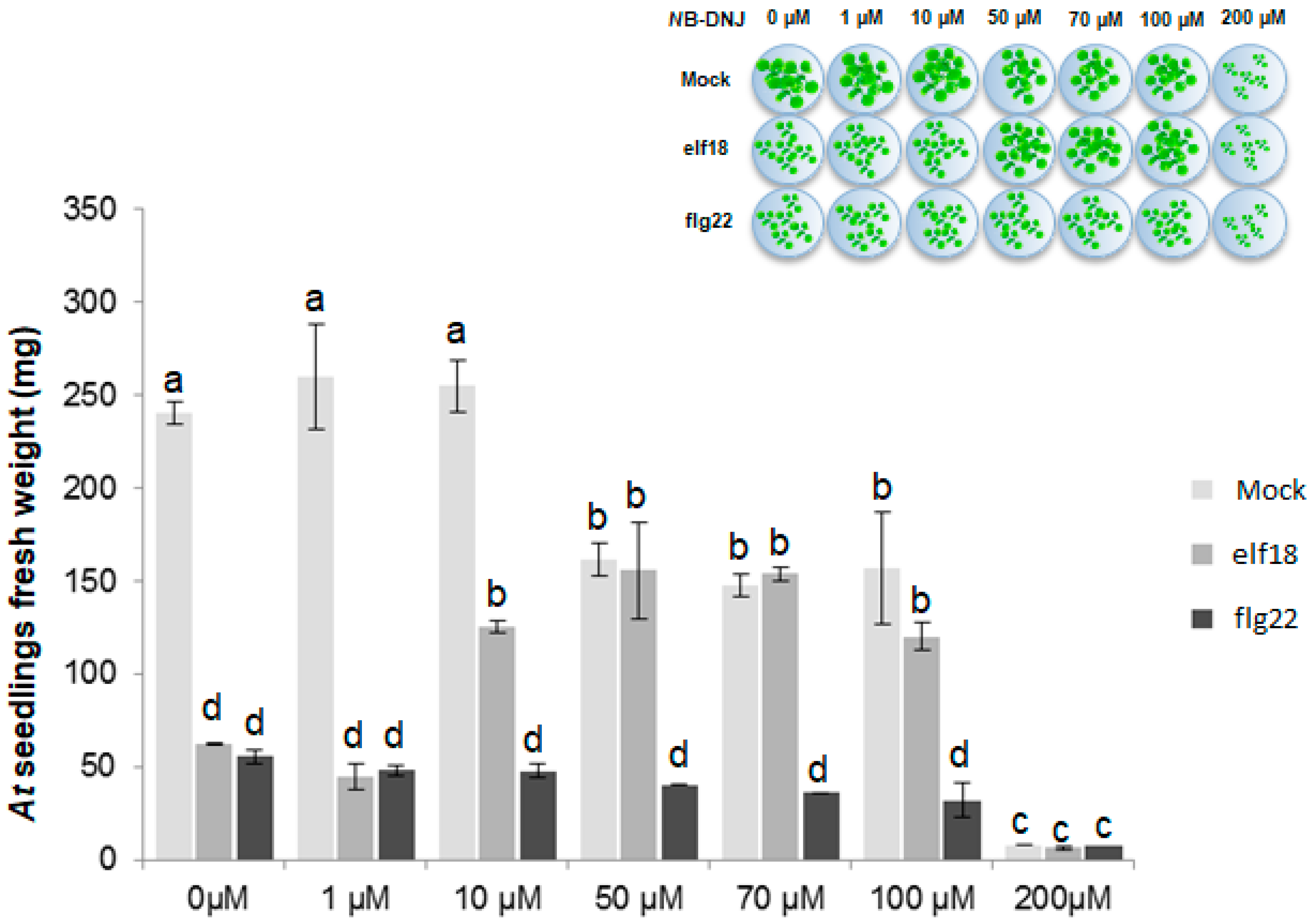
2.1. The Iminosugar NB-DNJ Is Toxic to Arabidopsis thaliana Plants at Concentrations above 200 µM

The iminosugar NB-DNJ [20,21] is well-tolerated in humans, with concentrations as high as 1 mg/mL (around 5 mM) displaying no cytotoxicity [22]. To test the toxicity of NB-DNJ in Arabidopsis thaliana, 15-day-old plants were treated with increasing concentrations of inhibitor, on the basis of a previously developed growth inhibition assay [17]. In the mock treatment experiments in Figure 1, the impact of NB-DNJ treatment on plant growth was measured. Arrest of wild-type seedling growth at NB-DNJ concentrations higher than 200 µM indicated toxicity. At lower NB-DNJ concentrations (1 and 10 µM), there was no impact on seedling growth. Supplementation of NB-DNJ at the intermediate concentrations of 50 µM, 70 µM and 100 µM resulted in reduced plant growth (about 30–40% of wild-type), with clear differences between elf18- and flg22-treated plants at the same concentrations. The concentration of 70 µM NB-DNJ was chosen to monitor α-Glu II activity.

2.2. NB-DNJ Doses Higher than 50 µM Render Arabidopsis thaliana Plants Insensitive to the elf18 Peptide

ER α-glucosidase II admits its client glycoproteins to the calnexin cycle and the mutations in the gene encoding it (At PSL5 in the plant) can cause client glycoprotein misfolding and loss of function. At ER α-Glu II impairment can be probed in the plant by studying the response to bacterial peptides elf18 and flg22. The At psl5-1 mutant carries a DNA mutation causing the S517F point substitution in the At ER α-glucosidase II α-subunit. The mutant plant is insensitive to elf18, suggesting that ER α-Glu II activity/inhibition can be monitored by measuring the responses induced by elf18 treatment [14]. In those experiments, flg22 treatment was used as a control because the response of the plant to this peptide is independent of ER α-Glu II activity [19]. The published data suggest that either EFR or another protein in the elf18 signalling pathway or involved in EFR localisation (but not FLS2 nor any proteins involved in flg22 signalling pathway/localisation) fold under ER α-glucosidase II control.
To check if NB-DNJ inhibits At ER α-glucosidase II in vivo, we supplemented A. thaliana plants with NB-DNJ at concentrations of 70 µM and treated the same plants with elf18 and flg22. Iminosugar treatment resulted in significant resistance to elf18 but not to flg22 (Figure 2). These NB-DNJ-induced elf18- and flg22-response phenotypes are similar to the ones of the At psl5-1 mutant plant with the same elicitors [19]. Taken together, our data suggest that NB-DNJ inhibits At ER α-glucosidase II in vivo at 70 µM.
To confirm that the defect in elf18 response is accompanied by reduced defense signaling, we investigated the transcription levels of two genes that are typically induced by elf18 and flg22. Phosphate-induced 1 (PHI1) and Reticulin-oxidase homologue (RET-OX) are indicated as early elicitor-induced genes because their transcription is maximal within 1 h after elicitor treatment [23]. Ten-day-old Col-0 seedlings, grown in absence or presence of 70 µM of NB-DNJ, and psl5-1 seedlings were treated with elf18 (or flg22, as a control) for 30 min. Low transcription of PHI1 and RET-OX in NB-DNJ treated seedlings were comparable with those recorded in the psl5-1 mutant in response to elf18 (Figure 3). Conversely, flg22 treated seedlings showed a normal defense response characterized by higher transcription levels of PHI1 and RET-OX (Figure 3).

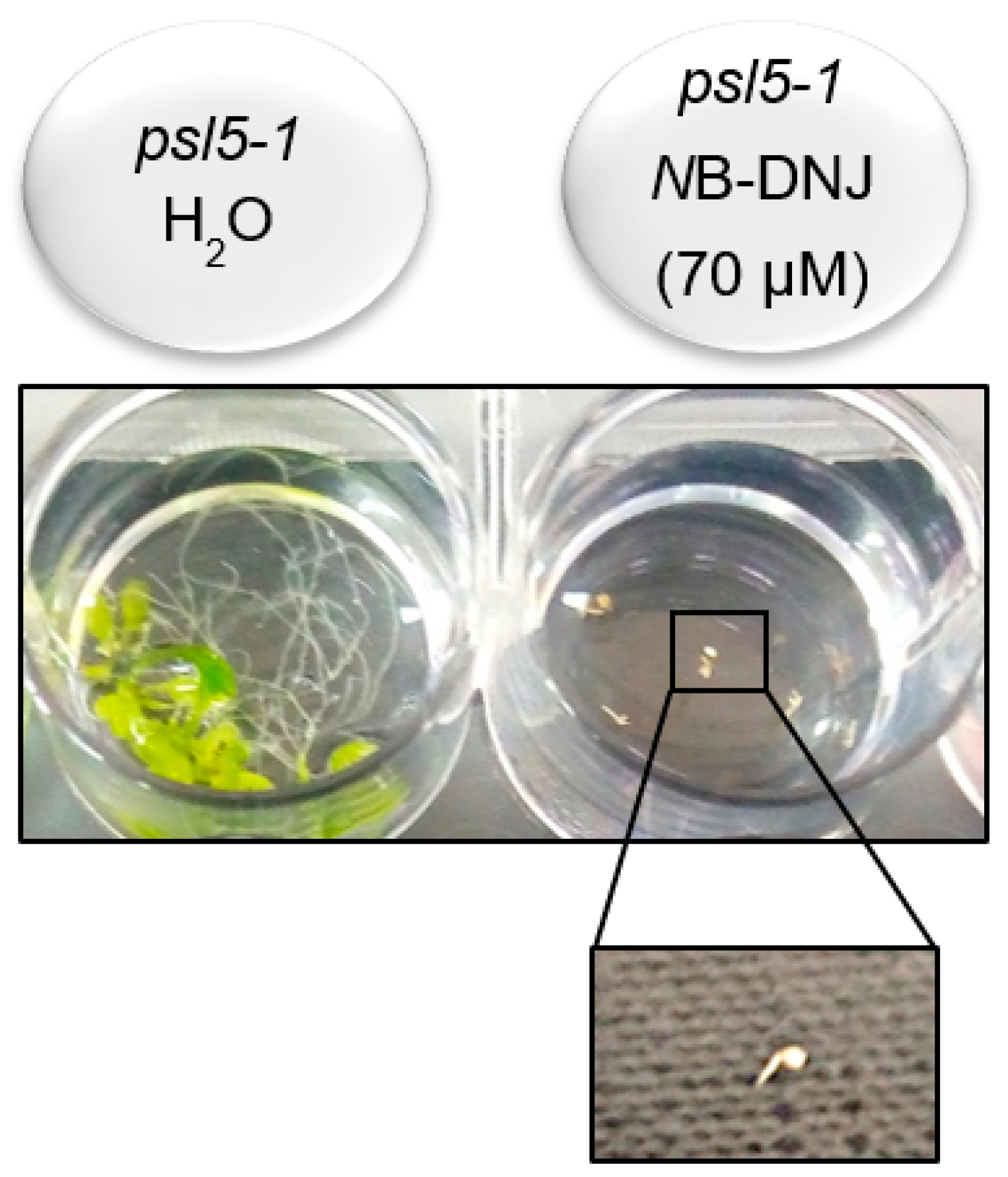
2.3. Treatment with 70 µM NB-DNJ Is Lethal to the At psl5-1 Mutant

ERQC plays a vital role in the development of multicellular organisms [24]. For example, a crucial developmental role of the ERQC checkpoint UGGT1 was demonstrated in mice embryos [25]. Furthermore, UGGT mutant plants show altered growth rates during vegetative development compared to wild type [26] although the UGGT gene can be knocked out in S. pombe without impairing cell growth [27]. Both known At ER α-glucosidase II missense mutations (the At psl5-1 S517F mutant and the At rsw3 S599F mutant) grow in a similar way to wild type [19], which raises questions as to the residual activity of the mutated proteins.
Homology modelling of the At ER α-glucosidase II protein, based on the recently determined crystal structure of the mouse enzyme [20], was used to gain insight on the reasons why the S517F and S599F point mutations affect At ER α–glucosidase II activity. In the At psl5-1 ER α-glucosidase II homology model (Figure 4), Ser517 is located about 8 Å from the nucleophilic catalytic residue Asp512 and about 9 Å from the catalytic acid/base Asp588 (Figure 4, inset). The At rsw3 S599F mutation is further away from the active site (Figure 4, inset). Neither mutant directly affects the catalytic residues of the enzyme. The possibility that either mutant carries residual ER α–glucosidase II activity cannot be ruled out.
To check if we could inhibit the putative residual At ER α-glucosidase II activity in At psl5-1, we grew At psl5-1 mutants in the presence of 70 µM NB-DNJ. Under these experimental conditions, the growth of germinated embryos is completely impaired and treatment with 70 µM NB-DNJ is lethal to the At psl5-1 mutant (Figure 5). As a result, we could not carry out experiments with the elf18 and flg22 peptides in the At psl5-1 plant.

3. Discussion

Iminosugars, currently the best known broad-spectrum antivirals, show cross-reactivity with intestinal glucosidases and enzymes of the glycosphingolipid pathway [12], creating a need to expand the search for new ERQC modulators. The development of low-cost platforms for the screening of molecules with potential pharmaceutical effects before testing them on animal models is only just emerging [28]. In this context, plants have been already proposed for preliminary screening of anti-cancer compounds [3]. The ERQC machinery is strongly conserved across eukaryotes, including plants, and many KO/mutant plants exist which can be used as negative controls; combined with the plant’s well characterised response to bacterial elicitors of the EFR/FLS2 receptors, it is clear that plants potentially afford a low-cost, easy and fast screening platform of ERQC modulators in vivo. In this work, we show that the ER α-glucosidase II inhibitor NB-DNJ influences the immune response of the plant to the bacterial peptide elf18, with a phenotype similar to the ER α-glucosidase II impaired plant, At psl5-1. The same iminosugar does not impair the plant response flagellin-derived flg22 peptide, again similarly to what observed in At psl5-1. This is the first observation that an iminosugar, which has reached clinical trial phase I as a mammalian ER α-glucosidase II inhibitor [20], also likely inhibits the plant enzyme in vivo. Alternatively, it is still conceivable that the molecule may act by interfering with either the trafficking or folding of the elf18-receptor and/or other proteins that are located downstream and involved in the elf18 signalling pathway.
Given the strong conservation of ERQC sequence and function across eukaryotes, the study strongly supports further efforts towards the development of the plant as a low-cost platform for early stage assays of ERQC modulators.

4. Materials and Methods

4.1. Plant Material, Growth Conditions and Treatments

For the growth inhibition assay, the seeds were surfaced, sterilized and sown in multi-well plates containing one-half strength Murashige and Skoog medium [29] (five seeds per 2 mL of liquid medium in the wells of 12-well-plates) supplemented with 0.5% (w/v) of sucrose. For NB-DNJ treatment, the seeds were germinated and grown in one-half-strength Murashige and Skoog medium containing the iminosugar NB-DNJ. After germination (3 day-post sowing), the medium was supplied with elicitors. The effect of treatment with the different peptides on seedling growth was analysed 15 days post sowing by photography and/or weighing (fresh weight). Variations in the weight of seedlings due to seed germination times that were not perfectly synchronized under slightly different environmental conditions (i.e., growth chamber temperature and light) did not affect the sensitivity of plants to elicitors after NB-DNJ treatment.
Elicitor treatment of seedlings was performed using elf18 (ac-SKEKFERTKPHVNVGTIG) and flg22 (QRLSTGSRINSAKDDAAGLQIA) at a concentration of 100 nM. Seedlings were grown at 22 °C and 70% relative humidity under a 16-h-light/8-h-dark cycle (approximately 120 µmol m–2 s–1). For gene transcription analysis, the seeds were surfaced, sterilized and sown in multi-well plates (approximately 10 seeds per well) containing one-half strength Murashige and Skoog medium (2 mL per well). After 9 days, the incubation medium was replaced with fresh medium, before the treatments with elicitors were performed after 24 h.

4.2. Homology Modelling

The protein sequences of the At ER α-glucosidase II α and β subunits were taken from the Uniprot database entries PSL5_ARATH and PSL4_ARATH, respectively. The sequences were aligned with the sequences of the mouse ER α-glucosidase II α and β subunits using Clustal omega [30]. The alignment was used in the program Modeller [31] together with PDB entry 5F0E, in order to produce a homology model of At ER α-glucosidase II α subunit residues 22–921 and β subunit residues 52–134. The At ER α-glucosidase II α psl5-1 S517F and rsw3 S599F point substitutions were modelled in PyMOL [32].

4.3. Gene Transcription Analysis

For gene transcription analysis, the seedlings were frozen in liquid nitrogen and homogenized with a MM301 Ball Mill (Retsch, Haan, Germany). Total RNA was extracted from at least two independent replicates, each having 20 seedlings, with NucleoZol reagent (Machery-Nagel, Duren, Germany) according to the manufacturer’s protocol. Total RNA (2 µg) was treated with RQ1 DNase (Promega, Madison, WI, USA), and the first-strand complementary DNA was synthesized using ImProm-II reverse transcriptase (Promega) according to the manufacturer’s instructions. qRT-PCR analysis was performed by using a CFX96 Real-Time System (Bio-Rad, Hercules, CA, USA). Complementary DNA (corresponding to 50 ng of total RNA) was amplified in a 20-µL reaction mix containing 1X GoTaq Real-Time PCR System (Promega) and 0.4 µM of each primer. Three technical replicates were performed for each sample, before data analysis was conducted using LinRegPCR software (Ruijter, Amsterdam, Netherlands). The transcription levels of each gene relative to UBIQUITIN5 were determined using a modification of the Pfaffl method [33] as described in reference [34] and expressed in arbitrary units. Primer sequences are shown in Table 1.

4.4. Statistical Analysis

The average weight of five plants per treatment was compared and statistically analyzed using ANOVA with Scheffè’s post hoc test. The statistical analysis on gene transcription data was performed using the Student’s t-test.

Author Contributions

L.M., A.L., P.R, N.Z. and A.S. conceived and designed the experiments; L.M., A.L. and I-B.R. performed the plant experiments; P.R. built the At ER α–glucosidase II homology model. L.M., A.L. and A.S. analysed the data; all authors contributed to the writing of the paper.

Acknowledgments

Arabidopsis thaliana psl5-1 mutants (in the Col-0 background) were kindly provided by Yusuke Saijo (Department of Plant Microbe Interactions, Max Planck Institute, Germany). N.Z. is a Fellow of Merton College, Oxford. PR is the recipient of a LISCB Wellcome Trust ISSF award, grant reference 204801/Z/16/Z.

Conflicts of Interest

The authors declare no conflict of interest.

References

  1. Gidalevitz, T.; Stevens, F.; Argon, Y. Orchestration of secretory protein folding by ER chaperones. Biochim. Biophys. Acta 2013, 1833, 2410–2424. [Google Scholar] [CrossRef] [PubMed] [Green Version]
  2. Alonzi, D.S.; Scott, K.A.; Dwek, R.A.; Zitzmann, N. Iminosugar antivirals: The therapeutic sweet spot. Biochem. Soc. Trans. 2017, 45, 571–582. [Google Scholar] [CrossRef] [PubMed]
  3. Dalziel, M.; Crispin, M.; Scanlan, C.N.; Zitzmann, N.; Dwek, R.A. Emerging principles for the therapeutic exploitation of glycosylation. Science 2014, 343, 1235681. [Google Scholar] [CrossRef] [PubMed]
  4. Butters, T.D.; Mellor, H.R.; Narita, K.; Dwek, R.A.; Platt, F.M. Small-molecule therapeutics for the treatment of glycolipid lysosomal storage disorders. Philos. Trans. R Soc. Lond. B Biol. Sci. 2003, 358, 927–945. [Google Scholar] [CrossRef] [PubMed]
  5. Roversi, P.; Marti, L.; Caputo, A.T.; Alonzi, D.S.; Hill, J.C.; Dent, K.C.; Kumar, A.; Levasseur, M.D.; Lia, A.; Waksman, T.; et al. Interdomain conformational flexibility underpins the activity of UGGT, the eukaryotic glycoprotein secretion checkpoint. Proc. Natl. Acad. Sci. USA 2017, 114, 8544–8549. [Google Scholar] [CrossRef] [PubMed]
  6. Vergara, D.; de Domenico, S.; Maffia, M.; Piro, G.; Sansebastiano, G.-P. Transgenic Plants as Low-Cost Platform for Chemotherapeutic Drugs Screening. IJMS 2015, 16, 2174–2186. [Google Scholar] [CrossRef] [PubMed] [Green Version]
  7. Lazar, C.; Durantel, D.; Macovei, A.; Zitzmann, N.; Zoulim, F.; Dwek, R.A.; Branza-Nichita, N. Treatment of hepatitis B virus-infected cells with alpha-glucosidase inhibitors results in production of virions with altered molecular composition and infectivity. Antivir. Res. 2007, 76, 30–37. [Google Scholar] [CrossRef] [PubMed]
  8. Steinmann, E.; Whitfield, T.; Kallis, S.; Dwek, R.A.; Zitzmann, N.; Pietschmann, T.; Bartenschlager, R. Antiviral effects of amantadine and iminosugar derivatives against hepatitis C virus. Hepatology 2007, 46, 330–338. [Google Scholar] [CrossRef] [PubMed] [Green Version]
  9. Pollock, S.; Dwek, R.A.; Burton, D.R.; Zitzmann, N. N-Butyldeoxynojirimycin is a broadly effective anti-HIV therapy significantly enhanced by targeted liposome delivery. AIDS 2008, 22, 1961–1969. [Google Scholar] [CrossRef] [PubMed] [Green Version]
  10. Warfield, K.L.; Plummer, E.M.; Sayce, A.C.; Alonzi, D.S.; Tang, W.; Tyrrell, B.E.; Hill, M.L.; Caputo, A.T.; Killingbeck, S.S.; Beatty, P.R.; et al. Inhibition of endoplasmic reticulum glucosidases is required for in vitro and in vivo dengue antiviral activity by the iminosugar UV-4. Antiviral Res. 2016. [Google Scholar] [CrossRef] [PubMed]
  11. Tyrrell, B.E.; Sayce, A.C.; Warfield, K.L.; Miller, J.L.; Zitzmann, N. Iminosugars: Promising therapeutics for influenza infection. Crit. Rev. Microbiol. 2017, 43, 521–545. [Google Scholar] [CrossRef] [PubMed]
  12. Sayce, A.C.; Alonzi, D.S.; Killingbeck, S.S.; Tyrrell, B.E.; Hill, M.L.; Caputo, A.T.; Iwaki, R.; Kinami, K.; Ide, D.; Kiappes, J.L.; et al. Iminosugars Inhibit Dengue Virus Production via Inhibition of ER Alpha-Glucosidases-Not Glycolipid Processing Enzymes. PLoS Negl. Trop. Dis. 2016, 10, e0004524. [Google Scholar] [CrossRef] [PubMed]
  13. Wilkinson, G.F.; Pritchard, K. In vitro screening for drug repositioning. J. Biomol. Screen. 2015, 20, 167–179. [Google Scholar] [CrossRef] [PubMed]
  14. Kunze, G.; Zipfel, C.; Robatzek, S.; Niehaus, K.; Boller, T.; Felix, G. The N terminus of bacterial elongation factor Tu elicits innate immunity in Arabidopsis plants. Plant Cell 2004, 16, 3496–3507. [Google Scholar] [CrossRef] [PubMed]
  15. Navarro, L.; Zipfel, C.; Rowland, O.; Keller, I.; Robatzek, S.; Boller, T.; Jones, J.D.G. The transcriptional innate immune response to flg22. Interplay and overlap with Avr gene-dependent defense responses and bacterial pathogenesis. Plant Physiol. 2004, 135, 1113–1128. [Google Scholar] [CrossRef] [PubMed]
  16. Zipfel, C.; Kunze, G.; Chinchilla, D.; Caniard, A.; Jones, J.D.G.; Boller, T.; Felix, G. Perception of the bacterial PAMP EF-Tu by the receptor EFR restricts Agrobacterium-mediated transformation. Cell 2006, 125, 749–760. [Google Scholar] [CrossRef] [PubMed]
  17. Gómez-Gómez, L.; Boller, T. FLS2: An LRR receptor-like kinase involved in the perception of the bacterial elicitor flagellin in Arabidopsis. Mol. Cell 2000, 5, 1003–1011. [Google Scholar] [CrossRef]
  18. Saijo, Y.; Tintor, N.; Lu, X.; Rauf, P.; Pajerowska-Mukhtar, K.; Häweker, H.H.A.; Dong, X.; Robatzek, S.; Schulze-Lefert, P. Receptor quality control in the endoplasmic reticulum for plant innate immunity. EMBO J. 2009, 28, 3439–3449. [Google Scholar] [CrossRef] [PubMed] [Green Version]
  19. Lu, X.; Tintor, N.; Mentzel, T.; Kombrink, E.; Boller, T.; Robatzek, S.; Schulze-Lefert, P.; Saijo, Y. Uncoupling of sustained MAMP receptor signaling from early outputs in an Arabidopsis endoplasmic reticulum glucosidase II allele. Proc. Natl. Acad. Sci. USA 2009, 106, 22522–22527. [Google Scholar] [CrossRef] [PubMed]
  20. Caputo, A.T.; Alonzi, D.S.; Marti, L.; Reca, I.-B.; Kiappes, J.L.; Struwe, W.B.; Cross, A.; Basu, S.; Lowe, E.D.; Darlot, B.; et al. Structures of mammalian ER α-glucosidase II capture the binding modes of broad-spectrum iminosugar antivirals. Proc. Natl. Acad. Sci. USA 2016, 113, E4630–E4638. [Google Scholar] [CrossRef] [PubMed]
  21. Karlsson, G.B.; Butters, T.D.; Dwek, R.A.; Platt, F.M. Effects of the imino sugar N-butyldeoxynojirimycin on the N-glycosylation of recombinant gp120. J. Biol. Chem. 1993, 268, 570–576. [Google Scholar] [PubMed]
  22. Platt, F.M.; Karlsson, G.B.; Jacob, G.S. Modulation of cell-surface transferrin receptor by the imino sugar N-butyldeoxynojirimycin. Eur. J. Biochem. 1992, 208, 187–193. [Google Scholar] [CrossRef] [PubMed] [Green Version]
  23. Gravino, M.; Savatin, D.V.; Macone, A.; De Lorenzo, G. Ethylene production in Botrytis cinerea- and oligogalacturonide-induced immunity requires calcium-dependent protein kinases. Plant J. 2015, 84, 1073–1086. [Google Scholar] [CrossRef] [PubMed] [Green Version]
  24. Silverblatt-Buser, E.W.; Frick, M.A.; Rabeler, C.; Kaplinsky, N.J. Genetic Interactions Between BOB1 and Multiple 26S Proteasome Subunits Suggest a Role for Proteostasis in Regulating Arabidopsis Development. G3 (Bethesda) 2018, 8, 1379–1390. [Google Scholar] [CrossRef] [PubMed]
  25. Molinari, M.; Galli, C.; Vanoni, O.; Arnold, S.M.; Kaufman, R.J. Persistent glycoprotein misfolding activates the glucosidase II/UGT1-driven calnexin cycle to delay aggregation and loss of folding competence. Mole. Cell 2005, 20, 503–512. [Google Scholar] [CrossRef] [PubMed]
  26. Blanco-Herrera, F.; Moreno, A.A.; Tapia, R.; Reyes, F.; Araya, M.; D’Alessio, C.; Parodi, A.; Orellana, A. The UDP-glucose: Glycoprotein glucosyltransferase (UGGT), a key enzyme in ER quality control, plays a significant role in plant growth as well as biotic and abiotic stress in Arabidopsis thaliana. BMC Plant Biol. 2015, 15, 127. [Google Scholar] [CrossRef] [PubMed]
  27. Fanchiotti, S.; Fernandez, F.; D’Alessio, C.; Parodi, A.J. The UDP-Glc: Glycoprotein glucosyltransferase is essential for Schizosaccharomyces pombe viability under conditions of extreme endoplasmic reticulum stress. J. Cell Biol. 1998, 143, 625–635. [Google Scholar] [CrossRef] [PubMed]
  28. Artal-Sanz, M.; de Jong, L.; Tavernarakis, N. Caenorhabditis elegans: A versatile platform for drug discovery. Biotechnol. J. 2006, 1, 1405–1418. [Google Scholar] [CrossRef] [PubMed]
  29. Murashige, T.; Skoog, F. A Revised Medium for Rapid Growth and Bio Assays with Tobacco Tissue Cultures. Physiol. Plant. 1962, 15, 473–497. [Google Scholar] [CrossRef]
  30. Aiyar, A. The use of CLUSTAL W and CLUSTAL X for multiple sequence alignment. Methods Mol. Biol. 2000, 132, 221–241. [Google Scholar] [PubMed]
  31. Eswar, N.; Eramian, D.; Webb, B.; Shen, M.-Y.; Sali, A. Protein structure modeling with MODELLER. Methods Mol. Biol. 2008, 426, 145–159. [Google Scholar] [PubMed]
  32. DeLano, W.L. The case for open-source software in drug discovery. Drug Discov. Today 2005, 10, 213–217. [Google Scholar] [CrossRef]
  33. Pfaffl, M.W.; Lange, I.G.; Daxenberger, A.; Meyer, H.H. Tissue-specific expression pattern of estrogen receptors (ER): Quantification of ER alpha and ER beta mRNA with real-time RT-PCR. APMIS 2001, 109, 345–355. [Google Scholar] [CrossRef] [PubMed]
  34. Ferrari, S.; Galletti, R.; Vairo, D.; Cervone, F.; De Lorenzo, G. Antisense expression of the Arabidopsis thaliana AtPGIP1 gene reduces polygalacturonase-inhibiting protein accumulation and enhances susceptibility to Botrytis cinerea. Mol. Plant Microbe Interact. 2006, 19, 931–936. [Google Scholar] [CrossRef] [PubMed]
Figure 1. NB-DNJ toxicity assay. Arabidopsis thaliana (At) seedlings, Col-0 ecotype, were grown in Murashige and Skoog (MS) half-strength liquid medium containing different concentrations of NB-DNJ in combination with 100 nM of elf18 or flg22 (inset). After 15 days of treatment, the fresh weight of seedlings was measured. Increasing concentrations of NB-DNJ resulted in increasing reduction in plant growth, with severe weight loss at 200 µM of NB-DNJ. Values represent the mean of at least five independent experiment (± s.e.; n = 5) with similar results in each experiment. Each letter (a, b, c and d) indicates a group of samples whose values are statistically equivalent to the ones in the same group, at the (p < 0.01) level determined by ANOVA with Scheffè’s post hoc test.
Ijms 19 02135 g001
Figure 2. In planta ER α–glucosidase II assay. Arabidopsis thaliana (At) Col-0 seedlings (wild-type, WT) were grown in presence or absence of NB-DNJ (70 µM) and their growth was compared to that of psl5-1 seedlings used as controls. NB-DNJ treatment was combined with that of elf18 or flg22. In presence of NB-DNJ, the growth trend of psl5-1 control seedlings was indistinguishable from WT ones, irrespective of treatment with elicitors. Inset shows representative pictures of 15-day-old seedlings (five seedlings per well) germinated and grown with or without NB-DNJ and elicitors. Values represent the mean of at least five independent experiments (± s.e.; n = 5) with similar results in each experiment. Each letter indicates a group of samples whose values are statistically equivalent to the ones in the same group, at the (p < 0.01) level determined by ANOVA with Scheffè’s post hoc test. Note: the differences in the weights of seedlings from those in the experiment in Figure 1, which may be due to slight variations in seed germination times (see methods), did not affect the sensitivity of plants to elicitors after NB-DNJ treatment.
Ijms 19 02135 g002
Figure 3. Transcription of defense genes. Arabidopsis thaliana WT (wild-type, empty bars), WT grown in the presence of NB-DNJ (70 µM, light grey bars) and psl5-1 (dark grey bars) 10-day-old seedlings (20 seedlings in each experiment) were treated with water or elf18 and flg22 elicitors. Transcription of PHI1 and RET-OX genes were determined by qRT-PCR 30 min after treatment. Transcription levels are shown as the mean of at least three independent experiments (± s.e.; n = 3) normalized to UBQ5 (ubiquitin 5) used as a reference. In both (a) and (b), asterisks indicate statistically significant differences between elf18-treated seedlings (WT + NB-DNJ and psl5-1) and corresponding treatment of the wild type according to Student’s t-test (3 asterisks = p < 0.001).
Ijms 19 02135 g003
Figure 4. Structural mapping of the psl5-1 and rsw3 missense mutations in At ER α-glucosidase II. Homology model of the At ER α-glucosidase II α subunit in green cartoon representation. The inset shows the At psl5-1 mutant S517F point substitution (orange sticks), and the At rsw3 mutant S599F substitution (magenta sticks). The catalytic Asp residue D512 and the acid–base catalyst residue D588 are depicted in red sticks. The distances between the mutated residues and the catalytic residues are indicated by dotted lines, with the distances in Å. Both the S517F and the S599F mutations are likely to destabilise the catalytic domain of the enzyme.
Ijms 19 02135 g004
Figure 5. At psl5-1 and NB-DNJ. Arabidopsis thaliana psl5-1 seedlings show impaired growth in presence of 70 µM NB-DNJ.
Ijms 19 02135 g005
Table 1. Primers used in this work.
GENEAG CODEFORWARD PRIMER (5′-3′)REVERSE PRIMER (5′-3′)
UBQ5AT3G62250GGAATCGACGCTTCATCTCGATGAAAGTCCCAGCTCCACA
PHI1AT1G35140TTGGTTTAGACGGGATGGTGACTCCAGTACAAGCCGATCC
RET-OXAT1G26380AGGTTCTCGAACCCTAACAACAGCACAGACGACACGTAAGAAAG

