Article

Characterization and Expression Profiling of Neuropeptides and G-Protein-Coupled Receptors (GPCRs) for Neuropeptides in the Asian Citrus Psyllid, Diaphorina citri (Hemiptera: Psyllidae)

1
Guangdong Engineering Research Center for Insect Behavior Regulation, College of Agriculture, South China Agricultural University, Guangzhou 510642, China
2
State Key Laboratory of Rice Biology; Institute of Insect Sciences, Zhejiang University, Hangzhou 310058, China
3
Department of Plant Protection, Faculty of Agricultural Sciences, Ghazi University, Dera Ghazi Khan 32200, Pakistan
*
Author to whom correspondence should be addressed.
Int. J. Mol. Sci. 2018, 19(12), 3912; https://doi.org/10.3390/ijms19123912
Submission received: 18 November 2018 / Revised: 30 November 2018 / Accepted: 3 December 2018 / Published: 6 December 2018
(This article belongs to the Section Molecular Neurobiology)

Abstract

:
Neuropeptides are endogenous active substances that widely exist in multicellular biological nerve tissue and participate in the function of the nervous system, and most of them act on neuropeptide receptors. In insects, neuropeptides and their receptors play important roles in controlling a multitude of physiological processes. In this project, we sequenced the transcriptome from twelve tissues of the Asian citrus psyllid, Diaphorina citri Kuwayama. A total of 40 candidate neuropeptide genes and 42 neuropeptide receptor genes were identified. Among the neuropeptide receptor genes, 35 of them belong to the A-family (or rhodopsin-like), four of them belong to the B-family (or secretin-like), and three of them are leucine-rich repeat-containing G-protein-coupled receptors. The expression profile of the 82 genes across developmental stages was determined by qRT-PCR. Our study provides the first investigation on the genes of neuropeptides and their receptors in D. citri, which may play key roles in regulating the physiology and behaviors of D. citri.

Graphical Abstract

1. Introduction

The central nervous system (CNS) and its neuropeptide messengers rank the highest in the entity level regulating endogenous biochemical control function [1]. Neuropeptides are a diverse set of signaling molecules in multicellular organisms. In insects, neuropeptides play a significant role in the regulation of fundamental events such as development, reproduction, feeding, courtship, olfaction, circadian rhythm, and many other processes [2,3]. Neuropeptides are processed from their larger, inactive precursors by enzymes [4], up activation, and then work on target cells by binding to the specific signal-transducing membrane receptors [5]. Most of these receptors are subordinate to the G-protein-coupled receptors (GPCRs), and the GPCRs have a similar topographical structure with seven transmembrane domains which are highly conservative through evolution and constitute the largest superfamily of cell surface proteins [6]. In vivo studies showed that neuropeptides and their receptors appear to have key roles in the regulation of physiology and behavior in insects; injection of kinin caused a significant reduction in weight gain and an increased mortality in Heliothis virescens [7], injection of tachykinin (TK) resulted in diminished olfactory responses in electroantennograms of Periplaneta americana [8], reduced levels of the receptor of TK in Drosophila melanogaster olfactory receptor neurons (ORNs) showed increased attraction to two food-related odors [9]. Neuropeptides and their receptors regulate the fundamental events in insect life cycle, hence, they were proposed as potential insecticides or targets to replace or complement neurotoxic compounds [10]. Owing to the sequence specificity of these molecules and the specific receptor, neuropeptide-based compounds would be species‒specific, and safe for non-target species. Moreover, resistance is not easy to develop, since a potential mutation in a neuropeptide precursor or receptor gene is typically detrimental [11].
The Asian citrus psyllid, Diaphorina citri Kuwayama (Hemiptera: Psyllidae), is one of the most important insect pests of citrus worldwide. It is the principal vector of Candidatus Liberibacter asiaticus (CLas), a phloem-inhabiting bacterium, which correlates with Huanglongbing (HLB) [12]. Huanglongbing, also known as citrus greening disease, poses the most destructive threat to citrus production worldwide with no known cure [13,14,15]. At present, the main ways to prevent and restrain the spread of the disease are employing chemical insecticides to control the vector; however, the application of conventional insecticides, including organophosphates, pyrethroids, and neonicotinoids, has led to the evolution of resistance to pesticides [16,17,18]. The primary means of blocking the transmission of HLB is to control D. citri, the vector of the pathogen; however, the development of insecticide resistance in D. citri also poses increased challenges for the persistent effectiveness of chemical control strategies. Furthermore, the more use of pesticides can also lead to problems with “resurgence” and “residue”. Pesticides with safe and novel ways of action are needed to alleviate the problems of pesticide resistance and environmental pollution. The primary task of developing neuroendocrine-based insecticides is to identify the structure and function of neuropeptides and the receptors involved in survival, development, and/or reproduction [11].
Previous studies have identified the neuropeptides or neuropeptide receptors in D. melanogaster [3], Bombyx mori [19], Chilo suppressalis [20], Tribolium castaneum [21], Apis mellifera [22], Nilaparvata lugens [23], and Rhodnius prolixus [24]. However, the neuropeptides and their GPCRs in D. citri are poorly characterized. For these reasons, in this study, to identify gene-encoding neuropeptides and the receptors of neuropeptides in D. citri, we analyzed the transcriptomes in twelve tissues of this pest. The expression profiles of these neuropeptides and the receptors were validated by quantitative real time-PCR (qRT-PCR). This is the first study on the neuropeptides and neuropeptide receptors of D. citri, and these results provide valuable information to develop an integrated pest management program for the Asian citrus psyllid.

2. Results

2.1. Sequencing and Unigene Assembly

A total of 143.37 Gb of raw data was acquired, and after removing low-quality, adaptor, and contaminated sequence reads, yielded 137.22 Gb of clean reads. These clean reads were assembled into 38,161,895 contigs (N50 = 48) of an average length of 50 bp by Trinity. Then we obtained 297,614 unigenes larger than 200 bp (N50 = 731) from non-redundant putative contigs, with a mean length of 575 bp. For annotations, the unigenes were searched against Nr (NCBI non-redundant protein sequences, http://www.ncbi.nlm.nih.gov/), Swiss-Prot (a manually annotated and reviewed protein sequence database, http://www.expasy.ch/sprot/), KEGG (Kyoto Encyclopedia of Genes and Genomes, http://www.genome.jp/kegg/), COG (Clusters of Orthologous Groups of proteins, http://www.ncbi.nlm.nih.gov/COG/), and GO (Gene Ontology, http://www.geneontology.org/) databases using BLASTx (search protein databases using a translated nucleotide query) with a cut-off E value of 10−5. As a result, 60,401 (20.29%) were annotated using the NCBI-Nr database; 34,380 (11.55%) by Swiss-Prot; 11,837 (3.98%) by KEGG; 30,808 (10.35%) by COG; 8691 (2.92%) by GO, which covered 60,999 (20.49%) of the total unigenes. Of the 297,614 unigenes, 8691 (2.92%) matched at least one GO term (Figure 1). Among these unigenes, 13,536, 11,569, and 10,489 transcripts were assigned to biological processes, molecular function, and cellular component, respectively. Cellular process and metabolic processes represented the most abundant GO terms in the biological process category, binding and catalytic activity were most represented in molecular function, and the most unigenes that accorded with the cellular component category were involved in cell parts and organelles (Figure 1). The raw data of the transcriptomic were submitted to the NCBI Short Read Archive (SRA) database as BioProject Accession Number SRP139008 (https://www.ncbi.nlm.nih.gov/sra/SRP139008).

2.2. Neuropeptide and Peptide Hormone Genes

Based on Nr-annotation and homology searches, 40 genes encoding neuropeptides and neurohormones were identified in D. citri (Table 1), including the neuropeptides involved in physiology and behavior, such as allatotropin (AT), crustacean cardioactive peptide (CCAP), CCHamide (CCH), ecdysis triggering hormone (ETH), eclosion hormone (EH), SIFamide (SIF), pigment-dispersing factor (PDF), tachykinin (TK) and natalisin (NTL). Elevenin-like peptide (ELP) is a molluscan neuropeptide and was first identified in Aplysia californica, and later ELP was found in many insect species with the exception of D. melanogaster and B. mori, in D. citri, the gene coding for ELP was found. Prothoracicotropic hormone (PTTH) has been proposed to regulate development in insects, and it has been identified in D. citri, however, previously, in Hemipteran, PTTH was only identified in N. lugens. The adipokinetic hormone (AKH)/corazonin-related peptide (ACP), bursicon alpha subunit (Burα), FMRFamide (FMRF), glycoprotein hormone alpha 2 (GPA2), IMFamide (IMF), neuropeptide F (NPF), sulfakinin (SK), and trissin (TR) were not found in the transcriptome databases of D. citri (Supplementary Table S1).

2.3. G-Protein-Coupled Receptors (GPCRs) for Neuropeptides

A total of 42 putative neuropeptide GPCRs genes were identified in the transcriptomes of D. citri based on homology analysis (Table 2). Of these receptors, 35 GPCRs belong to the A family, four belong to B family, and three belong to leucine-rich repeat-containing GPCRs (LGRs). In order to assign putative functions of these GPCRs, we compared them with those of D. melanogaster, N. lugens, and other arthropods. The results are presented as a neighbor-joining tree in Figure 2, Figure 3 and Figure 4.

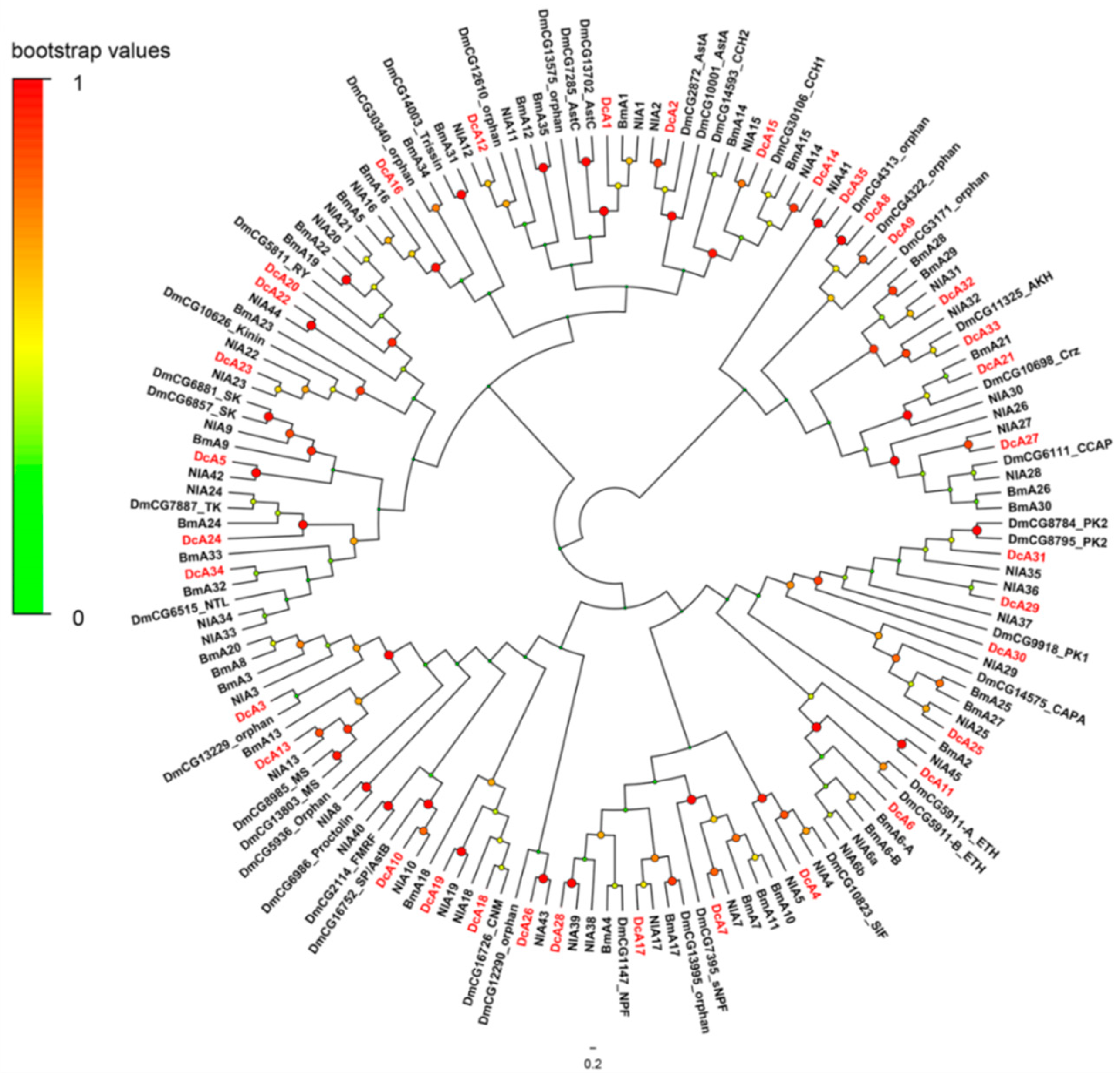
2.3.1. A-Family GPCRs

The A-family GPCRs are also known as the rhodopsin family. Thirty-five A-family neuropeptide GPCRs were identified in D. citri. In the phylogenetic tree of A-family GPCRs (Figure 2), DcA5, DcA20, DcA22-24, and DcA34 were clustered in a clade with DmCG5811 (RYamide receptor, RYR), DmCG10626 (Kinin receptor, KinR), DmCG6857 (Sulfakinin-like receptor, SKR), DmCG6881 (Sulfakinin-like receptor, SKR), CG7887 (Tachykinin receptor, TKR), DmCG6515 (Natalisin receptor, NTLR). DcA5, DcA23, DcA24, and DcA34 were identified as SKR, KinR, TKR, and NTLR respectively. DcA22 and DcA20 are two orthologs of DmRYaR. DcA7, DcA17, and DcA28 stay in the clade of NPF and short Neuropeptide F (sNPF) receptors, DcA28 is the receptor of NPF (NPFR), and DcA7 and DcA17 were identified as sNPF receptors (sNPFR). Besides that, there is another big clade locating in the phylogenetic tree, and it contains the Pyrokinin receptor (PKR), cardio acceleratory peptide (CAPAR), ecdysis-triggering hormone receptor (ETHR). DcA6 was predicted as ETHR, and DcA25 acts as a CAPAR, based on the Blastp analysis and phylogenetic analysis. DcA25 and DcA30 were identified as PK1R, DcA31 was identified as PK2R. The receptors of the GnRH-related peptides and CCAP were clustered in a clade, including DcA21, DcA27, DcA32, and DcA33. DcA21 located in the subtree of the corazonin receptor with BmA21, DcA27 was identified as CCAPR. DcA32 and DcA33 act as the receptor of AKH. DcA8, DcA9, and DcA35 are paralogs of CCAPR and AKHR; however, DcA8 and DcA9 are similar to two orphan receptors, DmCG4313 and DmCG4322. DcA35 is similar to NlA41, and NlA41 is an ortholog of the receptor for AVLP of T. castaneum, further based on blastp analysis, DcA35 has a high homology with AVLPR of T. castaneum (ABX00684, e-value 3e-112). As above, DcA35 was characterized as DcAVLPR. DcA11 is an ortholog of NlA45, and NlA45 is remarkably similar to the vertebrate thyrotropin-releasing hormone receptors (TRHRs). The receptors for CNMamide (CNM), FMRF, Myosuppressin (MS), sex peptide (SP), Proctolin (Pro), and 3 orphan receptors, DmCG12290, DmCG5986, and DmCG13229 were clustered in a clade. This clade included DcA3, DcA10, and DcA13. DcA10 was identified as the receptor of SP (SPR), and DcA13 acts as MS receptor (MSR). The predicted function of DcA3 was not identified. At last, DcA1, DcA2, DcA4, and DcA15 were obviously identified as receptors of AstC, AstA, SIF, and CCH, respectively.

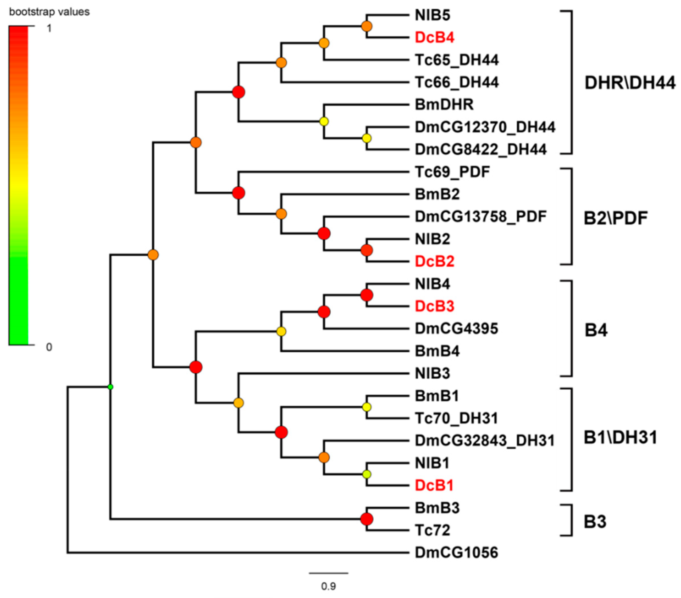
2.3.2. B-Family GPCRs

B-family GPCRs contain three subfamilies: subfamily B1, subfamily B2, and subfamily B3. Here the B-family neuropeptide GPCRs belong to the subfamily B1. Four B-family GPCRs associated with neuropeptide recognition were confirmed in D. citri. Each amino acid sequence of these candidate receptors contains the characteristic hormone receptor domain. In the phylogenetic tree of B-family GPCRs (Figure 3), four receptors were classified into four branches. DcB1 and DcB4 were identified as receptors of DH31 and DH44. DcB1 is the ortholog of DmCG32843, and DcB4 is the ortholog of CG12370 and DmCG8422. DcB2 was annotated as the receptor of PDF, which is involved in prolonging mating duration. DcB3 is an ortholog of DmCG4395, and it shows high similarity to the calcitonin-like diuretic hormone receptor 2 of R. prolixus (AHB86571, e-value 0.0).

2.3.3. Leucine-Rich Repeat-Containing GPCRs (LGRs)

Leucine-rich repeat-containing GPCRs are a class of GPCRs containing leucine-rich repeats (LRRs) at the N-terminus. Based on the number of LRRs, the type-specific hinge region and the presence or absence of a low-density lipoprotein receptor-like cysteine-rich motif (LDLa), LGRs can be identified as three main types (type A, B, and C). Furthermore, according to the number of LDLa motifs, Type C LGRs can be divided into two subtypes: Type C1, which contain only one LDLa, and Type C2, which contain multiple LDLa motifs. Three LGRs were identified in D. citri, two belong to Type A and one belong to Type C2. Both DcLGR1 and DcLGR2 contain eight LRRs, and this is a typical feature of Type A. In the phylogenetic tree of leucine-rich repeat-containing GPCRs (Figure 4), DcLGR1 and DcLGR2 are homologous to DmCG7665 which is the receptor of glycoprotein hormones in Drosophila. DcLGR3 contains eight LDLa motifs and two LRRs, and it belongs to Type C2. DcLGR3 is an ortholog of NlA47 which is also known as NlGRL101.

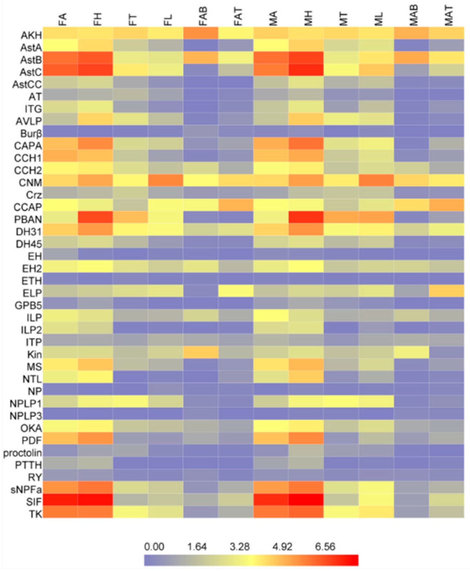
2.4. Tissue-Specific Expression Profiles of the Neuropeptides and Neuropeptide Receptors

In order to understand the potential function of the neuropeptides and neuropeptide receptors in D. citri, the expressions of the neuropeptides and neuropeptide receptors were profiled for different tissues of D. citri based on the transcriptome data (Figure 5). AKH, DH31, SIF, AstB, CCH2, ILP, EH2, CNM, ELP, CCAP, and Kin are ubiquitous in the tissues of the female and male adult. AstA-C, TK, CAPA, ITG, CCH1-2, MS, sNPF, OKA, ILP1-2, SIF, NTL, EH2, AVLP, PBAN, NPLP1, and CNM show high expression levels in head, and AstA, CAPA, ITG, IPL2, NTL; PDF express in head and antenna; CNM also shows the highest expression level in legs of all tissues; and ELP and CCAP have a high expression level in abdomen terminal.
The overwhelming majority of the neuropeptide receptors appear to be lowly expressed in all tissues in addition to A4, A7, A9, A12, A18, A23, A26, A27, A31, A33, B2, LGR1 (Figure 6). LGR1 (glycoprotein hormones), A26, A27 (CCAP) have the highest expression levels of all the receptors and exist in all tissues of the female and male adult. A7 (sNPF), A9, A25 (CAPA), and A31 (PBAN) express broadly in the head, thorax, and abdomen except the abdomen terminal. A4 (SIF), A12, and B2 (PDF) express mainly in the head and antenna. A23 (kinin) show the specific expression in the abdomen of female and male. A18 (CNM) especially expresses in the abdomen and abdomen terminal of the male. A24 (TK) have higher expressions in the head and antenna of males than other tissues.

2.5. Developmental Stages Expression Analysis by RT-qPCR

Developmental stages expression profiles of the neuropeptides and neuropeptide receptors were confirmed by the RT-qPCR (Figure 7 and Figure 8). AKH, AVLP, Burβ, DH31, DH45, PBAN, and OK had the highest expressional level in the 1st and 2nd instar nymphs. AstB, AstC, CAP2b, CCH1, CNM, GPb5, MS, and TK showed the highest expression of the adults at five days after eclosion (sexual maturity). The expression level of Kin, NP, and PTTH decreased gradually from egg to sexual maturity. The neuropeptide receptors A3, A5, A12, A13, A16, A18, A21–24, A28–A30, A32, A34, B1, B3, and LGR3 were predominately expressed in egg, and A14, A25–27, and B4 mainly expressed in adults. A35 and two orphan receptors (A9 and A17) both expressed highly in egg and adult.

3. Discussion

Control of D. citri is the key component of integrated control for citrus Huanglongbing. However, effective control measures are not currently available. As potential pesticides and new targets emerge on account of continuing development of existing insecticide resistance [1,25], the primary task of developing neuroendocrine-based insecticides is to identify the structure and function of neuropeptides and receptors, especially those involved in survival, development, and/or reproduction [11]. However, the molecular basis of the behavior of D. citri is poorly understood. Besides, the lack of genomic information prevents us from understanding the regulatory network of the neuropeptides. Fortunately, transcriptome analysis has provided the methods to identify and characterize multiple genes in insects [23], and using RNA-seq a number of genes encoding neuropeptides and their receptors were identified from D. citri.
The present transcriptional sequences of D. citri appear to contain most of the genes for insect neuropeptides and GPCRs except for several genes. Several neuropeptides (e.g., Trissin (TR) and IMFamide (IMF) orthologs) were also not identified in other Hemipteran insects such as A. pisum [26], N. lugens [23], and R. prolixus [11]. In fact, IMF is a unique neuropeptide of Lepidoptera and has not been found in other insects [20]. Neuropeptide F (NPF) is identified in almost all insect species and plays roles in feeding, metabolism, reproduction, and stress responses [20], whereas the BLAST search of transcriptomic data failed to find NPF in D. citri. Similarly, we have identified an ortholog of Proctolin; however, the receptor was not found. This may be due to the incomplete analyses of transcriptome information. Indeed, the gene coding for natalisin (NTL) and CNMamide (CNM) were identified from R. prolixus by reinvestigation [11] contrary to the previous suggestion that NTL and CNM were absent [24].

3.1. Neuropeptides Involved in Ecdysis and Development

Ecdysis-triggering hormone (ETH), eclosion hormone (EH), and crustacean cardioactive peptide (CCAP) are main players of the peptidergic circuit controlling ecdysis in insects. The functions of these peptides have been reported in the Drosophila and other insects [27,28]. In insects, EH is expressed in CNS, ETH production in endocrine cells in the epitracheal gland (Inka cells), and the CCAP-expressing neurons are located in abdominal ganglia [27]. Both ETH and EH regulate the release of CCAP from central CCAP neurons which inhibits pre-ecdysis [2]. In D. citri, one ETH encoding gene was identified, and two different genes encoding EH were identified. According to the expression profiles, ETH and EH1 show high expression levels in the nymph stage, and we can presume that ETH and EH1 play a vital role in molting processes.
Juvenile hormone (JH) is an important hormone and regulates development and growth in insects. Traditionally JH production in the corpora allata has been considered to be controlled by the peptides Allatotropins (AT) [29] and Allatostatins (Ast) [2]. The first insect AT was isolated from head extracts of Manduca sexta. This peptide was shown to stimulate JH biosynthesis by the corpora allata (CA) of adult females [29]. Allatostatins are diverse peptides derived from three different genes in insects, which were designated as AstA, AstB, and AstC. These peptides act as the inhibitors of JH production in corpora allata (CA) and have antagonism to AT [30]. Feeding the Lepidopteran AstC led to reduced growth and fecundity and caused significant mortality in A. pisum and in Myzus persicae [31,32]. The function of AstC receptors has been characterized in A. aegypti and T. castaneum, and they express in the central nervous system and gut [33,34]. Besides that, AstCC is a Type-C Ast and shows strong similarities to AstC. AstCC also has the function to regulate the biosynthesis of JH [35]. In D. citri, from 3rd instar nymphs to sexual maturity, the expression of AstCC shows a decreasing trend, and AT shows a growth trend. This result suggests that AT and AstCC might be involved in the regulation of JH production in D. citri.

3.2. Neuropeptides Control of Metabolism

Insulin-like peptides (ILPs) widely exist in insects and the insulin-like signaling pathway is conserved across higher multicellular animals [36]. In Drosophila, ablation of the insulin-producing cells, or deactivation of the ILPR leads to a series of phenotypes including grow logy, increased starvation resistance, increased levels of circulating carbohydrates cycling, elevated lipid storage, and lifespan extension [2]. Adipokinetic hormone (AKH) is suggested to be similar to mammalian glucagon and acts antagonistically to insulin by activating glycogen phosphorylase and mobilizing carbohydrates [37]. When the AKH-producing neuroendocrine cells were ablated in Drosophila, the trehalose levels of larvae and starved adults decreased, and these adults without AKH-cells become hypoactive, suggesting that AKH is involved in maintaining normal levels of circulating carbohydrates [38]. Ectopic expression of AKH in the fat body resulted in both increased circulating trehalose and a decrease in stored lipids [2]. In D. citri, two genes encoding ILP1 and ILP2 and one gene encoding AKH were identified, and more research is needed to reveal the functions of these genes.
The insect kinins are multifunctional neuropeptides shown to modulate hindgut contractions [39,40], diuretic activity [41,42], digestive enzyme release [40,43], and inhibit weight gain in larvae Lepidoptera [43,44]. Feeding the analogs of kinin to the pea aphid demonstrated that three of the biostable analogs showed antifeedant activity [45]. In D. citri, we identified one kinin precursor and one kinin receptor (DcA23), the kinin and the kinin receptor both express in the abdomens (Figure 5 and Figure 6). On the present understanding of the expression profile of Kin and the receptor, we speculate that kinin and DcA23 may be involved in regulating the digestion process in D. citri and they represent a potential pesticide and target.

3.3. Reproductive-Related Neuropeptides

SIFamide (SIF) is strictly conserved and widespread in insects and modulates sexual behavior, and it was first found in 1995 in HPLC (high-performance liquid chromatography) fractions [46]. In Drosophila, the expression of SIF was restricted to only four neurons of the pars intercerebralis, when the SIF neurons was ablated, the lacking SIF-less males perform vigorous and indiscriminate courtship directed at either sex, while females appear sexually hyper-receptive. When the SIF gene was knocked down via RNAi, the decrease of SIF also led to a similar change of behavior [47]. SIFamides show a conserved sequence, X1-X2-RKPPFNGSIFamide, and the SIFs differ only in their N-terminal amino [46], in D. citri, SIF and the receptor of SIF (DcA4) were detected.
Neuropeptide pigment-dispersing factor (PDF) is involved in maintaining behavioral rhythms in D. melanogaster [48]. In addition, males usually prolong mating duration in the presence of other males to increase the chance of successful gene transfer, this effect in D. melanogaster requires both Neuropeptide F receptor 1 (NPFR1) and PDF expressing in four small ventrolateral neurons as well as the PDF receptor and expressing Neuropeptide F (NPF) in two dorsolateral neurons [49]. Apparently, PDF and NPF work together to regulate prolonged mating duration in D. melanogaster. In this study, PDF was identified in D. citri; however, NPF was not found.
Natalisin (NTL) is an arthropod-specific neuropeptide which was recently identified in three holometabolous insect species: D. melanogaster, T. castaneum, and B. mori, and was proven to be involved in regulating mating behavior in D. melanogaster and T. castaneum [50]. The latest research on NTL shows that NTL is involved in modulating the mating of the oriental fruit fly, Bactrocera dorsalis [51]. The NTL precursors generally contain multiple repeats sequences of F-X1-X2-X3-Ra at the C-terminus. In hemipteran, X1, is usually W, and X2 is P [52]. In D. citri, NTL precursors and the receptor of NTL (DcA34) was identified, according to the amino acid sequences of the NTL precursor, four mature peptides were predicted (Supplementary Figure S1). The conservatism of NTLs sequences implies the conservatism of the functions; NTL will be a worthwhile option in developing novel control methods against D. citri.

3.4. Neuropeptides in Olfaction

Tachykinin (TK) is a multifunctional peptide, and it has been identified in many vertebrate and invertebrate species. In all vertebrate and a few invertebrate the TKs share a common C-terminal sequence motif, F-X-G-L-Ra [53]. Tachykinin is important in odor-based searching behavior of fruit flies, and several olfactory neurons contain high TK levels [54]. In Drosophila, (3R,11Z,19Z)-3-acetoxy-11,19-octacosadien-1-ol (CH503) is a gustatory sex pheromone. CH503 is detected by gustatory neurons on the male foreleg, a cluster of 8–10 neurons within the subesophageal region which mediates the pheromone response through the release of the TKs [55]. In D. citri, we identified four mature peptides of TK; all mature peptides have conserved sequences at the C-terminus (Supplementary Figure S1). Tachykinin shows a high expression level in antenna and DcA24 shows high expression in male antenna and head in D. citri, but beyond that, TK also showed the highest expression in adults at five days after sexual maturity. Based on the expression profile, it is probable that TK and DcA24 are involved in the recognition of the odor by adults. Therefore, TK and the receptor can be considered as a potential behavioral regulatory pesticide or target.
CCH1 and CCH2 were initially identified in the tsetse fly Glossina morsitans [56]. In Drosophila, CCH1 and CCH2 played a role in appetite regulation by activating their receptors respectively [57,58]. The receptor of CCH1 was also an important factor governing starvation-induced olfactory modifications [59]. Here, both CCH1 and CCH2 were identified in D. citri, and their corresponding receptors were excavated, A14 (CCH1) and A15 (CCH2), respectively. In D. citri, CCH1 and A14 presented high expression in adulthood, besides that, CCH1 is highly expressed in antennae and head. According to the results, it was assumed that CCH1 and A14 may play an important role in regulating starvation-induced host recognition behavior at the adult stage of D. citri. In addition to TK and CCHs, AstA, sNPF, and SIF also show high expression level in antenna and head, and it is consistent with neuropeptides expressed in brain and the antennal lobe of other insects as in previous reports [47,60,61].

4. Materials and Methods

4.1. Insect Rearing and RNA Extraction

The insects were collected from the Murraya exotica in the campus of South China Agricultural University, Guangzhou, Guangdong Province, China in 2013. The laboratory population of D. citri was reared in a greenhouse (26 °C, 80% RH) and 14:10 h (light:dark) photoperiod. D. citri adults were transferred to cages (40 × 40 × 50 cm) which contained the saplings of M. exotica for oviposition and feeding 4 days, the plants with eggs were transferred to new cages. When the eggs hatched, each instar of D. citri was collected during the process of growth and tissues of insect dissected from newly emerged adults (3 days old). Samples were frozen in liquid nitrogen and stored at −80 °C until extraction.

4.2. RNA-seq

For the small size of D. citri, we concentrated the tissues of the insects by dissecting newly emerged adults (3 days old). A total of 2000 antennas (includes a modicum of tissues of heads), 200 heads (remove antennas), 150 thoraxes, 300 legs, 150 abdomens, and 1000 terminal abdomens (cut from the 5th abdominal segments) were collected from males, and the tissues from females had equal numbers. Total RNA of each sample was extracted using TRIzol Reagent (Invitrogen, Waltham, MA, USA). Total RNA of each sample was quantified and qualified by Agilent 2100 Bioanalyzer (Agilent Technologies, Palo Alto, CA, USA), NanoDrop (Thermo Fisher Scientific, Waltham, MA, USA). NEBNext® Ultra™ RNA Library Prep Kit for Illumina® (Illumina, San Diego, CA, USA) was used for next-generation sequencing library preparations. Then, we employed Agilent 2100 Bioanalyzer (Agilent Technologies, Palo Alto, CA, USA) for library quality evaluation. The libraries were sequenced on an Illumina HiSeq2500 the clean data was assembled by Trinity [62].

4.3. Identification of the Neuropeptides and Their Putative G-Protein-Coupled Receptors in D. citri

The amino acid sequences of D. melanogaster, B. mori, N. lugens, C. suppressalis and other arthropods were used as BLAST queries to search for the candidate neuropeptides and neuropeptides receptor genes from D. citri transcriptomic data. The E-value threshold for neuropeptides was 10, and the E-value threshold for receptors was 10−5. The candidate neuropeptides and neuropeptides receptor genes were reconfirmed by means of BLASTX analysis with the non-redundant protein sequence (NR) at NCBI (http://www.ncbi.nlm.nih.gov/).

4.4. Structure and Domain Analysis

The open reading frames (ORFs) of the candidate genes were predicted by the ORF finder (http://www.ncbi.nlm.nih.gov/). SignalP 4.1 Server (http://www.cbs.dtu.dk/services/SignalP/) was employed to identify neuropeptide signal peptide [63]. The transmembrane domains of putative neuropeptides receptors genes were notarized by DAS-TMfilter server (http://mendel.imp.ac.at/sat/DAS/) [64]. Pfam (http://pfam.xfam.org) [65] and Conserved Domains (https://www.ncbi.nlm.nih.gov/Structure/cdd/wrpsb.cgi?) [66] were used to search the domain.

4.5. Phylogenetic Analysis

Phylogenetic analysis of D. citri neuropeptides receptors was performed by comparing the neuropeptide GPCRs with those of D. melanogaster, B. mori, N. lugens, and other arthropods. For the Drosophila, the CG numbers of the sequences were used, and for the other species the original names of GPCRs in the publications were used, and the names of ligands were added if identified, the amino acid sequences can be seen in Supplementary Text S1. Sequences were aligned by ClustalW [67], and Neighbor-Joining trees were constructed in MEGA6 with 1000 bootstrap replicates [68]. The dendrograms were viewed in FigTree and edited in Adobe PhotoShop CS6.

4.6. Gene Expression Profiling of the Neuropeptides and Neuropeptide Receptors

Gene expression levels for each tissues sample of male and female antenna (included a modicum of tissues of heads), head (remove antenna), thorax, leg, abdomen, and abdomen terminal were estimated by RSEM (RNA-Seq by Expectation-Maximization) (v1.2.6) [69]. The expression levels were given as FPKM values (fragments per kilobase of transcript, per million fragments sequenced). Each of the FPKM values were transformed into log2 (RPKM + 1) values, and the expression profiling of the putative genes was generated and visualized by Heatmap Illustrator version 1.0 (http://hemi.biocuckoo.org/) [70]. Differential expression analysis used the DESeq package [71], six genes were selected to verify the accuracy of expression level by qRT-PCR (Supplementary Figure S2). The data analysis was conducted using GraphPad Prism 7.01. Statistical significance was evaluated using a one-way ANOVA followed by Tukey’s multiple range test at the 0.05 level.

4.7. Developmental Stages Expression Analysis

qRT-PCR was used with SYBR-green fluorescence. Total RNA was extracted from eggs, nymphs (1st to 5th-instars), and adults of one day and five days (sexual maturity) after eclosion. The PrimeScript™ RT reagent Kit with gDNA Eraser (Takara, Dalian, China) was used for cDNA synthesis following manufacturer’s instructions, reference gene Dcitactin-1 was used as the internal controls [72]. The gene-specific primers were designed by Primer 3 program (http://elixir.ut.ee/Main/Services) [73] (Supplementary Table S2). The qRT-PCR reactions run on a CFX96 Touch™ Real-Time PCR System (Bio-Rad, California, USA), and the following program was adopted: 95 °C for 3 min, 40 cycles of 95 °C for 5 s and 59 °C for 30 s, and a final melting cycle (from 60 to 95 °C). Each experiment consisted of three biological replicates and three technical replicates. The relative values of mRNA expression were calculated by The 2−ΔΔCT method [74]. Data analysis was conducted using GraphPad Prism 7.01. Statistical significance was evaluated using a one-way ANOVA followed by the Tukey’s multiple range test at the 0.05 level.

5. Conclusions

The main purpose of this study was to identify the genes that encoding neuropeptides and their putative receptors. Transcriptomic analysis of these genes revealed the neuropeptide system in D. citri. In total, 40 neuropeptide and 42 neuropeptide receptor genes were identified. Most of these genes were annotated for the first time in D. citri. The expression of all these genes in different tissues was analyzed based on transcriptome profiling using RNA-seq data. The genes show high expression in the antenna and/or abdomen was speculated as representative potential pesticides or targets. Furthermore, these data contain more genes that are appropriate as new pesticides or targets which we did not mention; future research will prove that they are valuable in insect pest management.

Supplementary Materials

Supplementary materials can be found at https://www.mdpi.com/1422-0067/19/12/3912/s1.

Author Contributions

Conceptualization, Z.W., W.Z., and X.Z.; Data curation, Z.W. and X.Z.; Formal analysis, Z.W.; Investigation, Z.W. and X.Z.; Methodology, W.Z.; Project administration, X.Z.; Resources, M.S.H. and J.L.; Supervision, X.Z.; Validation, X.Z.; Visualization, Z.W.; Writing—original draft, Z.W.; Writing—review & editing, Z.W. and W.Z.

Funding

This research was funded by the National Natural Science Foundation of China, 31572314; National Key Research and Development Program of China, 2017YFD0202005; Department of Science and Technology of Guangdong Province, 2015B090903076.

Acknowledgments

We thank Syed Arif Hussain Rizvi and Fajun Tian for editorial assistance on the manuscript, and Xiaoyue Sang for the construction of dendrograms.

Conflicts of Interest

The authors declare no conflict of interest.

References

  1. Menn, J.J.; Borkovec, A.B. Insect neuropeptides: Potential new insect control agents. J. Agric. Food Chem. 1989, 37, 271–278. [Google Scholar] [CrossRef]
  2. Nässel, D.R.; Winther, Å.M.E. Drosophila neuropeptides in regulation of physiology and behavior. Prog. Neurobiol. 2010, 92, 42–104. [Google Scholar] [CrossRef] [PubMed]
  3. Hewes, R.S.; Taghert, P.H. Neuropeptides and neuropeptide receptors in the Drosophila melanogaster genome. Genome Res. 2001, 11, 1126–1142. [Google Scholar] [CrossRef] [PubMed]
  4. Fricker, L.D. Neuropeptide-processing enzymes: Applications for drug discovery. In Drug Addiction: From Basic Research to Therapy; Rapaka, R.S., Sadée, W., Eds.; Springer: New York, NY, USA, 2008; pp. 497–509. ISBN 978-0-387-76678-2. [Google Scholar]
  5. Zupanc, G.K.H. Peptidergic transmission: From morphological correlates to functional implications. Micron 1996, 27, 35–91. [Google Scholar] [CrossRef]
  6. Caers, J.; Verlinden, H.; Zels, S.; Vandersmissen, H.P.; Vuerinckx, K.; Schoofs, L. More than two decades of research on insect neuropeptide GPCRs: An overview. Front. Endocrinol. 2012, 3, 151. [Google Scholar] [CrossRef] [PubMed]
  7. Seinsche, A.; Dyker, H.; Lösel, P.; Backhaus, D.; Scherkenbeck, J. Effect of helicokinins and ACE inhibitors on water balance and development of Heliothis virescens larvae. J. Insect Physiol. 2000, 46, 1423–1431. [Google Scholar] [CrossRef]
  8. Jung, J.W.; Kim, J.H.; Pfeiffer, R.; Ahn, Y.J.; Page, T.L.; Kwon, H.W. Neuromodulation of olfactory sensitivity in the peripheral olfactory organs of the American cockroach, Periplaneta americana. PLoS ONE 2013, 8, e81361. [Google Scholar] [CrossRef] [PubMed]
  9. Ignell, R.; Root, C.M.; Birse, R.T.; Wang, J.W.; Nässel, D.R.; Winther, Å.M.E. Presynaptic peptidergic modulation of olfactory receptor neurons in Drosophila. Proc. Natl. Acad. Sci. USA 2009, 106, 13070–13075. [Google Scholar] [CrossRef] [PubMed]
  10. Verlinden, H.; Vleugels, R.; Zels, S.; Dillen, S.; Lenaerts, C.; Crabbé, K.; Spit, J.; Vanden Broeck, J. Receptors for neuronal or endocrine signalling molecules as potential targets for the control of insect pests. In Advances in Insect Physiology; Cohen, E., Ed.; Academic Press: New York, NY, USA, 2014; pp. 167–303. ISBN 0065-2806. [Google Scholar]
  11. Ons, S. Neuropeptides in the regulation of Rhodnius prolixus physiology. J. Insect Physiol. 2017, 97, 77–92. [Google Scholar] [CrossRef]
  12. Étienne, J.; Quilici, S.; Marival, D.; Franck, A. Biological control of Diaphorina citri (Hemiptera: Psyllidae) in Guadeloupe by imported Tamarixia radiata (Hymenoptera: Eulophidae). Fruits 2001, 56, 307–315. [Google Scholar] [CrossRef]
  13. Grafton-Cardwell, E.E.; Stelinski, L.L.; Stansly, P.A. Biology and management of Asian citrus psyllid, vector of the Huanglongbing pathogens. Annu. Rev. Entomol. 2013, 58, 413–432. [Google Scholar] [CrossRef] [PubMed]
  14. Halbert, S.E.; Manjunath, K.L. Asian citrus psyllids (Sternorrhyncha: Psyllidae) and greening disease of citrus: A literature review and assessment of risk in Florida. Fla. Entomol. 2004, 87, 330–353. [Google Scholar] [CrossRef]
  15. Manjunath, K.L.; Halbert, S.E.; Ramadugu, C.; Webb, S.; Lee, R.F. Detection of ‘Candidatus Liberibacter asiaticus’ in Diaphorina citri and its importance in the management of Citrus Huanglongbing in Florida. Phytopathology 2008, 98, 387–396. [Google Scholar] [CrossRef] [PubMed]
  16. Tiwari, S.; Clayson, P.J.; Kuhns, E.H.; Stelinski, L.L. Effects of buprofezin and diflubenzuron on various developmental stages of Asian citrus psyllid, Diaphorina citri. Pest Manag. Sci. 2012, 68, 1405–1412. [Google Scholar] [CrossRef] [PubMed]
  17. Sétamou, M.; Rodriguez, D.; Saldana, R.; Schwarzlose, G.; Palrang, D.; Nelson, S.D. Efficacy and uptake of soil-applied imidacloprid in the control of Asian citrus psyllid and a citrus leafminer, two foliar-feeding citrus pests. J. Econ. Entomol. 2010, 103, 1711–1719. [Google Scholar] [CrossRef] [PubMed]
  18. Tiwari, S.; Mann, R.S.; Rogers, M.E.; Stelinski, L.L. Insecticide resistance in field populations of Asian citrus psyllid in Florida. Pest Manag. Sci. 2011, 67, 1258–1268. [Google Scholar] [CrossRef] [PubMed]
  19. Roller, L.; Yamanaka, N.; Watanabe, K.; Daubnerová, I.; Žitňan, D.; Kataoka, H.; Tanaka, Y. The unique evolution of neuropeptide genes in the silkworm Bombyx mori. Insect Biochem. Mol. Biol. 2008, 38, 1147–1157. [Google Scholar] [CrossRef] [PubMed]
  20. Xu, G.; Gu, G.; Teng, Z.; Wu, S.; Huang, J.; Song, Q.; Ye, G.; Fang, Q. Identification and expression profiles of neuropeptides and their G protein-coupled receptors in the rice stem borer Chilo suppressalis. Sci. Rep. 2016, 6, 28976. [Google Scholar] [CrossRef]
  21. Li, B.; Predel, R.; Neupert, S.; Hauser, F.; Tanaka, Y.; Cazzamali, G.; Williamson, M.; Arakane, Y.; Verleyen, P.; Schoofs, L.; et al. Genomics, transcriptomics, and peptidomics of neuropeptides and protein hormones in the red flour beetle Tribolium castaneum. Genome Res. 2007, 18, 113–122. [Google Scholar] [CrossRef]
  22. Hummon, A.B.; Richmond, T.A.; Verleyen, P.; Baggerman, G.; Huybrechts, J.; Ewing, M.A.; Vierstraete, E.; Rodriguez-Zas, S.L.; Schoofs, L.; Robinson, G.E.; et al. From the genome to the proteome: Uncovering peptides in the Apis brain. Science 2006, 314, 647–649. [Google Scholar] [CrossRef]
  23. Tanaka, Y.; Suetsugu, Y.; Yamamoto, K.; Noda, H.; Shinoda, T. Transcriptome analysis of neuropeptides and G-protein coupled receptors (GPCRs) for neuropeptides in the brown planthopper Nilaparvata lugens. Peptides 2014, 53, 125–133. [Google Scholar] [CrossRef] [PubMed]
  24. Ons, S.; Sterkel, M.; Diambra, L.; Urlaub, H.; Rivera-Pomar, R. Neuropeptide precursor gene discovery in the Chagas disease vector Rhodnius prolixus. Insect Mol. Biol. 2011, 20, 29–44. [Google Scholar] [CrossRef] [PubMed]
  25. Audsley, N.; Down, R.E. G protein coupled receptors as targets for next generation pesticides. Insect Biochem. Mol. Biol. 2015, 67, 27–37. [Google Scholar] [CrossRef] [PubMed]
  26. Huybrechts, J.; Bonhomme, J.; Minoli, S.; Prunier-Leterme, N.; Dombrovsky, A.; Abdel-Latief, M.; Robichon, A.; Veenstra, J.A.; Tagu, D. Neuropeptide and neurohormone precursors in the pea aphid, Acyrthosiphon pisum. Insect Mol. Biol. 2010, 19, 87–95. [Google Scholar] [CrossRef] [PubMed]
  27. Kim, Y.; Žitňan, D.; Galizia, C.G.; Cho, K.; Adams, M.E. A command chemical triggers an innate behavior by sequential activation of multiple peptidergic ensembles. Curr. Biol. 2006, 16, 1395–1407. [Google Scholar] [CrossRef] [PubMed]
  28. Zitnan, D.; Adams, M.E. Neuroendocrine regulation of ecdysis. In Insect Endocrinology; Gilbert, L.I., Ed.; Academic Press: San Diego, CA, USA, 2012; pp. 253–309. ISBN 978-0-12-384749-2. [Google Scholar]
  29. Kataoka, H.; Toschi, A.; Li, J.P.; Carney, R.L.; Schooley, D.A.; Kramer, S.J. Identification of an allatotropin from adult Manduca sexta. Science 1989, 243, 1481. [Google Scholar] [CrossRef] [PubMed]
  30. Nachman, R.J.; Moyna, G.; Williams, H.J.; Tobe, S.S.; Scott, A.I. Synthesis, biological activity, and conformational studies of insect allatostatin neuropeptide analogues incorporating turn-promoting moieties. Bioorg. Med. Chem. 1998, 6, 1379–1388. [Google Scholar] [CrossRef]
  31. Down, R.E.; Matthews, H.J.; Audsley, N. Effects of Manduca sexta allatostatin and an analog on the pea aphid Acyrthosiphon pisum (Hemiptera: Aphididae) and degradation by enzymes from the aphid gut. Peptides 2010, 31, 489–497. [Google Scholar] [CrossRef]
  32. Matthews, H.J.; Down, R.E.; Audsley, N. Effects of Manduca sexta allatostatin and an analogue on the peach-potato aphid Myzus persicae (hemiptera: Aphididae) and degradation by enzymes in the aphid gut. Arch. Insect Biochem. Physiol. 2010, 75, 139–157. [Google Scholar] [CrossRef]
  33. Mayoral, J.G.; Nouzova, M.; Brockhoff, A.; Goodwin, M.; Hernandez-Martinez, S.; Richter, D.; Meyerhof, W.; Noriega, F.G. Allatostatin-C receptors in mosquitoes. Peptides 2010, 31, 442–450. [Google Scholar] [CrossRef] [Green Version]
  34. Audsley, N.; Vandersmissen, H.P.; Weaver, R.; Dani, P.; Matthews, J.; Down, R.; Vuerinckx, K.; Kim, Y.; Vanden Broeck, J. Characterisation and tissue distribution of the PISCF allatostatin receptor in the red flour beetle, Tribolium castaneum. Insect Biochem. Mol. Biol. 2013, 43, 65–74. [Google Scholar] [CrossRef] [PubMed]
  35. Dong, Y.; Wang, Z.; Jing, T. Functional characterization of ASTC (allatostatin C) and ASTCC (allatostatin double C) in Clostera anastomosis (Lepidoptera: Notodontidae). Gene 2017, 598, 1–8. [Google Scholar] [CrossRef] [PubMed]
  36. Wu, Q.; Brown, M.R. Signaling and function of insulin-like peptides in insects. Annu. Rev. Entomol. 2006, 51, 1–24. [Google Scholar] [CrossRef] [PubMed]
  37. Kim, S.K.; Rulifson, E.J. Conserved mechanisms of glucose sensing and regulation by Drosophila corpora cardiaca cells. Nature 2004, 431, 316. [Google Scholar] [CrossRef]
  38. Isabel, G.; Martin, J.; Chidami, S.; Veenstra, J.A.; Rosay, P. AKH-producing neuroendocrine cell ablation decreases trehalose and induces behavioral changes in Drosophila. Am. J. Physiol. Regul. Integr. Comp. Physiol. 2005, 288, R531–R538. [Google Scholar] [CrossRef] [PubMed]
  39. Holman, G.M.; Nachman, R.J.; Wright, M.S. Insect neuropeptides. Annu. Rev. Entomol. 1990, 35, 201–217. [Google Scholar] [CrossRef] [PubMed]
  40. Holman, G.M.; Cook, B.J.; Nachman, R.J. Primary structure and synthesis of a blocked myotropic neuropeptide isolated from the cockroach, Leucophaea maderae. Comp. Biochem. Physiol. Part C Pharmacol. Toxicol. Endocrinol. 1986, 85, 219–224. [Google Scholar] [CrossRef]
  41. Coast, G.M.; Holman, G.M.; Nachman, R.J. The diuretic activity of a series of cephalomyotropic neuropeptides, the achetakinins, on isolated Malpighian tubules of the house cricket, Acheta domesticus. J. Insect Physiol. 1990, 36, 481–488. [Google Scholar] [CrossRef]
  42. Hayes, T.K.; Pannabecker, T.L.; Hinckley, D.J.; Holman, G.M.; Nachman, R.J.; Petzel, D.H.; Beyenbach, K.W. Leucokinins, a new family of ion transport stimulators and inhibitors in insect Malpighian tubules. Life Sci. 1989, 44, 1259–1266. [Google Scholar] [CrossRef]
  43. Nachman, R.J.; Strey, A.; Isaac, E.; Pryor, N.; Lopez, J.D.; Deng, J.; Coast, G.M. Enhanced in vivo activity of peptidase-resistant analogs of the insect kinin neuropeptide family. Peptides 2002, 23, 735–745. [Google Scholar] [CrossRef]
  44. Nachman, R.J.; Coast, G.M.; Douat, C.; Fehrentz, J.; Kaczmarek, K.; Zabrocki, J.; Pryor, N.W.; Martinez, J. A C-terminal aldehyde insect kinin analog enhances inhibition of weight gain and induces significant mortality in Helicoverpa zea larvae. Peptides 2003, 24, 1615–1621. [Google Scholar] [CrossRef] [PubMed]
  45. Smagghe, G.; Mahdian, K.; Zubrzak, P.; Nachman, R.J. Antifeedant activity and high mortality in the pea aphid Acyrthosiphon pisum (Hemiptera: Aphidae) induced by biostable insect kinin analogs. Peptides 2010, 31, 498–505. [Google Scholar] [CrossRef] [PubMed]
  46. Verleyen, P.; Huybrechts, J.; Schoofs, L. SIFamide illustrates the rapid evolution in Arthropod neuropeptide research. Gen. Comp. Endocrinol. 2009, 162, 27–35. [Google Scholar] [CrossRef] [PubMed]
  47. Terhzaz, S.; Rosay, P.; Goodwin, S.F.; Veenstra, J.A. The neuropeptide SIFamide modulates sexual behavior in Drosophila. Biochem. Biophys. Res. Commun. 2007, 352, 305–310. [Google Scholar] [CrossRef] [PubMed]
  48. Lin, Y.; Stormo, G.D.; Taghert, P.H. The neuropeptide pigment-dispersing factor coordinates pacemaker interactions in the Drosophila circadian system. J. Neurosci. 2004, 24, 7951–7957. [Google Scholar] [CrossRef] [PubMed]
  49. Kim, W.J.; Jan, L.Y.; Jan, Y.N. A PDF/NPF neuropeptide signaling circuitry of male Drosophila melanogaster controls rival-induced prolonged mating. Neuron 2013, 80, 1190–1205. [Google Scholar] [CrossRef] [PubMed]
  50. Jiang, H.; Lkhagva, A.; Daubnerová, I.; Chae, H.; Šimo, L.; Jung, S.; Yoon, Y.; Lee, N.; Seong, J.Y.; Žitňan, D.; et al. Natalisin, a tachykinin-like signaling system, regulates sexual activity and fecundity in insects. Proc. Natl. Acad. Sci. USA 2013, 110, E3526–E3534. [Google Scholar] [CrossRef] [Green Version]
  51. Gui, S.H.; Jiang, H.B.; Liu, X.Q.; Xu, L.; Wang, J.J. Molecular characterizations of natalisin and its roles in modulating mating in the oriental fruit fly, Bactrocera dorsalis (Hendel). Insect Mol. Biol. 2017, 26, 103–112. [Google Scholar] [CrossRef]
  52. Leal, W.S.; Choo, Y.M.; Xu, P.; Da Silva, C.S.B.; Ueira-Vieira, C. Differential expression of olfactory genes in the southern house mosquito and insights into unique odorant receptor gene isoforms. Proc. Natl. Acad. Sci. USA 2013, 110, 18704–18709. [Google Scholar] [CrossRef]
  53. Vanden Broeck, J.; Torfs, H.; Poels, J.; Poyer, W.; Swinnen, E.; Ferket, K.; Loof, A. Tachykinin-like peptides and their receptors: A review. Ann. N. Y. Acad. Sci. 1999, 897, 374–387. [Google Scholar] [CrossRef]
  54. Winther, Å.M.E.; Acebes, A.; Ferrús, A. Tachykinin-related peptides modulate odor perception and locomotor activity in Drosophila. Mol. Cell. Neurosci. 2006, 31, 399–406. [Google Scholar] [CrossRef] [PubMed]
  55. Shankar, S.; Chua, J.Y.; Tan, K.J.; Calvert, M.E.; Weng, R.; Ng, W.C.; Mori, K.; Yew, J.Y. The neuropeptide tachykinin is essential for pheromone detection in a gustatory neural circuit. eLife 2015, 4, e6914. [Google Scholar] [CrossRef] [PubMed]
  56. Zdarek, J.; Nachman, R.J.; Denlinger, D.L. Parturition hormone in the tsetse Glossina morsitans: Activity in reproductive tissues from other species and response of tsetse to identified neuropeptides and other neuroactive compounds. J. Insect Physiol. 2000, 46, 213–219. [Google Scholar] [CrossRef]
  57. Hansen, K.K.; Hauser, F.; Williamson, M.; Weber, S.B.; Grimmelikhuijzen, C.J.P. The Drosophila genes CG14593 and CG30106 code for G-protein-coupled receptors specifically activated by the neuropeptides CCHamide-1 and CCHamide-2. Biochem. Biophys. Res. Commun. 2011, 404, 184–189. [Google Scholar] [CrossRef] [PubMed]
  58. Ida, T.; Takahashi, T.; Tominaga, H.; Sato, T.; Sano, H.; Kume, K.; Ozaki, M.; Hiraguchi, T.; Shiotani, H.; Terajima, S.; et al. Isolation of the bioactive peptides CCHamide-1 and CCHamide-2 from Drosophila and their putative role in appetite regulation as ligands for G protein-coupled receptors. Front. Endocrinol. 2012, 3, 177. [Google Scholar] [CrossRef] [PubMed]
  59. Farhan, A.; Gulati, J.; Groβe-Wilde, E.; Vogel, H.; Hansson, B.S.; Knaden, M. The CCHamide 1 receptor modulates sensory perception and olfactory behavior in starved Drosophila. Sci. Rep. 2013, 3, 2765. [Google Scholar] [CrossRef] [PubMed]
  60. Berg, B.G.; Schachtner, J.; Utz, S.; Homberg, U. Distribution of neuropeptides in the primary olfactory center of the heliothine moth Heliothis virescens. Cell Tissue Res. 2006, 327, 385–398. [Google Scholar] [CrossRef] [PubMed]
  61. Utz, S.; Huetteroth, W.; Wegener, C.; Kahnt, J.; Predel, R.; Schachtner, J. Direct peptide profiling of lateral cell groups of the antennal lobes of Manduca sexta reveals specific composition and changes in neuropeptide expression during development. Dev. Neurobiol. 2007, 67, 764–777. [Google Scholar] [CrossRef]
  62. Grabherr, M.G.; Haas, B.J.; Yassour, M.; Levin, J.Z.; Thompson, D.A.; Amit, I.; Adiconis, X.; Fan, L.; Raychowdhury, R.; Zeng, Q.; et al. Full-length transcriptome assembly from RNA-Seq data without a reference genome. Nat. Biotechnol. 2011, 29, 644–652. [Google Scholar] [CrossRef] [Green Version]
  63. Petersen, T.N.; Brunak, S.; von Heijne, G.; Nielsen, H. SignalP 4.0: Discriminating signal peptides from transmembrane regions. Nat. Methods 2011, 8, 785–786. [Google Scholar] [CrossRef]
  64. Cserzo, M.; Eisenhaber, F.; Eisenhaber, B.; Simon, I. On filtering false positive transmembrane protein predictions. Protein Eng. 2002, 15, 745–752. [Google Scholar] [CrossRef] [PubMed] [Green Version]
  65. Finn, R.D.; Coggill, P.; Eberhardt, R.Y.; Eddy, S.R.; Mistry, J.; Mitchell, A.L.; Potter, S.C.; Punta, M.; Qureshi, M.; Sangrador-Vegas, A.; et al. The Pfam protein families database: Towards a more sustainable future. Nucleic Acids Res. 2016, 44, D279–D285. [Google Scholar] [CrossRef] [PubMed]
  66. Marchler-Bauer, A.; Bo, Y.; Han, L.; He, J.; Lanczycki, C.J.; Lu, S.; Chitsaz, F.; Derbyshire, M.K.; Geer, R.C.; Gonzales, N.R.; et al. CDD/SPARCLE: Functional classification of proteins via subfamily domain architectures. Nucleic Acids Res. 2017, 45, D200–D203. [Google Scholar] [CrossRef] [PubMed]
  67. Thompson, J.D.; Higgins, D.G.; Gibson, T.J. CLUSTAL W: Improving the sensitivity of progressive multiple sequence alignment through sequence weighting, position-specific gap penalties and weight matrix choice. Nucleic Acids Res. 1994, 22, 4673–4680. [Google Scholar] [CrossRef] [PubMed]
  68. Tamura, K.; Stecher, G.; Peterson, D.; Filipski, A.; Kumar, S. MEGA6: Molecular evolutionary genetics analysis version 6.0. Mol. Biol. Evol. 2013, 30, 2725–2729. [Google Scholar] [CrossRef] [PubMed]
  69. Li, B.; Dewey, C.N. RSEM: Accurate transcript quantification from RNA-Seq data with or without a reference genome. BMC Bioinformatics 2011, 12, 323. [Google Scholar] [CrossRef] [PubMed]
  70. Deng, W.; Wang, Y.; Liu, Z.; Cheng, H.; Xue, Y. HemI: A toolkit for illustrating heatmaps. PLoS ONE 2014, 9, e111988. [Google Scholar] [CrossRef]
  71. Xu, Y.; Gao, S.; Yang, Y.; Huang, M.; Cheng, L.; Wei, Q.; Fei, Z.; Gao, J.; Hong, B. Transcriptome sequencing and whole genome expression profiling of chrysanthemum under dehydration stress. BMC Genomics 2013, 14, 662. [Google Scholar] [CrossRef]
  72. Wu, Z.; Zhang, H.; Bin, S.; Chen, L.; Han, Q.; Lin, J. Antennal and abdominal transcriptomes reveal chemosensory genes in the Asian citrus psyllid, Diaphorina citri. PLoS ONE 2016, 11, e159372. [Google Scholar] [CrossRef]
  73. Untergasser, A.; Cutcutache, I.; Koressaar, T.; Ye, J.; Faircloth, B.C.; Remm, M.; Rozen, S.G. Primer3—new capabilities and interfaces. Nucleic Acids Res. 2012, 40, e115. [Google Scholar] [CrossRef]
  74. Livak, K.J.; Schmittgen, T.D. Analysis of relative gene expression data using real-time quantitative PCR and the 2−ΔΔCT method. Methods 2001, 25, 402–408. [Google Scholar] [CrossRef] [PubMed]
Figure 1. Gene Ontology (GO) classifications of unigenes. The y-axis represents the number of the unigenes, the x-axis shows three categories and their subcategories.
Ijms 19 03912 g001
Figure 2. Phylogenetic tree of the A-family neuropeptide GPCRs. The neighbor-joining trees were constructed using MEGA 6 software with 1000-fold bootstrap repetitions. The A-family neuropeptide GPCRs of D. citri are shown in red text. Dc, D. citri; Dm, D. melanogaster; Nl, N. lugens.
Ijms 19 03912 g002
Figure 3. Phylogenetic tree of the B-family neuropeptide GPCRs. The neighbor-joining trees were constructed using MEGA 6 software with 1000-fold bootstrap repetitions. The B-family neuropeptide GPCRs of D. citri are shown in red text. Dc, D. citri; Dm, D. melanogaster; Nl, N. lugens; Bm, B. mori; Tc, T. castaneum.
Ijms 19 03912 g003
Figure 4. Phylogenetic tree of leucine-rich repeat-containing GPCRs (LGRs). The neighbor-joining trees were constructed using MEGA 6 software with 1000-fold bootstrap repetitions. The LGRs of D. citri are shown in red text. Dc, D. citri; Dm, D. melanogaster; Nl, N. lugens; Bm, B. mori; Tc, T. castaneum. Ap, Acyrthosiphon pisum; Am, A. mellifera; tetur, Tetranychus urticae.
Ijms 19 03912 g004
Figure 5. Expression profiles of neuropeptides in various tissues of D. citri based on FPKM values (fragments per kilobase per million reads). The mRNA levels, as represented by log2 (FPKM + 1) values, are shown in a heat map with colors ranging from blue (low expression) to red (high expression). MA, male antenna; MH, male head; MT, male thorax; ML, male leg; MAB, male abdomen; MAT, male abdomen terminal; FA, female antenna; FH, female head; FT, female thorax; FL, female leg; FAB, female abdomen; FAT, female abdomen terminal.
Ijms 19 03912 g005
Figure 6. Expression profiles of neuropeptide receptors in various tissues of D. citri based on FPKM values. The mRNA levels, as represented by log2 (FPKM + 1) values, are shown in the heat map with colors ranging from blue (low expression) to red (high expression). MA, male antenna; MH, male head; MT, male thorax; ML, male leg; MAB, male abdomen; MAT, male abdomen terminal; FA, female antenna; FH, female head; FT, female thorax; FL, female leg; FAB, female abdomen; FAT, female abdomen terminal.
Ijms 19 03912 g006
Figure 7. qRT-PCR results of neuropeptides throughout the D. citri life cycle. The y-axis represents the relative expression level and the x-axis the life cycle. The standard error is represented by the error bar and significant differences are represented by the different letters (p < 0.05). E, egg; N1-2, 1st- and 2nd-instar nymphs; N3, 3rd instar nymphs; N4, 4th instar nymphs; N5, 5th instar nymphs; 1D-A, the adults of one day after eclosion; 5D-A, five days after eclosion.
Ijms 19 03912 g007
Figure 8. The gene expression level of neuropeptide receptors throughout the D. citri life cycle. The y-axis represents the relative expression level and the x-axis the life cycle. The standard error is represented by the error bar and significant differences are represented by the different letters (p < 0.05). E, egg; N1-2, 1st- and 2nd-instar nymphs; N3, 3rd instar nymphs; N4, 4th instar nymphs; N5, 5th instar nymphs; 1D-A, the adults of one day after eclosion; 5D-A, five days after eclosion.
Ijms 19 03912 g008
Table 1. Neuropeptides identified from D. citri. ORF, open reading frame; SP, signal peptide; aa, amino acid.
Neuropeptide NameAccession No.AcronymORF (aa)SP (aa)Homology Search with Known Protein
SpeciesProtein IDE-Value
Adipokinetic hormoneMG550150AKH7424Nilaparvata lugensAFN26934.16.00e-17
Allatostatin AMG550151AstA18121Rhodnius prolixusACX47066.12.00e-21
Allatostatin BMG550152AstB21822Plautia staliBAU88428.12.00e-34
Allatostatin CMG550153AstC9823Pseudomyrmex gracilisXP_020296756.11.00e-20
Allatostatin CCMG550154AstCC11721Plautia staliBAV78791.16.00e-27
AllatotropinMG550155AT15228Nilaparvata lugensBAO00936.11.00e-08
Arginine vasopressin-like peptideMG550156AVLP17021Nilaparvata lugensBAO00934.11.00e-04
Bursicon beta subunitMG550157Burβ13419Nilaparvata lugensBAO00938.15.90e-52
Cardio acceleratory peptide 2b/Pyrokinin 1MG550158CAP2b18929Nilaparvata lugensBAO00941.11.00e-13
CCHamide 1MG550159CCH116526Nilaparvata lugensBAO00942.18.00e-13
CCHamide 2MG550160CCH216928Bombyx moriBAG55002.16.00e-08
CNMamideMG550161CNM16520Plautia staliBAV78799.19.00e-07
CorazoninMG550162Crz15526Solenopsis invictaXP_011172976.12.00e-03
Crustacean cardioactive peptideMG550163CCAP14925Plautia staliBAV78802.14.00e-42
Pyrokinin 2/Pheromone biosynthesis activating neruopeptideMG550164PBAN20029Bemisia tabaciXP_018895833.15.00e-20
Diuretic hormone 31MG550165DH3111929Nilaparvata lugensBAO00939.12.00e-35
Diuretic hormone 45/splicing variant of CRF-DHMG550166DH4524624Nilaparvata lugensBAO00945.16.00e-15
Eclosion hormone 1MG550167EH18025Diuraphis noxiaXP_015365040.11.00e-34
Eclosion hormone 2MG550168EH28428Nilaparvata lugensBAO00951.12.00e-19
Ecdysis triggering hormoneMG550169ETH15220Plautia staliBAV78804.12.00e-06
Elevenin-like peptideMG550170ELP12826Nilaparvata lugensBAO00952.13.00e-06
Glycoprotein hormone beta 5MG550171GPβ513818Nilaparvata lugensBAO00956.13.00e-53
ITG-containing peptideMG550172ITG21621Agrilus planipennisXP_018325057.12.00e-81
Insulin-like peptide 1MG550173ILP113625Nilaparvata lugensAIY24645.11.00e-03
Insulin-like peptide 2MG550174ILP214720Nilaparvata lugensBAO00958.14.00e-04
Ion transport peptide likeMG550175ITPL12426Harpegnathos saltatorXP_011137372.12.00e-51
KininMG550176Kin32728Plautia staliBAV78814.16.00e-17
MyosuppressinMG550177MS10022Nilaparvata lugensBAO00963.16.00e-32
NatalisinMG550178NTL21425Chilo suppressalisALM30330.18.70e-02
NeuroparsinMG550179NP9619Bombyx moriBAG50366.11.00e-13
Neuropeptide-like precursor 1MG550180NPLP161719Nilaparvata lugensBAO00966.13.00e-05
neuropeptide-like precursor 3MG550181NPLP38316Anoplophora glabripennisXP_018572912.17.00e-03
OrcokininMG550182OK16417Nilaparvata lugensBAO00969.12.00e-30
Pigment dispersing factorMG550183PDF8225Nilaparvata lugensBAO00970.11.00e-07
ProctolinMG550184Pro7228Nilaparvata lugensBAO00972.11.20e-02
Prothoracicotropic hormoneMG550185PTTH16831Nilaparvata lugensBAO00973.11.00e-09
RYamideMG550186RY16524Chilo suppressalisALM30346.11.80e-02
Short neuropeptide FMG550187sNPF11027Nilaparvata lugensBAO00976.12.00e-17
SIFamideMG550188SIF7522Tribolium castaneumEFA07409.13.00e-17
TachykininMG550189TK19920Tribolium castaneumKYB25859.13.00e-14
Table 2. Neuropeptide G-protein-coupled receptor (GPCR) genes identified from D. citri. ORF, open reading frame; aa, amino acid; TMDs, transmembrane domains.
Receptor NameAccession No.ORF (aa)TMD (No.)Putative IdentificationSpeciesMatched Gene (Accession No.)E-Value
Neuropeptide receptor A1MG5501904398Allatostatin receptor type CCarausius morosusAOV81581.12e-179
Neuropeptide receptor A2MG5501913927Allatostatin-A receptor-likeNilaparvata lugensXP_022192722.16e-168
Neuropeptide receptor A3MG5501924347Neuropeptide GPCR A3Nilaparvata lugensBAO01052.13e-136
Neuropeptide receptor A4MG5501935427SIFamide receptorOnthophagus taurusXP_022919670.19e-162
Neuropeptide receptor A5MG5501943817QRFP-like peptide receptorHalyomorpha halysXP_014285975.11e-137
Neuropeptide receptor A6MG55019511856Neuropeptide GPCR A6bNilaparvata lugensBAO01056.17e-143
Neuropeptide receptor A7MG5501964397Neuropeptide receptor A7Nilaparvata lugensBAO01057.15e-163
Neuropeptide receptor A8MG5501975207G-protein coupled receptor moodyCimex lectulariusXP_014257264.10
Neuropeptide receptor A9MG5501983837G-protein coupled receptor moody isoform X1Halyomorpha halysXP_014287792.12e-152
Neuropeptide receptor A10MG5501994067Sex peptide receptorBemisia tabaciXP_018916635.10
Neuropeptide receptor A11MG5502004107Thyrotropin-releasing hormone receptor-likeDiaphorina citriXP_008487532.20
Neuropeptide receptor A12MG5502014387Neuropeptide receptorDiaphorina citriAWT50635.10
Neuropeptide receptor A13MG5502024117Myosuppressin receptorRhodnius prolixusAGT02812.18e-146
Neuropeptide receptor A14MG5502034147CCHamide-1 receptorCimex lectulariusXP_014250319.13e-173
Neuropeptide receptor A15MG5502043787CCHamide-2 receptorMelanaphis sacchariXP_025191408.12e-177
Neuropeptide receptor A16MG5502054397Neuropeptide GPCR A16Nilaparvata lugensBAO01066.15e-137
Neuropeptide receptor A17MG5502063625Neuropeptide GPCR A17Nilaparvata lugensBAO01067.17e-56
Neuropeptide receptor A18MG5502074327Neuropeptide GPCR A18Nilaparvata lugensBAO01068.13e-126
Neuropeptide receptor A19MG5502084607Neuropeptide GPCR A19Nilaparvata lugensBAO01069.11e-132
Neuropeptide receptor A20MG5502094267RYamide receptorZootermopsis nevadensisXP_021924354.12e-125
Neuropeptide receptor A21MG5502104057Corazonin receptorPyrrhocoris apterusARV86500.12e-156
Neuropeptide receptor A22MG5502114617RYamide receptor-likeNilaparvata lugensXP_022187836.14e-153
Neuropeptide receptor A23MG5502124487Neuropeptide GPCR A23Nilaparvata lugensBAO01073.10
Neuropeptide receptor A24MG5502134237Tachykinin receptorPeriplaneta americanaARK07245.10
Neuropeptide receptor A25MG5502144637Capa receptor-likeBemisia tabaciXP_018899674.18e-178
Neuropeptide receptor A26MG5502153497Neuropeptide GPCR A43Nilaparvata lugensBAO01093.16e-109
Neuropeptide receptor A27MG5502163227Cardio acceleratory peptide receptorNilaparvata lugensXP_022190770.11e-167
Neuropeptide receptor A28MG5502175437Neuropeptide F receptorRhodnius prolixusAKO62911.12e-48
Neuropeptide receptor A29MG5502183837Pyrokinin-1 receptorRhodnius prolixusAFO73269.11e-154
Neuropeptide receptor A30MG5502195527Pyrokinin-1 receptorCimex lectulariusXP_014246737.18e-122
Neuropeptide receptor A31MG5502205207Pyrokinin-1 receptorCephus cinctusXP_024938460.16e-113
Neuropeptide receptor A32MG5502214936AKH receptorRhodnius prolixusAKO62857.11e-96
Neuropeptide receptor A33MG5502223657AKH receptorPseudoregma bambucicolaAKH80288.12e-151
Neuropeptide receptor A34MG5502234197Natalisin receptorBactrocera dorsalisAQM36729.17e-144
Neuropeptide receptor A35MG5502244237Neuropeptide GPCR A41Nilaparvata lugensBAO01091.12e-123
Neuropeptide receptor B1MG5502284087Neuropeptide GPCR B1Nilaparvata lugensBAO01101.14e-164
Neuropeptide receptor B2MG5502294637PDF receptorNilaparvata lugensXP_022204430.10
Neuropeptide receptor B3MG5502304207Neuropeptide receptor B4Chilo suppressalisALM88344.14e-139
Neuropeptide receptor B4MG5502314157Diuretic hormone receptorBemisia tabaciXP_018897695.10
Leucine-rich repeat G-protein-coupled receptor 1MG5502257077Lutropin-choriogonadotropic hormone receptorDrosophila rhopaloaXP_016976568.10
Leucine-rich repeat G-protein-coupled receptor 2MG5502268177Lutropin-choriogonadotropic hormone receptor-likeCryptotermes secundusXP_023716984.10
Leucine-rich repeat G-protein-coupled receptor 3MG55022610117Neuropeptide GPCR A47Nilaparvata lugensBAO01097.10

