Article

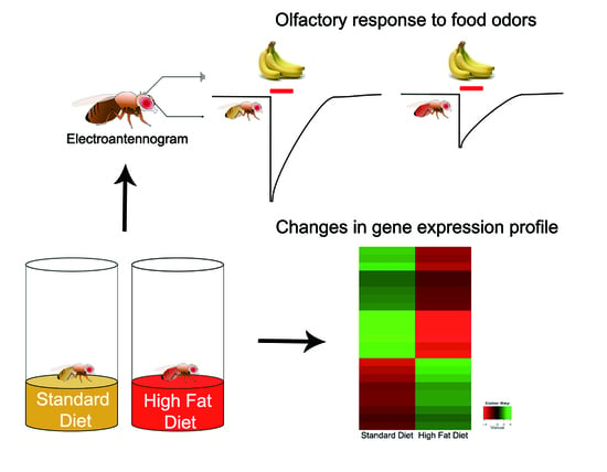
The Effects of High Fat Diet-Induced Stress on Olfactory Sensitivity, Behaviors, and Transcriptional Profiling in Drosophila melanogaster

Department of Life Sciences and Convergence Research Center for Insect Vectors, Incheon National University, 119 Academy-ro, Yeonsu-gu, Incheon 22012, Korea
*
Author to whom correspondence should be addressed.
Int. J. Mol. Sci. 2018, 19(10), 2855; https://doi.org/10.3390/ijms19102855
Submission received: 17 July 2018 / Revised: 10 September 2018 / Accepted: 12 September 2018 / Published: 20 September 2018
(This article belongs to the Section Biochemistry)

Abstract

:
High-fat diet (HFD) often causes obesity and it has detrimental effects on the sensory system. In particular, sensory-mediated responses are crucial for maintaining energy balance, as they are involved in a metabolic regulation; however, there is still no clear explanation about the relationship between HFD-induced stress and sensory system. To gain insight on how HFD-induced stress affects olfactory sensitivity and behavioral responses, we have used a Drosophila melanogaster model for olfactory and nutrient-related signaling and accessed physiological, behavioral, and transcriptional changes. We demonstrated that lifespan and climbing ability in HFD-treated flies decreased and that olfactory sensitivity and behavioral responses to odorants were changed. Olfactory sensitivity to eight of ten odorants after 14 days on HFD treatment were reduced, while behavioral attraction was increased to benzaldehyde in flies that were treated with HFD. This behavioral and physiological modification in HFD-treated flies for 14 days was accompanied by a significant decrease in DmOrco gene expression in a peripheral olfactory organ, suggesting that is could be involved in the action of metabolic and sensory signal. Gene expression profiles of antennae showed significant differences on the olfactory receptors, odorant-binding proteins, and insulin signaling. Our results suggested that olfactory sensitivity and behavioral responses to HFD-induced stress are mediated through olfactory and nutrient-related signaling pathways.

Graphical Abstract

1. Introduction

Most organisms maintain energy homeostasis to survive and function effectively under the influence of internal and external factors for biological processes of growth, development, metabolism, and aging [1,2]. Many environmental conditions, including nutrients, temperature, and light can influence food intake and cause consequences for health, including obesity [3,4]. Obesity causing a myriad of complex diseases is known to be related with an imbalance in the homeostasis of energy metabolism [5,6,7]. A previous study reported that simultaneous processing in taste and olfaction, the pivotal senses for survival, is associated with human obesity [8]. For instance, food intake controlled by external sensory signals has an effect on energy balance assessed by internal metabolic signals [9]. These fundamental responses are made through physiological changes, metabolic hormones, in particular, which regulate appetite [10].
Sensory-mediated responses are remarkably complex network [11,12]. In the natural environment, olfaction is related to finding food sources, recognizing predators, and locating mates [13,14]. In particular, feeding behaviors are closely linked and initiated with the finding of food sources in response to sensory inputs, for instance, olfactory information [12]. In mammals, such metabolic regulation in the olfactory cortex is changed by internal metabolic states where an increased number of insulin receptors (InR) on the olfactory bulb was found in starved mice [15] suggesting that the regulation of insulin signaling by food intake was attributed to olfaction.
Drosophila melanogaster, the fruit fly that is a powerful tool for studying gain- and loss-of-function by observing phenotypic changes and behavioral analysis, possesses evolutionarily conserved pathways in common with mammals [16,17]. Evidence for the link between olfactory response and insulin signaling has been shown by flies fed high-fat diet (HFD) exhibiting decreased olfactory sensitivity and odor-driven food searching behavior in comparison with flies that were fed with normal diet [18]. However, little is known about the impact of food stress on olfactory signaling and nutrient-related genes in the main peripheral olfactory organ, the antennae, of Drosophila melanogaster. It has been suggested that preferential odorant of over-weight people appeared to be a food-related rather than a non-food related [8]. Given this evidence, the alteration of metabolism and enhancement of stress resistance by the olfactory system in relation to food-derived odors might be involved in the modulation of the lifespan. Furthermore, these findings are likely to be correlated with feeding behavior and the energy balance through sensory processing [19,20,21].
To gain insight into how HFD-induced stress affects olfactory sensitivity and behaviors that mediate nutrient-related signaling, this study centers on the understanding of behavioral and sensory modification by food stress, such as HFD treatment in D. melanogaster, and it employs lifespan analysis, behavioral tests such as a climbing test, a choice test for food-related odorants, and electrophysiological recordings from antennae (Electroantennogram, EAG). Moreover, to pinpoint any modifications at the molecular level in the main olfactory organ, we examined the expression levels of DmOrco gene by quantitative real-time PCR (qRT-PCR) and overall transcriptional profiles of antennal tissues to identify genes including olfactory and nutrient-related signaling genes whose levels fluctuated with HFD treatment. These approaches provide a general overview of relationships between food stress and sensory modulation machineries in the peripheral olfactory organ based on Gene Ontology (GO).

2. Results

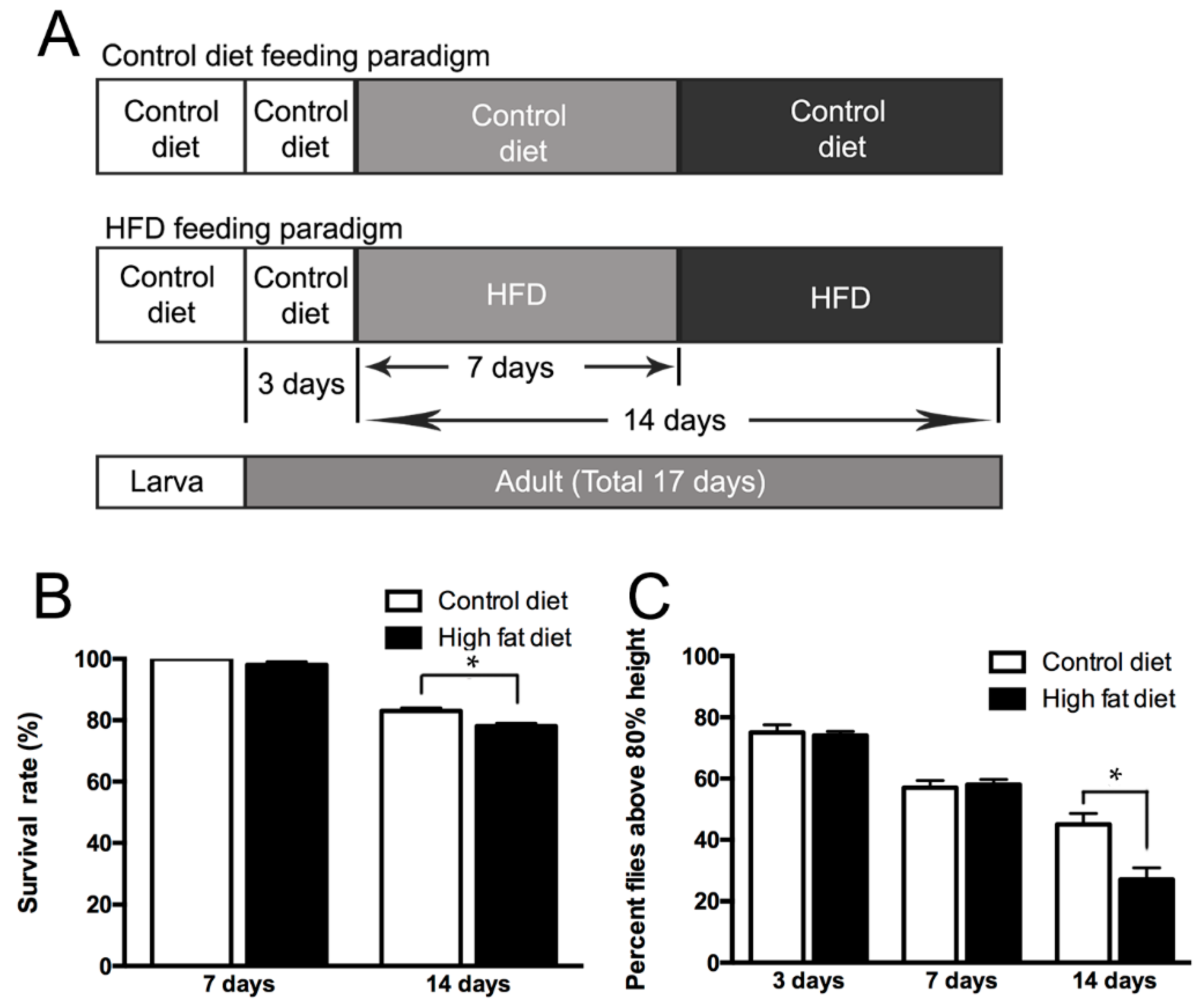
2.1. Life Span and Climbing Abilities of Flies Fed with HFD

We hypothesized that high fat diet (HFD), causing the alteration of physiological states [22,23], might reduce the life span and climbing ability of flies. To confirm the hypothesis, we fed a control diet and high-fat diet (HFD) ad libitum to male Canton-S adult flies four days after eclosion for seven days or 14 days (Figure 1A), with slight modification from the previous study [22]. The results showed that the life span of the flies fed with HFD diet started to decrease at day 7 and it was significantly reduced by approximately 12% at day 14 in comparison with those receiving the control diet (Figure 1B). Consistent with this, there were also indications that the locomotor activity of the flies declined after HFD intake. Since the diet appeared to be associated with climbing ability [24], we measured the climbing ability of flies fed with a control diet or HFD for seven or 14 days. Significant changes in climbing ability were observed for the HFD-treated groups (Figure 1C), suggesting that a HFD has a detrimental influence on fly mobility.

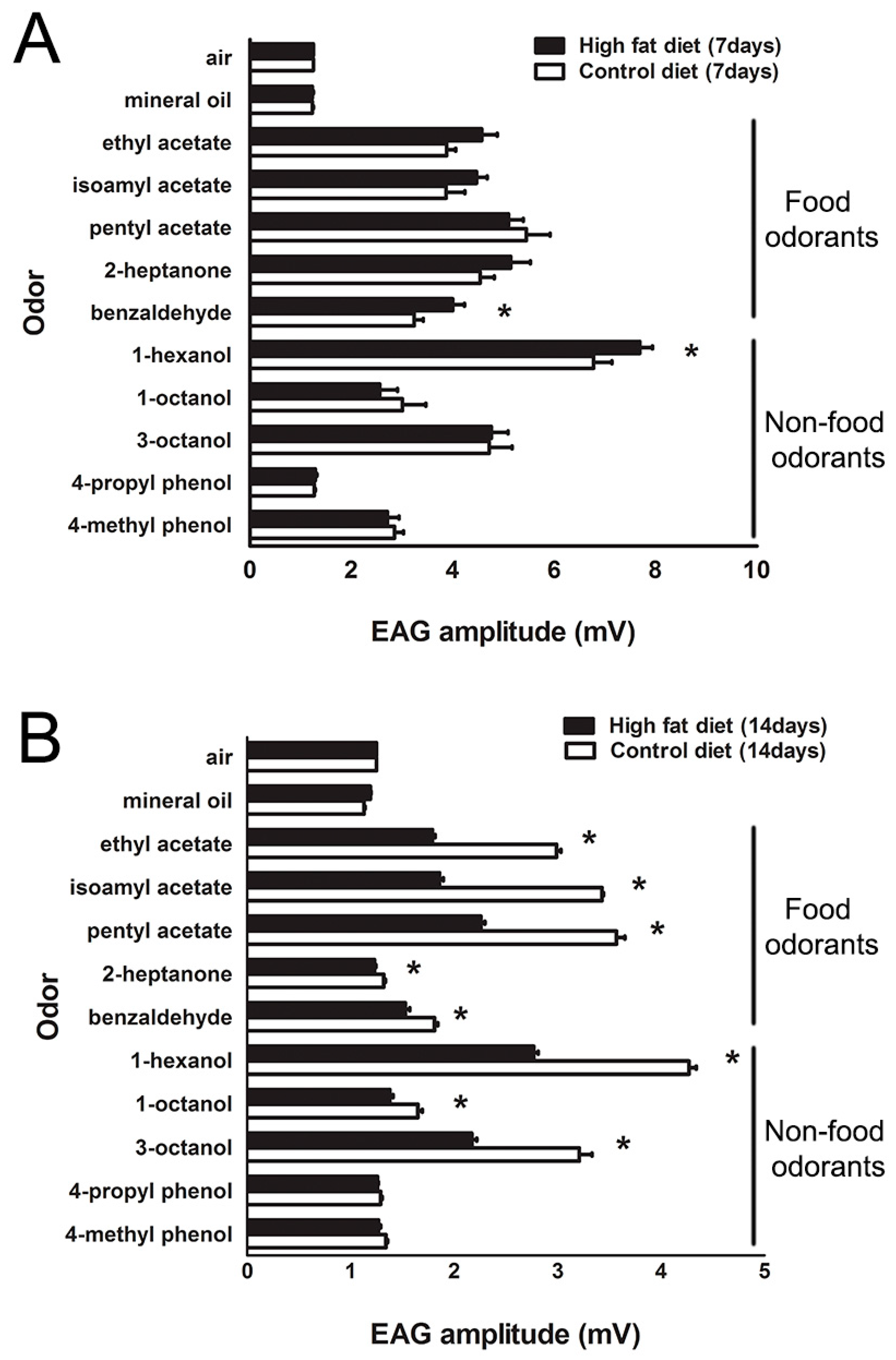
2.2. Modification of Olfactory Sensitivity by HFD Treatment

Olfactory sensitivity is known to be affected by internal and external factors [25]. Therefore, we asked whether the HFD affects olfactory sensitivity, including attraction to food odors, such as ethyl acetate, isoamyl acetate, pentyl acetate, 2-heptanone, and benzaldehyde when compared to non-food odors such as 1-hexanol, 3-octanol, 1-octanol, 4-methyl phenol, and 4-propyl phenol. A significant difference in EAG responses was found in flies fed with HFD for seven or 14 days (Figure 2A,B). Flies treated with HFD for 14 days showed decreases in olfactory sensitivities for the food odorants and three of five non-food odorants (Figure 2B); however, the responses of acetate odorants on HFD treatment for seven days had no significant differences between flies on a HFD and standard diet (Figure 2A). It was noting that olfactory sensitivity to benzaldehyde and 1-hexanol increased substantially in flies fed with HFD for seven days (Figure 2A).

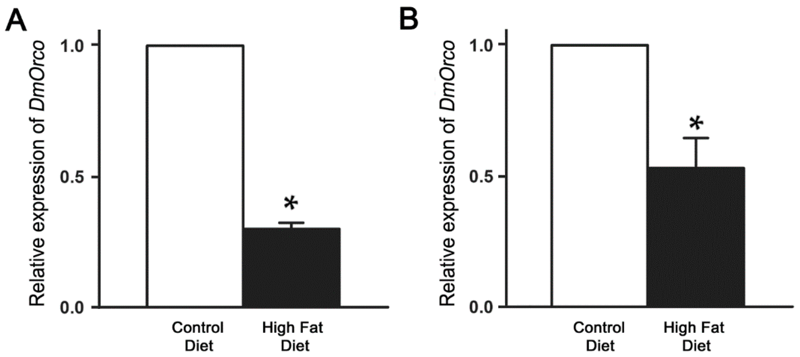
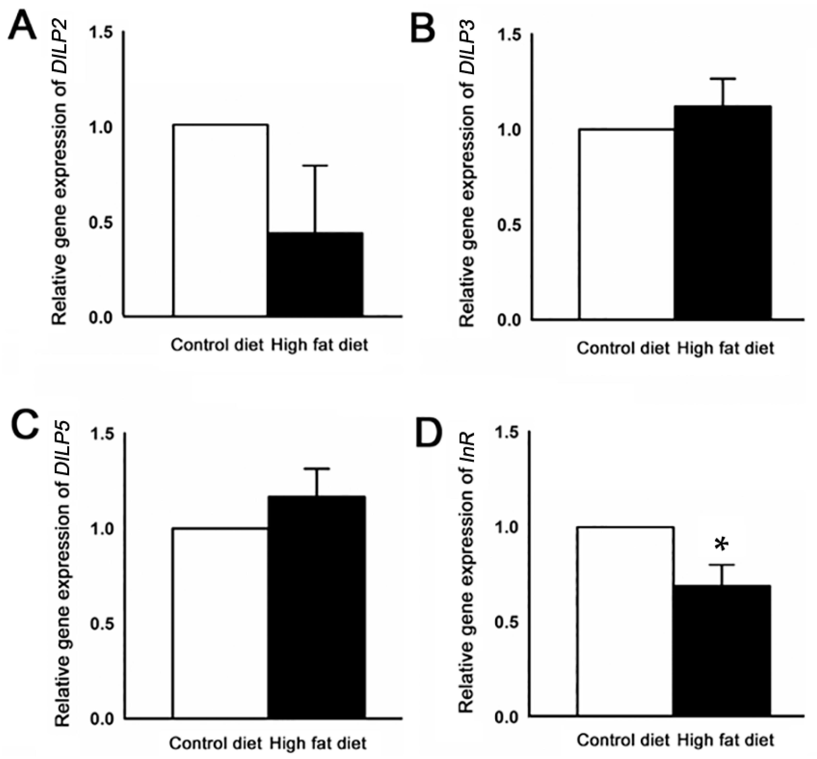
2.3. Reduction of DmOrco Gene Expression in the Fly Antenna after HFD Treatment

We next asked whether the odorant receptor co-receptor gene of D. melanogaster (DmOrco), expressed in most olfactory receptor neurons (ORNs) [25], is affected in flies by HFD treatment. The results showed that the level of DmOrco transcripts was significantly reduced by approximately 70% in flies fed with HFD for seven days when compared with those of control flies, while it was about a 47% reduction at 14 days (Figure 3A,B).

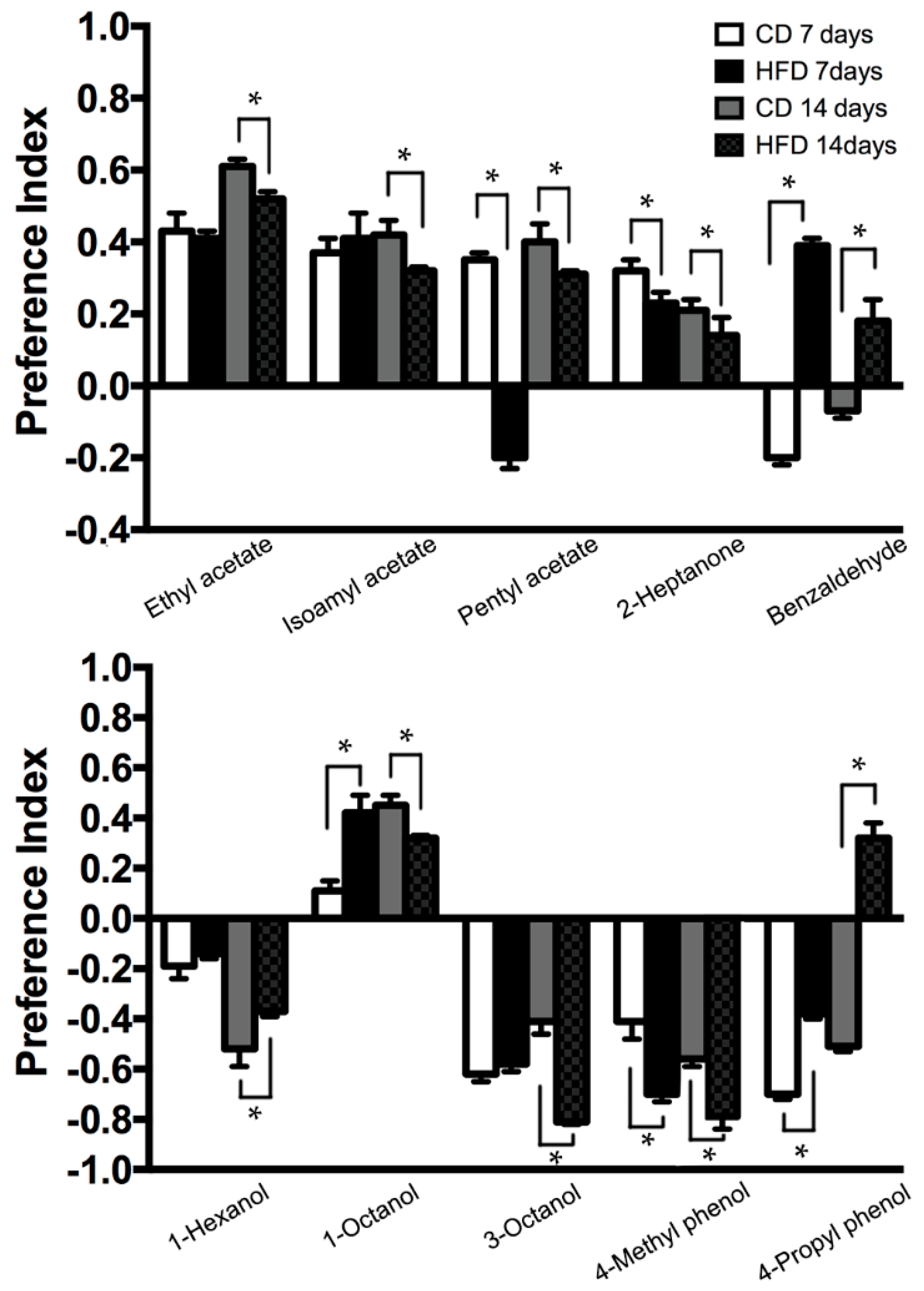
2.4. Modification of Odorant Choice Behaviors by HFD Treatment

HFD-fed flies showed different responses in terms of attraction and repulsion to the experimental odors as compared with those on the control diet. First, flies on a HFD at 14 days showed a significant decrease in appetitive behavior to ethyl acetate, isoamyl acetate, pentyl acetate, and 2-heptanone. On the other hand, there was an increase in repulsive behavioral responses to 3-octenol (14 days), 4-methyl phenol (7 and 14 days) in flies fed with HFD (Figure 4). In addition, the behavioral responses of HFD-fed flies to benzaldehyde and pentyl acetate showed a transition between repulsive and attractive behavior. In detail, there was an attractive behavioral response to benzaldehyde by flies fed with HFD for seven and 14 days, while the response to pentyl acetate disappeared with HFD for seven days (Figure 4). We found that the behavior of HFD-fed flies to 4-propyl phenol shift from repulsive response to appetitive response (Figure 4). In addition, while HFD-fed flies at seven days exhibit increased attractive response to 1-octanol, flies exposed to HFD at seven days show decreased attractive response to 1-octanol when compared to the response of control diet-fed files (Figure 4). Taken together, the exposure to high levels of dietary fat affects fruit fly olfactory related behavior.

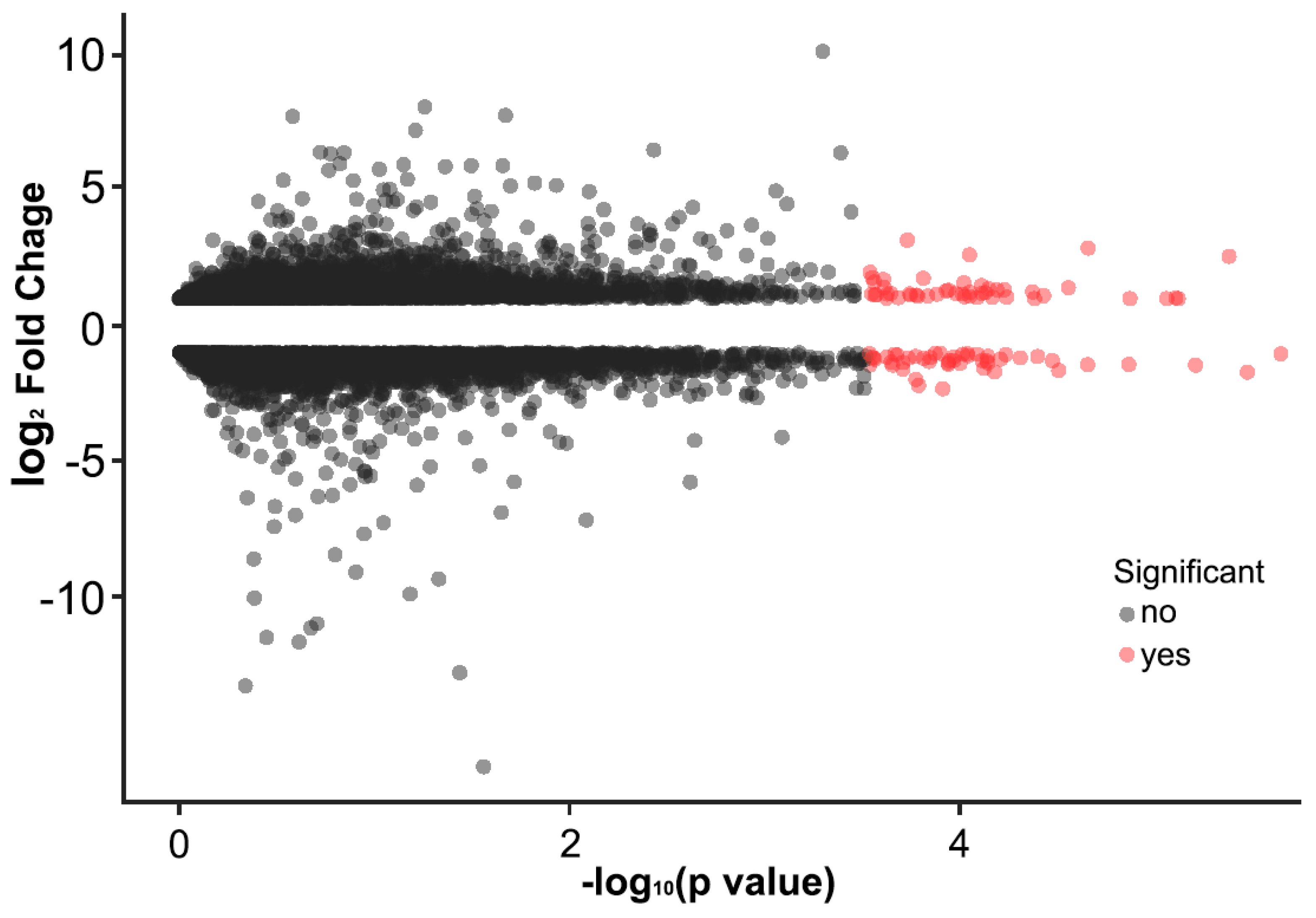
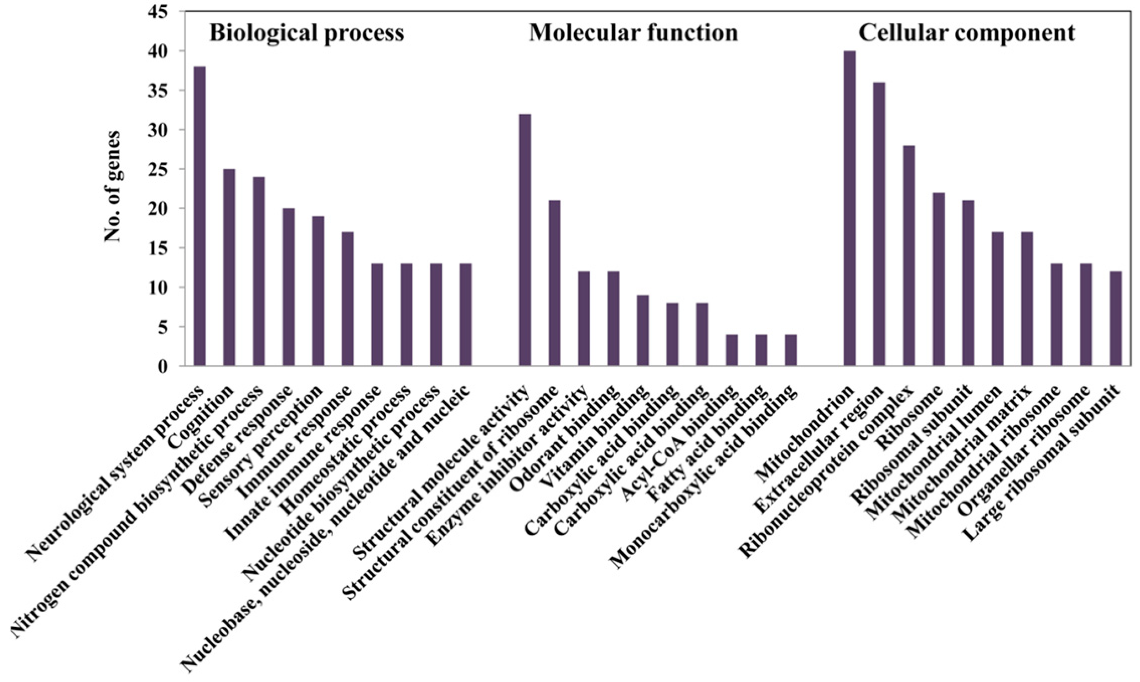
2.5. Alteration of Gene Expression Profiles by HFD Treatment

While considering the modification in patterns of olfactory sensitivity and behavior to odorants, we subsequently examined the gene expression levels in the antenna from flies fed with HFD for 14 days. Our Illumina paired-end sequencing yielded a total of 13,287,258 and 11,103,566 raw reads with the length of 101 bp from antenna tissue in flies fed with control diet (CD) and high fat diet (HFD), respectively. Firstly, we quantified our sequencing data by mapping against files transcript sequences. This analysis revealed that 72% of reads were mapped in the control diet-treated flies, while 67% were aligned in HFD-treated flies. Collectively, over two-thirds of the Drosophila melanogaster transcriptome data was expressed in both the control and HFD-treated flies, which is a similar observation for gene expression in other insects [26].
Then, we attempted to identify differentially expressed genes between control diet and HFD. A significant change in the level of gene expression after HFD treatment was indicated for a total of 732 genes, of which 379 genes were up-regulated and 343 genes down-regulated (Figure 5). From functional annotation, we noticeably identified genes that are related to olfactory related genes and neuronal sensor, which were down-regulated (Table 1). A total of 74 olfactory genes, including 34 odorant receptors (ORs) and 30 odorant-binding proteins (OBPs) were up-regulated and down-regulated from antennae of flies on high fat diet (Table 2 and Table 3).
Next, we analyzed the expression of the genes associated with insulin signaling pathways (Table S1). Normalization of qRT-PCR was performed for four reference genes that are involved with insulin signaling genes to increase accuracy. We measured the transcript variation of DILP2, DILP3, DILP5, and InR genes for data normalization. Although the relative levels of DILP2, DILP3, and DILP5 expression showed no significant differences, InR expression demonstrated a significant decrease (Figure 6).
To find out the main functional process that is affected by HFD treatment, we categorized the genes whose expression exhibited the substantial changes after HFD treatment. We analyzed the genes with a more than log two-fold change in expression in up- and down-regulation. These data were established according to the functional groups of GO, including biological process (BP), molecular function (MF), and cellular component (CC) (Figure 7). In particular, the largest number of genes was representative of several categories of BP, namely neurological system process, cognition, and sensory perception (Figure 7), which suggests a relevance to olfactory responses in both the peripheral and central nervous systems. Furthermore, genes of defense response and immune response were associated with response to HFD treatment.

3. Discussion

A High-Fat Diet Leads to Olfactory Dysfunction in Homeostatic Processing in Drosophila

High fat diet (HFD) is associated with an increased risk for disease, including obesity, heart disease, diabetes, and cancer [5,6,7]. In the present study, HFD for 14 days lead to a loss of climbing ability and reduction of life span of flies. Although consumption of HFD are found to cause nervous system dysfunction, whether such diets affect peripheral sensory system is largely unknown. We discovered a relationship between HFD and olfactory dysfunction in the fruit fly. Excessive stress by a HFD has a crucial impact on olfactory sensitivity. HFD-treated flies for 14 days were less sensitive to five food odors and three non-food odors. This is consistent with a recent reported that mice on HFD exhibited decreased olfactory discrimination [27]. This reduction in sensory sensitivity, which is exposed by a chronic fat diet, may be linked to the significantly decrease of olfactory related genes or other neuromodulators. For example, our DEG analysis indicated that 27 olfactory receptor genes and 21 odorant binding protein genes were down regulated in flies on fat diet. Because ORs and OBPs have been demonstrated to play an important role in olfactory receptor neurons maturation and axon guidance [28], one speculation is that high fat diet induced the down regulation of OR and OBP expression in olfactory receptor neuron that might thereby interfere with appropriate gene processing and targeting olfactory receptor neuronal axons to the proper antennal lobe region [29]. The cellular mechanisms underlying the HFD induced regulation of olfactory receptor gene expression are largely unknown. Recent evidence has emerged that proinflammatory responses are evoked in the olfactory epithelium in response to altered energy consumption [30]. HFD induce an increase in the number of macrophages and neuronal death, which resulted in a loss of connections from the olfactory epithelium and olfactory bulb [31]. Our DEG data showed that there was an upregulation of two genes that are known to be involved in apoptotic processes: Traf6 and Decay. Increased expression of these genes may induce apoptotic death in response to HFD. Further experiments are necessary to demonstrate whether Traf6 and Decay can induce apoptotic death of olfactory receptor neurons. Our DEG analysis provides the possibility that some mediators may regulate expression of the olfactory receptor genes and odorant binding proteins. Certain genes were observed in functional changes, which is associated with olfactory and nutrient-related pathways that modulate olfactory responses in flies. Previous studies demonstrated microarray analysis after starvation [32] or heat treatment [26] that affected olfactory responses and feeding behaviors, depending on nutritional status in adult flies. In both cases, the analysis included the antennal part, which is the main olfactory receptor organ, to make clear the importance of the internal state of olfaction.
One of the most prominent changes we found was a reduced appetitive behavioral response to olfactory stimuli, suggesting that HFD feeding may cause the alteration in olfactory perception. This is consistent with a recent report that mice with HFD-induced obesity showed deficits in olfactory learning and memory formation and altered behaviors related to olfaction and taste [33]. Besides, obese people have been shown to prefer a food-related odorant rather than a non-food related odorant when compared with normal weight people as same ages, implying that these people might not be able to regulate food intake by their internal state through appropriate odor processing [8]. Although molecular and neural mechanisms underlying the HFD-induced modification of olfactory behavior are not addressed yet, many lines of the research have been focused on insulin resistance. It has been reported that the activation of food stress responses is involved in the induction of insulin resistance and interestingly these results are observed in the olfactory bulb [34], implying that food stress-induced insulin resistance may affect the sensory cognition ability in animals.
In Drosophila, local signals by the neuropeptides and global metabolic cues by insulin are modified at specific olfactory sensory neuron to change olfactory responses [21,35,36]. For example, insulin as a metabolic factor modulates olfactory responses in fly antennae by enhancing the pre-synaptic facilitation and regulating odor preference in accordance with the internal state [18]. Furthermore, Drosophila insulin-like peptide 2 (DILP2) was shown to modulate a short neuropeptide F receptor (sNPFR1), which regulates olfactory driven behaviors in antenna and is also stimulated in starved flies [37,38,39]. Similarly, several studies demonstrate the importance of neuropeptides in the regulation of olfactory behavior [40,41]. Our digital gene expression analysis detected transcripts of DILP2 and insulin receptor (InR) in significant changes of down-regulation, in contrast with other insulin-related genes. This could be partially explained by the reduction of olfactory sensitivity caused by HFD treatment, and this may cause a loss of controlling ability of attractive behaviors to food odors. Recent research has demonstrated that DILP2 shows different expression levels depending on the type of diet [42]. Therefore, high fat diet condition must be function with other peripheral receptors, including those for olfaction. Olfactory responses to HFD treatment are affected by the over-expression of certain insulin signaling genes; therefore, communication through olfactory modulation could contribute to peripheral organs and subsequently be linked to the central nervous system [13,43].
Interestingly, the behavioral responses of HFD-fed flies to benzaldehyde and pentyl acetate showed a transition between repulsive and attractive behavior, which is contrary to the expected physiological responses of the fly. This defective olfactory behavior might be associated with the misexpression of specific genes. Our DEG analysis showed that transcript levels of several olfactory receptor genes and odorant binding protein genes were changed. Among these genes, misexpression of the or43a, one of the benzaldehyde receptors, caused a reduction of behavioral avoidance responses to benzaldehyde [44]. In addition, lush is a soluble odorant binding protein and it is expressed exclusively in the chemosensory system in flies. Mutants of this gene have been reported to a loss of the avoidance behaviors [45] and defects for pheromone-evoked behaviors [46]. Since the genetic dissection by which genes modulate olfactory behavior about each odorant remains unclear, further genetic experiments are necessary to demonstrate the relevance of specific olfactory sensory input and subsequent processing for olfactory driven behaviors.

4. Materials and Methods

4.1. Drosophila Stocks and Diet Treatment

The Canton-S wild-type strain of Drosophila melanogaster that was obtained from Bloomington Stock Center (Bloomington, IN, USA) was used in this study. All of the adult flies were maintained at 25 °C with 60% relative humidity and a 12 h light/12 h dark cycle. Food media for a standard diet was prepared by mixing 10% sugar, 10% yeast extract, and 1.5% agar with 1% Tween 80 (w/v) added in case of toxicity. To make the high-fat diet, 2% palmitic acid (w/v) and 1% Tween 80 (w/v) were added into the standard diet in accordance with a previous study [22].
In order to obtain adult flies for the experiments, newly enclosed flies were collected into new bottles and kept for two days on the standard diet. Next day, male and female flies were sorted under CO2 anesthesia and only male flies were placed on the standard diet for one more day. Test male flies were treated with the standard diet and a high-fat diet (HFD) for seven days and 14 days, respectively. Each vial contained 20 male flies.

4.2. Life Span and Climbing Assays

For life span assays, flies were kept at a density of 20 male flies per each vial on the standard diet and HFD. Each group of flies was transferred to a vial with fresh food medium every three days and dead flies in each group were counted every day. Five replicates were conducted for this experiment.
For climbing assays, a behavioral paradigm that was reported in a previous study was employed with slight modifications [24]. Twenty male flies were transferred to a 10 cm glass vial and placed at the bottom of the vial by gently tapping the flies down to the bottom. The number of flies that climbed above 80% height within 30 s was counted. Experimental trials were repeated ten times independently for each group. The climbing index (%) was calculated as the ratio of the number of flies climbing above 80% height to the total number of flies multiplied by 100. All of the climbing assay experiments were performed at 25 °C with 60% humidity.

4.3. Odor Stimulation

All the odorants tested in this study (ethyl acetate, isoamyl acetate, pentyl acetate, benzaldehyde, 2-heptanone, 1-hexanol, 3-octanol, 1-octanol, 4-propyl phenol, and 4-methyl phenol) were commercially available products at the highest purity (>98%, Sigma-Aldrich, Milwaukee, WI, USA). These odorants were dissolved in mineral oil (Sigma-Aldrich-330760) or ethanol at 1% dilution (v/v). A glass tube was prepared with a continuous, humidified air stream, and 20 μL of odorant solution was soaked onto a filter paper (4 × 4 mm, Toyo Roshi Kaisha, Tokyo, Japan) that was placed into a 5 mL disposable syringe with air pressure. These chemicals were used as odorant sources for EAG recordings and for behavioral assays.

4.4. Electrophysiological Recordings

Transepithelial electrophysiological recordings (electroantennograms, EAGs) from antennae in male Canton-S flies were made after individual diet treatment while using AgCl-coated silver wire inserted in a glass micropipette filled with 0.1 M KCl. The experimental fly was immobilized in a truncated 200 μL plastic pipette tip and trimmed to show an anterior aspect of the fly’s head. The third segments of the antennae were exposed for EAG recordings. A reference electrode was inserted into a compound eye and a recording electrode was placed on the dorso-medial surface of the third antennal segment, as described previously [47]. Electrical signals were amplified with an analog 10× active probe and conveyed to an acquisition system (IDAC4, Syntech, Hilversum, The Netherlands). Signals were then further recorded and analyzed while using EAG Pro software (Syntech). A constant air stream of 40 mL/min was delivered to the fly head by using a stimulus controller (CS-55, Syntech). Pulses of the odorants were produced during a 1 s stimulation through the Pasteur pipette to the syringe pipette. Signal amplitude of olfactory responses (mV) was measured from a baseline before stimulation to the trough of the electrical signals after odorant stimulation. Control EAG experiments loaded with no odorant and mineral oil were used. The interval between each odorant stimulus was about 60 s to prevent the adaptation of the test animal to a given odorant.

4.5. Quantitative RT-PCR

Flies were collected according to the procedures that are presented above for the diet treatment. Total RNA extraction was carried out while using an RNeasy Mini Kit (Qiagen, Valencia, CA, USA), according to the manufacturer’s protocol. For each treatment, at least three independent extractions were conducted while using 100 fly antennae. The quality and quantity of total RNA was measured by a Nanodrop 2000 (Thermo Scientific Inc., Wilmington, DE, USA). cDNA was prepared from the extracted RNA while using the Superscript III First-Strand Synthesis kit (Invitrogen Inc., Carlsbad, CA, USA). Quantitative RT-PCR was performed using a StepOnePlus machine (Applied Biosystems Inc., Foster City, CA, USA) according to the manufacturer’s protocol with SYBR green qRT-PCR Master Mix (Fermentas, Burlington, ON, Canada) to measure DmOrco gene expression. qRT-PCR measurement for each gene was repeated with three independent biological samples and quantitative analysis was conducted by StepOne plus Software V. 2.0 (Applied Biosystems). The transcript level of the gene was calculated by the standard curve method and normalized to the control gene ribosomal protein 49 (rp49) primer that was described in a previous paper [48]. The primer sequences for DmOrco were forward: 5′-GGTGGACCATGAGACGAACT-3′; reverse: 5′-CATCACGTCGCATAGATTGG-3′.

4.6. Behavioral Assay

Flies were placed under light CO2 anesthesia. Separated male flies were placed into vials (20 flies per vial) containing each diet treatment. Flies were transferred to new vials every two to three days and behavioral assays performed on days 7 and 14 after diet treatment. One day before the assay, flies were moved into empty vials containing only water on Kimwipes (Kimberly-Clark Worldwide, Neenah, WI, USA) for at least 15 h to produce a starved state [49]. For each measurement, the flies were gently tapped to place into a T-maze. The same odorants as used for the EAGs were used for this behavioral assay. Each vial at both sides contained two traps, an odor trap, and a control trap (water). Flies were allowed to have 30 s to choose one of the sides in the T-maze that contained odorant. The attraction index (AI) was calculated, as follows: (number of flies moving toward the odor trap − number of flies in the control trap)/(total number of flies). Ten replicates were performed for each treatment. All T-maze assay experiments were conducted at 25 °C with 60% humidity.

4.7. Analysis for Differentially Expressed Genes (DEG) and Gene Ontology of HFD-Fed Flies

Approximately 3000 samples of third antennal segments of male adult flies were prepared to achieve a sufficient representation of genes. The flies in each diet treatment were collected from 11:00 to 14:00 and 16:00 to 19:00 to prevent the possible modification of gene expression by circadian rhythms [26]. All the samples were incorporated to minimize any random factors that affect gene expression other than the treatment. Gene sequences were downloaded from the Drosophila Genome database (www.flybase.org) for the analysis of differentially expressed genes (DEG).
For the RNA sequencing experiment, 4 μg of total RNA was extracted while using the same protocol, as described above in the quantitative RT-PCR procedure. In order to convert mRNA in total RNA into a library of template molecules for subsequent cluster generation, a Illumina® TruSeq™ RNA sample preparation kit was used in accordance with the manufacturer’s instructions (Illumina Inc., San Diego, CA, USA). The total RNA sample was used for poly-A mRNA selection using poly-T oligo-attached magnetic beads with two rounds of purification. The resulting mRNA sample from the antennae was subjected to thermal mRNA fragmentation while using Elute, Prime, Fragment Mix from the Illumina® TruSeq™ RNA sample preparation kit. The mRNA fragments were reverse transcribed to synthesize the first-strand cDNA using a combination of reverse transcriptase and random primers. The mRNA template strand was removed and double-stranded cDNA (ds cDNA) was generated using DNA polymerase І. The ds cDNA was purified using Ampure XP beads to separate the ds cDNA from the second-strand reaction mix.
The cDNA fragments were then blunt-ended through an end-repair reaction while using an End Repair (ERP) mix (Illumina Inc). The 3′ to 5′ exonuclease activity of this mix removed the 3′ overhangs and the polymerase activity filled in the 5′ overhangs. Next, “A” nucleotide was added to the 3′ ends of the blunt fragments to prevent them from ligating to each other and then ligated to platform-specific double-stranded bar-coded adapters that provided a complementary overhang during the adapter ligation reaction. DNA libraries were sequenced using an Illumina HiSeq2000. The sequencing reads were deposited to the NCBI SRA (Short Read Archive) site (https://www.ncbi.nlm.nih.gov/sra) under the accession number SRR1501071 for the control diet and SRR1501074 for the high fat diet.
DEG analysis was performed using edgeR bioconductor package and RSEM software [50]. Quality check, normalization, and statistical analysis were performed to develop significantly matched signals. Genes with false discovery rate (FDR) value of at most 0.001 and fold change value of ≥2 were considered as significant differentially expressed genes. In addition, log2 (FPKM) expressional difference was manually calculated for all of the genes based on FPKM (Fragments Per Kilobase Million) values. After obtaining general information on the gene expression, gene ontology (GO) analysis was carried out for genes that showed log2 fold digital gene expression difference after diet treatment using the DAVID tool (http://david.abcc.ncifcrf.gov/). In the DAVID annotation system, modified Fisher Exact p value (EASE score) was adopted to measure the gene-enrichment in annotation terms. All data analysis and visualization of differentially expressed genes were conducted while using R 2.15.1 (http://www.r-project.org).

4.8. Statistical Analysis

Statistical analyses were performed by Student’s t-test (SPSS, Version 20, IBM, New York, NY, USA) on the results from the lifespan assay, climbing ability test, EAGs, and T-maze tests to determine any significant differences between the control diet and HFD treatment. Data were shown as means ± standard error.

5. Conclusions

High-fat diet affects sensory abilities in Drosophila melanogaster such as olfactory-driven behaviors and olfactory sensitivity to several odorants, which in turn makes some changes in molecular levels of gene expressions. Integration of internal and external factors provides crucial evidence to olfactory modulation in animals. Our present study demonstrates that high fat diet alters olfactory perception and changes of gene expression profiles in Drosophila melanogaster. A better understanding of the complex mechanisms that are underlying olfactory modulation might help to characterize the olfactory systems that are affected by HFD treatment. Identifying novel regulators of the olfactory system and examining them to find out specific odorant receptors to odors at the intracellular level affected by HFD treatment may provide an understanding of the mechanisms and circuits from antenna to brain in flies.

Supplementary Materials

Supplementary materials can be found at https://www.mdpi.com/1422-0067/19/10/2855/s1.

Author Contributions

J.J., D.-I.K., G.-Y.H., and H.W.K. designed experiments and J.J., D.-I.K., and G.-Y.H. carried out the experiments. J.J. and H.W.K. prepared the manuscript.

Funding

This research was supported by Basic Science Research Program through the National Research Foundation of Korea (NRF) funded by the Ministry of Science, ICT & Future Planning (2016R1A2B3011742) and Cooperative Research Program for Rural Development Administration (Project No. PJ012526012018). This study also partly supported by Incheon National University internal grant (2016) to H.K.

Acknowledgments

We are grateful to Murukarthick Jayakodi assisting a differentially expressed gene (DEG) analysis and to Soo Ho Lim for helping graphical art work.

Conflicts of Interest

The authors declare no conflict of interest.

References

  1. Rajan, A.; Perrimon, N. Drosophila as a model for interorgan communication: Lessons from studies on energy homeostasis. Dev. Cell 2011, 21, 29–31. [Google Scholar] [CrossRef] [PubMed]
  2. Rajan, A.; Perrimon, N. Of flies and men: Insights on organismal metabolism from fruit flies. BMC Biol. 2013, 11, 38. [Google Scholar] [CrossRef] [PubMed] [Green Version]
  3. Baker, K.D.; Thummel, C.S. Diabetic larvae and obese flies-emerging studies of metabolism in Drosophila. Cell Metab. 2007, 6, 257–266. [Google Scholar] [CrossRef] [PubMed]
  4. Skorupa, D.A.; Dervisefendic, A.; Zwiener, J.; Pletcher, S.D. Dietary composition specifies consumption, obesity, and lifespan in Drosophila melanogaster. Aging Cell 2008, 7, 478–490. [Google Scholar] [CrossRef] [PubMed]
  5. Gale, S.M.; Castracane, V.D.; Mantzoros, C.S. Energy homeostasis, obesity and eating disorders: Recent advances in endocrinology. J. Nutr. 2004, 134, 295–298. [Google Scholar] [CrossRef] [PubMed]
  6. Trinh, I.; Boulianne, G.L. Modeling obesity and its associated disorders in Drosophila. Physiology 2013, 28, 117–124. [Google Scholar] [CrossRef] [PubMed]
  7. Teleman, A.A.; Ratzenbock, I.; Oldham, S. Drosophila: A model for understanding obesity and diabetic complications. Exp. Clin. Endocrinol. Diabetes 2012, 120, 184–185. [Google Scholar] [CrossRef] [PubMed]
  8. Thompson, D.A.; Moskowitz, H.R.; Campbell, R.G. Taste and olfaction in human obesity. Physiol. Behav. 1977, 19, 335–337. [Google Scholar] [CrossRef]
  9. Withcher, D. Metabolic regulation and behavior: How hunger produces arousal-an insect study. Endocr. Metab. Immune 2007, 7, 304–310. [Google Scholar]
  10. Crowley, V.E. Overview of human obesity and central mechanisms regulating energy homeostasis. Ann. Clin. Biochem. 2008, 45, 245–255. [Google Scholar] [CrossRef] [PubMed]
  11. Melcher, C.; Bader, R.; Pankratz, M.J. Amino acids, taste circuits, and feeding behavior in Drosophila: Towards understanding the psychology of feeding in flies and man. J. Endocrinol. 2007, 192, 467–472. [Google Scholar] [CrossRef] [PubMed]
  12. Herrero, P. Fruit fly behavior in response to chemosensory signals. Peptides 2012, 38, 228–237. [Google Scholar] [CrossRef] [PubMed]
  13. Sato, K.; Touhara, K. Insect olfaction: Receptors, signal transduction, and behavior. Results Probl. Cell Differ. 2009, 47, 121–138. [Google Scholar] [PubMed]
  14. Carlson, J.R. Olfaction in Drosophila: From odor to behavior. Trends Genet. 1996, 12, 175–180. [Google Scholar] [CrossRef]
  15. Lacroix, M.C.; Badonnel, K.; Meunier, N.; Tan, F.; Schlegel-Le Poupon, C.; Durieux, D.; Monnerie, R.; Baly, C.; Congar, P.; Salesse, R.; et al. Expression of insulin system in the olfactory epithelium: First approaches to its role and regulation. J. Neuroendocrinol. 2008, 20, 1176–1190. [Google Scholar] [CrossRef] [PubMed]
  16. Vosshall, L.B. Into the mind of a fly. Nature 2007, 450, 193–197. [Google Scholar] [CrossRef] [PubMed] [Green Version]
  17. Diop, S.B.; Bodmer, R. Drosophila as a model to study the genetic mechanisms of obesity-associated heart dysfunction. J. Cell. Mol. Med. 2012, 16, 966–971. [Google Scholar] [CrossRef] [PubMed]
  18. Root, C.M.; Ko, K.I.; Jafari, A.; Wang, J.W. Presynaptic facilitation by neuropeptide signaling mediates odor-driven food search. Cell 2011, 145, 133–144. [Google Scholar] [CrossRef] [PubMed]
  19. Palouzier-Paulignan, B.; Lacroix, M.C.; Aime, P.; Baly, C.; Caillol, M.; Congar, P.; Julliard, A.K.; Tucker, K.; Fadool, D.A. Olfaction under metabolic influences. Chem. Senses 2012, 37, 769–797. [Google Scholar] [CrossRef] [PubMed]
  20. Nassel, D.R.; Winther, A.M. Drosophila neuropeptides in regulation of physiology and behavior. Prog. Neurobiol. 2010, 92, 42–104. [Google Scholar] [CrossRef] [PubMed]
  21. Libert, S.; Zwiener, J.; Chu, X.; Vanvoorhies, W.; Roman, G.; Pletcher, S.D. Regulation of Drosophila life span by olfaction and food-derived odors. Science 2007, 315, 1133–1137. [Google Scholar] [CrossRef] [PubMed]
  22. Sun, X.; Seeberger, J.; Alberico, T.; Wang, C.; Wheeler, C.T.; Schauss, A.G.; Zou, S. Acai palm fruit (Euterpe oleracea Mart.) pulp improves survival of flies on a high fat diet. Exp. Gerontol. 2010, 45, 243–251. [Google Scholar] [CrossRef] [PubMed]
  23. Boyd, O.; Weng, P.; Sun, X.; Alberico, T.; Laslo, M.; Obenland, D.M.; Kern, B.; Zou, S. Nectarine promotes longevity in Drosophila melanogaster. Free Radic. Biol. Med. 2011, 50, 1669–1678. [Google Scholar] [CrossRef] [PubMed]
  24. Birse, R.T.; Choi, J.; Reardon, K.; Rodriguez, J.; Graham, S.; Diop, S.; Ocorr, K.; Bodmer, R.; Oldham, S. High-fat-diet-induced obesity and heart dysfunction are regulated by the TOR pathway in Drosophila. Cell Metab. 2010, 12, 533–544. [Google Scholar] [CrossRef] [PubMed]
  25. Leinwand, S.G.; Chalasani, S.H. Olfactory networks: From sensation to perception. Curr. Opin. Genet. Dev. 2011, 21, 806–811. [Google Scholar] [CrossRef] [PubMed]
  26. Riveron, J.; Boto, T.; Alcorta, E. Transcriptional basis of the acclimation to high environmental temperature at the olfactory receptor organs of Drosophila melanogaster. BMC Genomics 2013, 14, 259. [Google Scholar] [CrossRef] [PubMed]
  27. Takase, K.; Tsuneoka, Y.; Oda, S.; Kuroda, M.; Funato, H. High-fat diet feeding alters olfactory-, social-, and reward-related behaviors of mice independent of obesity. Obesity 2016, 24, 886–894. [Google Scholar] [CrossRef] [PubMed] [Green Version]
  28. Feinstein, P.; Bozza, T.; Rodriguez, I.; Vassalli, A.; Mombaerts, P. Axon guidance of mouse olfactory sensory neurons by odorant receptor sand the β2 adrenergic receptor. Cell 2004, 117, 833–846. [Google Scholar] [CrossRef] [PubMed]
  29. Barish, S.; Volkan, P.C. Mechanisms of olfactory receptorneuron specification in Drosophila. WIREs. Dev. Biol. 2015, 4, 609–621. [Google Scholar] [CrossRef] [PubMed]
  30. Gregor, M.F.; Hotamisligil, G. Inflammatory mechanisms in obesity. Ann. Rev. Immunol. 2011, 29, 415–445. [Google Scholar] [CrossRef] [PubMed]
  31. Moraes, J.C.; Coope, A.; Morari, J.; Cintra, D.E.; Roman, E.A.; Pauli, J.R.; Romanatto, T.; Carvalheira, J.B.; Oliveira, A.L.R.; Saad, M.J.; et al. High-fat diet induces apoptosis of hypothalamic neurons. PLoS ONE 2009, 4, e5045. [Google Scholar] [CrossRef] [PubMed]
  32. Farhadian, S.F.; Suarez-Farinas, M.; Cho, C.E.; Pellegrino, M.; Vosshall, L.B. Post-fasting olfactory, transcriptional, and feeding responses in Drosophila. Physiol. Behav. 2012, 105, 544–553. [Google Scholar] [CrossRef] [PubMed]
  33. Thiebaud, N.; Johnson, M.C.; Butler, J.L.; Bell, G.A.; Ferguson, K.L.; Fadool, A.R.; Fadool, J.C.; Gale, A.M.; Gale, D.S.; Fadool, D.A. Hyperlipidemic diet causes loss of olfactory sensory neurons, reduces olfactory discrimination, and disrupts odor-reversal learning. J. Neurosci. 2014, 34, 6970–6984. [Google Scholar] [CrossRef] [PubMed]
  34. Fadool, D.A.; Tucker, K.; Pedarzani, P. Mitral cells of the olfactory bulb perform metabolic sensing and are disrupted by obesity at the level of the Kv1.3 ion channel. PLoS ONE 2011, 6, e24921. [Google Scholar] [CrossRef] [PubMed]
  35. Musselman, L.P.; Fink, J.L.; Narzinski, K.; Ramachandran, P.V.; Hathiramani, S.S.; Cagan, R.L.; Baranski, T.J. A high-sugar diet produces obesity and insulin resistance in wild-type Drosophila. Dis. Mod. Mech. 2011, 4, 842–849. [Google Scholar] [CrossRef] [PubMed]
  36. Heinrichsen, E.T.; Haddad, G.G. Role of high-fat diet in stress response of Drosophila. PLoS ONE 2012, 7, e42587. [Google Scholar] [CrossRef] [PubMed]
  37. Morris, S.N.; Coogan, C.; Chamseddin, K.; Fernandez-Kim, S.O.; Kolli, S.; Keller, J.N.; Bauer, J.H. Development of diet-induced insulin resistance in adult Drosophila melanogaster. Biochim. Biophys. Acta 2012, 1822, 1230–1237. [Google Scholar] [CrossRef] [PubMed]
  38. Benton, R. On the ORigin of smell: Odorant receptors in insects. Cell. Mol. Life Sci. 2006, 63, 1579–1585. [Google Scholar] [CrossRef] [PubMed]
  39. Lee, K.S.; Kwon, O.Y.; Lee, J.H.; Kwon, K.; Min, K.J.; Jung, S.A.; Kim, A.K.; You, K.H.; Tatar, M.; Yu, K. Drosophila short neuropeptide F signalling regulates growth by ERK-mediated insulin signalling. Nat. Cell Biol. 2008, 10, 468–475. [Google Scholar] [CrossRef] [PubMed]
  40. Winther, A.M.; Acebes, A.; Ferrus, A. Tachykinin-related peptides modulate odor perception and locomotor activity in Drosophila. Mol. Cell. Neurosci. 2006, 31, 399–406. [Google Scholar] [CrossRef] [PubMed]
  41. Kahsai, L.; Kapan, N.; Dircksen, H.; Winther, A.M.; Nassel, D.R. Metabolic stress responses in Drosophila are modulated by brain neurosecretory cells that produce multiple neuropeptides. PLoS ONE 2010, 5, e11480. [Google Scholar] [CrossRef] [PubMed]
  42. Carlsson, M.A.; Diesner, M.; Schachtner, J.; Nassel, D.R. Multiple neuropeptides in the Drosophila antennal lobe suggest complex modulatory circuits. J. Comp. Neurol. 2010, 518, 3359–3380. [Google Scholar] [CrossRef] [PubMed]
  43. Carlsson, M.A.; Enell, L.E.; Nassel, D.R. Distribution of short neuropeptide F and its receptor in neuronal circuits related to feeding in larval Drosophila. Cell Tissue Res. 2013, 353, 511–523. [Google Scholar] [CrossRef] [PubMed]
  44. Störtkuhl, K.F.; Kettler, R.; Fischer, S.; Hovemann, B. An increased receptive field of olfactory receptor Or43a in the antennal lobe of Drosophila reduces benzaldehyde-driven avoidance behavior. Chem. Senses 2005, 30, 81–87. [Google Scholar] [CrossRef] [PubMed]
  45. Kim, M.S.; Repp, A.; Smith, D.P. LUSH odorant-binding protein mediates chemosensory responses to alcohols in Drosophila melanogaster. Genetics 1998, 150, 711–721. [Google Scholar] [PubMed]
  46. Xu, P.; Atkinson, R.; Jones, D.N.M.; Smith, D.P. Drosophila OBP LUSH is required for activity of pheromone-sensitive neurons. Neuron 2005, 45, 193–200. [Google Scholar] [CrossRef] [PubMed]
  47. Tom, W.; de Bruyne, M.; Haehnel, M.; Carlson, J.R.; Ray, A. Disruption of olfactory receptor neuron patterning in Scutoid mutant Drosophila. Mol. Cell. Neurosci. 2011, 46, 252–261. [Google Scholar] [CrossRef] [PubMed]
  48. Hong, S.H.; Lee, K.S.; Kwak, S.J.; Kim, A.K.; Bai, H.; Jung, M.S.; Kwon, O.Y.; Song, W.J.; Tatar, M.; Yu, K. Minibrain/Dyrk1a regulates food intake through the Sir2-FOXO-sNPF/NPY pathway in Drosophila and mammals. PLoS Genet. 2012, 8, e1002857. [Google Scholar] [CrossRef]
  49. Min, S.; Ai, M.; Shin, S.A.; Suh, G.S. Dedicated olfactory neurons mediating attraction behavior to ammonia and amines in Drosophila. Proc. Natl. Acad. Sci. USA 2013, 110, E1321–E1329. [Google Scholar] [CrossRef] [PubMed]
  50. Trapnell, C.; Roberts, A.; Goff, L.; Pertea, G.; Kim, D.; Kelley, D.R.; Pimentel, H.; Salzberg, S.L.; Rinn, J.L.; Pachter, L. Differential gene and transcript expression analysis of RNA-seq experiments with TopHat and Cufflinks. Nat. Protoc. 2012, 7, 562–578. [Google Scholar] [CrossRef] [PubMed] [Green Version]
Figure 1. Physiological effects of high-fat diet (HFD) intake on wild type Canton-S flies. (A) Food treatment condition and feeding paradigm. The control diet and HFD were treated ad lib for seven and 14 days in male Canton-S flies. (B) Lifespan of wild flies fed with control diet and HFD. (n = 5, * p < 0.05). Error bars represent SEM. (C) Locomotor activity of wild type Canton-S flies fed with control diet and HFD. (n = 10, * p < 0.05). Error bars represent SEM.
Ijms 19 02855 g001
Figure 2. Olfactory responses of wild type Canton-S flies fed with control diet and HFD for seven days and 14 days. (A) Flies fed with HFD for seven days showed a reduction of olfactory responses to several odors. (n = 15 flies, * p < 0.05). Error bars represent SEM. (B) Diet treatment for 14 days demonstrated that olfactory sensitivity was mostly decreased to most odorants. Flies fed with HFD for 14 days showed a decline of olfactory responses to several odors, such as ethyl acetate, isoamyl acetate, pentyl acetate, 1-hexanol, and 3-octanol. (n = 15 flies, * p < 0.05). Error bars represent SEM.
Ijms 19 02855 g002
Figure 3. Relative expression of DmOrco in antenna of wild type Canton-S flies fed control diet and HFD for seven days and 14 days. (A) Relative DmOrco mRNA transcript levels after different food treatment for seven days. The level of DmOrco transcript was significantly reduced in HFD-fed flies 7 days after food treatment, showing an approximately 70% decrease compared with control flies. (B) Relative DmOrco mRNA transcript levels after different food treatment for 14 days. All qRT-PCRs were carried out in triplicate (* p < 0.05). Error bars represent SEM.
Ijms 19 02855 g003
Figure 4. Alteration of attraction behavioral responses of wild type Canton-S flies fed with control diet and HFD for seven days and 14 days to several odors in a T-maze. Flies fed with HFD showed different responses in terms of attraction and repulsion to odorants compared with those fed with the control diet. 20 male flies were tested in each trial. The numbers of flies trapped in the control trap were indicated on the right side of the T-maze, while those in the test trap were indicated on the left side. * p < 0.05.
Ijms 19 02855 g004
Figure 5. Gene expression changes in antennae of files on high fat diet. The x-axis is the −log10 p-value and the y axis is the fold-change value. Using the p-value 0.05 as the threshold cutoff, 97 genes in the left and right are selected. Red spots indicate the statistically significant differentially expressed genes (DEGs).
Ijms 19 02855 g005
Figure 6. Gene expression levels of some target genes related to insulin signaling identified by DEG analysis. (A) Relative DILP2 mRNA transcript levels after different food treatments for 14 days. (B) Relative DILP3 and (C) DILP5 mRNA transcript levels after different food treatments for 14 days. Both of the gene expressions were increased by approximately 10% in high fat diet (HFD)-fed flies as compared with control flies. (D) Relative InR mRNA transcript levels after different food treatments for 14 days. InR gene expression level was significantly decreased by approximately 30% compared with control flies. All qRT-PCRs were carried out in triplicate (* p < 0.05). Error bars represent SEM.
Ijms 19 02855 g006
Figure 7. Gene Ontology (GO) analysis of expressed genes showing log2 fold changes between control diet (CD) and high fat diet (HFD). GO terms in the biological process, molecular function, and cellular component were shown.
Ijms 19 02855 g007
Table 1. Functional description for differentially expressed genes.
Gene IDFunctional DescriptionGene IDFunctional Description
Upregulated Downregulated
FBtr0086664IM2FBtr0025595Akhr
FBtr0028381DecayFBgn0052523Serine protease
FBtr0070099Cyp4g1FBgn0026389Or43a
FBtr0100231RpL41FBtr0082607GstD1
FBtr0086662IM1FBtr0073062Drsl5
FBtr0074176sunFBgn0053532Lectin-37Da
FBtr0088035eEF1α1FBgn0037324Orco
FBtr0073468antdhFBtr0085805RpL6
FBtr0087992Cyp6g1FBgn0036926CG7646
FBgn0001179HayFBgn0026385Or47b
FBtr0072924RpL8FBtr0074969lush
FBtr0307212CG16978FBtr0079455Obp28a
FBtr0086477Obp56dFBtr0076229Sod1
FBtr0072185RpL39FBgn0036009Or67a
FBgn0013343Syntaxin-1AFBtr0081427CG9336
FBgn0035505Teh2FBtr0081855COX7A
FBtr0113742RpL15FBtr0086292Obp57c
FBtr0086216CG18067FBtr0087494CG30197
FBtr0081597Obp84aFBgn0038798Or92a
FBgn0033483EgrFBgn0034474Obp56g
FBtr0080306CG6770FBtr0071135RpS6
FBtr0265464Traf6FBtr0078769RpL35A
FBtr0084410RpS3FBgn0034475Obp56h
FBtr0300322Sfp84EFBgn0033789CG13324
FBtr0085463Obp99cFBgn0038203Or88a
FBgn0053502CG33502FBtr0088525RpL31
FBtr0070983RpL17FBtr0082370RpS25
FBtr0087747CG4716FBtr0086672GstE4
FBgn0041625Or65aFBgn0028416Met75Ca
FBgn0036078Or67cFBtr0071935RpS24
FBtr0071096RpS14bFBtr0075217CG7630
FBtr0075884RpS4FBgn0032008CG14277
FBtr0300635CG42502FBtr0070290sta
FBtr0087105RpLP2FBtr0307166Obp19a
FBtr0072805RpL23AFBtr0302899CG6503
FBtr0305965CG14661FBtr0073097RpL28
FBtr0075839Nplp2FBtr0078656Obp83a
FBtr0075955Obp69aFBtr0072164EbpIII
FBtr0075066RpL26FBtr0112740ND-MWFE
FBtr0077470RpL40FBtr0071392CG9691
FBtr0076423RpS9FBtr0307366lncRNA:CR34335
FBtr0082962His4rFBtr040734CG15065
FBtr0081920CG8369FBtr0077922a5
FBtr0072173eEF5FBtr0040735CG16386
FBtr0071360RpS28bFBtr0083969RpS30
FBtr0076273CG6409FBtr0075290a10
FBtr0078655Obp83bFBtr0300828RpS15Aa
FBtr0300321Os-CFBtr0076032RpL10Ab
FBtr0332183OS9
Table 2. Olfactory receptor (Or) genes: Down- and up-regulated after 14 days HFD treatment.
Gene IDFunctional DescriptionGene IDFunctional Description
Upregulated Downregulated
FBgn0026398Or22aFBgn0037324Orco
FBgn0026397Or22bFBgn0030204Or9a
FBgn0041625Or65aFBgn0041626Or19a
FBgn0036078Or67cFBgn0062565Or19b
FBgn0037399Or83cFBgn0026395Or23a
FBgn0037685Or85fFBgn0026392Or33a
FBgn0026391Or33b
FBgn0028946Or35a
FBgn0033041Or42a
FBgn0033043Or42b
FBgn0026389Or43a
FBgn0033404Or45a
FBgn0026386Or47a
FBgn0026385Or47b
FBgn0028963Or49b
FBgn0034473Or56a
FBgn0041624Or65b
FBgn0041623Or65c
FBgn0036009Or67a
FBgn0036019Or67b
FBgn0041622Or69a
FBgn0037576Or85a
FBgn0026399Or85e
FBgn0038203Or88a
FBgn0038798Or92a
FBgn0039551Or98a
FBgn0038798Or92a
Table 3. Odorant-binding protein (Obp) genes: Down- and up-regulated after 14 days HFD treatment.
Gene IDFunctional DescriptionGene IDFunctional description
Upregulated Downregulated
FBgn0050067Obp50aFBgn0030103Obp8a
FBgn0033931Obp50eFBgn0030985Obp18a
FBgn0034468Obp56aFBgn0031109Obp19a
FBgn0034470Obp56dFBgn0031110Obp19b
FBtr0075955Obp69aFBgn0031111Obp19c
FBgn0046876Obp83efFBgn0050052Obp49a
FBtr0078655Obp83bFBgn0043530Obp51a
FBtr0081597Obp84aFBgn0046879Obp56c
FBgn0039682Obp99cFBgn0034471Obp56e
FBgn0043533Obp56f
FBgn0034474Obp56g
FBgn0034475Obp56h
FBgn0034509Obp57c
FBgn0050145Obp57e
FBgn0034768Obp58b
FBgn0034769Obp58c
FBgn0034770Obp58d
FBgn0034766Obp59a
FBgn0046875Obp83a
FBgn0039685Obp99b
FBgn0039684Obp99d

