Review

Chaperones and the Proteasome System: Regulating the Construction and Demolition of Striated Muscle

Department of Biological Sciences, University of Alberta, Edmonton, AB T6G 2E9, Canada
*
Author to whom correspondence should be addressed.
Int. J. Mol. Sci. 2018, 19(1), 32; https://doi.org/10.3390/ijms19010032
Submission received: 21 October 2017 / Revised: 28 November 2017 / Accepted: 18 December 2017 / Published: 22 December 2017
(This article belongs to the Special Issue Molecular Chaperones)

Abstract

:
Protein folding factors (chaperones) are required for many diverse cellular functions. In striated muscle, chaperones are required for contractile protein function, as well as the larger scale assembly of the basic unit of muscle, the sarcomere. The sarcomere is complex and composed of hundreds of proteins and the number of proteins and processes recognized to be regulated by chaperones has increased dramatically over the past decade. Research in the past ten years has begun to discover and characterize the chaperones involved in the assembly of the sarcomere at a rapid rate. Because of the dynamic nature of muscle, wear and tear damage is inevitable. Several systems, including chaperones and the ubiquitin proteasome system (UPS), have evolved to regulate protein turnover. Much of our knowledge of muscle development focuses on the formation of the sarcomere but recent work has begun to elucidate the requirement and role of chaperones and the UPS in sarcomere maintenance and disease. This review will cover the roles of chaperones in sarcomere assembly, the importance of chaperone homeostasis and the cooperation of chaperones and the UPS in sarcomere integrity and disease.

1. Introduction

The unique tertiary and quaternary structure of a protein is critical for its function. Under the right conditions of pH, temperature, solute concentration, intracellular ions and solvent, many proteins are capable of spontaneously folding due to the intramolecular forces of the amino acids of the linear protein chain [1,2,3,4]. However, the majority of proteins require assistance to achieve their tertiary structure, with estimates of 30% of newly synthesized proteins being targeted for degradation due to improper folding [5]. These numbers increase when taking into account proteins that need to be refolded in the event of damage or denaturation. Molecular chaperones aid in the folding of thousands of proteins by binding the hydrophobic amino acids of client proteins and preventing aggregates in a process that we have recognized for several decades [6,7,8,9].
Proteins can become damaged in a variety of ways, from changes in the environmental conditions (temperature, oxidative stress & pH, salts, intracellular ions), wear and tear damage over time, or incomplete folding due to an absence of necessary chaperones [3,4]. Once proteins become misfolded, aggregation may occur due to the exposure of hydrophobic amino acid residues. At its most severe state, these aggregates can overwhelm cellular processes and machinery such that the cell undergoes apoptosis [10,11,12]. Many diseases arise from the aggregation of cellular proteins caused by their misfolding, the most well studied being neurodegenerative diseases including Alzheimer’s, Parkinson’s, Huntington’s, as well as prion diseases [13,14]. However, many muscle diseases (myopathies; see Table 1) are associated with protein aggregation, including inclusion body myopathy [15] and protein aggregate myopathies (PAM) [16]. While the cooperation of chaperones and the proteasome system have been well studied in the neurodegenerative diseases listed above, the resulting models and concepts discovered in these systems have not been applied to striated muscle despite the abundance of protein aggregation related myopathies listed in Table 1. Therefore, one of the purposes of this review is to highlight our current knowledge of the roles of chaperone and UPS related responses in striated muscle with respect to what has been studied in other organ systems.
As the diversity of cell and tissue types increases in complex organisms, novel contexts created the need to combat aggregation of misfolded or damaged proteins increase accordingly. In dynamic tissues, a new layer of complexity is added to this process as protein homeostasis (proteostasis) must occur as the tissue moves. Striated muscle exemplifies this complexity, as hundreds of components must be properly synthesized, folded and incorporated into its basic contractile unit, the sarcomere, for the tissue to function.
The sarcomere is composed of four main parts: The Z-disc, the I-band, the A-band and the M line (Figure 1; [35,36]). The Z disk forms the boundaries of the sarcomere and while it is composed mainly of α-actinin, it houses and anchors hundreds of proteins, which gives this structure a diversity of functions. For a thorough review of Z-disc components, their roles and their resulting diseases, see reviews [37,38]. The Z-disc anchors nebulin, desmin and titin, which lead to the initial belief that the Z-disc had a purely sarcomere stabilizing function. However, its physical connections to the sarcolemma and reservoir of chaperones, co-chaperones and E3 ligases have led to an expansion of Z-disc roles to include signaling and mechanosensing. The I band is composed of troponin, tropomyosin and actin thin filaments [35]. Actin is anchored at the Z-disc by alpha-actinin and assembled in to filaments (F-actin) along the protein nebulin. In an ATP and calcium dependent manner, actin binds and releases myosin thick filaments, the main component of the A-band, to undergo muscle contraction [39]. Besides myosin, the A-band is home to MyBP-C and MyBP-H, the former of which is implicated in regulating thick filament thickness [35]. Also implicated in thick filaments assembly and register is the giant protein titin, which stretches one half of the sarcomere from Z-disc to M-line. Considered to be the third filament system of the sarcomere, titin has many functions [40,41] and whose role in sarcomere assembly, maintenance and disease, is still being uncovered. The final component of the sarcomere, the M-line, resides in the center of two Z-discs and is proposed to regulate sarcomere stability and monitor sarcomere integrity [24,25]. Components of the M-line are still being uncovered but include various isoforms of myomesin, creatine kinase and the C terminus of titin.
After construction of the sarcomere, damage resulting from repeated contractions forces the tissue to undergo constant dynamic remodeling to remain healthy. In skeletal muscle, multiple systems have been implicated in protein quality control, including the muscle chaperone system [42], the autophagy-lysosome system [43] and auxiliary factors such as proteases and calpains [44] and the ubiquitin proteasome system (UPS). The UPS has been widely studied for decades as a mechanism by which damaged proteins are degraded [45,46,47]. In short, the UPS relies on three enzymes, E1, E2 and E3, which together activate and attach ubiquitin to an internal lysine residue on a damaged client protein. E1 enzymes activate ubiquitin, a highly conserved, 76 amino acid protein [48], in an ATP dependent process. Once activated, ubiquitin is transferred to an E2 ubiquitin carrier protein and then either independently or with the help of an E3 ubiquitin ligase protein or complex, ubiquitin is attached to a client protein. This process repeats until the client protein is poly-ubiquitinated. While only a few E1 enzymes exist, there are an abundance of E2 or E3 enzymes, as they dictate client protein specificity. It has been proposed that up to 5% of the eukaryotic genome encodes UPS related components, with the human genome containing genes for approximately 600 different E3 enzymes [49]. Once ubiquitin tagged, the client protein is degraded by the proteasome, an enzyme composed of a 20S catalytic core and one 19S regulatory cap, forming the 26S proteasome, or two 19S regulatory caps, forming the 30S proteasome [50,51] UPS mediated degradation occurs through the 26S proteasome in striated muscle. The 19S regulatory cap is responsible for unwinding the protein, where it is fed into the 20S core and degraded. Ubiquitin, however, is removed by de-ubiquitinating enzymes and recycled for further use. It is important to note that ubiquitin tagging is reversible [52] and involved in processes outside of proteasome degradation. Furthermore, other ubiquitin-independent mechanisms exist of labelling damaged proteins for UPS degradation, as exemplified by the N-end rule [53,54]. Together, this indicates multiple pathways of protein turnover [55].
While proteases, calpains and autophagy are vastly important to understanding muscle turnover and proteostasis, in this review we choose to focus on the muscle chaperone system and its cooperation with the UPS as their communication has been described for many neurological systems but largely ignored in striated muscle. Therefore, we will discuss known muscle chaperones, with specific focus on their regulation and interaction with the UPS and the roles of both in client protein homeostasis during sarcomere assembly and maintenance. An overview of the discussed factors can be found in Table 2.

2. The Chaperones

A chaperone is defined by its ability to bind and help fold client proteins and prevent the aggregation of proteins in the cytoplasm [7,9,56]. Co-chaperones are non-client proteins that assist chaperones in protein folding and stimulate the ATP cycling of molecular chaperones [57,58]. As assembly factors, chaperones also aid in the building of oligomers (e.g., sarcomere thin and thick filaments in muscle cells). Chaperones can be ubiquitously expressed or tissue specific (see Table 2) and have hundreds of client or target proteins as the steps involved in protein folding are generally the same (e.g., members of the heat shock protein family) [59,60]. Chaperones fold client proteins but their specificity for targets within tissues and stages of development is often determined by their co-chaperones; therefore, the concentration and localization of these factors become important in determining folding or degradation outcomes. As such, the gene regulatory pathways controlling chaperones and their co-chaperones are not yet fully elucidated in muscle but some work has been done with respect to the myosin chaperones Hsp90, Unc45b and Smyd1b [61].
The most well-known chaperone-client protein relationship is myosin heavy chains and their chaperones [58,62]. Through ATP hydrolysis (HSP protein family) and repeated binding and release of their client protein, these myosin chaperones allow controlled folding of myosin heavy chains (Figure 2).

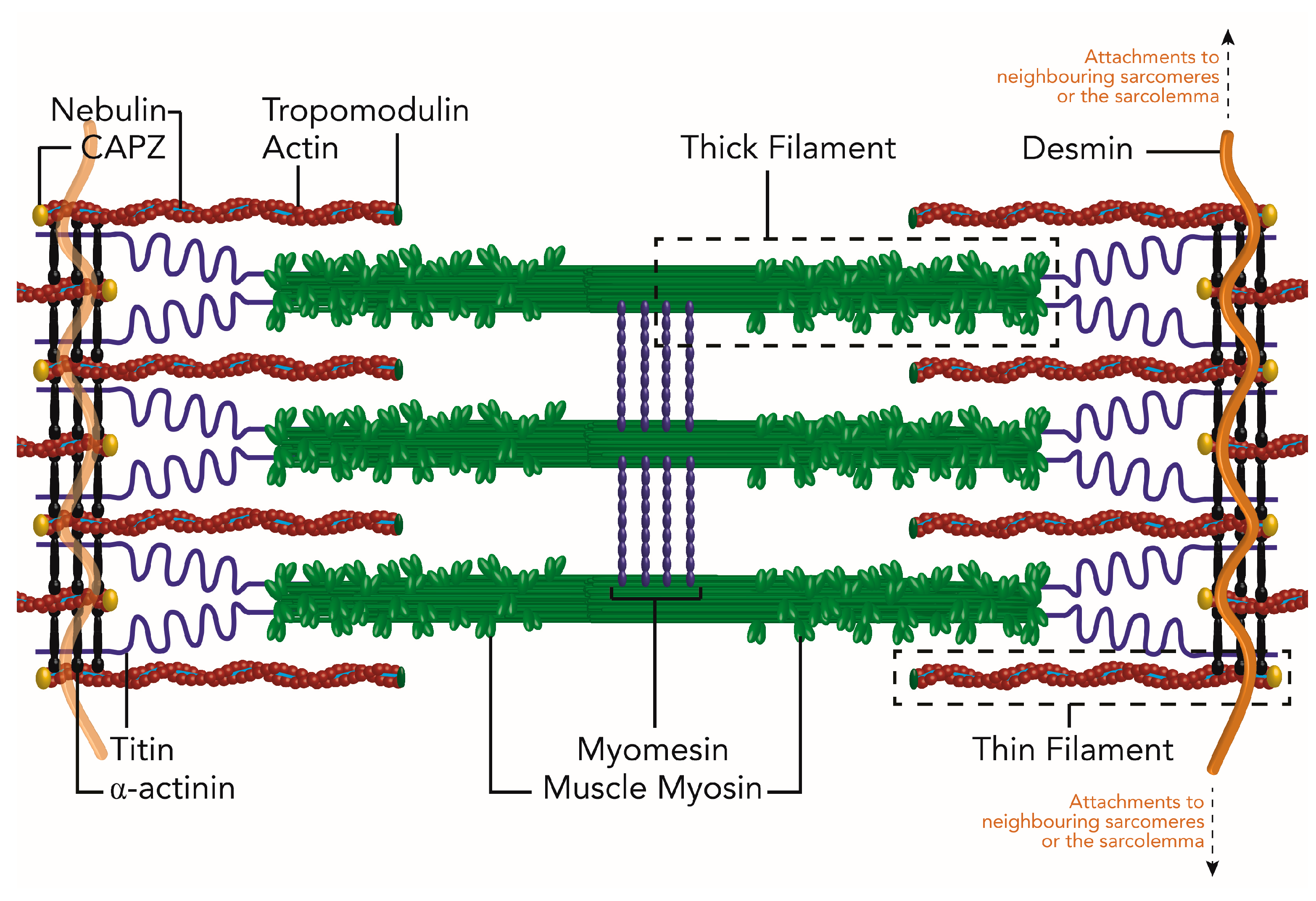
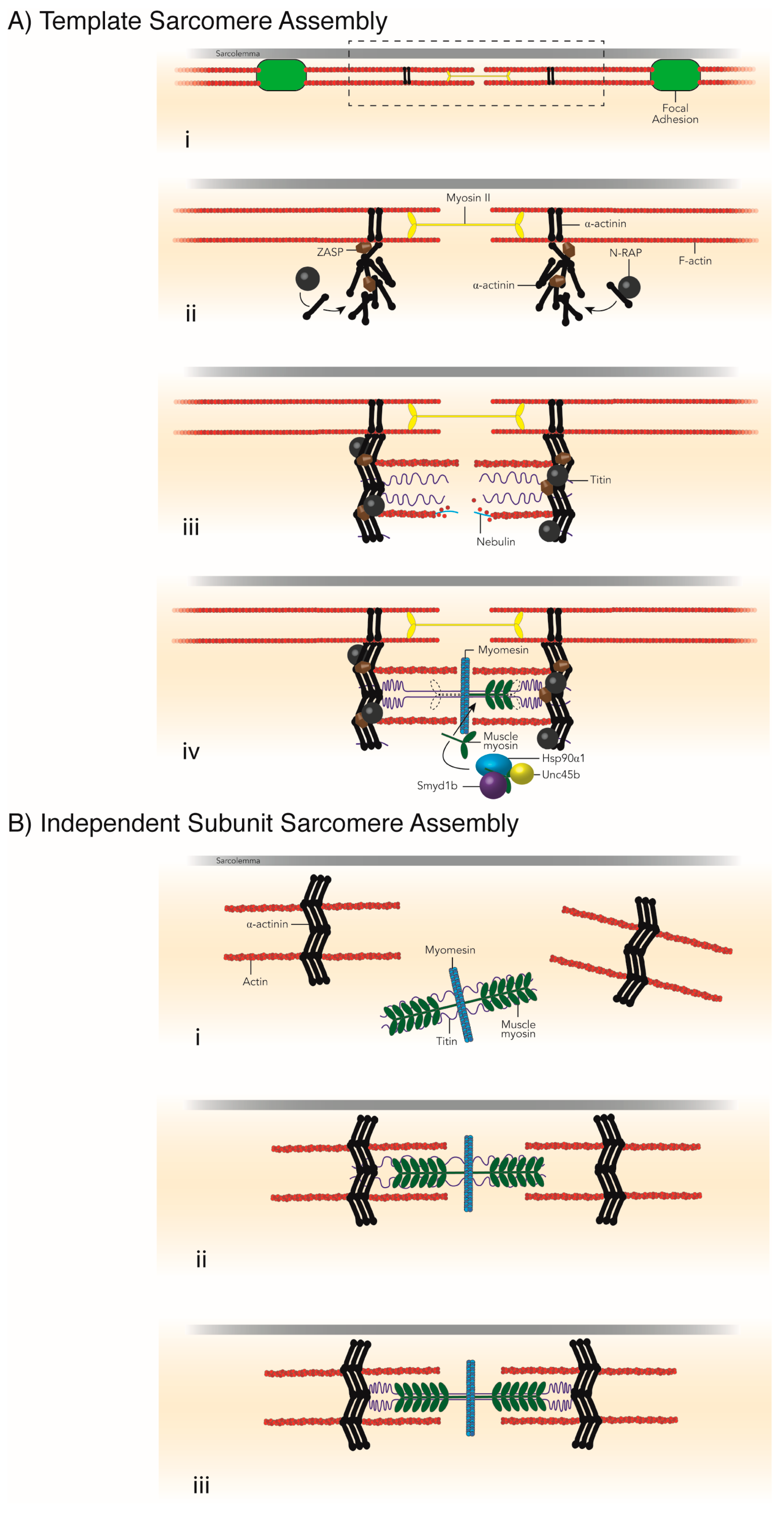
3. Necessity of Chaperones for Sarcomere Assembly

Of the various models of sarcomere assembly, the premyofibril model has the most supporting evidence both in vitro [47] and in vivo [63]. For an excellent review detailing these various models and the evidence for and against each, see [64]. In the premyofibril model of sarcomere assembly, α-actinin z-bodies and non-muscle myosin filaments form immature pre-sarcomeres, or premyofibrils (Figure 3) [64,65,66]. Incorporation of titin, muscle myosin and proteins of the M-line, mark the transition from premyofibrils to nascent and finally mature myofibrils as Z-discs, A-bands and M-lines are completed and the sarcomeres reach their final length and width. In addition to the evidence that supports the premyofibril model, this model is attractive as it is consistent with the idea that other factors are necessary for the growth and maturation of the sarcomere. This is in contrast to other models of sarcomere assembly, notably the Template [47,67,68,69] and Independent Subunit [47,69,70] sarcomere assembly models (Figure 4). Respectively, these models suggest sarcomere formation occurs along alpha-actinin, tropononin, tropomodulin and tropomyosin stress fiber templates [67] (Figure 4A), or form independently and are joined together along the length of titin [70,71] (Figure 4B). Neither, however account for the evidence that suggests that many sarcomere proteins themselves, such as myosin, a-actinin and the protein giant titin, cannot reach their native conformation without the assistance of chaperones. Furthermore, structures of the sarcomere require chaperones to help link components together, such as thin filament anchorage to the Z-disc or thick filament attachment to the M-line [72,73]. In this section, we will focus on the chaperones required for sarcomere protein folding and assembly of sarcomere structures.

3.1. Z-Disc Assembly

The assembly of the Z-disc occurs after the recruitment of talin, viniculin and ZASP to integrin at the muscle cell membrane [74,75,76,77,78]. Alpha-actinin localization requires ZASP and its organization into premature Z-discs, or Z-bodies, depends on N-RAP [35,79]. At present, no factor has been identified that is necessary for folding α-actinin. N-RAP binds α-actinin in the Z-disc and acts as a scaffold for thin filament assembly, acting as a bridge between Z-discs and thin filaments to help organize the I-Z-I structures of the sarcomere (Figure 3C–E) [72]. After the initial assembly of the sarcomere, desmin is incorporated into the Z-disc, by the molecular chaperone, αβ-crystallin, in order to attach sarcomeres of parallel myofibrils, anchor sarcomeres to the sarcolemma and maintain the structure and rigidity of the contractile unit [80,81].

3.2. Thin Filament Formation

Following Z-disc formation, the thin filaments begin to assemble along the nebulin scaffold that extends from the Z-disc toward the sarcomere center field [82,83]. The actin co-chaperone, GimC, binds actin as it is synthesized and prevents actin from aggregating in the cytoplasm. GimC then passes actin on to TriC, which completes actin folding and thin filament assembly (Figure 3E). Leiomodin 2 is important for elongating thin filaments to reach their mature length during sarcomere assembly [46]. Other chaperones, such as αβ-crystallin, associate with actin thin filaments but the function of this association is not well understood.

3.3. Non-Muscle Myosin Filaments

Non-muscle myosin II (NmmII) is incorporated into the developing myofibril between premature Z-discs simultaneously or immediately after Z-body formation and the beginning of thin filament assembly [47]. It has been suggested that NmmII aids in the alignment and assembly of actin thin filaments within the sarcomere but how NmmII integrates into the sarcomere is not understood [47,67,70,84,85]. Studies of NmmII are limited due to antibody specificity and the abundance of non-muscle myosins in every cell. Unc45b is a promising candidate as an NmmII chaperone as UNC45 in worms associates with NMM [86]. Non-muscle myosin II is replaced by muscle myosins, which may require an assembly chaperone or a member of the UPS system to target and remove NmmII from the sarcomere so that myosin thick filaments can be integrated into the sarcomere instead (Figure 3F,G).

3.4. Titin Folding and Incorporation

Titin, the largest known protein, stretches from Z-disc to the M-line and maintains sarcomere length. Due to its size, it is likely that titin has a host of assembly chaperones but the only chaperone required for titin filament assembly to date is αβ-crystallin [87,88]. Other chaperones, such as Hsp90α1, in addition to αβ-crystallin, localize to titin to help maintain titin integrity throughout sarcomere formation and muscle development [89,90]. It is likely that titin acts as a signaling hub of the sarcomere, with important roles in both sarcomere assembly and maintenance pathways, as well as serving as a platform for other chaperones involved in these roles [91].

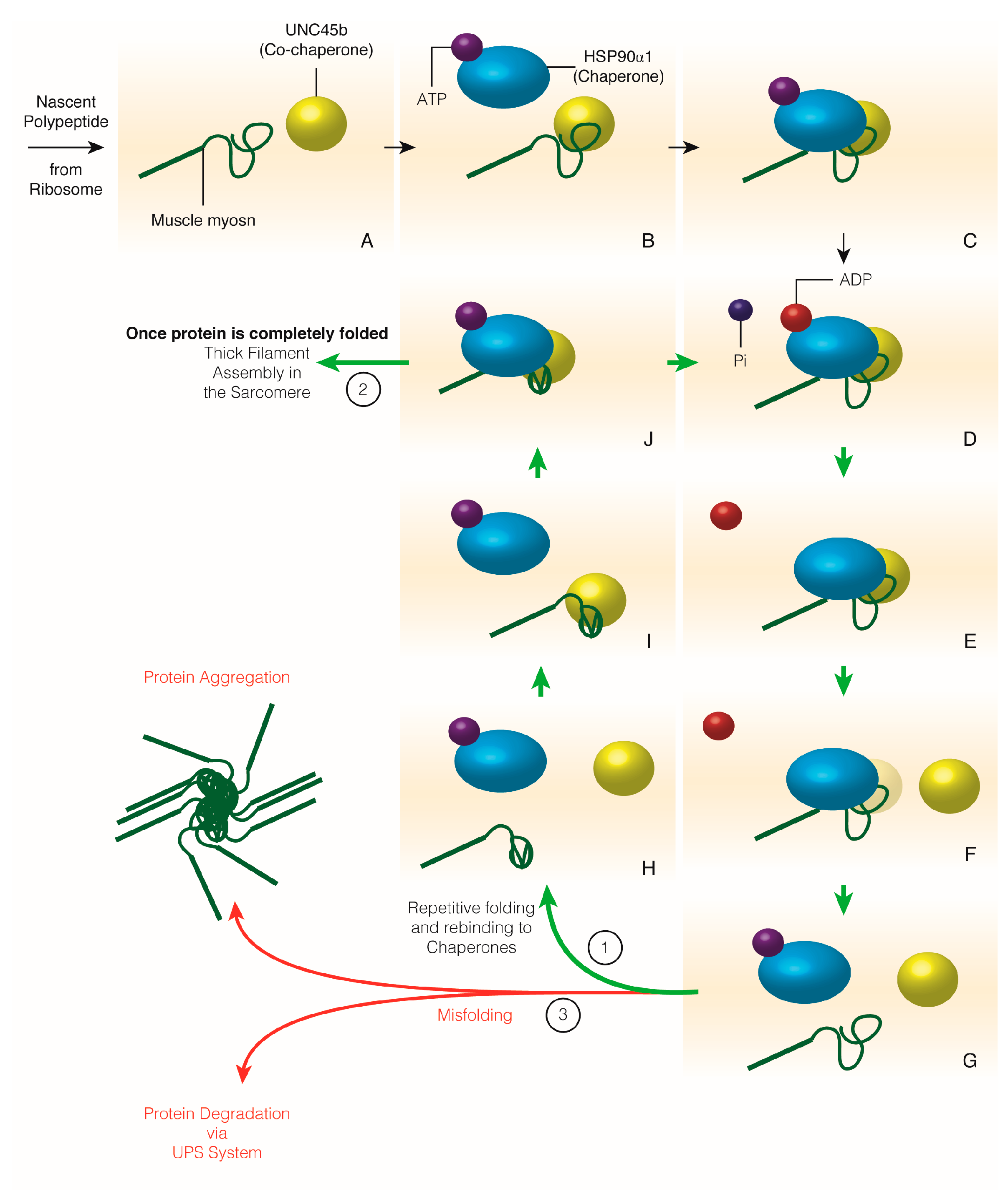
3.5. Muscle Myosin Folding and Thick Filament Assembly

The abundant sarcomere protein, muscle myosin II has provided a target for elucidating the roles and coordination of molecular chaperones and the Ubiquitin Proteasome System. Many chaperones and co-chaperones are required to incorporate active myosin into the sarcomere [92,93,94,95,96]. Hsp40 is one of the first co-chaperones to bind myosin II and recruit Hsp70 [97,98]. Hsp90α1 and its co-chaperone, Unc45b, bind the hydrophobic amino acids of the myosin globular head domain and prevent it from misfolding [96,99,100]. Myosin II tails are capable of spontaneous dimerization but this dimerization appears to be timed for construction of the antiparallel thick filaments [99]. Once the myosin globular domain is folded, myosin heavy chains are organized into the hexamers of the thick filaments and anchored by the M-line (Figure 3G), replacing non-muscle myosins in the sarcomere. The HSP family are a major class of chaperones but they do not encompass all factors required for protein folding and assembly. For instance, Smyd1b (which has the protein structure of a histone methyltransferase and a myosin-binding domain) is also required for myosin thick filament assembly in the developing myofibril by making physical contacts with myosin, Unc45b and Hsp90α1 [73,95,101]. Whether Smyd1b is necessary for folding or incorporation, or both, has yet to be determined. Smyd1b may have a similar role to a co-chaperone as it binds skeletal muscle nascent polypeptide associated complex, or skNAC, which is found on ribosomes to bind newly synthesized proteins. It could be that skNAC recruits Smyd1b to myosin and this subsequently recruits Unc45b and Hsp90α1 respectively. The combination of these chaperones varies in the type of muscle tissue; Hsp90α1 is not required for cardiac myosin folding and Smyd1b is not required for slow myosin thick filament assembly [73,94,95,101,102,103].

3.6. M-Line Assembly

The assembly of the protein complexes that make up the M-line of the sarcomere is not well understood. No chaperones have yet been identified for 4 of the major components of the M-line: the C-terminus of titin, myomesin, obscurin and obscurin-like 1 [104,105,106,107]. The coiled tails of muscle myosins are part of the M-line acting as anchorage points for the thick filaments of the sarcomere. The C-terminus of titin recruits myomesin to the M-line and then localizes obscurin and obscurin-like 1 [105]. Similar to the localization of Z-disc components by assembly chaperones, the recruitment and incorporation of M-line proteins could be due to unidentified assembly chaperones. It is likely, based on the size and complexity of the M-line in comparison to other sarcomere structures/regions, that the M-line complexes require aid in protein folding and assembly.
This section covered the formation of the sarcomere with respect to chaperones and co-chaperones that are required for protein folding and assembly of the sarcomere. These assembly factors are necessary for the formation of the sarcomere and mutants of these factors result in various myopathies with many degrees of severity and onset [95,108,109]. It should be noted, that several myopathies result from mutations of sarcomere structural components and these mutations can have a wide effect on the function or stability of other sarcomere proteins (e.g., titin) [110].

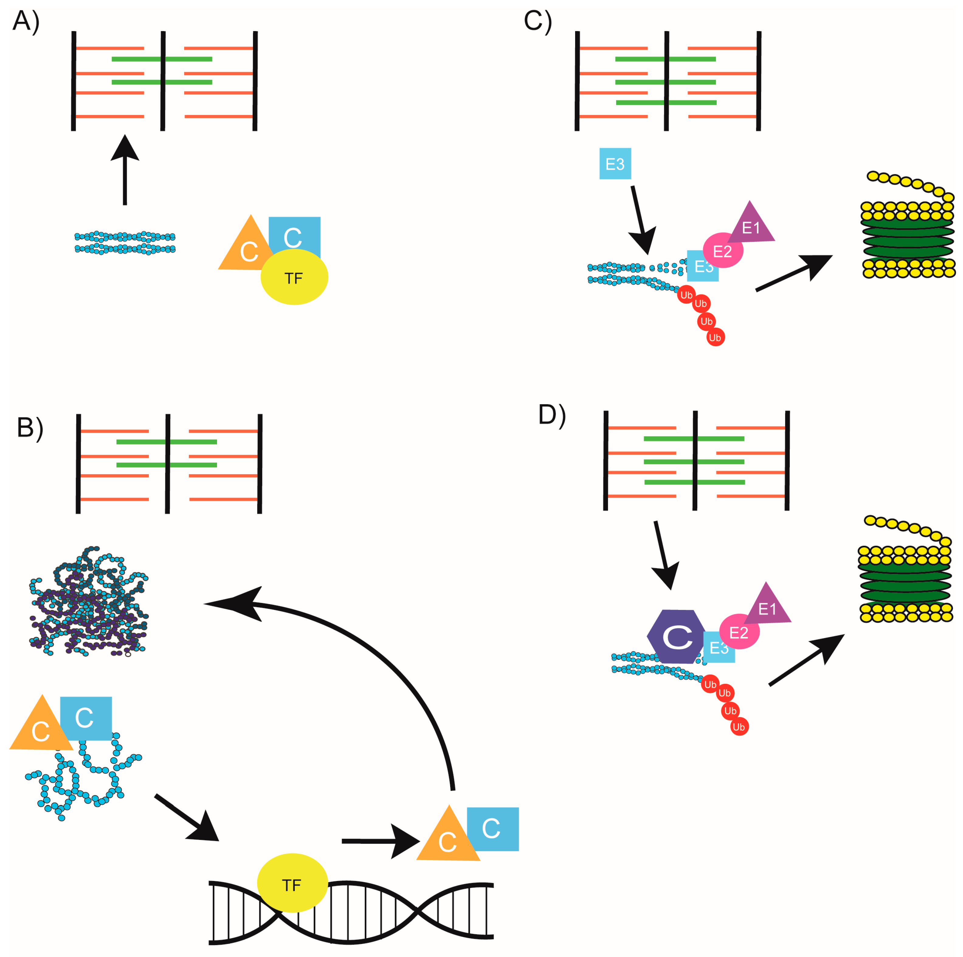
4. Chaperone Homeostasis

The above sections discussed the overwhelming evidence that specific chaperones are required for proper sarcomere assembly and therefore survival of the organism. However, it is less intuitive but possibly equally important, that chaperone expression is regulated, as an overabundance of a particular chaperone can cause equally severe muscle defects. C. elegans UNC-45, zebrafish Unc45b and human skeletal muscle Unc45 (SM-Unc45) have been shown to cause muscle disorganization when overexpressed either transgenically or by blocking its degradation [15,111,112]. This suggests a model where chaperone homeostasis is a highly regulated process. In invertebrates, a mechanism for Unc45 removal has been elucidated by which Unc45, UFD2, CDC-48 and CHN-1 (C. elegans orthologue of CHIP [carboxyl terminus of Hsc70 interacting protein]) form a complex that is able to polyubiquitinate Unc45 [15,112]. The integrity of this system is important in humans as well, as mutations in p97 (human CDC-48), cause Inclusion Body Myopathy, which is associated with Paget disease of bone and Frontotemporal Dementia (IBMPFD). IBMPFD results in sarcomere assembly defects, which is attributed to excess SM-Unc45 [15]. While the mechanisms behind the sarcomere defects in Unc45/Unc45b/SM-Unc45 overexpression models are still being uncovered, some suggestions have been put forth. Willis and colleagues (2009) suggest that these defects result from too many Unc45 molecules attached to myosin, which (a) impedes sarcomere assembly and (b) causes the formation of aggregates [113]. Other models suggest that Unc45 overexpression may cause the sarcomere to assemble incorrectly, which is later recognized and disassembled [92].
Although Unc45 is known to work with Hsp90 and Smyd1b to fold myosin [95,101,114], no similar complexes have yet been reported for control of Hsp90 or Smyd1b turnover. As no data yet exists for Smyd1b overexpression, it is unsurprising that no complex regulating Smyd1b expression has yet been discovered. Importantly, the sarcomere defects associated with excess Unc45/Unc45b/SM-Unc45 also appear to proceed through an Hsp90 independent pathway [111]. While it does appear that Hsp90a/b isoform homeostasis is important for proper muscle specification [115], no consequences to sarcomere assembly have been described to date when Hsp90 is overexpressed in muscle [111]. This could be due to the general expression of Hsp90 such that it requires Unc45 cooperation in striated muscle, or it may indicate that the myosin chaperone actions of Hsp90 and Unc45 differ enough that overexpression of one is detrimental but the other is benign, such that only homeostasis of Unc45 is tightly regulated.

5. Chaperones beyond Assembly

The term “molecular chaperone” was first coined after observations that certain proteins required additional factors to facilitate proper binding and prevent precipitation [116]. The role of chaperones was later extended to include binding and folding client proteins after their synthesis, preventing the aggregation of misfolded proteins and aiding in the formation of macromolecular structures [117]. Recently, connections between chaperones and protein degradation have been established [55], linking chaperones with both autophagy and the UPS.
Chaperone-Mediated Autophagy (CMA) and Chaperone-Assisted Selective Autophagy (CASA) connect the chaperone system to protein quality control. CMA involves the delivery of a chaperone substrate across a lysosomal membrane in a ubiquitin-independent manner [118] and although Hsp70 is involved in this process [119], CMA has not been widely explored in skeletal muscle [42]. CASA involves the ubiquitin dependent degradation of a chaperone substrate by the lysosome [120]. Unlike CMA, there is strong evidence for CASA in skeletal muscle [42]. CASA was first described in the Drosophila Z-disc as a mechanism by which Starvin (Stv; Drosophila Bag-3) mediates the autophagic turnover of filamin in a CHIP ubiquitination dependent manner [120]. It is likely that this role of Bag-3 (Bcl-2-associated athanogene-3) is conserved in mammals as well, as Bag-3 homozygous null mice display normal Z-disc assembly, which deteriorates over time and results in eventual apoptosis [121]. Bag-3 also appears to be important in humans, with a severe muscular dystrophy linked to loss of Bag-3 [25]. Because Bag-3 is considered to be an Hsp70 (or ubiquitous Hsc70) co-chaperone [122,123] along with its many other proposed roles [123] the role of Hsp70 in muscle maintenance becomes an important and currently under-investigated question.
Evidence for chaperone and UPS collaboration comes from Chaperone-Assisted Proteasomal degradation (CAP), which describes the process by which a chaperone client is ubiquitinated and subsequently degraded by the proteasome [55,124,125]. Evidence for CAP comes from the Hsc/Hsp70 co-chaperone Bag-1, which associates with the 26S proteasome and has been proposed to physically link chaperone and proteasome systems [126]. Bag-1 has been shown to interact with CHIP [127], which provides a mechanism by which chaperone substrates could be ubiquitinated and targeted directly to the proteasome. Although CHIP is the only E3 ligase identified so far that binds to this complex, there are likely other E3 ligases involved in this role as CHIP knockout mice do not have severe muscle defects [128]. While it is very likely that this process is abundant in skeletal muscle, it has not been widely studied to date.
Together, the collaboration of chaperones with both autophagic and proteasome degradation systems suggests a hypothesis by which chaperones respond to their client protein when damaged regardless of when the damage occurs. As many chaperones remain associated with their client proteins or are localized on nearby sarcomere structures after sarcomere assembly, this hypothesis makes sense. However, recent evidence that myosin damage may occur without a subsequent upregulated response of myosin chaperones contradicts this model [61]. Differing from zebrafish with chaperone loss of function mutations—steif (Unc45b, [61] and still heart (smyd1b; [95], zebrafish with mutations outside the chaperone pathway but with similar muscle disorganization do not display an upregulation in myosin chaperones [61]. The zebrafish mutation, sofa potato (sopfixe), affects an acetylcholine receptor, which results in grossly normal muscle ultrastructure but smaller myofibrils and disorganized slow myosin [129]. Ache has a mutation in acetyl cholinesterase, which results in gradual disorganization of myofibrils and a loss of slow myosin over time [130]. Finally, herzschlag (hel) mutants carry a mutation somewhere between the I-band and A-band in the titin paralog ttna (ttn2; [131]). This mutation results in initially normal sarcomere assembly, which degenerates over time [131]. The absence of myosin chaperone upregulation in these mutants despite the myosin disorganization they display suggests that myosin chaperones are not directly responding to the disorganized protein itself [61]. Instead, it appears that Hsf-1 is responsible for inducing the myosin chaperone response [61]. However, it is still unclear why these chaperones do not react in sop, ache, or hel embryos. One difference between these zebrafish and those with mutations in myosin chaperones is that the initial folding and assembly of myosin into the sarcomere is unimpeded. The difference in myosin chaperone response is potentially due to the stage in which damage occurs, with myosin chaperones responding to initial folding and assembly defects through an Hsf-1 mediated response (Figure 5A,B). Should damage occur post assembly due to normal wear and tear, an unc45b/hsp90/smyd1b/hsf-1 independent response to myosin occurs. (Figure 5C,D) Whether this response is entirely chaperone independent, or involves a subset of muscle maintenance chaperones

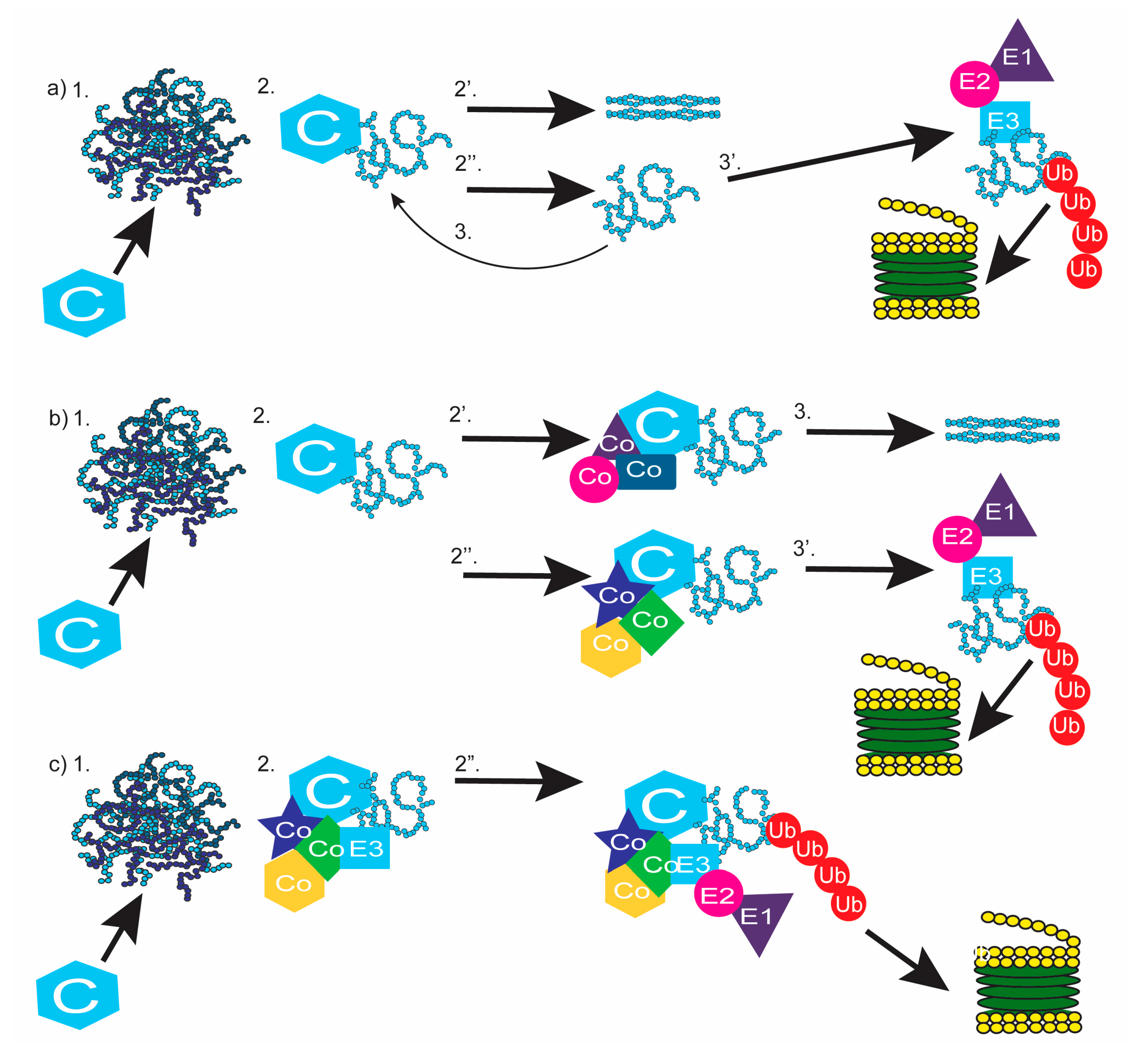
6. Completing the Chain: Communication between Chaperones and the Proteasome

Although the misfolded protein response is still being uncovered, the above section discussed the increasing evidence that chaperones and the UPS cooperate throughout this process. Chaperones and the proteasome system are also linked by the discovery of the E3 ligase, CHIP, that can physically bind both Hsp70 [132] and Hsp90 [133] and is associated with the proteasome [133,134]. Furthermore, when Hsp90 is prevented from folding a client protein, the client protein is degraded by the proteasome [133]. Together, this strongly suggests that chaperones have roles beyond initial protein folding and link protein folding and protein degradation. Therefore, when a chaperone encounters a misfolded client protein, a decision must be made to either refold or degrade the protein. These decisions are referred to as protein triage decisions [135,136].
How does the UPS recognize misfolded proteins? Many models have been presented [19] ranging from chaperones and the UPS existing as separate, competing, entities [135,136], to their coordination such that E3 enzymes deliver chaperone complexes and their ubiquitinated substrate, to the proteasome [124] (Figure 6). The simplest model of protein turnover is the “kinetic model of protein triage” which describes chaperones and the UPS as competing entities [135,136] (Figure 6A). In this model, chaperones respond to their misfolded client protein, likely recognizing exposed hydrophobic domains and attempt to refold it to its proper conformation. A misfolded protein that is not successfully refolded may be bound again by its chaperone, or by an E3 enzyme that also recognizes exposed hydrophobic domains [19]. In this model, whether a chaperone or an E3 enzyme binds the misfolded protein, is random [135,136], however concentrations of chaperones or E3 enzymes and damage to the client protein, would play an important role in which outcome is favored. If the misfolded protein is bound by its chaperone, the attempt to refolded cycle continues. If an E3 enzyme binds the misfolded protein, it is targeted for degradation.
An alternate model of protein turnover suggests that when a chaperone binds a misfolded client protein, it is the recruitment of specific co-chaperones that determines whether the client protein will be refolded or degraded [19,55]; (Figure 6B). Evidence for this model is abundant, mainly centers on the chaperones Hsp70 and Hsp90 and their substrates [19,55]. Co-factors Hip and Hop binding Hsp90 or Hsp70 has been shown to promote substrate folding [20,133,137], while binding of CHIP or Bag-1 promote substrate degradation [126,133,138,139] (For a schematic of Hsp/Hsc70 and Hsp90 associated co-chaperones and the refold/degradation consequence of the substrate, [55]). A lingering question remains as to how the appropriate co-chaperones are recruited. Like the previous model, refolding or degradation of the client protein likely depends on the initial damage to the protein as well as the presence and concentrations of its chaperone and co-chaperones. With all components being equal, this process will be stochastic [135]. In support of this, the addition of CHIP to lysates containing HOP and Hsp90 significantly decreases the number of HOP/Hsp90 complexes [133], suggesting that HOP can be outcompeted by CHIP. Furthermore, CHIP and HOP appear to bind the same location of Hsp70/Hsp90 [133] as do Bag-1 and HIP, which sterically hinder the binding of its competitor [55,140].
A third model of protein turnover suggests that the binding of a co-factor such as CHIP results in the transformation of a chaperone/client complex into a degradation complex, which is then transported to the proteasome [19,124,139]. Rather than CHIP and Bag-1 being thought of as co-factors in this model, these proteins and the chaperones they bind form a complex that is thought of as one big E3 ligase [124]. Substrate specificity will come from the chaperone’s affinity for their client [49], CHIP ubiquitinates proteins in the complex and then the complex is transported to and docked on the 26S proteasome by Bag-1 [126]; Figure 6C. This model differs from the “cofactor” model by the conceptualization of the entire complex as an active E3 ligase. Although these models are presented separately, they do not have to be mutually exclusive and it is feasible that certain co-factors promote folding, while others promote the transformation of the chaperone/substrate complex into a degradation complex. The binding and releasing of the folding co-factors or the degradation complex would be subject to competition, as outlined in the “kinetic model” making the best model of protein triage likely a combination of all three.
Much of the work done to formulate the models described above have been done in yeast, bacteria, or in vitro. Although these models have not been extensively studied in striated muscle, or in organisms containing striated muscle, it is possible that they could apply to this tissue as well. Further examination into these pathways in striated muscle could elucidate previously ignored treatments for myopathies listed in Table 1 and are therefore worth exploring. However, due to the tightly organized structure of the sarcomere, it is likely that an additional component in muscle is required to sense and report sarcomere damage. The protein titin, which stretches one-half sarcomere from Z-disc to M-line has been associated with sarcomere monitoring functions [91,141] and is a good candidate for regulating proteostasis. Links between titin and protein turnover have been established with the C terminal titin- kinase domain shown to be a binding site of the muscle specific ubiquitin ligases, MuRF1 [142], MuRF2 [143], as well as the protease Calpain 3 [26,144,145]. One role of this titin-kinase domain is to regulate gene expression in a contraction dependent manner, with MuRF2 and SRF translocating to the nucleus during an absence of contractions and regulating expression of genes such as myomesin [143]. However, as this domain also behaves as a reservoir for proteins involved in muscle maintenance, there is an obvious connection between titin and protein turnover that has yet to be fully elucidated.
MuRF2 belongs to the RING finger family of ubiquitin ligases and is closely related to MuRF1 [146], which has been shown to ubiquitin skeletal muscle myosin [147,148]. MuRF1 knockout mice are resistance to muscle atrophy [149], which is attributed to its RING domain [150] but are otherwise healthy and indistinguishable from wildtype animals [151]. Likewise, mouse knockout models of a third member of the MuRF family, MuRF3 do not display any abnormalities from wildtype animals, unless subjected to myocardial infarctions [148]. However, double-knockout (MuRF1−/−;MuRF3−/−) mice display a skeletal and cardiac muscle myopathy presenting similarly to human Myosin Storage Myopathies [148], which coupled with similar binding targets, [152] suggests that some functional redundancy exists between MuRF family members. As two members of this family are associated with titin [142,143], it is possible that titin regulates sarcomere protein turnover by docking ubiquitin ligases when muscle is healthy and releasing them when paralyzed [143]. This would constitute a chaperone independent response to sarcomeric damage, explaining the absence of myosin chaperone upregulation in zebrafish mutants with muscle damage [61]. However, it is impossible to eliminate the idea of a maintenance specific chaperone with this data alone and it is likely that both chaperone dependent and chaperone independent quality control systems exist together in striated muscle. We suspect that further research into the role of Hsp70 in muscle maintenance could fill in this gap. Likewise, “reporter” genes similar to titin could exist all over the sarcomere, including calsarcin at the Z-disc and myomesin and Mef2C at the M-line. How these systems work together and the signal transduction pathways regulating them will be an imperative focus of future muscle research.

7. Summary

Although the importance of protein turnover to muscle assembly and maintenance has become clear over the last few decades, the mechanisms and signaling pathways involved in this process still remain elusive. Both chaperone dependent and chaperone independent responses to protein damage have been identified but neither studied extensively in striated muscle. The onset and severity of the physiological and health consequences that occur when muscle proteostasis is interrupted (see Table 1), demands that understanding these processes are paramount to developing effective treatments and cures.

Acknowledgments

This work was supported by research funding from the Natural Sciences and Engineering Research Council of Canada.

Author Contributions

Casey Carlisle and Kendal Prill contributed equally to this paper.

Conflicts of Interest

The authors declare no conflict of interest.

References

  1. Bianchi, T.; Gelosa, L. On the preventive detection of pathogenic staphylococci in the rhinopharynx of employees in the food industry in the province of Milan during the 5-year period of 1967–1971. Ann. Sclavo 1973, 15, 83–98. [Google Scholar] [PubMed]
  2. Van den Berg, B.; Wain, R.; Dobson, C.M.; Ellis, R.J. Macromolecular crowding perturbs protein refolding kinetics: Implications for folding inside the cell. EMBO J. 2000, 19, 3870–3875. [Google Scholar] [CrossRef] [PubMed]
  3. Rose, G.D.; Fleming, P.J.; Banavar, J.R.; Maritan, A. A backbone-based theory of protein folding. Proc. Natl. Acad. Sci. USA 2006, 103, 16623–16633. [Google Scholar] [CrossRef] [PubMed]
  4. Kony, D.B.; Hunenberger, P.H.; van Gunsteren, W.F. Molecular dynamics simulations of the native and partially folded states of ubiquitin: Influence of methanol cosolvent, pH, and temperature on the protein structure and dynamics. Protein Sci. 2007, 16, 1101–1118. [Google Scholar] [CrossRef] [PubMed]
  5. Schubert, U.; Antón, L.C.; Gibbs, J.; Norbury, C.C.; Yewdell, J.W.; Bennink, J.R. Rapid degradation of a large fraction of newly synthesized proteins by proteasomes. Nature 2000, 404, 770–774. [Google Scholar] [CrossRef] [PubMed]
  6. Epstein, C.J.; Goldberger, R.F.; Anfinsen, C.B. The genetic control of tertiary protein structure: Studies with model systems. Cold Spring Harb. Symp. Quant. Biol. 1963, 28, 439–449. [Google Scholar] [CrossRef]
  7. Frydman, J.; Nimmesgern, E.; Ohtsuka, K.; Hartl, F.U. Folding of nascent polypeptide chains in a high molecular mass assembly with molecular chaperones. Nature 1994, 370, 111–117. [Google Scholar] [CrossRef] [PubMed]
  8. Hendrick, J.P.; Hartl, F.U. Molecular chaperone functions of heat-shock proteins. Ann. Rev. Biochem. 1993, 62, 349–384. [Google Scholar] [CrossRef] [PubMed]
  9. Ellis, R.J.; Hemmingsen, S.M. Molecular chaperones: Proteins essential for the biogenesis of some macromolecular structures. Trends Biochem. Sci. 1989, 14, 339–342. [Google Scholar] [CrossRef]
  10. Malicdan, M.C.; Noguchi, S.; Nonaka, I.; Saftig, P.; Nishino, I. Lysosomal myopathies: An excessive build-up in autophagosomes is too much to handle. Neuromuscul. Disord. 2008, 18, 521–529. [Google Scholar] [CrossRef] [PubMed]
  11. Masiero, E.; Sandri, M. Autophagy inhibition induces atrophy and myopathy in adult skeletal muscles. Autophagy 2010, 6, 307–309. [Google Scholar] [CrossRef] [PubMed]
  12. Nishino, I. Autophagic vacuolar myopathy. Semin. Pediatr. Neurol. 2006, 13, 90–95. [Google Scholar] [CrossRef] [PubMed]
  13. Paul, S. Dysfunction of the ubiquitin-proteasome system in multiple disease conditions: Therapeutic approaches. BioEssays 2008, 30, 1172–1184. [Google Scholar] [CrossRef] [PubMed]
  14. Aguzzi, A.; O’Connor, T. Protein aggregation diseases: Pathogenicity and therapeutic perspectives. Nat. Rev. Drug Discov. 2010, 9, 237–248. [Google Scholar] [CrossRef] [PubMed]
  15. Janiesch, P.C.; Kim, J.; Mouysset, J.; Barikbin, R.; Lochmuller, H.; Cassata, G.; Krause, S.; Hoppe, T. The ubiquitin-selective chaperone CDC-48/p97 links myosin assembly to human myopathy. Nat. Cell Biol. 2007, 9, 379–390. [Google Scholar] [CrossRef] [PubMed]
  16. Sharma, M.C.; Goebel, H.H. Protein aggregate myopathies. Neurol. India 2005, 53, 273–279. [Google Scholar] [PubMed]
  17. Askanas, V.; Engel, W.K.; Nogalska, A. Inclusion body myositis: A degenerative muscle disease associated with intra-muscle fiber multi-protein aggregates, proteasome inhibition, endoplasmic reticulum stress and decreased lysosomal degradation. Brain Pathol. 2009, 19, 493–506. [Google Scholar] [CrossRef] [PubMed]
  18. Garlepp, M.J.; Mastaglia, F.L. Inclusion body myositis. J. Neurol. Neurosurg. Psychiatry 1996, 60, 251–255. [Google Scholar] [CrossRef] [PubMed]
  19. Esser, C.; Alberti, S.; Hohfeld, J. Cooperation of molecular chaperones with the ubiquitin/proteasome system. Biochim. Biophys. Acta 2004, 1695, 171–188. [Google Scholar] [CrossRef] [PubMed]
  20. Nollen, E.A.; Kabakov, A.E.; Brunsting, J.F.; Kanon, B.; Hohfeld, J.; Kampinga, H.H. Modulation of in vivo HSP70 chaperone activity by Hip and Bag-1. J. Biol. Chem. 2001, 276, 4677–4682. [Google Scholar] [CrossRef] [PubMed]
  21. Abdulhaq, U.N.; Daana, M.; Dor, T.; Fellig, Y.; Eylon, S.; Schuelke, M.; Shaag, A.; Elpeleg, O.; Edvardson, S. Nemaline body myopathy caused by a novel mutation in troponin T1 (TNNT1). Muscle Nerve 2016, 53, 564–569. [Google Scholar] [CrossRef] [PubMed]
  22. Friedman, B.; Simpson, K.; Tesi-Rocha, C.; Zhou, D.; Palmer, C.A.; Suchy, S.F. Novel large deletion in the ACTA1 gene in a child with autosomal recessive nemaline myopathy. Neuromuscul. Disord. 2014, 24, 331–334. [Google Scholar] [CrossRef] [PubMed]
  23. Ilkovski, B.; Cooper, S.T.; Nowak, K.; Ryan, M.M.; Yang, N.; Schnell, C.; Durling, H.J.; Roddick, L.G.; Wilkinson, I.; Kornberg, A.J.; et al. Nemaline myopathy caused by mutations in the muscle alpha-skeletal-actin gene. Am. J. Hum. Genet. 2001, 68, 1333–1343. [Google Scholar] [CrossRef] [PubMed]
  24. Wallefeld, W.; Krause, S.; Nowak, K.J.; Dye, D.; Horvath, R.; Molnar, Z.; Szabo, M.; Hashimoto, K.; Reina, C.; De Carlos, J.; et al. Severe nemaline myopathy caused by mutations of the stop codon of the skeletal muscle alpha actin gene (ACTA1). Neuromuscul. Disord. 2006, 16, 541–547. [Google Scholar] [CrossRef] [PubMed]
  25. Selcen, D.; Muntoni, F.; Burton, B.K.; Md, E.P.; Sewry, C.; Bite, A.V.; Engel, A.G. Mutation in BAG3 Causes Severe Dominant Childhood Muscular Dystrophy. Ann. Neurol. 2009, 65, 83–89. [Google Scholar] [CrossRef] [PubMed]
  26. Keira, Y.; Noguchi, S.; Minami, N.; Hayashi, Y.K.; Nishino, I. Localization of Calpain 3 in Human Skeletal Muscle and Its Alteration in Limb-Girdle Muscular Dystrophy 2A Muscle. J. Biochem. 2003, 133, 659–664. [Google Scholar] [CrossRef] [PubMed]
  27. Garvey, S.M.; Rajan, C.; Lerner, A.P.; Frankel, W.N.; Cox, G.A. The muscular dystrophy with myositis (mdm) mouse mutation disrupts a skeletal muscle-specific domain of titin. Genomics 2002, 79, 146–149. [Google Scholar] [CrossRef] [PubMed]
  28. Nigro, V.; Savarese, M. Genetic basis of limb-girdle muscular dystrophies: The 2014 update. Acta Myol. 2014, 33, 1–12. [Google Scholar] [PubMed]
  29. Hackman, P.; Marchand, S.; Sarparanta, J.; Vihola, A.; Penisson-Besnier, I.; Eymard, B.; Pardal-Fernandez, J.M.; Hammouda, E.-H.; Richard, I.; Illa, I.; et al. Truncating mutations in C-terminal titin may cause more severe tibial muscular dystrophy (TMD). Neuromuscul. Disord. 2008, 18, 922–928. [Google Scholar] [CrossRef] [PubMed]
  30. Hackman, P.; Vihola, A.; Haravuori, H.; Marchand, S.; Sarparanta, J.; De Seze, J.; Labeit, S.; Witt, C.; Peltonen, L.; Richard, I.; et al. Tibial muscular dystrophy is a titinopathy caused by mutations in TTN, the gene encoding the giant skeletal-muscle protein titin. Am. J. Hum. Genet. 2002, 71, 492–500. [Google Scholar] [CrossRef] [PubMed]
  31. Sarparanta, J.; Blandin, G.; Charton, K.; Vihola, A.; Marchand, S.; Milic, A.; Hackman, P.; Ehler, E.; Richard, I.; Udd, B. Interactions with M-band titin and calpain 3 link myospryn (CMYA5) to tibial and limb-girdle muscular dystrophies. J. Biol. Chem. 2010, 285, 30304–30315. [Google Scholar] [CrossRef] [PubMed]
  32. Herman, D.S.; Lam, L.; Taylor, M.R.; Wang, L.; Teekakirikul, P.; Christodoulou, D.; Conner, L.; DePalma, S.R.; McDonough, B.; Sparks, E.; et al. Truncations of titin causing dilated cardiomyopathy. N. Engl. J. Med. 2012, 366, 619–628. [Google Scholar] [CrossRef] [PubMed]
  33. Gerull, B.; Gramlich, M.; Atherton, J.; McNabb, M.; Trombitas, K.; Sasse-Klaassen, S.; Seidman, J.G.; Seidman, C.; Granzier, H.; Labeit, S.; et al. Mutations of TTN, encoding the giant muscle filament titin, cause familial dilated cardiomyopathy. Nat. Genet. 2002, 30, 201–204. [Google Scholar] [CrossRef] [PubMed]
  34. Thottakara, T.; Friedrich, F.W.; Reischmann, S.; Braumann, S.; Schlossarek, S.; Kramer, E.; Juhr, D.; Schluter, H.; van der Velden, J.; Munch, J.; et al. The E3 ubiquitin ligase Asb2beta is downregulated in a mouse model of hypertrophic cardiomyopathy and targets desmin for proteasomal degradation. J. Mol. Cell. Cardiol. 2015, 87, 214–224. [Google Scholar] [CrossRef] [PubMed]
  35. Au, Y. The muscle ultrastructure: A structural perspective of the sarcomere. Cell. Mol. Life Sci. 2004, 61, 3016–3033. [Google Scholar] [CrossRef] [PubMed]
  36. Hwang, P.M.; Sykes, B.D. Targeting the sarcomere to correct muscle function. Nat. Rev. Drug Discov. 2015, 14, 313–328. [Google Scholar] [CrossRef] [PubMed]
  37. Knoll, R.; Buyandelger, B.; Lab, M. The sarcomeric Z-disc and Z-discopathies. J. Biomed. Biotechnol. 2011, 2011, 569628. [Google Scholar] [CrossRef] [PubMed]
  38. Luther, P.K. The vertebrate muscle Z-disc: Sarcomere anchor for structure and signalling. J. Muscle Res. Cell Motil. 2009, 30, 171–185. [Google Scholar] [CrossRef] [PubMed]
  39. Holmes, K.C.; Geeves, M.A. The structural basis of muscle contraction. Philos. Trans. R. Soc. Lond. Ser. B Biol. Sci. 2000, 355, 419–431. [Google Scholar] [CrossRef] [PubMed]
  40. Tskhovrebova, L.; Trinick, J. Roles of titin in the structure and elasticity of the sarcomere. J. Biomed. Biotechnol. 2010, 2010, 612482. [Google Scholar] [CrossRef] [PubMed]
  41. Myhre, J.L.; Pilgrim, D. A Titan but not necessarily a ruler: Assessing the role of titin during thick filament patterning and assembly. Anat. Rec. 2014, 297, 1604–1614. [Google Scholar] [CrossRef] [PubMed]
  42. Smith, D.A.; Carland, C.R.; Guo, Y.; Bernstein, S.I. Getting Folded: Chaperone Proteins in Muscle Development, Maintenance and Disease. Anat. Rec. 2014, 297, 1637–1649. [Google Scholar] [CrossRef] [PubMed]
  43. Mizushima, N. Autophagy: Process and function. Genes Dev. 2007, 21, 2861–2873. [Google Scholar] [CrossRef] [PubMed]
  44. Huang, J.; Zhu, X. The molecular mechanisms of calpains action on skeletal muscle atrophy. Physiol. Res. 2016, 65, 547–560. [Google Scholar] [PubMed]
  45. Myung, J.; Kim, K.B.; Crews, C.M. The ubiquitin-proteasome pathway and proteasome inhibitors. Med. Res. Rev. 2001, 21, 245–273. [Google Scholar] [CrossRef] [PubMed]
  46. Chereau, D.; Boczkowska, M.; Skwarek-Maruszewska, A.; Fujiwara, I.; Hayes, D.B.; Rebowski, G.; Lappalainen, P.; Pollard, T.D.; Dominguez, R. Leiomodin is an actin filament nucleator in muscle cells. Science 2008, 320, 239–243. [Google Scholar] [CrossRef] [PubMed]
  47. Rhee, D. The premyofibril: Evidence for its role in myofibrillogenesis. Cell Motil. Cytoskelet. 1994, 28, 1–24. [Google Scholar] [CrossRef] [PubMed]
  48. Vijay-Kumar, S.; Bugg, C.E.; Wilkinson, K.D.; Vierstra, R.D.; Hatfield, P.M.; Cook, W.J. Comparison of the three-dimensional structures of human, yeast, and oat ubiquitin. J. Biol. Chem. 1987, 262, 6396–6399. [Google Scholar] [PubMed]
  49. Kriegenburg, F.; Ellgaard, L.; Hartmann-Petersen, R. Molecular chaperones in targeting misfolded proteins for ubiquitin-dependent degradation. FEBS J. 2012, 279, 532–542. [Google Scholar] [CrossRef] [PubMed]
  50. Tanaka, K. The proteasome: Overview of structure and functions. Proc. Jpn. Acad. Ser. B Phys. Biol. Sci. 2009, 85, 12–36. [Google Scholar] [CrossRef] [PubMed]
  51. Nandi, D.; Tahiliani, P.; Kumar, A.; Chandu, D. The ubiquitin-proteasome system. J. Biosci. 2006, 31, 137–155. [Google Scholar] [CrossRef] [PubMed]
  52. Amerik, A.Y.; Hochstrasser, M. Mechanism and function of deubiquitinating enzymes. Biochim. Biophys. Acta 2004, 1695, 189–207. [Google Scholar] [CrossRef] [PubMed]
  53. Bachmair, A.; Finley, D.; Varshavsky, A. In vivo half-life of a protein is a function of its amino-terminal residue. Science 1986, 234, 179–186. [Google Scholar] [CrossRef] [PubMed]
  54. Varshavsky, A. The N-end rule pathway and regulation by proteolysis. Protein Sci. 2011, 20, 1298–1345. [Google Scholar] [CrossRef] [PubMed]
  55. Kettern, N.; Dreiseidler, M.; Tawo, R.; Hohfeld, J. Chaperone-assisted degradation: Multiple paths to destruction. Biol. Chem. 2010, 391, 481–489. [Google Scholar] [CrossRef] [PubMed]
  56. Ellis, R.J. The molecular chaperone concept. Semin. Cell Biol. 1990, 1, 1–9. [Google Scholar] [PubMed]
  57. Caplan, A.J. What is a co-chaperone? Cell Stress Chaperones 2003, 8, 105–107. [Google Scholar] [CrossRef]
  58. Langer, T.; Lu, C.; Echols, H.; Flanagan, J.; Hayer, M.K.; Hartl, F.U. Successive action of DnaK, DnaJ and GroEL along the pathway of chaperone-mediated protein folding. Nature 1992, 356, 683–689. [Google Scholar] [CrossRef] [PubMed]
  59. Li, Z.; Srivastava, P. Heat-shock proteins. Curr. Protoc. Immunol. 2014. Appendix 1, Appendix 1T. [Google Scholar]
  60. Craig, E.A.; Gambill, B.D.; Nelson, R.J. Heat shock proteins: Molecular chaperones of protein biogenesis. Microbiol. Rev. 1993, 57, 402–414. [Google Scholar] [PubMed]
  61. Etard, C.; Armant, O.; Roostalu, U.; Gourain, V.; Ferg, M.; Strähle, U. Loss of function of myosin chaperones triggers Hsf1-mediated transcriptional response in skeletal muscle cells. Genome Biol. 2015, 16, 267. [Google Scholar] [CrossRef] [PubMed]
  62. Siegert, R.; Leroux, M.R.; Scheufler, C.; Hartl, F.U.; Moarefi, I. Structure of the molecular chaperone prefoldin: Unique interaction of multiple coiled coil tentacles with unfolded proteins. Cell 2000, 103, 621–632. [Google Scholar] [CrossRef]
  63. Sanger, J.W.; Wang, J.; Holloway, B.; Du, A.; Sanger, J.M. Myofibrillogenesis in skeletal muscle cells in zebrafish. Cell Motil. Cytoskelet. 2009, 66, 556–566. [Google Scholar] [CrossRef] [PubMed]
  64. Sanger, J.W.; Kang, S.; Siebrands, C.C.; Freeman, N.; Du, A.; Wang, J.; Stout, A.L.; Sanger, J.M. How to build a myofibril. J. Muscle Res. Cell Motil. 2005, 26, 343–354. [Google Scholar] [CrossRef] [PubMed]
  65. Sanger, J.W.; Wang, J.; Fan, Y.; White, J.; Sanger, J.M. Assembly and dynamics of myofibrils. J. Biomed. Biotechnol. 2010, 2010, 858606. [Google Scholar] [CrossRef] [PubMed]
  66. Sparrow, J.C.; Schock, F. The initial steps of myofibril assembly: Integrins pave the way. Nat. Rev. Mol. Cell Biol. 2009, 10, 293–298. [Google Scholar] [CrossRef] [PubMed]
  67. Dlugosz, A.A.; Antin, P.B.; Nachmias, V.T.; Holtzer, H. The relationship between stress fiber-like structures and nascent myofibrils in cultured cardiac myocytes. J. Cell Biol. 1984, 99, 2268–2278. [Google Scholar] [CrossRef] [PubMed]
  68. Sanger, J.M.; Mittal, B.; Pochapin, M.B.; Sanger, J.W. Myofibrillogenesis in living cells microinjected with fluorescently labeled alpha-actinin. J. Cell Biol. 1986, 102, 2053–2066. [Google Scholar] [CrossRef] [PubMed]
  69. Sanger, J.W.; Mittal, B.; Sanger, J.M. Analysis of myofibrillar structure and assembly using fluorescently labeled contractile proteins. J. Cell Biol. 1984, 98, 825–833. [Google Scholar] [CrossRef] [PubMed]
  70. Lu, M.H.; DiLullo, C.; Schultheiss, T.; Holtzer, S.; Murray, J.M.; Choi, J.; Fischman, D.A.; Holtzer, H. The vinculin/sarcomeric-alpha-actinin/alpha-actin nexus in cultured cardiac myocytes. J. Cell Biol. 1992, 117, 1007–1022. [Google Scholar] [CrossRef] [PubMed]
  71. Holtzer, H.; Hijikata, T.; Lin, Z.X.; Zhang, Z.Q.; Holtzer, S.; Protasi, F.; Franzini-Armstrong, C.; Sweeney, H.L. Independent assembly of 1.6 microns long bipolar MHC filaments and I-Z-I bodies. Cell Struct. Funct. 1997, 22, 83–93. [Google Scholar] [CrossRef] [PubMed]
  72. Carroll, S.L.; Herrera, A.H.; Horowits, R. Targeting and functional role of N-RAP, a nebulin-related LIM protein, during myofibril assembly in cultured chick cardiomyocytes. J. Cell Sci. 2001, 114, 4229–4238. [Google Scholar] [PubMed]
  73. Just, S.; Meder, B.; Berger, I.M.; Etard, C.; Trano, N.; Patzel, E.; Hassel, D.; Marquart, S.; Dahme, T.; Vogel, B.; et al. The myosin-interacting protein SMYD1 is essential for sarcomere organization. J. Cell Sci. 2011, 124, 3127–3136. [Google Scholar] [CrossRef] [PubMed]
  74. Jani, K.; Schock, F. Zasp is required for the assembly of functional integrin adhesion sites. J. Cell Biol. 2007, 179, 1583–1597. [Google Scholar] [CrossRef] [PubMed]
  75. Humphries, J.D.; Wang, P.; Streuli, C.; Geiger, B.; Humphries, M.J.; Ballestrem, C. Vinculin controls focal adhesion formation by direct interactions with talin and actin. J. Cell Biol. 2007, 179, 1043–1057. [Google Scholar] [CrossRef] [PubMed]
  76. Tokuyasu, K.T. Immunocytochemical studies of cardiac myofibrillogenesis in early chick embryos. III. Generation of fasciae adherentes and costameres. J. Cell Biol. 1989, 108, 43–53. [Google Scholar] [CrossRef] [PubMed]
  77. Volk, T.; Fessler, L.I.; Fessler, J.H. A role for integrin in the formation of sarcomeric cytoarchitecture. Cell 1990, 63, 525–536. [Google Scholar] [CrossRef]
  78. Hilenski, L.L.; Ma, X.H.; Vinson, N.; Terracio, L.; Borg, T.K. The role of beta 1 integrin in spreading and myofibrillogenesis in neonatal rat cardiomyocytes in vitro. Cell Motil. Cytoskelet. 1992, 21, 87–100. [Google Scholar] [CrossRef] [PubMed]
  79. Manisastry, S.M.; Zaal, K.J.; Horowits, R. Myofibril assembly visualized by imaging N-RAP, alpha-actinin, and actin in living cardiomyocytes. Exp. Cell Res. 2009, 315, 2126–2139. [Google Scholar] [CrossRef] [PubMed]
  80. Vicart, P.; Caron, A.; Guicheney, P.; Li, Z.; Prevost, M.C.; Faure, A.; Chateau, D.; Chapon, F.; Tome, F.; Dupret, J.M.; et al. A missense mutation in the alphaB-crystallin chaperone gene causes a desmin-related myopathy. Nat. Genet. 1998, 20, 92–95. [Google Scholar] [CrossRef] [PubMed]
  81. Milner, D.J.; Weitzer, G.; Tran, D.; Bradley, A.; Capetanaki, Y. Disruption of muscle architecture and myocardial degeneration in mice lacking desmin. J. Cell Biol. 1996, 134, 1255–1270. [Google Scholar] [CrossRef] [PubMed]
  82. Littlefield, R.; Almenar-Queralt, A.; Fowler, V.M. Actin dynamics at pointed ends regulates thin filament length in striated muscle. Nat. Cell Biol. 2001, 3, 544–551. [Google Scholar] [CrossRef] [PubMed]
  83. Mardahl-Dumesnil, M.; Fowler, V.M. Thin filaments elongate from their pointed ends during myofibril assembly in Drosophila indirect flight muscle. J. Cell Biol. 2001, 155, 1043–1053. [Google Scholar] [CrossRef] [PubMed]
  84. Ferrari, M.B.; Ribbeck, K.; Hagler, D.J.; Spitzer, N.C. A calcium signaling cascade essential for myosin thick filament assembly in Xenopus myocytes. J. Cell Biol. 1998, 141, 1349–1356. [Google Scholar] [CrossRef] [PubMed]
  85. Du, A.; Sanger, J.M.; Linask, K.K.; Sanger, J.W. Myofibrillogenesis in the first cardiomyocytes formed from isolated quail precardiac mesoderm. Dev. Biol. 2003, 257, 382–394. [Google Scholar] [CrossRef]
  86. Kachur, T.; Ao, W.; Berger, J.; Pilgrim, D. Maternal UNC-45 is involved in cytokinesis and colocalizes with non-muscle myosin in the early Caenorhabditis elegans embryo. J. Cell Sci. 2004, 117, 5313–5321. [Google Scholar] [CrossRef] [PubMed]
  87. Inagaki, N.; Hayashi, T.; Arimura, T.; Koga, Y.; Takahashi, M.; Shibata, H.; Teraoka, K.; Chikamori, T.; Yamashina, A.; Kimura, A. Alpha B-crystallin mutation in dilated cardiomyopathy. Biochem. Biophys. Res. Commun. 2006, 342, 379–386. [Google Scholar] [CrossRef] [PubMed]
  88. Bullard, B.; Ferguson, C.; Minajeva, A.; Leake, M.C.; Gautel, M.; Labeit, D.; Ding, L.; Labeit, S.; Horwitz, J.; Leonard, K.R.; et al. Association of the chaperone alphaB-crystallin with titin in heart muscle. J. Biol. Chem. 2004, 279, 7917–7924. [Google Scholar] [CrossRef] [PubMed]
  89. Voelkel, T.; Andresen, C.; Unger, A.; Just, S.; Rottbauer, W.; Linke, W.A. Lysine methyltransferase Smyd2 regulates Hsp90-mediated protection of the sarcomeric titin springs and cardiac function. Biochim. Biophys. Acta 2013, 1833, 812–822. [Google Scholar] [CrossRef] [PubMed]
  90. Donlin, L.T.; Andresen, C.; Just, S.; Rudensky, E.; Pappas, C.T.; Kruger, M.; Jacobs, E.Y.; Unger, A.; Zieseniss, A.; Dobenecker, M.W.; et al. Smyd2 controls cytoplasmic lysine methylation of Hsp90 and myofilament organization. Genes Dev. 2012, 26, 114–119. [Google Scholar] [CrossRef] [PubMed]
  91. Krüger, M.; Linke, W.A. The giant protein titin: A regulatory node that integrates myocyte signaling pathways. J. Biol. Chem. 2011, 286, 9905–9912. [Google Scholar] [CrossRef] [PubMed]
  92. Kim, J.; Lowe, T.; Hoppe, T. Protein quality control gets muscle into shape. Trends Cell Biol. 2008, 18, 264–272. [Google Scholar] [CrossRef] [PubMed]
  93. Etard, C.; Behra, M.; Fischer, N.; Hutcheson, D.; Geisler, R.; Strahle, U. The UCS factor Steif/Unc-45b interacts with the heat shock protein Hsp90a during myofibrillogenesis. Dev. Biol. 2007, 308, 133–143. [Google Scholar] [CrossRef] [PubMed]
  94. Hawkins, T.A.; Haramis, A.P.; Etard, C.; Prodromou, C.; Vaughan, C.K.; Ashworth, R.; Ray, S.; Behra, M.; Holder, N.; Talbot, W.S.; et al. The ATPase-dependent chaperoning activity of Hsp90a regulates thick filament formation and integration during skeletal muscle myofibrillogenesis. Development 2008, 135, 1147–1156. [Google Scholar] [CrossRef] [PubMed]
  95. Prill, K.; Reid, P.W.; Wohlgemuth, S.L.; Pilgrim, D.B. Still Heart Encodes a Structural HMT, SMYD1b, with Chaperone-Like Function during Fast Muscle Sarcomere Assembly. PLoS ONE 2015, 10, e0142528. [Google Scholar] [CrossRef] [PubMed]
  96. Liu, L.; Srikakulam, R.; Winkelmann, D.A. Unc45 activates Hsp90-dependent folding of the myosin motor domain. J. Biol. Chem. 2008, 283, 13185–13193. [Google Scholar] [CrossRef] [PubMed]
  97. Qiu, X.B.; Shao, Y.M.; Miao, S.; Wang, L. The diversity of the DnaJ/Hsp40 family, the crucial partners for Hsp70 chaperones. Cell. Mol. Life Sci. 2006, 63, 2560–2570. [Google Scholar] [CrossRef] [PubMed]
  98. Wittung-Stafshede, P.; Guidry, J.; Horne, B.E.; Landry, S.J. The J-domain of Hsp40 couples ATP hydrolysis to substrate capture in Hsp70. Biochemistry 2003, 42, 4937–4944. [Google Scholar] [CrossRef] [PubMed]
  99. Srikakulam, R.; Liu, L.; Winkelmann, D.A. Unc45b forms a cytosolic complex with Hsp90 and targets the unfolded myosin motor domain. PLoS ONE 2008, 3, e2137. [Google Scholar] [CrossRef] [PubMed]
  100. Bujalowski, P.J.; Nicholls, P.; Oberhauser, A.F. UNC-45B chaperone: The role of its domains in the interaction with the myosin motor domain. Biophys. J. 2014, 107, 654–661. [Google Scholar] [CrossRef] [PubMed]
  101. Li, H.; Zhong, Y.; Wang, Z.; Gao, J.; Xu, J.; Chu, W.; Zhang, J.; Fang, S.; Du, S.J. Smyd1b is required for skeletal and cardiac muscle function in zebrafish. Mol. Biol. Cell 2013, 24, 3511–3521. [Google Scholar] [CrossRef] [PubMed]
  102. Du, S.J.; Li, H.; Bian, Y.; Zhong, Y. Heat-shock protein 90alpha1 is required for organized myofibril assembly in skeletal muscles of zebrafish embryos. Proc. Natl. Acad. Sci. USA 2008, 105, 554–559. [Google Scholar] [CrossRef] [PubMed]
  103. Sass, J.B.; Martin, C.C.; Krone, P.H. Restricted expression of the zebrafish hsp90alpha gene in slow and fast muscle fiber lineages. Int. J. Dev. Biol. 1999, 43, 835–838. [Google Scholar] [PubMed]
  104. Agarkova, I.; Perriard, J.C. The M-band: An elastic web that crosslinks thick filaments in the center of the sarcomere. Trends Cell Biol. 2005, 15, 477–485. [Google Scholar] [CrossRef] [PubMed]
  105. Fukuzawa, A.; Lange, S.; Holt, M.; Vihola, A.; Carmignac, V.; Ferreiro, A.; Udd, B.; Gautel, M. Interactions with titin and myomesin target obscurin and obscurin-like 1 to the M-band: Implications for hereditary myopathies. J. Cell Sci. 2008, 121, 1841–1851. [Google Scholar] [CrossRef] [PubMed]
  106. Kontrogianni-Konstantopoulos, A.; Catino, D.H.; Strong, J.C.; Sutter, S.; Borisov, A.B.; Pumplin, D.W.; Russell, M.W.; Bloch, R.J. Obscurin modulates the assembly and organization of sarcomeres and the sarcoplasmic reticulum. FASEB J. 2006, 20, 2102–2111. [Google Scholar] [CrossRef] [PubMed]
  107. Lange, S.; Agarkova, I.; Perriard, J.C.; Ehler, E. The sarcomeric M-band during development and in disease. J. Muscle Res. Cell Motil. 2005, 26, 375–379. [Google Scholar] [CrossRef] [PubMed]
  108. Gottlieb, P.D.; Pierce, S.A.; Sims, R.J.; Yamagishi, H.; Weihe, E.K.; Harriss, J.V.; Maika, S.D.; Kuziel, W.A.; King, H.L.; Olson, E.N.; et al. Bop encodes a muscle-restricted protein containing MYND and SET domains and is essential for cardiac differentiation and morphogenesis. Nat. Genet. 2002, 31, 25–32. [Google Scholar] [CrossRef] [PubMed]
  109. Sims, R.J.; Weihe, E.K.; Zhu, L.; O’Malley, S.; Harriss, J.V.; Gottlieb, P.D. m-Bop, a repressor protein essential for cardiogenesis, interacts with skNAC, a heart- and muscle-specific transcription factor. J. Biol. Chem. 2002, 277, 26524–26529. [Google Scholar] [CrossRef] [PubMed]
  110. Bonnemann, C.G.; Laing, N.G. Myopathies resulting from mutations in sarcomeric proteins. Curr. Opin. Neurol. 2004, 17, 529–537. [Google Scholar] [CrossRef] [PubMed]
  111. Bernick, E.P.; Zhang, P.J.; Du, S. Knockdown and overexpression of Unc-45b result in defective myofibril organization in skeletal muscles of zebrafish embryos. BMC Cell Biol. 2010, 11, 70. [Google Scholar] [CrossRef] [PubMed]
  112. Hoppe, T.; Cassata, G.; Barral, J.M.; Springer, W.; Hutagalung, A.H.; Epstein, H.F.; Baumeister, R. Regulation of the myosin-directed chaperone UNC-45 by a novel E3/E4-multiubiquitylation complex in C. elegans. Cell 2004, 118, 337–349. [Google Scholar] [CrossRef] [PubMed]
  113. Willis, M.S.; Schisler, J.C.; Portbury, A.L.; Patterson, C. Build it up-Tear it down: Protein quality control in the cardiac sarcomere. Cardiovasc. Res. 2009, 81, 439–448. [Google Scholar] [CrossRef] [PubMed]
  114. Barral, J.M.; Hutagalung, A.H.; Brinker, A.; Hartl, F.U.; Epstein, H.F. Role of the myosin assembly protein UNC-45 as a molecular chaperone for myosin. Science 2002, 295, 669–671. [Google Scholar] [CrossRef] [PubMed]
  115. Echeverria, P.C.; Briand, P.A.; Picard, D. A Remodeled Hsp90 Molecular Chaperone Ensemble with the Novel Cochaperone Aarsd1 Is Required for Muscle Differentiation. Mol. Cell. Biol. 2016, 36, 1310–1321. [Google Scholar] [CrossRef] [PubMed]
  116. Laskey, R.A.; Honda, B.M.; Mills, A.D.; Finch, J.T. Nucleosomes are assembled by an acidic protein which binds histones and transfers them to DNA. Nature 1978, 275, 416–420. [Google Scholar] [CrossRef] [PubMed]
  117. Ellis, R.J. Molecular chaperones: Assisting assembly in addition to folding. Trends Biochem. Sci. 2006, 31, 395–401. [Google Scholar] [CrossRef] [PubMed]
  118. Kaushik, S.; Cuervo, A.M. Chaperone-mediated autophagy: A unique way to enter the lysosome world. Trends Cell Biol. 2012, 22, 407–417. [Google Scholar] [CrossRef] [PubMed]
  119. Chiang, H.L.; Terlecky, S.R.; Plant, C.P.; Dice, J.F. A role for a 70-kilodalton heat shock protein in lysosomal degradation of intracellular proteins. Science 1989, 246, 382–385. [Google Scholar] [CrossRef] [PubMed]
  120. Arndt, V.; Dick, N.; Tawo, R.; Dreiseidler, M.; Wenzel, D.; Hesse, M.; Furst, D.O.; Saftig, P.; Saint, R.; Fleischmann, B.K.; et al. Chaperone-assisted selective autophagy is essential for muscle maintenance. Curr. Biol. 2010, 20, 143–148. [Google Scholar] [CrossRef] [PubMed]
  121. Homma, S.; Iwasaki, M.; Shelton, G.D.; Engvall, E.; Reed, J.C.; Takayama, S. BAG3 deficiency results in fulminant myopathy and early lethality. Am. J. Pathol. 2006, 169, 761–773. [Google Scholar] [CrossRef] [PubMed]
  122. Takayama, S.; Bimston, D.N.; Matsuzawa, S.; Freeman, B.C.; Aime-Sempe, C.; Xie, Z.; Morimoto, R.I.; Reed, J.C. BAG-1 modulates the chaperone activity of Hsp70/Hsc70. EMBO J. 1997, 16, 4887–4896. [Google Scholar] [CrossRef] [PubMed]
  123. Behl, C. Breaking BAG: The Co-Chaperone BAG3 in Health and Disease. Trends Pharmacol. Sci. 2016, 37, 672–688. [Google Scholar] [CrossRef] [PubMed]
  124. Arndt, V.; Rogon, C.; Hohfeld, J. To be, or not to be—Molecular chaperones in protein degradation. Cell. Mol. Life Sci. 2007, 64, 2525–2541. [Google Scholar] [CrossRef] [PubMed]
  125. Bercovich, B.; Stancovski, I.; Mayer, A.; Blumenfeld, N.; Laszlo, A.; Schwartz, A.L.; Ciechanover, A. Ubiquitin-dependent degradation of certain protein substrates in vitro requires the molecular chaperone Hsc70. J. Biol. Chem. 1997, 272, 9002–9010. [Google Scholar] [CrossRef] [PubMed]
  126. Luders, J.; Demand, J.; Hohfeld, J. The ubiquitin-related BAG-1 provides a link between the molecular chaperones Hsc70/Hsp70 and the proteasome. J. Biol. Chem. 2000, 275, 4613–4617. [Google Scholar] [CrossRef] [PubMed]
  127. Demand, J.; Alberti, S.; Patterson, C.; Hohfeld, J. Cooperation of a ubiquitin domain protein and an E3 ubiquitin ligase during chaperone/proteasome coupling. Curr. Biol. 2001, 11, 1569–1577. [Google Scholar] [CrossRef]
  128. Dai, Q.; Zhang, C.; Wu, Y.; McDonough, H.; Whaley, R.A.; Godfrey, V.; Li, H.H.; Madamanchi, N.; Xu, W.; Neckers, L.; et al. CHIP activates HSF1 and confers protection against apoptosis and cellular stress. EMBO J. 2003, 22, 5446–5458. [Google Scholar] [CrossRef] [PubMed]
  129. Etard, C.; Behra, M.; Ertzer, R.; Fischer, N.; Jesuthasan, S.; Blader, P.; Geisler, R.; Strahle, U. Mutation in the delta-subunit of the nAChR suppresses the muscle defects caused by lack of Dystrophin. Dev. Dyn. 2005, 234, 1016–1025. [Google Scholar] [CrossRef] [PubMed]
  130. Behra, M.; Cousin, X.; Bertrand, C.; Vonesch, J.L.; Biellmann, D.; Chatonnet, A.; Strahle, U. Acetylcholinesterase is required for neuronal and muscular development in the zebrafish embryo. Nat. Neurosci. 2002, 5, 111–118. [Google Scholar] [CrossRef] [PubMed]
  131. Myhre, J.L.; Hills, J.A.; Prill, K.; Wohlgemuth, S.L.; Pilgrim, D.B. The titin A-band rod domain is dispensable for initial thick filament assembly in zebrafish. Dev. Biol. 2014, 387, 93–108. [Google Scholar] [CrossRef] [PubMed]
  132. Ballinger, C.A.; Connell, P.; Wu, Y.; Hu, Z.; Thompson, L.J.; Yin, L.Y.; Patterson, C. Identification of CHIP, a novel tetratricopeptide repeat-containing protein that interacts with heat shock proteins and negatively regulates chaperone functions. Mol. Cell. Biol. 1999, 19, 4535–4545. [Google Scholar] [CrossRef] [PubMed]
  133. Connell, P.; Ballinger, C.A.; Jiang, J.; Wu, Y.; Thompson, L.J.; Hohfeld, J.; Patterson, C. The co-chaperone CHIP regulates protein triage decisions mediated by heat-shock proteins. Nat. Cell Biol. 2001, 3, 93–96. [Google Scholar] [PubMed]
  134. Meacham, G.C.; Patterson, C.; Zhang, W.; Younger, J.M.; Cyr, D.M. The Hsc70 co-chaperone CHIP targets immature CFTR for proteasomal degradation. Nat. Cell Biol. 2001, 3, 100–105. [Google Scholar] [PubMed]
  135. Gottesman, S.; Wickner, S.; Maurizi, M.R. Protein quality control: Triage by chaperones and proteases. Genes Dev. 1997, 11, 815–823. [Google Scholar] [CrossRef] [PubMed]
  136. Wickner, S.; Maurizi, M.R.; Gottesman, S. Posttranslational quality control: Folding, refolding, and degrading proteins. Science 1999, 286, 1888–1893. [Google Scholar] [CrossRef] [PubMed]
  137. Odunuga, O.O.; Longshaw, V.M.; Blatch, G.L. Hop: More than an Hsp70/Hsp90 adaptor protein. BioEssays 2004, 26, 1058–1068. [Google Scholar] [CrossRef] [PubMed]
  138. Murata, S.; Minami, Y.; Minami, M.; Chiba, T.; Tanaka, K. CHIP is a chaperone-dependent E3 ligase that ubiquitylates unfolded protein. EMBO Rep. 2001, 2, 1133–1138. [Google Scholar] [CrossRef] [PubMed]
  139. Wiederkehr, T.; Bukau, B.; Buchberger, A. Protein turnover: A CHIP programmed for proteolysis. Curr. Biol. 2002, 12, R26–R28. [Google Scholar] [CrossRef]
  140. Kanelakis, K.C.; Murphy, P.J.; Galigniana, M.D.; Morishima, Y.; Takayama, S.; Reed, J.C.; Toft, D.O.; Pratt, W.B. hsp70 interacting protein Hip does not affect glucocorticoid receptor folding by the hsp90-based chaperone machinery except to oppose the effect of BAG-1. Biochemistry 2000, 39, 14314–14321. [Google Scholar] [CrossRef] [PubMed]
  141. Gautel, M. Cytoskeletal protein kinases: Titin and its relations in mechanosensing. Pflug. Arch. 2011, 462, 119–134. [Google Scholar] [CrossRef] [PubMed]
  142. Mrosek, M.; Labeit, D.; Witt, S.; Heerklotz, H.; von Castelmur, E.; Labeit, S.; Mayans, O. Molecular determinants for the recruitment of the ubiquitin-ligase MuRF-1 onto M-line titin. FASEB J. 2007, 21, 1383–1392. [Google Scholar] [CrossRef] [PubMed]
  143. Lange, S.; Xiang, F.; Yakovenko, A.; Vihola, A.; Hackman, P.; Rostkova, E.; Kristensen, J.; Brandmeier, B.; Franzen, G.; Hedberg, B.; et al. The kinase domain of titin controls muscle gene expression and protein turnover. Science 2005, 308, 1599–1603. [Google Scholar] [CrossRef] [PubMed]
  144. Beckmann, J.S.; Spencer, M. Calpain 3, the “gatekeeper” of proper sarcomere assembly, turnover and maintenance. Neuromuscul. Disord. 2008, 18, 913–921. [Google Scholar] [CrossRef] [PubMed]
  145. Sorimachi, H.; Imajoh-Ohmi, S.; Emori, Y.; Kawasaki, H.; Ohno, S.; Minami, Y.; Suzuki, K. Molecular cloning of a novel mammalian calcium-dependent protease distinct from both m- and mu-types. Specific expression of the mRNA in skeletal muscle. J. Biol. Chem. 1989, 264, 20106–20111. [Google Scholar] [PubMed]
  146. Centner, T.; Yano, J.; Kimura, E.; McElhinny, A.S.; Pelin, K.; Witt, C.C.; Bang, M.L.; Trombitas, K.; Granzier, H.; Gregorio, C.C.; et al. Identification of muscle specific ring finger proteins as potential regulators of the titin kinase domain. J. Mol. Biol. 2001, 306, 717–726. [Google Scholar] [CrossRef] [PubMed]
  147. Clarke, B.A.; Drujan, D.; Willis, M.S.; Murphy, L.O.; Corpina, R.A.; Burova, E.; Rakhilin, S.V.; Stitt, T.N.; Patterson, C.; Latres, E.; et al. The E3 Ligase MuRF1 degrades myosin heavy chain protein in dexamethasone-treated skeletal muscle. Cell Metab. 2007, 6, 376–385. [Google Scholar] [CrossRef] [PubMed]
  148. Fielitz, J.; Kim, M.S.; Shelton, J.M.; Latif, S.; Spencer, J.A.; Glass, D.J.; Richardson, J.A.; Bassel-Duby, R.; Olson, E.N. Myosin accumulation and striated muscle myopathy result from the loss of muscle RING finger 1 and 3. J. Clin. Investig. 2007, 117, 2486–2495. [Google Scholar] [CrossRef] [PubMed]
  149. Moriscot, A.S.; Baptista, I.L.; Bogomolovas, J.; Witt, C.; Hirner, S.; Granzier, H.; Labeit, S. MuRF1 is a muscle fiber-type II associated factor and together with MuRF2 regulates type-II fiber trophicity and maintenance. J. Struct. Biol. 2010, 170, 344–353. [Google Scholar] [CrossRef] [PubMed]
  150. Cohen, S.; Brault, J.J.; Gygi, S.P.; Glass, D.J.; Valenzuela, D.M.; Gartner, C.; Latres, E.; Goldberg, A.L. During muscle atrophy, thick, but not thin, filament components are degraded by MuRF1-dependent ubiquitylation. J. Cell Biol. 2009, 185, 1083–1095. [Google Scholar] [CrossRef] [PubMed] [Green Version]
  151. Bodine, S.C.; Latres, E.; Baumhueter, S.; Lai, V.K.; Nunez, L.; Clarke, B.A.; Poueymirou, W.T.; Panaro, F.J.; Na, E.; Dharmarajan, K.; et al. Identification of ubiquitin ligases required for skeletal muscle atrophy. Science 2001, 294, 1704–1708. [Google Scholar] [CrossRef] [PubMed]
  152. Witt, S.H.; Granzier, H.; Witt, C.C.; Labeit, S. MURF-1 and MURF-2 target a specific subset of myofibrillar proteins redundantly: Towards understanding MURF-dependent muscle ubiquitination. J. Mol. Biol. 2005, 350, 713–722. [Google Scholar] [CrossRef] [PubMed]
Figure 1. Simplified schematic of the mature sarcomere in striated muscle. The major components of the mature sarcomere are shown. The Z-disc, composed mainly of α-actinin, denote the boundaries of individual sarcomeres, add stability and act as attachment sites for signaling factors, neighboring myofibrils and the myocyte cell membrane (sarcolemma). Thin filaments made of actin, nebulin and tropomodulin extend from the Z-disc to interact with the globular head domains of the myosin thick filaments that protrude out from the M-line toward the Z-disc. The binding of myosin to actin, of the thin filaments, allows the thick filaments to pull the Z-discs toward the center of the sarcomere resulting in contraction/shortening of the sarcomere length. The protein giant, titin and the elastic properties of myomesin in the M-line buffer the contraction of the sarcomere. Desmin is incorporated into the Z-discs of sarcomeres to help stabilize sarcomere structure, align sarcomeres in neighboring myofibrils and connect the contractile structures to the sarcolemma.
Ijms 19 00032 g001
Figure 2. The folding of nascent client proteins by their chaperone and co-chaperones. Exemplifying the folding of the muscle myosin II globular head domain by Hsp90α1 and Unc45b as an example of molecular chaperones folding their client proteins. As proteins are synthesized by the ribosome, co-chaperones such as Unc45b can bind to the nascent polypeptide as it emerges from the ribosome (A,B). ATP-bound Hsp90α1 is recruited to nascent myosin polypeptides by Unc45b (B,C). Binding of Hsp90α1 causes a conformational change that hydrolyzes ATP to ADP that disassociates from Hsp90α1 (CE). Unc45b releases Hsp90α1 and myosin, as Hsp90α1 rebinds cytoplasmic ATP, which allows the controlled folding of segments of unbound myosin (FH). Hsp90α1 and Unc45b repeatedly rebind and fold myosin until it is completely folded (I,J; 1 & 2). If myosin is unable to be folded due to missing chaperones or incomplete binding, myosin (or any protein requiring folding) misfolds and can aggregate within the cell or be degraded via the ubiquitin proteasome system (3). Green arrows indicate the sequence of folding events that leads to a correctly folded protein to be incorporated into the sarcomere. Red arrows indicate the outcomes of misfolded proteins. Note that proteins can be discarded from any step of nascent protein folding and aggregate or be degraded.
Ijms 19 00032 g002
Figure 3. The Premyofibril Model of Sarcomere Assembly and Necessary Chaperones. Sarcomere assembly begins with the dimerization of integrins in the sarcolemma. Integrins recruit talin and viniculin to the sarcolemma to form protocostameres (A). ZASP localizes to the protocostameres to recruit α-actinin, which is folded and incorporated by its chaperone, N-RAP (B,C). The organization of α-actinin into Z-bodies likely recruits the protein giants nebulin and titin to the developing Z-discs (D). Nebulin and titin extend out from the Z-discs to the sarcomere center as Z-discs migrate away from one another to reach mature sarcomere length (D). GimC and TRiC fold actin before incorporating into thin filaments along the nebulin scaffold (E). Titin folding and integrity is maintained, in part, by αβ-crystallin during sarcomere assembly and muscle development (E). Non-muscle myosin II is proposed to aid in the alignment and formation of the thin filaments but the factors required for non-muscle myosin folding and incorporation are unknown ((F); dotted shape). In the final stages of sarcomere assembly, non-muscle myosin is replaced by muscle myosin II to form the thick filaments, which are assembled by Hsp90α1, Unc45b and Smyd1b (G). The M-line assembles either immediately after or simultaneously to thick filament formation and incorporates the tails of myosin heavy chains and the C-terminus of titin (G).
Ijms 19 00032 g003
Figure 4. The Template and Independent Subunit Models of Sarcomere Assembly. The template model of sarcomere assembly suggests that sarcomeres require a template to form (A). The cell’s stress fibers, which have simple contractile structures, are proposed to be the templates for the formation of sarcomeres in myofibrils (A, i). Very similar to the premyofibril model, components of the Z-disc (such as α-actinin) form at stress fiber contractile sites (A, ii). Nebulin and titin extend from the Z-disc as actin is assembled into thin filaments along the nebulin scaffold (A, iii). Muscle myosin is folded and incorporated as thick filaments into the developing sarcomere by its chaperones and co-chaperones (iv). The independent subunit model for sarcomere assembly described sarcomere formation by the joining of pre-assembled subunits, or sections, of the sarcomere (B). These pre-assembled units consist of the Z-discs with attached actin thin filaments and the M-line combined with thick filaments and the protein giant, titin (B, iiii). These units come together and physically connect at the sarcolemma to create the mature contractile sarcomere (B, ii,iii).
Ijms 19 00032 g004
Figure 5. Damaged protein response in sarcomere assembly and maintenance. (A) In a healthy assembling sarcomere, chaperones (C) fold and incorporate muscle proteins; (B) when protein damage occurs during sarcomere assembly, chaperones dissociate from their complex to bind client proteins. Transcription factor (TF) translocates to the nucleus and initiates transcription of chaperones [61]. When protein damage occurs during sarcomere maintenance, either (C) a chaperone independent response occurs by which E3 ligases target damaged proteins and mark them for degradation or (D) a chaperone dependent response occurs by which chaperones and the UPS cooperate to target damaged proteins to the proteasome.
Ijms 19 00032 g005
Figure 6. Models of chaperone and UPS cooperation in protein quality control. (a) Kinetic Model of Protein Triage. 1. Protein aggregate forms. Chaperones (C) are recruited. 2. Chaperones bind client protein and either succeeds in refolding (2′) or fail (2′′). 3. Misfolded proteins either rebinds chaperone or is targeted for degradation by the UPS; (b). cofactor mediated model of protein turnover. 1. Protein aggregate forms. Chaperones (C) are recruited. 2. Chaperone binds client protein and co-chaperones (co) are recruited and either promotes refolding of client protein (2′, 3. or degradation 2′′, 3′); (c) degradation complex model of protein triage. 1. Protein aggregate forms and Chaperones (C) are recruited. 2. Chaperone binds client protein and co-chaperones (co) are recruited, transforming the chaperone complex into the E3 ligase complex. 3. E3 complex is targeted to the proteasome for degradation.
Ijms 19 00032 g006
Table 1. Sarcomere protein with assembly and maintenance factors and associated myopathy.
Sarcomere Protein or StructureChaperones Required for AssemblyChaperones and UPS Members Required for Maintenance Muscle Disease
fast myosinHsp40
Hsp70
Hsp90α1
Unc45b
Smyd1b
CHIP
MURF2
Inclusion Body Myopathy [17,18]
slow myosinHsp40
Hsp70
Hsp90α1
Unc45b
CHIPInclusion Body Myopathy [17,18,19,20]
actinGimC
TriC
Leiomodin 2
Leiomodin 3
αβ-crystallinNemaline Myopathy [21,22,23,24]
α-actininZASP
N-RAP
MURF1 (associates with N-RAP in Yeast-2-hybrid)Muscular Dystrophy [25]
Nemaline Myopathy [24]
titinαβ-crystallinHsp90α1
Smyd2
αβ-crystallin
MURF1 (associates with titin in Yeast-2-hybrid)
Calpain3
Limb Girdle Muscular Dystrophy [26,27,28]
Tibial Muscular Dystrophy [29,30,31]
Dilated Cardiomyopathy [32,33]
nebulinUnknownUnknownNemaline Myopathy
non-muscle myosinUnc45 is a likely chaperone candidateUnknownUnknown
desminαβ-crystallinAsb2βDesmin Related Myopathies (DRM)
Hypertrophic Cardiomyopathy [34]
troponin IUnknownMURF1Nemaline Myopathy [21]
Table 2. Factors implicated in sarcomere assembly and maintenance.
Factor NameHomologues Implicated in MuscleFactor TypeExpressionModel Organism
UNC45(SM-Unc45) Unc45bChaperone/Co-chaperoneCardiac, fast and slow skeletal muscleHuman, Mouse, Xenopus, Zebrafish, C. elegans
(GC-Unc45) Unc45aCo-chaperoneGenerally ExpressedHuman, Mouse, Zebrafish
Hsp90Hsp90α1ChaperoneCardiac, Skeletal muscle and Neural TissueZebrafish
Hsp90α2Proposed chaperoneCardiac, Skeletal muscle and Neural TissueZebrafish
Hsp90αβUnknownGenerally ExpressedZebrafish
Smyd1m-Bop/Smyd1bChaperone/Co-chaperoneCardiac, Fast skeletal muscleMouse/Zebrafish
Smyd1aProposed chaperoneSkeletal MuscleZebrafish
Smyd2Smyd2aMethyltransferase Generally ExpressedMouse, Zebrafish
αβ-crystallinαβ-crystallin/CRYAB/Hspb1ChaperoneCardiac and Skeletal muscleHuman, Mouse, Zebrafish
MuRF1/Trim63MuRF1/Trim63E3 enzymeCardiac and Skeletal muscleMouse, Zebrafish (Zebrafish have Trim63a and Trim63b)
MuRF2/Trim55MuRF2/Trim55E3 enzymeCardiac and Skeletal muscleMouse, Zebrafish (Zebrafish have Trim55a and Trim55b)
MuRF3/Trim54MuRF3/Trim54E3 enzymeCardiac and Skeletal muscleMouse, Zebrafish
MuRF4/Trim101MuRF4/Trim101E3 enzymeSkeletal MuscleZebrafish
Bag-1Bag-1Co-chaperoneGenerally ExpressedHuman, Mouse, Zebrafish
Bag-3/StarvinBag-3/StarvinCo-chaperoneStriated muscle Z-discHuman, Mouse, Zebrafish, Drosophila
Stub1/CHIPStub1/CHIPCo-chaperone/E3 enzymeCytosol, ERHuman, Zebrafish, Mouse, C. elegans
Hsf-1HSf-1Transcription FactorGenerally ExpressedHumans, Mice, Zebrafish, C. elegans
Hsc-70Hsp70ChaperoneGenerally Expressed Under Heat ShockHuman, Mice, Zebrafish, E. coli
Hsp70-1ChaperoneGenerally Expressed Under Heat ShockHuman, Mouse, Zebrafish
Hsp70-3ChaperoneUnknownHuman, Mouse, Zebrafish
ST13/HipHipCo-chaperoneCytoplasmHuman, Mouse, Zebrafish, Drosophila
STIP1/HopSTIP1/HopCo-chaperoneNucleus, CytoplasmHuman, Mouse, Zebrafish, C. elegans, Drosophila
Calpain-3Calpain-3ProteaseGenerally ExpressedHuman, Mouse, Zebrafish
SRFSRFTranscription factorNucleusHuman, Mouse, Zebrafish
UFD2/Ube4bUFD2/Ub24bE3 enzymeNucleus, CytoplasmC. elegans, Human, Mouse, Zebrafish
CHN-1CHN-1GTPase activating proteinCytosolHuman, C. elegans
p97/Vcp/CDC48p97/CDC48ATPaseGenerally ExpressedHuman, Mouse, Zebrafish, C. elegans

