Review

Calcium Dynamics Mediated by the Endoplasmic/Sarcoplasmic Reticulum and Related Diseases

Department of Chemistry, Center for Diagnostics and Therapeutics (CDT), Georgia State University, Atlanta, GA 30303, USA
*
Author to whom correspondence should be addressed.
Int. J. Mol. Sci. 2017, 18(5), 1024; https://doi.org/10.3390/ijms18051024
Submission received: 11 February 2017 / Revised: 28 April 2017 / Accepted: 1 May 2017 / Published: 10 May 2017
(This article belongs to the Special Issue Calcium Regulation and Sensing)

Abstract

:
The flow of intracellular calcium (Ca2+) is critical for the activation and regulation of important biological events that are required in living organisms. As the major Ca2+ repositories inside the cell, the endoplasmic reticulum (ER) and the sarcoplasmic reticulum (SR) of muscle cells are central in maintaining and amplifying the intracellular Ca2+ signal. The morphology of these organelles, along with the distribution of key calcium-binding proteins (CaBPs), regulatory proteins, pumps, and receptors fundamentally impact the local and global differences in Ca2+ release kinetics. In this review, we will discuss the structural and morphological differences between the ER and SR and how they influence localized Ca2+ release, related diseases, and the need for targeted genetically encoded calcium indicators (GECIs) to study these events.

Graphical Abstract

1. Introduction

From transcription [1] to cell growth and proliferation [2], intracellular calcium (Ca2+) transients activate various internal processes that sustain the life of an organism. The efficiency and speed of Ca2+ signaling is powered by the ability of cells to maintain the near 20,000-fold gradient between intracellular and extracellular Ca2+ concentrations [3]. Resting cytosolic Ca2+ concentration in non-excitable cells is about 0.1 µM. When cells are activated, the level of cytosolic Ca2+ rises up to 1 µM, triggering many consequent processes [4]. In skeletal muscle cells, cytosolic Ca2+ can far exceed 1 µM during activation [5]. This rise in cytosolic Ca2+ can control a vast repertoire of cellular functions because of the adaptable nature and organization of the Ca2+ signaling system. The Ca2+ channels, pumps, and calcium-binding proteins (CaBPs) expressed in different tissues, i.e., the Ca2+ signaling toolkit, are tailored to yield Ca2+ signaling mechanisms that produce varied spatial-temporal patterns for the initiation of slow processes such as gene expression [6] to fast processes such as neurotransmitter release [7,8]. The large pool of intracellular Ca2+ that initiates these subsequent processes comes from the intracellular Ca2+ storage organelles, primarily the endoplasmic reticulum (ER) and its specialized form, the sarcoplasmic reticulum (SR), in muscle cells.
The ER is a dynamic organelle that is purposed with vital tasks within the cell such as protein synthesis and folding and intracellular signaling [9]. The ER reacts to intracellular cues from inositol 1,4,5-trisphosphate (IP3), Ca2+, and reactive oxygen species (ROS), to name a few, by transmitting a Ca2+ signal or stress signal to recruit subsequent components to produce a global effect. The ER is central to intracellular Ca2+ signaling. The inositol 1,4,5-trisphosphate receptor (IP3R) and the ryanodine receptor (RyR) are the primary Ca2+ release channels situated on the ER and SR membrane [10,11]. Calcium activation of these receptors creates a regenerative process of calcium release called calcium induced calcium release (CICR), which contributes largely to rapid intracellular Ca2+ transients. CICR has been studied extensively in muscle cells and neurons [8,12,13,14] including cardiac neurons [15]. Ligands such as IP3 and cyclic ADP-ribose (cADPR) are known to sensitize IP3R and RyR to calcium, respectively, where calcium alone can elicit calcium release through the RyR [16,17]. The sarco-endoplasmic reticulum Ca2+ ATPase (SERCA) pump is located on the ER/SR membrane and functions to restore resting cytosolic [Ca2+] by refilling the ER/SR with Ca2+ upon signal termination [18]. Intraluminal ER Ca2+ also mediates the function of the ER [19]. Fluctuations in ER Ca2+ impact protein folding and can trigger the unfolded protein response (UPR) [20].
The heterogeneity and plasticity of the ER allows for the modulation of its function [21]. The ER exists as a membranous network of sheets and tubules [22]. Due to its vast network, the ER forms connections with other membrane systems through membrane contact sites (MCS) in the cell. These associations are formed with organelles such as the Golgi and mitochondria and with larger bilayers such as the plasma membrane (PM). Transport of proteins from the ER to the Golgi is the first step in the secretory pathway. The sorting of these proteins occurs at the ER–Golgi interface. These contacts are primarily formed between areas of flattened ER and the Golgi cisternae [23,24]. In the mitochondria, tubule regions of the ER associate with the organelle at the mitochondria-associated ER membrane (MAM). These regions are linked to such cellular processes as metabolism, lipid synthesis, cell death, and Ca2+ signaling [25]. Outer ER membranes form adjacent contacts with the PM. ER–PM junctions vary in their inherent features depending on the cell type [26,27,28,29]. The uniqueness of these junctions highlight their role in exocytosis, endocytosis, lipid homeostasis, and Ca2+ signaling [30]. The distribution of the ER/SR in the cell and associated Ca2+ signaling proteins control the spatial signaling ability of the organelle [23]. This review will discuss the morphology and structural organization of the ER/SR and how it contributes to the local differences in Ca2+ release, the role of the ER/SR in disease, and the development of targeted genetically encoded calcium indicators (GECIs) to study ER/SR-mediated Ca2+ signaling, especially related to rapid calcium transients.

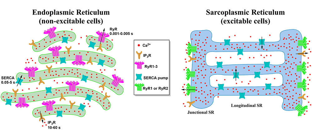
2. Morphology of the ER and the SR

The ER appears as an uninterrupted network with three distinct forms and accompanied specialized functions [22] (Figure 1a). The rough ER (RER) appears as flattened sacs and is speckled with ribosomes for protein synthesis. The smooth ER (SER) is an elongated, cylindrical network with key functions in Ca2+ storage and release. Surrounding the nucleus is an extension of the ER called the nuclear envelope (NE). The NE is also dotted with ribosomes and supplies the nucleus with Ca2+ for gene transcription [22,31]. The ER extends throughout the cell taking up more than 10% of the cell capacity. As such the ER is able to interact with several key organelles and membranes, namely the plasma membrane, mitochondria, lysosomes, and endosomes. The local regulation of these microdomain interactions with the ER allow for comprehensive control of cellular Ca2+ dynamics [31,32].
The SR is a morphologically distinct version of the smooth ER specialized for Ca2+ release to fuel muscle contraction [33] (Figure 1b). It consists of large sink portions referred to as terminal cisternae (TC) connected to elongated tubes termed the longitudinal SR. RyRs are located solely in the TC, while SERCA pumps are found exclusively in the longitudinal SR. The distribution of these primary receptors within the SR divide this organelle into two divergent subdomains tasked with the release and the uptake of Ca2+ [34]. Within the skeletal muscle cell, the TC is distributed to be in proximity to the tubule invaginations of the sarcolemma membrane called the transverse tubules (t tubules) with triads consisting of a t tubule segment between two TC [35]. Located on the t tubule membrane are the dihydropyridine receptors (DHPRs) that allow Ca2+ to flow into the myoplasm in response to depolarization. The space between the SR TC membrane and the t tubule membrane is referred to as the junctional zone [36]. This extended SR network consisting of the TC, longitudinal SR, and the t tubules surrounds the myofibrils in repeating units near the sarcomere ensuring delivery of Ca2+ for contraction to occur [37].

3. ER/SR Mediated Calcium Signaling

The Ca2+ released from the ER/SR makes up the bulk of the Ca2+ signal. The IP3R and the RyR are the main Ca2+ release channels on the ER/SR membrane. For cardiac muscle or skeletal muscle, Ca2+ activates the RyR through CICR via interaction with the DHPR, allowing more Ca2+ to discharge from the ER/SR to propagate the Ca2+ signal [38,39,40]. The SERCA pump is the main mode of Ca2+ transport back from the cytosol into the ER/SR [41]. Here we will discuss the distribution and function of these calcium signaling constituents and their interaction with other proteins.

3.1. The Ryanodine Receptor

The RyR exists in three isoforms in mammals: RyR1, RyR2, and RyR3. RyR1 and RyR2 are the predominant isoforms found in skeletal and cardiac muscle, respectively, while RyR3 is dominant in the thalamus, hippocampus, corpus striatum, and smooth muscle [42,43]. RyR3 is also present in mammalian skeletal muscle cells during development [44]. High-resolution cryo-electron microscopy structures of the RyR show it exists as a homotetramer with a total mass over 2 MDa making it the largest ion channel. The large cytosolic portion of the RyR has a mushroom-like shape with the rest of the channel embedded in the SR membrane [45,46]. This large cytosolic portion of the RyR is where many proteins bind to regulate its function such as FKBP12 (RyR1), FKBP12.6 (RyR2), calmodulin (CaM), CaMKII, S100A proteins, and DHPRs [47]. Ca2+ activates and inhibits RyR1 activity. When Ca2+ concentration in the cytosol is ~1 µM, the receptor is active, and it is inhibited when the Ca2+ concentration reaches 1 mM. This activation occurs in a [Ca2+] dependent bell shaped curve [11]. Myoplasmic free magnesium (Mg2+) is a potent inhibitor of RyR1 in resting skeletal muscle cells. If the physiological concentration of magnesium is lowered from 1 to 0.05 mmol/L, the receptor opens resulting in a steep decrease in SR Ca2+. Cytosolic adenosine triphosphate (ATP) also triggers the Ca2+ release channel activity [48]. Caffeine exerts a stimulatory effect on RyRs by enhancing their affinity for Ca2+ without disrupting magnesium binding [49]. 4-Chloro-m-cresol (4-CmC) is a potent non-native agonist of the RyR, having a 10-fold higher sensitivity than caffeine for inducing SR Ca2+ release with both regularly employed to study RyR-mediated Ca2+ release in healthy and diseased cells [50]. While the three isoforms of RyR are predominately found in their specific cell types, they can be found in varying concentrations in all cells. Their location in the cell, specifically on the ER/SR membrane, varies between each isoform. RyR1 is highly localized in the terminal cisternae region of the SR membrane facing the t tubules in skeletal muscle cells. As mentioned previously, he DHPRs, situated on the t tubule membrane, interact directly with RyR1 to activate Ca2+ release when membrane depolarization occurs [36]. Residues 1635–2636 of RyR1 were shown to interact with the DHPR to mediate Ca2+ release and excitation–contraction (E-C) coupling [51]. Later, it was discovered that residues 1–1680 at the N-terminal portion of RyR1 facilitate E-C coupling [52]. RyR1 also enriches the activity of the DHPR through a backward current [53]. RyR1 and RyR2 are also controlled luminally through interactions with calsequestrin, junctin, and triadin [54]. RyR3 was found to co-localize with lysosomes in the perinuclear region of cardiac muscle cells, whereas RyR1 and RyR2 also co-localize with lysosomes they are 2-fold lower and mainly in the sub-plasmalemmal and extra-perinuclear, respectively, where 4- and 60-fold less lysosomal co-localization occurs [55].

3.2. The Inositol 1,4,5-Trisphosphate Receptor

The IP3R is the major Ca2+ release channel on the ER membrane of non-excitable cells [56]. Its presence in excitable cells serves to amplify the Ca2+ signal generated from depolarization [57]. The IP3R is also present in cardiac and skeletal muscle cells where it functions to increase gene expression through excitation-transcription coupling for fiber growth and stabilization of the neuromuscular junction [58,59]. The IP3R is a member of a vast ion channel superfamily [60]. Like RyR, the IP3R functions as a tetramer having a large cytosolic domain resembling a mushroom and six transmembrane segments with the Ca2+ binding portion homologous to that of the RyR [61]. Three isoforms of the IP3R exist with IP3R1 being the most thoroughly studied [62]. IP3 is generated as a second messenger from the breakdown of phosphoinositol-4,5-bisphosphate (PIP2) by phospholipase C (PLC) through activation of G-protein coupled receptors (GPCRs) [56]. Channel opening is activated by both Ca2+ and IP3, where IP3 increases the response of the channel to Ca2+ giving it a bell-shaped response curve similar to RyR [4]. Channel opening allows the release of Ca2+ from the ER and other internal stores expressing the receptor [56]. Several molecules interact indirectly or directly with the IP3R triggering its activation. ATP increases IP3-mediated Ca2+ release through the IP3R at concentrations of 100 µM [63]. ATP binds to the purinergic receptor P2YR, a GPCR, triggering production of IP3 that binds to the IP3R to release Ca2+ from the ER [64,65]. IP3R1 was also found to be activated by thiol modification with less than 10 µM of thimerosal, having more than this concentration actually caused an inhibitory effect [66]. Additionally, IP3R can be phosphorylated through protein kinase A (PKA), protein kinase B (PKB), cycline-dependent kinase 1 (CDK1), and MAP kinases [66].

3.3. The Sarco-Endoplasmic Reticulum Ca2+ ATPase Pump

The role of Ca2+ pumps is to direct Ca2+ ions out of the cell or back into organelles once the initial stimulus for the signaling event is removed. The major Ca2+ pump for the ER/SR is the SERCA pump found on the membrane. The SERCA pump is powered by ATP hydrolysis to move Ca2+ ions against the concentration gradient. This pump helps to restore cytosolic Ca2+ concentration to 0.1 µM [3,38]. The SERCA pump is a P-type ATPase pump that reduces cytosolic Ca2+ and refills the ER/SR by actively pumping the ion into the ER/SR lumen [41]. The pump functions as a monomer with a mass of 110 kDa. Three genes encode SERCA1, SERCA2, and SERCA3 isoforms whose amino acid sequences are highly conserved. From these three genes, alternative splicing generates over 10 variants of the pump adding to the diversity of its function in different tissues [67]. SERCA1 is primarily distributed in fast twitch skeletal muscle. SERCA2 is found in fast twitch and slow twitch muscle, cardiac muscle, smooth muscle, and non-muscle cells. SERCA3 is expressed in non-muscle cells [68]. The crystal structure of rabbit SERCA1a revealed a large cytosolic portion consisting of the Asp 351 phosphorylation site (domain P), the nucleotide-binding site (domain N), and the anchor portion (domain A). The transmembrane (domain M) portion of the pump contains 10 α-helices where two Ca2+ ions bind [69]. The binding sites are side by side and are formed by M4, M5, M6, and M8 helices. When Ca2+ binds to the pump, the large cytosolic head changes from a closed to open conformation to facilitate translocation of the ions. For each ATP molecule consumed, the pump transports two Ca2+ ions into the ER/SR lumen [68,70]. Specific inhibitors of the SERCA pump include thapsigargin, from Thapsia garganica, and cyclopiazonic acid (CPA), from Aspergillus and Penicillum. CPA has a low affinity for the pump and reversibly blocks the Ca2+ access point [71]. Thapsigargin, on the other hand, irreversibly binds to the Ca2+ free pump at residue F256 in the M3 helix with nanomolar affinity [41].

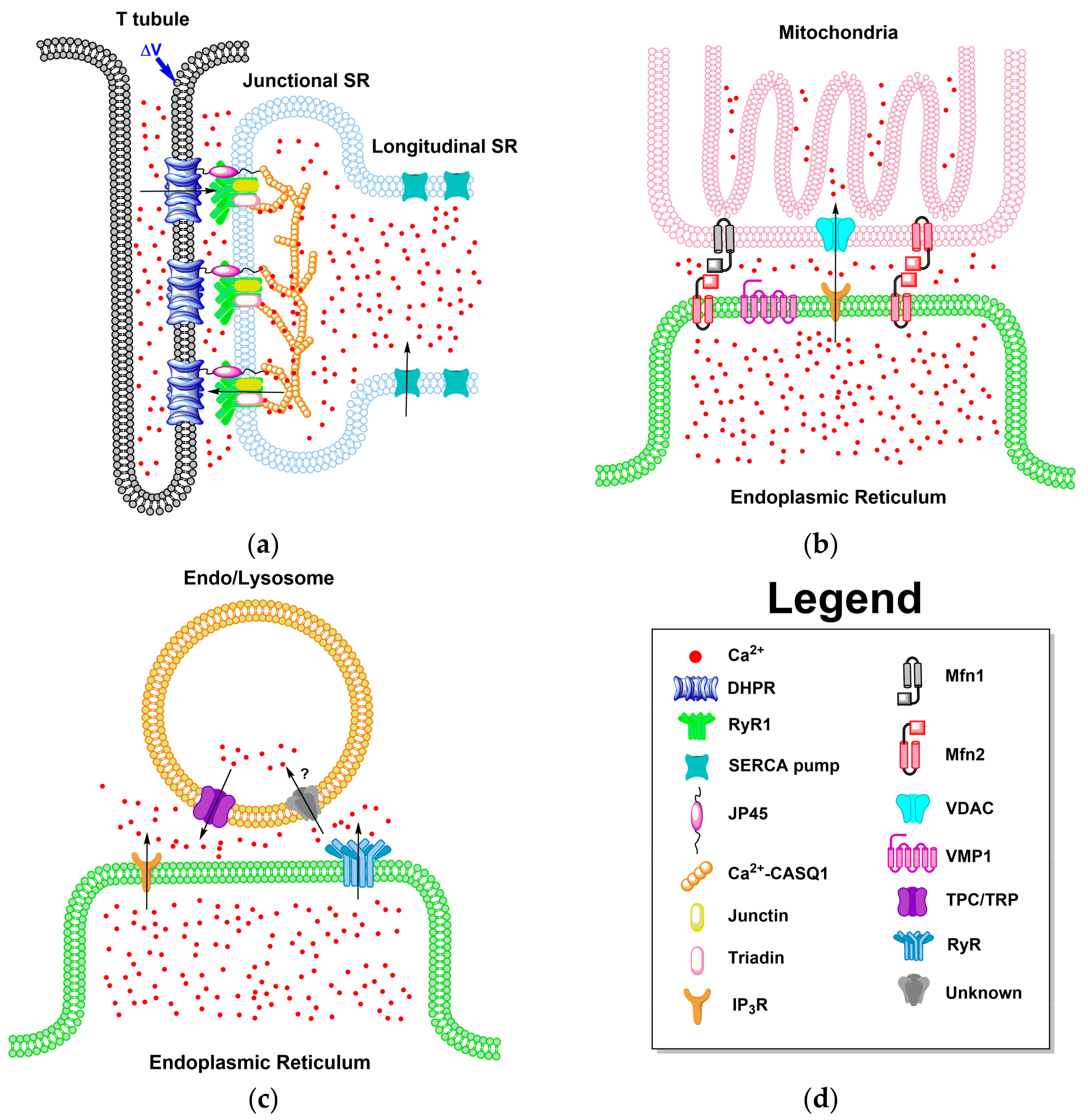
4. Local Domains of ER/SR Calcium Release

Intracellular calcium signaling is composed of both local and global events. Ca2+ channel activation produces momentary localized elevations in cytosolic Ca2+. These short-lived plumes, puffs, and sparks of Ca2+ are limited to 1–6 μm around the mouth of the channel. The diffusion of these brief bursts of Ca2+ sensitize nearby channels which aids signal amplification [72]. The recruitment and coordinated release of calcium from several calcium channels define global calcium signals. In skeletal muscle, synchronous channel opening is achieved through membrane depolarization that leads to the conformational change in the DHPR sensed by the coupled RyRs. The initial fast calcium release is via the DHPR coupled with RyR1. For cardiac muscle cells, calcium sparks facilitate CICR activating surrounding uncoupled RyRs to propagate the calcium signal [72]. The IP3R is also present in cardiac and skeletal muscle with IP3-associated calcium transients contributing to skeletal muscle growth and stabilization of the neuromuscular junction through excitation-transcription coupling, as mentioned in a previous section [58,59]. In non-excitable cells, Ca2+ puffs are responsible for producing regenerative Ca2+ waves and oscillations from the IP3R using CICR to recruit nearby clusters of IP3R when IP3 is present [10,73]. The structural framework and heterogeneous distribution of the ER/SR and associated proteins form localized Ca2+ signaling domains for the rapid mobilization of intracellular Ca2+ [21].

4.1. ER-PM Junction of Skeletal Muscle and Cardiac Cells

As discussed in previous sections, the junctional zone is a highly specialized ER-PM junction found in skeletal muscle and cardiac cells. The structure of this interface with associated proteins allows an immense amount of Ca2+ to be released from the SR in response to action potentials, fueling E-C coupling [29,36,54] This large reservoir of Ca2+ required to generate contractile force repetitively from a train of action potentials in skeletal muscle cells is maintained by the low affinity, high capacity Ca2+ binding and release of calsequestrin (CASQ1) [74]. CASQ1 is concentrated in the SR TC where it forms long polymers near the opening of RyR1 in a Ca2+ dependent manner [75,76]. Polymerized CASQ1 can bind 40–50 mol of Ca2+/mol of CASQ1 with a 103 M−1 affinity over a high Ca2+ concentration range of 0.01–1 M. Its exceptional buffering function makes CASQ1 the sole mechanism for fast Ca2+ binding and release from the SR [74]. CASQ1 is an important regulator of SR Ca2+ release through its association with integral membrane RyR associated proteins junctin and triadin (Figure 2a), but it is not required for luminal Ca2+ sensing. Under basal conditions, SR Ca2+ release and contraction were observed in studies of CASQ2-null mice [77]. When junctin and triadin are present, CASQ inhibits RyR function when luminal Ca2+ is at 1 mM. [78]. Research done by Manno and colleagues shows that CASQ polymerizes in the SR of adult mouse myofibrils at rest and depolymerizes fully in response to electrical stimulation or maximal depletion of Ca2+ from the SR by treatment with a high amount of 4-CmC supporting the luminal Ca2+ sensing role of CASQ [79]. In skeletal muscle cells, an additional protein associated with the junctional zone, having an important role in E-C coupling, was discovered called junctional protein 45 (JP45) [80]. JP45 interacts with the DHPR at the cytosolic portion of its N-terminal and with CASQ1 at its C-terminal situated in the SR lumen [81]. Deletion studies of JP45 from young mice resulted in a loss of skeletal muscle strength due to decreased expression of the DHPR, which is essential for E-C coupling [82]. These local Ca2+ signals that activate the contractile proteins in the junctional zone are fueled by global changes in SR Ca2+ and are critical for E-C coupling to occur [83]. The structure of this junction differs in cardiac cells. In heart cells, contraction is produced by calcium release from dyads composed of an adjacent TC and t tubule segment. The DHPR and the RyR isoform 2 (RyR2) are located in the dyad on the surfaces of the t tubule and SR membrane, respectively. Junctin, triadin, and CASQ all co-localize with the RyR2 as seen in skeletal muscle [84]. A lower density of DHPRs exists in cardiac cells. The DHPRs do not interact directly with RyR2 in response to depolarization. Instead, a steady current of extracellular Ca2+ comes in and activates RyR2 [85]. The Ca2+ sensor GCaMP6f, fused to the N-terminal of junctin and triadin, has been applied to study calcium release in the dyads of rat cardiomyocytes. Confocal imaging with the probe was able to elucidate Ca2+ nanosparks that are much smaller than Ca2+ sparks [86]. GCaMP2.2 and GCaMP2.2low were applied to study local Ca2+ release in the cleft region between the sarcolemma and junctional SR membrane in cardiac cells. Fusing the indicators with FKBP12.6 tagged them to the RyR microdomains. Using the targeted and non-targeted versions of the probe, cleft [Ca2+] was larger than the global, or bulk, [Ca2+] (194 nmol/L versus 100 nmol/L) [87].

4.2. ER Junctions with Other Organelles

The ER also has local junctions with other organelles such as mitochondria, lysosomes, and endosomes (Figure 2). The methods of communication between these organelles are being consistently researched and new mechanisms of interaction are elucidated frequently. The mitochondria and ER communication is as complex as it is vital. The IP3R on the ER releases Ca2+ from internal stores. If the mitochondria are within the critical distance, Ca2+ is taken into the mitochondria through the voltage-dependent anion channel (VDAC) and then into the cristae by the mitochondrial Ca2+ uniporter (MCU) [88,89]. The closer the distance between IP3R and MCU, the more Ca2+ is taken into the mitochondria from the ER/SR. Additionally, the further apart the two channels are, the less Ca2+ is shuttled between the organelles [90]. This critical distance, as well as ER structure, is shown to be regulated by the vacuole membrane protein 1 (VMP1), an ER transmembrane protein which assists in keeping the distance between mitochondria and the ER MCS [91]. The ER-mitochondria complex is further stabilized by the connection of mitofusion proteins, Mfn1 and Mfn2. The ER has been shown to have this transmembrane protein Mfn2 on the membrane forming a complex with either Mfn1 or Mfn2, both expressed on the mitochondrial membrane [92,93].
The MCS between the ER/SR and the endosomal and lysosomal pathway is as complex as it is biologically relevant. The Ca2+ signaling between these organelles is less readily understood, while being expanded upon through vigorous research daily. While studies have shown that the endo/lysosome contact with the ER/SR is extremely complex and intricate, the Ca2+ signaling is marginally more simplistic yet mysterious. In the MCS between the ER/SR and endo/lysosome, as shown in Figure 2c, IP3Rs and RyRs release Ca2+ from internal ER/SR stores, meanwhile the transient receptor channels (TRP) and two-pore channels (TPC) release Ca2+ from the endo/lysosome after activation by Ca2+ activator nicotinic acid adenine dinucleotide phosphate (NAADP) [94,95,96,97,98]. While the shutting of Ca2+ from the internal stores of ER/SR, as well as endo/lysosomes, the mechanism for pumping Ca2+ into the endo/lysosome is still unknown. Research shows that Ca2+ is brought into endo/lysosomes, but the protein which regulates this process and its functions is still being investigated, although based on the rates of uptake it seems a Ca2+ exchanger is more likely than a Ca2+ pump [99].

5. Diseases Associated with ER/SR Calcium Signaling

As cells grow and separate into their respective tissue types during the developmental process, diverse elements of the Ca2+ signaling machinery will be expressed that will render different properties to the generated Ca2+ signal [7,38]. These components are under continuous modification to adjust to environmental changes and ensure the preservation of the Ca2+-mediated response for the particular cell type. When problems arise with constituents of the signaling network, Ca2+ itself will trigger transcription of the defunct components to restore the signal [38]. Various diseases are attributed to dysfunctional elements in the Ca2+ signaling network [100,101], primarily the IP3R and RyR that mediate Ca2+ release from the ER/SR and the SERCA pump [16]. A few of the diseases and conditions that involve these major Ca2+ signaling elements will be discussed in this section.

5.1. Brody’s Disease

Brody’s disease is an uncommon genetic disorder of skeletal muscle [41]. The symptoms of this musculo-skeletal disease are not exact but are commonly described as cramps, reduced muscle relaxation, and exercise-induced stiffness. Since the symptoms are broad, diagnosing this disease in a clinical setting is problematic. Brody’s disease originates from mutations to SERCA1 through the ATP2A1 gene. The function of the pump, not its expression level, is decreased in suffering patients [18,102]. The SERCA pump is responsible for sequestering cytosolic Ca2+ into the ER/SR after the occurrence of the signaling event [103]. As a result of pump malfunction, cytosolic [Ca2+] remains high after stimulus removal. This sustained level of cytosolic Ca2+ contributes directly to muscle stiffness. Administration of the drugs dantrolene and verapamil was shown to restore cytosolic Ca2+ levels back to basal [102].

5.2. Catecholaminergic Polymorphic Ventricular Tachycardia

Ca2+ is a critical component of the various aspects of the signaling system in cardiomyocytes that work together to generate the contractile force needed to pump blood throughout the body [104]. Some of these areas include the electrophysiological mechanisms used to generate the action potential, E-C coupling, myofilament activation, energy manufacture and metabolism, cell death, and transcriptional control of cardiac machinery [105]. A breakdown or mutation in any of the components in the previously mentioned areas will cause electrical and mechanical problems within the heart [104]. Catecholaminergic polymorphic ventricular tachycardia (CPVT) is a heart disorder that causes abnormal heart rhythm [100]. CPVT is characterized by rapid heart rate through β-adrenergic receptor stimulation. Commonly occurring in kids and teenagers, CPVT causes fainting and sudden death. When left untreated, CPVT has a 31% mortality rate in patients 30 years of age [106]. The clinical characterization of CPVT is fainting connected to seizures brought on by stress or exercise. The primary form of CPVT, CPVT1, is caused by dominant mutations in RyR2. Mutations in CASQ2 also cause CPVT [100,107]. RyR2 plays a major role in Ca2+ regulation and release from the SR in cardiomyocytes [108]. Activation of the L-type Ca2+ channel allows Ca2+ to flow into the cytosol from the extracellular environment. The circulating Ca2+ opens RyR2, through CICR, allowing prompt discharge of Ca2+ from the SR. The elevated cytosolic Ca2+ level induces contraction and is taken back up into the SR [104,109]. Improper regulation of this cycle is associated with many heart disorders such as CPVT [106].

5.3. Malignant Hyperthermia

Malignant hyperthermia (MH) is a genetic skeletal muscle condition caused by mutations in RyR1 [110,111]. Commonly induced by certain anesthetics, MH is the underlying cause of anesthesia-related deaths in patients who are seemingly healthy. The anesthetics known to initiate MH include isoflurane, sevoflurane, haloethane, desflurane, enflurane, and the muscle relaxer succinylcholine [112]. In some reported cases of MH, abrupt changes in temperature and stressors triggered the disease. Clinical hallmarks of MH include rapid heart rate, high blood pressure, sustained muscle contractions, increased CO2 levels, trouble breathing, and severe increase in core temperature. Untimely diagnosis of MH leads to death. The hypermetabolic state mirrored in MH is attributed to escalated Ca2+ release from the SR caused by defects in RyR1 situated on the SR membrane [111,112]. Hundreds of mutations to human RyR1 have been linked to MH, thus far, with research still ongoing. The majority of these mutations are confined to three locales in RyR1: C35-R614 in the N-Terminal, D2129-R2458 in the central region, and I3916-G4942 in the carboxy terminal, but several mutations still exist outside of these areas [113]. MH mutations have also been identified in the human CACNL1A3 gene coding the α1 subunit of the DHPR located on the sarcolemma [114]. Different mechanisms have been revealed for the molecular basis of this condition. Dirksen and Avila found that RyR1 mutations, that only produce MH, slightly increased RyR1 activity without changing the overall concentration of SR Ca2+, whereas those that produce MH and central core disease (CCD) increase RyR1 activity resulting in depletion of the SR and an increase in intracellular Ca2+ [115]. Other studies reveal that MH mutations to RyR1 and CASQ1 decrease the threshold for store overload induced calcium release (SOICR). Exposure to volatile anesthetics further decreases the SOICR barrier, making RyR1 more sensitive to small increases in luminal Ca2+. CASQ1 MH mutants exhibit reduced buffering ability causing SR Ca2+ to increase over the threshold. This reduced threshold for Ca2+ activation of RyR1 leads to an increase in cytosolic Ca2+, causing the characteristic hypermetabolic state depicted in MH patients [116]. The abnormal handling of SR Ca2+ leads to muscle rigidity caused by elevated myoplasmic Ca2+ and increased glycogen and glucose breakdown sparked by phosphorylase kinase galvanization by Ca2+ [113]. These frenzied metabolic reactions consume O2, ATP, and glycogen reserves and create exorbitant amounts of metabolic waste products, leading to an eventual disturbance in ion levels and ensuing cellular destruction [117]. Clinical treatment of MH involves the administration of the muscle relaxer dantrolene, the only pharmacological agent known to treat the disease [118]. The mechanism for the action of dantrolene has been widely debated. Dantrolene imposes a hindrance on the DHPR, blocking L-type currents and diminishing Ca2+ release from the SR; however, expression of RyR1 was found necessary for dantrolene to exert an inhibitory effect on the DHPR through modification of the coupling between these channels [119]. Recently, dantrolene was shown to inhibit RyR1 and RyR2 function in the presence of 100 nM CaM in single channel recordings [120].

5.4. Alzheimer’s Disease

Alzheimer’s disease (AD) is medically defined as the continuous deterioration of mental capacity leading to noticeable declines in behavior and memory associated with aging [121]. Currently, the prevalence of AD is ranked highest among all other neurological maladies and has no cure. The majority of AD cases that arise are late onset or sporadic AD (SAD). Familial AD (FAD), or early onset AD, constitutes a small percentage of cases and has genetic origins. In both cases, AD manifests in the brain as lesions of amyloid-beta (Aβ) protein, fibrous tangles of tau (τ) protein, and cell death resulting in the reduction of brain mass and ultimately death [122,123]. Amyloid plaque formation is caused by the improper cleavage of the neuroprotective amyloid precursor protein (APP) by secretase enzymes resulting in copious amounts of the cytotoxic, 42 residue fragment Aβ [124]. Mutations in APP and presenilins 1 and 2 (PS1 and PS2), which are located in the ER membrane of neurons, lead to a buildup of Aβ in cases of FAD. A common denominator in all cases of AD is the mishandling of neuronal intracellular Ca2+, primarily IP3R and RyR-mediated Ca2+ release from the ER [121,125]. Resting free intracellular Ca2+ levels were dramatically increased in the neurons of transgenic mice exhibiting AD compared to normal neurons. This increase was due to the influx of Ca2+ through voltage-gated channels and its release from the ER through the IP3R and RyR [126]. PS1 and PS2 mutants of FAD were shown to excite IP3R activity causing amplified Ca2+ signaling from the ER, which increased Aβ production [127]. Injection of Aβ aggregates into Xenopus oocytes alone elicited IP3R stimulation through GPCR production of IP3 resulting in vigorous Ca2+ release from the ER and cytotoxicity [128]. Reduction in IP3R levels and blocking of RyR activity was shown to alleviate AD symptoms in AD mouse models solidifying IP3R and RyR-mediated Ca2+ release from the ER in neurons as therapeutic targets for AD [129,130].

6. ER/SR-Targeted GECIs

To decipher the irregularities in Ca2+ signaling that lead to the aforementioned diseases, specific tools are essential to study them. The progress made in the creation of synthetic calcium dyes yielded significant advances in the understanding of intracellular calcium signaling [131]. MagFura-2 and Fluo-5N are frequently used to study ER Ca2+, having dissociation constants (Kd) of 25 and 90 µM, respectively [132,133,134,135]. The application of synthetic indicators for studying Ca2+ changes in specific organelles is plagued with the compartmentalization propensity of the dye in non-specific organelles [131]. The ability to monitor calcium transients, in situ and in vivo, in targeted intracellular locations was revolutionized by the discovery and manipulation of fluorescent proteins [136]. Using site-directed mutagenesis, circular permutation, and fused FP constructs, a variety of GECIs have been created to monitor cytosolic and organellar Ca2+, becoming powerful tools in the field of calcium imaging [137,138]. To measure luminal Ca2+ within a specific organelle like the ER/SR, there are preferable properties the indicator should have. First, the GECI must have specificity for Ca2+, over other physiological ions and small molecules, with a Kd between 0.5–5× the [Ca2+] of the target organelle. It must have good folding and bright fluorescence at 37 °C for mammalian cell application. It should possess a large signal enhancement upon Ca2+ binding with a high signal to noise ratio (SNR). It should display rapid binding and release kinetics for measuring fast calcium fluctuations. The binding stoichiometry of the GECI–Ca2+ complex should be 1:1 for simple, quantitative interpretation of the fluorescence signal. Additionally, the probe should be unaffected by the pH of the environment being monitored [137,138,139]. Here, we give a brief history on GECIs with a primary focus on ER/SR-targeted GECIs. For further reading, a few in depth reviews on calcium dyes and GECIs are referenced here [134,137,138].
GECIs fall into two major categories: single fluorophore and Förster resonance energy transfer (FRET) pair. Single fluorophore sensors are non-ratiometric and experience a change in fluorescence intensity when calcium binds. Most of the single fluorophore sensors are large chimeras of a fluorescent protein (FP) connected to a native CaBP, such as CaM or troponin C (TnC). Chelation of calcium by the CaBP induces a conformational change that rearranges the chromophore environment, inducing fluorescence change. For single fluorophore sensors, calcium binding produces changes either in absorbance or the quantum yield that lead to the change in fluorescence intensity. Single fluorophore sensors are commonly referred to as intensiometric because the fluorescence intensity change at only one wavelength is measured [137]. FRET pair, or ratiometric, sensors involve the fusion of two FPs with a CaBP in the center of the construct. In these indicators, the emission energy of one FP (donor) is used as the excitation energy for the other FP (acceptor). The efficiency of energy transfer is dependent on the distance between the two FPs and the extent of overlap between the emission and excitation of the donor and acceptor, respectively. The conformational change that occurs between the CaBP when calcium binds brings the FPs closer together for FRET to occur [137,140]. Cameleons are genetically encoded FRET pair sensors consisting of two fluorescent proteins, normally cyan fluorescent protein (CFP) and yellow fluorescent protein (YFP), with calmodulin and M13, from myosin light chain kinase, in the center of the construct. These sensors have been applied in different cell lines to monitor Ca2+ dynamics from the ER/SR and cytosol [141] but primary application is cytosolic due to their strong affinity for Ca2+. D1ER and D4cpv-CASQ1 are cameleons that utilize CFP and Citrine, a type of YFP, and CFP and circularly permutated Venus, respectively. D1ER and D4cpv-CASQ1 are targeted to the ER/SR and have been used to study Ca2+ release from these organelles [142,143]. Recently, ER-targeted cameleons with a red hue were developed based off of the optimal cameleon D1ER [142]. The new red shifted cameleon called D1ERCmR2 has the fluorescent proteins Clover positioned at the N-terminal acting as the FRET pair donor and mRuby2 at the C-terminal acting as the FRET pair acceptor. D1ERCmR2 has two in vitro Kds of 0.8 and 60 µM with an in situ Kd of 220 µM determined in HeLa cells. D1ERCmR2 can be used in tandem with fura-2 to measure ER and cytosolic Ca2+ concentrations [144]. As for the kinetics of cameleon sensors, not much data has been reported. Cameleons YC2.0, one of the first yellow cameleons, has a dissociation time constant (Ƭrt) of 83 ms. YC-Nano140 has a Ƭrt of 303 ms [137].
Because of the large size of FRET pairs and limited signal intensity, probes called GCaMP were developed using a single circularly permuted enhanced green fluorescent protein (cpEGFP) fused to calmodulin and M13. The use of cpEGFP increased the signal intensity of the probes for use in cells but their affinities for Ca2+ are high with Kd values in the nanomolar range [145,146]. The original GCaMP indicator, published in 2001, has a Ca2+ Kd of 235 nM with a dissociation time constant of ~200 ms [145]. The Ca2+ induced fluorescence change mechanism was discovered from crystal structure analysis of GCaMP2. The Ca2+ induced conformational change in CaM creates new contacts between CaM and cpGFP. CaM residues change the chromophore environment of cpGFP, preventing solvent access to the chromophore aiding fluorescence increase [146]. Since its unveiling, several variants of GCaMP have been created and used to monitor Ca2+ transients in different environments such as mouse cardiac cells in vivo (GCaMP2) [147], chemosensory neurons of Caenorhabditis elegans and Drosophila melanogaster (GCaMP3) [148], and in the brain cells of mice, Caenorhabditis elegans, Drosophila melanogaster, and zebra fish (GCaMP5s) [149]. As mentioned in a previous section, GCaMP6f is a newer construct in the GCaMP family with the ability to sense Ca2+ nanosparks that initiate EC coupling in the junctional zone dyad space in rat cardiomyocytes. By targeting the sensor to the junctional zone using resident proteins junctin and triadin, Ca2+ nanosparks that are 50 times smaller than standard sparks were seen [86]. All of the initial GCaMP variants mentioned have strong affinities for Ca2+ that limit their use to cellular environments with minimal Ca2+ concentrations like the cytosol, and only use cpGFP. None of the initial GCaMPs have Ca2+ affinities low enough to target to organelles like the ER/SR [145,147,149]. A new subfamily of GCaMPs was created, from random mutations to GCaMP3, termed genetically encoded Ca2+ indicators for optical imaging (GECOs). Green (G-GECO), red (R-GECO), and ratiometric blue-green (GEM-GECO) variants were created. Although the color palette was expanded, the new GECO variants still have high Ca2+ sensitivity [150]. Robert Campbell and colleagues created low affinity red (LAR) GECOs, LAR-GECO1 and LAR-GECO1.2, with Ca2+ Kds of 24 and 12 µM, respectively. LAR-GECO1 was used to monitor thapsigargin inhibition of ER refilling in HeLa, HEK293, and U2-OS cells co-transfected with CatchER. LAR-GECO had larger decreases in intensity over all three cell lines [151]. In 2014, new low affinity, GECO-type indicators were created based on cfGCaMP2. The lead variant, from a library of 58 mutants, had a Ca2+ Kd of 368 µM and a large dynamic range. This new variant termed Ca2+-measuring organelle-Entrapped Protein IndicAtor 1 in the ER (CEPIA1er) was able to monitor ER Ca2+ dynamics in HeLa cells with thapsigargin and histamine treatment [152]. Variants of CEPIA were also created with different emission wavelengths such as red (R-CEPIAer), green (G-CEPIAer), and ratiometric blue-green (GEM-CEPIAer) with Ca2+ affinities of 565, 672, and 558 µM, respectively [152]. Table 1 lists properties of the discussed GECIs. Many of the mentioned and reported GECIs, such as CEPIA, do not state their Ca2+ binding and release kinetics. For more localized measurements of rapid Ca2+ transients, in processes like muscle contraction, it is crucial to utilize a Ca2+ biosensor with reported, fast kinetics [153].
In the past, we demonstrated our knowledge and skill for designing novel CaBPs using non-native CaBPs as the scaffold [154,155], and manipulating the binding affinity of these designed CaBPs by altering the number of charged residues in the metal binding site [156,157]. Using these previously published results and subsequent statistical analysis of coordination chemistry in native CaBPs [158,159], a novel EGFP-based sensor called CatchER (Ca2+ sensor for detecting high concentration in the ER) was designed [160] (Figure 3). CatchER has a similar absorption profile as GFP with 395 nm and 488 nm excitation peaks and single wavelength emission at 510 nm. CatchER has a large fluorescence intensity change induced by Ca2+ binding and a Kd between 120–180 µM. Its unmatched Ca2+ off rate (koff) of 700 s−1 (Figure 3b), which is unique for a GECI, makes it capable of measuring fast Ca2+ release from the ER/SR in various cell types [160]. In response to electrical stimulation of flexor digitorum brevis (FDB) muscle fibers electroporated with CatchER, the probe had a faster response time and larger dynamic range compared to cells electroporated with D1ER [160] (Figure 3c). Although CatchER demonstrates 1:1 binding stoichiometry for Ca2+, crystal structure analysis revealed two positions for the Ca2+ ion bound within the designed site created by incorporating mutated residues S147E, S202D, Q204E, F223E and T225E. The multiple occupancies for a single Ca2+ ion, within the binding site, were deemed responsible for the rapid kinetic properties of the sensor [161]. CatchER also has a 44% in fluorescence lifetime with Ca2+ bound [162], a property not found in current ER/SR-targeted GECIs. This Ca2+-induced fluorescence lifetime increase points to its future application in fluorescence lifetime imaging microscopy (FLIM). FLIM provides better spatial resolution on smaller timescales extending its quantitative ability beyond standard fluorescence microscopy [163].

7. Summary and Perspective

The intricacies of the intracellular calcium signaling network are impacted by the structural organization of the ER/SR. Events such as muscle contraction are greatly influenced by the local organization of the RyR, CASQ, JP45, and other regulatory proteins that initiate Ca2+ release from the SR lumen. A better understanding of local Ca2+ signaling and the essential components that comprise the underlying machinery will guide the development of innovative methods to treat the numerous diseases that develop as a result of impaired ER/SR Ca2+ signaling.

Acknowledgments

This work was funded by National Institutes of Health (NIH) GM62999, NIH EB007268, NIH AG15820, B&B Seed Grant, and a NIH Supplemental Grant to Florence N. Reddish, B&B fellowship to Cassandra L. Miller, CDT fellowship to Rakshya Gorkhali.

Conflicts of Interest

The authors declare no conflict of interest.

Abbreviations

Ca2+Calcium
EREndoplasmic reticulum
SRSarcoplasmic reticulum
RERRough ER
SERSmooth ER
CaBPCalcium-binding proteins
GECIGenetically encoded calcium indicators
IP3Inositol 1,4,5-triphosphate
IP3RInositol 1,4,5-triphosphate receptor
RyRRyanodine receptor
cADPRCyclic ADP-ribose
CICRCalcium induced calcium release
SERCASarco-endoplasmic reticulum Ca2+ ATPase
UPRUnfolded protein response
MCSMembrane contact sites
PMPlasma membrane
MAMMitochondria-associated ER membrane
NENuclear envelope
TCTerminal cisternae
CaMCalmodulin
4-CmC4-Chloro-m-cresol
E-CExcitation-contraction
DHPRDihydropyridine receptor
PIP2Phosphoinositol-4 5-bisphosphate
Mg2+Magnesium
PLCPhospholipase C
GPCRG-protein coupled receptors
PKAProtein kinase A
PKBProtein kinase B
CDK1Cycline-dependent kinase 1
CPACyclopiazonic acid
JP45Junctional protein 45
CASQ1Calsequestrin (skeletal muscle)
VDACVoltage-dependent anion channel
MCUMitochondrial Ca2+ uniporter
VMP1Vacuole membrane protein 1
TRPTransient receptor channels
MfnMitofusion proteins
TPCTwo-pore channels
NAADPNicotinic acid adenine dinucleotide phosphate
CPVTCatecholaminergic polymorphic ventricular tachycardia
MHMalignant hyperthermia
ATPAdenosine triphosphate
ADAlzheimer’s disease
SADSporadic AD
FADFamilial AD
Amyloid-beta
τTau
APPAmyloid precursor protein
PS1Presenilins 1
PS2Presenilins 2
SNRSignal to noise ratio
TnCTroponin C
FPFluorescent protein
ƬrtDissociation time constant
cpEGFPSingle circularly permuted enhanced green fluorescent protein
FDBFlexor digitorum brevis
FLIMFluorescence lifetime imaging microscopy
FRETFörster resonance energy transfer
CFPCyan fluorescent protein
YFPYellow fluorescent protein

References

  1. Al-Shanti, N.; Stewart, C.E. Ca2+/calmodulin-dependent transcriptional pathways: Potential mediators of skeletal muscle growth and development. Biol. Rev. 2009, 84, 637–652. [Google Scholar] [CrossRef] [PubMed]
  2. Lipskaia, L.; Hulot, J.S.; Lompré, A.M. Role of sarco/endoplasmic reticulum calcium content and calcium ATPase activity in the control of cell growth and proliferation. Pflüg. Arch. Eur. J. Physiol. 2009, 457, 673–685. [Google Scholar] [CrossRef] [PubMed]
  3. Clapham, D.E. Calcium signaling. Cell 2007, 131, 1047–1058. [Google Scholar] [CrossRef] [PubMed]
  4. Berridge, M.J.; Lipp, P.; Bootman, M.D. The versatility and universality of calcium signalling. Nat. Rev. Mol. Cell Biol. 2000, 1, 11–21. [Google Scholar] [CrossRef] [PubMed]
  5. Baylor, S.; Hollingworth, S. Sarcoplasmic reticulum calcium release compared in slow-twitch and fast-twitch fibres of mouse muscle. J. Physiol. 2003, 551, 125–138. [Google Scholar] [CrossRef] [PubMed]
  6. Dolmetsch, R.E.; Xu, K.; Lewis, R.S. Calcium oscillations increase the efficiency and specificity of gene expression. Nature 1998, 392, 933–936. [Google Scholar] [PubMed]
  7. Berridge, M.J.; Bootman, M.D.; Roderick, H.L. Calcium signalling: Dynamics, homeostasis and remodelling. Nat. Rev. Mol. Cell Biol. 2003, 4, 517–529. [Google Scholar] [CrossRef] [PubMed]
  8. Berridge, M.J. Neuronal calcium signaling. Neuron 1998, 21, 13–26. [Google Scholar] [CrossRef]
  9. Chen, S.; Novick, P.; Ferro-Novick, S. ER structure and function. Curr. Opin. Cell Biol. 2013, 25, 428–433. [Google Scholar] [CrossRef] [PubMed]
  10. Berridge, M.J. Inositol trisphosphate and calcium signalling mechanisms. Biochim. Biophys. Acta 2009, 1793, 933–940. [Google Scholar] [CrossRef] [PubMed]
  11. Lanner, J.T.; Georgiou, D.K.; Joshi, A.D.; Hamilton, S.L. Ryanodine receptors: Structure, expression, molecular details, and function in calcium release. Cold Spring Harb. Perspect. Biol. 2010, 2. [Google Scholar] [CrossRef] [PubMed]
  12. Marrion, N.; Adams, P. Release of intracellular calcium and modulation of membrane currents by caffeine in bull-frog sympathetic neurones. J. Physiol. 1992, 445, 515. [Google Scholar] [CrossRef] [PubMed]
  13. Hua, S.Y.; Tokimasa, T.; Takasawa, S.; Furuya, Y.; Nohmi, M.; Okamoto, H.; Kuba, K. Cyclic ADP-ribose modulates Ca2+ release channels for activation by physiological Ca2+ entry in bullfrog sympathetic neurons. Neuron 1994, 12, 1073–1079. [Google Scholar] [CrossRef]
  14. Verkhratsky, A.; Shmigol, A. Calcium-induced calcium release in neurones. Cell Calcium 1996, 19, 1–14. [Google Scholar] [CrossRef]
  15. Parsons, R.L.; Barstow, K.L.; Scornik, F.S. Spontaneous miniature hyperpolarizations affect threshold for action potential generation in mudpuppy cardiac neurons. J. Neurophysiol. 2002, 88, 1119–1127. [Google Scholar] [PubMed]
  16. Seo, M.D.; Enomoto, M.; Ishiyama, N.; Stathopulos, P.B.; Ikura, M. Structural insights into endoplasmic reticulum stored calcium regulation by inositol 1,4,5-trisphosphate and ryanodine receptors. Biochim. Biophys. Acta Mol. Cell Res. 2015, 1853, 1980–1991. [Google Scholar] [CrossRef] [PubMed]
  17. Endo, M. Calcium-induced calcium release in skeletal muscle. Physiol. Rev. 2009, 89, 1153–1176. [Google Scholar] [CrossRef] [PubMed]
  18. Vandecaetsbeek, I.; Vangheluwe, P.; Raeymaekers, L.; Wuytack, F.; Vanoevelen, J. The Ca(2+) pumps of the endoplasmic reticulum and Golgi apparatus. Cold Spring Harb. Perspect. Biol. 2011, 3, a004184. [Google Scholar] [CrossRef] [PubMed]
  19. Burdakov, D.; Petersen, O.H.; Verkhratsky, A. Intraluminal calcium as a primary regulator of endoplasmic reticulum function. Cell Calcium 2005, 38, 303–310. [Google Scholar] [CrossRef] [PubMed]
  20. Malhotra, J.D.; Kaufman, R.J. The endoplasmic reticulum and the unfolded protein response. Semin. Cell Dev. Biol. 2007, 18, 716–731. [Google Scholar] [CrossRef] [PubMed]
  21. Rizzuto, R.; Pozzan, T. Microdomains of intracellular Ca2+: Molecular determinants and functional consequences. Physiol. Rev. 2006, 86, 369–408. [Google Scholar] [CrossRef] [PubMed]
  22. Pozzan, T.; Rizzuto, R.; Volpe, P.; Meldolesi, J. Molecular and cellular physiology of intracellular calcium stores. Physiol. Rev. 1994, 74, 595–636. [Google Scholar] [PubMed]
  23. Zhang, H.; Hu, J. Shaping the endoplasmic reticulum into a social network. Trends Cell Biol. 2016, 26, 934–943. [Google Scholar] [CrossRef] [PubMed]
  24. Gomez-Navarro, N.; Miller, E. Protein sorting at the ER–Golgi interface. J. Cell Biol. 2016, 215, 769–778. [Google Scholar] [CrossRef] [PubMed]
  25. Filadi, R.; Theurey, P.; Pizzo, P. The endoplasmic reticulum-mitochondria coupling in health and disease: Molecules, functions and significance. Cell Calcium 2017, 6, 1–15. [Google Scholar] [CrossRef] [PubMed]
  26. Carrasco, S.; Meyer, T. Stim proteins and the endoplasmic reticulum-plasma membrane junctions. Annu. Rev. Biochem. 2011, 80, 973–1000. [Google Scholar] [CrossRef] [PubMed]
  27. Rosenbluth, J. Subsurface cisterns and their relationship to the neuronal plasma membrane. J. Cell Biol. 1962, 13, 405–421. [Google Scholar] [CrossRef] [PubMed]
  28. West, M.; Zurek, N.; Hoenger, A.; Voeltz, G.K. A 3D analysis of yeast ER structure reveals how ER domains are organized by membrane curvature. J. Cell Biol. 2011, 193, 333–346. [Google Scholar] [CrossRef] [PubMed]
  29. Rossi, A.E.; Dirksen, R.T. Sarcoplasmic reticulum: The dynamic calcium governor of muscle. Muscle Nerve 2006, 33, 715–731. [Google Scholar] [CrossRef] [PubMed]
  30. Stefan, C.J.; Manford, A.G.; Emr, S.D. ER–PM connections: Sites of information transfer and inter-organelle communication. Curr. Opin. Cell Biol. 2013, 25, 434–442. [Google Scholar] [CrossRef] [PubMed]
  31. Lam, A.K.M.; Galione, A. The endoplasmic reticulum and junctional membrane communication during calcium signaling. Biochim. Biophys. Acta 2013, 1833, 2542–2559. [Google Scholar] [CrossRef] [PubMed]
  32. Bravo, R.; Parra, V.; Gatica, D.; Rodriguez, A.E.; Torrealba, N.; Paredes, F.; Wang, Z.V.; Zorzano, A.; Hill, J.A.; Jaimovich, E.; et al. Endoplasmic reticulum and the unfolded protein response: Dynamics and metabolic integration. Int. Rev. Cell Mol. Biol. 2013, 301, 215–290. [Google Scholar] [PubMed]
  33. Sorrentino, V. Molecular determinants of the structural and functional organization of the sarcoplasmic reticulum. Biochim. Biophys. Acta 2004, 1742, 113–118. [Google Scholar] [CrossRef] [PubMed]
  34. Rossi, D.; Barone, V.; Giacomello, E.; Cusimano, V.; Sorrentino, V. The sarcoplasmic reticulum: An organized patchwork of specialized domains. Traffic 2008, 9, 1044–1049. [Google Scholar] [CrossRef] [PubMed]
  35. Franzini-Armstrong, C.; Protasi, F.; Ramesh, V. Shape, size, and distribution of Ca2+ release units and couplons in skeletal and cardiac muscles. Biophys. J. 1999, 77, 1528–1539. [Google Scholar] [CrossRef]
  36. Sorrentino, V. Sarcoplasmic reticulum: Structural determinants and protein dynamics. Int. J. Biochem. Cell Biol. 2011, 43, 1075–1078. [Google Scholar] [CrossRef] [PubMed]
  37. Porter, K.R.; Palade, G.E. Studies on the endoplasmic reticulum III. Its form and distribution in striated muscle cells. J. Biophys. Biochem. Cytol. 1957, 3, 269–300. [Google Scholar] [CrossRef] [PubMed]
  38. Berridge, M.J. Calcium signalling remodelling and disease. Biochem. Soc. Trans. 2012, 40, 297–309. [Google Scholar] [CrossRef] [PubMed]
  39. Tsugorka, A.; Rios, E.; Blatter, L.A. Imaging elementary events of calcium release in skeletal muscle cells. Science 1995, 269, 1723. [Google Scholar] [CrossRef] [PubMed]
  40. Shirokova, N.; García, J.; Ríos, E. Local calcium release in mammalian skeletal muscle. J. Physiol. 1998, 512, 377–384. [Google Scholar] [CrossRef] [PubMed]
  41. Brini, M.; Carafoli, E. Calcium pumps in health and disease. Physiol. Rev. 2009, 89, 1341–1378. [Google Scholar] [CrossRef] [PubMed]
  42. FranziniArmstrong, C.; Protasi, F. Ryanodine receptors of striated muscles: A complex channel capable of multiple interactions. Physiol. Rev. 1997, 77, 699–729. [Google Scholar]
  43. Hakamata, Y.; Nakai, J.; Takeshima, H.; Imoto, K. Primary structure and distribution of a novel ryanodine receptor/calcium release channel from rabbit brain. FEBS Lett. 1992, 312, 229–235. [Google Scholar] [CrossRef]
  44. Tarroni, P.; Rossi, D.; Conti, A.; Sorrentino, V. Expression of the ryanodine receptor type 3 calcium release channel during development and differentiation of mammalian skeletal muscle cells. J. Biol. Chem. 1997, 272, 19808–19813. [Google Scholar] [CrossRef] [PubMed]
  45. Zalk, R.; Clarke, O.B.; des Georges, A.; Grassucci, R.A.; Reiken, S.; Mancia, F.; Hendrickson, W.A.; Frank, J.; Marks, A.R. Structure of a mammalian ryanodine receptor. Nature 2015, 517, 44–49. [Google Scholar] [CrossRef] [PubMed]
  46. Yan, Z.; Bai, X.C.; Yan, C.Y.; Wu, J.P.; Li, Z.Q.; Xie, T.; Peng, W.; Yin, C.C.; Li, X.M.; Scheres, S.H.W.; et al. Structure of the rabbit ryanodine receptor RyR1 at near-atomic resolution. Nature 2015, 517, 50–55. [Google Scholar] [CrossRef] [PubMed]
  47. Capes, E.M.; Loaiza, R.; Valdivia, H.H. Ryanodine receptors. Skelet. Muscle 2011, 1. [Google Scholar] [CrossRef] [PubMed]
  48. Lamb, G.; Stephenson, D. Effect of Mg2+ on the control of Ca2+ release in skeletal muscle fibres of the toad. J. Physiol. 1991, 434, 507. [Google Scholar] [CrossRef] [PubMed]
  49. Herrmann-Frank, A.; Lüttgau, H.C.; Stephenson, D.G. Caffeine and excitation–contraction coupling in skeletal muscle: A stimulating story. J. Muscle Res. Cell Motil. 1999, 20, 223–236. [Google Scholar] [CrossRef] [PubMed]
  50. HerrmannFrank, A.; Richter, M.; Sarkozi, S.; Mohr, U.; LehmannHorn, F. 4-Chloro-m-cresol, a potent and specific activator of the skeletal muscle ryanodine receptor. Biochim. Biophys. Acta Gen. Subj. 1996, 1289, 31–40. [Google Scholar] [CrossRef]
  51. Nakai, J.; Sekiguchi, N.; Rando, T.A.; Allen, P.D.; Beam, K.G. Two regions of the ryanodine receptor involved in coupling withl-type Ca2+ channels. J. Biol. Chem. 1998, 273, 13403–13406. [Google Scholar] [CrossRef] [PubMed]
  52. Perez, C.F.; Mukherjee, S.; Allen, P.D. Amino acids 1–1,680 of ryanodine receptor type 1 hold critical determinants of skeletal type for excitation-contraction coupling—Role of divergence domain D2. J. Biol. Chem. 2003, 278, 39644–39652. [Google Scholar] [CrossRef] [PubMed]
  53. Nakai, J.; Dirksen, R.T.; Nguyen, H.T.; Pessah, I.N.; Beam, K.G.; Allen, P.D. Enhanced dihydropyridine receptor channel activity in the presence of ryanodine receptor. Nature 1996, 380, 72–75. [Google Scholar] [CrossRef] [PubMed]
  54. Divet, A.; Paesante, S.; Bleunven, C.; Anderson, A.; Treves, S.; Zorzato, F. Novel sarco(endo) plasmic reticulum proteins and calcium homeostasis in striated muscles. J. Muscle Res. Cell Motil. 2005, 26, 7–12. [Google Scholar] [CrossRef] [PubMed]
  55. Evans, A.M.; Fameli, N.; Ogunbayo, O.A.; Duan, J.; Navarro-Dorado, J. From contraction to gene expression: Nanojunctions of the sarco/endoplasmic reticulum deliver site- and function-specific calcium signals. Sci. China Life Sci. 2016, 59, 749–763. [Google Scholar] [CrossRef] [PubMed]
  56. Patterson, R.L.; Boehning, D.; Snyder, S.H. Inositol 1,4,5-trisphosphatereceptors as signal integrators. Annu. Rev. Biochem. 2004, 73, 437–465. [Google Scholar] [CrossRef] [PubMed]
  57. Zhu, H.P.; Bhattacharyya, B.J.; Lin, H.; Gomez, C.M. Skeletal muscle IP3R1 receptors amplify physiological and pathological synaptic calcium signals. J. Neurosci. 2011, 31, 15269–15283. [Google Scholar] [CrossRef]
  58. Powell, J.A.; Molgó, J.; Adams, D.S.; Colasante, C.; Williams, A.; Bohlen, M.; Jaimovich, E. IP3 receptors and associated Ca2+ signals localize to satellite cells and to components of the neuromuscular junction in skeletal muscle. J. Neurosci. 2003, 23, 8185–8192. [Google Scholar] [PubMed]
  59. Wu, X.; Zhang, T.; Bossuyt, J.; Li, X.; McKinsey, T.A.; Dedman, J.R.; Olson, E.N.; Chen, J.; Brown, J.H.; Bers, D.M. Local InsP 3-dependent perinuclear Ca2+ signaling in cardiac myocyte excitation-transcription coupling. J. Clin. Investig. 2006, 116, 675–682. [Google Scholar] [CrossRef] [PubMed]
  60. Mikoshiba, K. Ip3 receptor/Ca2+ channel: From discovery to new signaling concepts. J. Neurochem. 2007, 102, 1426–1446. [Google Scholar] [CrossRef] [PubMed]
  61. Serysheva, I.I. Toward a high-resolution structure of IP3R channel. Cell Calcium 2014, 56, 125–132. [Google Scholar] [CrossRef] [PubMed]
  62. Bezprozvanny, I. The inositol 1,4,5-trisphosphate receptors. Cell Calcium 2005, 38, 261–272. [Google Scholar] [CrossRef] [PubMed]
  63. Ferris, C.D.; Huganir, R.L.; Bredt, D.S.; Cameron, A.M.; Snyder, S.H. Inositol trisphosphate receptor: Phosphorylation by protein kinase c and calcium calmodulin-dependent protein kinases in reconstituted lipid vesicles. Proc. Natl. Acad. Sci. USA 1991, 88, 2232–2235. [Google Scholar] [CrossRef] [PubMed]
  64. Dubyak, G.R.; el-Moatassim, C. Signal transduction via P2-purinergic receptors for extracellular ATP and other nucleotides. Am. J. Physiol. Cell Physiol. 1993, 265, C577–C606. [Google Scholar] [PubMed]
  65. Song, Z.; Vijayaraghavan, S.; Sladek, C.D. Atp increases intracellular calcium in supraoptic neurons by activation of both P2X and P2Y purinergic receptors. Am. J. Physiol. Regul. Integr. Comp. Physiol. 2007, 292, R423–R431. [Google Scholar] [CrossRef] [PubMed]
  66. Shah, S.Z.; Zhao, D.; Khan, S.H.; Yang, L. Regulatory mechanisms of endoplasmic reticulum resident IP3 receptors. J. Mol. Neurosci. MN 2015, 56, 938–948. [Google Scholar] [CrossRef] [PubMed]
  67. Grover, A.K.; Khan, I. Calcium-pump isoforms—Diversity, selectivity and plasticity. Cell Calcium 1992, 13, 9–17. [Google Scholar] [CrossRef]
  68. Periasamy, M.; Kalyanasundaram, A. Serca pump isoforms: Their role in calcium transport and disease. Muscle Nerve 2007, 35, 430–442. [Google Scholar] [CrossRef] [PubMed]
  69. Toyoshima, C.; Nomura, H.; Sugita, Y. Crystal structures of Ca2+-ATPase in various physiological states. In Na,k-Atpase and Related Cation Pumps: Structure, Function, and Regulatory Mechanisms; Jorgensen, P.L., Karlish, S.J.D., Maunsbach, A.B., Eds.; New York Academy of Sciences: New York, NY, USA, 2003; Volume 986, pp. 1–8. [Google Scholar]
  70. Toyoshima, C.; Nakasako, M.; Nomura, H.; Ogawa, H. Crystal structure of the calcium pump of sarcoplasmic reticulum at 2.6 Å resolution. Nature 2000, 405, 647–655. [Google Scholar] [CrossRef] [PubMed]
  71. Michelangeli, F.; East, J.M. A diversity of serca Ca2+ pump inhibitors. Biochem. Soc. Trans. 2011, 39, 789–797. [Google Scholar] [CrossRef] [PubMed]
  72. Bootman, M.D.; Rietdorf, K.; Hardy, H.; Dautova, Y.; Corps, E.; Pierro, C.; Stapleton, E.; Kang, E.; Proudfoot, D. Calcium signalling and regulation of cell function. eLS 2006. [Google Scholar] [CrossRef]
  73. Bootman, M.D.; Lipp, P.; Berridge, M.J. The organisation and functions of local Ca2+ signals. J. Cell Sci. 2001, 114, 2213–2222. [Google Scholar] [PubMed]
  74. Sanchez, E.J.; Lewis, K.M.; Danna, B.R.; Kang, C. High-capacity Ca2+ binding of human skeletal calsequestrin. J. Biol. Chem. 2012, 287, 11592–11601. [Google Scholar] [CrossRef] [PubMed]
  75. Royer, L.; Rios, E. Deconstructing calsequestrin. Complex buffering in the calcium store of skeletal muscle. J. Physiol. Lond. 2009, 587, 3101–3111. [Google Scholar] [CrossRef] [PubMed]
  76. Park, H.; Wu, S.; Dunker, A.K.; Kang, C. Polymerization of calsequestrin: Implications for Ca2+ regulation. J. Biol. Chem. 2003, 278, 16176–16182. [Google Scholar] [CrossRef] [PubMed]
  77. Knollmann, B.C.; Chopra, N.; Hlaing, T.; Akin, B.; Yang, T.; Ettensohn, K.; Knollmann, B.E.; Horton, K.D.; Weissman, N.J.; Holinstat, I. Casq2 deletion causes sarcoplasmic reticulum volume increase, premature Ca2+ release, and catecholaminergic polymorphic ventricular tachycardia. J. Clin. Investig. 2006, 116, 2510–2520. [Google Scholar] [CrossRef] [PubMed]
  78. Beard, N.A.; Laver, D.R.; Dulhunty, A.F. Calsequestrin and the calcium release channel of skeletal and cardiac muscle. Prog. Biophys. Mol. Biol. 2004, 85, 33–69. [Google Scholar] [CrossRef] [PubMed]
  79. Manno, C.; Figueroa, L.C.; Gillespie, D.; Fitts, R.; Kang, C.; Franzini-Armstrong, C.; Rios, E. Calsequestrin depolymerizes when calcium is depleted in the sarcoplasmic reticulum of working muscle. Proc. Natl. Acad. Sci. USA 2017, 114, E638–E647. [Google Scholar] [CrossRef] [PubMed]
  80. Zorzato, F.; Anderson, A.A.; Ohlendieck, K.; Froemming, G.; Guerrini, R.; Treves, S. Identification of a novel 45 kDa protein (JP-45) from rabbit sarcoplasmic-reticulum junctional-face membrane. Biochem. J. 2000, 351, 537–543. [Google Scholar] [CrossRef] [PubMed]
  81. Anderson, A.A.; Treves, S.; Biral, D.; Betto, R.; Sandona, D.; Ronjat, M.; Zorzato, F. The novel skeletal muscle sarcoplasmic reticulum JP-45 protein—Molecular cloning, tissue distribution, developmental expression, and interaction with α1.1 subunit of the voltage-gated calcium channel. J. Biol. Chem. 2003, 278, 39987–39992. [Google Scholar] [CrossRef] [PubMed]
  82. Delbono, O.; Xia, J.Y.; Treves, S.; Wang, Z.M.; Jimenez-Moreno, R.; Payne, A.M.; Messi, M.L.; Briguet, A.; Schaerer, F.; Nishi, M.; et al. Loss of skeletal muscle strength by ablation of the sarcoplasmic reticulum protein JP45. Proc. Natl. Acad. Sci. USA 2007, 104, 20108–20113. [Google Scholar] [CrossRef] [PubMed]
  83. Hernandez-Ochoa, E.O.; Pratt, S.J.P.; Lovering, R.M.; Schneider, M.F. Critical role of intracellular RyR1 calcium release channels in skeletal muscle function and disease. Front. Physiol. 2016, 6, 11. [Google Scholar] [CrossRef] [PubMed]
  84. Zhang, L.; Kelley, J.; Schmeisser, G.; Kobayashi, Y.M.; Jones, L.R. Complex formation between junction, triadin, calsequestrin, and the ryanodine receptor—Proteins of the cardiac junctional sarcoplasmic reticulum membrane. J. Biol. Chem. 1997, 272, 23389–23397. [Google Scholar] [CrossRef] [PubMed]
  85. Lamb, G.D. Excitation–contraction coupling in skeletal muscle: Comparisons with cardiac muscle. Clin. Exp. Pharmacol. Physiol. 2000, 27, 216–224. [Google Scholar] [CrossRef] [PubMed]
  86. Shang, W.; Lu, F.; Sun, T.; Xu, J.; Li, L.L.; Wang, Y.; Wang, G.; Chen, L.; Wang, X.; Cannell, M.B.; et al. Imaging Ca2+ nanosparks in heart with a new targeted biosensor. Circ. Res. 2014, 114, 412–420. [Google Scholar] [CrossRef] [PubMed]
  87. Despa, S.; Shui, B.; Bossuyt, J.; Lang, D.; Kotlikoff, M.I.; Bers, D.M. Junctional cleft Ca2+ (I) measurements using novel cleft-targeted Ca2+ sensors. Circ. Res. 2014, 115, 339. [Google Scholar] [CrossRef] [PubMed]
  88. Szabadkai, G.; Bianchi, K.; Varnai, P.; De Stefani, D.; Wieckowski, M.R.; Cavagna, D.; Nagy, A.I.; Balla, T.; Rizzuto, R. Chaperone-mediated coupling of endoplasmic reticulum and mitochondrial Ca2+ channels. J. Cell Biol. 2006, 175, 901–911. [Google Scholar] [CrossRef] [PubMed]
  89. De Stefani, D.; Raffaello, A.; Teardo, E.; Szabo, I.; Rizzuto, R. A forty-kilodalton protein of the inner membrane is the mitochondrial calcium uniporter. Nature 2011, 476, 336–340. [Google Scholar] [CrossRef] [PubMed]
  90. Qi, H.; Li, L.; Shuai, J. Optimal microdomain crosstalk between endoplasmic reticulum and mitochondria for Ca2+ oscillations. Sci. Rep. 2015, 5, 7984. [Google Scholar] [CrossRef] [PubMed]
  91. Tabara, L.C.; Escalante, R. Vmp1 establishes ER-microdomains that regulate membrane contact sites and autophagy. PLoS ONE 2016, 11, e0166499. [Google Scholar] [CrossRef] [PubMed]
  92. Chen, H.; Detmer, S.A.; Ewald, A.J.; Griffin, E.E.; Fraser, S.E.; Chan, D.C. Mitofusins Mfn1 and Mfn2 coordinately regulate mitochondrial fusion and are essential for embryonic development. J. Cell Biol. 2003, 160, 189–200. [Google Scholar] [CrossRef] [PubMed]
  93. de Brito, O.M.; Scorrano, L. Mitofusin 2: A mitochondria-shaping protein with signaling roles beyond fusion. Antioxid. Redox Signal. 2008, 10, 621–633. [Google Scholar] [CrossRef] [PubMed]
  94. Morgan, A.J.; Platt, F.M.; Lloyd-Evans, E.; Galione, A. Molecular mechanisms of endolysosomal Ca2+ signalling in health and disease. Biochem. J. 2011, 439, 349–374. [Google Scholar] [CrossRef] [PubMed]
  95. Grimm, C.; Hassan, S.; Wahl-Schott, C.; Biel, M. Role of trpml and two-pore channels in endolysosomal cation homeostasis. J. Pharmacol. Exp. Ther. 2012, 342, 236–244. [Google Scholar] [CrossRef] [PubMed]
  96. Ruas, M.; Rietdorf, K.; Arredouani, A.; Davis, L.C.; Lloyd-Evans, E.; Koegel, H.; Funnell, T.M.; Morgan, A.J.; Ward, J.A.; Watanabe, K.; et al. Purified tpc isoforms form naadp receptors with distinct roles for Ca(2+) signaling and endolysosomal trafficking. Curr. Biol. CB 2010, 20, 703–709. [Google Scholar] [CrossRef] [PubMed]
  97. Wang, X.; Zhang, X.; Dong, X.P.; Samie, M.; Li, X.; Cheng, X.; Goschka, A.; Shen, D.; Zhou, Y.; Harlow, J.; et al. Tpc proteins are phosphoinositide-activated sodium-selective ion channels in endosomes and lysosomes. Cell 2012, 151, 372–383. [Google Scholar] [CrossRef] [PubMed]
  98. Abe, K.; Puertollano, R. Role of trp channels in the regulation of the endosomal pathway. Physiology 2011, 26, 14–22. [Google Scholar] [CrossRef] [PubMed]
  99. Lopez-Sanjurjo, C.I.; Tovey, S.C.; Prole, D.L.; Taylor, C.W. Lysosomes shape ins(1,4,5)P3-evoked Ca2+ signals by selectively sequestering Ca2+ released from the endoplasmic reticulum. J. Cell Sci. 2013, 126, 289–300. [Google Scholar] [CrossRef] [PubMed]
  100. Ríos, E.; Figueroa, L.; Manno, C.; Kraeva, N.; Riazi, S. The couplonopathies: A comparative approach to a class of diseases of skeletal and cardiac muscle. J. Gen. Physiol. 2015, 145, 459–474. [Google Scholar] [CrossRef] [PubMed]
  101. Mikoshiba, K. Role of IP3 receptor signaling in cell functions and diseases. Adv. Biol. Regul. 2015, 57, 217–227. [Google Scholar] [CrossRef] [PubMed]
  102. Benders, A.A.; Veerkamp, J.H.; Oosterhof, A.; Jongen, P.J.; Bindels, R.J.; Smit, L.M.; Busch, H.F.; Wevers, R.A. Ca2+ homeostasis in brody’s disease. A study in skeletal muscle and cultured muscle cells and the effects of dantrolene an verapamil. J. Clin. Investig. 1994, 94, 741–748. [Google Scholar] [CrossRef] [PubMed]
  103. Bublitz, M.; Musgaard, M.; Poulsen, H.; Thøgersen, L.; Olesen, C.; Schiøtt, B.; Morth, J.P.; Møller, J.V.; Nissen, P. Ion pathways in the sarcoplasmic reticulum Ca2+-ATPase. J. Biol. Chem. 2013, 288, 10759–10765. [Google Scholar] [CrossRef] [PubMed]
  104. Bers, D.M. Calcium cycling and signaling in cardiac myocytes. Annu. Rev. Physiol. 2008, 70, 23–49. [Google Scholar] [CrossRef] [PubMed]
  105. Bers, D.M.; Guo, T. Calcium signaling in cardiac ventricular myocytes. In Communicative Cardiac Cell; Sideman, S., Beyar, R., Landesberg, A., Eds.; Wiley-Blackwell: Hoboken, NJ, USA, 2005; Volume 1047, pp. 86–98. [Google Scholar]
  106. Leenhardt, A.; Denjoy, I.; Guicheney, P. Catecholaminergic polymorphic ventricular tachycardia. Circ. Arrhythm. Electrophysiol. 2012, 5, 1044–1052. [Google Scholar] [CrossRef] [PubMed]
  107. Faggioni, M.; Kryshtal, D.O.; Knollmann, B.C. Calsequestrin mutations and catecholaminergic polymorphic ventricular tachycardia. Pediatr. Cardiol. 2012, 33, 959–967. [Google Scholar] [CrossRef] [PubMed]
  108. Yano, M.; Yamamoto, T.; Kobayashi, S.; Matsuzaki, M. Role of ryanodine receptor as a Ca2+ regulatory center in normal and failing hearts. J. Cardiol. 2009, 53, 1–7. [Google Scholar] [CrossRef] [PubMed]
  109. Bers, D.M.; Shannon, T.R. Calcium movements inside the sarcoplasmic reticulum of cardiac myocytes. J. Mol. Cell. Cardiol. 2013, 58, 59–66. [Google Scholar] [CrossRef] [PubMed]
  110. Robinson, R.; Carpenter, D.; Shaw, M.A.; Halsall, J.; Hopkins, P. Mutations in RyR1 in malignant hyperthermia and central core disease. Hum. Mutat. 2006, 27, 977–989. [Google Scholar] [CrossRef] [PubMed]
  111. Mickelson, J.R.; Louis, C.F. Malignant hyperthermia: Excitation-contraction coupling, Ca2+ release channel, and cell Ca2+ regulation defects. Physiol. Rev. 1996, 76, 537–592. [Google Scholar] [PubMed]
  112. Rosenberg, H.; Pollock, N.; Schiemann, A.; Bulger, T.; Stowell, K. Malignant hyperthermia: A review. Orphanet J. Rare Dis. 2015, 10, 93. [Google Scholar] [CrossRef] [PubMed]
  113. Betzenhauser, M.J.; Marks, A.R. Ryanodine receptor channelopathies. Pflug. Arch. 2010, 460, 467–480. [Google Scholar] [CrossRef] [PubMed]
  114. Monnier, N.; Procaccio, V.; Stieglitz, P.; Lunardi, J. Malignant-hyperthermia susceptibility is associated with a mutation of the α1-subunit of the human dihydropyridine-sensitive L-type voltage-dependent calcium-channel receptor in skeletal muscle. Am. J. Hum. Genet. 1997, 60, 1316–1325. [Google Scholar] [CrossRef] [PubMed]
  115. Dirksen, R.T.; Avila, G. Distinct effects on Ca2+ handling caused by malignant hyperthermia and central core disease mutations in RyR1. Biophys. J. 2004, 87, 3193–3204. [Google Scholar] [CrossRef] [PubMed]
  116. MacLennan, D.H.; Chen, S. Store overload-induced Ca2+ release as a triggering mechanism for cpvt and mh episodes caused by mutations in RyR and Casq genes. J. Physiol. 2009, 587, 3113–3115. [Google Scholar] [CrossRef] [PubMed]
  117. MacLennan, D.H. Ca2+ signalling and muscle disease. Eur. J. Biochem. 2000, 267, 5291–5297. [Google Scholar] [CrossRef] [PubMed]
  118. Krause, T.; Gerbershagen, M.U.; Fiege, M.; Weisshorn, R.; Wappler, F. Dantrolene—A review of its pharmacology, therapeutic use and new developments. Anaesthesia 2004, 59, 364–373. [Google Scholar] [CrossRef] [PubMed]
  119. Bannister, R.A. Dantrolene-induced inhibition of skeletal L-type Ca2+ current requires RyR1 expression. BioMed Res. Int. 2013, 2013, 390493. [Google Scholar] [CrossRef] [PubMed]
  120. Oo, Y.W.; Gomez-Hurtado, N.; Walweel, K.; van Helden, D.F.; Imtiaz, M.S.; Knollmann, B.C.; Laver, D.R. Essential role of calmodulin in ryr inhibition by dantrolene. Mol. Pharmacol. 2015, 88, 57–63. [Google Scholar] [CrossRef] [PubMed]
  121. LaFerla, F.M. Calcium dyshomeostasis and intracellular signalling in Alzheimer’s disease. Nat. Rev. Neurosci. 2002, 3, 862–872. [Google Scholar] [CrossRef] [PubMed]
  122. Supnet, C.; Bezprozvanny, I. The dysregulation of intracellular calcium in alzheimer disease. Cell Calcium 2010, 47, 183–189. [Google Scholar] [CrossRef] [PubMed]
  123. Wojda, U.; Salinska, E.; Kuznicki, J. Calcium ions in neuronal degeneration. IUBMB Life 2008, 60, 575–590. [Google Scholar] [CrossRef] [PubMed]
  124. Mattson, M.P.; Chan, S.L. Neuronal and glial calcium signaling in Alzheimer’s disease. Cell Calcium 2003, 34, 385–397. [Google Scholar] [CrossRef]
  125. Liang, J.Y.; Kulasiri, D.; Samarasinghe, S. Ca2+ dysregulation in the endoplasmic reticulum related to Alzheimer’s disease: A review on experimental progress and computational modeling. Biosystems 2015, 134, 1–15. [Google Scholar] [CrossRef] [PubMed]
  126. Lopez, J.R.; Lyckman, A.; Oddo, S.; LaFerla, F.M.; Querfurth, H.W.; Shtifman, A. Increased intraneuronal resting Ca2+ in adult Alzheimer’s disease mice. J. Neurochem. 2008, 105, 262–271. [Google Scholar] [CrossRef] [PubMed]
  127. Cheung, K.H.; Shineman, D.; Muller, M.; Cardenas, C.; Mei, L.J.; Yang, J.; Tomita, T.; Iwatsubo, T.; Lee, V.M.Y.; Foskett, J.K. Mechanism of Ca2+ disruption in Alzheimer’s disease by presenilin regulation of InsP(3) receptor channel gating. Neuron 2008, 58, 871–883. [Google Scholar] [CrossRef] [PubMed]
  128. Demuro, A.; Parker, I. Cytotoxicity of intracellular a β42 amyloid oligomers involves Ca2+ release from the endoplasmic reticulum by stimulated production of inositol trisphosphate. J. Neurosci. 2013, 33, 3824–3833. [Google Scholar] [CrossRef] [PubMed]
  129. Shilling, D.; Muller, M.; Takano, H.; Mak, D.O.D.; Abel, T.; Coulter, D.A.; Foskett, J.K. Suppression of InsP3 receptor-mediated Ca2+ signaling alleviates mutant presenilin-linked familial Alzheimer’s disease pathogenesis. J. Neurosci. 2014, 34, 6910–6923. [Google Scholar] [CrossRef] [PubMed]
  130. Oules, B.; Del Prete, D.; Greco, B.; Zhang, X.X.; Lauritzen, I.; Sevalle, J.; Moreno, S.; Paterlini-Brechot, P.; Trebak, M.; Checler, F.; et al. Ryanodine receptor blockade reduces amyloid-β load and memory impairments in Tg2576 mouse model of alzheimer disease. J. Neurosci. 2012, 32, 11820–11834. [Google Scholar] [CrossRef] [PubMed]
  131. Gerasimenko, J.V.; Petersen, O.H.; Gerasimenko, O.V. Monitoring of intra-ER free Ca2+. Wiley Interdiscip. Rev. Membr. Transp. Signal. 2014, 3, 63–71. [Google Scholar] [CrossRef]
  132. Hofer, A.M.; Schulz, I. Quantification of intraluminal free ca in the agonist-sensitive internal calcium store using compartmentalized fluorescent indicators: Some considerations. Cell Calcium 1996, 20, 235–242. [Google Scholar] [CrossRef]
  133. Ziman, A.P.; Ward, C.W.; Rodney, G.G.; Lederer, W.J.; Bloch, R.J. Quantitative measurement of Ca2+ in the sarcoplasmic reticulum lumen of mammalian skeletal muscle. Biophys. J. 2010, 99, 2705–2714. [Google Scholar] [CrossRef] [PubMed]
  134. Paredes, R.M.; Etzler, J.C.; Watts, L.T.; Zheng, W.; Lechleiter, J.D. Chemical calcium indicators. Methods 2008, 46, 143–151. [Google Scholar] [CrossRef] [PubMed]
  135. Raju, B.; Murphy, E.; Levy, L.A.; Hall, R.D.; London, R.E. A fluorescent indicator for measuring cytosolic free magnesium. Am. J. Physiol. Cell Physiol. 1989, 256, C540–C548. [Google Scholar]
  136. Chudakov, D.M.; Matz, M.V.; Lukyanov, S.; Lukyanov, K.A. Fluorescent proteins and their applications in imaging living cells and tissues. Physiol. Rev. 2010, 90, 1103–1163. [Google Scholar] [CrossRef] [PubMed]
  137. Koldenkova, V.P.; Nagai, T. Genetically encoded Ca2+ indicators: Properties and evaluation. Biochim. Biophys. Acta Mol. Cell Res. 2013, 1833, 1787–1797. [Google Scholar] [CrossRef] [PubMed]
  138. Suzuki, J.; Kanemaru, K.; Iino, M. Genetically encoded fluorescent indicators for organellar calcium imaging. Biophys. J. 2016, 111, 1119–1131. [Google Scholar] [CrossRef] [PubMed]
  139. McCombs, J.E.; Palmer, A.E. Measuring calcium dynamics in living cells with genetically encodable calcium indicators. Methods 2008, 46, 152–159. [Google Scholar] [CrossRef] [PubMed]
  140. Newman, R.H.; Fosbrink, M.D.; Zhang, J. Genetically encodable fluorescent biosensors for tracking signaling dynamics in living cells. Chem. Rev. 2011, 111, 3614–3666. [Google Scholar] [CrossRef] [PubMed]
  141. Miyawaki, A.; Llopis, J.; Heim, R.; McCaffery, J.M.; Adams, J.A.; Ikura, M.; Tsien, R.Y. Fluorescent indicators for Ca2+ based on green fluorescent proteins and calmodulin. Nature 1997, 388, 882–887. [Google Scholar] [PubMed]
  142. Palmer, A.E.; Jin, C.; Reed, J.C.; Tsien, R.Y. Bcl-2-mediated alterations in endoplasmic reticulum Ca2+ analyzed with an improved genetically encoded fluorescent sensor. Proc. Natl. Acad. Sci. USA 2004, 101, 17404–17409. [Google Scholar] [CrossRef] [PubMed]
  143. Sztretye, M.; Yi, J.X.; Figueroa, L.; Zhou, J.S.; Royer, L.; Rios, E. D4cpv-calsequestrin: A sensitive ratiometric biosensor accurately targeted to the calcium store of skeletal muscle. J. Gen. Physiol. 2011, 138, 211–229. [Google Scholar] [CrossRef] [PubMed]
  144. Waldeck-Weiermair, M.; Bischof, H.; Blass, S.; Deak, A.; Klec, C.; Graier, T.; Roller, C.; Rost, R.; Eroglu, E.; Gottschalk, B.; et al. Generation of red-shifted cameleons for imaging Ca2+ dynamics of the endoplasmic reticulum. Sensors 2015, 15, 13052–13068. [Google Scholar] [CrossRef] [PubMed]
  145. Nakai, J.; Ohkura, M.; Imoto, K. A high signal-to-noise Ca2+ probe composed of a single green fluorescent protein. Nat. Biotechnol. 2001, 19, 137–141. [Google Scholar] [CrossRef] [PubMed]
  146. Akerboom, J.; Rivera, J.D.V.; Guilbe, M.M.R.; Malave, E.C.A.; Hernandez, H.H.; Tian, L.; Hires, S.A.; Marvin, J.S.; Looger, L.L.; Schreiter, E.R. Crystal structures of the gcamp calcium sensor reveal the mechanism of fluorescence signal change and aid rational design. J. Biol. Chem. 2009, 284, 6455–6464. [Google Scholar] [CrossRef] [PubMed]
  147. Tallini, Y.N.; Ohkura, M.; Choi, B.R.; Ji, G.J.; Imoto, K.; Doran, R.; Lee, J.; Plan, P.; Wilson, J.; Xin, H.B.; et al. Imaging cellular signals in the heart in vivo: Cardiac expression of the high-signal Ca2+ indicator GCaMP2. Proc. Natl. Acad. Sci. USA 2006, 103, 4753–4758. [Google Scholar] [CrossRef] [PubMed]
  148. Tian, L.; Hires, S.A.; Mao, T.; Huber, D.; Chiappe, M.E.; Chalasani, S.H.; Petreanu, L.; Akerboom, J.; McKinney, S.A.; Schreiter, E.R.; et al. Imaging neural activity in worms, flies and mice with improved gcamp calcium indicators. Nat. Methods 2009, 6, 875–881. [Google Scholar] [CrossRef] [PubMed]
  149. Akerboom, J.; Chen, T.W.; Wardill, T.J.; Tian, L.; Marvin, J.S.; Mutlu, S.; Calderon, N.C.; Esposti, F.; Borghuis, B.G.; Sun, X.R.; et al. Optimization of a GCaMP calcium indicator for neural activity imaging. J. Neurosci. 2012, 32, 13819–13840. [Google Scholar] [CrossRef] [PubMed]
  150. Yamada, Y.; Mikoshiba, K. Quantitative comparison of novel GCaMP-type genetically encoded Ca2+ indicators in mammalian neurons. Front. Cell. Neurosci. 2012, 6, 41. [Google Scholar] [CrossRef] [PubMed]
  151. Wu, J.H.; Prole, D.L.; Shen, Y.; Lin, Z.H.; Gnanasekaran, A.; Liu, Y.J.; Chen, L.D.; Zhou, H.; Cheng, S.R.W.; Usachev, Y.M.; et al. Red fluorescent genetically encoded Ca2+ indicators for use in mitochondria and endoplasmic reticulum. Biochem. J. 2014, 464, 13–22. [Google Scholar] [CrossRef] [PubMed]
  152. Suzuki, J.; Kanemaru, K.; Ishii, K.; Ohkura, M.; Okubo, Y.; Iino, M. Imaging intraorganellar Ca2+ at subcellular resolution using CEPIA. Nat. Commun. 2014, 5, 4153. [Google Scholar] [CrossRef] [PubMed]
  153. Tang, S.; Reddish, F.; Zhuo, Y.; Yang, J.J. Fast kinetics of calcium signaling and sensor design. Curr. Opin. Chem. Biol. 2015, 27, 90–97. [Google Scholar] [CrossRef] [PubMed]
  154. Yang, W.; Wilkins, A.L.; Ye, Y.M.; Liu, Z.R.; Li, S.Y.; Urbauer, J.L.; Hellinga, H.W.; Kearney, A.; van der Merwe, P.A.; Yang, J.J. Design of a calcium-binding protein with desired structure in a cell adhesion molecule. J. Am. Chem. Soc. 2005, 127, 2085–2093. [Google Scholar] [CrossRef] [PubMed]
  155. Yang, W.; Jones, L.M.; Isley, L.; Ye, Y.M.; Lee, H.W.; Wilkins, A.; Liu, Z.R.; Hellinga, H.W.; Malchow, R.; Ghazi, M.; et al. Rational design of a calcium-binding protein. J. Am. Chem. Soc. 2003, 125, 6165–6171. [Google Scholar] [CrossRef] [PubMed]
  156. Maniccia, A.W.; Yang, W.; Li, S.Y.; Johnson, J.A.; Yang, J.J. Using protein design to dissect the effect of charged residues on metal binding and protein stability. Biochemistry 2006, 45, 5848–5856. [Google Scholar] [CrossRef] [PubMed]
  157. Maniccia, A.L.W.; Yang, W.; Johnson, J.; Li, S.; Harianto, T.; Zhou, H.X.; Shaket, L.A.; Yang, J.J. Inverse tuning of metal binding affinity and protein stability by altering charged coordination residues in designed calcium binding proteins. PMC Biophys. 2009, 2, 11. [Google Scholar] [CrossRef] [PubMed]
  158. Kirberger, M.; Wang, X.; Deng, H.; Yang, W.; Chen, G.T.; Yang, J.J. Statistical analysis of structural characteristics of protein Ca2+-binding sites. J. Biol. Inorg. Chem. 2008, 13, 1169–1181. [Google Scholar] [CrossRef] [PubMed]
  159. Kirberger, M.; Wang, X.; Zhao, K.; Tang, S.; Chen, G.T.; Yang, J.J. Integration of diverse research methods to analyze and engineer Ca2+-binding proteins: From prediction to production. Curr. Bioinform. 2010, 5, 68–80. [Google Scholar] [CrossRef] [PubMed]
  160. Tang, S.; Wong, H.C.; Wang, Z.M.; Huang, Y.; Zou, J.; Zhuo, Y.; Pennati, A.; Gadda, G.; Delbono, O.; Yang, J.J. Design and application of a class of sensors to monitor Ca2+ dynamics in high Ca2+ concentration cellular compartments. Proc. Natl. Acad. Sci. USA 2011, 108, 16265–16270. [Google Scholar] [CrossRef] [PubMed]
  161. Zhang, Y.; Reddish, F.; Tang, S.; Zhuo, Y.; Wang, Y.F.; Yang, J.J.; Weber, I.T. Structural basis for a hand-like site in the calcium sensor catcher with fast kinetics. Acta Crystallogr. Sect. D Biol. Crystallogr. 2013, 69, 2309–2319. [Google Scholar] [CrossRef] [PubMed]
  162. Zhuo, Y.; Solntsev, K.M.; Reddish, F.; Tang, S.; Yang, J.J. Effect of Ca2+ on the steady-state and time-resolved emission properties of the genetically encoded fluorescent sensor CatchER. J. Phys. Chem. B 2014, 119, 2103. [Google Scholar] [CrossRef] [PubMed]
  163. Gadella, T.W.J.; Jovin, T.M.; Clegg, R.M. Fluorescence lifetime imaging microscopy (FLIM)—Spatial-resolution of microstructures on the nanosecond time-scale. Biophys. Chem. 1993, 48, 221–239. [Google Scholar] [CrossRef]
  164. Griesbeck, O.; Baird, G.S.; Campbell, R.E.; Zacharias, D.A.; Tsien, R.Y. Reducing the environmental sensitivity of yellow fluorescent protein. Mechanism and applications. J. Biol. Chem. 2001, 276, 29188–29194. [Google Scholar] [CrossRef] [PubMed]
  165. Kabbara, A.A.; Allen, D.G. The use of the indicator fluo-5N to measure sarcoplasmic reticulum calcium in single muscle fibres of the cane toad. J. Physiol. 2001, 534, 87–97. [Google Scholar] [CrossRef] [PubMed]
Figure 1. A comparison of the ER and SR morphology and the arrangement of the common surface receptors and pumps. (a) The ER, found in all cell types, excitable and non-excitable but mainly non-excitable cells, have an even distribution of the receptors that release Ca2+ from the organelle, IP3R and RyR, and the pump which refills this organelle, the SERCA pump. Notably, the isoforms of the receptors and pumps expressed hinge upon the tissue they are located; (b) The SR, found only in excitable cells, has a higher distribution of RyR, primarily concentrated in the TC, and the SERCA pump being on the longitudinal SR. Additionally, the RyR isoforms 1 and 2 are more predominately found in the SR than isoform 3. The black arrows indicate the direction Ca2+ flows through the respective receptor or pump.
Ijms 18 01024 g001
Figure 2. Membrane contact sites within the ER/SR. The black arrows indicate the direction Ca2+ flows through the respective receptor or pump. (a) Representation of the organization of the junctional zone and the channels, receptors, pumps, and proteins involved in the E-C coupling process in skeletal muscle cells. The ΔV here represents a voltage change applied to the cell membrane; (b) Representation of the mitochondria and ER/SR microdomain found in excitable and non-excitable cells; (c) Representation of the endo/lysosome and ER/SR microdomain found in excitable and non-excitable cells. The receptor/mechanism that returns Ca2+ to the endo/lysosome is indicated here by a “?” symbol, as it is still unknown; (d) Legend of symbols used in (ac). DHPR (dihydropyridine receptor), JP45 (junctional protein 45), CASQ1 (calsequestrin), Mfn1 and Mfn2 (mitofusion proteins 1 and 2), VDAC (voltage-dependent anion channel), VMP1 (vacuole membrane protein 1), TPC (two-pore channels), and TRP (transient receptor channels).
Ijms 18 01024 g002
Figure 3. Capturing fast Ca2+ dynamics with CatchER. (a) Cartoon representation of the crystal structure of Ca2+ bound CatchER (PDB: 4l1i). Using site-directed mutagenesis, a Ca2+ binding site consisting of mutated residues S147E, S202D, Q204E, F223E and T225E was created on the surface of enhanced green fluorescent protein (EGFP); (b) koff traces measured by stopped-flow. Fluorescence decreased when Ca2+ bound CatchER was mixed with 200 µM ethylene glycol-bis(β-aminoethyl ether)-N,N,N′,N′-tetraacetic acid (EGTA). Measurements were carried out in 10 mM Tris (pH 7.4) at room temperature; (c) CatchER and D1ER kinetics in response to 100-ms pulses to 20 mV in FDB fibers under patch clamp. Time to peak, half recovery time, and response range normalized to basal fluorescence (ΔF/F) were analyzed for both indicators. (* p < 0.01).
Ijms 18 01024 g003
Table 1. Properties of ER/SR Ca2+ indicators.
Sensor Namekon (M−1·s−1)koff (s−1)Kd (µM)λEx (nm)λEm (nm)Reference
CatchER3.89 × 106700120–180395, 488510[160]
D1ER3.86 × 1062560.8–60436 (CFP)465 (CFP)[164]
500 (YFP)535 (YFP)
D1ERCmR2--200490510,560[144]
Fluo-5N--90494516[165]
G-CEPIA1er--672(−Ca2+) 402,498(−Ca2+) 499[152]
(+Ca2+) 401,497(+Ca2+) 498
GEM-CEPIA1er--558(−Ca2+) 401(−Ca2+) 381,395[152]
(+Ca2+) 391(+Ca2+) 381,394
LAR-GECO1--24(−Ca2+) 574598[151]
(+Ca2+) 561
LAR-GECO1.2--12(−Ca2+) 570(−Ca2+) 594[151]
(+Ca2+) 557(+Ca2+) 584
Mag-Fura-27.5 × 108600, 26, 76050345490[135]
R-CEPIA1er--565(−Ca2+) 445,576(−Ca2+) 570[152]
(+Ca2+) 448,562(+Ca2+) 561
kon—Rate of calcium association to the indicator. Obtained using stopped-flow. koff—Rate of calcium dissociation from the indicator. Obtained using stopped flow. Kd—dissociation constant. λEx—excitation wavelength(s) with (+Ca2+) and without (−Ca2+) calcium. λEm—emission wavelength(s) with (+Ca2+) and without (−Ca2+) calcium.

