Review

Adverse Intrauterine Environment and Cardiac miRNA Expression

1
Early Origins of Adult Health Research Group; School of Pharmacy & Medical Sciences, Sansom Institute for Health Research, University of South Australia, Adelaide, SA 5001, Australia
2
CSIRO Agriculture, 306 Carmody Rd, St. Lucia, QLD 4067, Australia
3
Mechanisms in Cell Biology and Disease Research Group School of Pharmacy & Medical Sciences, Sansom Institute for Health Research, University of South Australia, Adelaide, SA 5001, Australia
*
Author to whom correspondence should be addressed.
Int. J. Mol. Sci. 2017, 18(12), 2628; https://doi.org/10.3390/ijms18122628
Submission received: 2 October 2017 / Revised: 10 November 2017 / Accepted: 28 November 2017 / Published: 6 December 2017
(This article belongs to the Special Issue Adaptation to Chronic Hypoxia: The Last Word Has Not yet Been Said)

Abstract

:
Placental insufficiency, high altitude pregnancies, maternal obesity/diabetes, maternal undernutrition and stress can result in a poor setting for growth of the developing fetus. These adverse intrauterine environments result in physiological changes to the developing heart that impact how the heart will function in postnatal life. The intrauterine environment plays a key role in the complex interplay between genes and the epigenetic mechanisms that regulate their expression. In this review we describe how an adverse intrauterine environment can influence the expression of miRNAs (a sub-set of non-coding RNAs) and how these changes may impact heart development. Potential consequences of altered miRNA expression in the fetal heart include; Hypoxia inducible factor (HIF) activation, dysregulation of angiogenesis, mitochondrial abnormalities and altered glucose and fatty acid transport/metabolism. It is important to understand how miRNAs are altered in these adverse environments to identify key pathways that can be targeted using miRNA mimics or inhibitors to condition an improved developmental response.

1. Introduction

Cardiovascular and metabolic disorders often present in adult life, but may have their origins in changes to the intrauterine environment during fetal development [1,2]. Given that the number of cardiomyocytes a human will have for life is set at birth, it is important to understand how cardiomyocyte endowment is regulated in the developing fetus. The importance of miRNA expression in cardiac development has been recognised, but how an adverse intrauterine environment can disrupt miRNA expression in the fetal heart and subsequently alter the critical process of heart development has not yet been explored. The prevalence of intrauterine growth restriction (IUGR) is as high as 10% worldwide [3] and the number of women of reproductive age who are classified as overweight/obese has increased to as many as 1 in 5 in pregnancy [4,5]. Therefore, it is important to determine the impact that these adverse fetal conditions have on cardiac health in adulthood. Herein we highlight a number of changes to the maternal and intrauterine environment and how these may impact on the expression of fetal cardiac miRNAs during development.

2. Developmental Programming in Early Life and Heart Disease

There were ~17.3 million deaths from cardiovascular disease in 2013, making it one of the leading causes of death globally [6,7,8]. Cardiovascular disease in adults is generally attributed to an unhealthy lifestyle with a focus on poor diet and smoking, interacting to a weak extent with genetic susceptibility [9,10]. However, emerging evidence suggests that cardiovascular and metabolic disorders in adult life can be initiated by physiological insults during fetal development (Figure 1) [1,2]. The associations between impaired development in utero and cardiovascular disease later in life can be explained by the concept of the developmental origins of health and disease (DOHaD) [11]. The DOHaD hypothesis is defined as the “setting” of a physiological system by an early stimulus or insult, during a critical period of development, when the manipulation of the environment, including oxygenation and nutritional factors, can have long-lasting, or programmed, consequences for the physiology of the fetus [1,11,12]. These insults can include placental insufficiency, living at high altitude, maternal obesity, diabetes, maternal undernutrition and stress, each of which can impact on the fetal intrauterine environment through changes in fetal oxygenation, nutrition and the hormonal environment. The DOHaD hypothesis may help explain how changes in utero can result in altered epigenetic regulation of cardiomyocyte endowment after an environmental insult and how this is subsequently linked to programming of cardiovascular disease in adult life.

3. Dysregulation of the Epigenome during Fetal Development

Epigenetic mechanisms involving mitotically heritable changes in gene expression without changes to the DNA sequence can regulate fetal development and fetal programming during pregnancy [13]. The control of fetal programming is mediated by at least three epigenetic pathways; (i) DNA methylation, [14]; (ii) histone modifications that change how DNA is packaged with histone proteins to form chromatin and (iii) the expression of non-coding RNAs including miRNAs and long noncoding RNAs (lncRNAs; Figure 2) [13,14]. Specific miRNAs regulate the expression of genes encoding epigenetic modifier enzymes while some lncRNAs bind to specific DNA sites and provide scaffolds for binding of epigenetic modifier enzymes [15]. Epigenetic processes are tightly regulated during embryonic and fetal growth and have important roles in the normal development of organs, including the heart. Interactions between these epigenetic processes create the unique gene expression programs that orchestrate the development of each tissue and this is particularly important in cardiac development as many of these processes impact on metabolism. However, the epigenome is vulnerable to dysregulation throughout life by environmental factors; with development and embryogenesis being the most vulnerable periods [16]. Changes in the intrauterine environment can cause epigenetic dysregulation, which is termed “environmental epigenomics” [16]. This reflects the continuous interaction between the epigenome and the environment, including both endogenous (e.g., hormone levels or immune response) and exogenous factors (e.g., oxygenation, nutritional and drug exposures) [17]. The interplay between early life environmental epigenomics and development may lead to altered tissue function later in life. It is therefore likely that changes in the intrauterine environment, such as hypoxemia and dysregulation of maternal nutrient intake will impact upon the epigenome during pregnancy potentially generating lifelong consequences. This review will focus on fetal cardiac miRNA expression in the context of several adverse maternal conditions including; maternal or fetal hypoxia, obesity, undernutrition and stress, and the potential negative postnatal physiological outcomes.

4. miRNA Expression is Essential for the Regulation of Cellular Function in the Heart

Epigenetics is involved in the control of fetal heart development via multiple pathways such as covalent modifications of DNA and histone proteins, as well as miRNA expression, which acts to repress gene expression by interfering with mRNA translation or stability. DNA methylation and histone modification not only regulate the expression of protein-encoding genes, but also miRNAs [18]. Conversely, a subset of miRNAs control the expression of other epigenetic regulators including DNA methyltransferases, histone deacetylases and polycomb group genes [18]. This feedback network between miRNAs and other epigenetic pathways forms an “epigenetics–miRNA regulatory circuit” that organizes the whole gene expression profile [18]. The human heart encodes over 700 miRNAs [19], which modulate the expression of more than 30% of the protein encoding genes [20]. These miRNAs are widely conserved across eukaryotes, and play an essential role in almost all aspects of cardiac development and postnatal heart function [21]. miRNA precursors are expressed in the nucleus (Figure 3) and this expression is often controlled in a cell type specific manner via transcription factors [22]. The miRNA profile of each organ, including the heart, is therefore unique. miRNA precursor transcripts are processed in the nucleus and cytoplasm (Figure 3) before the functional ~22 nucleotide miRNAs are formed. The mature miRNA then binds to the Argonaute protein to direct the RNA-induced silencing complex (Figure 3; RISC; also referred to as miRNA-ribonucleoprotein complex) to the target mRNA [22]. The miRISC complex regulates gene expression by translational repression or RISC-mediated cleavage of the mRNA. Many miRNAs are highly conserved between species and often retain the same mRNA targets. Since the human genome expresses a large variety of different miRNAs in heart tissue, and bioinformatics algorithms predict that each miRNA has hundreds of target transcripts, intricate regulation of essential cardiac developmental pathways is therefore possible [23].
Although the exact role of miRNAs in the epigenetic regulation of cardiac development has not been fully elucidated, the importance of these non-coding RNAs has been evaluated by manipulating the enzymes required to form the mature miRNA. Dicer, the enzyme required to process pre-miRNAs into functional miRNA, is encoded for by a single gene Dicer1. Mutations in the Dicer1 gene are embryonically lethal at a gestational age of 7.5 days in rodents [24] and lead to generalized growth arrest in zebrafish embryos, which rarely survive beyond 13–14 days post-fertilization [25]. Due to the lethality of global Dicer1 silencing, tissue specific deletion methods have helped determine the role of this enzyme in cardiac development. Conditional knockout models utilising Cre-recombinase in cardiac progenitor cells exhibit lethality or a poorly developed myocardium, indicating that cardiac specific Dicer1 expression is essential in the morphogenesis and development of the fetal heart [26,27]. Dicer activity is also required for the maintenance of postnatal cardiac function, as demonstrated by cardiomyocyte-specific Dicer1 ablation, which results in reduced mature miRNA in neonatal mouse heart and associated dilative cardiomyopathy and heart failure by postnatal day 4 [28]. Furthermore, Cre mediated cardiac deletion of Dicer1 leads to spontaneous cardiac remodelling in three week old mice, resulting in increased rates of sudden death or a significant reduction in cardiac function by the end of week 4 [29]. Dicer1 deletion in adult mice also results in cardiac remodeling. However, unlike their younger counterparts, adult mice do not display premature sudden death. Instead Dicer1 deletion in adulthood results in severe histopathological changes including pathological hypertrophy, extensive inflammation and interstitial fibrosis [29]. Dicer1 deletion therefore demonstrates the integral role of miRNAs in the regulation of cardiac development as well as postnatal maintenance of heart function.
Some of the most highly expressed miRNAs in the developing heart; miR-1 and miR-133a, have served as excellent examples of the regulatory requirement for miRNAs during heart development. In vivo studies have demonstrated the requirement for each of these miRNAs, with targeted deletion and knockdown resulting in either embryonic lethality, pericardial edema, ventricular septal defects or chamber dilatation in mouse models [30]. In addition to animal studies, several congenital cardiac defects have been associated with altered expression of specific miRNAs. Within the hearts of patients of the most common genetic disorder that leads to cardiac abnormalities, trisomy 21 (Down Syndrome), five miRNAs were found to be overexpressed (miR-99a, let-7c, miR-125b-2, miR-155 and miR-802), all of which are present on chromosome 21 [31]. Children with non-syndromic tetralogy of fallot have 61 miRNAs significantly changed in their myocardium compared to normally developing subjects [32]. Lastly, in the congenital heart condition of single ventricle malformation; 38 miRNAs were downregulated and 10 upregulated compared to normal control cardiac tissue, with WNT and mTOR signaling pathways as the most significantly affected by these changes [33]. These animal model and clinical studies provide a strong basis for the fundamental requirement of miRNA expression during fetal development, as well as demonstrate the consequences to their dysregulation in utero.

5. Chronic Hypoxemia Alters Heart Development and Changes the Expression of Cardiac miRNAs

Although fetal hearts have a remarkable ability to grow, function and survive in a low oxygen environment, chronic hypoxemia is associated with many cardiac related complications, and can cause both short and long term effects [34]. Chronic hypoxemia during fetal development can significantly delay heart growth and this is of particular importance in high altitude pregnancies, with ~140 million people living at high altitude worldwide [35]. Pregnant women living at high altitude have lower PaO2 (50 mm Hg) and thus there is altered placental growth compared to a pregnancy at sea level (PaO2, 95 mm Hg) [36,37]. These pregnancies are also at an increased risk of developing IUGR (birth weight < 10th centile for gestational age) and/or delivering a low birth weight (LBW; birth weight < 2.5 kg) infant, which has been related to increased infant mortality, premature birth, and development of cardiovascular related diseases in adulthood [38,39]. Chronic hypoxemia in utero is not limited to high altitude pregnancies, with additional causes that can include: umbilical cord compression, placental insufficiency resulting from aberrant placental development, pre-eclampsia, smoking, pollution and hemoglobinopathy [40]. LBW associated with reduced fetal growth is at least twice as common in developing countries, and reflects poor maternal nutrition, although maternal smoking is an important risk factor [40]. These factors contribute to reduced oxygen delivery and in some cases nutrients, to fetal tissues, which induces chronic hypoxemia in utero, resulting in adaptations to fetal development due to environmental stress.
The adverse conditions created by chronic hypoxemia in utero are particularly disruptive to the development of cardiac tissue, due to its high metabolic demand [34]. Insufficient oxygen in utero results in myocardial thinning and ventricle dilation in addition to epicardium detachment in fetal rat hearts [41]. Similarly, hypoxemia delayed fetal heart maturation, when compared to controls in both chickens and mice [41,42], with either myocardial hypoplasia or cardiomyocyte hypertrophy in other animal models including sheep, as well as in humans [34,41,43,44,45,46,47]. The increased size of cardiomyocytes postnatally is likely to be compensatory for the reduced total number of cardiomyocytes [3,48,49] and reinforced by the increased heart to body weight ratio when exposed to prenatal hypoxemia [43,44,47]; suggesting either reduced growth of other non-essential organs in favour of heart development or cardiac enlargement. This decrease in cardiomyocyte number in hypoxemic fetuses is likely influenced via increased programmed cell death and autophagy during critical windows of heart development [48]. Hypoxia mediated apoptosis is controlled by increased cell death signalling through elevated caspase 3 activity and Fas activation, and suppressed by cell survival pathways, as indicated by Bcl-2 and Hsp70 expression in fetal hearts [44]. The reduction of proliferation is a result of premature exit from the cell cycle, as seen by the higher percentage of terminally differentiated cardiomyocytes in the heart at birth [44,50,51,52,53]. Prolonged insufficient oxygen supply can therefore cause abnormal fetal heart structural development, due to reduced cardiomyocyte proliferation and increased apoptosis and autophagy.
The mechanisms underpinning the changes in gene expression and function of hypoxemic fetal hearts are complex and not completely understood, however, it is likely that miRNA expression plays a key role. The chief regulator of oxygen homeostasis in the hypoxemic heart are the hypoxia inducible factors (HIFs), which mediate the expression of genes with hypoxia response elements [48]. During acute hypoxia, HIFs recruit mechanisms to increase oxygen supply (angiogenesis, erythropoiesis and vasodilation) and decrease oxygen demand (decreased oxidative metabolism and increased glycolysis), as well as regulating the cell cycle, apoptosis and autophagy [48,54]. Although the mechanisms underpinning HIF stabilization in hypoxemia are well understood, many mechanisms involved in the regulation of its expression remain unclear. miRNAs have been implicated in the regulation of HIFs, with microarray analysis revealing several hypoxia-inducible miRNAs. Among these are miR-155, miR-138, miR-26, miR-22, miR-34a, miR-214, miR-199a, miR-696, miR-484, and miR-210 (Figure 4), which target HIFs, or have confirmed hypoxia response elements (HRE) in their promoter regions, implicating them in a network of miRNAs that may regulate or are regulated by HIFs [55,56,57,58,59,60,61,62,63,64,65,66,67]. Vascular endothelial growth factor (VEGF) also has a HRE and is a positive regulator of angiogenesis, as well as a pivotal growth factor in fetal development and is regulated by many factors [68], but the role of miRNAs in its regulation is not clear. Some miRNAs that have predicted targets to angiogenic factors are sharply down-regulated after hypoxia treatment in cell culture, including miR-15b, miR-16, miR-20a and miR-20b [69], suggesting that they are involved in VEGF regulation. Chronic hypoxemia can therefore alter the amount of myocyte proliferation, apoptosis and hypertrophy that occurs in the fetal heart [48], but the exact role of miRNA expression in this process is yet to be fully elucidated.
Interestingly, a specific set of miRNAs are expressed almost exclusively in the placenta and secreted into the fetal circulation via exosomes [70,71,72]. These circulating miRNAs act in a similar mechanism to hormones, and can be detected in both the maternal and fetal compartments, including the fetal heart [72]. The expression of miRNAs was shown to be disrupted in placental tissue from small for gestational age pregnancies and placental pathologies such as IUGR [73,74] as well as mothers exposed to environmental pollutants [75]. The role of these placental specific miRNAs in heart development have yet to be fully explored, but there is some evidence that altered maternal nutrition as well as placental pathologies such as placental ablation, umbilical cord compression, and placental insufficiency alter circulating miRNA expression and this may impact fetal heart development.

6. Changes in Heart Development and miRNA Expression as a Result of Altered Maternal Nutrition or Diabetes

Low birth weight and undernutrition have been associated with increased risk of chronic heart disease in adult life [83,84,85]. This could possibly be linked to epigenetic adaptations that have occurred in utero to ensure survival of the fetus, but lead to adverse health outcomes in adulthood. Nutrition may be the largest non-genetic influence in fetal development, including factors such as maternal body composition, diet, and the efficiency of nutrient transport to the fetus [86]. The fetal heart is sensitive to changes in glucose availability as glucose oxidation is the main source of energy in the developing heart [87]. Throughout pregnancy there is a dominance of glucose transporter 1 (GLUT1) in cardiac tissue, however its expression can be modulated by maternal diet. In cell culture, GLUT1 mRNA is down-regulated as a result of reduced nutrients, and this transcript has been identified as a target of several miRNAs such as miR-130b, miR-19a, miR-19b and miR-301a [88]. Conversely, when GLUT1 is up-regulated as seen in cell culture models of maternal diabetes and maternal overnutrition, there is a correlation with decreased miR-199a, miR-138, miR-150 and miR-532-5p (Figure 5) [88]. After birth, glucose transport in the heart is mostly achieved by glucose transporter 4 (GLUT4) [89] and regulated by the insulin receptor (IR), insulin receptor substrate 1 (IRS-1) and PI3K, which in turn phosphorylates phosphoinositide-dependent kinase-1 (PDPK-1) and/or Akt. The activation of these processes can translocate GLUT4 to the plasma membrane to facilitate postnatal glucose uptake. GLUT4 expression is regulated by miR-133 and miR-195-5p, both of which have been implicated in the inhibition of cardiomyocyte proliferation (Figure 5) [90]. High glucose concentrations also increase the expression of miR-1 and miR-206 in cardiomyocytes, which have been implicated in myocyte development [91] and high levels of apoptosis in H9c2 cells [92]. The diabetic heart experiences a number of metabolic changes characterized by insulin resistance, reduced cellular glucose import and oxidation, and increased mitochondrial fatty acid import and oxidation. A number of miRNA have been identified to play a role in regulating these metabolic changes including miR-216a, miR-199a, miR-195 and miR-34a [93,94]. In addition to metabolic changes, miRNAs may also play a role in diabetic cardiomyocyte apoptosis and cell survival signaling [95]. miRNA expression in the fetal heart may be significantly modulated by maternal nutrition and plasma glucose concentrations, and it is therefore important to identify the key miRNAs and their target genes, which are affected by this process and how this may influence cardiomyocyte endowment.
Shortly after birth there is a transition from mainly glucose metabolism to fatty acid oxidation as a source of cardiac energy, and the programmed effects acquired during fetal development may alter the expression of key postnatal factors in the regulation of fatty acid metabolism. Fatty acid oxidation in the heart is regulated by the phosphorylation of AMP-activated protein kinase (AMPK) and acetyl CoA carboxykinase (ACC) [100,101,102]. Whereas β-oxidation in the heart is also regulated by peroxisome proliferator activated receptor (PPARα; promotes uptake, utilization, and catabolism of fatty acids [103]), carnitine palmitoyltransferase-1 (CPT1; facilitates the transport of fatty acids into the mitochondria [104]), and pyruvate dehydrogenase kinase-4 (PDK-4; inhibits PDH, upregulating fatty acid β-oxidation [105,106]). Cpt1α expression is controlled by miR-370, which in turn is regulated by miR-122 (Figure 6) [107]. There are several other miRNAs associated with this pathway, including miR-451, which has higher expression in glucose rich environments, controlling AMPK expression [108]; miR-22, which regulates PPARα [109]; and miR-107, which regulates PDK-4 expression (Figure 6) [110]. Altered maternal nutrition may prime the fetal heart for exposure to stress in the postnatal environment, and it is therefore important to understand the underlying mechanisms that are regulated via miRNA expression.

7. Altered Cardiac miRNA Expression in Response to Maternal Disease and Stress

In addition to gestational diabetes (as discussed in Section 6), there are a number of diseases prevalent in pregnant women what may alter fetal miRNA expression. Gestational hypertension effects up to 8% of pregnancies and has been associated with a number of changes in miRNA expression in peripheral blood, including highly expressed cardiac miRNAs; miR-1 and miR-133a, (Figure 7) and have a strong correlation with severity of disease [115]. These circulating miRNAs can transfer across the placenta to regulate fetal gene expression [116]. Maternal infections during pregnancy often pose a significant threat to the heath of the fetus. Individual viral, bacterial and parasitic infections have specific sets of miRNAs associated with their physiological response [117,118]. Although the current dogma is that the fetus is kept sterile from maternal bacterial infection, miRNAs that are altered in response to maternal infection can transfer to the fetus, altering fetal gene expression. In addition, viruses can also gain access to the placenta and fetus through a variety of means, including transmission through trophoblasts, transfer of infected macrophages, invasive surgical procedures or vaginal infection [119]. Maternal inflammation from allergy may also play a role in transfer of circulating miRNAs to the fetus. Asthma for example has been associated with a specific subset of miRNAs in biological fluids, including miR-629-3p, miR-223-3p, miR-142-3p, and miR-146a (Figure 7) [120,121].
Placental disorders remain as one of the largest impacts on fetal development, including those found in ectopic pregnancies, placenta previa, intrauterine bleeding and placental abruption. Previous studies have proven distinct dysregulation of placental miRNAs in response to these disorders. For example, placenta previa and placental abruption have both been associated with altered circulating concentrations of miR-517a and miR-517c (Figure 7) [122], pregnancy-associated, placenta-specific miRNAs. As discussed above, these placentally expressed miRNAs are released into the fetal circulation via exosomes and act somewhat like hormones, though the effect on cardiac development has not yet been explored [70,71,72]. It is also important to note that these disorders may also interrupt placental transfer of maternal miRNAs to the fetal tissues, impairing any exosomal miRNA signaling that may be important to fetal development.
Maternal stress can be caused by a range of factors including psychological stress (e.g., grief, anxiety and depression) and physical stress (e.g., shift work, undernutrition or environmental mishaps) [123,124]. If sufficiently severe, these factors may alter the intrauterine environment, pre-emptively priming the fetus for an anticipated stressful postnatal environment. Maternal stress during pregnancy has been extensively studied in relation to the development of the fetal brain and the hypothalamo–pituitary–adrenal (HPA) axis. These studies demonstrate changes in molecular and behavioral differences in offspring of chronically stressed mothers [123,125,126,127]. The major stress hormone, cortisol, increases in the maternal circulation two to four fold over the course of gestation. Fetal exposure to the increasing concentrations of maternal cortisol is regulated by 11BHSD2 in the placenta, which acts to convert active cortisol into the inactive form cortisone [127]. The expression of 11BHSD2 in the placenta also increases across gestation, providing partial protection of the fetus from maternal cortisol. However, the placental expression of this enzyme decreases towards the end of pregnancy, allowing a large portion of maternal cortisol to reach the fetus. This normal decrease in placental 11BHSD2 expression works in concert with the developing fetal adrenal gland to ensure that the fetus has sufficient cortisol during the third trimester of pregnancy, when many organs—especially the lungs—require this for complete maturation. Although the fetus is somewhat protected from maternal cortisol for most of the time during gestation, in some situations where cortisol is over-secreted or placental transport is impaired, excess levels of maternal cortisol can reach the fetus.
Several miRNAs have been identified as regulators of steroid production and the stress response. For example, miR-24 is a key regulator of CYP11B1 (11β-hydroxylase) and CYP11B2 (aldosterone synthase), responsible for the final stages of cortisol and aldosterone biosynthesis [128]. Several other miRNAs, including miR-29, miR-208, miR-499, miR-16, miR-20b, miR-126 and miR-144, (Figure 7) have altered cardiac expression or circulating concentrations in response to stress [129,130,131]. High concentrations of cortisol during pregnancy can negatively impact on heart development with evidence of hypertrophy in myocytes from the left ventricular free wall from fetal sheep and altered glucose transport and angiotensin expression [132]. It is likely that miRNAs play a role in these important changes. However, the expression of miRNAs in heart tissue under high cortisol stimulation during fetal development is yet to be investigated in detail.

8. Conclusions

Cardiovascular and metabolic disorders in adult life may derive their origins from changes to the intrauterine environment during fetal development. Given that the number of cardiomyocytes a human will have for life is set at birth, it is important to understand how cardiomyocyte endowment is regulated during early fetal life, which may lead to novel therapies. Intrauterine growth restriction and fetal hypoxemia can negatively impact heart development and the effects may persist into adult life. Dysregulation of miRNA pathways as a result of hypoxia may contribute to these negative outcomes. Although currently the molecular pathways resulting in adverse outcomes have not been elucidated in overweight, obese or diabetic pregnancies, we have identified key pathways by which changes to glucose metabolism may dysregulate the expression of key miRNAs involved in developing a healthy heart. Since there are thousands of miRNAs, each regulating multiple target genes, this complex network of gene regulation will require further investigation to tease apart the impact of each altered miRNA on their regulatory networks. Taken together, it is clear that changes in miRNA expression as a result of altered intrauterine environment may contribute to negative cardiac outcomes as a result of fetal hypoxemia, maternal overnutrition and maternal stress. Future research may identify key miRNAs that can be targeted using mimics or inhibitors to condition an improved response in adverse developmental conditions.

Acknowledgments

We thank Enzo Porrello (Murdoch Children’s Research Institute) for discussion of this area and reading of the manuscript. Janna L. Morrison was supported by a NHMRC fellowship (APP1066916). Mitchell C. Lock was by funded by an Australian Government Research Training Program (RTP) scholarship.

Author Contributions

Mitchell C. Lock and Janna L. Morrison were responsible for the conception and design of the article. Mitchell C. Lock, Kimberley J. Botting, Doug Brooks, Ross L. Tellam and Janna L. Morrison were involved in analysis and interpretation of the data. Mitchell C. Lock and Janna L. Morrison drafted the article and all authors contributed to the final version.

Conflicts of Interest

The authors declare no conflict of interest.

References

  1. McMillen, I.C.; Robinson, J.S. Developmental origins of the metabolic syndrome: Prediction, plasticity, and programming. Physiol. Rev. 2005, 85, 571–633. [Google Scholar] [CrossRef] [PubMed]
  2. Barker, D.J.; Osmond, C.; Golding, J.; Kuh, D.; Wadsworth, M.E. Growth in utero, blood pressure in childhood and adult life, and mortality from cardiovascular disease. BMJ 1989, 298, 564–567. [Google Scholar] [CrossRef] [PubMed]
  3. Master, J.S.; Zimanyi, M.A.; Yin, K.V.; Moritz, K.M.; Gallo, L.A.; Tran, M.; Wlodek, M.E.; Black, M.J. Transgenerational left ventricular hypertrophy and hypertension in offspring after uteroplacental insufficiency in male rats. Clin. Exp. Pharmacol. Physiol. 2014, 41, 884–890. [Google Scholar] [CrossRef] [PubMed]
  4. LaCoursiere, D.Y.; Bloebaum, L.; Duncan, J.D.; Varner, M.W. Population-based trends and correlates of maternal overweight and obesity, Utah 1991–2001. Am. J. Obstet. Gynecol. 2005, 192, 832–839. [Google Scholar] [CrossRef] [PubMed]
  5. Australian Institute of Health and Welfare (AIHW). Australia’s Mothers and Babies 2014—In Brief; Perinatal Statistics Series No. 32; AIHW: Canberra, Australia, 2016; Volume 32 (PER 72).
  6. Alwan, A.D.; Galea, G.; Stuckler, D. Development at risk: Addressing noncommunicable diseases at the United Nations high-level meeting. Bull. World Health Organ. 2011, 89, 546–546A. [Google Scholar] [CrossRef] [PubMed]
  7. Australian Institute of Health and Welfare (AIHW). Cardiovascular Disease, Diabetes and Chronic Kidney Disease—Australian Facts: Risk Factors; Cardiovascular, Diabetes and Chronic Kidney Disease Series No. 4; Australian Institute of Health and Welfare: Canberra, Australian, 2015.
  8. Benjamin, E.J.; Blaha, M.J.; Chiuve, S.E.; Cushman, M.; Das, S.R.; Deo, R.; de Ferranti, S.D.; Floyd, J.; Fornage, M.; Gillespie, C.; et al. Heart Disease and Stroke Statistics-2017 Update: A Report from the American Heart Association. Circulation 2017, 135, e146–e603. [Google Scholar] [CrossRef] [PubMed]
  9. Stampfer, M.J.; Hu, F.B.; Manson, J.E.; Rimm, E.B.; Willett, W.C. Primary prevention of coronary heart disease in women through diet and lifestyle. N. Engl. J. Med. 2000, 343, 16–22. [Google Scholar] [CrossRef] [PubMed]
  10. Roger, V.L.; Go, A.S.; Lloyd-Jones, D.M.; Benjamin, E.J.; Berry, J.D.; Borden, W.B.; Bravata, D.M.; Dai, S.; Ford, E.S.; Fox, C.S.; et al. Executive summary: Heart disease and stroke statistics-2012 update: A report from the American Heart Association. Circulation 2012, 125, 188–197. [Google Scholar] [CrossRef] [PubMed]
  11. Barker, D.J. The developmental origins of adult disease. J. Am. Coll. Nutr. 2004, 23, S588–S595. [Google Scholar] [CrossRef]
  12. Lucas, A. Programming by early nutrition in man. Ciba Found. Symp. 1991, 156, 38–50. [Google Scholar] [PubMed]
  13. Jaenisch, R.; Bird, A. Epigenetic regulation of gene expression: How the genome integrates intrinsic and environmental signals. Nat. Genet. 2003, 33, 245–254. [Google Scholar] [CrossRef] [PubMed]
  14. Perera, F.; Herbstman, J. Prenatal environmental exposures, epigenetics, and disease. Reprod. Toxicol. 2011, 31, 363–373. [Google Scholar] [CrossRef] [PubMed]
  15. Greenland, S.; Senn, S.; Rothman, K.; Carlin, J.; Poole, C.; Goodman, S.; Altman, D. Statistical tests, P values, confidence intervals, and power: A guide to misinterpretations. Eur. J. Epidemiol. 2016, 31, 337–350. [Google Scholar] [CrossRef] [PubMed]
  16. Jirtle, R.L.; Skinner, M.K. Environmental epigenomics and disease susceptibility. Nat. Rev. Genet. 2007, 8, 253–262. [Google Scholar] [CrossRef] [PubMed]
  17. Reamon-Buettner, S.M.; Borlak, J. A new paradigm in toxicology and teratology: Altering gene activity in the absence of DNA sequence variation. Reprod. Toxicol. 2007, 24, 20–30. [Google Scholar] [CrossRef] [PubMed]
  18. Sato, F.; Tsuchiya, S.; Meltzer, S.J.; Shimizu, K. MicroRNAs and epigenetics. FEBS J. 2011, 278, 1598–1609. [Google Scholar] [CrossRef] [PubMed]
  19. Flecknoe, S.; Wallace, M.; Cock, M.; Harding, R.; Hooper, S. Changes in alveolar epithelial cell proportions during fetal and postnatal development in sheep. Am. J. Physiol. Lung Cell. Mol. Physiol. 2003, 285, L664–L670. [Google Scholar] [CrossRef] [PubMed]
  20. Lewis, B.P.; Burge, C.B.; Bartel, D.P. Conserved seed pairing, often flanked by adenosines, indicates that thousands of human genes are microRNA targets. Cell 2005, 120, 15–20. [Google Scholar] [CrossRef] [PubMed]
  21. Van Rooij, E. The art of microRNA research. Circ. Res. 2011, 108, 219–234. [Google Scholar] [CrossRef] [PubMed]
  22. Kim, V.N.; Han, J.; Siomi, M.C. Biogenesis of small RNAs in animals. Nat. Rev. Mol. Cell. Biol. 2009, 10, 126–139. [Google Scholar] [CrossRef] [PubMed]
  23. Porrello, E.R. microRNAs in cardiac development and regeneration. Clin. Sci. (Lond.) 2013, 125, 151–166. [Google Scholar] [CrossRef] [PubMed]
  24. Bernstein, E.; Kim, S.Y.; Carmell, M.A.; Murchison, E.P.; Alcorn, H.; Li, M.Z.; Mills, A.A.; Elledge, S.J.; Anderson, K.V.; Hannon, G.J. Dicer is essential for mouse development. Nat. Genet. 2003, 35, 215–217. [Google Scholar] [CrossRef] [PubMed]
  25. Wienholds, E.; Koudijs, M.J.; van Eeden, F.J.; Cuppen, E.; Plasterk, R.H. The microRNA-producing enzyme Dicer1 is essential for zebrafish development. Nat. Genet. 2003, 35, 217–218. [Google Scholar] [CrossRef] [PubMed]
  26. Saxena, A.; Tabin, C.J. miRNA-processing enzyme Dicer is necessary for cardiac outflow tract alignment and chamber septation. Proc. Natl. Acad. Sci. USA 2010, 107, 87–91. [Google Scholar] [CrossRef] [PubMed]
  27. Zhao, Y.; Ransom, J.F.; Li, A.; Vedantham, V.; von Drehle, M.; Muth, A.N.; Tsuchihashi, T.; McManus, M.T.; Schwartz, R.J.; Srivastava, D. Dysregulation of cardiogenesis, cardiac conduction, and cell cycle in mice lacking miRNA-1-2. Cell 2007, 129, 303–317. [Google Scholar] [CrossRef] [PubMed]
  28. Chen, J.F.; Murchison, E.P.; Tang, R.; Callis, T.E.; Tatsuguchi, M.; Deng, Z.; Rojas, M.; Hammond, S.M.; Schneider, M.D.; Selzman, C.H.; et al. Targeted deletion of Dicer in the heart leads to dilated cardiomyopathy and heart failure. Proc. Natl. Acad. Sci. USA 2008, 105, 2111–2116. [Google Scholar] [CrossRef] [PubMed]
  29. Da Costa Martins, P.A.; Bourajjaj, M.; Gladka, M.; Kortland, M.; van Oort, R.J.; Pinto, Y.M.; Molkentin, J.D.; De Windt, L.J. Conditional dicer gene deletion in the postnatal myocardium provokes spontaneous cardiac remodeling. Circulation 2008, 118, 1567–1576. [Google Scholar] [CrossRef] [PubMed]
  30. Tian, J.; An, X.; Niu, L. Role of microRNAs in cardiac development and disease. Exp. Ther. Med. 2017, 13, 3–8. [Google Scholar] [CrossRef] [PubMed]
  31. Latronico, M.V.; Catalucci, D.; Condorelli, G. MicroRNA and cardiac pathologies. Physiol. Genom. 2008, 34, 239–242. [Google Scholar] [CrossRef] [PubMed]
  32. O’Brien, J.E., Jr.; Kibiryeva, N.; Zhou, X.G.; Marshall, J.A.; Lofland, G.K.; Artman, M.; Chen, J.; Bittel, D.C. Noncoding RNA expression in myocardium from infants with tetralogy of Fallot. Circ. Cardiovasc. Genet. 2012, 5, 279–286. [Google Scholar] [CrossRef] [PubMed]
  33. Yu, Z.B.; Han, S.P.; Bai, Y.F.; Zhu, C.; Pan, Y.; Guo, X.R. microRNA expression profiling in fetal single ventricle malformation identified by deep sequencing. Int. J. Mol. Med. 2012, 29, 53–60. [Google Scholar] [CrossRef] [PubMed]
  34. Patterson, A.; Zhang, L. Hypoxia and Fetal Heart Development. Curr. Mol. Med. 2010, 10, 653–666. [Google Scholar] [CrossRef] [PubMed]
  35. Moore, L.G.; Niermeyer, S.; Zamudio, S. Human adaptation to high altitude: Regional and life-cycle perspectives. Am. J. Phys. Anthropol. 1998, 107 (Suppl. S27), 25–64. [Google Scholar] [CrossRef]
  36. Zamudio, S. The placenta at high altitude. High Alt. Med. Biol. 2003, 4, 171–191. [Google Scholar] [CrossRef] [PubMed]
  37. Kametas, N.A.; McAuliffe, F.; Krampl, E.; Chambers, J.; Nicolaides, K.H. Maternal cardiac function during pregnancy at high altitude. BJOG Int. J. Obstet. Gynaecol. 2004, 111, 1051–1058. [Google Scholar] [CrossRef] [PubMed]
  38. Jensen, G.M.; Moore, L.G. The effect of high altitude and other risk factors on birthweight: Independent or interactive effects? Am. J. Public Health 1997, 87, 1003–1007. [Google Scholar] [CrossRef] [PubMed]
  39. Moore, L.G. Fetal growth restriction and maternal oxygen transport during high altitude pregnancy. High Alt. Med. Biol. 2003, 4, 141–156. [Google Scholar] [CrossRef] [PubMed]
  40. Delisle, H. Programming of Chronic Disease by Impaired Fetal Nutrition: Evidence and Implications for Policy and Intervention Strategies; World Health Organization: Geneva, Switzerland, 2001; pp. 1–93. [Google Scholar]
  41. Ream, M.; Ray, A.M.; Chandra, R.; Chikaraishi, D.M. Early fetal hypoxia leads to growth restriction and myocardial thinning. Am. J. Physiol. Regul. Integr. Comp. Physiol. 2008, 295, R583–R595. [Google Scholar] [CrossRef] [PubMed]
  42. Sharma, S.K.; Lucitti, J.L.; Nordman, C.; Tinney, J.P.; Tobita, K.; Keller, B.B. Impact of hypoxia on early chick embryo growth and cardiovascular function. Pediatr. Res. 2006, 59, 116–120. [Google Scholar] [CrossRef] [PubMed]
  43. Martin, C.; Yu, A.Y.; Jiang, B.H.; Davis, L.; Kimberly, D.; Hohimer, A.R.; Semenza, G.L. Cardiac hypertrophy in chronically anemic fetal sheep: Increased vascularization is associated with increased myocardial expression of vascular endothelial growth factor and hypoxia-inducible factor 1. Am. J. Obstet. Gynecol. 1998, 178, 527–534. [Google Scholar] [CrossRef]
  44. Bae, S.; Xiao, Y.; Li, G.; Casiano, C.A.; Zhang, L. Effect of maternal chronic hypoxic exposure during gestation on apoptosis in fetal rat heart. Am. J. Physiol. Heart Circ. Physiol. 2003, 285, H983–H990. [Google Scholar] [CrossRef] [PubMed]
  45. Bubb, K.J.; Cock, M.L.; Black, M.J.; Dodic, M.; Boon, W.M.; Parkington, H.C.; Harding, R.; Tare, M. Intrauterine growth restriction delays cardiomyocyte maturation and alters coronary artery function in the fetal sheep. J. Physiol. 2007, 578 Pt 3, 871–881. [Google Scholar] [CrossRef] [PubMed]
  46. Louey, S.; Jonker, S.S.; Giraud, G.D.; Thornburg, K.L. Placental insufficiency decreases cell cycle activity and terminal maturation in fetal sheep cardiomyocytes. J. Physiol. 2007, 580 Pt 2, 639–648. [Google Scholar] [CrossRef] [PubMed]
  47. Morrison, J.L.; Botting, K.J.; Dyer, J.L.; Williams, S.J.; Thornburg, K.L.; McMillen, I.C. Restriction of placental function alters heart development in the sheep fetus. Am. J. Physiol. Regul. Integr. Comp. Physiol. 2007, 293, R306–R313. [Google Scholar] [CrossRef] [PubMed]
  48. Botting, K.J.; McMillen, I.C.; Forbes, H.; Nyengaard, J.R.; Morrison, J.L. Chronic hypoxemia in late gestation decreases cardiomyocyte number but does not change expression of hypoxia-responsive genes. J. Am. Heart Assoc. 2014, 3, e000531. [Google Scholar] [CrossRef] [PubMed]
  49. Black, M.J.; Siebel, A.L.; Gezmish, O.; Moritz, K.M.; Wlodek, M.E. Normal lactational environment restores cardiomyocyte number after uteroplacental insufficiency: Implications for the preterm neonate. Am. J. Physiol. Regul. Integr. Comp. Physiol. 2012, 302, R1101–R1110. [Google Scholar] [CrossRef] [PubMed]
  50. Li, G.; Bae, S.; Zhang, L. Effect of prenatal hypoxia on heat stress-mediated cardioprotection in adult rat heart. Am. J. Physiol. Heart Circ. Physiol. 2004, 286, H1712–H1719. [Google Scholar] [CrossRef] [PubMed]
  51. Li, G.; Xiao, Y.; Estrella, J.L.; Ducsay, C.A.; Gilbert, R.D.; Zhang, L. Effect of fetal hypoxia on heart susceptibility to ischemia and reperfusion injury in the adult rat. J. Soc. Gynecol. Investig. 2003, 10, 265–274. [Google Scholar] [CrossRef]
  52. Patterson, A.J.; Chen, M.; Xue, Q.; Xiao, D.L.; Zhang, L. Chronic prenatal hypoxia induces epigenetic programming of PKC{epsilon} gene repression in rat hearts. Circ. Res. 2010, 107, 365–373. [Google Scholar] [CrossRef] [PubMed]
  53. Xue, Q.; Zhang, L. Prenatal hypoxia causes a sex-dependent increase in heart susceptibility to ischemia and reperfusion injury in adult male offspring: Role of protein kinase C epsilon. J. Pharmacol. Exp. Ther. 2009, 330, 624–632. [Google Scholar] [CrossRef] [PubMed]
  54. Semenza, G.L. HIF-1: Mediator of physiological and pathophysiological responses to hypoxia. J. Appl. Physiol. (1985) 2000, 88, 1474–1480. [Google Scholar]
  55. Bruning, U.; Cerone, L.; Neufeld, Z.; Fitzpatrick, S.F.; Cheong, A.; Scholz, C.C.; Simpson, D.A.; Leonard, M.O.; Tambuwala, M.M.; Cummins, E.P.; et al. MicroRNA-155 Promotes Resolution of Hypoxia-Inducible Factor 1α Activity during Prolonged Hypoxia. Mol. Cell. Biol. 2011, 31, 4087–4096. [Google Scholar] [CrossRef] [PubMed]
  56. Nallamshetty, S.; Chan, S.Y.; Loscalzo, J. Hypoxia: A Master Regulator of MicroRNA Biogenesis and Activity. Free Radic. Biol. Med. 2013, 64, 20–30. [Google Scholar] [CrossRef] [PubMed]
  57. He, S.; Liu, P.; Jian, Z.; Li, J.; Zhu, Y.; Feng, Z.; Xiao, Y. miR-138 protects cardiomyocytes from hypoxia-induced apoptosis via MLK3/JNK/c-jun pathway. Biochem. Biophys. Res. Commun. 2013, 441, 763–769. [Google Scholar] [CrossRef] [PubMed]
  58. Han, M.; Yang, Z.; Sayed, D.; He, M.; Gao, S.; Lin, L.; Yoon, S.; Abdellatif, M. GATA4 expression is primarily regulated via a miR-26b-dependent post-transcriptional mechanism during cardiac hypertrophy. Cardiovasc. Res. 2012, 93, 645–654. [Google Scholar] [CrossRef] [PubMed]
  59. Gurha, P.; Abreu-Goodger, C.; Wang, T.; Ramirez, M.O.; Drumond, A.L.; van Dongen, S.; Chen, Y.; Bartonicek, N.; Enright, A.J.; Lee, B.; et al. Targeted deletion of microRNA-22 promotes stress-induced cardiac dilation and contractile dysfunction. Circulation 2012, 125, 2751–2761. [Google Scholar] [CrossRef] [PubMed]
  60. Tabuchi, T.; Satoh, M.; Itoh, T.; Nakamura, M. MicroRNA-34a regulates the longevity-associated protein SIRT1 in coronary artery disease: Effect of statins on SIRT1 and microRNA-34a expression. Clin. Sci. (Lond.) 2012, 123, 161–171. [Google Scholar] [CrossRef] [PubMed]
  61. Boon, R.A.; Iekushi, K.; Lechner, S.; Seeger, T.; Fischer, A.; Heydt, S.; Kaluza, D.; Treguer, K.; Carmona, G.; Bonauer, A.; et al. MicroRNA-34a regulates cardiac ageing and function. Nature 2013, 495, 107–110. [Google Scholar] [CrossRef] [PubMed]
  62. Bernardo, B.C.; Gao, X.M.; Winbanks, C.E.; Boey, E.J.; Tham, Y.K.; Kiriazis, H.; Gregorevic, P.; Obad, S.; Kauppinen, S.; Du, X.J.; et al. Therapeutic inhibition of the miR-34 family attenuates pathological cardiac remodeling and improves heart function. Proc. Natl. Acad. Sci. USA 2012, 109, 17615–17620. [Google Scholar] [CrossRef] [PubMed]
  63. El Azzouzi, H.; Leptidis, S.; Dirkx, E.; Hoeks, J.; van Bree, B.; Brand, K.; McClellan, E.A.; Poels, E.; Sluimer, J.C.; van den Hoogenhof, M.M.; et al. The hypoxia-inducible microRNA cluster miR-199a approximately 214 targets myocardial PPARdelta and impairs mitochondrial fatty acid oxidation. Cell. Metab. 2013, 18, 341–354. [Google Scholar] [CrossRef] [PubMed]
  64. Rane, S.; He, M.; Sayed, D.; Vashistha, H.; Malhotra, A.; Sadoshima, J.; Vatner, D.E.; Vatner, S.F.; Abdellatif, M. Downregulation of miR-199a derepresses hypoxia-inducible factor-1alpha and Sirtuin 1 and recapitulates hypoxia preconditioning in cardiac myocytes. Circ. Res. 2009, 104, 879–886. [Google Scholar] [CrossRef] [PubMed]
  65. Wenz, T. Regulation of mitochondrial biogenesis and PGC-1alpha under cellular stress. Mitochondrion 2013, 13, 134–142. [Google Scholar] [CrossRef] [PubMed]
  66. Das, S.; Ferlito, M.; Kent, O.A.; Fox-Talbot, K.; Wang, R.; Liu, D.; Raghavachari, N.; Yang, Y.; Wheelan, S.J.; Murphy, E.; et al. Nuclear miRNA regulates the mitochondrial genome in the heart. Circ. Res. 2012, 110, 1596–1603. [Google Scholar] [CrossRef] [PubMed]
  67. Wang, K.; Long, B.; Jiao, J.Q.; Wang, J.X.; Liu, J.P.; Li, Q.; Li, P.F. miR-484 regulates mitochondrial network through targeting Fis1. Nat. Commun. 2012, 3. [Google Scholar] [CrossRef] [PubMed]
  68. Ferrara, N.; Gerber, H.P.; LeCouter, J. The biology of VEGF and its receptors. Nat. Med. 2003, 9, 669–676. [Google Scholar] [CrossRef] [PubMed]
  69. Hua, Z.; Lv, Q.; Ye, W.; Wong, C.K.A.; Cai, G.; Gu, D.; Ji, Y.; Zhao, C.; Wang, J.; Yang, B.B.; et al. MiRNA-Directed Regulation of VEGF and Other Angiogenic Factors under Hypoxia. PLoS ONE 2006, 1, e116. [Google Scholar] [CrossRef] [PubMed]
  70. Donker, R.B.; Mouillet, J.F.; Chu, T.; Hubel, C.A.; Stolz, D.B.; Morelli, A.E.; Sadovsky, Y. The expression profile of C19MC microRNAs in primary human trophoblast cells and exosomes. Mol. Hum. Reprod. 2012, 18, 417–424. [Google Scholar] [CrossRef] [PubMed]
  71. Ouyang, Y.; Mouillet, J.-F.; Coyne, C.B.; Sadovsky, Y. Placenta-specific microRNAs in exosomes—Good things come in nano-packages. Placenta 2014, 35, S69–S73. [Google Scholar] [CrossRef] [PubMed]
  72. Chang, G.; Mouillet, J.F.; Mishima, T.; Chu, T.; Sadovsky, E.; Coyne, C.B.; Parks, W.T.; Surti, U.; Sadovsky, Y. Expression and trafficking of placental microRNAs at the feto-maternal interface. FASEB J. 2017, 31, 2760–2770. [Google Scholar] [CrossRef] [PubMed]
  73. Cindrova-Davies, T.; Herrera, E.A.; Niu, Y.; Kingdom, J.; Giussani, D.A.; Burton, G.J. Reduced Cystathionine γ-Lyase and Increased miR-21 Expression Are Associated with Increased Vascular Resistance in Growth-Restricted Pregnancies: Hydrogen Sulfide as a Placental Vasodilator. Am. J. Pathol. 2013, 182, 1448–1458. [Google Scholar] [CrossRef] [PubMed]
  74. Maccani, M.A.; Padbury, J.F.; Marsit, C.J. miR-16 and miR-21 Expression in the Placenta Is Associated with Fetal Growth. PLoS ONE 2011, 6, e21210. [Google Scholar] [CrossRef] [PubMed]
  75. Li, Q.; Kappil, M.A.; Li, A.; Dassanayake, P.S.; Darrah, T.H.; Friedman, A.E.; Friedman, M.; Lambertini, L.; Landrigan, P.; Stodgell, C.J.; et al. Exploring the associations between microRNA expression profiles and environmental pollutants in human placenta from the National Children’s Study (NCS). Epigenetics 2015, 10, 793–802. [Google Scholar] [CrossRef] [PubMed]
  76. Semenza, G.L. Hypoxia-Inducible Factor 1 and Cardiovascular Disease. Annu. Rev. Physiol. 2014, 76, 39–56. [Google Scholar] [CrossRef] [PubMed]
  77. Planavila, A.; Dominguez, E.; Navarro, M.; Vinciguerra, M.; Iglesias, R.; Giralt, M.; Lope-Piedrafita, S.; Ruberte, J.; Villarroya, F. Dilated cardiomyopathy and mitochondrial dysfunction in Sirt1-deficient mice: A role for Sirt1-Mef2 in adult heart. J. Mol. Cell. Cardiol. 2012, 53, 521–531. [Google Scholar] [CrossRef] [PubMed]
  78. Dorn, G.W.; Vega, R.B.; Kelly, D.P. Mitochondrial biogenesis and dynamics in the developing and diseased heart. Genes Dev. 2015, 29, 1981–1991. [Google Scholar] [CrossRef] [PubMed]
  79. Oka, T.; Maillet, M.; Watt, A.J.; Schwartz, R.J.; Aronow, B.J.; Duncan, S.A.; Molkentin, J.D. Cardiac-specific deletion of Gata4 reveals its requirement for hypertrophy, compensation, and myocyte viability. Circ. Res. 2006, 98, 837–845. [Google Scholar] [CrossRef] [PubMed]
  80. Fasanaro, P.; D’Alessandra, Y.; Di Stefano, V.; Melchionna, R.; Romani, S.; Pompilio, G.; Capogrossi, M.C.; Martelli, F. MicroRNA-210 modulates endothelial cell response to hypoxia and inhibits the receptor tyrosine kinase ligand Ephrin-A3. J. Biol. Chem. 2008, 283, 15878–15883. [Google Scholar] [CrossRef] [PubMed]
  81. Ong, S.-B.; Hausenloy, D.J. Mitochondrial morphology and cardiovascular disease. Cardiovasc. Res. 2010, 88, 16–29. [Google Scholar] [CrossRef] [PubMed]
  82. Barish, G.D.; Narkar, V.A.; Evans, R.M. PPARδ: A dagger in the heart of the metabolic syndrome. J. Clin. Investig. 2006, 116, 590–597. [Google Scholar] [CrossRef] [PubMed]
  83. Forrester, T.E.; Wilks, R.J.; Bennett, F.I.; Simeon, D.; Osmond, C.; Allen, M.; Chung, A.P.; Scott, P. Fetal growth and cardiovascular risk factors in Jamaican schoolchildren. BMJ 1996, 312, 156–160. [Google Scholar] [CrossRef] [PubMed]
  84. Barker, D.J. Mothers, Babies, and Disease in Later Life; British Medical Journal Group, Wiley Inc.: London, UK, 1994; Volume 7. [Google Scholar]
  85. Stein, C.E.; Fall, C.H.; Kumaran, K.; Osmond, C.; Cox, V.; Barker, D.J. Fetal growth and coronary heart disease in south India. Lancet 1996, 348, 1269–1273. [Google Scholar] [CrossRef]
  86. Phillips, D.I. External influences on the fetus and their long-term consequences. Lupus 2006, 15, 794–800. [Google Scholar] [CrossRef] [PubMed]
  87. Lopaschuk, G.D.; Jaswal, J.S. Energy metabolic phenotype of the cardiomyocyte during development, differentiation, and postnatal maturation. J. Cardiovasc. Pharmacol. 2010, 56, 130–140. [Google Scholar] [CrossRef] [PubMed]
  88. Chen, B.; Li, H.; Zeng, X.; Yang, P.; Liu, X.; Zhao, X.; Liang, S. Roles of microRNA on cancer cell metabolism. J. Transl. Med. 2012, 10, 228. [Google Scholar] [CrossRef] [PubMed]
  89. Hay, W.W., Jr. Placental transport of nutrients to the fetus. Horm. Res. 1994, 42, 215–222. [Google Scholar] [PubMed]
  90. Macheda, M.L.; Rogers, S.; Best, J.D. Molecular and cellular regulation of glucose transporter (GLUT) proteins in cancer. J. Cell. Physiol. 2005, 202, 654–662. [Google Scholar] [CrossRef] [PubMed]
  91. Bauersachs, J.; Thum, T. Biogenesis and regulation of cardiovascular microRNAs. Circ. Res. 2011, 109, 334–347. [Google Scholar] [CrossRef] [PubMed]
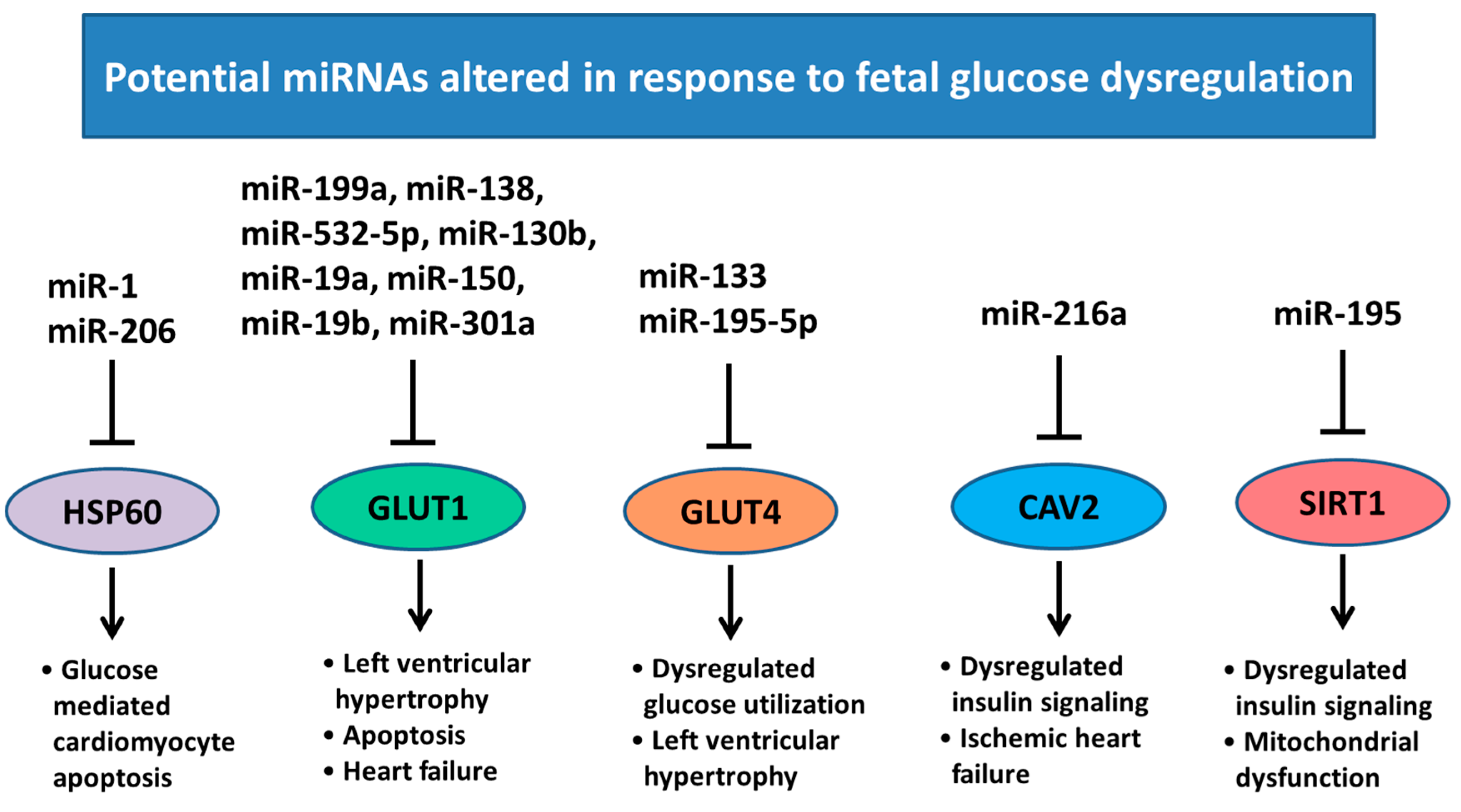
  92. Yu, X.Y.; Song, Y.H.; Geng, Y.J.; Lin, Q.X.; Shan, Z.X.; Lin, S.G.; Li, Y. Glucose induces apoptosis of cardiomyocytes via microRNA-1 and IGF-1. Biochem. Biophys. Res. Commun. 2008, 376, 548–552. [Google Scholar] [CrossRef] [PubMed]
  93. Hathaway, Q.A.; Pinti, M.V.; Durr, A.J.; Waris, S.; Shepherd, D.L.; Hollander, J.M. Regulating MicroRNA Expression: At the Heart of Diabetes Mellitus and the Mitochondrion. Am. J. Physiol. Heart Circ. Physiol. 2017. [Google Scholar] [CrossRef] [PubMed]
  94. Greco, S.; Fasanaro, P.; Castelvecchio, S.; D’Alessandra, Y.; Arcelli, D.; Di Donato, M.; Malavazos, A.; Capogrossi, M.C.; Menicanti, L.; Martelli, F. MicroRNA dysregulation in diabetic ischemic heart failure patients. Diabetes 2012, 61, 1633–1641. [Google Scholar] [CrossRef] [PubMed]
  95. Zhao, F.; Li, B.; Wei, Y.Z.; Zhou, B.; Wang, H.; Chen, M.; Gan, X.D.; Wang, Z.H.; Xiong, S.X. MicroRNA-34a regulates high glucose-induced apoptosis in H9c2 cardiomyocytes. J. Huazhong Univ. Sci. Technol. 2013, 33, 834–839. [Google Scholar] [CrossRef] [PubMed]
  96. Shao, D.; Tian, R. Glucose Transporters in Cardiac Metabolism and Hypertrophy. Compr. Physiol. 2015, 6, 331–351. [Google Scholar] [CrossRef] [PubMed]
  97. Liao, R.; Jain, M.; Cui, L.; D’Agostino, J.; Aiello, F.; Luptak, I.; Ngoy, S.; Mortensen, R.M.; Tian, R. Cardiac-specific overexpression of GLUT1 prevents the development of heart failure attributable to pressure overload in mice. Circulation 2002, 106, 2125–2131. [Google Scholar] [CrossRef] [PubMed]
  98. Kaczmarczyk, S.J.; Andrikopoulos, S.; Favaloro, J.; Domenighetti, A.A.; Dunn, A.; Ernst, M.; Grail, D.; Fodero-Tavoletti, M.; Huggins, C.E.; Delbridge, L.M.; et al. Threshold effects of glucose transporter-4 (GLUT4) deficiency on cardiac glucose uptake and development of hypertrophy. J. Mol. Endocrinol. 2003, 31, 449–459. [Google Scholar] [CrossRef] [PubMed]
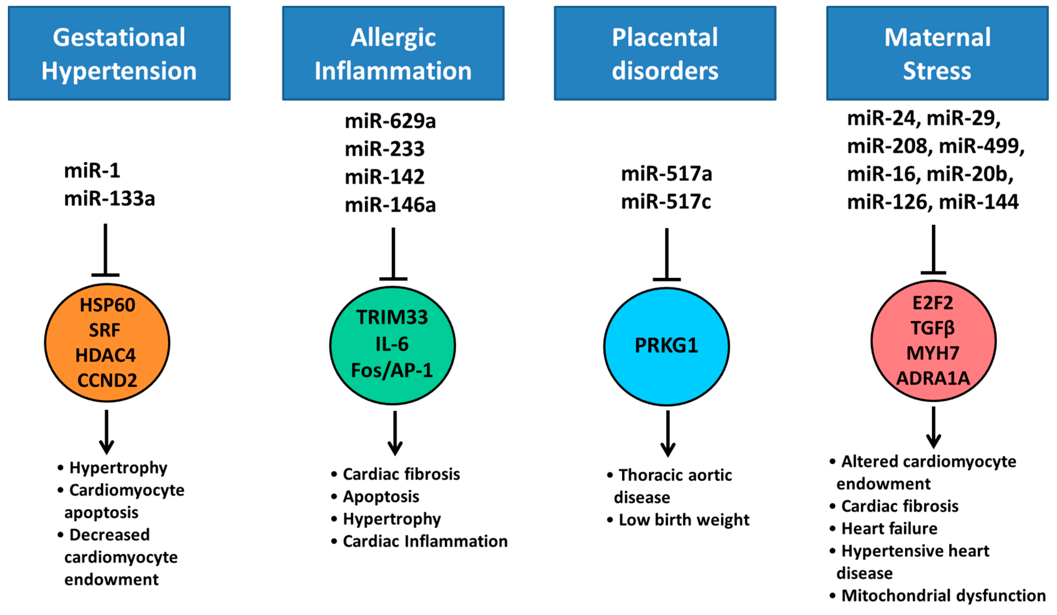
  99. Shan, Z.X.; Lin, Q.X.; Deng, C.Y.; Zhu, J.N.; Mai, L.P.; Liu, J.L.; Fu, Y.H.; Liu, X.Y.; Li, Y.X.; Zhang, Y.Y.; et al. miR-1/miR-206 regulate Hsp60 expression contributing to glucose-mediated apoptosis in cardiomyocytes. FEBS Lett. 2010, 584, 3592–3600. [Google Scholar] [CrossRef] [PubMed]
  100. Pineiro, R.; Iglesias, M.J.; Gallego, R.; Raghay, K.; Eiras, S.; Rubio, J.; Dieguez, C.; Gualillo, O.; Gonzalez-Juanatey, J.R.; Lago, F. Adiponectin is synthesized and secreted by human and murine cardiomyocytes. FEBS Lett. 2005, 579, 5163–5169. [Google Scholar] [CrossRef] [PubMed]
  101. Yamauchi, T.; Kamon, J.; Ito, Y.; Tsuchida, A.; Yokomizo, T.; Kita, S.; Sugiyama, T.; Miyagishi, M.; Hara, K.; Tsunoda, M.; et al. Cloning of adiponectin receptors that mediate antidiabetic metabolic effects. Nature 2003, 423, 762–769. [Google Scholar] [CrossRef] [PubMed]
  102. Park, S.H.; Gammon, S.R.; Knippers, J.D.; Paulsen, S.R.; Rubink, D.S.; Winder, W.W. Phosphorylation-activity relationships of AMPK and acetyl-CoA carboxylase in muscle. J. Appl. Physiol. (1985) 2002, 92, 2475–2482. [Google Scholar] [CrossRef] [PubMed]
  103. Van Raalte, D.H.; Li, M.; Pritchard, P.H.; Wasan, K.M. Peroxisome proliferator-activated receptor (PPAR)-alpha: A pharmacological target with a promising future. Pharm. Res. 2004, 21, 1531–1538. [Google Scholar] [CrossRef] [PubMed]
  104. McGarry, J.D. The mitochondrial carnitine palmitoyltransferase system: Its broadening role in fuel homoeostasis and new insights into its molecular features. Biochem. Soc. Trans. 1995, 23, 321–324. [Google Scholar] [CrossRef] [PubMed]
  105. Wu, P.; Sato, J.; Zhao, Y.; Jaskiewicz, J.; Popov, K.M.; Harris, R.A. Starvation and diabetes increase the amount of pyruvate dehydrogenase kinase isoenzyme 4 in rat heart. Biochem. J. 1998, 329 Pt 1, 197–201. [Google Scholar] [CrossRef] [PubMed]
  106. Sugden, M.C.; Holness, M.J. Mechanisms underlying regulation of the expression and activities of the mammalian pyruvate dehydrogenase kinases. Arch. Physiol. Biochem. 2006, 112, 139–149. [Google Scholar] [CrossRef] [PubMed]
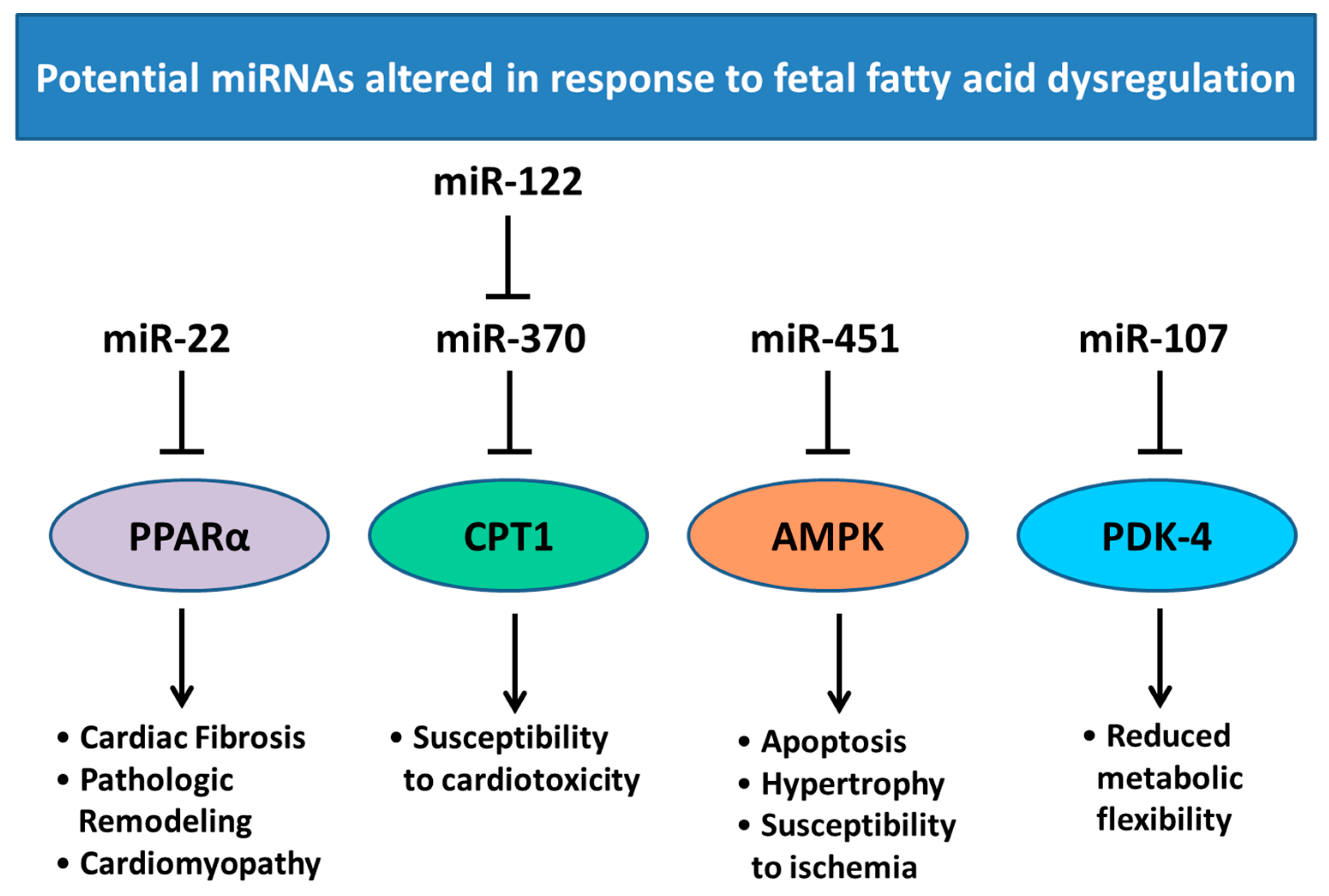
  107. Iliopoulos, D.; Drosatos, K.; Hiyama, Y.; Goldberg, I.J.; Zannis, V.I. MicroRNA-370 controls the expression of microRNA-122 and Cpt1alpha and affects lipid metabolism. J. Lipid Res. 2010, 51, 1513–1523. [Google Scholar] [CrossRef] [PubMed]
  108. Ansari, K.I.; Ogawa, D.; Rooj, A.K.; Lawler, S.E.; Krichevsky, A.M.; Johnson, M.D.; Chiocca, E.A.; Bronisz, A.; Godlewski, J. Glucose-based regulation of miR-451/AMPK signaling depends on the OCT1 transcription factor. Cell Rep. 2015, 11, 902–909. [Google Scholar] [CrossRef] [PubMed]
  109. Gurha, P.; Wang, T.; Larimore, A.H.; Sassi, Y.; Abreu-Goodger, C.; Ramirez, M.O.; Reddy, A.K.; Engelhardt, S.; Taffet, G.E.; Wehrens, X.H.; et al. microRNA-22 promotes heart failure through coordinate suppression of PPAR/ERR-nuclear hormone receptor transcription. PLoS ONE 2013, 8, e75882. [Google Scholar] [CrossRef] [PubMed]
  110. Safdar, A.; Abadi, A.; Akhtar, M.; Hettinga, B.P.; Tarnopolsky, M.A. miRNA in the Regulation of Skeletal Muscle Adaptation to Acute Endurance Exercise in C57Bl/6J Male Mice. PLoS ONE 2009, 4, e5610. [Google Scholar] [CrossRef] [PubMed]
  111. Finck, B.N. The PPAR regulatory system in cardiac physiology and disease. Cardiovasc. Res. 2007, 73, 269–277. [Google Scholar] [CrossRef] [PubMed]
  112. Sayed-Ahmed, M.M.; Aldelemy, M.L.; Al-Shabanah, O.A.; Hafez, M.M.; Al-Hosaini, K.A.; Al-Harbi, N.O.; Al-Sharary, S.D.; Al-Harbi, M.M. Inhibition of gene expression of carnitine palmitoyltransferase I and heart fatty acid binding protein in cyclophosphamide and ifosfamide-induced acute cardiotoxic rat models. Cardiovasc. Toxicol. 2014, 14, 232–242. [Google Scholar] [CrossRef] [PubMed]
  113. Dyck, J.R.B.; Lopaschuk, G.D. AMPK alterations in cardiac physiology and pathology: Enemy or ally? J. Physiol. 2006, 574 Pt 1, 95–112. [Google Scholar] [CrossRef] [PubMed]
  114. Zhang, S.; Hulver, M.W.; McMillan, R.P.; Cline, M.A.; Gilbert, E.R. The pivotal role of pyruvate dehydrogenase kinases in metabolic flexibility. Nutr. Metab. (Lond.) 2014, 11, 10. [Google Scholar] [CrossRef] [PubMed]
  115. Hromadnikova, I.; Kotlabova, K.; Hympanova, L.; Krofta, L. Gestational hypertension, preeclampsia and intrauterine growth restriction induce dysregulation of cardiovascular and cerebrovascular disease associated microRNAs in maternal whole peripheral blood. Thromb. Res. 2016, 137, 126–140. [Google Scholar] [CrossRef] [PubMed]
  116. Li, J.; Zhang, Y.; Li, D.; Liu, Y.; Chu, D.; Jiang, X.; Hou, D.; Zen, K.; Zhang, C.-Y. Small non-coding RNAs transfer through mammalian placenta and directly regulate fetal gene expression. Protein Cell 2015, 6, 391–396. [Google Scholar] [CrossRef] [PubMed]
  117. Staedel, C.; Darfeuille, F. MicroRNAs and bacterial infection. Cell. Microbiol. 2013, 15, 1496–1507. [Google Scholar] [CrossRef] [PubMed]
  118. Roberts, A.P.; Lewis, A.P.; Jopling, C.L. The role of microRNAs in viral infection. Prog. Mol. Biol. Transl. Sci. 2011, 102, 101–139. [Google Scholar] [CrossRef] [PubMed]
  119. Mouillet, J.F.; Ouyang, Y.; Bayer, A.; Coyne, C.B.; Sadovsky, Y. The role of trophoblastic microRNAs in placental viral infection. Int. J. Dev. Biol. 2014, 58, 281–289. [Google Scholar] [CrossRef] [PubMed]
  120. Maes, T.; Cobos, F.A.; Schleich, F.; Sorbello, V.; Henket, M.; De Preter, K.; Bracke, K.R.; Conickx, G.; Mesnil, C.; Vandesompele, J.; et al. Asthma inflammatory phenotypes show differential microRNA expression in sputum. J. Allergy Clin. Immunol. 2016, 137, 1433–1446. [Google Scholar] [CrossRef] [PubMed]
  121. Perry, M.M.; Adcock, I.M.; Chung, K.F. Role of microRNAs in allergic asthma: Present and future. Curr. Opin. Allergy Clin. Immunol. 2015, 15, 156–162. [Google Scholar] [CrossRef] [PubMed]
  122. Hasegawa, Y.; Miura, K.; Higashijima, A.; Abe, S.; Miura, S.; Yoshiura, K.; Masuzaki, H. Increased Levels of Cell-Free miR-517a and Decreased Levels of Cell-Free miR-518b in Maternal Plasma Samples From Placenta Previa Pregnancies at 32 Weeks of Gestation. Reprod. Sci. 2015, 22, 1569–1576. [Google Scholar] [CrossRef] [PubMed]
  123. Kinsella, M.T.; Monk, C. Impact of Maternal Stress, Depression & Anxiety on Fetal Neurobehavioral Development. Clin. Obstet. Gynecol. 2009, 52, 425–440. [Google Scholar] [CrossRef] [PubMed]
  124. DiPietro, J.A. Maternal stress in pregnancy: Considerations for fetal development. J. Adolesc. Health 2012, 51, S3–S8. [Google Scholar] [CrossRef] [PubMed]
  125. Zucchi, F.C.; Yao, Y.; Ward, I.D.; Ilnytskyy, Y.; Olson, D.M.; Benzies, K.; Kovalchuk, I.; Kovalchuk, O.; Metz, G.A. Maternal stress induces epigenetic signatures of psychiatric and neurological diseases in the offspring. PLoS ONE 2013, 8, e56967. [Google Scholar] [CrossRef] [PubMed]
  126. Antolic, A.; Feng, X.; Wood, C.E.; Richards, E.M.; Keller-Wood, M. Increased maternal nighttime cortisol concentrations in late gestation alter glucose and insulin in the neonatal lamb. Physiol. Rep. 2015, 3, e12548. [Google Scholar] [CrossRef] [PubMed]
  127. Davis, E.P.; Sandman, C.A. The Timing of Prenatal Exposure to Maternal Cortisol and Psychosocial Stress is Associated with Human Infant Cognitive Development. Child Dev. 2010, 81, 131–148. [Google Scholar] [CrossRef] [PubMed]
  128. Robertson, S.; MacKenzie, S.M.; Alvarez-Madrazo, S.; Diver, L.A.; Lin, J.; Stewart, P.M.; Fraser, R.; Connell, J.M.; Davies, E. MicroRNA-24 Is a Novel Regulator of Aldosterone and Cortisol Production in the Human Adrenal Cortex. Hypertension 2013, 62, 572–578. [Google Scholar] [CrossRef] [PubMed]
  129. Honda, M.; Kuwano, Y.; Katsuura-Kamano, S.; Kamezaki, Y.; Fujita, K.; Akaike, Y.; Kano, S.; Nishida, K.; Masuda, K.; Rokutan, K. Chronic Academic Stress Increases a Group of microRNAs in Peripheral Blood. PLoS ONE 2013, 8, e75960. [Google Scholar] [CrossRef] [PubMed]
  130. Hu, Z.; Shen, W.-J.; Cortez, Y.; Tang, X.; Liu, L.-F.; Kraemer, F.B.; Azhar, S. Hormonal Regulation of MicroRNA Expression in Steroid Producing Cells of the Ovary, Testis and Adrenal Gland. PLoS ONE 2013, 8, e78040. [Google Scholar] [CrossRef] [PubMed]
  131. Cho, J.H.; Lee, I.; Hammamieh, R.; Wang, K.; Baxter, D.; Scherler, K.; Etheridge, A.; Kulchenko, A.; Gautam, A.; Muhie, S.; et al. Molecular evidence of stress-induced acute heart injury in a mouse model simulating posttraumatic stress disorder. Proc. Natl. Acad. Sci. USA 2014, 111, 3188–3193. [Google Scholar] [CrossRef] [PubMed]
  132. Lumbers, E.R.; Boyce, A.C.; Joulianos, G.; Kumarasamy, V.; Barner, E.; Segar, J.L.; Burrell, J.H. Effects of cortisol on cardiac myocytes and on expression of cardiac genes in fetal sheep. Am. J. Physiol. Regul. Integr. Comp. Physiol. 2005, 288, R567–R574. [Google Scholar] [CrossRef] [PubMed]
  133. Yu, H.; Lu, Y.; Li, Z.; Wang, Q. microRNA-133: Expression, function and therapeutic potential in muscle diseases and cancer. Curr. Drug Targets 2014, 15, 817–828. [Google Scholar] [CrossRef] [PubMed]
  134. Jingushi, K.; Ueda, Y.; Kitae, K.; Hase, H.; Egawa, H.; Ohshio, I.; Kawakami, R.; Kashiwagi, Y.; Tsukada, Y.; Kobayashi, T.; et al. miR-629 Targets TRIM33 to Promote TGFbeta/Smad Signaling and Metastatic Phenotypes in ccRCC. Mol. Cancer Res. 2015, 13, 565–574. [Google Scholar] [CrossRef] [PubMed]
  135. Sun, Y.; Varambally, S.; Maher, C.A.; Cao, Q.; Chockley, P.; Toubai, T.; Malter, C.; Nieves, E.; Tawara, I.; Wang, Y.; et al. Targeting of microRNA-142-3p in dendritic cells regulates endotoxin-induced mortality. Blood 2011, 117, 6172–6183. [Google Scholar] [CrossRef] [PubMed]
  136. Palomer, X.; Capdevila-Busquets, E.; Botteri, G.; Davidson, M.M.; Rodriguez, C.; Martinez-Gonzalez, J.; Vidal, F.; Barroso, E.; Chan, T.O.; Feldman, A.M.; et al. miR-146a targets Fos expression in human cardiac cells. Dis. Model Mech. 2015, 8, 1081–1091. [Google Scholar] [CrossRef] [PubMed]
  137. Gago-Diaz, M.; Blanco-Verea, A.; Teixido, G.; Huguet, F.; Gut, M.; Laurie, S.; Gut, I.; Carracedo, A.; Evangelista, A.; Brion, M. PRKG1 and genetic diagnosis of early-onset thoracic aortic disease. Eur. J. Clin. Investig. 2016, 46, 787–794. [Google Scholar] [CrossRef] [PubMed]
  138. Barchitta, M.; Maugeri, A.; Quattrocchi, A. The Role of miRNAs as Biomarkers for Pregnancy Outcomes: A Comprehensive Review. Int. J. Genom. 2017, 2017, 8067972. [Google Scholar] [CrossRef] [PubMed]
  139. Lal, A.; Navarro, F.; Maher, C.; Maliszewski, L.E.; Yan, N.; O’Day, E.; Chowdhury, D.; Dykxhoorn, D.M.; Tsai, P.; Hofman, O.; et al. miR-24 inhibits cell proliferation by suppressing expression of E2F2, MYC and other cell cycle regulatory genes by binding to “seedless” 3′UTR microRNA recognition elements. Mol. Cell 2009, 35, 610–625. [Google Scholar] [CrossRef] [PubMed]
  140. Van Rooij, E.; Sutherland, L.B.; Thatcher, J.E.; DiMaio, J.M.; Naseem, R.H.; Marshall, W.S.; Hill, J.A.; Olson, E.N. Dysregulation of microRNAs after myocardial infarction reveals a role of miR-29 in cardiac fibrosis. Proc. Natl. Acad. Sci. USA 2008, 105, 13027–13032. [Google Scholar] [CrossRef] [PubMed]
  141. Montgomery, R.L.; Hullinger, T.G.; Semus, H.M.; Dickinson, B.A.; Seto, A.G.; Lynch, J.M.; Stack, C.; Latimer, P.A.; Olson, E.N.; van Rooij, E. Therapeutic Inhibition of miR-208a Improves Cardiac Function and Survival During Heart Failure. Circulation 2011, 124, 1537–1547. [Google Scholar] [CrossRef] [PubMed]
  142. Shieh, J.T.; Huang, Y.; Gilmore, J.; Srivastava, D. Elevated miR-499 levels blunt the cardiac stress response. PLoS ONE 2011, 6, e19481. [Google Scholar] [CrossRef] [PubMed]
  143. He, L.; Huang, C. MiR-19b and miR-16 cooperatively signaling target the regulator ADRA1A in Hypertensive heart disease. Biomed. Pharmacother. 2017, 91, 1178–1183. [Google Scholar] [CrossRef] [PubMed]
  144. Zhang, X.; Wang, X.; Zhu, H.; Zhu, C.; Wang, Y.; Pu, W.T.; Jegga, A.G.; Fan, G.-C. Synergistic Effects of the GATA-4-Mediated miR-144/451 Cluster in Protection against Simulated Ischemia/Reperfusion-Induced Cardiomyocyte Death. J. Mol. Cell. Cardiol. 2010, 49, 841–850. [Google Scholar] [CrossRef] [PubMed]
  145. Zhu, S.; Hu, X.; Yu, Z.; Peng, Y.; Zhu, J.; Liu, X.; Li, M.; Han, S.; Zhu, C. Effect of miR-20b on Apoptosis, Differentiation, the BMP Signaling Pathway and Mitochondrial Function in the P19 Cell Model of Cardiac Differentiation In Vitro. PLoS ONE 2015, 10, e0123519. [Google Scholar] [CrossRef] [PubMed]
Figure 1. Summary of adverse intrauterine environments during pregnancy that may impact miRNA expression resulting in dysregulation of heart development and adverse postnatal physiological outcomes.
Ijms 18 02628 g001
Figure 2. Epigenetic mechanisms. Epigenetic regulation can occur through several different pathways. These include DNA methylation, histone modification and non-coding RNAs. Non-coding RNAs such as miRNAs and lncRNAs are capable of modulating gene expression through suppression of mRNA expression or by direct regulation of epigenetic modifying enzymes.
Ijms 18 02628 g002
Figure 3. Processing of miRNAs: (1) miRNAs are transcribed as 5′-capped large polyadenylated precursor transcripts (pri-miRNA) primarily in a Polymerase II-dependent manner. Approximately 40% of human miRNAs are co-transcribed as clusters encoding up to eight distinct miRNA sequences in a single pri-miRNA transcript; (2) Pri-miRNAs are cleaved within the nucleus by the microprocessor complex containing Drosha (an RNaseIII-type nuclease) and a protein co-factor, DiGeorge syndrome critical region 8 gene (DGCR8) in humans; (3) The resulting 60–70 nucleotide hairpin structure (pre-miRNA) encodes for a single miRNA sequence that is exported from the nucleus to the cytoplasm by Exportin-5; (4) Cytoplasmic pre-miRNAs are further cleaved by another RNaseIII-nuclease, Dicer, in concert with cofactors (TRBP and PACT in humans), to eliminate the pre-miRNA loop sequence forming a short-lived asymmetric duplex intermediate (miRNA: miRNA*); (5) The mature miRNA is then loaded into the miRISC complex with Argonaute proteins; (6) miRNA guided suppression of the target mRNA. Identification of the target mRNA is assisted by the “seed sequence”, which is essential for the binding of the miRNA to the mRNA. The seed sequence is a conserved heptametrical sequence that is typically situated at positions 2–7 from the miRNA 5′-end. Even if the base pairing of miRNA with its target mRNA does not match perfectly, the “seed sequence” has to be perfectly complementary.
Ijms 18 02628 g003
Figure 4. Regulation of miRNA expression in response to hypoxia. miR-138, miR-155 and miR-199a are induced in response to hypoxia, but can also modulate HIF1α; a major regulator of the hypoxia response and associated with vascular remodeling [54,76]. miR-22 regulates SIRT1 and HDAC4 expression that are associated with dilated cardiomyopathy, hypertrophy, and mitochondrial dysfunction [77]. miR-696 regulates PGC1α, dysfunction of this can result in mitochondrial dysfunction and increased susceptibility to pressure overload [78]. GATA4 plays an important role in the response pressure overload [79] and is a target of miR-26. miR-210 targets EFNA3, PTPN1 and HIF3α that have been associated with vascular remodeling [80]. miR-484 regulates expression of FIS1 which is associated with mitochondrial fragmentation and apoptotic cell death [81]. PPARδ plays a major role in the onset of cardiac failure [82] and is a target of miR-214. miR-34a also regulates SIRT1 as well as PNUTS that plays a role in DNA damage responses and cardiomyocyte apoptosis [61]. (SIRT1; Sirtuin 1, HDAC4; Histone deacetylase 4, PGC1α; PPARG coactivator 1 α, GATA4; GATA binding protein 4, EFNA3; Ephrin A3, PTPN1; Protein-tyrosine phosphatase 1B, HIF3α; Hypoxia inducible factor 3 α subunit, FIS1; Fission, Mitochondrial 1, PPARδ; Peroxisome proliferator activated receptor delta, PNUTS; Protein phosphatase 1 regulatory subunit 10). Pointed arrow represent downstream effects, blocked arrows represent downregulation of target genes.
Ijms 18 02628 g004
Figure 5. Regulation of miRNA expression in response to altered glucose transport. miR-199a, miR-138, miR-150, miR-532-5p, miR-130b, miR-19a, miR-19b and miR-301a have been implicated in regulating GLUT1 expression; associated with left ventricular hypertrophy and heart failure [96,97]. miR-133 and 195-5p regulate GLUT4 expression, which has been associated with hypertrophy and glucose homeostasis within the heart [98]. Expression of miR-1 and miR-206 are upregulated by increased glucose concentrations and target HSP60 what has a role in glucose mediated apoptosis in cardiomyocytes [99]. miR-216a regulates CAV2 expression, which plays a role in ischemic heart failure [94]. miR-195 expression is dysregulated by diabetes and acts on SIRT1, involved in insulin signaling and mitochondrial function [95]. HSP60; heat shock protein 60, GLUT1; glucose transporter 1, GLUT4; glucose transporter 4 CAV2; caveolin 2, SIRT1; sirtuin 1. Pointed arrow represent downstream effects, blocked arrows represent downregulation of target genes.
Ijms 18 02628 g005
Figure 6. Regulation of miRNA expression in response to altered fatty acid dysregulation. PPARα has been associated with cardiac fibrosis, pathologic remodeling and cardiomyopathy during development [111] and is a target of miR-22 in the heart. miR-122 regulates miR-370 which in turn regulates CPT1 expression which has been associated with susceptibly to cardiotoxicity [112]. miR-451 mediates AMPK expression which is associated with apoptosis, hypertrophy and increased susceptibility to ischemia [113]. PDK-4 expression has been associated with loss of metabolic flexibility and exacerbation of cardiomyopathy [114] and is a predicted target of miR-107. PPARα; peroxisome proliferator activated receptor, CPT1; carnitine palmitoyltransferase-1, AMPK; AMP-activated protein kinase, PDK-4; pyruvate dehydrogenase kinase-4. Pointed arrow represent downstream effects, blocked arrows represent downregulation of target genes.
Ijms 18 02628 g006
Figure 7. Regulation of miRNA expression in response to maternal disease and stress. HSP60, SRF, HDAC4 and CCND2 have been associated with cardiomyocyte hypertrophy, apoptosis and regulation of cardiomyocyte endowment [133] and are targets of miR-1 and miR-133a in the heart, that are dysregulated by maternal hypertension [91,115]. miR-629a regulates TRIM33 expression that in turn regulates the TGFβ/Smad signaling pathway which has been associated with cardiac fibrosis, hypertrophy and apoptosis [134]. miR-142 mediates a number of target genes including IL-6 [135], a potent myokine. The Fos/AP-1 pathway is a key component of cardiac inflammation and is a predicted target of miR-146a [136]. Placenta produced miR-517a has been associated with low birth fetuses and targets PRKG1 which has been associated with thoracic aortic disease [137,138]. miR-24 is a modulator of cell cycle, targeting E2F2 [139]. miR-29 targets TGFβ expression and has been associated with increased cardiac fibrosis after myocardial infarction [140]. miR-208 and miR-499 has been shown to regulate MYH7 a contractile subunit in the heart, and is associated with heart failure [141,142]. miR-16 targets ADRA1A and is associated with hypertensive heart disease [143]. GATA4 expression has been shown regulate miR-144 expression and modulate the response to ischemia/reperfusion cardiomyocyte injury [144]. miR-20b is an important regulator of apoptosis, differentiation and mitochondrial function [145]. HSP60; heat shock protein 60, SRF; serum response factor; HDAC4; histone deacetylase 4, CCND2; cyclin D2, TRIM33; tripartite motif containing 33, IL-6; interleukin 6, AP-1; activator protein 1, PRKG1; cGMP-dependent protein kinase 1, E2F2; E2F transcription factor 2, TGFβ; transforming growth factor β, MYH7; myosin heavy chain 7, ADRA1A; α-1A adrenergic receptor.
Ijms 18 02628 g007

