2. Results and Discussion
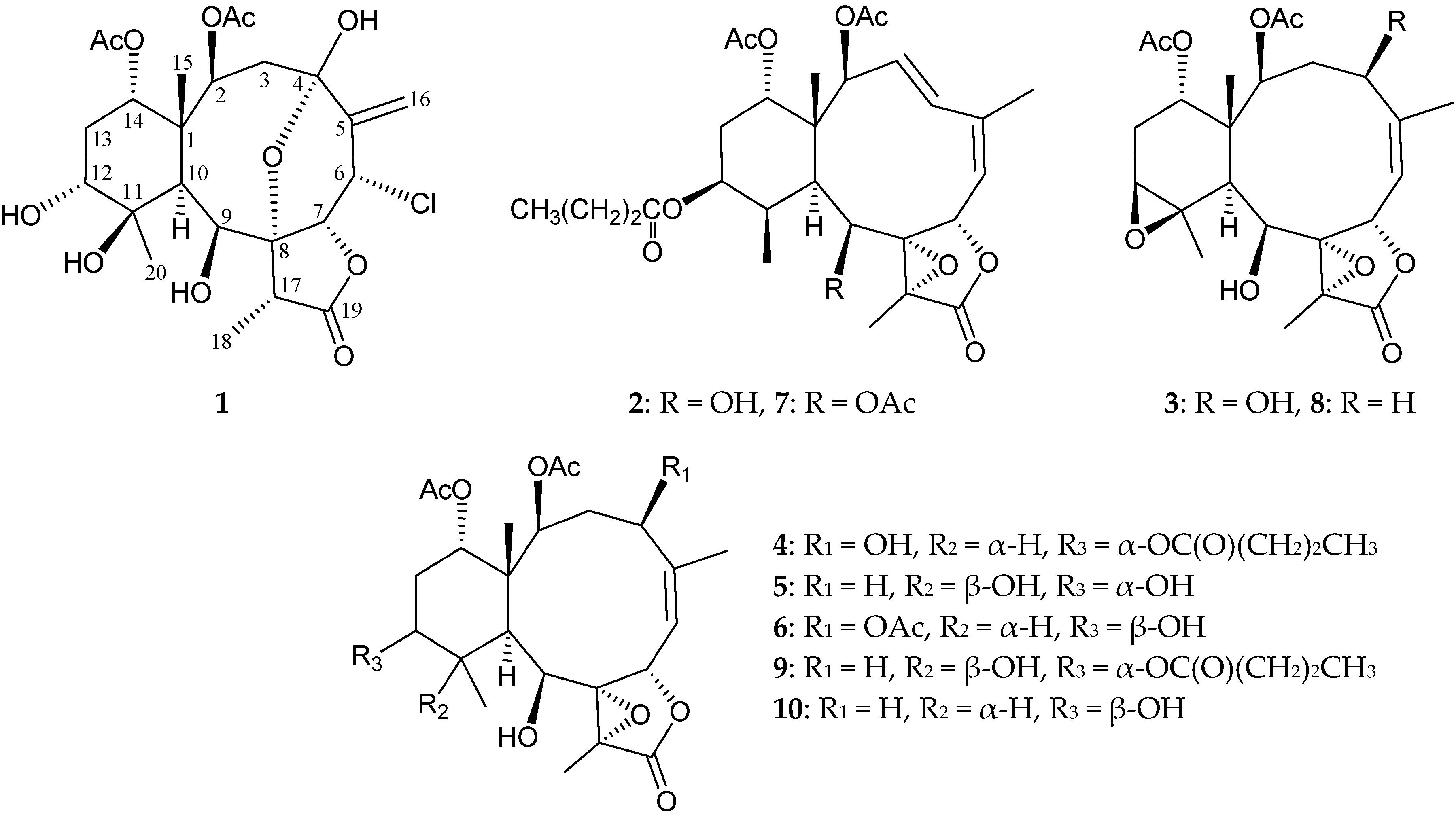
The molecular formula of a new briarane, briarenolide ZI (
1), was determined as C
24H
33ClO
11 (eight degrees of unsaturation) by high-resolution electrospray ionization mass spectrum (HRESIMS) at
m/
z 555.16025 (calcd. for C
24H
33ClO
11 + Na, 555.16036). The IR of
1 showed absorptions at 1715, 1769 and 3382 cm
−1, which were consistent with the presence of ester, γ-lactone and hydroxy groups. The
13C NMR spectrum (
Table 1) suggested that
1 possessed an exocyclic carbon-carbon double bond based on signals at δ
C 138.6 (C-5) and 116.9 (CH
2-16), which was confirmed by the
1H NMR spectrum of
1 (
Table 1), which showed two olefin proton signals at δ
H 5.88 (1H, dd,
J = 2.4, 1.2 Hz, H-16a) and 5.64 (1H, dd,
J = 2.4, 1.2 Hz, H-16b). Three carbonyl resonances at δ
C 175.3 (C-19), 173.4 and 169.3 (2 × ester carbonyls) revealed the presence of one γ-lactone and two ester groups in
1; two acetyl methyls (δ
H 2.06, s, 2 × 3H) were also observed. According to the overall unsaturation data, it was concluded that
1 was a diterpenoid molecule possessing four rings.
1H NMR coupling information in the
1H–
1H correlation spectroscopy (COSY) spectrum of
1 enabled identification of the H-2/H
2-3, H-6/H-7, H-12/H
2-13/H-14, H-6/H
2-16 (by allylic coupling) and H-17/H
3-18 units (
Table 1). The heteronuclear multiple bond coherence (HMBC) correlations between protons and quaternary carbons of
1 (H-2, H
2-3, H-10, H
2-13, H
3-15/C-1; H-2, H
2-3, H
2-16, OH-4/C-4; H-16b, OH-4/C-5; H-10, H
3-18, OH-9/C-8; H
3-20/C-11 and H-17, H
3-18/C-19) permitted elucidation of the carbon skeleton (
Table 1). HMBC correlations between H
2-16/C-4, -5 and -6 indicated an exocyclic double bond at C-5, which was further confirmed by the allylic coupling between H
2-16/H-6. HMBC correlations between H
3-15/C-1, -2, -10 and -14 and H-2 and H-10/C-15, revealed that the ring junction C-15 methyl group was located at C-1. Furthermore, an HMBC correlation between H-2 (δ
H 5.09) and the acetate carbonyl (δ
C 173.4) revealed the presence of an acetate ester at C-2; and an HMBC correlation between a hydroxy proton (δ
H 6.50) and C-4 oxygenated quaternary carbon suggested the presence of a hydroxy group at C-4. The C-4 hydroxy group was determined to be part of a hemiketal constellation on the basis of a characteristic carbon signal at δ
C 96.7.
1H–
1H COSY correlations between OH-9/H-9 and OH-12/H-12 suggested the presence of the hydroxy groups at C-9 and C-12. A carbon signal at δ
C 81.8 (C-8) indicated
3J-coupling with protons at δ
H 2.23 (H-10), 1.33 (H
3-18) and 2.73 (OH-9). Therefore, the remaining hydroxy and acetoxy groups had to be positioned at C-11 and C-14, respectively, as indicated by analysis of
1H–
1H COSY correlations and characteristic NMR signal analysis. The intensity of the sodiated molecules [M + 2 + Na]
+ isotope peak observed in the ESIMS and HRESIMS spectra ([M + Na]
+:[M + 2 + Na]
+ = 3:1) was evidence of the presence of one chlorine atom in
1. The methine unit at δ
C 56.2 was more shielded than expected for an oxygenated carbon and was correlated to the methine proton at δ
H 5.54 (H-6) in the heteronuclear multiple quantum coherence (HMQC) spectrum, and this proton signal was
3J-correlated with H-7 (δ
H 4.73) in the
1H–
1H COSY spectrum, which proved that a chlorine atom was attached at C-6. These data, together with the HMBC correlations between H-17/C-9, -18 and -19 and H
3-18/C-8, -17 and -19, established the molecular framework of
1.
Table 1.
1H (400 MHz, CDCl3) and 13C (100 MHz, CDCl3) NMR data and 1H–1H COSY (correlation spectroscopy) and HMBC (heteronuclear multiple bond coherence) correlations for briarane 1.
Table 1.
1H (400 MHz, CDCl3) and 13C (100 MHz, CDCl3) NMR data and 1H–1H COSY (correlation spectroscopy) and HMBC (heteronuclear multiple bond coherence) correlations for briarane 1.
| Position | δH (J in Hz) | δC, Multiple | 1H–1H COSY | HMBC |
|---|
| 1 | – | 45.6, C | – | – |
| 2 | 5.09 d (6.4) | 73.4, CH | H2-3 | C-1, -4, -15, acetate carbonyl |
| 3 | 3.73 dd (16.0, 6.4); 1.46 d (16.0) | 41.7, CH2 | H-2 | C-1, -2, -4 |
| 4 | – | 96.7, C | – | – |
| 5 | – | 138.6, C | – | – |
| 6 | 5.54 dt (2.8, 2.4) | 56.2, CH | H-7, H2-16 | n. o. a |
| 7 | 4.73 d (2.8) | 79.8, CH | H-6 | n. o. |
| 8 | – | 81.8, C | – | – |
| 9 | 4.88 d (3.2) | 76.9, CH | H-10, OH-9 | n. o. |
| 10 | 2.23 s | 40.5, CH | H-9 | C-1, -2, -8, -9, -15 |
| 11 | – | 78.5, C | – | – |
| 12 | 3.50 br s | 76.1, CH | H2-13, OH-12 | n. o. |
| 13 | 2.44 ddd (15.6, 4.0, 2.8); 1.98 ddd (15.6, 3.2, 2.8) | 28.0, CH2 | H-12, H-14 | C-1 |
| 14 | 5.22 t (2.8) | 76.3, CH | H2-13 | n. o. |
| 15 | 1.55 s | 16.5, CH3 | – | C-1, -2, -10, -14 |
| 16a/b | 5.88 dd (2.4, 1.2); 5.64 dd (2.4, 1.2) | 116.9, CH2 | H-6 | C-4, -5, -6 |
| 17 | 2.58 q (7.2) | 50.4, CH | H3-18 | C-9, -18, -19 |
| 18 | 1.33 d (7.2) | 8.2, CH3 | H-17 | C-8, -17, -19 |
| 19 | – | 175.3, C | – | – |
| 20 | 1.56 s | 28.9, CH3 | – | C-10, -11, -12 |
| OAc-2 | – | 173.4, C | – | – |
| | 2.06 s | 21.3, CH3 | – | Acetate carbonyl |
| OAc-14 | – | 169.3, C | – | – |
| | 2.06 s | 21.1, CH3 | – | Acetate carbonyl |
| OH-4 | 6.50 s | – | – | C-3, -4, -5 |
| OH-9 | 2.73 d (3.2) | – | H-9 | C-8 |
| OH-12 | 2.67 br s | – | H-12 | n. o. |
The relative configuration of
1 was elucidated on the basis of a nuclear Overhauser effect spectroscopy (NOESY) experiment and by vicinal
1H–
1H proton coupling constant analysis. Most naturally-occurring briarane natural products have Me-15 in the β-orientation and H-10 in the α-orientation [
2,
3,
4,
5,
6], which were verified by the absence of a correlation between these two groups. In the NOESY experiment of
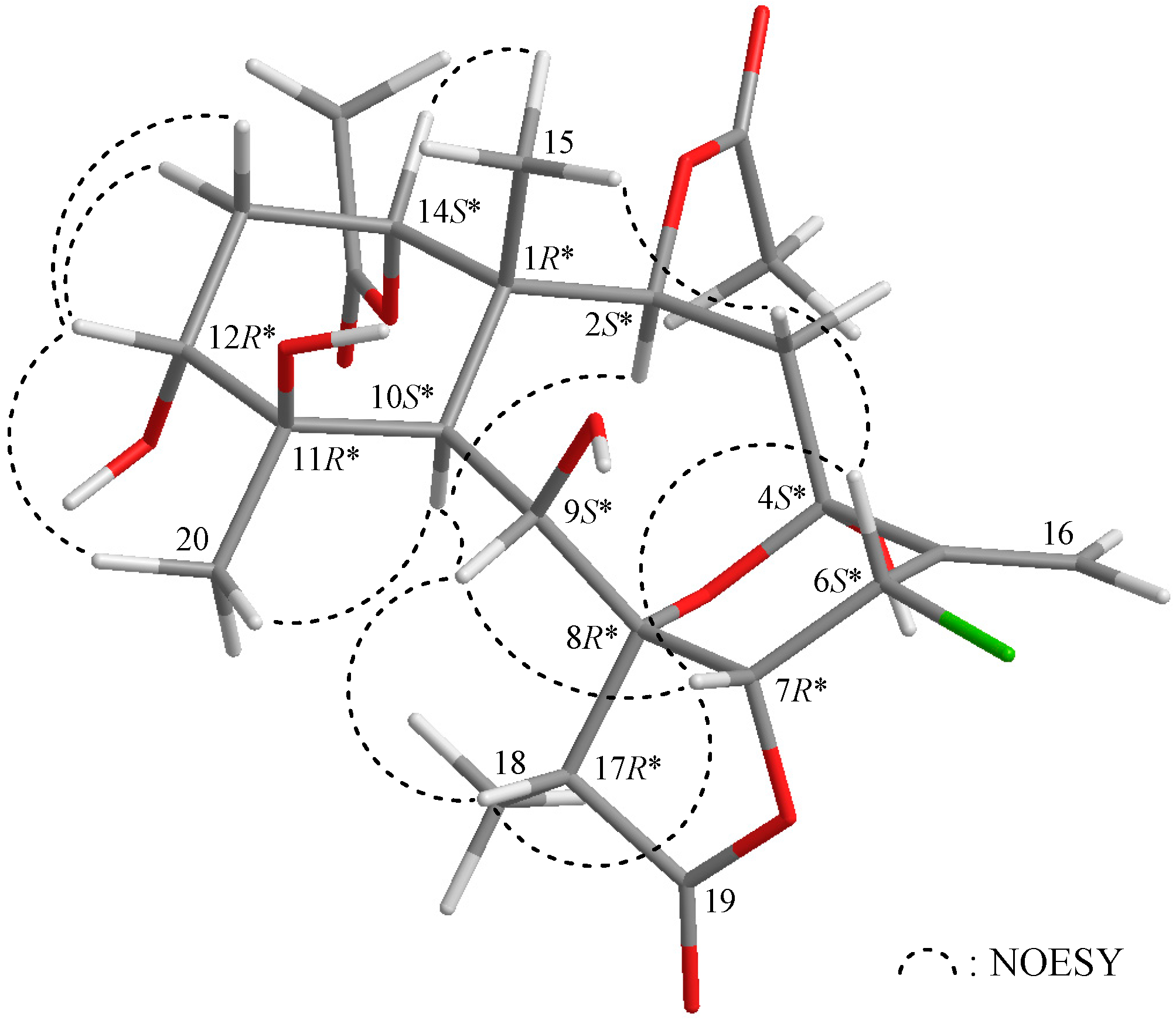
1 (
Figure 2), H-10 correlated with H-2, H-9 and H
3-20, indicating that these protons were situated on the same face; they were assigned as α protons, as C-15 methyl was β-oriented at C-1. The oxymethine proton H-14 was found to exhibit a response with H
3-15, but not with H-10, revealing that H-14 was β-oriented. H-12 correlated with each of the C-13 methylene protons and H
3-20, but not with H-10, indicating that H-12 was β-oriented and was positioned on the equatorial direction in the cyclohexane ring by modeling analysis. H-17 exhibited correlations with H-9 and H-7 and was also found to be reasonably close to H-9 and H-7 by modeling analysis; thus, H-17 could therefore be placed on the β face in
1, and H-7 was β-oriented. One of the C-3 methylene protons (δ
H 3.73) displayed a correlation with H
3-15; therefore, it was assigned as the H-3β proton, and the other was assigned as H-3α (δ
H 1.46). H-6 displayed correlations with H-3β and H-7, which confirmed that this proton was in the β-orientation, and the oxygen bridge between C-4 and C-8 was found to be α-oriented by modeling analysis. Based on the aforementioned results, the structure, including the relative configuration, of
1 was elucidated unambiguously.
Figure 2.
Selected protons with key nuclear Overhauser effect spectroscopy (NOESY) correlations of 1.
Figure 2.
Selected protons with key nuclear Overhauser effect spectroscopy (NOESY) correlations of 1.
Briarenolide ZII (
2) was isolated as a white powder and had a molecular formula of C
28H
38O
10 on the basis of HRESIMS at
m/
z 557.23552 (calcd. for C
28H
38O
10 + Na, 557.23572). Carbonyl resonances in the
13C NMR spectrum of
2 (
Table 2) at δ
C 173.0, 170.7, 170.4 and 169.9 demonstrated the presence of a γ-lactone and three other esters in
2. It was found that the NMR signals of
2 were similar to those of a known briarane analogue, excavatolide F (
7) [
7] (
Figure 1), except that the signals corresponding to the 9-acetoxy group in
7 were replaced by signals for a hydroxy group in
2. The correlations from a NOESY experiment of
2 also revealed that the stereochemistry of this metabolite was identical to that of
7. Thus, briarenolide ZII (
2) was found to be the 9-
O-deacetyl derivative of
7.
Briarenolide ZIII (
3) had a molecular formula C
24H
32O
10 as deduced from HRESIMS at
m/
z 503.18858 (calcd. for C
24H
32O
10 + Na, 503.18877). The IR spectrum of
1 showed three bands at 3444, 1779 and 1732 cm
−1, which were in agreement with the presence of hydroxy, γ-lactone and ester groups. Carbonyl resonances in the
13C NMR spectrum of
3 at δ
C 171.8, 170.7 and 170.6 revealed the presence of a γ-lactone and two esters (
Table 3). Both esters were identified as acetates by the presence of two acetyl methyl resonances in the
1H (δ
H 2.01, 1.98, each 3H × s) and
13C (δ
C 21.1, 21.1) NMR spectra (
Table 3).
It was found that the NMR data of
3 were similar to those of a known briarane analogue, 2β-acetoxy-2-(debutyryloxy)-stecholide E (
8) [
1] (
Figure 1), except that the signals corresponding to the 4-hydroxy group in
3 were not present in
8. A correlation from the NOESY signals of
3 showed that H-4 correlated with H-2, but not with H
3-15, indicating that the hydroxy group at C-4 was β-oriented. The results of
1H–
1H COSY and HMBC correlations fully supported the positions of functional groups, and hence, briarenolide ZIII (
3) was found to be the 4β-hydroxy derivative of
8.
Table 2.
1H (400 MHz, CDCl3) and 13C (100 MHz, CDCl3) NMR data and 1H–1H COSY and HMBC correlations for briarane 2.
Table 2.
1H (400 MHz, CDCl3) and 13C (100 MHz, CDCl3) NMR data and 1H–1H COSY and HMBC correlations for briarane 2.
| Position | δH (J in Hz) | δC, Multiple | 1H–1H COSY | HMBC |
|---|
| 1 | – | 45.6, C | – | – |
| 2 | 5.39 d (10.0) | 75.9, CH | H-3 | C-1, -3, -4, -14, -15, acetate carbonyl |
| 3 | 5.76 dd (16.0, 10.0) | 126.0, CH | H-2, H-4 | C-5 |
| 4 | 6.82 d (16.0) | 139.0, CH | H-3, H-6, H3-16 | C-2, -3, -5, -6 |
| 5 | – | 140.4, C | – | – |
| 6 | 5.44 dq (4.4, 1.6) | 118.4, CH | H-4, H-7, H3-16 | C-4, -8 |
| 7 | 5.10 d (4.4) | 76.8, CH | H-6 | C-5, -6 |
| 8 | – | 69.9, C | – | – |
| 9 | 4.36 d (9.6) | 74.6, CH | H-10 | C-1, -8, -10, -11, -17 |
| 10 | 2.08 d (4.8) | 38.5, CH | H-9, H-11 | C-1, -2, -8, -9, -11, -14, -15, -20 |
| 11 | 2.23 m | 39.2, CH | H-10, H-12, H3-20 | C-1, -10, -12, -13, -20 |
| 12 | 4.98 m | 70.3, CH | H-11, H2-13 | C-20, -1′ |
| 13 | 2.03 m; 1.84 dt (14.4, 3.2) | 26.3, CH2 | H-12, H-14 | C-12 |
| 14 | 4.95 t (3.2) | 74.3, CH | H2-13 | C-15, acetate carbonyl |
| 15 | 1.43 s | 16.1, CH3 | – | C-1, -2, -10, -14 |
| 16 | 1.89 br s | 23.5, CH3 | H-4, H-6 | C-4, -5, -6 |
| 17 | – | 63.4, C | H3-18 | – |
| 18 | 1.52 s | 10.0, CH3 | H-17 | C-7, -8, -19 |
| 19 | – | 170.7, C | – | – |
| 20 | 1.15 d (7.2) | 10.5, CH3 | H-11 | C-10, -11, -12 |
| OAc-2 | – | 169.9, C | – | – |
| | 1.98 s | 21.2, CH3 | – | Acetate carbonyl |
| OAc-14 | – | 170.4, C | – | – |
| | 2.09 s | 21.3, CH3 | – | Acetate carbonyl |
| OC(O)Pr-12 1′2′3′4′ | – | – | – | – |
| 1′ | – | 173.0, C | – | – |
| 2′ | 2.26 t (7.2) | 36.3, CH2 | H2-3′ | C-1′, -3′, -4′ |
| 3′ | 1.61 sext (7.2) | 18.4, CH2 | H2-2′, H3-4′ | C-1′, -2′, -4′ |
| 4′ | 0.94 t (7.2) | 13.7, CH3 | H2-3′ | C-2′, -3′ |
Table 3.
1H (400 MHz, CDCl3) and 13C (100 MHz, CDCl3) NMR data and 1H–1H COSY and HMBC correlations for briarane 3.
Table 3.
1H (400 MHz, CDCl3) and 13C (100 MHz, CDCl3) NMR data and 1H–1H COSY and HMBC correlations for briarane 3.
| Position | δH (J in Hz) | δC, Multiple | 1H–1H COSY | HMBC |
|---|
| 1 | – | 45.7, C | – | – |
| 2 | 4.72 d (6.0) | 73.8, CH | H2-3 | C-1, -4, -10, -14, -15, acetate carbonyl |
| 3 | 3.05 m; 1.92 m | 40.8, CH2 | H-2, H-4 | C-1, -4, -5 |
| 4 | 4.23 dd (12.4, 5.2) | 71.3, CH | H2-3 | C-5, -6, -16 |
| 5 | – | 147.5, C | – | – |
| 6 | 5.49 dt (9.6, 1.2) | 122.0, CH | H-7, H3-16 | C-4, -16 |
| 7 | 6.22 d (9.6) | 73.4, CH | H-6 | C-5, -6 |
| 8 | – | 71.0, C | – | – |
| 9 | 4.45 dd (6.0, 3.6) | 72.2, CH | H-10, OH-9 | C-7, -8, -11 |
| 10 | 2.29 d (3.6) | 42.5, CH | H-9 | C-1, -8, -9, -11, -15 |
| 11 | – | 63.6, C | – | – |
| 12 | 3.05 d (2.8) | 61.4, CH | H2-13 | n. o. a |
| 13 | 2.08 m | 25.2, CH2 | H-12, H-14 | n. o. |
| 14 | 4.73 br s | 73.8, CH | H2-13 | C-1, -2, -10, -12, -15, acetate carbonyl |
| 15 | 1.19 s | 16.0, CH3 | – | C-1, -10, -14 |
| 16 | 2.11 d (1.2) | 25.5, CH3 | H-6 | C-4, -5, -6 |
| 17 | – | 62.5, C | – | – |
| 18 | 1.67 s | 9.4, CH3 | – | C-8, -17, -19 |
| 19 | – | 171.8, C | – | – |
| 20 | 1.35 s | 24.5, CH3 | – | C-10, -11, -12 |
| OAc-2 | – | 170.7, C | – | – |
| | 1.98 s | 21.1, CH3 | – | Acetate carbonyl |
| OAc-14 | – | 170.6, C | – | – |
| | 2.01 s | 21.1, CH3 | – | Acetate carbonyl |
| OH-19 | 2.89 d (6.0) | – | H-9 | C-8 |
Briarenolide ZIV (
4) was obtained as a white powder, and the molecular formula of
4 was determined to be C
28H
40O
11 (9° of unsaturation) by HRESIMS at
m/
z 575.24645 (calcd. for C
28H
40O
11 + Na, 575.24628). The IR spectrum of
4 showed three bands at 3444, 1778 and 1732 cm
−1, consistent with the presence of hydroxy, γ-lactone and ester carbonyl groups. Carbonyl resonances in the
13C NMR spectrum of
4 showed signals at δ
C 173.9, 173.2, 170.8 and 170.4, which revealed the presence of a γ-lactone and three esters in
4 (
Table 4), of which, two of the esters were identified as acetates based on the presence of two acetyl methyl resonances in the
1H NMR spectrum of
4 at δ
H 1.97 (2 × 3H, s) (
Table 4). The other ester was found to be an
n-butyrate group based on
1H NMR studies, which revealed seven contiguous protons (δ
H 0.94, 3H, t,
J = 7.2 Hz; 1.65, 2H, sextet,
J = 7.2 Hz; 2.23, 2H, t,
J = 7.2 Hz). According to the
1H and
13C NMR spectra,
4 was found to have a γ-lactone moiety (δ
C 173.9, C-19) and a trisubstituted olefin (δ
C 145.4, C-5; 121.6, CH-6; δ
H 5.32, 1H, d,
J = 8.8 Hz, H-6). The presence of a tetrasubstituted epoxide that contained a methyl substituent was established based on the signals of two oxygenated quaternary carbons at δ
C 71.8 (C-8) and 63.7 (C-17) and confirmed by the proton signals of a methyl singlet at δ
H 1.51 (3H, s, H
3-18). Thus, from the NMR data, five degrees of unsaturation were accounted for, and
4 was identified as a tetracyclic compound. From the
1H–
1H COSY spectrum of
4 (
Table 4), three different structural units, including C-2/-3/-4, C-6/-7 and C-9/-10/-11/-12/-13/-14, were identified. From these data and the HMBC correlation results (
Table 4), the connectivity from C-1 to C-14 could be established. A methyl attached at C-5 was confirmed by an allylic coupling between H
3-16/H-6 and by the HMBC correlations between H
3-16/C-4, -5 and -6. The C-15 and C-20 methyl groups were identified as being positioned at C-1 and C-11 from the HMBC correlations between H
3-15/C-1, -2, -10, -14 and H
3-20/C-10, -11, -12, respectively. Furthermore, the acetate esters positioned at C-2 and C-14 were established by the HMBC correlations between δ
H 4.97 (H-2) and 4.70 (H-14) and the acetate carbonyls at δ
C 170.4 and 170.8, respectively. The location of an
n-butyrate group in
4 was verified by an HMBC correlation between H-12 (δ
H 4.83) and the n-butyrate carbonyl carbon (δ
C 173.2) (
Table 4). These data, together with the HMBC correlations between H
3-18/C-8, -17 and -19, established the main molecular framework of
4. The NMR data of
4 were found to be similar to those of a known briarane, excavatolide Z (
9) [
8] (
Figure 1), except that the signals corresponding to the 4-hydroxy group in
4 were not present in
9, and an 11β-hydroxy group was found in
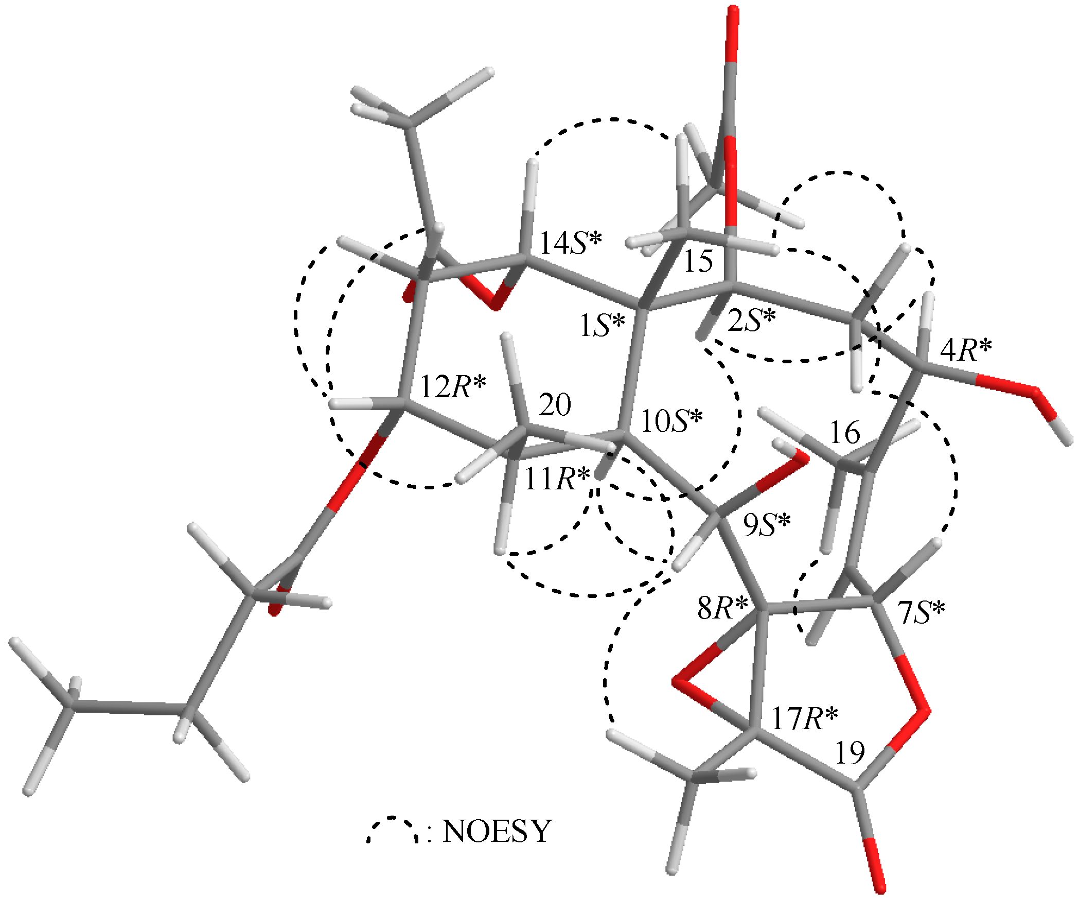
9. The correlations from NOESY signals of
4 (
Figure 3) also showed that the relative configurations of most chiral centers of
4 were similar to those of
9. H-10 exhibited interactions with H-2 and H-11, and H-2 correlated with H-4, indicating that the hydroxy group at C-4 and the methyl group at C-11 were β-oriented; additionally, briarenolide ZIV (
4) was found to be the 4β-hydroxy-11-dehydroxy-11β-methyl derivative of
9.
Figure 3.
Selected protons with key NOESY correlations of 4.
Figure 3.
Selected protons with key NOESY correlations of 4.
Table 4.
1H (400 MHz, CDCl3) and 13C (100 MHz, CDCl3) NMR data and 1H–1H COSY and HMBC correlations for briarane 4.
Table 4.
1H (400 MHz, CDCl3) and 13C (100 MHz, CDCl3) NMR data and 1H–1H COSY and HMBC correlations for briarane 4.
| Position | δH (J in Hz) | δC, Multiple | 1H–1H COSY | HMBC |
|---|
| 1 | – | 46.1, C | – | – |
| 2 | 4.97 d (8.0) | 74.9, CH | H2-3 | C-1, -4, -10, -15, acetate carbonyl |
| 3 | 3.22 dd (15.2, 12.0); 1.93 m | 39.7, CH2 | H-2, H-4 | C-1, -4 |
| 4 | 4.16 dd (12.0, 5.2) | 71.3, CH | H2-3 | C-3, -5, -6, -16 |
| 5 | – | 145.4, C | – | – |
| 6 | 5.32 d (8.8) | 121.6, CH | H-7, H3-16 | C-4, -16 |
| 7 | 6.14 d (8.8) | 75.4, CH | H-6 | C-5, -6, -19 |
| 8 | – | 71.8, C | – | – |
| 9 | 3.79 br s | 74.1, CH | H-10 | C-1, -7, -8, -10, -11, -17 |
| 10 | 2.39 d (5.2) | 37.2, CH | H-9, H-11 | C-1, -2, -8, -9, -11, -12, -14, -15, -20 |
| 11 | 1.88 m | 43.2, CH | H-10, H-12, H3-20 | C-1, -10, -12, -20 |
| 12 | 4.83 br s | 72.1, CH | H-11, H2-13 | C-10, -14, -1′ |
| 13 | 2.11 m; 1.95 m | 24.6, CH2 | H-12, H-14 | C-11, -12, -14 |
| 14 | 4.70 br s | 74.2, CH | H2-13 | C-1, -2, -10, -12, -15, acetate carbonyl |
| 15 | 1.32 s | 15.2, CH3 | – | C-1, -2, -10, -14 |
| 16 | 2.05 d (1.2) | 25.3, CH3 | H-6 | C-4, -5, -6 |
| 17 | – | 63.7, C | – | – |
| 18 | 1.51 s | 9.7, CH3 | – | C-8, -17, -19 |
| 19 | – | 173.9, C | – | – |
| 20 | 1.25 d (7.2) | 15.2, CH3 | H-11 | C-10, -11, -12 |
| OAc-12 | – | 170.4, C | – | – |
| | 1.97 s | 21.2, CH3 | – | Acetate carbonyl |
| OAc-14 | – | 170.8, C | – | – |
| | 1.97 s | 21.5, CH3 | – | Acetate carbonyl |
| OC(O)Pr-12 1′2′3′4′ | – | – | – | – |
| 1′ | – | 173.2, C | | – |
| 2′ | 2.23 t (7.2) | 36.6, CH2 | H2-3′ | C-1′, -3′, -4′ |
| 3′ | 1.65 sext (7.2) | 18.5, CH2 | H2-2′, H3-4′ | C-1′, -2′, -4′ |
| 4′ | 0.94 t (7.2) | 13.6, CH3 | H2-3′ | C-2′, -3′ |
Briarenolide ZV (
5) was obtained as a white powder and had the molecular formula C
24H
30O
10, as determined by HRESIMS at
m/
z 505.20460 (calcd. for C
24H
30O
10 + Na, 505.20442) (10° of unsaturation). The IR spectrum of
5 showed bands at 3445, 1770 and 1732 cm
−1, consistent with the presence of hydroxy, γ-lactone and ester carbonyl groups. Comparison of the
1H and distortioneless enhancement by polar transfer (DEPT) spectra with the molecular formula revealed that there must be three exchangeable protons, requiring the presence of three hydroxy groups. In addition, it was found that the spectral data (IR,
1H and
13C NMR) of
5 (
Table 5) were similar to those of a known briarane, excavatolide Z (
9) [
8] (
Figure 1), except that
9 exhibited signals representing an
n-butyrate substitution, which were replaced by a hydroxy group in
5. The results of
1H–
1H COSY and HMBC correlations fully supported the positions of functional groups, and hence, briarenolide ZV (
5) was found to be the 12-
O-debutyryl derivative of
9.
The new briarane, briarenolide ZVI (
6), had a molecular formula of C
26H
36O
11 as determined by HRESIMS at
m/
z 547.21473 (calcd. for C
26H
36O
11 + Na, 547.21498). Thus, nine degrees of unsaturation were therefore determined for the molecule of
6. In addition, the spectral data (IR,
1H and
13C NMR) (
Table 6) of
6 were found to be similar to those of a known briarane, excavatolide E (
10) [
9] (
Figure 1). However, the NMR spectra revealed that the signals representing the C-4 methylene group in
10 were replaced by those of an additional acetoxy group. In the NOESY experiment of
6, H-10 gives correlations to H-2, H-9, H-11 and H-12, but not to H
3-15 and H
3-20, and H-2 was found to show a correlation with H-4, indicating that these protons (H-2, H-4, H-9, H-10, H-11 and H-12) are located on the same face of the molecule and assigned as α-protons, since the C-15 and C-20 methyls are the β-substituents at C-1 and C-11, respectively. The signal of H
3-20 showed a correlation with H
3-18, indicating that H
3-18 and 8,17-epoxide group were β- and α-oriented, respectively, in the γ-lactone ring in
6. H-4 correlated with H-2, but not with H-7 and H
3-15, indicating that H-7 was β-oriented. H-14 was found to exhibit nuclear Overhauser effect (NOE) responses with H-2 and H
3-15, but not with H-10, revealing the β-orientation of this proton. Thus, based on the above findings, Compound
6 was found to be the 4β-acetoxy derivative of
10, with a structure as described by Formula
6. Furthermore, the chemical shifts for H
3-18 in briaranes
4,
5 and
6 were found to appear at δ
H 1.51, 1.68 and 1.57, respectively, indicating that the 11β-hydroxy group in
5 led to a downfield chemical shift for H
3-18.
Table 5.
1H (400 MHz, CDCl3) and 13C (100 MHz, CDCl3) NMR data and 1H–1H COSY and HMBC correlations for briarane 5.
Table 5.
1H (400 MHz, CDCl3) and 13C (100 MHz, CDCl3) NMR data and 1H–1H COSY and HMBC correlations for briarane 5.
| Position | δH (J in Hz) | δC, Multiple | 1H–1H COSY | HMBC |
|---|
| 1 | – | 48.6, C | – | – |
| 2 | 5.02 d (7.2) | 75.7, CH | H2-3 | C-1, -3, -4, -10, -14, -15, acetate carbonyl |
| 3 | 2.86 td (15.2, 5.2); 1.59 m | 32.5, CH2 | H-2, H2-4 | n. o. a |
| 4 | 2.50 br d (15.2); 1.91 m | 28.7, CH2 | H2-3 | n. o. |
| 5 | – | 146.0, C | – | – |
| 6 | 5.28 d (9.6) | 117.9, CH | H-7, H3-16 | C-4 |
| 7 | 5.50 d (9.6) | 75.1, CH | H-6 | C-5 |
| 8 | – | 71.1, C | – | – |
| 9 | 4.65 dd (5.6, 2.0) | 69.7, CH | H-10, OH-9 | C-7, -8, -10, -11, -17 |
| 10 | 2.13 br s | 44.0, CH | H-9 | C-9 |
| 11 | – | 78.6, C | – | – |
| 12 | 3.43 br d (10.0) | 76.6, CH | H2-13, OH-12 | n. o. |
| 13 | 2.32 m; 1.92 m | 26.5, CH2 | H-12, H-14 | n. o. |
| 14 | 4.99 t (2.8) | 77.5, CH | H2-13 | C-1, -10, -15, acetate carbonyl |
| 15 | 1.42 s | 15.9, CH3 | – | C-1, -2, -10, -14 |
| 16 | 2.00 s | 26.9, CH3 | H-6 | C-4, -5, -6 |
| 17 | – | 63.4, C | – | – |
| 18 | 1.68 s | 9.6, CH3 | – | C-8, -17, -19 |
| 19 | – | 171.6, C | – | |
| 20 | 1.41 s | 31.1, CH3 | – | C-10, -11, -12 |
| OAc-2 | – | 170.8, C | – | – |
| | 1.99 s | 21.4, CH3 | – | Acetate carbonyl |
| OAc-14 | – | 169.8, C | – | – |
| | 2.03 s | 21.7, CH3 | – | Acetate carbonyl |
| OH-9 | 2.45 br s | – | H-9 | n. o. |
| OH-12 | 2.74 d (10.0) | – | H-12 | n. o. |
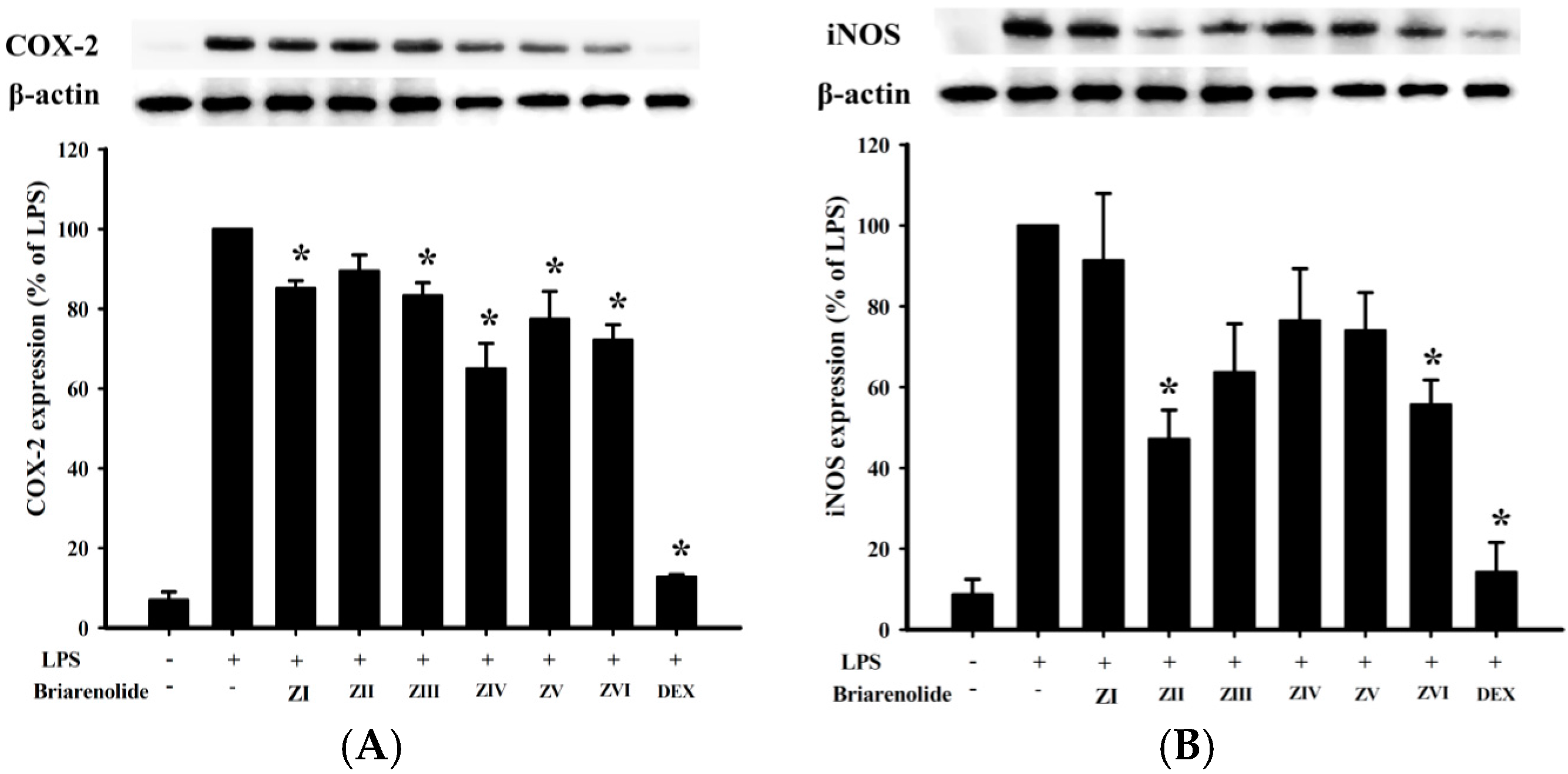
In an
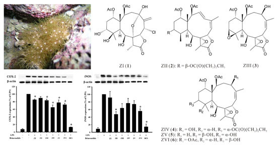
in vitro anti-inflammatory activity assay, Western blot analysis was used to evaluate the upregulation of the pro-inflammatory cyclooxygenase 2 (COX-2) and inducible nitric oxide synthase (iNOS) protein expressions in lipopolysaccharide (LPS)-stimulated RAW264.7 macrophage cells. At a concentration of 10 μM, briarenolides ZII (
2) and ZVI (
6) were found to significantly reduce the levels of iNOS to 47.2% and 55.7%, respectively, in comparison to the control cells stimulated with LPS only (
Figure 4 and
Table 7). By using trypan blue staining, it was observed that briarenolides ZI–ZVI (
1–
6) did not induce significant cytotoxicity in RAW264.7 macrophage cells.
Table 6.
1H (400 MHz, CDCl3) and 13C (100 MHz, CDCl3) NMR data and 1H–1H COSY and HMBC correlations for briarane 6.
Table 6.
1H (400 MHz, CDCl3) and 13C (100 MHz, CDCl3) NMR data and 1H–1H COSY and HMBC correlations for briarane 6.
| Position | δH (J in Hz) | δC, Multiple | 1H–1H COSY | HMBC |
|---|
| 1 | – | 46.1, C | – | – |
| 2 | 4.87 d (8.0) | 73.6, CH | H2-3 | C-1, -3, -4, -10, -14, -15, acetate carbonyl |
| 3 | 3.16 dd (15.6, 12.8); 1.91 m | 37.6, CH2 | H-2, H-4 | C-1, -2, -4, -5 |
| 4 | 5.01 dd (12.8, 5.6) | 72.7, CH | H2-3 | C-3, -5, -6, -16, acetate carbonyl |
| 5 | – | 144.1, C | – | – |
| 6 | 5.39 d (9.2) | 122.7, CH | H-7, H3-16 | C-4, -16 |
| 7 | 5.92 d (9.2) | 74.5, CH | H-6 | C-5, -6, -19 |
| 8 | – | 71.7, C | – | – |
| 9 | 3.91 br s | 74.7, CH | H-10, OH-9 | n. o. a |
| 10 | 2.20 dd (4.8, 2.2) | 41.6, CH | H-9, H-11 | C-1, -2, -11, -15, -20 |
| 11 | 1.99 m | 44.7, CH | H-10, H-12, H3-20 | n. o. |
| 12 | 4.04 dt (8.8, 3.6) | 67.0, CH | H-11, H2-13 | n. o. |
| 13 | 1.84 m | 29.0, CH2 | H-12, H-14 | C-1, -12 |
| 14 | 4.78 t (2.8) | 76.2, CH | H2-13 | C-10, -12, acetate carbonyl |
| 15 | 1.31 s | 15.4, CH3 | – | C-1, -2, -10, -14 |
| 16 | 2.13 s | 25.3, CH3 | H-6 | C-4, -5, -6 |
| 17 | – | 63.3, C | – | – |
| 18 | 1.57 s | 10.2, CH3 | – | C-8, -17, -19 |
| 19 | – | 172.0, C | – | – |
| 20 | 1.19 d (7.2) | 9.5, CH3 | H-11 | C-10, -11, -12 |
| OAc-2 | – | 170.2, C | – | – |
| | 1.99 s | 21.5, CH3 | – | Acetate carbonyl |
| OAc-4 | – | 170.4, C | – | – |
| | 2.01 s | 21.0, CH3 | – | Acetate carbonyl |
| OAc-14 | – | 170.5, C | – | – |
| | 1.99 s | 21.2, CH3 | – | Acetate carbonyl |
| OH-9 | 2.95 br s | – | H-9 | n. o. |
Figure 4.
Effects of briarenolides ZI–ZVI (1–6) on pro-inflammatory cyclooxygenase 2 (COX-2) and inducible nitric oxide synthase (iNOS) protein expressions in lipopolysaccharide (LPS)-stimulated murine macrophage cell line RAW264.7. (A) Relative density of the COX-2 Western blot; (B) relative density of the iNOS Western blot. The relative intensity of the LPS-stimulated group was taken to be 100%. Band intensities were quantified by densitometry and are indicated as the percentage change relative to that of the LPS-stimulated group. Briarenolides ZII (2) and ZVI (6) and DEX significantly inhibited LPS-induced iNOS protein expression (<60%) in macrophages. The experiments were repeated three times (* p < 0.05, significantly different from the LPS-stimulated group).
Table 7.
The effect of briarenolides ZI–ZVI (1–6) on LPS-induced COX-2 and iNOS protein expression in macrophage.
Table 7.
The effect of briarenolides ZI–ZVI (1–6) on LPS-induced COX-2 and iNOS protein expression in macrophage.
| Compounds | COX-2 | iNOS |
|---|
| Expression (% of LPS) | Expression (% of LPS) |
|---|
| Control | 6.9 ± 2.1 | 8.7 ± 3.8 |
| LPS | 100 ± 0 | 100 ± 0 |
| ZI (1) | 85.1 ± 1.9 | 91.4 ± 16.6 |
| ZII (2) | 89.5 ± 4.0 | 47.2 ± 7.2 |
| ZIII (3) | 83.3 ± 3.3 | 63.7 ± 12.0 |
| ZIV (4) | 65.0 ± 6.4 | 76.4 ± 13.0 |
| ZV (5) | 77.5 ± 6.9 | 74.0 ± 9.4 |
| ZVI (6) | 72.2 ± 3.8 | 55.7 ± 6.1 |
| DEX a | 12.8 ± 0.6 | 14.2 ± 7.3 |
