Review

Virulence Factors of Erwinia amylovora: A Review

1
Departament de Microbiologia i Parasiologia Sanitàries, Facultat de Farmàcia, Universitat de Barcelona, Av. Joan XXIII s/n, 08028 Barcelona, Spain
2
Departament de Microbiologia, Facultat de Biologia, Universitat de Barcelona, Av. Diagonal 643, 08071 Barcelona, Spain
*
Author to whom correspondence should be addressed.
Int. J. Mol. Sci. 2015, 16(6), 12836-12854; https://doi.org/10.3390/ijms160612836
Submission received: 8 February 2015 / Revised: 22 May 2015 / Accepted: 26 May 2015 / Published: 5 June 2015
(This article belongs to the Section Biochemistry)

Abstract

:
Erwinia amylovora, a Gram negative bacteria of the Enterobacteriaceae family, is the causal agent of fire blight, a devastating plant disease affecting a wide range of host species within Rosaceae and a major global threat to commercial apple and pear production. Among the limited number of control options currently available, prophylactic application of antibiotics during the bloom period appears the most effective. Pathogen cells enter plants through the nectarthodes of flowers and other natural openings, such as wounds, and are capable of rapid movement within plants and the establishment of systemic infections. Many virulence determinants of E. amylovora have been characterized, including the Type III secretion system (T3SS), the exopolysaccharide (EPS) amylovoran, biofilm formation, and motility. To successfully establish an infection, E. amylovora uses a complex regulatory network to sense the relevant environmental signals and coordinate the expression of early and late stage virulence factors involving two component signal transduction systems, bis-(3′-5′)-cyclic di-GMP (c-di-GMP) and quorum sensing. The LPS biosynthetic gene cluster is one of the relatively few genetic differences observed between Rubus- and Spiraeoideae-infecting genotypes of E. amylovora. Other differential factors, such as the presence and composition of an integrative conjugative element associated with the Hrp T3SS (hrp genes encoding the T3SS apparatus), have been recently described. In the present review, we present the recent findings on virulence factors research, focusing on their role in bacterial pathogenesis and indicating other virulence factors that deserve future research to characterize them.

1. Introduction

Erwinia amylovora is the type species of the genus Erwinia that belongs to the family Enterobacteriaceae. As is common in bacterial taxonomy in recent years, there have been a continuous description of novel Erwinia species as well as some species have been reclassified and transferred to other genera (http://www.bacterio.net). Most members of this genus cause diseases in plants and, historically, it is important to remember that E. amylovora was the first bacterium demonstrated to cause disease in plant, a discovery made in the late 1800s, at the same period as a similar discovery with human and animal diseases [1,2].
E. amylovora causes fire blight, a devastating plant disease affecting a wide range of host species within the Rosaceae subfamily Spiraeoideae, and is a major global threat to commercial apple and pear production. Moreover strains infecting plants in the genus Rubus belonging to the subfamily Rosoideae, including blackberry and raspberry, have also been reported [3,4,5,6,7].
In the last two centuries this pathogen has spread worldwide [6,7,8] and, in consequence, E. amylovora has been cataloged as a quarantine organism in the European Union, where it is subject to phytosanitary legislation [8], and, recently, has been included in the top 10 plant pathogenic bacteria published in the journal Molecular Plant Pathology [9].
Among the limited number of control options currently available, prophylactic application of antibiotics (e.g., streptomycin or oxytetracycline) during the bloom period appears most effective [10]. However, regulatory restriction, public health concerns, and pathogen resistance development severely limit the long-term prospects of antibiotic use [11,12]. Biological control measures may offer promising alternatives to minimize or even substitute the use of antibiotics, and to mitigate occurrence of resistance [5,13].
In the pathogenesis of E. amylovora, pathogen cells enter plants through the nectarthodes of flowers and other natural openings, such as wounds, and are capable of rapid movement within plants and the establishment of systemic infections [14,15,16,17]. In susceptible hosts, bacteria first move through the intercellular spaces of parenchyma and, in a later stage, in the xylem vessels, thus provoking extensive lesions, and sometimes complete dieback of the tree, under favorable climatic conditions. The diseased parts of the plant become brown or black, as if they had been swept by fire [17].
Many virulence determinants of E. amylovora have been characterized, including the Type III secretion system (T3SS), the exopolysaccharide (EPS) amylovoran, biofilm formation, and motility [15,18]. To successfully establish an infection, E. amylovora uses a complex regulatory network to sense the relevant environmental signals and coordinate the expression of early and late stage virulence factors involving two component signal transduction systems, bis-(3′-5′)-cyclic di-GMP (c-di-GMP) and quorum sensing [15]. In the present review, we present the recent findings on virulence factors of E. amylovora research, focusing on their role in bacterial pathogenesis and on the aspects that deserve future research.

2. Virulence Factors

E. amylovora is highly virulent and capable of rapid systemic movement within plant hosts and of rapid dissemination among rosaceous species, including apple and pear trees, when environmental conditions are favorable. The internal movement of the pathogen through the vascular system of plants and the ability of the pathogen to infect flowers, actively growing shoots, and rootstocks makes the management of fire blight difficult [14,19].
It has been shown that two major virulence determinants are required for E. amylovora to infect and cause disease on host plants: the EPS amylovoran and the Hrp type III secretion system (T3SS) [20]. Previous results demonstrated that E. amylovora forms a biofilm in vitro and in planta [19,21,22].

2.1. Exopolysaccharides (EPS) Amylovoran and Levan

Exopolysaccharides (EPS) have been suggested to play a key role in bypassing the plant defense system, in disturbing and obstructing the vascular system of the plant and in protecting the bacteria against water and nutrient loss during dry conditions [19,23,24,25]. Previous work has demonstrated that EPS is an important element in the biofilm formation of E. amylovora, enabling the bacteria to attach to several surfaces and to each other [19,21,26]. Amylovoran has been shown to be the main factor necessary for biofilm formation, and levan is a contributing factor [21,24], the quantity of amylovoran produced by individual E. amylovora strains being correlated with the degree of virulence [24,27].
Amylovoran is a polymer of a pentasaccharide repeating unit that generally consists of four galactose residues and one glucuronic acid residue [24,27,28]. The molecular size of amylovoran is influenced by several environmental conditions and cell-metabolism-related factors [24,29]. The strains of E. amylovora that do not have the capacity to produce amylovoran are non-pathogenic and are unable to spread in plant vessels [24,30].
The amylovoran synthesis (ams) gene cluster involved in the biosynthesis of amylovoran produces 12 ams-encoded gene products (AmsA to AmsL). AmsC, AmsH, and AmsL are believed to be involved in oligosaccharide transport and assembly, while AmsA possesses a tyorisine kinase activity. AmsB, AmsD, AmsE, AmsG, AmsJ, and AmsK proteins appear to play a part in annealing the different galactose, glucuronic acid, and pyruvyl subunits to the lipid carrier in order to form an amylovoran unit. AmsF may process newly synthesized repeating units and/or be involved in their polymerization by adding them to an existing amylovoran chain. Finally, AmsI seems to have a distinct function in recycling of the diphosphorylated lipid carrier after release of the synthesized repeating unit [24,31,32,33].
Levan is another EPS produced by E. amylovora, considered as a virulence factor [19,34]. It has been shown that the lack of levan synthesis can result in a slow development of symptoms in the host plant [24,35]. The specific role of levan in pathogenesis is still unknown and deserves further research.
Interestingly, in recent studies, it has been demonstrated that the depolymerase (DpoL1) encoded by the T7-like E. amylovora phage L1 efficiently degrades amylovoran. Exposure of the bacteria to either L1 phage or recombinant DpoL1 led to EPS degradation and a phenotype similar to the one observed with EPS mutants. The enzyme strips the cells, thereby paving the way for infection by providing access of L1 phage to the cell wall, and also facilitating the infection by other phages, as Dpo-negative Y2 phages [5].

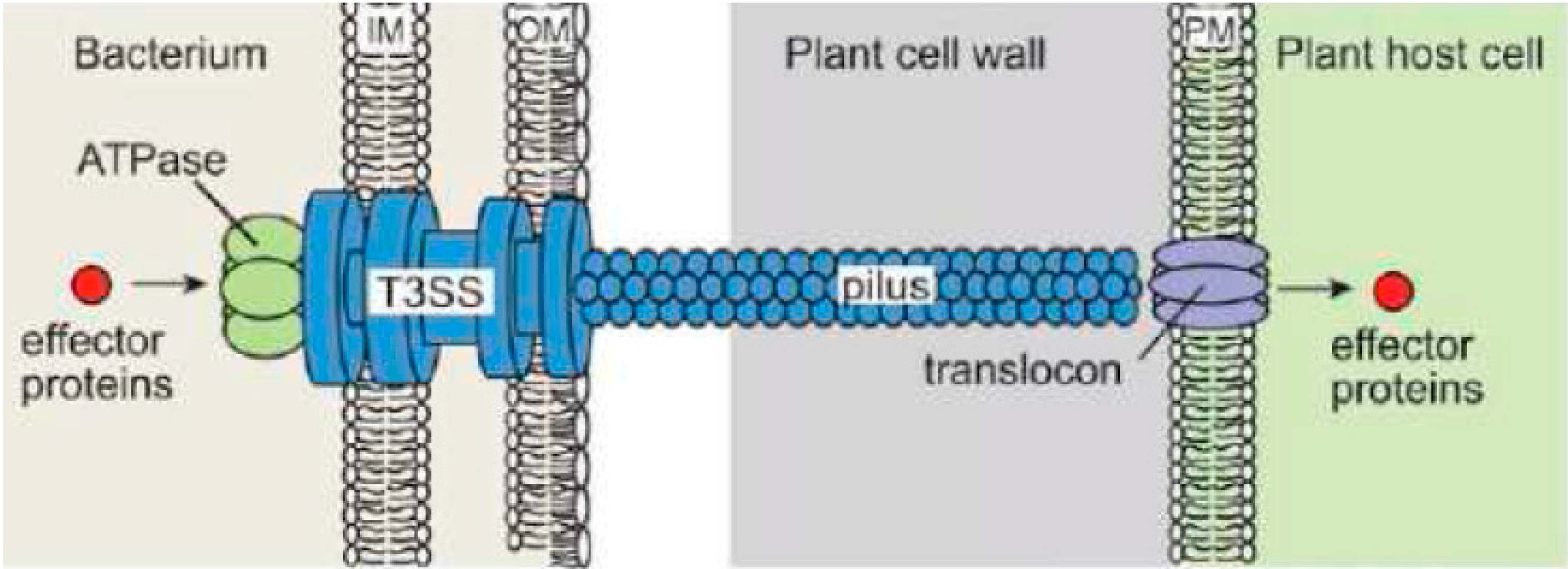
2.2. Type III Secretion System (T3SS)

The type III secretion system (T3SS) is one of the important virulence factors used by E. amylovora in order to successfully infect its hosts [24,36]. As with other Gram negative phytopathogenic bacteria, E. amylovora uses this evolutionarily conserved secretion system to export and deliver effector proteins into the cytosol of host plant cells through a pilus-like structure, which forms the central core element of T3SS [24] (Figure 1).
E. amylovora pathogenicity relies on a type III secretion system and on a single effector DspA/E. This effector belongs to the widespread AvrE family of effectors whose biological function is not fully understood [37].
T3SS is composed of a large, cylindrically shaped macromolecular complex organized into a series of ring-like structures with inner rings, outer rings and neck structure. It is embedded in the inner and outer membrane of the bacteria, while spanning the periplasmic membrane and extending into the extracellular environment with a pilus filament [24,37] (Figure 1). It has been shown that T3SS occurs only at the site of Hrp pilus assembly and that pilus guides the transfer of effector proteins outside the bacterial cell, favoring the “conduit/guiding filament” model [37] (Figure 1).
The T3SS of plant-pathogenic bacteria is mainly made out of Hrc proteins, encoded by hrp-conserved (hrc) genes among plant-pathogenic bacteria and Hrp proteins, encoded by hypersensitive response and pathogenicity (hrp) genes. In E. amylovora, hrc and hrp genes are clustered in a pathogenicity island, which contains four regions, i.e., an hrc/hrp region, an Hrc effectors and elicitors region, an Hrp-associated enzymes region, and an island transfer region [24,38].
Figure 1. Schematic representation of the T3SS from plant pathogenic bacteria [39] (modified from [39], with permission from American Society of Plant Biologists).
Ijms 16 12836 g001

2.3. Curli

Curli are the major proteinaceous component of a complex extra-cellular matrix produced by many Enterobacteriaceae, including E. amylovora. Curli fibres are known to be involved in adhesion to surfaces, cell aggregation and biofilm formation. Curli can also mediate host cell adhesion and invasion and they stimulate the host inflammatory responses [19,40,41]. The structure and biogenesis of curli are unique among the bacterial fibres that have been described to date, belonging to a growing class of fibres, known as amyloids, which are associated with diverse neurological diseases in humans. Curli fibres are 4–6 nm-wide and, like other amyloid fibres, are β sheet-rich self-assembling protein polymers that are resistant to chemical and temperature denaturation, and to digestion by proteinases [41].
Recent results have showed reductions in virulence due to deletion of a regulator of curli genes, crl, suggesting that not only functional attachment but also mature biofilm formation is needed for full virulence in the host [19]. Further research is necessary to charaterize the chemical structures and genes encoding this structure and its specific role in pathogenicity, as it is also in process for other Enterobacteria as E. coli or Salmonella [40].

2.4. Biofilm Formation

In addition to the specific role of the exopolysaccharides amylovoran and levanin in the biofilm formation of E. amylovora, as mentioned above, a recent study has suggested that type I fimbriae, flagella, type IV pili, and curli of E. amylovora may contribute to biofilm formation in static and flowing environments and that defects in many of the genes encoding these appendages result in decreased virulence in planta [19].
Previous results of the same group demonstrated that E. amylovora forms a biofilm in vitro and in planta [19,21]. Pathogenesis and biofilm formation appear to be linked, but without identifying genes encoding traits independent of amylovoran production, the mechanistic role in virulence of biofilm formation in E. amylovora could not be studied. Interestingly, mutants with reduced biofilm-formation ability appear unable to successfully establish large populations in apple xylem. Colonization of xylem is critical to the systemic movement of the pathogen through plants [19,22]. Therefore, biofilm-deficient mutants remain localized within an inoculated leaf and are strongly impaired in the ability to invade the rest of the plant [19].
In a study using a bioinformatic approach and the recently sequenced genome of E. amylovora [19,42,43], genes encoding putative cell surface attachment structures were identified [19]. A time course assay indicated that type I fimbriae function earlier in attachment, while type IV pilus structures appeared to function later in attachment. Deficiencies in type I fimbriae lead to an overall reduction in E. amylovora virulence. By deletion of individual genes and gene clusters and using a combination of in vitro attachment assays and plant virulence assays it was demonstrated that multiple attachment structures are present in E. amylovora and play a role in mature biofilm formation, which is critical to pathogenesis and systemic movement in the host (Figure 2). In contrast, a fully functional biofilm was not necessary to survival and growth in planta [19].
Although the mechanistic details behind biofilm formation remain largely unknown and genetic and structural analyses should be done for full characterization of these structures, it is suggested that they are formed in response to environmental triggers [24,44] and quorum sensing signals [24,45].
Figure 2. Images of putative attachment structures of E. amylovora [19] (adapted from [19], with permission from American Society for Microbiology). (A) TEM imaging of a planktonic E. amylovora cell grown in broth culture and negatively stained. Peritrichous flagella are indicated by arrows (scale: 1 µm); (B) TEM image of E. amylovora in planta. Putative attachment structures connect bacterial cells to host cells (scale: 1 µm); (C) SEM image of E. amylovora cells found within a biofilm, with multiple appendages that protrude from the bacterial cell and attach to the host surface, as indicated by the arrows (scale: 2 µm).
Ijms 16 12836 g002

2.5. Motility

E. amylovora is a nonobligate pathogen able to survive outside the host under starvation conditions, allowing its spread by various means, such as rainwater [46]. In fact, motility mechanisms have been shown to be of relevance for the survival of the bacteria ouside the host [46] and also for the attachment to the host cell surfaces, together with other virulence factors as type IV pili, type I fimbriae and curli by biofilm formation [19]. In different studies, long peritrichous flagella have been observed (Figure 2) [19,46], although chemical structural data have not been published yet.
In a study where two of the four gene clusters encoding the production and regulation of flagella in E. amylovora were deleted, various effects of flagella on biofilm formation and virulence were demonstrated. Results obtained also showed that flagellum production appeared to be controlled by multiple gene clusters that may function independently. Consequently, it was suggested that the flagella of E. amylovora have multifaceted functions in the biofilm formation process [19].
Swarming motility has also been described in E. amylovora [15,47], although, as in other Enterobacteria, its role remains unknown, requiring further research.
E. amylovora responses to starvation revealed that cells lost motility but most of them exhibited long peritrichous flagella attached to the bacterial surface. Motility inactivation during starvation would be reversible since flagella could enable a rapid recovery of motility under favorable conditions [46].

2.6. Lipopolysaccharide (LPS)

LPS is a factor that has been described recently to be involved in the virulence of E. amylovora [48], although the complete chemical structure of LPS and its relationship with the pathogenicity has not yet been described.
Interestingly, in Erwinia carotovora, a comprehensive physicochemical analysis of highly purified mono- and bis-phosphoryl hexa- to heptaachyl and pentaacyl lipid A part structures has been published. The distinct differences observed between different Lipid A structures could explain differences in the biological activity, such as the cytokine inducing activity [49].
At the genetic level, comparative genomic analysis revealed differences in the LPS biosynthesis gene cluster between the Rubus-infecting strain ATCC BAA-2158 and the Spiraeoideae-infecting strain CFBP 1430 of E. amylovora. These differences were restricted to the core region that could be involved in a process of adaptation to the new host. Genetic differences observed in the LPS biosynthetic gene cluster corroborate rpoB-based phylogenetic clustering of E. amylovora into four different groups and enable the discrimination of Spiraeoideae- and Rubus-infecting strains [48].

2.7. Metalloprotease PrtA

A metalloprotease with a molecular mass of 48 kDa secreted by E. amylovora has been characterized [50]. The protease is apparently secreted into the external medium through the type I secretion pathway via PrtD, PrtE, and PrtF, which share more than 90% identity with the secretion apparatus for lipase of S. marcescens [50].
A gene cluster encodes four genes connected to protease expression, including a structural gene (prtA) and three genes (prtD, prtE, prtF) for secretion of the protease, which are transcribed in the same direction. The organization of the protease gene cluster in E. amylovora is different from that in other Gram-negative bacteria, such as Erwinia chrysanthemi, Pseudomonas aeruginosa, and Serratia marcescens. On the basis of the conservative motif of metalloproteases, PrtA has been identified to be a member of the metzincin subfamily of zinc-binding metalloproteases, and has been confirmed to be the 48 kDa protease on gels by sequencing of tryptic peptide fragments derived from the protein [50]. In a protease mutant created by mutations with Tn 5–insertions in the prtD gene, the lack of protease reduced colonization of an E. amylovora secretion mutant, labeled with the gene for the green fluorescent protein (gfp) in the parenchyma of apple leaves [50].
Further research is necessary, however, to assess its structural characteristics, activity, substrates, the specific role of this metalloprotease in the pathogenic mechanisms of E. amylovora, and also the possible practical applications of this protein, based on the differences observed with other Gram negative bacteria, as already proposed for this interesting wide family of proteins [51].

2.8. Iron-Scavenging Siderophore Desferrioxamine

Under iron-limiting conditions, E. amylovora CFBP 1430 produces hydroxamate-type siderophores [52], characterized as cyclic desferrioxamines (DFOs), mostly DFO E [53,54] and the specific receptor FoxR, a 70,000-Da protein needed for the passage of ferric complexes across the outer membrane [17,54].
Several iron uptake negative mutants of E. amylovora CFBP 1430 isolated by insertional mutagenesis have been identified. In mutant VD61, the mutation (dfo-61::MudIIpR13) disrupts the DFO biosynthetic pathway; in VD17, the mutation (foxR-17::MudIIpR13) affects the synthesis of the FoxR receptor and the mutant accumulates DFO in the external medium because of its failure to transport back the DFO ferric complex [17,54]. In the pathogenic analysis of these mutants, results obtained demonstrated that, in E. amylovora, production of DFOs is a critical function for bacterial iron acquisition during pathogenesis, as well as for bacterial induction of electrolyte leakage from plant cells [17]. Based on this, a dual role of DFO during plant/E. amylovora interactions has been proposed [17], which deserve further research.

2.9. Multidrug Efflux Pump AcrAB

During pathogenesis, E. amylovora is exposed to a variety of plant-borne antimicrobial compounds, which in the case of plants of Rosaceae, many are constitutively synthesized as isoflavonoids [22]. Then, bacterial multidrug efflux transporters, which mediate resistance toward structurally-unrelated compounds, might confer tolerance to these antimicrobial compounds (phytoalexins) [22]. In this regard, it has been observed that clonation of the acrAB locus from E. amylovora, encoding a resistance nodulation division-type transport system, in E. coli conferred resistance to hydrophobic and amphiphilic toxins [22]. An acrB-deficient E. amylovora mutant was impaired in virulence on apple rootstock MM 106 and was susceptible toward extracts of leaves of MM 106, as well as to the apple phytoalexins phloretin, naringenin, quercetin, and (+)-catechin [22]. These results strongly suggest that the AcrAB transport system plays an important role as a protein complex required for the virulence of E. amylovora in resistance toward apple phytoalexins and that it is required for successful colonization of a host plant [22].

2.10. Other Virulence Factors

Other virulence factors of E. amylovora have been proposed, as the case of the sorbitol transporter, encoded by the srl operon [55]. It has been shown that expression of the srl operon in E. amylovora is high in the presence of sorbitol in medium and is repressed by glucose. Mutants with a sorbitol deficiency were still virulent on slices of immature pears, but were unable to cause significant fire blight symptoms on apple shoots. Since sorbitol is used for carbohydrate transport in host plants of E. amylovora, this sugar alcohol has been proposed to be an important factor in determining host specificity for the fire blight pathogen [55].
Moreover, Rosaceous plants also contain sucrose as storage and transport carbohydrates [56,57,58]. The regulation and biochemistry of sucrose metabolism of E. amylovora has also been studied, demonstrating that sucrose mutants (in the scr regulon), created by site-directed mutagenesis, did not produce significant fire blight symptoms on apple seedlings, thus, indicating the importance of sucrose metabolism for colonization of host plants by E. amylovora [58].

3. Genetics

The genome of E. amylovora consists of a circular chromosome of 3,805,874 bp and two plasmids: AMYP1 (28,243 bp), also reported as PEA29, and the larger plasmid AMYP2 (71,487 bp), also named pEA72. The small size of the E. amylovora genome (3.8 Mb) in comparison with most free-living enterobacteria, including plant pathogens, with genomes of 4.5–5.5 Mb, and the preponderance of pseudogenes, genome reduction can have occurred via mutational inactivation and subsequent deletion and, as a consequence, has led to the loss of most of the genes involved in anaerobic respiration and fermentation found in typical, related enterobacteria, with the consecuent reduction of the capacity to live in anaerobic environments [42]. The genome sequence of E. amylovora has revealed clear signs of pathoadaptation to the rosaceous plant environment. For example, T3SS-related proteins are more similar to proteins of other plant pathogens than to proteins of closely related enterobacteria [42].
Comparative genomic analysis of 12 strains representing distinct populations (e.g., geographic, temporal, host origin) of E. amylovora has allowed to describe the pan-genome of this species. The pan-genome contains 5751 coding sequences and is a highly conserved relative to other phytopathogenic bacteria, comprising on average 89% conserved core genes. While the chromosones of Spiraeoideae-infecting strains are highly homogeneous, greater genetic diversity was observed between Spiraeoideae- and Rubus-infecting strains (and among individual Rubus-infecting strains) [20].
Using molecular approaches based on the study of repetitive elements, such as Multiple Loci Variable Number of Tandem Repeats Analysis (MLVA) [59] or sequencing of Clustered Regularly Interspaced Short Palindromic Repeats (CRISPR) [60], this diversity has also been especially observed among strains isolated from Rubus plants and, to a lesser extent, in Spiraeoideae-infecting strains [48].
In comparison with other plant pathogens, however, E. amylovora has relatively low genetic diversity, with a limited genetic recombination, this being associated with its exposure to limited selection pressures due to pome fruit breeding strategically favoring only high-valued varieties, which are often highly susceptible to fire blight [20,61,62].
On the basis of the current available data, it has been hypothesized that the critical event for adaptation to Rubus spp. took place after species separation of E. amylovora and E. pyrifoliae, as the Spiraeoideae infecting isolates of E. amylovora and E. pyrifoliae (including Japanese strains), as well as E. tasmaniensis and E. piriflorinigrans, all sharing the Spiraeoideae-type LPS biosynthetic cluster [48]. Interestingly, genome sequencing of three Mexican E. amylovora strains have revealed an rpsL chromosal mutation conferring high-level of streptomycin resistance [63]. This chromosomal mutation is the predominant E. amylovora mechanism, typically appearing after years of intensive application with long persistence in populations [10,63], explaining the observed inefficacy of streptomycin in Mexico [63,64] and underscoring the need of revision of pesticide use strategies to avoid similar resistance evolution against other antimicrobial drugs as oxytetracycline or gentamicin [63].
The expression of genes related to starvation, oxidative stress, motility, pathogenicity, and virulence have been detected during the entire experimental period with different regulation patterns observed during the first 24 h. Further, starved cells remained as virulent as nonstressed cells. Overall, these results provide new knowledge on the biology of E. amylovora under conditions prevailing in nature, which could contribute to a better understanding of the life cycle of this pathogen [46].
No major differences among 12 strains of E. amylovora were reported in the amylovoran biosynthesis cluster (>98% amino acid indentity across the whole region) [20]. Variation, however, has been observed in the Hrp cluster, a pathogenicity island that encodes the hypersensitive response and pathogenicity (hrp) T3SS and the majority of known T3SS effector proteins [20,38]. Variation was identified in HrpK, the putative chaperones OrfA and OrfC (which varied between host specific grouping of Rubus- and Spiraeoideae-infecting strains) and, more significantly, Eop 1, which has been shown to function as a host-limiting factor [20,38,65,66].
Three gene clusters related to the Type 6 Secretion System (T6SS) have also been identified in E. amylovora [20,43], although their exact role in this species is unknown [20] and should deserve further research. In other bacteria, T6SS has been shown to play a significant role in bacterial-bacterial and bacterial-host interactions, suggesting a role for niche specialization [67].

4. Regulation of Virulence Factors

4.1. Two Component Transduction Systems

The phoPQ two component transduction system has been genetically characterized in E. amylovora [68] and it has been demonstrated that this system in E. amylovora plays major roles in virulence on immature pear fruit and in regulating amylovoran biosynthesis and swarming motility [47]. It has also been shown that phoPQ mutants were more resistant to strong acidic conditions (pH 4.5 or 5) than that of the wild-type (WT) strain, suggesting that this system in E. amylovora may negatively regulate acid resistance gene expression. Furthermore, the PhoPQ system negatively regulated gene expression of two novel T3SS in E. amylovora. These results are in contrast to those reported for the PhoPQ system in Salmonella and Xanthomonas, where it positively regulates T3SS and acid resistance. In addition, survival of phoPQ mutants was about 10-fold lower than that of WT when treated with cecropin A at pH 5.5, suggesting that the PhoPQ system renders the pathogen more resistant to cecropin A [68].
These results suggest that the the PhoPQ system may act as a part of a regulatory network that governs E. amylovora in a wide variety of cellular functions, thus enhancing its survival under the changing environments of different hosts or in different environments, such as the acidic plant apoplast, a major barrier for plant pathogenic bacteria to cause disease [68].

4.2. Small RNA

Recently, different regulatory small RNAs (sRNAs) requiring the RNA chaperone Hfq for both stability and functional activation have been identified in E. amylovora, and further, being some of them involved in the regulation of various virulence traits including motility, amylovoran EPS production, biofilm formation, and T3SS. This discovery is of relevance since, although sRNAs have been increasingly recognized as pivotal regulators in bacteria, genome-wide identification of sRNAs has only been performed in a limited number of bacteria [15].

4.3. Quorum Sensing

Quorum sensing systems have been described in E. amylovora, both QS-1, relying on an N-homoserine lactone autoinducer-1 signal (AI-1) [69,70,71] and QS-2 reliant on LuxS as the enzyme responsible for signal (AI-2) production [72]. However, a global regulatory function has not yet been explored. Although previous studies reported that luxS did not affect quorum sensing in E. amylovora [71], recent results have indicated that some strains of E. amylovora have luxS-dependent AI-2 activity, and that inactivation of luxS affects some phenotypes, including virulence in planta [72], although the molecular basis of luxS-dependent regulation remains to be determined [72]. It was observed that AI-2 produced in E. amylovora activated the expression of bioluminescence in the Vibrio harveyi BB170 reporter strain. Findings showed that AI-2 in Ea1665 was dependent on EaluxS because expression of EaluxS in the luxS frame-shift mutation of E. coli DH5α demonstrated its importance in AI-2 synthesis. The pattern of AI-2 production with respect to growth phase and environmental changes deserves further research, and may give some insight into the function of this signaling system [72].
Moreover, using comparative genomic analyses to search homologs of genes involved, several genes were identified corresponding to the Autoinducer-3/Epinephrine/Norepinephrine signaling system in E. coli. Particularly, the putative homologs to the two component system QseEF and the transcriptional regulator QseA are highly conserved in E. amylovora. Gene-knockout experiments revealed that the QseEFEa system is involved in the regulation of swarming motility, biosynthesis of amylovoran, and biofilm formation [73].

4.4. c-di-GMP

It has been shown that E. amylovora encodes eight genes involved in the biosynthesis and/or degradation of cyclic-di-GMP (c-di-GMP), a second messenger signaling compound. Individual knockout mutations in each of these genes revealed that c-di-GMP positively regulated biofilm formation and negatively regulated motility. These results demonstrated that E. amylovora possesses several regulatory networks that govern the expression of virulence genes [73,74].

4.5. ppGpp

In a recent study, the role of the bacterial alarmone ppGpp in activating the T3SS and virulence of E. amylovora was investigated using ppGpp mutants generated by Red recombinase cloning. The virulence of a ppGpp-deficient mutant (ppGpp(0)), as well as a dksA mutant of E. amylovora, was completely impaired, and bacterial growth was significantly reduced, suggesting that ppGpp is required for full virulence of E. amylovora. Expression of T3SS genes was greatly downregulated in the ppGpp(0) and dksA mutants. Western blotting showed that accumulations of the HrpA protein in the ppGpp(0) and dksA mutants were about 10% and 4%, respectively, of that in the wild-type strain. Furthermore, higher levels of ppGpp resulted in a reduced cell size of E. amylovora. Moreover, serine hydroxamate and α-methylglucoside, which induce amino acid and carbon starvation, respectively, activated hrpA and hrpL promoter activities in hrp-inducing minimal medium. These results demonstrated that ppGpp and DksA play central roles in E. amylovora virulence and indicated that E. amylovora utilizes ppGpp as an internal messenger to sense environmental/nutritional stimuli for regulation of the T3SS and virulence [75].

4.6. Type 3 Effectors

There are several effectors in E. amylovora that have been described [36], although recent findings have been focused on DspA/E since it has been shown that the ability of E. amylovora to promote disease mainly depends on this single injected T3E [76]. DspA/E belongs to the widespread AvrE family of type III effectors that suppress plant defense responses and promote bacterial growth following infection [76,77,78]. In recent studies, expression of dspA/E in the yeast Saccharomyces cerevisiae inhibited cell growth, this effect associated with perturbations of the actin cytoskeleton and endocytosis [77], activation of phosphatase 2A, and downregulation of the sphingolipid biosynthetic pathway leading to growth arrest [76].
In nonhost Arabidopsis thaliana leaves, DspA/E was required for transient bacterial growth, as an E. amylovora dspA/E mutant was unable to grow. Study of transgenic Arabidopsis lines expressing DspA/E indicated that DspA/E promotes modifications of plant cell metabolism among which the repression of protein synthesis could be determinant in the facilitation of necrosis and bacterial growth [78].

4.7. Hrp X/Y, HrpS and HrpL Cascade Leading to Activation of hrpT3SS

Two regulatory components, hrpX and hrpY, of the hrp system of Erwinia amylovora have been identified [79]. The hrpXY operon is expressed in rich media, but its transcription is increased threefold by low pH, nutrient, and temperature levels, conditions that mimic the plant apoplast. hrpXY is autoregulated and directs the expression of hrpL, which, in turn, activates transcription of other loci in the hrp gene cluster [80].
The deduced amino-acid sequences of hrpX and hrpY are similar to bacterial two-component regulators including VsrA/VsrD of Pseudomonas (Ralstonia) solanacearum, DegS/DegU of Bacillus subtilis, and UhpB/UhpA and NarX/NarP, NarL of Escherichia coli. The N-terminal signal-input domain of HrpX contains PAS domain repeats [79].
hrpS, located downstream of hrpXY, encodes a protein with homology to WtsA (HrpS) of Erwinia (Pantoea) stewartii, HrpR and HrpS of Pseudomonas syringae, and other σ54-dependent, enhancer binding proteins. Transcription of hrpS also is induced under conditions that mimic the plant apoplast. However, hrpS is not autoregulated, and its expression is not affected by hrpXY [79]. As a member of the NtrC family, HrpS is unusual in that it lacks a long N-terminal receiver domain [79].
The structure of the input domain of E. amylovora HrpX appears to be exceptional, compared with sensor proteins involved in other type III systems, which contain two transmembrane regions and a periplasmic domain [79]. Thus, at least two types of transmitter-receiver systems appear to have evolved for control of type III systems in response to environmental stimuli in hosts [79].

4.8. Regulatory Cascade for Amylovoran Synthesis

The biosynthesis of amylovoran is regulated by another two-component signal transduction system, the RcsCDB phosphorelay system, which has been found to be essential for virulence in E. amylovora [47].

5. Discussion

Nowadays, there is a high amount of interest to find and apply biological measures to combat and prevent plant diseases to avoid the use of chemical compounds in agriculture [81,82]. In this context, it is fundamental to understand the host (plant)-parasite interactions [81].
In this review, we have summarized the most recent findings regarding virulence factors of E. amylovora, having seen that, although the most relevant virulence mechanisms have been described, there is substantial further research to completely characterize, at the chemical and functional levels, the different factors involved in pathogenicity. In this regard, recent genome sequencing should accelerate the knowledge on this species, particularly in regards to pathogenicity, considering the limited genetic diversity observed in the different strains. Overall, these data will help to understand the whole mechanism of the specific interactions of E. amylovora with its hosts, thus, allowing better diagnosis and control strategies, which can include biological measures, such as the design of lytic bacteriophages, competitive attenuated strains, etc.
For most of the virulence factors identified, even for the two main factors (the EPS amylovoran and T3SS), further research is necessary to fully understand their role in the pathogenic network of the bacteria, including the main mechanism of adhesion via the biofilm and the interaction of the different elements with the plant structures, including the acidic plant apoplast and the plant defenses, under different environmental conditions.
Complete characterization of the structures involved in virulence, and their regulation mechanisms and further genetic analysis, will allow to design more precise diagnostic and control strategies (including a more rational use of antimicrobials), together with a more precise phylogenetic classification of E. amylovora, across the genus, and the Enterobacterial family.

Acknowledgments

This work was supported by Plan Nacional de I + D+ i (Ministerio de Educación, Ciencia y Deporte and Ministerio de Sanidad, Spain) and from Generalitat de Catalunya (Centre de Referència en Biotecnologia, Barcelona, Spain).

Conflicts of Interest

The authors declare no conflict of interest.

References

  1. Griffith, C.S.; Sutton, T.B.; Peterson, P.D. Fire Blight: The Foundation of Phytobacteriology; APS Press: St. Paul, MN, USA, 2003. [Google Scholar]
  2. Hauben, L.; Swings, J. Genus XIII Erwinia. In Bergey’s Manual of Systematic Bacteriology, 2nd ed.; Brenner, D.J., Krieg, N.R., Staley, J.T., Garrity, G.M., Eds.; Springer: New York, NY, USA, 2005; Volume 2B, pp. 670–679. [Google Scholar]
  3. Bonn, W.G.; van der Zwet, T. Distribution and economic importance of fire blight. In Fire Blight: The Disease and Its Causative Agent, Erwinia Amylovora; Vanneste, J.L., Ed.; CAB International: Wallingford, CT, USA, 2000; pp. 37–53. [Google Scholar]
  4. Thomson, S.V. Epidemiology of fire blight. In Fire Blight: The Disease and Its Causative Agent, Erwinia Amylovora; Vanneste, J.L., Ed.; CAB International: Wallingford, UK, 2000; pp. 9–36. [Google Scholar]
  5. Born, Y.; Fieseler, L.; Klumpp, J.; Eugster, M.R.; Zurfluh, K.; Duffy, B.; Loessner, M.J. The tail-associated depolymerase of Erwinia amylovora phage L1 mediates host cell adsorption and enzymatic capsule removal, which can enhance infection by other phage. Environ. Microbiol. 2014, 16, 2168–2180. [Google Scholar] [CrossRef] [PubMed]
  6. Vanneste, J.L. Fire blight: The disease and its causative agent. In Erwinia Amylovora; CABI Publishing: Oxfordshire, UK, 2000. [Google Scholar]
  7. Van der Zwet, T.; Orolaza-Halbrendt, N.; Zeller, W. Fire Blight: History, Biology, and Management; APS Press: St. Paul, MN, USA, 2012. [Google Scholar]
  8. Santander, R.D.; Catala-Senent, J.F.; Marco-Noales, E.; Biosca, E.G. In planta recovery of Erwinia amylovora viable but nonculturable cells. Trees 2012, 26, 75–82. [Google Scholar] [CrossRef]
  9. Mansfield, J.; Genin, S.; Magori, S.; Citovsky, V.; Sriariyanum, M.; Ronald, P.; Dow, M.; Verdier, V.; Beer, S.V.; Machado, M.A.; et al. Top 10 plant pathogenic bacteria in molecular plant pathology. Mol. Plant Pathol. 2012, 13, 614–629. [Google Scholar] [CrossRef] [PubMed]
  10. McManus, P.S.; Stockwell, V.O.; Sundin, G.W.; Jones, A.L. Antibiotic use in plant agriculture. Annu. Rev. Phytopathol. 2002, 40, 443–465. [Google Scholar] [CrossRef] [PubMed]
  11. Duffy, B.; Schärer, H.-J.; Bünter, M.; Klay, A.; Holliger, E. Regulatory measures against Erwinia amylovora in Switzerland. EPPO Bull. 2005, 35, 239–244. [Google Scholar] [CrossRef]
  12. Stockwell, V.O.; Duffy, B. Use of antibiotics in plant agriculture. Rev. Sci. Tech. 2012, 31, 199–210. [Google Scholar] [PubMed]
  13. Johnson, K.B.; Stockwell, V.O. Management of fire blight: A case study in microbial ecology. Annu. Rev. Phytopathol. 1998, 36, 227–248. [Google Scholar] [CrossRef] [PubMed]
  14. Norelli, J.L.; Jones, A.L.; Aldwinckle, H.S. Fire blight management in the twenty-first century: Using new technologies that enhance host resistance in apple. Plant Dis. 2003, 87, 756–765. [Google Scholar] [CrossRef]
  15. Zeng, Q.; Sundin, G.W. Genome-wide identification of Hfq-regulated small RNAs in the fire blight pathogen Erwinia amylovora discovered small RNAs with virulence regulatory function. BMC Genomics 2014, 15. [Google Scholar] [CrossRef] [PubMed]
  16. Thomson, S.V. The role of the stigma in fire blight infections. Phytopathology 1986, 76, 476–482. [Google Scholar] [CrossRef]
  17. Dellagi, A.; Brisset, M.N.; Paulin, J.P.; Expert, D. Dual role of desferrioxamine in Erwinia amylovora pathogenicity. Mol. Plant Microbe Interact. 1998, 11, 734–742. [Google Scholar] [CrossRef] [PubMed]
  18. Malnoy, M.; Martens, S.; Norelli, J.L.; Barny, M.A.; Sundin, G.W.; Smits, T.H.; Duffy, B. Fire blight: Applied genomic insights of the pathogen and host. Annu. Rev. Phytopathol. 2012, 50, 475–494. [Google Scholar] [CrossRef] [PubMed]
  19. Koczan, J.M.; Lenneman, B.R.; McGrath, M.J.; Sundin, G.W. Cell surface attachment structures contribute to biofilm formation and xylem colonization by Erwinia amylovora. Appl. Environ. Microbiol. 2011, 77, 7031–7039. [Google Scholar] [CrossRef] [PubMed]
  20. Mann, R.A.; Smits, T.H.; Bühlmann, A.; Blom, J.; Goesmann, A.; Frey, J.E.; Plummer, K.M.; Beer, S.V.; Luck, J.; Duffy, B.; et al. Comparative genomics of 12 strains of Erwinia amylovora identifies a pan-genome with a large conserved core. PLoS ONE 2013, 8, e55644. [Google Scholar] [CrossRef] [PubMed]
  21. Koczan, J.M.; McGrath, M.J.; Zhao, Y.; Sundin, G.W. Contribution of Erwinia amylovora exopolysaccharides amylovoran and levan to biofilm formation: Implications in pathogenicity. Phytopathology 2009, 99, 1237–1244. [Google Scholar] [CrossRef] [PubMed]
  22. Burse, A.; Weingart, H.; Ullrich, M.S. NorM, an Erwinia amylovora multidrug efflux pump involved in in vitro competition with other epiphytic bacteria. Appl. Environ. Microbiol. 2004, 70, 693–703. [Google Scholar] [CrossRef] [PubMed]
  23. Ordax, M.; Marco-Noales, E.; Lopez, M.M.; Biosca, E.G. Exopolysaccharides favor the survival of Erwinia amylovora under copper stress through different strategies. Res. Microbiol. 2010, 161, 549–555. [Google Scholar] [CrossRef] [PubMed]
  24. Vrancken, K.; Holtappels, M.; Schoofs, H.; Deckers, T.; Valcke, R. Pathogenicity and infection strategies of the fire blight pathogen Erwinia amylovora in Rosaceae: State of the art. Microbiology 2013, 159, 823–832. [Google Scholar] [CrossRef] [PubMed]
  25. Bellemann, P.; Bereswill, S.; Berger, S.; Geider, K. Visualization of capsule formation by Erwinia amylovora and assays to determine amylovoran synthesis. Int. J. Biol. Macromol. 1994, 16, 290–296. [Google Scholar] [CrossRef]
  26. Ramey, B.E.; Koutsoudis, M.; von Bodman, S.B.; Fuqua, C. Biofilm formation in plant-microbe associations. Curr. Opin. Microbiol. 2004, 7, 602–609. [Google Scholar] [CrossRef] [PubMed]
  27. Maes, M.; Orye, K.; Bobev, S.; Devreese, B.; van Beeumen, J.; de Bruyn, A.; Busson, R.; Herdewijn, P.; Morreel, K.; Messens, E. Influence of amylovoran production on virulence of Erwinia amylovora and a different amylovoran structure in E. amylovora isolates from Rubus. Eur. J. Plant Pathol. 2001, 107, 839–844. [Google Scholar] [CrossRef]
  28. Nimtz, M.; Mort, A.; Domke, T.; Wray, V.; Zhang, Y.; Qiu, F.; Coplin, D.; Geider, K. Structure of amylovoran, the capsular exopolysaccharide from the fire blight pathogen Erwinia amylovora. Carbohydr. Res. 1996, 287, 59–76. [Google Scholar] [CrossRef]
  29. Schollmeyer, M.; Langlotz, C.; Huber, A.; Coplin, D.L.; Geider, K. Variations in the molecular masses of the capsular exopolysaccharides amylovoran, pyrifolan and stewartan. Int. J. Biol. Macromol. 2012, 50, 518–522. [Google Scholar] [CrossRef] [PubMed]
  30. Bellemann, P.; Geider, K. Localization of transposon insertions in pathogenicity mutants of Erwinia amylovora and their biochemical characterization. J. Gen. Microbiol. 1992, 138, 931–940. [Google Scholar] [CrossRef] [PubMed]
  31. Bugert, P.; Geider, K. Characterization of the amsI gene product as a low molecular weight acid phosphatase controlling exopolysaccharide synthesis of Erwinia amylovora. FEBS Lett. 1997, 400, 252–256. [Google Scholar] [CrossRef]
  32. Eastgate, J.A. Erwinia amylovora: The molecular basis of fire blight disease. Mol. Plant Pathol. 2000, 1, 325–329. [Google Scholar] [CrossRef] [PubMed]
  33. Langlotz, C.; Schollmeyer, M.; Coplin, D.; Nimtz, M.; Geider, K. Biosynthesis of the repeating units of the exopolysaccharides amylovoran from Erwinia amylovora and steewartan from Pantoea stewartii. Physiol. Mol. Plant Pathol. 2011, 75, 163–169. [Google Scholar] [CrossRef]
  34. Gross, M.; Geier, G.; Rudolph, K.; Geider, K. Levan and levansucrase synthesized by the fire blight pathogen Erwinia amylovora. Physiol. Mol. Plant Pathol. 1992, 40, 371–381. [Google Scholar] [CrossRef]
  35. Geier, G.; Geider, K. Characterization and influence on virulence of the levansucrase gene from the fireblight pathogen Erwinia amylovora. Phys. Mol. Plant Pathol. 1993, 42, 387–404. [Google Scholar] [CrossRef]
  36. Khokhani, D.; Zhang, C.; Li, Y.; Wang, Q.; Zeng, Q.; Yamazaki, A.; Hutchins, W.; Zhou, S.S.; Chen, X.; Yang, C.H. Discovery of plant phenolic compounds that act as type III secretion system inhibitors orinducers of the fire blight pathogen, Erwinia amylovora. Appl. Environ. Microbiol. 2013, 79, 5424–5436. [Google Scholar] [CrossRef] [PubMed]
  37. Jin, Q.; Hu, W.; Brown, I.; McGhee, G.; Hart, P.; Jones, A.L.; He, S.Y. Visualization of secreted Hrp and Avr proteins along the Hrp pilus during type III secretion in Erwinia amylovora and Pseudomonas syringae. Mol. Microbiol. 2001, 40, 1129–1139. [Google Scholar] [CrossRef] [PubMed]
  38. Oh, C.-S.; Beer, S.V. Molecular genetics of Erwinia amylovora involved in the development of fire blight. FEMS Microbiol. Lett. 2005, 253, 185–192. [Google Scholar] [CrossRef] [PubMed]
  39. Büttner, D.; He, S.Y. Type III protein secretion in plant pathogenic bacteria. Plant Physiol. 2009, 150, 1656–1664. [Google Scholar] [CrossRef] [PubMed]
  40. Barnhart, M.M.; Chapman, M.R. Curli biogenesis and function. Annu. Rev. Microbiol. 2006, 60, 131–147. [Google Scholar] [CrossRef] [PubMed]
  41. Epstein, E.A.; Chapman, M.R. Polymerizing the fibre between bacteria and host cells: The biogenesis of functional amyloid fibres. Cell Microbiol. 2008, 10, 1413–1420. [Google Scholar] [CrossRef] [PubMed]
  42. Sebaihia, M.; Bocsanczy, A.M.; Biehl, B.S.; Quail, M.A.; Perna, N.T.; Glasner, J.D.; DeClerck, G.A.; Cartinhour, S.; Schneider, D.J.; Bentley, S.D.; et al. Complete genome sequence of the plant pathogen Erwinia amylovora strain ATCC 49946. J. Bacteriol. 2010, 192, 2020–2021. [Google Scholar] [CrossRef] [PubMed]
  43. Smits, T.H.; Rezzonico, F.; Kamber, T.; Blom, J.; Goesmann, A.; Frey, J.E.; Duffy, B. Complete genome sequence of the fire blight pathogen Erwinia amylovora CFBP 1430 and comparison to other Erwinia spp. Mol. Plant Microbe Interact. 2010, 23, 384–393. [Google Scholar] [CrossRef] [PubMed]
  44. Davey, M.E.; O’Toole, G.A. Microbial biofilms: From ecology to molecular genetics. Microbiol. Mol. Biol. Rev. 2000, 64, 847–867. [Google Scholar] [CrossRef] [PubMed]
  45. Sauer, K.; Camper, A.K.; Ehrlich, G.D.; Costerton, J.W.; Davies, D.G. Pseudomonas aeruginosa displays multiple phenotypes during development as a biofilm. J. Bacteriol. 2002, 184, 1140–1154. [Google Scholar] [CrossRef] [PubMed]
  46. Santander, R.D.; Oliver, J.D.; Biosca, E.G. Cellular, physiological, and molecular adaptive responses of Erwinia amylovora to starvation. FEMS Microbiol. Ecol. 2014, 88, 258–271. [Google Scholar] [CrossRef] [PubMed]
  47. Zhao, Y.; Wang, D.; Nakka, S.; Sundin, G.W.; Korban, S.S. Systems level analysis of two-component signal transduction systems in Erwinia amylovora: Role in virulence, regulation of amylovoran biosynthesis and swarming motility. BMC Genomics 2009, 10. [Google Scholar] [CrossRef] [PubMed]
  48. Rezzonico, F.; Braun-Kiewnick, A.; Mann, R.A.; Goesmann, A.; Rodoni, B.; Duffy, B.; Smits, T.H.M. Lipopolysaccharide biosynthesis genes discriminate between Rubus- and Spiraeoideae-infective genotypes of Erwinia amylovora. Mol. Plant Pathol. 2012, 13, 975–984. [Google Scholar] [CrossRef] [PubMed]
  49. Fukuoka, S.; Brandenburg, K.; Müller, M.; Lindner, B.; Koch, M.H.; Seydel, U. Physico-chemical analysis of lipid A fractions of lipopolysaccharide from Erwinia carotovora in relation to bioactivity. Biochim. Biophys. Acta 2001, 1510, 185–197. [Google Scholar] [CrossRef]
  50. Zhang, Y.; Bak, D.D.; Heid, H.; Geider, K. Molecular characterization of a protease secreted by Erwinia amylovora. J. Mol. Biol. 1999, 289, 1239–1251. [Google Scholar] [CrossRef] [PubMed]
  51. Wu, J.W.; Chen, X.L. Extracellular metalloproteases from bacteria. Appl. Microbiol. Biotechnol. 2011, 92, 253–262. [Google Scholar] [CrossRef] [PubMed]
  52. Vanneste, J.L. Pathogenesis and host specificity in plant diseases: Histopathological, biochemical, genetic and molecular bases. In Procaryotes; Singh, U.S., Singh, R.P., Kohmoto, K., Eds.; Pergamon Press: Oxford, UK, 1995; Volume 1, pp. 21–41. [Google Scholar]
  53. Feistner, G.J.; Stahl, D.C.; Gabrik, A.H. Proferrioxamine siderophores of Erwinia amylovora. A capillary liquid chromatographic/electrospray tandem mass spectrometry study. Org. Mass Spectrom. 1993, 28, 163–175. [Google Scholar] [CrossRef]
  54. Kachadourian, R.; Dellagi, A.; Laurent, J.; Bricard, L.; Kunesch, G.; Expert, D. Desferrioxamine-dependent iron transport in Erwinia amylovora CFBP 1430: Cloning of the gene encoding the ferrioxamine receptor FoxR. BioMetals 1996, 9, 143–150. [Google Scholar] [CrossRef] [PubMed]
  55. Aldridge, P.; Metzger, M.; Geider, K. Genetics of sorbitol metabolism in Erwinia amylovora and its influence on bacterial virulence. Mol. Gen. Genet. 1997, 256, 611–619. [Google Scholar] [CrossRef] [PubMed]
  56. Grant, C.R.; Rees, T. Sorbitol metabolism by apple seedlings. Phytochemistry 1981, 20, 1505–1511. [Google Scholar] [CrossRef]
  57. Wallart, R.A. Distribution of sorbitol in Rosaceae. Phytochemistry 1980, 19, 2603–2610. [Google Scholar] [CrossRef]
  58. Bogs, J.; Geider, K. Molecular analysis of sucrose metabolism of Erwinia amylovora and influence on bacterial virulence. J. Bacteriol. 2000, 182, 5351–5358. [Google Scholar] [CrossRef] [PubMed]
  59. Dreo, T.; Ravnikar, M.; Frey, J.E.; Smits, T.; Duffy, B. In silico analysis of variable number of tandem repeats in Erwinia amylovora genomes. Acta Hortic. 2011, 896, 115–118. [Google Scholar]
  60. Rezzonico, F.; Smits, T.H.; Duffy, B. Diversity, evolution, and functionality of clustered regularly interspaced short palindromic repeat (CRISPR) regions in the fire blight pathogen Erwinia amylovora. Appl. Environ. Microbiol. 2011, 77, 3819–3829. [Google Scholar] [CrossRef] [PubMed]
  61. McManus, P.S.; Jones, A.L. Genetic fingerprinting of Erwinia amylovora strains isolated from tree-fruit crops and Rubus spp. Phytopathology 1995, 85, 1547–1553. [Google Scholar] [CrossRef]
  62. Smits, T.H.; Rezzonico, F.; Duffy, B. Evolutionary insights from Erwinia amylovora genomics. J. Biotechnol. 2011, 155, 34–39. [Google Scholar] [CrossRef] [PubMed]
  63. Smits, T.H.; Guerrero-Prieto, V.M.; Hernández-Escarcega, G.; Blom, J.; Goesmann, A.; Rezzonico, F.; Duffy, B.; Stockwell, V.O. Whole-Genome Sequencing of Erwinia amylovora Strains from Mexico Detects Single Nucleotide Polymorphisms in rpsL Conferring Streptomycin Resistance and in the avrRpt2 Effector Altering Host Interactions. Genome Announc. 2014, 2. [Google Scholar] [CrossRef] [PubMed]
  64. De Léon-Door, A.P.; Romo Chacón, A.; Acosta Muñiz, C. Detection of streptomycin resistance in Erwinia amylovora strains isolated from Apple ochards in Chihuahua, Mexico. Eur. J. Plant Pathol. 2013, 137, 223–229. [Google Scholar] [CrossRef]
  65. Asselin, J.E.; Bonasera, J.M.; Kim, J.F.; Oh, C.S.; Beer, S.V. Eop1 from a Rubus strain of Erwinia amylovora functions as a host-range limiting factor. Phytopathology 2011, 101, 935–944. [Google Scholar] [CrossRef] [PubMed]
  66. Mann, R.A.; Blom, J.; Bühlmann, A.; Plummer, K.M.; Beer, S.V.; Luck, J.E.; Goesmann, A.; Frey, J.E.; Rodoni, B.C.; Duffy, B.; et al. Comparative analysis of the Hrp pathogenicity island of Rubus- and Spiraeoideae-infecting Erwinia amylovora strains identifies the IT region as a remnant of an integrative conjugativeelement. Gene 2012, 504, 6–12. [Google Scholar] [CrossRef] [PubMed]
  67. Egan, F.; Reen, F.J.; O’Gara, F. Tle Distribution and Diversity in Metagenomic Datasets Reveals Niche Specialisation. Environ. Microbiol. Rep. 2015, 7, 194–203. [Google Scholar] [CrossRef] [PubMed]
  68. Nakka, S.; Qi, M.; Zhao, Y. The PmrAB system in Erwinia amylovora renders the pathogen more susceptible to polymyxin B and more resistant to excess iron. Res. Microbiol. 2010, 161, 153–157. [Google Scholar] [CrossRef] [PubMed]
  69. Molina, L.; Rezzonico, F.; Défago, G.; Duffy, B. Autoinduction in Erwinia amylovora: Evidence of an acyl-homoserine lactone signal in the fire blight pathogen. J. Bacteriol. 2005, 187, 3206–3213. [Google Scholar] [CrossRef] [PubMed]
  70. Venturi, V.; Venuti, C.; Devescovi, G.; Lucchese, C.; Friscina, A.; Degrassi, G.; Aguilar, C.; Mazzucchi, U. The plant pathogen Erwinia amylovora produces acyl-homoserine lactone signal molecules in vitro and in planta. FEMS Microbiol. Lett. 2004, 241, 179–183. [Google Scholar] [CrossRef] [PubMed]
  71. Rezzonico, F.; Duffy, B. The role of luxS in the fire blight pathogen Erwinia amylovora is limited to metabolism and does not involve quorum sensing. Mol. Plant Microbe Interact. 2007, 20, 1284–1297. [Google Scholar] [CrossRef] [PubMed]
  72. Gao, Y.; Song, J.; Hu, B.; Zhang, L.; Liu, Q.; Liu, F. The luxS gene is involved in AI-2 production, pathogenicity, and some phenotypes in Erwinia amylovora. Curr. Microbiol. 2009, 58, 1–10. [Google Scholar] [CrossRef] [PubMed]
  73. Castiblanco, L.F.; Edmunds, A.C.; Waters, C.M.; Sundin, G.W. Characterization of quorum sensing and cyclic-di-GMP signaling systems in Erwinia amylovora. Phytopathology 2011, 101, S2. [Google Scholar]
  74. Edmunds, A.C.; Castiblanco, L.F.; Sundin, G.W.; Waters, C.M. Cyclic Di-GMP modulates the disease progression of Erwinia amylovora. J. Bacteriol. 2013, 195, 2155–2165. [Google Scholar] [CrossRef] [PubMed]
  75. Ancona, V.; Lee, J.H.; Chatnaparat, T.; Oh, J.; Hong, J.I.; Zhao, Y. The Bacterial Alarmone (p)ppGpp Activates the Type III Secretion System in Erwinia amylovora. J. Bacteriol. 2015, 197, 1433–1443. [Google Scholar] [CrossRef] [PubMed]
  76. Siamer, S.; Guillas, I.; Shimobayashi, M.; Kunz, C.; Hall, M.N.; Barny, M.A. Expression of the bacterial type III effector DspA/E in Saccharomyces cerevisiae down-regulates the sphingolipid biosynthetic pathway leading to growth arrest. J. Biol. Chem. 2014, 289, 18466–18477. [Google Scholar] [CrossRef] [PubMed]
  77. Siamer, S.; Patrit, O.; Fagard, M.; Belgareh-Touzé, N.; Barny, M.A. Expressing the Erwinia amylovora type III effector DspA/E in the yeast Saccharomyces cerevisiae strongly alters cellular trafficking. FEBS Open Biol. 2011, 1, 23–28. [Google Scholar] [CrossRef] [PubMed]
  78. Degrave, A.; Moreau, M.; Launay, A.; Barny, M.A.; Brisset, M.N.; Patrit, O.; Taconnat, L.; Vedel, R.; Fagard, M. The bacterial effector DspA/E is toxic in Arabidopsis thaliana and is required for multiplication and survival of fire blight pathogen. Mol. Plant Pathol. 2013, 14, 506–517. [Google Scholar] [CrossRef] [PubMed] [Green Version]
  79. Wei, Z.; Kim, J.F.; Beer, S.V. Regulation of hrp genes and type III protein secretion in Erwinia amylovora by HrpX/HrpY, a novel two-component system, and HrpS. Mol. Plant Microbe Interact. 2000, 13, 1251–1262. [Google Scholar] [CrossRef] [PubMed]
  80. Wei, Z.M.; Beer, S.V. hrpL activates Erwinia amylovora hrp gene transcription and is a member of the ECF subfamily of s factors. J. Bacteriol. 1995, 177, 6201–6210. [Google Scholar] [PubMed]
  81. Pal, K.K.; McSpadden Gardener, B. Biological control of plant pathogens. Plant Health Instr. 2006. [Google Scholar] [CrossRef]
  82. Choi, S.K.; Jeong, H.; Kloepper, J.W.; Ryu, C.M. Genome Sequence of Bacillus amyloliquefaciens GB03, an Active Ingredient of the First Commercial Biological Control Product. Genome Announc. 2014, 2. [Google Scholar] [CrossRef] [PubMed]

