Article

Fingolimod (FTY720-P) Does Not Stabilize the Blood–Brain Barrier under Inflammatory Conditions in an in Vitro Model

1
Department of Neurology, University of Würzburg, Würzburg 97080, Germany
2
Department of Neurology, University Medical Center of the Johannes Gutenberg-University Mainz, Mainz 55131, Germany
3
Department of Neurology, University of Münster, Münster 48149, Germany
4
Department of Physiology I-Neuropathophysiology, University of Münster, Münster 48149, Germany
*
Author to whom correspondence should be addressed.
Int. J. Mol. Sci. 2015, 16(12), 29454-29466; https://doi.org/10.3390/ijms161226177
Submission received: 11 June 2015 / Revised: 16 November 2015 / Accepted: 1 December 2015 / Published: 10 December 2015
(This article belongs to the Special Issue Advances in Multiple Sclerosis)

Abstract

:
Breakdown of the blood-brain barrier (BBB) is an early hallmark of multiple sclerosis (MS), a progressive inflammatory disease of the central nervous system. Cell adhesion in the BBB is modulated by sphingosine-1-phosphate (S1P), a signaling protein, via S1P receptors (S1P1). Fingolimod phosphate (FTY720-P) a functional S1P1 antagonist has been shown to improve the relapse rate in relapsing-remitting MS by preventing the egress of lymphocytes from lymph nodes. However, its role in modulating BBB permeability—in particular, on the tight junction proteins occludin, claudin 5 and ZO-1—has not been well elucidated to date. In the present study, FTY720-P did not change the transendothelial electrical resistance in a rat brain microvascular endothelial cell (RBMEC) culture exposed to inflammatory conditions and thus did not decrease endothelial barrier permeability. In contrast, occludin was reduced in RBMEC culture after adding FTY720-P. Additionally, FTY720-P did not alter the amount of endothelial matrix metalloproteinase (MMP)-9 and MMP-2 in RBMEC cultures. Taken together, our observations support the assumption that S1P1 plays a dual role in vascular permeability, depending on its ligand. Thus, S1P1 provides a mechanistic basis for FTY720-P-associated disruption of endothelial barriers—such as the blood-retinal barrier—which might result in macular edema.

Graphical Abstract

1. Introduction

Multiple sclerosis (MS) is a progressive inflammatory disease of the central nervous system (CNS) [1]. This disabling disease is characterized by multifocal demyelination, axonal loss, activation of glial cells, and infiltration by immune cells [1]. An early key event in the pathogenesis of MS is the loss of blood-brain barrier (BBB) integrity. Thereby, T helper lymphocytes secrete interleukin 17, which disrupts the BBB allowing efficient penetration of inflammatory cells into the brain [2]. Expression of matrix metalloproteinases (MMPs) further facilitates the migration of inflammatory cells [3].
The BBB consists of specialized brain endothelial cells which are supported in their barrier function by surrounding glial cells [4]. The function of the BBB is highly dependent on the expression and appropriate localization of tight junction (TJ) and adherens junction (AJ) complexes between the brain endothelial cells [5]. TJ proteins include claudins, occludin, junctional adhesion molecules (JAMs), and zonula occludens protein (ZO)-1 and ZO-2 [6]. The main function of occludin appears to be in TJ regulation [7]. In the BBB, expression of the proteins claudin 3, claudin 5, and possibly claudin 12 are considered to contribute to the high transendothelial electrical resistance (TEER) [8]. In MS lesions, abnormal occludin, ZO-1 and JAM-A expression correlated with active demyelination and BBB leakage [9,10].
Cell adhesion in the BBB is modulated by sphingosine-1-phosphate (S1P)—a signaling protein—via S1P receptors. In particular, endothelial S1P receptor-1 (S1P1) is involved in formation of intercellular adherens junctions and maintenance of BBB integrity [11]. Fingolimod (Gilenya™, FTY720-P)—a functional S1P1-antagonist—has been shown to improve the relapse rate in relapsing-remitting MS by preventing the egress of lymphocytes from the lymph nodes [12]. However, the role of FTY720-P in modulating TJ—in particular on occludin, claudin 5, and ZO-1—has not been elucidated so far. Furthermore, studies addressing the effect of FTY720-P on an in vitro BBB model are sparse and most often performed under non-inflammatory conditions [13,14,15]. This study aims to investigate whether FTY720-P (i) alters endothelial permeability in an in vitro BBB model under inflammatory conditions via S1P1 modulation; (ii) changes expression of MMP-2 and MMP-9; (iii) influences the amount of TJ proteins (occludin; claudin 5; ZO-1); and (iv) acts on the S1P/extracellular signal-regulated protein kinase (erk) 1 and 2 signaling pathway.

2. Results

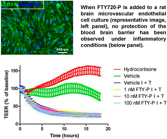
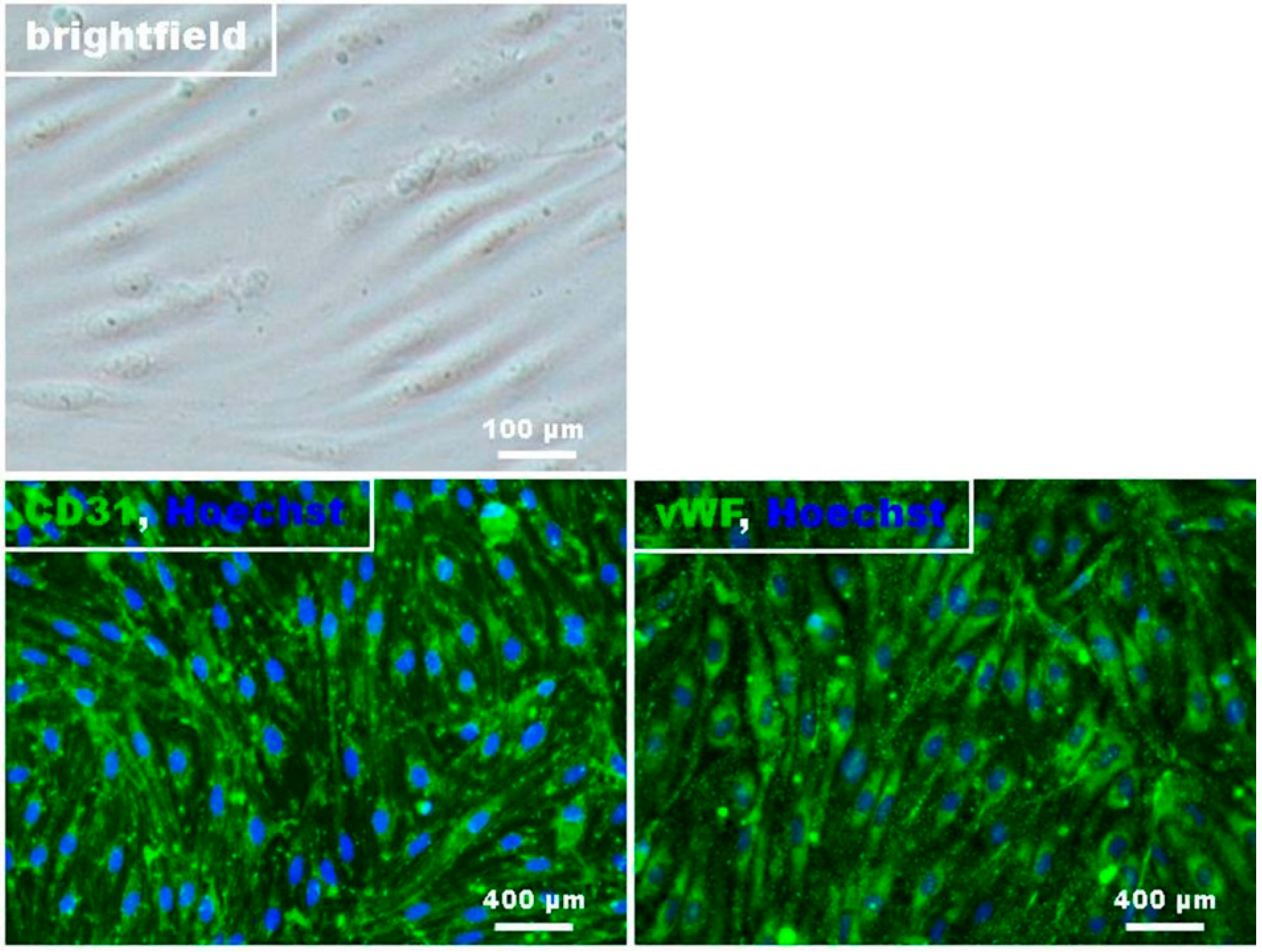
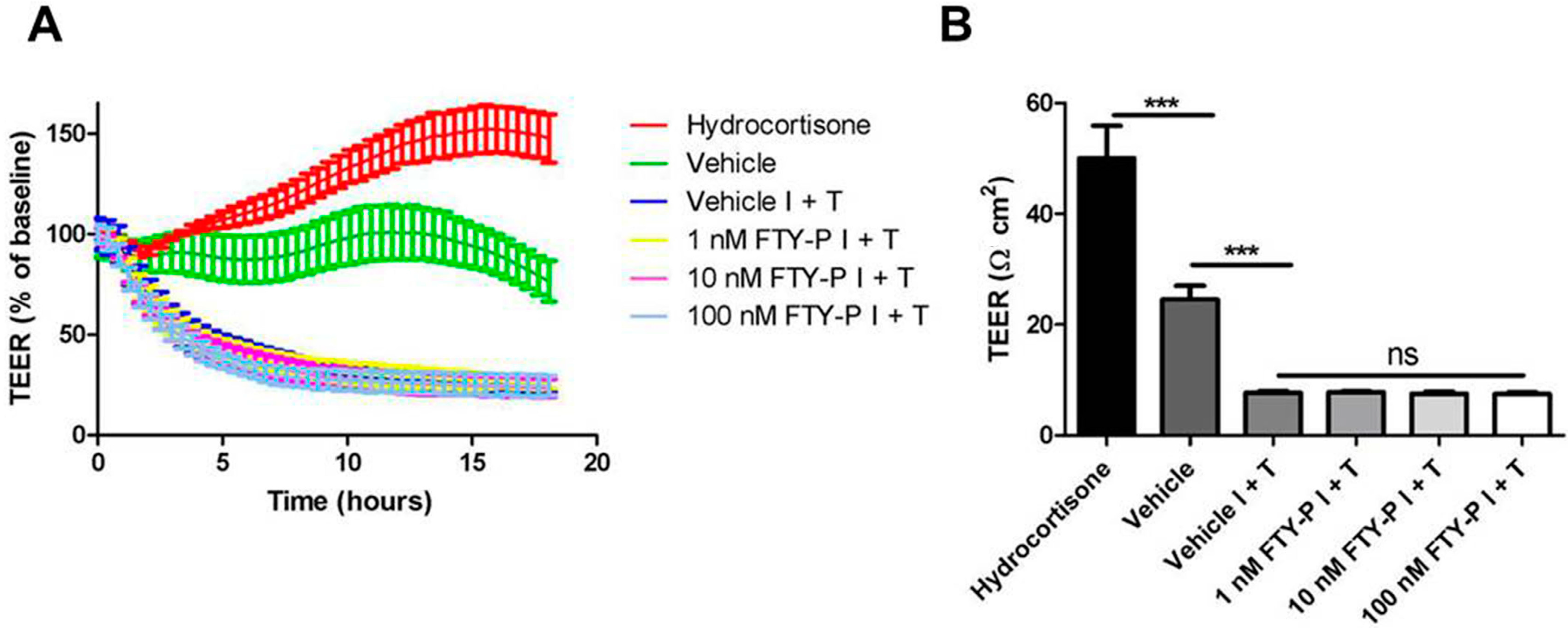
2.1. FTY720-P Does Not Enhance Endothelial Barrier Function in Rat Brain Microvascular Endothelial Cell (RBMEC) Cultures

The main effect of FTY720-P in MS is the alteration of lymphocyte trafficking via modulation of S1P1 [16]. Brain microvascular endothelial cells (BMECs) represent a possible additional target for FTY720-P in MS patients by directly interfering with the function of the BBB, since BMECs also express S1P1 [17]. In a first set of experiments, the endothelial phenotypic morphology and the presence of a monolayer was confirmed by phase-contrast microscopy and immunostaining of the cells with the endothelial cell markers CD31 and von Willebrand factor (vWF) (Figure 1). About 95% of all RBMECs were vWF-positive indicating a high degree of cell culture purity. The barrier function of the present cell cultures was verified by the detection of the TJ-proteins claudin-5 and occludin.
Figure 1. Histological characterization of rat brain endothelial cells. Phase contrast image of confluent RBMEC revealed that the cells express a spindle shaped morphology. Micrographs of immunofluorescence staining against Hoechst (blue), CD31, vWF, claudin-5, and occludin (green).
Ijms 16 26177 g001aIjms 16 26177 g001b
First, the effect of FTY720-P on RBMECs under inflammatory conditions was examined. Five days after seeding cell cultures, RBMECs were exposed to an inflammatory milieu of interferon γ (IFNγ) and tumor necrosis factor α (TNFα) (100 IU each) and incubated with either a control medium (vehicle) or FTY720-P in three different doses (1, 10, and 100 nM) for 18 h. TEER was continuously measured during these 18 h, which enabled the integrity of the BBB to be determined in vitro. Incubation of RBMEC cultures with 550 nM hydrocortisone served as a positive control.
TEER of vehicle-treated RBMECs decreased significantly after exposure to IFNγ and TNFα compared with RBMECs under homeostatic conditions (7.8 ± 0.3 Ω·cm2 vs. 24.6 ± 2.5 Ω·cm2; p < 0.001). Adding FTY720-P in three different doses to RBMECs exposed to an inflammatory milieu did not increase TEER compared with vehicle-treated RBMECs under the same conditions (Figure 2A,B).
Figure 2. Transendothelial electrical resistance (TEER) of rat brain microvascular endothelial cell (RBMEC) cultures exposed to inflammatory conditions. (A) Time- and dose-dependent effect of FTY720-P (FTY-P; 1, 10 and 100 nM; n = 4) on TEER in RBMECs exposed to interferon γ and tumor necrosis factor α (I + T; 100 IU each) for 18 h compared with TEER of RBMECs in an inflammatory milieu alone (Vehicle I + T; n = 4) and under homeostatic conditions (Vehicle; n = 4). Hydrocortisone treatment (HC; 550 nM; n = 3) was used as a positive control; (B) Absolute TEER values of RBMECs 18 h after exposure to I + T (100 IU each) and FTY720-P treatment (n = 3 or 4). *** p < 0.001; ns, not significant.
Ijms 16 26177 g002
Caspase-mediated apoptosis of cerebral endothelial cells might contribute to changes of their barrier function [18]. In this context, apoptosis might be due to high concentrations of IFNγ and TNFα or induced by a high (i.e., 100 nM) concentration of FTY720-P. Therefore, a caspase 3-mediated apoptotic effect of these cytokines alone or in combination with FTY720-P on RBMECs was investigated using Western blot analysis followed by denistometric quantification. Neither the inflammatory conditions nor FTY720-P added to the inflammatory milieu induced caspase 3 activation or endothelial cell death (Figure 3A,B).
Figure 3. Analysis of apoptosis in rat brain microvascular endothelial cells (RBMECs) exposed to inflammatory conditions and incubated with FTY720-P. (A,B) Western blot analyses and densitometric quantification of the amount of full-length caspase 3 protein in RBMEC cultures after exposure to interferon γ and tumor necrosis factor α (I + T; 100 IU each) and treatment with FTY720-P (FTY-P; 1, 10 and 100 nM; n = 4) for 18 h compared with untreated cultures under inflammatory conditions (Vehicle I + T; n = 4) as well as with cultures under a homeostatic milieu (Vehicle; n = 4); (C) Staurosporine treatment (Stauro; 1 μM for 2 h) was used as a positive control. Ctrl, control; ns, not significant; Veh, vehicle.
Ijms 16 26177 g003

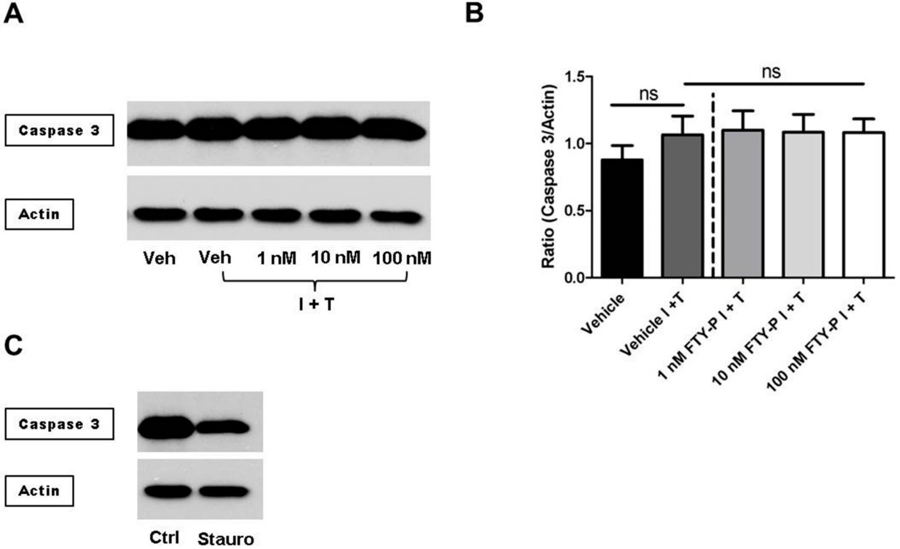
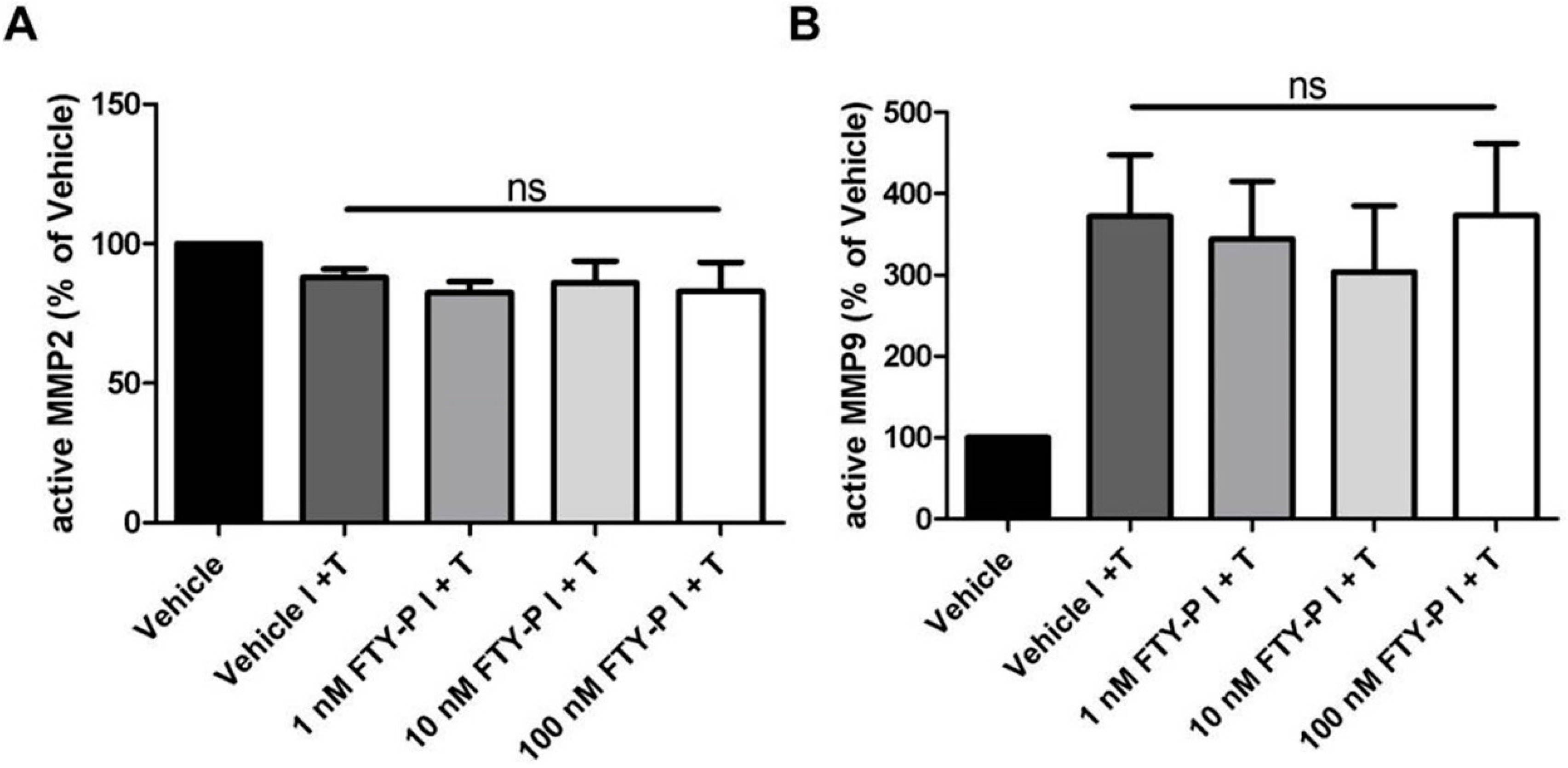
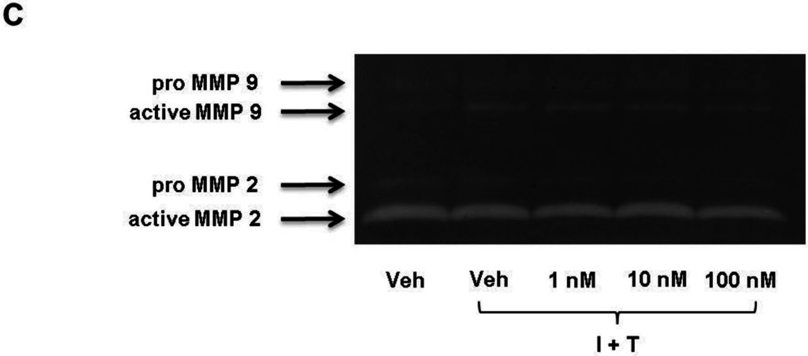
2.2. FTY720-P Does Not Alter the Amount of MMP-2 and MMP-9 Proteins in RBMEC Cultures

MMPs increase the permeability of the blood-brain barrier by attacking the extracellular matrix, basal lamina and TJs in endothelial cells, resulting in the final neuroinflammatory damage [19]. To examine whether FTY720-P alters MMP-2 or MMP-9 expression of endothelial cells, we performed a zymographic analysis of RBMEC supernatants 18 h after FTY720-P treatment. We detected no difference between FTY720-P- and vehicle-treated BMEC cultures regarding the amount of MMP-2 and MMP-9 (Figure 4A–C).
Figure 4. Secretion of matrix metalloproteinase (MMP)-2 (A) and MMP-9 (B) by rat brain microvascular endothelial cells after exposure to interferon γ and tumor necrosis factor α (I + T; 100 IU each) and treatment with FTY720-P (FTY-P; 1, 10 and 100 nM; n = 4) for 18 h compared with untreated cultures under inflammatory conditions (Vehicle I + T; n = 4) as well as with cultures in a homeostatic milieu (Vehicle; n = 4); (C) Representative experiment of zymography bands. ns, not significant; Veh, vehicle.
Ijms 16 26177 g004aIjms 16 26177 g004b

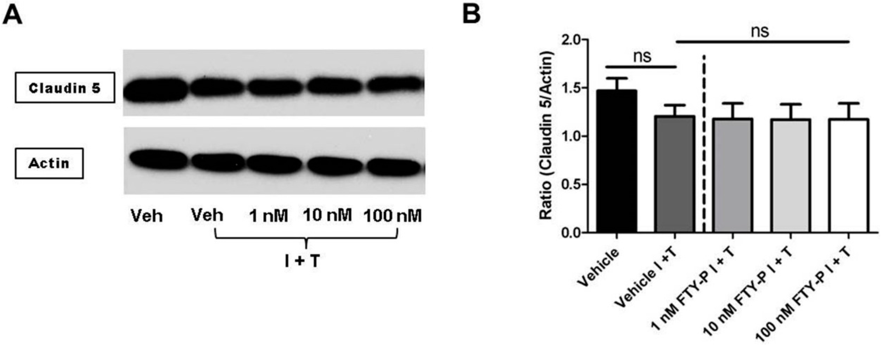
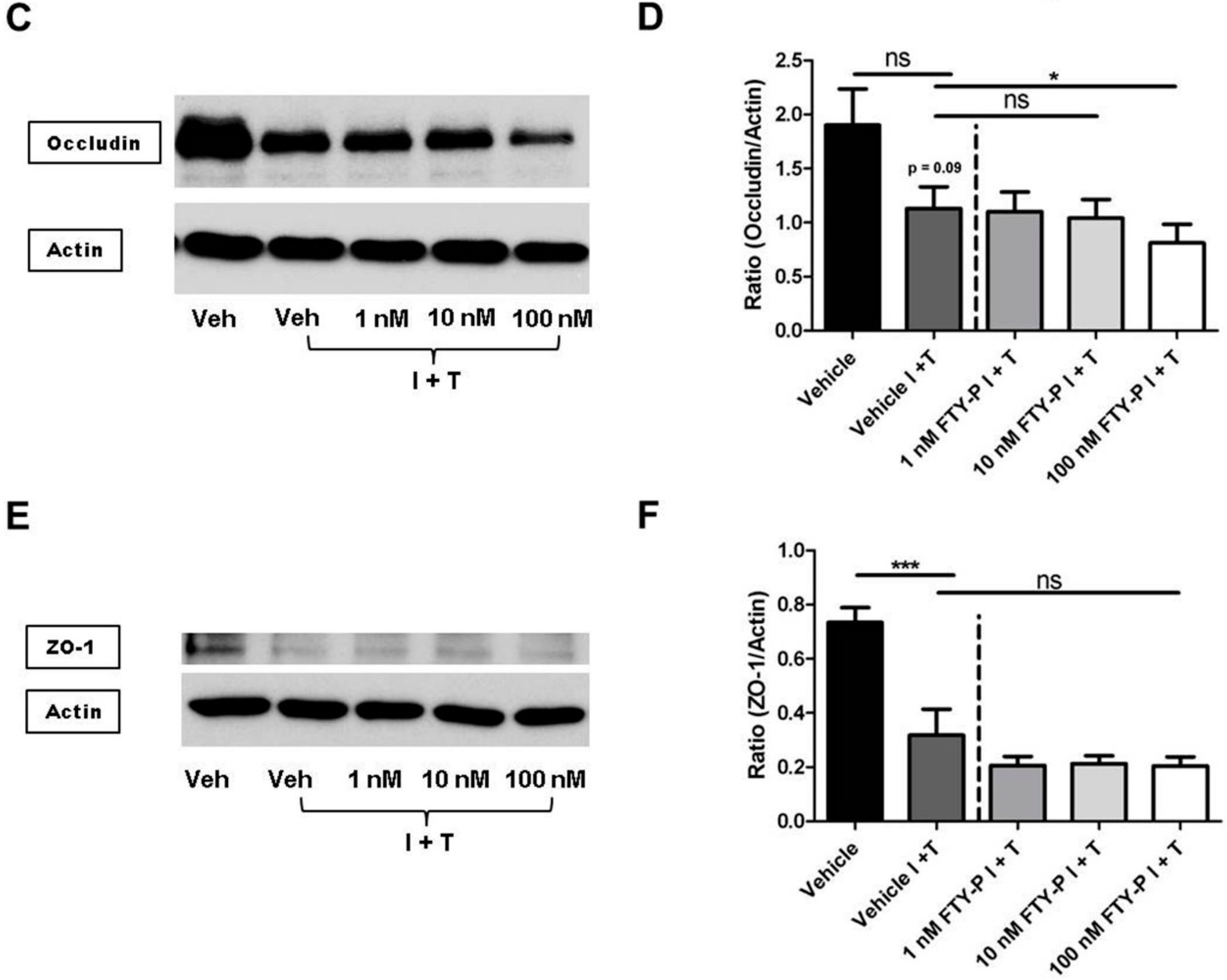
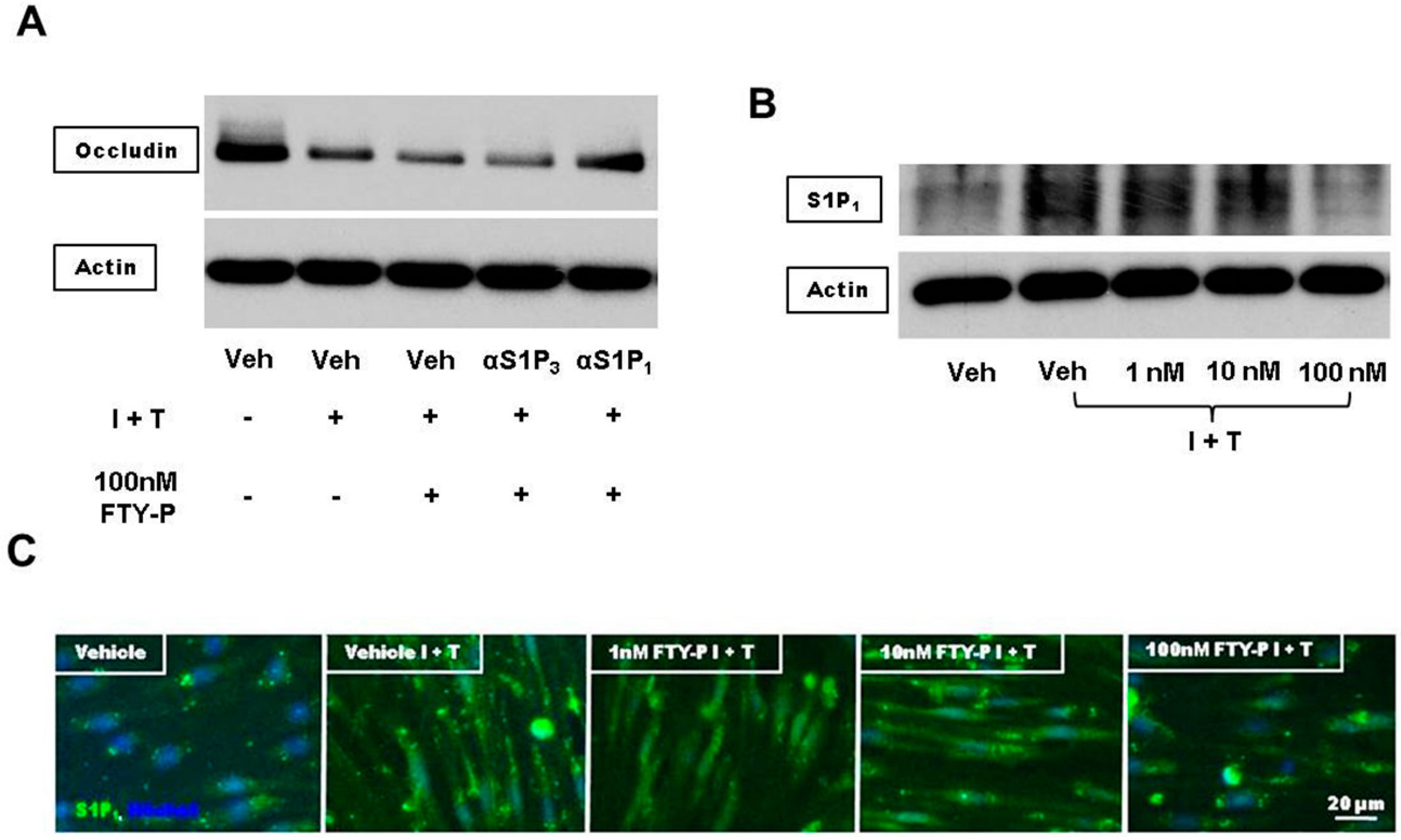
2.3. FTY720-P Reduces Occludin in RBMEC Cultures

According to the aforementioned experiments, FTY720-P did not alter BBB permeability in an RBMEC culture exposed to inflammatory conditions. This finding raised the question whether FTY720-P might even have the opposite effect, i.e., might further destabilize the endothelial barrier function—an eventuality which is not necessarily depicted by measurements of the TEER. Therefore, we investigated the effect of FTY720-P on the TJ proteins occludin, claudin 5, and ZO-1 in RBMEC cultures which were exposed to inflammatory conditions for 18 h. Quantitative assessment of TJ proteins was performed using Western blot analysis followed by densitometric quantification; a qualitative determination of these proteins was enabled by immunocytochemistry.
When FTY720-P was added to RBMEC cultures under inflammatory conditions, a dose-dependent and significant decrease of occludin was detected compared with non-treated cell cultures (occludin/actin 1.1 ± 0.2 vs. 0.8 ± 0.2; p < 0.05). In contrast, FTY720-P-treatment did not change the amount of claudin 5 in RBMEC cutlures when exposed to an inflammatory milieu (Figure 5, Figure S1). Additionally, ZO-1 was significantly decreased in RBMEC cultures after exposure to IFNγ and TNFα. However, FTY720-P-treatment did not alter the amount of ZO-1.
Figure 5. Effect of FTY720-P (FTY) on the amount of junctional protein of rat brain microvascular endothelial cell (RBMEC) cultures under inflammatory conditions. Western blot analysis and densitometric quantification of the amount of claudin 5 (A,B); occludin (C,D); and ZO-1 (E,F) proteins in RBMECs after exposure to interferon γ and tumor necrosis factor α (I + T; 100 IU each) and treatment with FTY-P (1, 10 and 100 nM; n = 4) for 18 h compared with untreated cultures under inflammatory conditions (Vehicle I + T; n = 4) as well as with cultures in a homeostatic milieu (Vehicle; n = 4). β-Actin was used as loading control. * p < 0.05; *** p < 0.001; ns, not significant; Veh, vehicle.
Ijms 16 26177 g005aIjms 16 26177 g005b

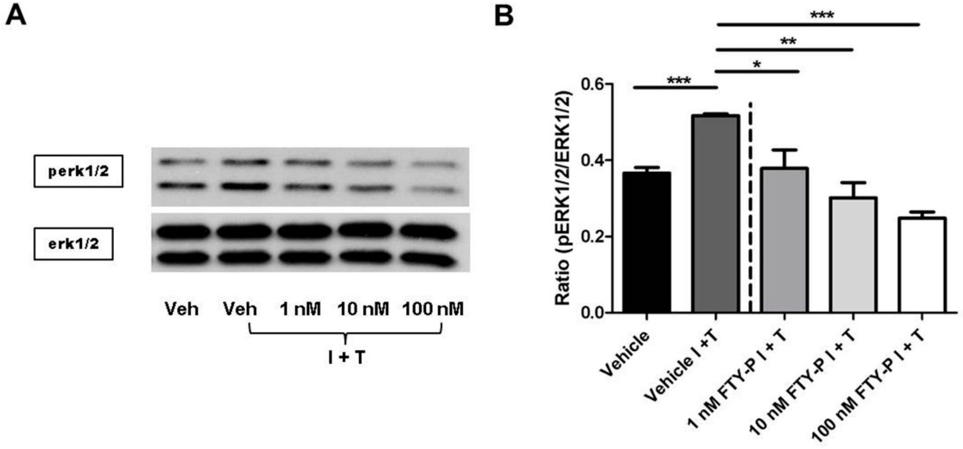
2.4. FTY720-P-Associated Decrease of Occludin Is Not Mediated via the erk1/2 Pathway

FTY720-P is a functional antagonist of all S1P receptors except S1P2 [20]. Therefore we next examined whether RBMEC expresses these receptors under inflammatory conditions. In contrast to the S1P1 and S1P3-receptor expression, we observed a sdownregulation of S1P4 and S1P5-receptors under inflammatory conditions (supplementary Figure S2). Thus, S1P4 and S1P5-receptors seem to play no crucial role in the regulation of the BBB integrity under inflammatory conditions and therefore no further investigations on these two receptors were performed. To analyze whether FTY720 impacts S1P1 or S1P3-receptor signaling we used both, specific S1P1 and S1P3-receptor antagonists. Our Western blot analyses yielded only a link between S1P1 signaling and the reduction of occludin (Figure 6A).
Downregulation of occludin is induced by activation of the extracellular-signal-regulated kinase (erk1/2) pathway leading to an increase in endothelial permeability [21]. erk1/2 phosphorylation (i.e., activation) is mediated by high-affinity antagonism of S1P1, including a ligand-induced receptor internalization [22]. We next assessed the effect of FTY720-P on the protein amount of S1P1. There was no change in the amount of S1P1 protein when cell cultures were treated with FTY720-P doses of 1 or 10 nM. However, treatment with 100 nM FTY720-P revealed a clear reduction in this amount (Figure 6B,C).
Ninety minutes after exposing RBMEC culture to IFNγ and TNFα, phosphorylation of erk1/2 decreased by adding FTY720-P to the cell cultures at doses of 1, 10, and 100 nM in a dose-dependent manner (Figure 7).
Figure 6. (A) Representative Western blot analysis of the amount of occludin and β-actin proteins in RBMECs either exposed to interferon γ and tumor necrosis factor α (I + T; 100 IU each) or cultured in physiological medium (Veh) after incubation with FTY720-P (100 nM) in the presence or absence of a specific S1P1 or S1P3-receptor antagonist (αS1P3, αS1P1; 10 μM each) for 18 h; (B) Representative Western blot analysis of the amount of S1P1 and β-actin proteins RBMECs either exposed to interferon γ and tumor necrosis factor α (I + T; 100 IU each) or cultured in physiological medium after incubation with FTY720-P (1, 10, and 100 nM) for 18 h; (C) Representative immunofluorescence staining against Hoechst (blue) and S1P1 (green) in RBMECs.
Ijms 16 26177 g006
Figure 7. Effect of FTY720-P (FTY-P) on erk1/2 signaling in rat brain microvascular endothelial cell (RBMEC) cultures exposed to inflammatory conditions and incubated with FTY720-P. (A,B) Western blot analyses and densitometric quantification of the amount of erk1/2 and perk1/2 proteins in RBMECs treated for 90 min with FTY-P (1, 10, and 100 nM; n = 3) and exposed to interferon γ and tumor necrosis factor α (I + T; 100 IU each) compared with untreated cultures under inflammatory conditions (Vehicle I + T; n = 3) as well as to cultures in homeostatic milieu (Vehicle; n = 3). * p < 0.05; ** p < 0.01; *** p < 0.001; Veh, vehicle.
Ijms 16 26177 g007

3. Discussion

In the present study, the role of FTY720-P on endothelial permeability has been examined in RBMEC cultures exposed to inflammatory conditions. As a main finding, FTY720-P at different dosages did not decrease endothelial permeability in RBMECs after exposure to IFNγ and TNFα. This is in contrast to recently published studies which revealed a protective effect of FTY720-P on BBB leakage in cell cultures and in an animal model [14,23]. However, following FTY720-P treatment in MS patients, macular edema was a prominent adverse event reported in two large randomized controlled trials, the TRANSFORMS [24] and FREEDOMS [12] trial. When comparing different studies of FTY720-P in MS [12,24] and renal transplant recipients [25,26], the incidence of macular edema was dose-dependent. Interestingly, whereas the ocular toxicity was dose-dependent in these studies, the therapeutic effect was not [27]. The pathogenesis of FTY720-P-associated macular edema is unspecified. There is some evidence that FTY720-P involves also the degradation of the S1P1 receptor and thus plays a role in regulating vascular permeability [28,29], providing a possible explanation of FTY720-P-associated macular edema.
Furthermore, we found a downregulation of the TJ protein occludin in RBMEC cultures exposed to inflammatory conditions and treated with FTY720-P. The S1P1 receptor is involved in the intercellular adhesion of endothelial cells via TJs, AJs and gap junctions [28,30]. ZO-1, a TJ-associated protein, forms a multiprotein complex with occludin [31,32] and thus is suggested to regulate occludin. S1P alters the distribution of ZO-1 protein via S1P1 which results in an enhanced endothelial barrier function [30,33]. FTY720-P is a functional antagonist to S1P1 and, as a consequence, might reduce occludin in RBMECs.
Redistribution of ZO-1 protein is mediated via the S1P1/Gi/Akt/Rac pathway [30], which finally results in erk1/2 phosphorylation. Activation of erk1/2 has been shown to downregulate occludin [21]. In the present study, assessment of erk1/2 revealed a dose dependent decrease in phosphorylation but also a decrease of occludin after FTY-720-P-treatment under inflammatory conditions. Thus, we hypothesize that regulation of occludin is also mediated by mechanisms that overcome the S1P1/Gi/Akt/Rac pathway. Increased MMP-9 activity promotes the disruption of occludin and ZO-1 in cerebral endothelial cells leading to the disruption of BBB integrity [34]. Additionally, MMP-9 is also able to degrade occludin in retinal epithelial cells [35]. However, in this study, the amount of MMP-9 and ZO-1 in BMECs did not change when FTY720-P was added to the medium, making this pathway unlikely. Alternatively, a reduction of occludin might be promoted via caveolin-1, a major structural component of membrane microdomains thought to be functionally complexed with TJs [36]. Caveolin-1 siRNA delivered to BMEC monolayers reduced the caveolin-1 protein level as well as occludin and ZO-1. In this context, occludin exhibited a dissociation from the cytoskeletal framework [37]. Downregulation of ZO-1/occludin is also associated with a decreased expression of connexin 43 (Cx43) [38], a gap junction protein, which is required to maintain endothelial barrier function [39]. Notably, the interaction of Cx43 and ZO-1/occludin is linked to S1P1 modulation [40].
In summary, FTY720-P did not contribute to a decreased leakage of endothelial barrier function of RBMECs after exposure to inflammatory conditions, which is in line with other studies [22,41,42]. In contrast, we found downregulation of the TJ protein occludin, which rather contributes to a further destabilization of the endothelial barrier. This observation might be due to S1P1-mediated signaling since this receptor intervenes in the regulation of endothelial permeability, providing a mechanistic basis for FTY720-P-associated disruption of barrier function. Conversely, S1P1 activation via S1P enhances endothelial barrier integrity through its action both on the cell cytoskeleton and on intercellular junctions [43]. Therefore, S1P1 has a dual role in immune and vascular cell biology. Depending on whether S1P or FTY720-P interacts with this receptor, S1P1-mediated signaling either enhances endothelial barrier function or inhibits lymphocyte egression from lymphoid tissues and increases vascular permeability.

4. Materials and Methods

4.1. Materials

FTY720-P was obtained from Biomol (Hamburg, Germany). FTY720-P was dissolved in the vehicle solution composed of DMSO/HCl (stock concentration: 50 mM) and diluted with methanol to 1 mM FTY720-P [44]. IFNγ was purchased from Miltenyi (Bergisch Gladbach, Germany). TNFα was obtained from Peprotech (Hamburg, Germany). Selective sphingosine-1-phosphate receptor S1P1 (W146) and S1P3 (TY 52156) antagonists were obtained from Tocris (Wiesbaden-Nordenstadt, Germany). Rabbit anti-caspase 3, anti-erk1/2, and anti-Phospho-erk1/2 antibodies were acquired from Cell Signaling (Leiden, The Netherlands). Rabbit anti-S1P1 was obtained from Santa Cruz (Heidelberg, Germany). Rabbit anti-vWF, and anti-occludin were purchased from Abcam (Cambridge, UK). Mouse anti-claudin 5 and rabbit anti-ZO-1 antibodies were obtained from Invitrogen (Darmstadt, Germany). Mouse anti-CD31 and rabbit anti-vWF antibodies were obtained from Invitrogen. Mouse anti-occludin antibody was acquired from Becton Dickinson (Heidelberg, Germany). All other reagents were purchased from Sigma–Aldrich (St. Louis, MO, USA).

4.2. Preparation and Cultivation of RBMECs

RBMECs were isolated and cultured as previously described for murine brain microvessel endothelial cells [45]. Briefly, brains of eight-week-old rats were homogenized and stepwise digested by two proteases, followed by further purification steps. RBMECs were either planted on collagen type IV/fibronectin-coated transwell polyester membrane inserts with 0.4-μm pores (Corning, Life Science, Kaiserslautern, Germany) or in 24-well plates (Nunc, Darmstadt, Germany). The cell cultures were maintained in serum-containing culture medium at 37 °C with a humidified atmosphere of 5% CO2/95% air, for five days. All experiments were conducted in serum-free medium.

4.3. Immunocytochemistry

RBMECs were cultured on collagen IV/fibronectin-coated glass coverslips or Transwell Polyester Membrane inserts with 0.4-μm pores. Cells were fixed with methanol for 10 min at −20 °C and stained with mouse anti-CD31, rabbit anti-vWF, rabbit anti-occludin, rabbit anti-S1P1, or mouse anti-claudin 5 according to standard protocols. RBMECs were imaged with a Nikon Eclipse 50i microscope equipped with a CCD camera (Nikon, Tokyo, Japan).

4.4. Western Blot Assays

RBMECs were cultured in 24-well plates until they reached confluency. After cell lysis, the amount of protein was determined using BCA protein assay. Denatured protein (1–3 μg) was electrophoresed and transferred to a nitrocellulose membrane. Membranes were blocked for 1 h and incubated with either an anti-erk1/2 antibody, anti-phosphoerk1/2, anti-S1P1, anti-caspase 3, anti-claudin 5, antu-ZO-1, or an anti-occludin antibody at 4 °C overnight. Thereafter, the membranes were incubated with a horseradish peroxidase-conjugated anti-rabbit or anti-mouse immunoglobulin G antibody (Dianova, Hamburg, Germany) at room temperature for 1 h. Immunoblots were detected using ECLplus (PerkinElmer, Waltham, MA, USA) and a Kodak X-OMAT 5000 RA developer (Kodak, Rochester, NY, USA). In order to control protein loading and transfer, membranes were incubated with a β-actin monoclonal antibody. Bands were quantified by densitometric analysis using ImageJ Analysis Software 1.45s [46] and normalized to the actin band.

4.5. Measurement of TEER

The electrical resistance across the RBMEC layers was measured using an automated long-term monitoring resistance meter (CellZcope, San Francisco, CA, USA). TEER was assessed on endothelial cell monolayers cultured on collagen IV/fibronectin-coated Transwell Pore Polyester Membrane inserts with 0.4-μm pores (Corning).

4.6. Zymography

The amount of secreted MMP-2 and MMP-9 in BMEC supernatants was detected by zymography as recently reported [47]. Briefly, BMEC supernatants were collected and mixed with equal volumes of sample buffer (20% glycin, 4% SDS, 2 mM EDTA, 0.01% bromophenol blue, 125 mM Tris–HCl pH 6.8). Next, samples were loaded on a 10% SDS-PAGE containing 0.1% gelatin. After electrophoresis, the gels were treated twice with 2.7% Triton X-100 solution for 30 min and thereafter were incubated in developing buffer (50 mM Tris, 200 mM NaCl, 5 mM CaCl2, 0.02% Brij-35, 40 mM Tris–HCl pH 7.5) at 37 °C overnight. Gels were stained with 0.25% (w/v) Coomassie brilliant blue in 25% isopropanol/10% acetic acid, and bleached with 50% methanol and 10% acetic acid until bands with diminished staining appeared. Images of gels were captured by EPSON Perfection V500 scanner (Epson, Nagano, Japan) and subsequently analyzed with ImageJ Analysis Software 1.45s National Institutes of Health, Bethesda, MD, USA.

4.7. Quantitative Real-Time PCR

Quantitative real-time PCR analysis was performed according to standard procedures. Relative gene expression levels of S1P1 (EDG1; assay ID: Rn02758712_s1; Applied Biosystems, Frankfurt, Germany), S1P3 (EDG3; assay ID: Rn02758880_s1, Applied Biosystems), S1P4 (EDG4; assay ID: Rn01408095_s1, Applied Biosystems), and S1P5 (EDG5; assay ID: Rn00572952_s1, Applied Biosystems) were quantified with fluorescent TaqMan technology. Gapdh (TaqMan Predeveloped Assay Reagents for gene expression, part number: 4351371, Applied Biosystems) was used as an endogenous control.

4.8. Statistical Analysis

All results are presented as mean ± SEM. To test for significant differences between multiple groups, one-way analysis of variance was used, with post hoc Bonferroni adjustment for p-values. Statistical analysis comparing two groups (Vehicle vs. Vehicle I + T) was performed using two-tailed Student t test. p-values <0.05 were considered significant with * p < 0.05; ** p < 0.01; *** p < 0.001.

Supplementary Materials

Supplementary materials can be found at https://www.mdpi.com/1422-0067/16/12/26177/s1.

Acknowledgments

We thank Andrea Sauer and Susanne Hellmig for excellent technical assistance. This work was supported by the Sonderforschungsbereich 688, the Interdisziplinäres Zentrum für klinische Forschung Würzburg and Novartis Pharma GmbH, Nürnberg. Christoph Kleinschnitz has received consulting fees, speaker’s honoraria, compensation for travel expenses, and financial support for scientific projects from Novartis Pharma GmbH, Nürnberg, Germany. Stefan Bittner has received financial research support from Novartis, funding for travel expenses for attending meetings from Novartis and Bayer Schering and honoraria from TEVA and Biogen Idec. Sven G. Meuth has received honoraria for lecturing and travel expenses for attending meetings and has received financial research support from Bayer, Bayer Schering, Biogen Idec, Genzyme, Merck Serono, MSD, Novartis, Novo Nordisk, Sanofi-Aventis, and Teva.

Author Contributions

Michael K. Schuhmann and Felix Fluri had the idea for the project. Michael K. Schuhmann conceived and designed the RBMEC experiments, immunohistochemistry and protein analysis. Michael K. Schuhmann and Felix Fluri performed the RBMEC cultures, immunocytochemistry and Western blot analyses. Michael K. Schuhmann conducted the TEER measurements. Michael K. Schuhmann, Christoph Kleinschnitz, and Felix Fluri analyzed the data. Michael K. Schuhmann, Stefan Bittner, Sven G. Meuth, Christoph Kleinschnitz, and Felix Fluri evaluated the data and co-wrote the paper.

Conflicts of Interest

The authors declare no conflict of interest.

References

  1. Compston, A.; Coles, A. Multiple sclerosis. Lancet 2008, 372, 1502–1517. [Google Scholar] [CrossRef]
  2. Kebir, H.; Kreymborg, K.; Ifergan, I.; Dodelet-Devillers, A.; Cayrol, R.; Bernard, M.; Giuliani, F.; Arbour, N.; Becher, B.; Prat, A. Human TH17 lymphocytes promote blood-brain barrier disruption and central nervous system inflammation. Nat. Med. 2007, 13, 1173–1175. [Google Scholar] [CrossRef] [PubMed]
  3. Alt, C.; Duvefelt, K.; Franzén, B.; Yang, Y.; Engelhardt, B. Gene and protein expression profiling of the microvascular compartment in experimental autoimmune encephalomyelitis in C57Bl/6 and SJL mice. Brain Pathol. Zurich Switz. 2005, 15, 1–16. [Google Scholar] [CrossRef]
  4. Neuwelt, E.A.; Bauer, B.; Fahlke, C.; Fricker, G.; Iadecola, C.; Janigro, D.; Leybaert, L.; Molnár, Z.; O’Donnell, M.E.; Povlishock, J.T.; et al. Engaging neuroscience to advance translational research in brain barrier biology. Nat. Rev. Neurosci. 2011, 12, 169–182. [Google Scholar] [CrossRef] [PubMed]
  5. Abbott, N.J.; Rönnbäck, L.; Hansson, E. Astrocyte–endothelial interactions at the blood–brain barrier. Nat. Rev. Neurosci. 2006, 7, 41–53. [Google Scholar] [CrossRef] [PubMed]
  6. Hawkins, B.T.; Davis, T.P. The Blood-Brain Barrier/Neurovascular Unit in Health and Disease. Pharmacol. Rev. 2005, 57, 173–185. [Google Scholar] [CrossRef] [PubMed]
  7. Yu, A.S.L.; McCarthy, K.M.; Francis, S.A.; McCormack, J.M.; Lai, J.; Rogers, R.A.; Lynch, R.D.; Schneeberger, E.E. Knockdown of occludin expression leads to diverse phenotypic alterations in epithelial cells. Am. J. Physiol. 2005, 288, C1231–C1241. [Google Scholar] [CrossRef] [PubMed]
  8. Wolburg, H.; Lippoldt, A. Tight junctions of the blood–brain barrier: development, composition and regulation. Vascul. Pharmacol. 2002, 38, 323–337. [Google Scholar] [CrossRef]
  9. Kirk, J.; Plumb, J.; Mirakhur, M.; McQuaid, S. Tight junctional abnormality in multiple sclerosis white matter affects all calibres of vessel and is associated with blood–brain barrier leakage and active demyelination. J. Pathol. 2003, 201, 319–327. [Google Scholar] [CrossRef] [PubMed]
  10. Padden, M.; Leech, S.; Craig, B.; Kirk, J.; Brankin, B.; McQuaid, S. Differences in expression of junctional adhesion molecule-A and β-catenin in multiple sclerosis brain tissue: Increasing evidence for the role of tight junction pathology. Acta Neuropathol. 2006, 113, 177–186. [Google Scholar] [CrossRef] [PubMed]
  11. Marsolais, D.; Rosen, H. Chemical modulators of sphingosine-1-phosphate receptors as barrier-oriented therapeutic molecules. Nat. Rev. Drug Discov. 2009, 8, 297–307. [Google Scholar] [CrossRef] [PubMed]
  12. Kappos, L.; Radue, E.-W.; O’Connor, P.; Polman, C.; Hohlfeld, R.; Calabresi, P.; Selmaj, K.; Agoropoulou, C.; Leyk, M.; Zhang-Auberson, L.; et al. A Placebo-Controlled Trial of Oral Fingolimod in Relapsing Multiple Sclerosis. N. Engl. J. Med. 2010, 362, 387–401. [Google Scholar] [CrossRef] [PubMed]
  13. Cartwright, T.A.; Campos, C.R.; Cannon, R.E.; Miller, D.S. Mrp1 is essential for sphingolipid signaling to p-glycoprotein in mouse blood–brain and blood–spinal cord barriers. J. Cereb. Blood Flow Metab. 2013, 33, 381–388. [Google Scholar] [CrossRef] [PubMed]
  14. Nishihara, H.; Shimizu, F.; Sano, Y.; Takeshita, Y.; Maeda, T.; Abe, M.; Koga, M.; Kanda, T. Fingolimod Prevents Blood-Brain Barrier Disruption Induced by the Sera from Patients with Multiple Sclerosis. PLoS ONE 2015, 10, e0121488. [Google Scholar] [CrossRef] [PubMed]
  15. Shimizu, F.; Tasaki, A.; Sano, Y.; Ju, M.; Nishihara, H.; Oishi, M.; Koga, M.; Kawai, M.; Kanda, T. Sera from Remitting and Secondary Progressive Multiple Sclerosis Patients Disrupt the Blood-Brain Barrier. PLoS ONE 2014, 9, e92872. [Google Scholar] [CrossRef] [PubMed]
  16. Mandala, S.; Hajdu, R.; Bergstrom, J.; Quackenbush, E.; Xie, J.; Milligan, J.; Thornton, R.; Shei, G.-J.; Card, D.; Keohane, C.; et al. Alteration of Lymphocyte Trafficking by Sphingosine-1-Phosphate Receptor Agonists. Science 2002, 296, 346–349. [Google Scholar] [CrossRef] [PubMed]
  17. Brinkmann, V.; Cyster, J.G.; Hla, T. FTY720: Sphingosine 1-Phosphate Receptor-1 in the Control of Lymphocyte Egress and Endothelial Barrier Function. Am. J. Transplant. 2004, 4, 1019–1025. [Google Scholar] [CrossRef] [PubMed]
  18. Lopez-Ramirez, M.A.; Fischer, R.; Torres-Badillo, C.C.; Davies, H.A.; Logan, K.; Pfizenmaier, K.; Male, D.K.; Sharrack, B.; Romero, I.A. Role of Caspases in Cytokine-Induced Barrier Breakdown in Human Brain Endothelial Cells. J. Immunol. 2012, 189, 3130–3139. [Google Scholar] [CrossRef] [PubMed]
  19. Rosenberg, G.A. Matrix metalloproteinases and their multiple roles in neurodegenerative diseases. Lancet Neurol. 2009, 8, 205–216. [Google Scholar] [CrossRef]
  20. Brinkmann, V.; Davis, M.D.; Heise, C.E.; Albert, R.; Cottens, S.; Hof, R.; Bruns, C.; Prieschl, E.; Baumruker, T.; Hiestand, P.; et al. The Immune Modulator FTY720 Targets Sphingosine 1-Phosphate Receptors. J. Biol. Chem. 2002, 277, 21453–21457. [Google Scholar] [CrossRef] [PubMed]
  21. Zeiller, C.; Mebarek, S.; Jaafar, R.; Pirola, L.; Lagarde, M.; Prigent, A.-F.; Némoz, G. Phospholipase D2 regulates endothelial permeability through cytoskeleton reorganization and occludin downregulation. Biochim. Biophys. Acta 2009, 1793, 1236–1249. [Google Scholar] [CrossRef] [PubMed]
  22. Sanna, M.G.; Wang, S.-K.; Gonzalez-Cabrera, P.J.; Don, A.; Marsolais, D.; Matheu, M.P.; Wei, S.H.; Parker, I.; Jo, E.; Cheng, W.-C.; et al. Enhancement of capillary leakage and restoration of lymphocyte egress by a chiral S1P1 antagonist in vivo. Nat. Chem. Biol. 2006, 2, 434–441. [Google Scholar] [CrossRef] [PubMed]
  23. Foster, C.A.; Mechtcheriakova, D.; Storch, M.K.; Balatoni, B.; Howard, L.M.; Bornancin, F.; Wlachos, A.; Sobanov, J.; Kinnunen, A.; Baumruker, T. FTY720 Rescue Therapy in the Dark Agouti Rat Model of Experimental Autoimmune Encephalomyelitis: Expression of Central Nervous System Genes and Reversal of Blood-Brain-Barrier Damage. Brain Pathol. 2009, 19, 254–266. [Google Scholar] [CrossRef] [PubMed]
  24. Cohen, J.A.; Barkhof, F.; Comi, G.; Hartung, H.-P.; Khatri, B.O.; Montalban, X.; Pelletier, J.; Capra, R.; Gallo, P.; Izquierdo, G.; et al. Oral Fingolimod or Intramuscular Interferon for Relapsing Multiple Sclerosis. N. Engl. J. Med. 2010, 362, 402–415. [Google Scholar] [CrossRef] [PubMed]
  25. Salvadori, M.; Budde, K.; Charpentier, B.; Klempnauer, J.; Nashan, B.; Pallardo, L.M.; Eris, J.; Schena, F.P.; Eisenberger, U.; Rostaing, L.; et al. FTY720 versus MMF with cyclosporine in de novo renal transplantation: a 1-year, randomized controlled trial in Europe and Australasia. Am. J. Transplant. 2006, 6, 2912–2921. [Google Scholar] [CrossRef] [PubMed]
  26. Tedesco-Silva, H.; Pescovitz, M.D.; Cibrik, D.; Rees, M.A.; Mulgaonkar, S.; Kahan, B.D.; Gugliuzza, K.K.; Rajagopalan, P.R.; de Esmeraldo, R.M.; Lord, H.; et al. Randomized controlled trial of FTY720 versus MMF in de novo renal transplantation. Transplantation 2006, 82, 1689–1697. [Google Scholar] [CrossRef] [PubMed]
  27. Jain, N.; Bhatti, M.T. Fingolimod-associated macular edema Incidence, detection, and management. Neurology 2012, 78, 672–680. [Google Scholar] [CrossRef] [PubMed]
  28. Lee, M.-J.; Thangada, S.; Claffey, K.P.; Ancellin, N.; Liu, C.H.; Kluk, M.; Volpi, M.; Sha’afi, R.I.; Hla, T. Vascular Endothelial Cell Adherens Junction Assembly and Morphogenesis Induced by Sphingosine-1-Phosphate. Cell 1999, 99, 301–312. [Google Scholar] [CrossRef]
  29. Garcia, J.G.; Liu, F.; Verin, A.D.; Birukova, A.; Dechert, M.A.; Gerthoffer, W.T.; Bamberg, J.R.; English, D. Sphingosine 1-phosphate promotes endothelial cell barrier integrity by Edg-dependent cytoskeletal rearrangement. J. Clin. Investig. 2001, 108, 689–701. [Google Scholar] [CrossRef] [PubMed]
  30. Lee, J.-F.; Zeng, Q.; Ozaki, H.; Wang, L.; Hand, A.R.; Hla, T.; Wang, E.; Lee, M.-J. Dual Roles of Tight Junction-associated Protein, Zonula Occludens-1, in Sphingosine 1-Phosphate-mediated Endothelial Chemotaxis and Barrier Integrity. J. Biol. Chem. 2006, 281, 29190–29200. [Google Scholar] [CrossRef] [PubMed]
  31. Schmidt, A.; Utepbergenov, D.I.; Mueller, S.L.; Beyermann, M.; Schneider-Mergener, J.; Krause, G.; Blasig, I.E. Occludin binds to the SH3-hinge-GuK unit of zonula occludens protein 1: Potential mechanism of tight junction regulation. Cell. Mol. Life Sci. 2004, 61, 1354–1365. [Google Scholar] [CrossRef] [PubMed]
  32. Fanning, A.S.; Anderson, J.M. Zonula Occludens-1 and -2 Are Cytosolic Scaffolds That Regulate the Assembly of Cellular Junctions. Ann. N. Y. Acad. Sci. 2009, 1165, 113–120. [Google Scholar] [CrossRef] [PubMed]
  33. Krump-Konvalinkova, V.; Yasuda, S.; Rubic, T.; Makarova, N.; Mages, J.; Erl, W.; Vosseler, C.; Kirkpatrick, C.J.; Tigyi, G.; Siess, W. Stable Knock-Down of the Sphingosine 1-Phosphate Receptor S1P1 Influences Multiple Functions of Human Endothelial Cells. Arterioscler. Thromb. Vasc. Biol. 2005, 25, 546–552. [Google Scholar] [CrossRef] [PubMed]
  34. Harkness, K.A.; Adamson, P.; Sussman, J.D.; Davies-Jones, G.A.B.; Greenwood, J.; Woodroofe, M.N. Dexamethasone regulation of matrix metalloproteinase expression in CNS vascular endothelium. Brain 2000, 123, 698–709. [Google Scholar] [CrossRef] [PubMed]
  35. Giebel, S.J.; Menicucci, G.; McGuire, P.G.; Das, A. Matrix metalloproteinases in early diabetic retinopathy and their role in alteration of the blood–retinal barrier. Lab. Investig. 2005, 85, 597–607. [Google Scholar] [CrossRef] [PubMed]
  36. Nusrat, A.; Parkos, C.A.; Verkade, P.; Foley, C.S.; Liang, T.W.; Innis-Whitehouse, W.; Eastburn, K.K.; Madara, J.L. Tight junctions are membrane microdomains. J. Cell Sci. 2000, 113, 1771–1781. [Google Scholar] [PubMed]
  37. Song, L.; Ge, S.; Pachter, J.S. Caveolin-1 regulates expression of junction-associated proteins in brain microvascular endothelial cells. Blood 2007, 109, 1515–1523. [Google Scholar] [CrossRef] [PubMed]
  38. Tien, T.; Barrette, K.F.; Chronopoulos, A.; Roy, S. Effects of high glucose-induced Cx43 downregulation on occludin and ZO-1 expression and tight junction barrier function in retinal endothelial cells. Investig. Ophthalmol. Vis. Sci. 2013, 54, 6518–6525. [Google Scholar] [CrossRef] [PubMed]
  39. Nagasawa, K.; Chiba, H.; Fujita, H.; Kojima, T.; Saito, T.; Endo, T.; Sawada, N. Possible involvement of gap junctions in the barrier function of tight junctions of brain and lung endothelial cells. J. Cell. Physiol. 2006, 208, 123–132. [Google Scholar] [CrossRef] [PubMed]
  40. Tencé, M.; Ezan, P.; Amigou, E.; Giaume, C. Increased interaction of connexin43 with zonula occludens-1 during inhibition of gap junctions by G protein-coupled receptor agonists. Cell Signal. 2012, 24, 86–98. [Google Scholar] [CrossRef] [PubMed]
  41. Tauseef, M.; Kini, V.; Knezevic, N.; Brannan, M.; Ramchandaran, R.; Fyrst, H.; Saba, J.; Vogel, S.M.; Malik, A.B.; Mehta, D. Activation of Sphingosine Kinase-1 Reverses the Increase in Lung Vascular Permeability Through Sphingosine-1-Phosphate Receptor Signaling in Endothelial Cells. Circ. Res. 2008, 103, 1164–1172. [Google Scholar] [CrossRef] [PubMed]
  42. Krump-Konvalinkova, V.; Chwalla, I.; Siess, W. FTY720 inhibits S1P-mediated endothelial healing: Relationship to S1P1-receptor surface expression. Biochem. Biophys. Res. Commun. 2008, 370, 603–608. [Google Scholar] [CrossRef] [PubMed]
  43. Wang, L.; Dudek, S.M. Regulation of vascular permeability by sphingosine 1-phosphate. Microvasc. Res. 2009, 77, 39–45. [Google Scholar] [CrossRef] [PubMed]
  44. Sensken, S.-C.; Stäubert, C.; Keul, P.; Levkau, B.; Schöneberg, T.; Gräler, M.H. Selective activation of G alpha i mediated signalling of S1P3 by FTY720-phosphate. Cell Signal. 2008, 20, 1125–1133. [Google Scholar] [CrossRef] [PubMed]
  45. Bittner, S.; Ruck, T.; Schuhmann, M.K.; Herrmann, A.M.; ou Maati, H.M.; Bobak, N.; Göbel, K.; Langhauser, F.; Stegner, D.; Ehling, P.; et al. Endothelial TWIK-related potassium channel-1 (TREK1) regulates immune-cell trafficking into the CNS. Nat. Med. 2013, 19, 1161–1165. [Google Scholar] [CrossRef] [PubMed]
  46. ImageJ, Image Processing and Analysis in Java. National Institutes of Health: Bethesda, MD, USA. Available online: http://rsb.info.nih.gov/ij/ (accessed on 29 October 2011).
  47. Zozulya, A.; Weidenfeller, C.; Galla, H.-J. Pericyte–endothelial cell interaction increases MMP-9 secretion at the blood–brain barrier in vitro. Brain Res. 2008, 1189, 1–11. [Google Scholar] [CrossRef] [PubMed]

